
8 Surgery of Primary Colon and Rectal Cancer
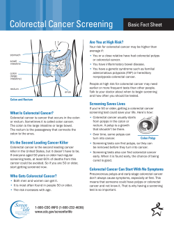
8
Surgery of Primary Colon
and Rectal Cancer
MARK J. OTT, MD
JEAN-PIERRE E.N. PIERIE, MD, PHD
The surgical treatment of colorectal cancer has multiple facets. Historically, surgery has been and continues to be the best method of effecting cure in this
disease. Along with this primary role, surgery is
often required for diagnosis and staging of disease,
along with the palliation of symptoms. The purpose
of this chapter is to outline and illustrate each of
these key components of surgical care in the treatment of these cancers of the lower gastrointestinal
tract. An understanding of the appropriate application of surgery in this disease is essential for both
surgeons and physicians involved in the treatment of
colorectal cancer. Only then can surgery be put to its
best use, which is to bring about a cure and/or
improve the quality and length of life for patients
with cancers of the colon and rectum.
SURGERY AS A DIAGNOSTIC TOOL
With the advent of radiologic and endoscopic methods, the need for surgical exploration to obtain diagnostic tissue has decreased considerably. Whereas
preoperative tissue diagnosis was not used in the
majority of colorectal tumors three decades ago, the
vast majority of patients today have histology prior to
definitive surgery. Since greater than 98 percent of
colon and rectal cancers are adenocarcinomas arising
from the mucosa and thus endoluminal, a thorough
lower endoscopy can determine the diagnosis prior to
any surgical exploration in the vast majority of cases.
In the 5 to 10 percent of cases where the colonoscope
is not able to reach the tumor due to technical reasons
(tortuous colon, poor preparation, etc.), a doublecontrast barium enema can provide a radiologic diagnosis. It should be emphasized that in the case of
colon carcinoma, an actual tissue diagnosis is often
not necessary prior to surgery. As long as the location
and appearance of the tumor, either endoscopic or
radiologic, are adequately obtained, the information
necessary to proceed with surgery is in hand. In the
case of rectal carcinoma, diagnostic tissue and tumor
depth and location are usually essential in planning
the appropriate definitive treatment.
For the more rare tumors of the colon and rectum, such as carcinoids, lymphomas, and gastrointestinal stromal tumors that are submucosal or extraluminal, standard endoscopic methods may not
make the diagnosis. In these situations, endoscopic
ultrasonography can often visualize and biopsy
these lesions. When this is not successful, ultrasonography or computed tomography (CT)-guided
biopsy techniques can often obtain diagnostic tissue.
However, with carcinoids, lymphomas, and gastrointestinal stromal tumors, it is common for the true
diagnosis to be made at surgical exploration. Figure
8–1 demonstrates a large gastrointestinal stromal
tumor of the sigmoid colon. This patient had normal
bowel activity and a normal colonoscopy. The CT
scan demonstrates the large primary tumor arising
from the sigmoid colon.
Surgery may also be necessary to obtain diagnostic tissue in the case of recurrent disease. A rea-
107
108
CANCER OF THE LOWER GASTROINTESTINAL TRACT
Figure 8–1. A high-grade gastrointestinal stromal tumor arising
from the sigmoid colon. The tumor (black arrow) fills most of the
pelvis. Ureters (white arrows) are in close proximity to the tumor.
sonably common scenario is that of a rising carcinoembryonic antigen (CEA) level and a CT scan
showing extraluminal thickened tissue in the area of
prior surgery. Endoscopic ultrasonography or radiologic-guided biopsies often return nondiagnostic
inflammatory or fibrous tissue. When distant disease
is ruled out, these patients warrant re-exploration for
diagnosis and possible resection. These patients can
still be cured through a combination of surgery,
radiation therapy, and chemotherapy, but a firm
diagnosis needs to be established.
SURGERY AS A CURATIVE
AND STAGING TECHNIQUE
The goal of resection for both colonic and rectal cancers is threefold: first, a complete resection of the
involved bowel segment with negative proximal, distal, and radial margins; second, a wide lymphadenectomy to encompass all draining lymph nodes for that
segment of bowel; and third, restoration of bowel continuity when possible. The restoration of bowel continuity is an important goal, but the bowel resection and
lymphadenectomy should never be compromised to
avoid an ostomy. All of these goals should be
achieved in conjunction with a thorough exploration
and a minimal amount of complications (ie, infection,
hemorrhage, and sexual or urinary dysfunction). The
actual methods of resection vary according to location and will be discussed separately.
Both the colon and especially the rectum normally carry high bacterial counts (ie, Bacteroides
fragilis and Escherichia coli 1010 and 107/g of wet
feces, respectively1) and are thus considered contaminated cases. In order to reduce infectious complications from an expected rate of 70 percent down to
less than 10 percent, preoperative decontamination
of the bowel is indicated. There are many methods of
decontamination but most involve a mechanical
washout to evacuate the colon followed by an antibiotic sterilization of residual anaerobic and gram
negative bacteria. Patients who undergo a colorectal
resection with primary anastomosis in an unprepared situation are more likely to have wound infections, anastomotic breakdown, and leakage. This has
traditionally prompted most surgeons to perform a
diverting colostomy in this situation. However,
surgery on the unprepared right or transverse colon,
which has a lower bacterial count and liquid stool,
can still be safely performed with either a right
colectomy or extended right colectomy and primary
ileocolonic anastomosis. When faced with a colorectal cancer that cannot undergo proper decontamination prior to surgery (ie, obstruction, severe
hemorrhage, or perforation and infection), the surgeon must weigh these risks and benefits in deciding
between a resection with colostomy or an anastomosis. In the case of a colostomy, it is useful for the surgeon to identify the distal bowel stump with a colored nonabsorbable suture at the time of the first
surgery to facilitate its identification at a subsequent
surgery to restore bowel continuity.
COLONIC RESECTIONS
Figures 8–2 and 8–3 demonstrate the classic
anatomy and vascular supply of the colon. While
variations exist, the lymphatic drainage parallels the
vascular supply. Thus, by excising the appropriate
segment of colon with its accompanying vascular
arcades back to their respective origins from either
the superior mesenteric or inferior mesenteric artery,
a wide lymphadenectomy will be achieved. There is
no oncologic benefit to high ligation of the inferior
mesenteric artery flush with the aorta. A ligation
below the origin of the left colic is equally curative.2
In terms of the actual tumor and bowel wall resec-
Surgery of Primary Colon and Rectal Cancer
Figure 8–2. Arterial supply to the colon with the colon mobilized and
displaced downward (below the marginal artery of Drummond). ICA =
ileocolic artery; LCA = left colic artery; RCA = right colic artery;
SA = sigmoid arteries; SRA = superior rectal artery. From Nyhus LM,
Baker RJ, eds. Mastery of surgery, 2nd ed. Boston: Little, Brown,
1992.
tion, it would be sufficient to resect the involved
segment of colon with a 1- to 2-cm margin of normal bowel. This would resect all gross and microscopic disease within the bowel wall. However, this
type of resection would not achieve a sufficient lymphadenectomy, and thus wider resections are generally performed. These resections follow the vascular
distributions of the colon (Figure 8–4). These hemicolectomies resect the associated lymphatics and
minimize the chance of an ischemic anastomosis
that can happen with segmental resections in
patients with atherosclerotic vessels.
Tumor location and lymphovascular supply
determine the appropriate resection. Lesions located
in the cecum should be resected to include a segment of terminal ileum and its mesentery. The right
colon is resected up to the right of the middle colic
vessels. Tumors located in the ascending colon are
resected with a standard right hemicolectomy.
Lesions at the hepatic flexure may require resection
by an extended right hemicolectomy (Figure 8–5).
Obviously, the surgeon’s judgment is critical in
determining the appropriate operation as there are
109
anatomic variations and other patient issues that
must be considered. Tumors in the mid-transverse
colon are often resected from the hepatic flexure to
the splenic flexure. This includes resection of the
contiguous mesentery of the middle colic vessels.
When transecting a vessel from the superior or inferior mesenteric artery, there is no curative benefit to
a flush transection. A 1-cm cuff of vessel to ligate
will give the same lymphadenectomy with less risk
of hemorrhage.
The attached segment of omentum should also be
resected with the specimen. This is done by transecting the omentum on the colonic side of the gastroepiploic vessels. The omentum also contains
lymph nodes, which may be involved with tumor
metastases. During right colonic resections, care
must be taken to visualize and protect the duodenum, right ureter, and right gonadal vessels. All
three of these structures are at risk as the right colon
is mobilized out of the retroperitoneum.
Figure 8–6 demonstrates the ligation of the inferior mesenteric artery in order to mobilize the colon
during a left hemicolectomy. As stated earlier, ligation of the inferior mesenteric artery does not add to
cure rate, but it does remove the inferior mesenteric
Figure 8–3. Venous drainage of colon. IMV = inferior mesenteric
vein; SMV = superior mesenteric vein; SRV = superior rectal vein.
From Nyhus LM, Baker RJ, eds. Mastery of surgery, 2nd ed. Boston:
Little, Brown, 1992.
110
CANCER OF THE LOWER GASTROINTESTINAL TRACT
Figure 8–4. Extent of surgical resection for cancer at various sites. A black disk
represents the cancer. Anastomosis of the bowel remaining after resection is
shown in the small insets. The extent of resection is determined by the distribution
of the regional lymph nodes along the blood supply. From Shrock TR. Large intestine. In: Way LW, ed. Current surgical diagnosis and treatment, 7th ed. Los Altos,
CA: Lange Medical Publications, 1985.
artery as a point of fixation. This in turn allows for better mobilization of the remaining colon and a tensionfree anastomosis. For more proximal lesions, the
superior rectal artery can often be preserved with no
compromise in cure rates. There is no curative or
staging benefit to a para-aortic lymphadenectomy.
During the resection of left colon lesions, the
splenocolic and renocolic ligaments will often need
to be incised to adequately mobilize the colon for
resection and or anastomosis. Small tears in the capsule of the spleen are easily created by too vigorous
retraction. Prevention is far more successful than
treatment. However, when bleeding occurs from
splenic capsular tears, it is important to incise the
remaining attachments between the colon and
spleen. This releases the tension on the capsule. It is
then more likely that topical hemostatic agents,
cautery, and direct pressure will be successful in
achieving hemostasis. Other structures that are easily damaged during mobilization of the left colon
from the retroperitoneum include the left ureter, left
gonadal vessels, and tail of the pancreas. The tail of
the pancreas can be inadvertently injured during
mobilization of the splenic flexure of the colon. This
can lead to either postoperative pancreatitis or a pancreatic fistula. Visualization of these structures will
help prevent their injury.
The surgeon’s judgment in determining the extent
of the resection is critical. A large premalignant
polyp not amenable to endoscopic removal can be
removed via a colotomy and polypectomy or via a
segmental resection, without lymphadenectomy.
Large bulky lesions that invade through the bowel
wall into adjacent organs should be resected with a
negative margin of the adjacent organ (ie, bladder,
small bowel, abdominal wall, etc.). Stage for stage,
Surgery of Primary Colon and Rectal Cancer
111
tumor. There certainly is evidence that manipulation
of the tumor during the operation leads to increased
tumor shedding of detectable tumor cells into the
blood supply.7 Turnbull’s initial series demonstrated
improvement in survival in patients resected with
this technique. However, subsequent controlled
studies have failed to demonstrate any clinical benefit in decreased local recurrence or increased survival.8 At the present time it cannot be considered
standard of care.
There is great variability in the frequency of
postoperative bowel movement following a hemicolectomy. Most patients will have a minimal
increase in frequency, but some patients will initially
have an increase of two to four movements per day.
These patients can have their frequency decreased
through the addition of fiber to the diet and, when
necessary, an antimotility agent. The remaining
colon will often adapt over a 4- to 6-month period to
return a patient to a more normal-for-them pattern.
Figure 8–5. The extended right hemicolectomy for a neoplasm at
the hepatic flexure side of the transverse colon. Includes a subtotal
colectomy with resection of the cecum and the ascending, transverse, and a portion of the descending colon with its mesentery to
the level of the left colic vessels. From Nyhus LM, Baker RJ, eds.
Mastery of surgery, 2nd ed. Boston: Little Brown, 1992.
these T4 lesions have no increase in complications
and an equal cure rate with en bloc surgical resection.3 The plane of invasion between the colonic
tumor and the invaded organ should not be fractured.
Disruption of this plane, even with subsequent surgical resection, decreases the cure rate by greater than
50 percent.4,5 In the case of invasion into the abdominal wall or retroperitoneum, the area of resection
should be marked with metal clips for subsequent
radiation therapy to help minimize the risk of a local
recurrence. When possible, the area should also be
covered by omentum to help exclude as much bowel
as possible from the subsequent radiation field.
The No-Touch technique, as popularized by
Turnbull and colleagues,6 involves the early division
and ligation of the vascular supply and occlusion of
the proximal and distal bowel lumen around the area
of the tumor. This is done to limit the shedding of
tumor cells into the blood and lymphatic systems
and into the bowel lumen during manipulation of the
Figure 8–6. Location of division of the inferior mesenteric artery
(arrow) 1 cm from the aorta during a left hemicolectomy. From Nyhus
LM, Baker RJ, eds. Mastery of surgery, 2nd ed. Boston: Little,
Brown, 1992.
112
CANCER OF THE LOWER GASTROINTESTINAL TRACT
RECTAL RESECTIONS
There is a wide array of treatment choices facing the
clinician managing rectal cancer. Surgical treatment
options can vary from transabdominal operations to
local excision. Newer techniques are being applied
to reduce the necessity for permanent colostomy,
improve functional results, and reduce local recurrence. Radiation therapy with chemotherapy, especially in the preoperative setting, has a much more
prominent role in the treatment of rectal cancer than
in colon cancer. The roles of adjuvant and neoadjuvant therapy are discussed in subsequent chapters in
this text.
Low Anterior and
Abdominoperineal Resection
The gold standard for removal of a rectal cancer is
via a transabdominal low anterior resection (LAR) or
a combined abdominoperineal resection (APR). Both
of these operations remove the involved bowel segment and the associated lymphatic tissue. There is no
curative benefit of the complete rectal resection and
permanent colostomy accomplished by an APR versus the LAR if the surgeon is able to obtain adequate
proximal and distal margins in combination with a
wide resection of the draining mesorectal lymphatic
tissue. Best was the first to demonstrate that mucosal
involvement 2 cm or more below the distal edge of
the lesion was found in less than 1 percent of cases.9
Others confirmed this finding in several retrospective studies that demonstrated that there was no benefit in local control or metastatic disease for bowel
margins greater than 2 cm.10,11 The question was
finally laid to rest by the National Surgical Adjuvant
Breast and Bowel Project (NSABP) [R-01] trial
showing no difference in recurrence between resections with distal margins greater than 3 cm versus
those within 2 cm.12 If an adequate distal margin
without excising the rectal sphincter mechanism is
possible, then a reconstruction via either a coloanal
or colorectal anastomosis is indicated (Figure 8–7).
However, Figures 8–8A and 8–8B show an example
of a specimen of a T3N0M0 rectal tumor that was situated too low to be eligible for a sphincter-saving
procedure. Consequently, an APR was performed.
A
B
C
Figure 8–7. Methods of anastomosis of the proximal colon to the
rectum or anus after a low anterior resection: A, end-to-end colorectal anastomosis; B, end-to-side colorectal anastomosis with J-pouch;
C, end-to-end coloanal anastomosis. From Enker WE. Total mesorectal excision with sphincter and autonomic-nerve preservation in the
treatment of rectal cancer. In: Condon RE, ed. Current techniques in
general surgery. New York: Lawrence DellaCorte, 1996.
The proximal extent of lymphatic resection has
likewise been contested but never answered in a
prospective study. However, several nonrandomized
studies would support a dissection to include the
nodal tissue up to the bifurcation of the left colic
from the inferior mesenteric artery. Division of the
artery just distal to this bifurcation preserves the left
colic blood supply and has not shown any survival
disadvantage relative to a more extensive proximal
lymphadenectomy of para-aortic nodes with ligation
of the inferior mesenteric artery at its origin. In a retrospective study from St. Mark’s Hospital of 784
patients with ligation distal to the left colic versus
586 patients with resection of the inferior mesenteric
artery and accompanying nodal tissue, there were no
differences in 5-year survival for any Dukes A, B, or
C patients.13 Similarly, in a series by Hojo and colleagues, there was only 1 survivor among 15 patients
who had extensive proximal lymphadenectomies
when there were pathologically positive nodes along
the inferior mesenteric artery.14 These studies support the practice of resecting proximal nodal tissue
up to and including the origin of the superior rectal
Surgery of Primary Colon and Rectal Cancer
113
artery and preserving the inferior mesenteric and left
colic artery.
The concept of a sharp total mesorectal excision
(TME) has increased the surgical awareness of the
importance of obtaining a wide radial margin by
encompassing the mesorectum as a defined packet.
In 1982, Heald and colleagues published his initial
description of the surgical techniques involved in
TME15 (Figure 8–9). He also emphasized the importance of identifying and preserving the sympathetic
and parasympathetic nerves to retain postoperative
bowel, bladder, and sexual function, the autonomic
nerve preservation (TME-ANP) (Figure 8–10). The
dissection is continued down to the levator ani muscles to achieve full mobilization of the rectum and
mesorectum (Figures 8–11, A and B). Incomplete
circumferential mobilization of the rectum limits dis-
A
B
Figure 8–8. A, Specimen after an APR and B, opened to show a
T3N0M0 rectal cancer just above the anal sphinter. It is situated too
low to be eligible for a sphincter-saving LAR.
Figure 8–9. Schematic of the critical pelvic structures encountered
during a sharp total mesorectal excision. The autonomic nerves and
rectal vessels are divided sharply and individually to preserve the
autonomic innervation necessary for bladder and sexual function.
From Enker WE. Total mesorectal excision with sphincter and autonomic-nerve preservation in the treatment of rectal cancer. In: Condon RE, ed. Current techniques in general surgery. New York:
Lawrence DellaCorte, 1996.
114
CANCER OF THE LOWER GASTROINTESTINAL TRACT
Figure 8–10. Mobilization of the rectum during a sharp mesorectal dissection demonstrating identification and preservation of autonomic nerve structures critical for bladder and sexual function. From
Enker WE. Total mesorectal excision with sphincter and autonomicnerve preservation in the treatment of rectal cancer. In: Condon RE,
ed. Current techniques in general surgery. New York: Lawrence
DellaCorte, 1996.
tal resection margins (Figure 8–11D), whereas full
mobilization will allow more distal resections (Figure 8–11C). Traditionally, much of the pelvic dissection below the peritoneal reflection for rectal cancer
was performed in a blunt fashion. This method of
dissection was rapid and identified lateral tissue containing vascular, lymphatic, and nervous tissue that
was then divided between clamps. The traditional
technique will also allow for adequate distal mobilization but is more likely to injure autonomic nerve
roots and disrupt the mesorectal lymphatic packet.
Table 8–1 demonstrates the results of Heald’s
personal, prospectively collected, consecutive, nonrandomized series of patients16 compared to the
results of the North Central Cancer Treatment Group
(NCCTG).17 While the two series cover similar time
periods, there are differences in patient tumor profiles that limit the value of a direct comparison. His
excellent results with local recurrence rates of less
than 5 percent and overall recurrence at 10 years of
22 percent in curatively operated patients have provoked intense discussions about the true merit of
this more time- and labor-intensive technique. This
study covers a 13-year period from 1978 to 1991,
with mean follow-up of 7.5 years. Of the 290
patients, 135 high-risk patients who were most likely
to recur after curative resection constituted the
group for analysis. There were 126 low anterior
resections and 9 abdominoperineal resections in this
group. High risk was defined according to the definition from the NCCTG. These investigators
excluded Dukes A (Astler Coller A + B1) lesions,
those more than 12 cm from the anal verge, and
those considered not curative by the surgeon. The
operation was considered curative when, at the end
of the procedure, the surgeon believed that all
grossly detectable cancer had been removed. None
of these patients received adjuvant therapy, which
reflects a long-term opinion of Heald’s that
improved survival and local control can be achieved
by optimal surgery alone. This is an obvious divergence from the recommendations of the 1990
National Institutes of Health (NIH) consensus conference where the combination of radiation therapy
and chemotherapy was recommended as standard of
care for stage 2 and 3 disease.18
There is a cost for this apparent decrease in local
and distal recurrence. Operative times are increased
A
B
C
D
Figure 8–11. Mobilization of the rectum below the peritoneal
reflection: A, the rectum prior to mobilization; B, full mobilization of
the rectum to the level of the levator ani muscles; C, full mobilization
of the rectum allows distal transection of the rectum with a generous
distal margin; D, incomplete mobilization of the rectum compromises
the distal margin of transection. From Enker WE. Total mesorectal
excision with sphincter and autonomic-nerve preservation in the
treatment of rectal cancer. In: Condon RE, ed. Current techniques in
general surgery. New York: Lawrence DellaCorte, 1996.
Surgery of Primary Colon and Rectal Cancer
Table 8–1. LOCAL AND TOTAL RECURRENCE RATES FOR
TOTAL MESORECTAL EXCISION AND NCCTG DATA
Treatment Group
TME
Conventional surgery
+ radiation
Conventional surgery
+ radiation + chemotherapy
Local
Recurrence
(%)
Overall
Recurrence
(%)
5
25
22
62.7
13.5
41.5
Data from MacFarlane JK, Ryall RDH, Heald RJ. Mesorectal excision for rectal cancer. Lancet 1993; 341:457–60.
by 2.5 hours, and there are increased blood transfusion requirements. There is an 11 percent clinical
and an additional 6.4 percent radiologic leak rate
with TME. Heald and many others now routinely use
a temporary diverting colostomy. Due to complications, 5 percent of these colostomies were never
reversed. A similar series of 350 patients in Norway
undergoing TME had an anastomotic leak rate
greater than 20 percent, leading to emergency
surgery, morbidity, and two deaths.19
While TME is touted as improving autonomic
nerve function postoperatively, no good randomized
studies exist to document this fact. The standard
blunt dissection technique with clamping of the lateral pedicles undoubtedly has resulted in numerous
unintended injuries to the pelvic autonomic nerve
plexus. It would seem reasonable that a more meticulous dissection with preservation of the defined
nerve roots would lead to better function.20
All of the preceding discussion serves to emphasize the point that surgical technique does make a
difference in patient outcome. Hermanek and colleagues surveyed rectal cancer outcomes in seven
major German cities and found that local recurrence
rates varied among individual surgeons from less
than 10 percent to more than 50 percent.21,22 Quirke
and colleagues demonstrated the importance of
radial margin clearance.23 In this prospective series
of 52 patients with rectal adenocarcinoma, careful
whole-mount sectioning of APR specimens revealed
spread to the lateral margins in 14 patients (27%).
Twelve of these patients (85%) developed local
recurrence whereas only 1 of 38 patients (3%) with
negative radial margins developed a local recurrence. Sharp dissection employed in TME to excise
115
the entire mesorectum reproducibly provides a maximal intact lateral resection margin and may explain
the beneficial effects on local control.
Local Excision of Rectal Carcinoma
and Sphincter-Sparing Techniques
The best operation in the treatment of rectal cancer
is one that achieves a curative resection with the
least morbidity and mortality. As the incidence of
large bowel cancer increases with each decade of
life, many patients diagnosed with cancer of the rectum are older than 70 years of age with coexistent
medical problems. An advantage of local excision is
that it is usually associated with a mortality rate near
zero and similar low morbidity rates.24,25 The avoidance of a permanent stoma is another great advantage to the patient if the cure of cancer is not jeopardized. Approximately 10 to 15 percent of all patients
with rectal carcinoma have tumors confined to the
rectal wall and might be eligible for local excision
after proper patient selection. The approach may be
transanal, transcoccygeal, or trans-sphincteric as
described in more detail later in this chapter.26–28
However, regardless of the surgical approach, the
resection must achieve a negative margin and a fullthickness excision; otherwise, therapy is inadequate
and further surgical therapy is indicated.
The most important criterion for local excision is
accessibility, which will influence surgical treatment.29 To maximize the chance of a good oncologic
procedure, several surgical groups have reported criteria for local excision, mostly in single institution,
retrospective studies.25,27 Ideal rectal tumors for this
approach are situated below the peritoneal reflection,
are less than 4 cm in diameter, take up less than 40
percent of the rectal circumference, have no palpable
or radiologically visible perirectal nodes, are mobile
on digital examination, and have a well-differentiated
histology without lymphatic or vascular invasion.30,31
All of these factors identify tumors that have a low
likelihood of nodal metastases. Since local resections
generally do not remove draining lymph nodes, these
unresected lymph nodes can lead to local and distant
recurrence if they in fact contain tumor. This is supported by the finding that local recurrence rates after
local resection and the frequency of lymph node
116
CANCER OF THE LOWER GASTROINTESTINAL TRACT
metastases found at APR are closely matched for similar stage diseases.29 Therefore, local excision with
curative intent is only appropriate when the risk of
nodal metastases is as near zero as possible. The
methods presently available to determine the depth of
tumor invasion of the rectal wall (the T status) and to
document regional lymph nodes (N status) are limited. A good digital rectal examination in experienced
hands can predict invasion of the rectal wall in up to
80 percent of cases but is less useful for detecting
regional tumor-positive lymph nodes.32 CT scan and
MRI are useful for identification of intra-abdominal
metastases and can accurately identify locally
advanced tumors that have invaded adjacent pelvic
structures, but the negative predictive value for
regional lymph nodes is only 50 to 70 percent. Endoluminal ultrasonography has been reported to be a
more accurate diagnostic instrument for assessment
of penetration of the rectal wall by carcinoma. It has
a negative predictive value of 70 to 95 percent in
experienced centers. The same expert groups report a
negative predicted value of 80 to 90 percent for detection of regional lymph nodes by ultrasonography.33,34
The caveats are that an expert sonographist is needed
for a reliable intraluminal sonographic examination
and, even then, not all lymph nodes containing tumor
are enlarged in colon and rectal carcinoma.35,36 Therefore, despite the best preoperative staging presently
available, the status of lymphatic involvement is often
still uncertain. This has important implications, as
lymph node involvement has the single greatest
impact on survival and is present in 40 to 60 percent
of all rectal cancer patients treated by surgery
alone.37,38 When deciding whether to perform a local
resection where lymph nodes are generally not
removed versus transabdominal resections where
lymph nodes are resected, there is always some uncertainty. When a patient is a candidate for a local resection (tumor confined to mucosa, submucosa, or muscularis propria), tumor biopsy information in terms of
tumor grade and evidence of lymphatic or vascular
invasion can be very important. Patients with poorly
differentiated tumors and evidence of lymphatic or
vascular invasion on biopsy have a 36 to 53 percent
rate of local recurrence after local resections alone
versus a 12 to 15 percent rate of local recurrence after
transabdominal resection.39 Because rectal cancer his-
tology can affect the type of operation, most rectal
cancers require a tissue diagnosis and thorough staging prior to definitive surgical treatment.
Steele and his co-workers have recently published the only multi-institutional prospective controlled trial of sphincter-sparing treatment for distal
rectal carcinoma in the literature, conducted by the
CALGB (Cancer and Leukemia Group B).24 Their
inclusion criteria for local excision of a rectal tumor
match the criteria mentioned earlier in this chapter,
although no effort was made to differentiate between
high-grade (including a histology of vascular and
lymphatic invasion) and low-grade tumors. After
local excision, patients with T1 tumors received no
further treatment and were followed for recurrence
and survival. Patients with T2 tumors were given
adjuvant radiation and chemotherapy. Any patient
with a local recurrence was salvaged with a formal
APR. Only patients who underwent excision with
negative margins were included. After 6 years of
follow-up, survival and failure-free survival rates of
the eligible patients are 85 and 78 percent, respectively, with no difference between the T1 and T2
(plus adjuvant treatment) groups. Comparing these
results to historical control patients who received a
formal APR, the authors conclude no significant
therapeutic disadvantage in the sphincter-sparing
approach thus far, when the strict selection criteria
are applied. The efficacy of surgical salvage after
local recurrence remains uncertain because too few
patients were salvaged and the follow-up was relatively short. The results concerning salvage surgery
following local recurrence (mostly a formal APR)
are of utmost importance, since disappointing
results from retrospective studies have been reported
until now.29
After local resection, postoperative radiation and
chemotherapy are usually given for lesions penetrating the rectal wall. The two prospective series that
have looked at this form of therapy indicate a local
recurrence rate of 6.5 to 8 percent and a survival rate
of 90 percent, which is equal to the expected results
of an abdominoperineal resection.40,41 Rectal function is reported as good or satisfactory in greater
than 80 percent of the patients.
Although local excision of rectal carcinoma can
be accomplished through a transsacral or trans-
Surgery of Primary Colon and Rectal Cancer
sphincteric approach, transanal exposure is the technique chosen most frequently. The prone jackknife
position is used for lesions in the anterior and lateral
locations. For lesions of the posterior rectum, the
lithotomy position is preferred.42 The preoperative
bowel preparation is the same as in other large bowel
procedures. In the operating room, the rectum is irrigated with an antiseptic solution to minimize contamination during the procedure. Exposure is achieved
by an anal speculum and/or retractor. The procedure
requires full-thickness excision, with an in vivo margin of 1 cm beyond the edge of the tumor (Figure
8–12, A and B). The positioning of traction sutures
A
117
helps in removal of the tumor and orients the specimen for the pathologist (Figure 8–13, A and B). To
minimize operative bleeding, electrocautery should
be used. Primary closure of the resulting defect
shortens the time required for healing, but this
aspect is not a strict requirement as long as the
defect lies caudad to the peritoneal reflection (Figure 8–14, A and B). Proctoscopic examination
should be performed at the completion of the procedure to ensure that an adequate lumen has been preserved. Figure 8–15 gives an endoscopic view of the
rectal wound 1 year after local excision.
For very low rectal tumors, at or near the anorec-
A
B
Figure 8–12. Transanal local excision of a low rectal tumor: A,
exposure of a posterior wall rectal carcinoma with Parks anal retractor and B, diagram of the same stage of the operation. The 1-cm margin is marked. From Bailey HR, Huval WV, Max E, et al.27
B
Figure 8–13. A, Full-thickness excision of the tumor using traction
sutures for presentation of the specimen, and B, diagram of the same
stage of the operation. From Bailey HR, Huval WV, Max E, et al.27
118
CANCER OF THE LOWER GASTROINTESTINAL TRACT
tal junction, an intersphincteric resection with partial excision of the internal anal sphincter might be
possible28 (Figure 8–16). In a small prospective
study, 5-year survival rates of 75 percent were
reported after this procedure for T2 and T3 tumors
plus adjuvant therapy in very low tumors between
2.5 cm and 4.5 cm from the anal verge. Continence
was normal in half of the patients and half of the
patients suffered from occasional minor leaks.
Kraske was the first to describe a transsacral
approach for midrectal cancers in 1885.26 Others
have modified this technique, but the principles are
the same.43 Tumors situated on the anterior side of
the midrectum are suitable for this approach. Post-
operative fistulae are the most frequent postoperative complications after this procedure and occur in
approximately 15 percent of cases. The technique
can be ideal for midrectal tumors and should be a
part of the surgeon’s armamentarium.
In conclusion, local excision as a sphincter-saving
procedure is possible in 10 to 15 percent of patients
with rectal cancers. Strict selection minimizes the
chance of regional lymph node metastases and is the
key to a good outcome.
PALLIATIVE COLORECTAL SURGERY
Not all colorectal surgery is performed with curative
intent. Patients presenting with truly unresectable
primary tumors or patients with metastatic disease
constitute this group. In the case of patients with
unresectable primary tumors, the goal of surgery is
usually to relieve obstruction. For distal tumors of the
rectum and sigmoid colon, this will usually involve a
diverting colostomy and probably a mucous fistula.
Patients with distal obstruction who are unresectable
should not have an end colostomy and blind pouch
proximal to the obstruction. The colon will continue
to secrete mucus and can potentially freely perforate
or fistulize into an adjacent organ from internal pressure. Patients with more proximal unresectable colon
cancers can usually be bypassed with a side-to-side
A
B
Figure 8–14. A, Primary longitudinal closure of the defect with
absorbable sutures, without narrowing of the lumen; and B, diagram
of the same stage of the operation, although a transverse closure is
depicted. From Bailey HR, Huval WV, Max E, et al.27
Figure 8–15. Endoscopic view of the healed rectal wound 1 year
after local excision of a rectal cancer.
Surgery of Primary Colon and Rectal Cancer
anastomosis between bowel proximal and distal to
the obstruction. This will circumvent the obstruction
and allow internal decompression of the potentially
obstructed segments while keeping the patient free of
a colostomy. A side-to-side anastomosis between the
cecum and sigmoid colon is usually easily performed
and causes only a minimal increase in the number of
bowel movements per day.
Patients with unresectable primary tumors and
bleeding from the tumor present a more difficult situation. These patients will often have a resolution or
decrease in bleeding with colonic diversion or
bypass. The continued presence of the fecal stream
contributes to the bleeding process. When this fails
to control the bleeding, external beam radiation will
often be successful.
Patients who present with distant metastases and
resectable primary colorectal tumors should generally have their primary tumor resected. Possible
exceptions to this include widespread carcinomatosis with expected survival of less than 6 months or
multiple medical problems that make the patient a
high-risk surgical candidate or that preclude any
systemic chemotherapy. Due to difference in tumor
biology, almost every surgical series has about a 5
percent, 5-year survival rate for these patients.44
LAPAROSCOPIC TECHNIQUES IN
COLORECTAL CANCER SURGERY
Laparoscopy has been used in colorectal surgery for
several decades.45 The initial clinical reports of
laparoscopic colorectal surgery (LCR) suggested
many advantages in comparison to conventional
(open) techniques, including reduced postoperative
pain, earlier recovery of bowel function, shorter
length of hospital stay, and earlier return to normal
activities.46 More recent studies have not been as
favorable.47 To date, there at least appear to be no significant differences in perioperative complications or
mortality between laparoscopic and open colorectal
surgery, and short-term outcomes seem equivalent.
Despite a trend toward a reduced length of stay, the
overall costs of laparoscopic resections have historically been higher. This in part has been related to both
a longer operative time and higher equipment costs.
Many of the expenses would be expected to decline,
119
as advances in both laparoscopic equipment design
and surgical technique reduce the surgeon’s operative
time and costs. Some of the specific complications
related to laparoscopic technique include port-site
herniation, inadvertent organ injury, complications of
pneumoperitoneum (respiratory acidosis), and resection of the wrong segment of bowel.48
Another aspect is the learning curve. There are
major differences in surgical technique between LCR
and other more common laparoscopic procedures like
laparoscopic cholecystectomy. These differences
include working in multiple anatomic regions, the
need to divide multiple mesenteric vessels, removal of
a fairly large specimen, and the performance of a safe
anastomosis. The number of supervised laparoscopic
colorectal resections needed to comfortably learn the
procedure is not well documented but is estimated to
be more than 40 completed cases, partly depending
on the laparoscopic experience of the surgeon.49,50
This implies that a second qualified surgeon and a
high-volume setting are needed to overcome this
problem. Furthermore, if conversion from LCR to an
open procedure is needed, one gets the worst of both
methods: higher cost and operative time combined
with a larger incision and an increased incidence of
postoperative complications.51
There are three potential uses of laparoscopy in
colorectal cancer. The first is an occasional need for
Figure 8–16. Intersphincteric resection of a very low rectal cancer at
the anorectal junction, with partial excision of the internal anal sphincter. From Rullier E, Zerbib F, Laurent C, et al.28
120
CANCER OF THE LOWER GASTROINTESTINAL TRACT
diagnostic or staging information prior to definitive
treatment. Given the success of endoscopic and radiologic studies, this is an extremely rare event. Nevertheless, laparoscopy allows inspection of the entire
abdominal cavity and permits biopsy of serosal
lesions. Furthermore, intraoperative laparoscopic
ultrasonography is especially useful for diagnosing
occult, small intraparenchymal liver metastasis from
colorectal cancer.
The second possible role is in palliative colorectal
surgery. In cases where advanced colorectal carcinoma is diagnosed preoperatively, laparoscopic
surgery permits a variety of palliative measures, such
as resection of the primary tumor, intestinal bypass,
and stomal diversion without the need for a large incision. Alternative procedures, including laparoscopically assisted feeding jejunostomies and cathether
placement for intraperitoneal chemotherapy, can be
performed in the same setting. As compared to open
surgery, LCR is generally believed to have a more
rapid return of bowel function, decreases length of
hospitalization, and results in less postoperative pain,
although there are also studies that refute these
claims.52 Given the specific disadvantages of LCR as
outlined earlier, the surgeon has to decide whether the
potential benefits outweigh these problems.
The role LCR in curative cancer surgery is still to
be defined and is presently being evaluated.
Although similar 5-year survival rates after LCR
and open procedures have been reported in a comparative prospective trial, there is still considerable
debate whether there will be a compromise in cure
rate.47,53 This question is currently being investigated in multiple randomized prospective studies,
conducted in several countries.50,54 There is some
experimental evidence that laparoscopic procedures
decrease tumor growth as compared with open procedures.55 On the other hand, a unique complication
related to laparoscopic cancer surgery seems to be
an altered pattern of tumor spread. This has led to
port-site metastases and is not well understood.56
Not only are such recurrences present at the site of
tumor extraction, but they have also been documented at port sites not involved in specimen
removal. Similar findings have been described for
other intra-abdominal malignancies, including pancreatic, ovarian, and gallbladder cancer. It is not
Figure 8–17. Laparoscopic view of intra-abdominal dissection with
a harmonic scalpel.
clear whether these types of recurrences are an
inevitable consequence of minimally invasive techniques or perhaps related to a learning experience.
Current ongoing studies indicate that the incidence
of trocar site recurrence seems to be decreasing,
suggesting that the learning curve may have influenced this adverse outcome.50
As stated earlier, there are specific techniques
and equipment for the laparoscopic procedure. Dissection is primarily performed using a laparoscopic
harmonic scalpel (Figure 8–17), and vascular struc-
Figure 8–18. Laparoscopic view of transsection of a vascular
structure in the mesentery of the colon using the laparoscopic vascular stapler device.
Surgery of Primary Colon and Rectal Cancer
121
tures are preferably divided by a laparoscopic vascular stapler (Figure 8–18). The bowel is transsected
with the help of a laparoscopic roticulating gastrointestinal stapler (Figure 8–19). After the bowel is dissected out and transsected distally (Figure 8–20), the
specimen is taken out of the abdominal cavity
through a 4- to 5-cm incision (Figure 8–21). Although
the anastomosis can be performed intracorporally,
the easier and therefore safer extracorporal technique is chosen by many surgeons, including the
Figure 8–21. The resected colon is brought extracorporally
through the 4-cm abdominal incision.
Figure 8–19. Laparoscopic view of the transsection of the distal
colon using a laparoscopic roticulating gastrointestinal stapler device.
authors, in case of a right-sided or transverse colectomy. However, when the anastomosis is to be performed between the distal left colon and rectum, a
stapled anastomosis can be carried out intracorporally using a circular stapling device.
Laparoscopy will certainly have an important
role in colorectal surgery, but until such time as
more prospective trials become conclusive, only
patients enrolled in one of these studies should be
offered a laparoscopic resection for cancer with
curative intent.
REFERENCES
Figure 8–20. Laparoscopic view of the pelvic cavity showing the
transsected distal colon and the uterus anterior of that.
1. Mackowiak PA. The normal microbial flora. N Engl J
Med 1982;307:83–93.
2. Grinnell RS. Results of ligation of inferior mesenteric
artery at the aorta in resections of carcinoma of the
descending and sigmoid colon and rectum. Surg
Gynecol Obstet 1965;120:1031–6.
3. Gebhardt C. Multi-visceral resection in colorectal carcinoma. Langenbecks Arch Chir Suppl Kongressbd
1998;115:327–30.
122
CANCER OF THE LOWER GASTROINTESTINAL TRACT
4. Gall FP, Tonak J, Altendorf A. Multivisceral resections
in colorectal cancer. Dis Colon Rectum 1987;30:
337–41.
5. Hunter JA, Ryan JA Jr, Schultz P. En block resection of
colon cancer adherent to other organs. Am J Surg
1987;154:67–71.
6. Turnbull RB, Kyle K, Watson FR, Spratt J. Cancer of
the colon: the influence of the no-touch isolation
technique on survival rates. Ann Surg 1967;166:
420–5.
7. Hayashi N, Egami H, Kai M, et al. No-touch isolation
technique reduces intraoperative shedding of tumor
cells into the portal vein during resection of colorectal cancer. Surgery 1999;125:369–74.
8. Wiggers T, Jeekel J, Arends JW, et al. No touch isolation
technique in colon cancer: a controlled prospective
trial. Br J Surg 1988;75:873–8.
9. Best RR. Rectosigmoidectomy with anastomosis in carcinoma of rectum and rectosigmoid. J Int Coll Surgeons 1950;13:203–8.
10. Hojo K. Anastomotic recurrence after sphincter-saving
resection for rectal cancer: length of distal clearance
of the bowel. Dis Colon Rectum 1986;29:11–4.
11. Pollett WG, Nicholls RJ. The relationship between the
extent of distal clearance and survival and local recurrence rates after curative anterior resection for carcinoma of the rectum. Ann Surg 1983;198:159–63.
12. Wolmark N, Fischer B. An analysis of survival and
treatment failure following abdominoperineal and
sphincter-saving resection in Dukes’ B and C rectal
carcinoma. Ann Surg 1986;204:480–9.
13. Pezim ME, Nicholls RJ. Survival after high or low ligation of the inferior mesenteric artery during curative
surgery for rectal cancer. Ann Surg 1984;200:729–33.
14. Hojo K, Koyama Y, Moriya Y. Lymphatic spread and its
prognostic value in patients with rectal cancer. Am J
Surg 1982;144:350–4.
15. Heald RJ, Husband EM, Ryall RDH. The mesorectum
in rectal cancer surgery – the clue to pelvic recurrence? Br J Surg 1982;69:613–6.
16. MacFarlane JK, Ryall RDH, Heald RJ. Mesorectal excision for rectal cancer. Lancet 1993;341:457–60.
17. Krook JE, Moertel CG, Gunderson LL, et al. Effective
surgical adjuvant therapy for high-risk rectal carcinoma. N Engl J Med 1991;324:709–15.
18. NIH consensus conference. Adjuvant therapy for
patients with colon and rectal cancer. JAMA 1990;
264:1444–50.
19. Heald RJ. Rectal cancer: the surgical options. Eur J
Cancer 1995;31A:1189–92.
20. Havenga K, Enker WE, McDermot K, et al. Male and
female sexual and urinary function after autonomic
nerve preservation for carcinoma of the rectum. J
Am Coll Surg 1996;182:495–502.
21. Hermanek P, Wiebelt H, Staimmer D, Riedl S. Prognostic factors of rectum carcinoma—experience of the
German multicenter study SGCRC. German study
group colo-rectal carcinoma. Tumori 1995;81:60–4.
22. Hermanek P, Hohenberger W. The importance of volume in colorectal cancer surgery. Eur J Surg Oncol
1996;22:213–5.
23. Quirke P, Durdey P, Dixon MF, Williams NS. Local
recurrence of rectal adenocarcinoma due to inadequate surgical resection. Lancet 1986;Nov:996–8.
24. Steele GD, Herndon JE, Bleday R, et al. Sphincter-sparing treatment for distal rectal adenocarcinoma. Ann
Surg Oncol 1999;6:433–41.
25. Murray JJ, Stahl TJ. Sphincter-saving alternatives for
treatment of adenocarcinoma involving distal rectum. Surg Clin North Am 1993;73:131–144.
26. Kraske P. Zur extirpation hochsitzender masdarmkrebs.
Verh Dtsch Ges Chir 1885;14:464.
27. Bailey HR, Huval WV, Max E, et al. Local excision of
carcinoma of the rectum for cure. Surgery 1992;111:
555–61.
28. Rullier E, Zerbib F, Laurent C, et al. Intersphincteric
resection with excision of internal anal sphincter for
conservative treatment of very low rectal cancer. Dis
Colon Rectum 1999;42:1168–75.
29. Killingback M. Local excision of carcinoma of the rectum: indications. World J Surg 1992;16:437–46.
30. Breen E, Bleday R. Preservation of the anus in the therapy of distal rectal cancers. Surg Clin North Am
1997;77:71–83.
31. Tanaka S, Yokota T, Saito D, et al. Clinicopathologic
features of early rectal carcinoma and indications for
endoscopic treatment. Dis Colon Rectum 1995;38:
959–63.
32. Nicholls RJ, Galloway DJ, Mason AY, et al. Clinical
local staging of rectal cancer. J Surg 1985;72(Suppl):
S51–2.
33. Rifkin MD, Ehrlich SM, Marks G. Staging of rectal carcinoma: prospective comparison of endorectal US
and CT. Radiology 1989;170:319–22.
34. Saitoh N, Okui K, Sarashina H, et al. Evaluation of
echographic diagnosis of rectal cancer using intrarectal ultrasonic examination. Dis Colon Rectum
1986;29:234–42.
35. Nicholls RJ, Mason AY, Morson BC, et al. The clinical
staging of rectal cancer. Br J Surg 1982;69:404–9.
36. Herrera-Ornelas L, Justiniano J, Castillo N, et al.
Metastases in small lymph nodes from colon cancer.
Arch Surg 1987;122:1253–6.
37. Dukes C, Bussey H. The spread of rectal cancer and its
effect on prognosis. Br J Cancer 1958;12:309–20.
38. Gabriel W, Dukes C, Bussey H. Lymphatic spread in
cancer of the rectum. Br J Surg 1935;23:395–413.
39. Willett C, Compton C, Shellito P, Efird J. Selection fac-
Surgery of Primary Colon and Rectal Cancer
40.
41.
42.
43.
44.
45.
46.
47.
48.
tors for local excision or abdominoperineal resection
of early stage rectal cancer. Cancer 1994;73:2716–20.
Bleday R, Breen E, Jessup JM, et al. Prospective evaluation of local excision for small rectal cancers. Dis
Colon Rectum 1997;40:388–92.
Ota D, Skibber J, Rich T. M.D. Anderson cancer center
experience with local excision and multimodality
therapy for rectal cancer. Surg Oncol Clin N Am
1992;1:147–52.
Nivatvongs S, Wolff BG. Technique of per anal excision
for carcinoma of the low rectum. World J Surg 1992;
16:447–50.
Hargrove WC, Gertner MH, Fitts WT. The Kraske operation for carcinoma of the rectum. Surg Gynecol
Obstet 1979;148:931–3.
Knoch M, Hohenberger W. Long-term survival after noncurative therapy of colorectal carcinomas. Langenbecks Arch Chir Suppl Kongressbd 1996;113:133–5.
Monson JRT, Hill AD, Dapzi A. Laparoscopic colonic
surgery. Br J Surg 1995;82:150–7.
Phillips EH, Franklin M, Carroll BJ, et al. Laparoscopic
colectomy. Ann Surg 1992;216:703–7.
Franklin ME, Rosenthal D, Abrego-Medina D, et al.
Prospective comparison of open versus laparoscopic
colon surgery for carcinoma: five year results. Dis
Colon Rectum. 1996;39:S35–46.
Wexner SD, Cohen SM, Ulrich, et al. Laparoscopic col-
49.
50.
51.
52.
53.
54.
55.
56.
123
orectal surgery: are we being honest with our
patients? Dis Colon Rectum 1995;38:723–7.
Agachan F, Joo JS, Weiss EG, Wexner SD. Intra-operative laparoscopic complications. Are we getting better? Dis Colon Rectum 1996;39:S14–9.
Stocchi L, Nelson H. Laparoscopic colectomy for colon
cancer: trial update. J Surg Oncol 1998;68:255–67.
Slim K, Pezet D, Riff Y, et al. High morbidity rate after
converted laparoscopic colorectal surgery. Br J Surg
1995;82:1406–8.
Ortiz H, Armendariz P, Yarnoz C. Early postoperative
feeding after elective colorectal surgery is not a benefit unique to laparoscopy assisted procedures. Int J
Colorectal Dis 1996;11:246–9.
Wexner SD, Cohen SM. Port site metastases after
laparoscopic colorectal surgery for cure of malignancy. Br J Surg 1995;82:295–8.
COST Study Group. Early results of laparoscopic
surgery for colorectal cancer. Dis Colon Rectum
1996;39:S53–8.
Bouvy ND, Marquet RL, Jeekel J, Bonjer HJ. Laparoscopic surgery is associated with less tumour growth
stimulation than conventional surgery: an experimental study. Br J Surg 1997;84:358–61.
Tomita H, Marcello PW, Jeffrey MD, Milsom MD.
Laparoscopic surgery of the colon and rectum.
World J Surg 1999;23:397–405.


© Copyright 2026