
Shrinking of Solid-state Nanopores by Direct Thermal Heating Open Access
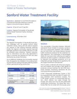
Asghar et al. Nanoscale Research Letters 2011, 6:372
http://www.nanoscalereslett.com/content/6/1/372
NANO EXPRESS
Open Access
Shrinking of Solid-state Nanopores by Direct
Thermal Heating
Waseem Asghar1,2, Azhar Ilyas1,2, Joseph Anthony Billo1,2 and Samir Muzaffar Iqbal1,2,3*
Abstract
Solid-state nanopores have emerged as useful single-molecule sensors for DNA and proteins. A novel and simple
technique for solid-state nanopore fabrication is reported here. The process involves direct thermal heating of 100
to 300 nm nanopores, made by focused ion beam (FIB) milling in free-standing membranes. Direct heating results
in shrinking of the silicon dioxide nanopores. The free-standing silicon dioxide membrane is softened and adatoms
diffuse to a lower surface free energy. The model predicts the dynamics of the shrinking process as validated by
experiments. The method described herein, can process many samples at one time. The inbuilt stress in the oxide
film is also reduced due to annealing. The surface composition of the pore walls remains the same during the
shrinking process. The linear shrinkage rate gives a reproducible way to control the diameter of a pore with
nanometer precision.
Background
The use of a-hemolysin protein nanopores inspired the
fabrication of solid-state nanopores. Solid-state nanopores have emerged as novel biosensors for single molecule analysis of DNA, proteins, etc. [1-7]. Solid-state
nanopores are more stable than protein nanopores
under various experimental conditions like pH, salinity,
and temperature [8-11]. When a single bio-molecule
electrophoretically passes through a nanopore, it gives
significant current blockage pulses.
The diameter of the nanopore should be almost at the
same scale as the size of the translocating species. The
pores fabricated with conventional processes result into
initial diameters larger than the size of species of interest [12-16]. The nanopore diameter is then reduced
using transmission electron microscope (TEM) or field
emission scanning electron microscope (FESEM) to
induce the shrinking [15,17] and FIB for the sculpting
processes [18]. During the TEM shrinking process, the
viscous flow of SiO 2 membrane is induced by an electron beam of optimal intensity. The nanopore shrinks or
expands based on the surface-tension-driven mass flow.
The nanopore, fulfilling the condition r < t/2, would
shrink under the electron beam at optimal conditions
* Correspondence: [email protected]
1
Department of Electrical Engineering, University of Texas at Arlington,
Arlington, TX 76019, USA
Full list of author information is available at the end of the article
where r is the radius of the pore and t is the thickness
of the membrane. TEM beam exposure depletes oxygen
from the oxide at depletion rate of about 10% per hour
[15]. Higher shrinking rates can be achieved through
FESEM induced shrinking [17].
The FESEM induced shrinking mechanism is putatively not surface tension driven, but explained by radiolysis. The crystalline structure of the nanopore is
disturbed under a high energy FESEM electron beam.
This results in pore shrinkage due to the diffusion of Si
and oxygen atoms toward the edge of the pore to overcome the crystalline defects present at the edge. The
stoicheometry of the SiO2 is expected to be different
than a normal oxide layer due to radiolysis. Different
shrinking rates were reported by using different acceleration voltages during FESEM exposure [17]. The
nanopore was found to be always shrinking independent
of the ratio of the pore’s diameter and membrane thickness under FESEM [17].
During the FIB sculpting process, the nanopore is
exposed to an energetic ion beam. The accelerating ions
drilled a nanopore in a thin oxide membrane due to
sputtering of the surface, or these reduced the pore diameter due to atom diffusion or surface tension driven
mass flow [18,19]. The FIB sculpting process is also
dependent on the substrate temperature. Under an
Argon ion beam, the pore closed at room temperature
while it opened at temperatures close to 0°C [18].
© 2011 Asghar et al; licensee Springer. This is an Open Access article distributed under the terms of the Creative Commons Attribution
License (http://creativecommons.org/licenses/by/2.0), which permits unrestricted use, distribution, and reproduction in any medium,
provided the original work is properly cited.
Asghar et al. Nanoscale Research Letters 2011, 6:372
http://www.nanoscalereslett.com/content/6/1/372
Page 2 of 6
Chemical composition of the material around the
nanopore periphery changes during TEM or FESEM
induced shrinking processes. This produces variable
modifications of nanopore surface properties. These
processes make the nanopore unfavorable for molecule
analysis due to increased surface charge and electrical
noise in the desired signal. In addition, all these shrinking processes are time consuming because they can only
process one nanopore at a time. In this article, we
report a simple and novel method to shrink nanopores
using direct thermal heating. High temperature treatment (>1000°C), or annealing, promotes the viscous
flow of the silicon dioxide (SiO2) membrane and results
in morphological changes that depend on the ratio of
nanopore diameter to membrane thickness. Residual
stress in the SiO 2 membrane is also reduced during
high temperature annealing. Surface composition of the
nanopore is maintained in this approach, as opposed to
being inevitably changed in the electron or ion irradiation approaches previously reported. Annealing has been
extensively used in semiconductor industry to reduce
leakage current in thin films [20], to repair gate oxide
damage from electrical stress [21], and to minimize residual stress in amorphous films [22].
Results and discussion
A boron-doped double-side-polished Si (100) wafer was
thermally oxidized to a thickness of 400 nm. Square
etch-start windows were opened in the SiO2 using standard photolithography. Free-standing SiO2 membranes
(30 × 30 μm2) were achieved using wet tetramethylammonium hydroxide (TMAH) anisotropic etching
through the whole wafer thickness. The schematic in
Figure 1(a) depicts the membrane formed after anisotropic etching. Bulk membrane composition was determined by energy dispersive X-ray spectroscopy (EDS).
The EDS analysis showed that the membranes contained
only Si and O, as shown in Figure 1(b). The EDS analysis revealed 31% Si and 69% O. This was in good agreement with the expected stoichiometric film ratio
of 33.33% Si and 66.66% O in SiO 2 . A FIB was then
employed to drill nanopores in free-standing SiO2 membranes operated at a 30 kV acceleration voltage [23].
A larger portion of the drilled nanopores were in the
diameter range of 100 to 300 nm. The high-resolution
transmission electron microscope operating at 300 kV
was used to image the nanopores after FIB drilling as
shown in the inset of Figure 1(b). The nanopore dyes
were kept in heating furnace at specific temperature for
pore shrinking. The nitrogen flow rate of 20 sccm was
maintained during this process.
We observed the nanopores shrinking or expanding
when subjected to high temperature (1000 to 1250°C),
contradicting previous findings [15]. The nanopores
Figure 1 Pore fabrication process and EDS analysis of
membrane. (a) The schematic showing a cross-section view of the
device. It consists of 250 nm thick free-standing SiO2 membrane, in
200 μm thick wafer. The small circle on the top of the membrane
depicts a nanopore drilled using FIB. (b) EDS spectrum from SiO2
membrane confirming the presence of only Si and O. TEM
micrograph (inset) shows the nanopore in free-standing membrane
drilled with FIB.
having an initial diameter of 250 nm were reduced to
3 nm at 1150°C as shown in Figure 2. The nanopores
were imaged with TEM after each temperature processing step to characterize the process. After loading the
dyes into the furnace, the temperature was allowed to
stabilize for 30 s before counting the actual processing
time. After the thermal process, the dyes were unloaded
from furnace and cooled down to room temperature.
When the dyes were processed at temperatures below
1000°C, it was observed that there was very little or no
change in the diameter of the nanopore. This can be
explained by the fact that at low temperature (<1000°C),
the oxide layer would not be relaxed to an extent that it
would start changing pore morphology. When the nanopores were processed at a higher temperature (>1250°
C), the oxide membranes either broke due to very high
thermal stress or the shrinking process was too fast to
control. This was especially so for pores smaller than
20 nm diameter [24]. As an example, a nanopore with
initial diameter of ~270 nm, processed at 1250°C, is
shown in Figure 3. The TEM images of the nanopore
Asghar et al. Nanoscale Research Letters 2011, 6:372
http://www.nanoscalereslett.com/content/6/1/372
Page 3 of 6
Figure 2 TEM micrographs showing thermal shrinking of silicon dioxide nanopore. (a) TEM micrograph of ~250 nm pore drilled with FIB
in 300 nm thick oxide membrane. (b) TEM micrograph of the nanopore after 5 min of thermal shrinking at 1150°C. The diameter of the
nanopore was ~150 nm. The wavy surface of the oxide at nanopore edges shows the shrinking process due to viscous flow of oxide. (c)
Nanopore after 10 min. The diameter is ~20 nm. (d) Nanopore after another 10 min and 40 s showing the diameter of ~3 nm.
show that the nanopore closed after 4 min due to an
increased shrinking rate. The shrinking or expansion
rate thus increased at higher temperature. When the
pore diameter was larger than the membrane thickness,
the nanopore started expanding in size instead of
shrinking. A 350 nm nanopore in a 300 nm thick membrane was processed at 1150°C for 50 min. The pore
expanded in size to 1.5 μm (Figure 4). It is interesting
to note that direct heating can be used to shrink or
expand the pore based only on the ratio of initial nanopore diameter to cylindrical length of the pore. The
temperature itself had no effect on whether the nanopore would shrink or expand. The pore shrinking and
expanding mechanism can be explained by the surface
Figure 3 High temperature shrinking process. (a) TEM micrograph of ~270 nm diameter nanopore before shrinking. (b) TEM micrograph of
nanopore after 4 min of thermal shrinking at 1250°C. The pore closed in just 4 min due to high shrinkage rate. The shrinking rate was about
70 nm/min.
Asghar et al. Nanoscale Research Letters 2011, 6:372
http://www.nanoscalereslett.com/content/6/1/372
Page 4 of 6
Figure 4 SEM micrographs show pore expansion. (a) The nanopore before thermal process. The initial diameter of nanopore was ~350 nm
and thickness of the oxide membrane was ~300 nm. (b) The nanopore after processing at 1150°C for 15 min. The diameter increased to 650
nm. (c) The nanopore further expanded to 1.5 μm after 50 min heating at 1150°C.
tension which induced viscous flow of oxide film as
described below.
The nanopore shrinking process was characterized at
different temperatures as shown in Figure 5. The nanopore had no shrinking or expansion at 900°C. When the
temperature was increased above 1000°C, the pore morphology started changing due to the diffusion and the
viscous flow of oxide. The average nanopore shrinking
rate was ~22 nm/min at 1150°C, which increased to 80
nm/min when the temperature was raised to 1250°C. At
higher temperatures, the shrinking process was difficult
to control precisely at the nano scale. When the nanopore diameter was reduced to tens of nanometers, low
Figure 5 Plot of pore diameter vs time at different
temperatures. This plot is based on TEM micrographs of different
nanopores processed at different temperatures. No change in pore
size is seen after 20 min at 900°C. The pore shrinkage rate increased
with increasing temperature. Note: average diameter of the
nanopore = sqrt (long axis × short axis). All the shrinking processes
show almost linear shrinking behavior.
processing temperature (<1150°C) was used to accurately
control the shrinking.
An obvious concern in the pore shrinking process
is the possibility of hydrocarbon contamination that
can affect pore shrinkage dynamics. All the dyes
were cleaned with Piranha solution (1:1, sulfuric acid:
hydrogen peroxide) before nanopore drilling with FIB.
The chips were cleaned with argon-oxygen plasma for 5
min before and after each shrinking step. The chips
were also cleaned with piranha solution for 5 min after
TEM images to see whether the cleaning had any effect
on the nanopore size. TEM images after cleaning
revealed that the pore diameter remained the same. The
local EDS analysis after each processing step showed no
traces of hydrocarbons as shown in Table 1. Secondly,
the nanopore shrank (Figure 2) or expanded (Figure 4)
based on the ratio of nanopore radius to oxide membrane thickness, which is a strong indication that hydrocarbon contamination is not involved in the shrinking
process. Thus, the process is not associated with hydrocarbon contamination.
The physics of nanopore shrinkage and expansion can
be explained by taking into account the surface tension
of the viscous oxide membrane [15]. At high temperature, the oxide membrane softens and deforms to find a
structural morphology with lower surface free energy
F. For simplicity, the nanopore is considered cylindrical
with radius r and oxide membrane thickness t. The
change in free energy with respect to radius can be calculated using the simple mathematical relation ΔF =
gΔA = 2πg (rt - r2), where g is the surface tension of the
fluid and ΔA is the change in the surface area [15,25].
From the above relation, it can be concluded that surface free energy of the nanopore having r <t/2 can be
lowered by reducing r, whereas for nanopores having
r >t/2, their surface free energy can be lowered by
Asghar et al. Nanoscale Research Letters 2011, 6:372
http://www.nanoscalereslett.com/content/6/1/372
Page 5 of 6
Table 1 EDS analysis of pore at different steps of the
process.
Processing condition
Element
Wt%
At.%
Before FIB drilling
O
55.97
69.06
Si
44.03
30.94
After FIB drilling
O
Si
55.60
44.40
68.73
31.27
After heating
O
56.33
69.37
Si
43.67
30.63
The analysis showed no O depletion during the process. The weight and
atomic percentages of the nanopore edges remained almost constant. No
traces of hydrocarbons were found at any step.
increasing size [15,25]. The ratio of radius to membrane
thickness along with the exact geometry of the nanopore, are considered important factors in estimating a
decision on whether the pore will shrink or expand. The
decisive ratio of nanopore radius and membrane thickness was also verified experimentally. A 250 nm diameter pore in a 300 nm membrane shrank (Figure 2),
while a 350 nm diameter pore in 300 nm membrane
expanded (Figure 4) at 1150°C. Experiments performed
on 150 nm thick membranes also showed similar results
(data not shown). Interestingly, nanopore shrinking
similar to TEM shrinking can be achieved at high temperature. The major advantage provided is that TEM
processes one pore at a time, whereas this approach can
process a whole wafer in one run. We believe that viscous flow is induced in the oxide membranes which
results in nanopores shrinking or expanding. Similar
dynamics of pore closing and opening have been
reported in films of mercury and air holes in water
sheets [26]. The holes used in these studies were of
micrometer scale. The larger holes increased in size
while the smaller holes closed down due to surface tension [26]. Similar kinetics have also been observed when
20 nm thick gold sheets with 10 to 30 nm pores were
subjected to an annealing process [25]. Mathematical
modeling and experiments proved that pores with diameters smaller than the gold film thickness tend to
shrink while pores with diameters larger than the film
thickness tend to expand during the thermal annealing
process [25]. Similar diffusion kinetics of oxide membranes to shrink or expand the nanopores during high
temperature annealing process may be applicable.
Methods
Nanopore fabrication and characterization process
The fabrication process started by oxidizing a doubleside-polished, boron-doped silicon (100) wafer. The
initial oxide thickness was 400 nm. Positive photoresist
(PR) S1813 (Shipley Microposit J2 PR, Marlborough,
MA, USA) was coated on one side of the wafer and
square windows were opened after development. PR was
coated on the other side followed by buffered hydrofluoric acid wet etching to remove oxide from square windows. The wafer was then washed with de-ionized (DI)
water and dried with nitrogen. The wafer was submerged in acetone to remove the remaining PR. In
order to make free-standing membranes, anisotropic
etching was performed using 20% TMAH in DI water at
90°C (Mallinckrodt Baker, Inc. Phllipsburg, NJ, USA).
Self-limiting etch was stopped once 30 × 30 μm2 square
windows were achieved in SiO 2. The thickness of the
SiO2 membranes were then reduced to 300 nm by reactive ion etching (RIE) using tetraflouromethane at 100
W and gas flow rate of 15 sccm. The etch rate of the
RIE was characterized using a reflectometer (Ocean
Optic, Dunedin, FL, USA). All samples were cleaned
with piranha solution before FIB (Carl Zeiss, Peabody,
MA, USA) drilling. The free-standing oxide membranes
were drilled with the FIB to create the initial pores. The
FIB process was optimized first in terms of drilling time
and milling current while the acceleration voltage of
30 kV was fixed. HRTEM (Hitachi High Technologies
America, Inc., Schaumburg, IL, USA) operating at
300 kV was used to image the nanopores and to characterize their diameters.
High temperature shrinking process
The heating furnace was first turned on to raise the
temperature to the desired range. All samples were put
together in a horizontal carrier inside the furnace. The
samples were allowed to heat up for 30 s before starting
the actual processing time. The nitrogen flow rate of 20
sccm was maintained throughout the shrinking process.
After the desired amount of time, the samples were
taken out of furnace to cool down to room temperature.
All the samples were cleaned with argon-oxygen plasma
for 5 min before and after every thermal processing step
to avoid hydrocarbon contamination.
Conclusions
We demonstrated a new technique to shrink nanopores
in oxide membranes with nanometer precision. The
shrinking process is controlled and repeatable. In contrast to TEM or FESEM shrinking methods, our process
can be used to shrink many napopore dyes in parallel.
We processed 5 to 10 dyes in one run and achieved
similar shrinking rates. Our technique has an additional
advantage in that it did not change the chemical composition of the pore walls. The oxide layer is softened
under high temperature and is allowed to diffuse due to
surface diffusion of viscous oxide.
Abbreviations
DI: de-ionized; EDS, energy dispersive X-ray spectroscopy; FESEM: field
emission scanning electron microscope; FIB: focused ion beam; HRTEM:
Asghar et al. Nanoscale Research Letters 2011, 6:372
http://www.nanoscalereslett.com/content/6/1/372
high-resolution transmission electron microscope; PR: photoresist; RIE,
reactive ion etching; TEM: transmission electron microscope; TMAH:
tetramethylammonium hydroxide.
Acknowledgements
We are thankful to the staff at Nanotechnology Research and Teaching
Facility for their help in fabrication. Partial chip characterization was carried
out at UTA Characterization Center for Materials and Biology (C2MB). The
work was supported by grants from The Metroplex Research Consortium for
Electronic Devices and Materials (MRCEDM), Arlington, Texas and National
Science Foundation CAREER grant number ECCS-0845669.
Author details
Department of Electrical Engineering, University of Texas at Arlington,
Arlington, TX 76019, USA 2Nanotechnology Research and Teaching Facility,
University of Texas at Arlington, Arlington, TX 76019, USA 3Joint Graduate
Committee of Bioengineering Program, University of Texas at Arlington and
University of Texas Southwestern Medical Center at Dallas, University of
Texas at Arlington, Arlington, TX 76019, USA
1
Authors’ contributions
WA fabricated the nanopores and carried out the thermal shrinking process.
WA and AI did the characterization and imaging of the nanopores. WA and
SMI developed the conceptual framework and wrote the paper. SMI
designed the experiments and supervised the work.
Competing interests
The authors declare that they have no competing interests.
Received: 11 February 2011 Accepted: 4 May 2011
Published: 4 May 2011
References
1. Venkatesan BM, Shah AB, Zuo JM, Bashir R: DNA Sensing using
nanocrystalline surface-enhanced Al2O3 nanopore sensors. Adv Funct
Mater 2010, 20:1266-1275.
2. Iqbal SM, Akin D, Bashir R: Solid-state nanopore channels with DNA
selectivity. Nat Nanotechnol 2007, 2:243-248.
3. Chang H, Kosari F, Andreadakis G, Alam MA, Vasmatzis G, Bashir R: DNAmediated fluctuations in ionic current through silicon oxide nanopore
channels. Nano Lett 2004, 4:1551-1556.
4. Chang H, Venkatesan BM, Iqbal SM, Andreadakis G, Kosari F, Vasmatzis G,
Peroulis D, Bashir R: DNA counterion current and saturation examined by
a MEMS-based solid state nanopore sensor. Biomed Microdev 2006,
8:263-269.
5. Singer A, Wanunu M, Morrison W, Kuhn H, Frank-Kamenetskii M, Meller A:
Nanopore based sequence specific detection of duplex DNA for
genomic profiling. Nano Lett 2010, 10:738-742.
6. Firnkes M, Pedone D, Knezevic J, Döblinger M, Rant U: Electrically
facilitated translocations of proteins through silicon nitride nanopores:
conjoint and competitive action of diffusion, electrophoresis, and
electroosmosis. Nano Lett 2010, 10:2162-2167.
7. Ramachandran A, Liu Y, Asghar W, Iqbal SM: Characterization of DNAnanopore interactions by molecular dynamics. Am J Biomed Sci 2009,
1:344-351.
8. DeGuzman VS, Lee CC, Deamer DW, Vercoutere WA: Sequence-dependent
gating of an ion channel by DNA hairpin molecules. Nucl Acids Res 2006,
34:6425-6437.
9. Smeets RMM, Keyser UF, Krapf D, Wu MY, Dekker NH, Dekker C: Salt
dependence of ion transport and DNA translocation through solid-state
nanopores. Nano Lett 2006, 6:89-95.
10. Fologea D, Gershow M, Ledden B, McNabb DS, Golovchenko JA, Li J:
Detecting single stranded DNA with a solid state nanopore. Nano Lett
2005, 5:1905-1909.
11. Bockelmann U, Viasnoff V: Theoretical study of sequence-dependent
nanopore unzipping of DNA. Biophys J 2008, 94:2716-2724.
12. Park SR, Peng H, Ling XS: Fabrication of nanopores in silicon chips using
feedback chemical etching. Small 2007, 3:116-119.
13. Lo CJ, Aref T, Bezryadin A: Fabrication of symmetric sub-5 nm nanopores
using focused ion and electron beams. Nanotechnology 2006, 17:3264.
Page 6 of 6
14. Lanyon YH, De Marzi G, Watson YE, Quinn AJ, Gleeson JP, Redmond G,
Arrigan DWM: Fabrication of nanopore array electrodes by focused ion
beam milling. Anal Chem 2007, 79:3048-3055.
15. Storm AJ, Chen JH, Ling XS, Zandbergen HW, Dekker C: Fabrication of
solid-state nanopores with single-nanometre precision. Nat Mater 2003,
2:537-540.
16. Wu S, Park SR, Ling XS: Lithography-free formation of nanopores in
plastic membranes using laser heating. Nano Lett 2006, 6:2571-2576.
17. Chang H, Iqbal SM, Stach EA, King AH, Zaluzec NJ, Bashir R: Fabrication
and characterization of solid-state nanopores using a field emission
scanning electron microscope. Appl Phys Lett 2006, 88:103109.
18. Li J, Stein D, McMullan C, Branton D, Aziz MJ, Golovchenko JA: Ion-beam
sculpting at nanometre length scales. Nature 2001, 412:166-169.
19. Cai Q, Ledden B, Krueger E, Golovchenko JA, Li J: Nanopore sculpting with
noble gas ions. J Appl Phys 2006, 100:024914.
20. Alers GB, Fleming RM, Wong YH, Dennis B, Pinczuk A, Redinbo G, Urdahl R,
Ong E, Hasan Z: Nitrogen plasma annealing for low temperature TaO
films. Appl Phys Lett 1998, 72:1308.
21. King JC, Hu C: Effect of low and high temperature anneal on processinduced damage of gate oxide. Electron Dev Lett, IEEE 2002, 15:475-476.
22. Ferrari AC, Kleinsorge B, Morrison NA, Hart A, Stolojan V, Robertson J: Stress
reduction and bond stability during thermal annealing of tetrahedral
amorphous carbon. J Appl Phys 1999, 85:7191.
23. Nilsson J, Lee JRI, Ratto TV, Létant SE: Localized functionalization of single
nanopores. Adv Mater 2006, 18:427-431.
24. Kaczer B, Degraeve R, Pangon N, Groeseneken G: The influence of
elevated temperature on degradation and lifetime prediction of thin
silicon-dioxide films. Electron Dev IEEE Trans 2002, 47:1514-1521.
25. Lanxner M, Bauer CL, Scholz R: Evolution of hole size and shape in {100},
{110} and {111} monocrystalline thin films of gold. Thin Solid Films 1987,
150:323-335.
26. Taylor GI, Michael DH: On making holes in a sheet of fluid. J Fluid Mech
1973, 58:625-639.
doi:10.1186/1556-276X-6-372
Cite this article as: Asghar et al.: Shrinking of Solid-state Nanopores by
Direct Thermal Heating. Nanoscale Research Letters 2011 6:372.
Submit your manuscript to a
journal and benefit from:
7 Convenient online submission
7 Rigorous peer review
7 Immediate publication on acceptance
7 Open access: articles freely available online
7 High visibility within the field
7 Retaining the copyright to your article
Submit your next manuscript at 7 springeropen.com


© Copyright 2026