
10 The Muscular System T
000200010270575674_R1_CH10_p0320-0384.qxd
12/2/2011 04:36 AM Page 320
10
The Muscular
System
Interactions of Skeletal Muscles in the
Body (p. 321)
Naming Skeletal Muscles (pp. 321–322)
Muscle Mechanics: Importance of
Fascicle Arrangement and Leverage
(pp. 322–324)
Arrangement of Fascicles (pp. 322–323)
Lever Systems: Bone-Muscle Relationships
(pp. 323–324)
Major Skeletal Muscles of the Body
(pp. 324–382)
320
T
he human body enjoys an incredibly wide range of movements.
The gentle blinking of your eye, standing on tiptoe, and wielding
a sledgehammer are just a small sample of the different activities
promoted by your muscular system. Muscle tissue includes all contractile
tissues (skeletal, cardiac, and smooth muscle), but when we study the
muscular system, skeletal muscles take center stage. These muscular “machines”that enable us to perform so many different activities are the focus
of this chapter. Before describing the individual muscles in detail, we will
describe the manner in which muscles “play”with or against each other to
bring about movements, consider the criteria used for naming muscles,
and explain the principles of leverage.
000200010270575674_R1_CH10_p0320-0384.qxd
12/2/2011 04:36 AM Page 321
Chapter 10 The Muscular System
Interactions of Skeletal Muscles
in the Body
䉴 Describe the function of prime movers, antagonists,
synergists, and fixators.
The arrangement of body muscles permits them to work either
together or in opposition to achieve a wide variety of movements. As you eat, for example, you alternately raise your fork to
your lips and lower it to your plate, and both sets of actions are
accomplished by your arm and hand muscles. But muscles can
only pull; they never push. Generally as a muscle shortens, its
insertion (attachment on the movable bone) moves toward its
origin (its fixed or immovable point of attachment). Whatever
one muscle or muscle group can do, another muscle or group of
muscles can “undo.”
Muscles can be classified into four functional groups: prime
movers, antagonists, synergists, and fixators. A muscle that has
the major responsibility for producing a specific movement is
a prime mover, or agonist (ag⬘o-nist; “leader”), of that movement. The biceps brachii muscle, which fleshes out the anterior arm (and inserts on the radius), is a prime mover of elbow
flexion.
Muscles that oppose, or reverse, a particular movement are
called antagonists (an-tag⬘o-nists; “against the leaders”). When
a prime mover is active, the antagonist muscles may be
stretched or may remain relaxed. Usually however, antagonists
help to regulate the action of a prime mover by contracting
slightly to provide some resistance, thus helping to prevent
overshooting the mark or to slow or stop the movement. As you
might expect, a prime mover and its antagonist are located on
opposite sides of the joint across which they act. Antagonists
can also be prime movers in their own right. For example, flexion of the forearm by the biceps brachii muscle of the arm is antagonized by the triceps brachii, the prime mover for extending
the forearm. As noted in Chapter 9, it is important that the two
members of any agonist/antagonist pair be challenged and developed equally to prevent undue tension on the less developed
muscle and joint inflexibility.
In addition to agonists and antagonists, most movements involve the action of one or more synergists (sin⬘er-jists; syn together, erg work). Synergists help prime movers by (1)
adding a little extra force to the same movement or (2) reducing
undesirable or unnecessary movements that might occur as the
prime mover contracts. This second function deserves more explanation. When a muscle crosses two or more joints, its contraction causes movement at all of the spanned joints unless
other muscles act as joint stabilizers. For example, the finger
flexor muscles cross both the wrist and the interphalangeal joints,
but you can make a fist without bending your wrist because synergistic muscles stabilize the wrist. Additionally, as some flexors
act, they may cause several other (undesirable) movements at
the same joint. Synergists can prevent this, allowing all of the
prime mover’s force to be exerted in the desired direction.
When synergists immobilize a bone, or a muscle’s origin so
that the prime mover has a stable base on which to act, they are
more specifically called fixators (fik⬘sa-terz). Recall from Chap-
321
ter 7 that the scapula is held to the axial skeleton only by muscles and is quite freely movable. The fixator muscles that run
from the axial skeleton to the scapula can immobilize the
scapula so that only the desired movements occur at the mobile
shoulder joint. Additionally, muscles that help to maintain upright posture are fixators.
In summary, although prime movers seem to get all the
credit for causing certain movements, antagonistic and synergistic muscles are also important in producing smooth, coordinated, and precise movements. Furthermore, a muscle may act
as a prime mover in one movement, an antagonist for another
movement, a synergist for a third movement, and so on.
Naming Skeletal Muscles
䉴 List the criteria used in naming muscles. Provide an
example to illustrate the use of each criterion.
Skeletal muscles are named according to a number of criteria,
each of which describes the muscle in some way. Paying attention to these cues can simplify the task of learning muscle
names and actions.
1. Location of the muscle. Some muscle names indicate the
bone or body region with which the muscle is associated.
For example, the temporalis (tempor-a˘⬘lis) muscle overlies the temporal bone, and intercostal (costal rib) muscles run between the ribs.
2. Shape of the muscle. Some muscles are named for their
distinctive shapes. For example, the deltoid (del⬘toid)
muscle is roughly triangular (deltoid triangle), and together the right and left trapezius (trah-pe⬘ze-us) muscles
form a trapezoid.
3. Relative size of the muscle. Terms such as maximus
(largest), minimus (smallest), longus (long), and brevis
(short) are often used in muscle names—as in gluteus
maximus and gluteus minimus (the large and small gluteus muscles, respectively).
4. Direction of muscle fibers. The names of some muscles reveal the direction in which their fibers (and fascicles) run
in reference to some imaginary line, usually the midline of
the body or the longitudinal axis of a limb bone. In muscles with the term rectus (straight) in their names, the
fibers run parallel to that imaginary line (axis).The terms
transversus and oblique indicate that the muscle fibers run
respectively at right angles and obliquely to that line. Specific examples include the rectus femoris (straight muscle
of the thigh, or femur) and transversus abdominis (transverse muscle of the abdomen).
5. Number of origins. When biceps, triceps, or quadriceps
forms part of a muscle’s name, you can assume that the
muscle has two, three, or four origins, respectively. For example, the biceps brachii (bra⬘ke-i) muscle of the arm has
two origins, or heads.
6. Location of the attachments. Some muscles are named
according to their points of origin and insertion. The origin
is always named first. For instance, the sternocleidomastoid
10
000200010270575674_R1_CH10_p0320-0384.qxd
322
12/2/2011 04:36 AM Page 322
UN I T 2 Covering, Support, and Movement of the Body
(sterno-klido-mas⬘toid) muscle of the neck has a dual
origin on the sternum (sterno) and clavicle (cleido), and it
inserts on the mastoid process of the temporal bone.
7. Action. When muscles are named for the movement they
produce, action words such as flexor, extensor, or adductor
appear in the muscle’s name. For example, the adductor
longus, located on the medial thigh, brings about thigh adduction, and the supinator (soo⬘pı˘-na⬙tor) muscle
supinates the forearm. (To review the terminology for various actions, see Chapter 8, Figures 8.5, 8.6, pp. 256–258.)
Often, several criteria are combined in the naming of a muscle. For instance, the name extensor carpi radialis longus tells us
the muscle’s action (extensor), what joint it acts on (carpi wrist), and that it lies close to the radius of the forearm (radialis);
it also hints at its size (longus) relative to other wrist extensor
muscles. Unfortunately, not all muscle names are this descriptive.
(a) Circular
(orbicularis oris)
(b) Convergent
(pectoralis major)
(a)
(g)
(b)
(f)
(c) Parallel
(sartorius)
C H E C K Y O U R U N D E R S TA N D I N G
10
1. The term “prime mover” is used in the business world to indicate people that get things done—the movers and shakers.
What is its physiological meaning?
2. What criteria are used in naming each of the following muscles: iliacus, adductor brevis, quadriceps femoris?
For answers, see Appendix G.
(c)
(d) Unipennate
(extensor digitorum
longus)
(e)
(d)
Muscle Mechanics: Importance
of Fascicle Arrangement
and Leverage
䉴 Name the common patterns of muscle fascicle arrangement
and relate these to power generation.
䉴 Define lever, and explain how a lever operating at a
mechanical advantage differs from one operating at a
mechanical disadvantage.
䉴 Name the three types of lever systems and indicate the
arrangement of effort, fulcrum, and load in each. Also note
the advantages of each type of lever system.
We discussed most factors contributing to muscle force and
speed (load, fiber type, etc.) in Chapter 9 with two important
exceptions—the patterns of fascicle arrangement in muscles
and lever systems. We attend to these factors next.
Arrangement of Fascicles
All skeletal muscles consist of fascicles (bundles of fibers), but
fascicle arrangements vary, resulting in muscles with different
shapes and functional capabilities. The most common patterns
of fascicle arrangement are circular, convergent, parallel, and
pennate (Figure 10.1).
The fascicular pattern is circular when the fascicles are
arranged in concentric rings (Figure 10.1a). Muscles with this
arrangement surround external body openings, which they
(e) Bipennate
(rectus femoris)
(f) Fusiform
(biceps brachii)
(g) Multipennate
(deltoid)
Figure 10.1 Patterns of fascicle arrangement in muscles.
close by contracting. A general term for such muscles is
sphincters (“squeezers”). Examples are the orbicularis muscles
surrounding the eyes and the mouth.
A convergent muscle has a broad origin, and its fascicles
converge toward a single tendon of insertion. Such a muscle is
triangular or fan shaped like the pectoralis major muscle of the
anterior thorax (Figure 10.1b).
In a parallel arrangement, the long axes of the fascicles run
parallel to the long axis of the muscle. Such muscles are either
straplike like the sartorius muscle of the thigh (Figure 10.1c), or
spindle shaped with an expanded belly (midsection), like the
biceps brachii muscle of the arm (Figure 10.1f). However, some
authorities classify the spindle-shaped muscles into a separate
class as fusiform muscles. This is the approach we use here.
000200010270575674_R1_CH10_p0320-0384.qxd
12/2/2011 04:36 AM Page 323
Chapter 10 The Muscular System
323
Effort x length of effort arm = load x length of load arm
(force x distance) = (resistance x distance)
Effort
10
kg
0.25 cm
Effort
25 cm
Fulcrum
10 x 25 = 1000 x 0.25
250 = 250
Load
1000 kg
Load
Fulcrum
(a) Mechanical advantage with a power lever
Figure 10.2 Lever systems operating at a mechanical advantage
and a mechanical disadvantage. The equation at the top expresses
the relationships among the forces and distances in any lever system.
(a) Mechanical advantage with a power lever. When using a jack, the
In a pennate (pen⬘¯at) pattern, the fascicles (and thus the
muscle fibers) are short and they attach obliquely (penna ⫽
feather) to a central tendon that runs the length of the muscle.
If, as in the extensor digitorum longus muscle of the leg, the fascicles insert into only one side of the tendon, the muscle is
unipennate (Figure 10.1d). If the fascicles insert into the tendon
from opposite sides, so that the muscle’s “grain” resembles a
feather, the arrangement is bipennate (Figure 10.1e). The rectus
femoris of the thigh is bipennate. A multipennate arrangement
looks like many feathers situated side by side, with all their quills
inserted into one large tendon. The deltoid muscle, which forms
the roundness of the shoulder, is multipennate (Figure 10.1g).
The arrangement of a muscle’s fascicles determines its range
of motion (the amount of movement produced when a muscle
shortens) and its power. Because skeletal muscle fibers may
shorten to about 70% of their resting length when they contract, the longer and the more nearly parallel the muscle fibers
are to a muscle’s long axis, the more the muscle can shorten.
Muscles with parallel fascicle arrangement shorten the most,
but they are not usually very powerful. Muscle power depends
more on the total number of muscle fibers in the muscle. The
greater the number of muscle fibers, the greater the power. The
stocky bipennate and multipennate muscles, which “pack in”
the most fibers, shorten very little but are very powerful.
C H E C K Y O U R U N D E R S TA N D I N G
3. Of the muscles illustrated in Figure 10.1, which could shorten
most? Which two would likely be most powerful? Why?
For answers, see Appendix G.
load lifted is greater than the applied muscular effort. Only 10 kg of
force (the effort) is used to lift a 1000-kg car (the load).
(Figure continues on p. 324)
Lever Systems: Bone-Muscle Relationships
The operation of most skeletal muscles involves the use of leverage and lever systems (partnerships between the muscular and
skeletal systems). A lever is a rigid bar that moves on a fixed
point called the fulcrum, when a force is applied to it. The applied force, or effort, is used to move a resistance, or load. In
your body, your joints are the fulcrums, and your bones act as
levers. Muscle contraction provides the effort which is applied
at the muscle’s insertion point on a bone. The load is the bone
itself, along with overlying tissues and anything else you are trying to move with that lever.
A lever allows a given effort to move a heavier load, or to
move a load farther or faster, than it otherwise could. If, as
shown in Figure 10.2a, the load is close to the fulcrum and the
effort is applied far from the fulcrum, a small effort exerted over
a relatively large distance can be used to move a large load over
a small distance. Such a lever is said to operate at a mechanical
advantage and is commonly called a power lever. For example,
as shown to the right in Figure 10.2a, a person can lift a car with
such a lever, in this case, a jack. The car moves up only a small
distance with each downward “push” of the jack handle, but relatively little muscle effort is needed.
If, on the other hand, the load is far from the fulcrum and the
effort is applied near the fulcrum, the force exerted by the muscle must be greater than the load to be moved or supported, as
Figure 10.2b shows. This lever system operates at a mechanical
disadvantage and is a speed lever. These levers are useful because they allow a load to be moved rapidly over a large distance
(with a wide range of motion). Wielding a shovel is an example.
As you can see, small differences in the site of a muscle’s insertion (relative to the fulcrum or joint) can translate into large
10
000200010270575674_R1_CH10_p0320-0384.qxd
324
12/2/2011 04:36 AM Page 324
UN I T 2 Covering, Support, and Movement of the Body
Effort
100 kg
Effort
Load
25 cm
50 cm
Fulcrum
100 x 25 = 50 x 50
2500 = 2500
Fulcrum
50 kg
Load
(b) Mechanical disadvantage with a speed lever
Figure 10.2 (continued) Lever systems operating at a mechanical
advantage and a mechanical disadvantage. (b) Mechanical disadvantage with a speed lever. When using a shovel to lift dirt, the muscular
force is greater than the load lifted. A muscular force (effort) of 100 kg
is used to lift 50 kg of dirt (the load). Levers operating at a mechanical
disadvantage are common in the body.
10
differences in the amount of force a muscle must generate to
move a given load or resistance.
Regardless of type, all levers follow the same basic principle:
Effort farther than lever operates at a
load from fulcrum mechanical advantage
Effort nearer than
lever operates at a
load to fulcrum mechanical disadvantage
Depending on the relative position of the three elements—
effort, fulcrum, and load—a lever belongs to one of three classes.
In first-class levers, the effort is applied at one end of the lever
and the load is at the other, with the fulcrum somewhere
between. Seesaws and scissors are first-class levers. First-class
leverage also occurs when you lift your head off your chest (Figure 10.3a). Some first-class levers in the body operate at a mechanical advantage (for strength), but others, such as the action
of the triceps muscle in extending the forearm against resistance,
operate at a mechanical disadvantage (for speed and distance).
In a second-class lever, the effort is applied at one end of the
lever and the fulcrum is located at the other, with the load between them. A wheelbarrow demonstrates this type of lever system. Second-class levers are uncommon in the body, but the
best example is the act of standing on your toes (Figure 10.3b).
All second-class levers in the body work at a mechanical advantage because the muscle insertion is always farther from the fulcrum than is the load. Second-class levers are levers of strength,
but speed and range of motion are sacrificed for that strength.
In third-class levers, the effort is applied between the load
and the fulcrum. These levers are speedy and always operate at a
mechanical disadvantage. Tweezers or forceps provide this type
of leverage. Most skeletal muscles of the body act in third-class
lever systems. An example is the activity of the biceps muscle of
the arm, lifting the distal forearm and anything carried in the
hand (Figure 10.3c). Third-class lever systems permit a muscle
to be inserted very close to the joint across which movement
occurs, which allows rapid, extensive movements (as in throwing) with relatively little shortening of the muscle. Muscles involved in third-class levers tend to be thicker and more powerful.
In conclusion, differences in the positioning of the three elements modify muscle activity with respect to (1) speed of contraction, (2) range of movement, and (3) the weight of the load
that can be lifted. In lever systems that operate at a mechanical
disadvantage (speed levers), force is lost but speed and range of
movement are gained. Systems that operate at a mechanical
advantage (power levers) are slower, more stable, and used
where strength is a priority.
C H E C K Y O U R U N D E R S TA N D I N G
4. Which of the three lever systems involved in muscle mechanics would be the fastest lever—first-, second-, or third-class?
5. What benefit is provided by a lever that operates at a mechanical advantage?
For answers, see Appendix G.
Major Skeletal Muscles
of the Body
䉴 Name and identify the muscles described in Tables 10.1 to
10.17. State the origin, insertion, and action of each.
The grand plan of the muscular system is all the more impressive because of the sheer number of skeletal muscles in the
body—more than 600 of them (many more than are shown in
Figures 10.4 and 10.5 combined)! Trying to remember all the
000200010270575674_R1_CH10_p0320-0384.qxd
12/2/2011 04:36 AM Page 325
Chapter 10 The Muscular System
(a)
First-class lever
(b)
Arrangement of the elements is
load-fulcrum-effort
Load
Second-class lever
(c) Third-class
Arrangement of the elements is
fulcrum-load-effort
Effort
L
Fulcrum
Load
L
lever
Arrangement of the elements is
load-effort-fulcrum
Load
Load
L
L
Effort
Fulcrum
Effort
Fulcrum
325
Load
Load
L
L
10
Fulcrum
Effort
Effort
Effort
Fulcrum
Fulcrum
Example: scissors
Example: wheelbarrow
Example: tweezers or forceps
Effort
Effort
Fulcrum
Load
Load
Effort
In the body: A first-class lever system
raises your head off your chest. The
posterior neck muscles provide the effort,
the atlanto-occipital joint is the fulcrum,
and the weight to be lifted is the facial
skeleton.
Figure 10.3 Lever systems.
Fulcrum
In the body: Second-class leverage is
exerted when you stand on tip-toe. The
effort is exerted by the calf muscles
pulling upward on the heel; the joints of
the ball of the foot are the fulcrum; and
the weight of the body is the load.
Load
Fulcrum
In the body: Flexing the forearm by the
biceps brachii muscle exemplifies
third-class leverage. The effort is exerted
on the proximal radius of the forearm, the
fulcrum is the elbow joint, and the load is
the hand and distal end of the forearm.
000200010270575674_R1_CH10_p0320-0384.qxd
326
12/2/2011 04:36 AM Page 326
UN I T 2 Covering, Support, and Movement of the Body
Facial
Epicranius, frontal belly
Head
Temporalis
Masseter
Orbicularis oculi
Zygomaticus
Orbicularis oris
Shoulder
Trapezius
Neck
Platysma
Sternohyoid
Sternocleidomastoid
Thorax
Deltoid
Pectoralis minor
Pectoralis major
Arm
Serratus anterior
Triceps brachii
Intercostals
Biceps brachii
Brachialis
10
Abdomen
Rectus abdominis
Forearm
Pronator teres
External oblique
Brachioradialis
Internal oblique
Flexor carpi radialis
Transversus abdominis
Palmaris longus
Pelvis/thigh
Thigh
Iliopsoas
Tensor fasciae
latae
Pectineus
Sartorius
Adductor longus
Thigh
Gracilis
Rectus femoris
Vastus lateralis
Vastus medialis
Leg
Fibularis longus
Extensor digitorum longus
Tibialis anterior
Leg
Gastrocnemius
Soleus
Figure 10.4 Anterior view of superficial
muscles of the body. The abdominal surface has been partially dissected on the right
side to show somewhat deeper muscles. (See
A Brief Atlas of the Human Body, Figure 33.)
000200010270575674_R1_CH10_p0320-0384.qxd
12/2/2011 04:36 AM Page 327
Chapter 10 The Muscular System
327
Neck
Epicranius, occipital belly
Sternocleidomastoid
Trapezius
Shoulder
Deltoid
Infraspinatus
Arm
Teres major
Triceps brachii
Brachialis
Rhomboid major
Forearm
Brachioradialis
Latissimus dorsi
10
Extensor carpi radialis
longus
Flexor carpi ulnaris
Extensor carpi ulnaris
Hip
Extensor digitorum
Gluteus medius
Gluteus maximus
Iliotibial tract
Thigh
Adductor magnus
Hamstrings:
Biceps femoris
Semitendinosus
Semimembranosus
Leg
Gastrocnemius
Soleus
Fibularis longus
Calcaneal
(Achilles) tendon
Figure 10.5 Posterior view of superficial muscles of the body. (See A Brief
Atlas of the Human Body, Figures 32, 35,
and 36.)
000200010270575674_R1_CH10_p0320-0384.qxd
328
10
12/2/2011 04:36 AM Page 328
UN I T 2 Covering, Support, and Movement of the Body
names, locations, and actions of these muscles is a monumental
task. Take heart; here we consider only the principal muscles
(approximately 125 pairs of them). Although this number is far
fewer than 600, the job of learning about all these muscles will
still require a concerted effort on your part.
Memorization will be easier if you can apply what you have
learned in a practical, or clinical, way; that is, with a functional
anatomy focus. Once you are satisfied that you have learned the
name of a muscle and can identify it on a cadaver, model, or diagram, you must then flesh out your learning by asking yourself,
“What does it do?” [It might be a good idea to review body
movements (pp. 253–259) before you get to this point.]
In the tables that follow, the muscles of the body have been
grouped by function and by location, roughly from head to
foot. Each table is keyed to a particular figure or group of figures
illustrating the muscles it describes. The legend at the beginning
of each table provides an overview of the types of movements
effected by the muscles listed and gives pointers on the way
those muscles interact with one another. The table itself describes each muscle’s shape, location relative to other muscles,
origin and insertion, primary actions, and innervation. (Because
some instructors want students to defer learning the muscle innervations until the nervous system has been studied, you
might want to check on what is expected of you in this regard.)
As you consider each muscle, be alert to the information its
name provides. After reading its entire description, identify the
muscle on the corresponding figure and, in the case of superficial muscles, also on Figure 10.4 or Figure 10.5. Doing so will
help you to link the descriptive material in the table to a visual
image of the muscle’s location in the body. Also try to relate a
muscle’s attachments and location to its actions. This will focus
your attention on functional details that often escape student
awareness. For example, both the elbow and knee joints are
hinge joints that allow flexion and extension. However, the knee
flexes to the dorsum of the body (the calf moves toward the posterior thigh), whereas elbow flexion carries the forearm toward
the anterior aspect of the arm. Therefore, leg flexors are located
on the posterior thigh, while forearm flexors are found on the
anterior aspect of the humerus. Because many muscles have
several actions, we have indicated the primary action of each
muscle in blue type in the tables.
Finally, keep in mind that the best way to learn muscle actions is to act out their movements yourself while feeling for the
muscles contracting (bulging) beneath your skin.
The sequence of the tables in this chapter is
■
■
■
■
■
■
■
■
■
■
■
■
■
■
■
■
■
Table 10.1 Muscles of the Head, Part I: Facial Expression
(Figure 10.6); pp. 329–331.
Table 10.2 Muscles of the Head, Part II: Mastication and
Tongue Movement (Figure 10.7); pp. 332–333.
Table 10.3 Muscles of the Anterior Neck and Throat: Swallowing (Figure 10.8); pp. 334–335.
Table 10.4 Muscles of the Neck and Vertebral Column: Head
Movements and Trunk Extension (Figure 10.9); pp. 336–339.
Table 10.5 Muscles of the Thorax: Breathing (Figure 10.10);
pp. 340–341.
Table 10.6 Muscles of the Abdominal Wall: Trunk Movements
and Compression of Abdominal Viscera (Figure 10.11);
pp. 342–343.
Table 10.7 Muscles of the Pelvic Floor and Perineum: Support
of Abdominopelvic Organs (Figure 10.12); pp. 344–345.
Table 10.8 Superficial Muscles of the Anterior and Posterior
Thorax: Movements of the Scapula (Figure 10.13);
pp. 346–349.
Table 10.9 Muscles Crossing the Shoulder Joint: Movements
of the Arm (Humerus) (Figure 10.14); pp. 350–352.
Table 10.10 Muscles Crossing the Elbow Joint: Flexion and
Extension of the Forearm (Figure 10.14); p. 353.
Table 10.11 Muscles of the Forearm: Movements of the
Wrist, Hand, and Fingers (Figures 10.15 and 10.16);
pp. 354–357.
Table 10.12 Summary of Actions of Muscles Acting on the
Arm, Forearm, and Hand (Figure 10.17); pp. 358–359.
Table 10.13 Intrinsic Muscles of the Hand: Fine Movements
of the Fingers (Figure 10.18); pp. 360–362.
Table 10.14 Muscles Crossing the Hip and Knee Joints:
Movements of the Thigh and Leg (Figures 10.19 and 10.20);
pp. 363–369.
Table 10.15 Muscles of the Leg: Movements of the Ankle and
Toes (Figures 10.21 to 10.23); pp. 370–375.
Table 10.16 Intrinsic Muscles of the Foot: Toe Movement and
Arch Support (Figure 10.24); pp. 376–379.
Table 10.17 Summary of Major Actions of Muscles Acting on
the Thigh, Leg, and Foot (Figure 10.25); pp. 380–381.
000200010270575674_R1_CH10_p0320-0384.qxd
12/2/2011 04:36 AM Page 329
Chapter 10 The Muscular System
329
MUSCLE GALLERY
TABLE 10.1
Muscles of the Head, Part I: Facial Expression (Figure 10.6)
The muscles that promote facial expression lie in the scalp and face
just deep to the skin. They are thin and variable in shape and
strength, and adjacent muscles tend to be fused. They are unusual
muscles in that they insert into skin (or other muscles), not bones.
In the scalp, the main muscle is the epicranius, which has distinct
anterior and posterior parts. The lateral scalp muscles are vestigial in
humans. Muscles clothing the facial bones lift the eyebrows, flare the
nostrils, open and close the eyes and mouth, and provide one of the
MUSCLE
DESCRIPTION
best tools for influencing others—the smile. The tremendous importance of facial muscles in nonverbal communication becomes especially clear when they are paralyzed, as in some stroke victims and in
the expressionless “mask” of patients with Parkinson’s disease. All muscles listed in this table are innervated by cranial nerve VII, the facial
nerve (see Table 13.2). The external muscles of the eyes, which act to
direct the eyeballs, and the levator palpebrae superioris muscles that
raise the eyelids are described in Chapter 15.
ORIGIN (O) AND
INSERTION (I)
ACTION
NERVE
SUPPLY
MUSCLES OF THE SCALP
Epicranius (occipitofrontalis)
(epı˘-kra⬘ne-us;
ok-sipı˘-to-fron-ta⬘lis)
(epi over; cran skull)
Bipartite muscle consisting of the frontal and occipital bellies connected by a cranial aponeurosis, the galea
aponeurotica; the alternate actions of these two muscles pull scalp forward and backward
10
■
Frontal belly
(fron⬘tal)
(front forehead)
Covers forehead and
dome of skull; no bony
attachments
O—galea aponeurotica
I—skin of eyebrows and
root of nose
With aponeurosis fixed,
raises the eyebrows (as in
surprise); wrinkles forehead skin horizontally
Facial nerve (cranial VII)
■
Occipital belly
(ok-sipı˘-tal⬘)
(occipito base of
skull)
Overlies posterior occiput;
by pulling on the galea,
fixes origin of frontalis
O—occipital and temporal bones
I—galea aponeurotica
Fixes aponeurosis and
pulls scalp posteriorly
Facial nerve
Small muscle; activity
associated with that of
orbicularis oculi
O—arch of frontal bone
above nasal bone
I—skin of eyebrow
Draws eyebrows together
and inferiorly; wrinkles
skin of forehead vertically
(as in frowning)
Facial nerve
MUSCLES OF THE FACE
Corrugator supercilii
(kor⬘ah-ga-ter
sooper-sı˘⬘le-i)
(corrugo wrinkle;
supercilium eyebrow)
➤
000200010270575674_R1_CH10_p0320-0384.qxd
330
12/2/2011 04:36 AM Page 330
UN I T 2 Covering, Support, and Movement of the Body
MUSCLE GALLERY
TABLE 10.1
Muscles of the Head, Part I: Facial Expression (Figure 10.6) (continued)
MUSCLE
10
DESCRIPTION
ORIGIN (O) AND
INSERTION (I)
ACTION
NERVE
SUPPLY
Orbicularis oculi
(or-bik⬘u-lar-is ok⬘u-li)
(orb ⫽ circular; ocul ⫽ eye)
Thin, tripartite sphincter
muscle of eyelid; surrounds rim of the orbit
O—frontal and maxillary
bones and ligaments
around orbit
I—tissue of eyelid
Closes eye; various parts
can be activated individually; produces blinking,
squinting, and draws eyebrows inferiorly
Facial nerve
Zygomaticus—major and
minor (zi-go-mat⬘ı˘-kus)
(zygomatic ⫽ cheekbone)
Muscle pair extending diagonally from cheekbone
to corner of mouth
O—zygomatic bone
I—skin and muscle at corner of mouth
Raises lateral corners of
mouth upward (smiling
muscle)
Facial nerve
Risorius (ri-zor⬘e-us)
(risor ⫽ laughter)
Slender muscle inferior
and lateral to zygomaticus
O—lateral fascia associated with masseter muscle
I—skin at angle of mouth
Draws corner of lip laterally; tenses lips; synergist
of zygomaticus
Facial nerve
Levator labii superioris
(le˘ -va⬘tor la⬘be-i
soo-per⬙e-or⬘is)
(leva ⫽ raise; labi ⫽ lip;
superior ⫽ above, over)
Thin muscle between
orbicularis oris and
inferior eye margin
O—zygomatic bone and
infraorbital margin of
maxilla
I—skin and muscle of
upper lip
Opens lips; raises and furrows the upper lip
Facial nerve
Depressor labii inferioris
(de-pres⬘or la⬘be-i
in-fer⬙e-or⬘is)
(depressor ⫽ depresses;
infer ⫽ below)
Small muscle running
from mandible to lower
lip
O—body of mandible lateral to its midline
I—skin and muscle of
lower lip
Draws lower lip inferiorly
(as in a pout)
Facial nerve
Depressor anguli oris
(ang⬘gu-li or-is)
(angul ⫽ angle, corner;
or ⫽ mouth)
Small muscle lateral to
depressor labii inferioris
O—body of mandible below incisors
I—skin and muscle at angle of mouth below insertion of zygomaticus
Draws corners of mouth
downward and laterally
(as in a “tragedy mask”
grimace); zygomaticus antagonist
Facial nerve
Orbicularis oris
Complicated, multilayered muscle of the lips
with fibers that run in
many different directions;
most run circularly
O—arises indirectly from
maxilla and mandible;
fibers blended with fibers
of other facial muscles associated with the lips
I—encircles mouth; inserts
into muscle and skin at
angles of mouth
Closes lips; purses and
protrudes lips; kissing and
whistling muscle
Facial nerve
Mentalis (men-ta⬘lis)
(ment ⫽ chin)
One of the muscle pair
forming a V-shaped muscle mass on chin
O—mandible below incisors
I—skin of chin
Wrinkles chin; protrudes
lower lip
Facial nerve
Buccinator
(bu⬘sı˘-na⬙ter)
(bucc ⫽ cheek or
“trumpeter”)
Thin, horizontal cheek
muscle; principal muscle
of cheek; deep to masseter (see also Figure 10.7)
O—molar region of maxilla and mandible
I—orbicularis oris
Compresses cheek (as in
whistling and sucking);
trampoline-like action
holds food between teeth
during chewing; draws
corner of mouth laterally;
well developed in nursing
infants
Facial nerve
Platysma (plah-tiz⬘mah)
(platy ⫽ broad, flat)
Unpaired, thin, sheetlike
superficial neck muscle;
not strictly a head muscle,
but plays a role in facial
expression
O—fascia of chest (over
pectoral muscles and
deltoid)
I—lower margin of
mandible, and skin and
muscle at corner of mouth
Tenses skin of neck (e.g.,
during shaving); helps depress mandible; pulls
lower lip back and down,
i.e., produces downward
sag of mouth
Facial nerve
000200010270575674_R1_CH10_p0320-0384.qxd
12/2/2011 04:36 AM Page 331
Chapter 10 The Muscular System
331
MUSCLE GALLERY
TABLE 10.1
(continued)
Galea
aponeurotica
Frontal
belly
Corrugator supercilii
Orbicularis oculi
Levator labii
superioris
Epicranius
Occipital
belly
Temporalis
Zygomaticus
minor and major
Buccinator
Risorius
Orbicularis oris
Mentalis
Depressor
labii inferioris
Depressor anguli oris
Platysma
Figure 10.6 Lateral view of muscles of the scalp, face, and neck.
10
Masseter
Sternocleidomastoid
Trapezius
Splenius
capitis
000200010270575674_R1_CH10_p0320-0384.qxd
12/2/2011 04:36 AM Page 332
MUSCLE GALLERY
TABLE 10.2
Muscles of the Head, Part II: Mastication and Tongue Movement (Figure 10.7)
Four pairs of muscles are involved in mastication (chewing and biting)
activities, and all are innervated by the mandibular branch of cranial
nerve V (the trigeminal nerve). The prime movers of jaw closure (and
biting) are the powerful masseter and temporalis muscles, which can
be palpated easily when the teeth are clenched (Figure 10.7a). Side-toside grinding movements are brought about by the pterygoid muscles
(Figure 10.7b). The buccinator muscles (see Table 10.1) also play a role
in chewing. Normally, gravity is sufficient to depress the mandible, but
if there is resistance to jaw opening, neck muscles such as the digastric
and mylohyoid muscles (see Table 10.3) are activated.
MUSCLE
DESCRIPTION
The tongue is composed of muscle fibers that curl, squeeze, and
fold the tongue during speaking and chewing. These intrinsic tongue
muscles, arranged in several planes, change the shape of the tongue
and contribute to its exceptional nimbleness, but they do not really
move the tongue. They are considered in Chapter 23 with the digestive
system. In this table, we consider only the extrinsic tongue muscles,
which anchor and move the tongue (Figure 10.7c). All extrinsic tongue
muscles are innervated by cranial nerve XII, the hypoglossal nerve (see
Table 13.2).
ORIGIN (O) AND
INSERTION (I)
ACTION
NERVE
SUPPLY
MUSCLES OF MASTICATION
10
Masseter (mah-se⬘ter)
(maseter chewer)
Powerful muscle that
covers lateral aspect of
mandibular ramus
O—zygomatic arch and
zygomatic bone
I—angle and ramus of
mandible
Prime mover of jaw closure; elevates mandible
Trigeminal nerve
(cranial V)
Temporalis (tem⬙por-a˘⬘lis)
(tempora time; pertaining
to the temporal bone)
Fan-shaped muscle
that covers parts of the
temporal, frontal, and
parietal bones
O—temporal fossa
I—coronoid process of
mandible via a tendon
that passes deep to
zygomatic arch
Closes jaw; elevates
and retracts mandible;
maintains position of the
mandible at rest; deep
anterior part may help
protract mandible
Trigeminal nerve
Medial pterygoid
(me⬘de-ul ter⬘ı˘-goid)
(medial toward
median plane;
pterygoid winglike)
Deep two-headed muscle
that runs along internal
surface of mandible and
is largely concealed by
that bone
O—medial surface of
lateral pterygoid plate of
sphenoid bone, maxilla,
and palatine bone
I—medial surface of
mandible near its angle
Acts with the lateral
pterygoid muscle to protrude (protract) mandible
and to promote sideto-side side (grinding)
movements; synergist of
temporalis and masseter
muscles in elevation of
the mandible
Trigeminal nerve
Lateral pterygoid
(lateral away from median
plane)
Deep two-headed muscle;
lies superior to medial
pterygoid muscle
O—greater wing and
lateral pterygoid plate of
sphenoid bone
I—condyle of mandible
and capsule of temporomandibular joint
Provides forward sliding
and side-to-side grinding
movements of the lower
teeth; protrudes mandible (pulls it anteriorly)
Trigeminal nerve
Buccinator
See Table 10.1
See Table 10.1
Compresses the cheek;
helps keep food between
grinding surfaces of teeth
during chewing
Facial nerve (cranial VII)
MUSCLES PROMOTING TONGUE MOVEMENTS (EXTRINSIC MUSCLES)
Genioglossus
(je⬙ne-o-glah⬘sus)
(geni chin;
glossus tongue)
Fan-shaped muscle; forms
bulk of inferior part of
tongue; its attachment
to mandible prevents
tongue from falling backward and obstructing respiration
O—internal surface of
mandible near symphysis
I—inferior aspect of the
tongue and body of hyoid
bone
Protracts tongue; can depress or act in concert
with other extrinsic muscles to retract tongue
Hypoglossal nerve
(cranial XII)
Hyoglossus (hi⬘o-glos⬙us)
(hyo pertaining to hyoid
bone)
Flat, quadrilateral muscle
O—body and greater
horn of hyoid bone
I—inferolateral tongue
Depresses tongue and
draws its sides downward
Hypoglossal nerve
Styloglossus
(sti-lo-glah⬘sus)
(stylo pertaining to styloid
process)
Slender muscle running
superiorly to and at right
angles to hyoglossus
O—styloid process of temporal bone
I—inferolateral tongue
Retracts (and elevates)
tongue
Hypoglossal nerve
000200010270575674_R1_CH10_p0320-0384.qxd
12/2/2011 04:36 AM Page 333
Chapter 10 The Muscular System
333
MUSCLE GALLERY
TABLE 10.2
(continued)
Temporalis
Lateral
pterygoid
Orbicularis
oris
Medial
pterygoid
Masseter
Buccinator
Masseter
pulled away
10
(a)
(b)
Tongue
Styloid process
Styloglossus
Genioglossus
Hyoglossus
Mandibular symphysis
Stylohyoid
Geniohyoid
Thyroid cartilage
(c)
Figure 10.7 Muscles promoting mastication and tongue movements. (a) Lateral view of
the temporalis, masseter, and buccinator muscles. (b) Lateral view of the deep chewing muscles,
the medial and lateral pterygoid muscles. (c) Extrinsic muscles of the tongue. Some suprahyoid
muscles of the throat are also illustrated.
Hyoid bone
Thyrohyoid
000200010270575674_R1_CH10_p0320-0384.qxd
334
12/2/2011 04:36 AM Page 334
UN I T 2 Covering, Support, and Movement of the Body
MUSCLE GALLERY
TABLE 10.3
Muscles of the Anterior Neck and Throat: Swallowing (Figure 10.8)
The neck is divided into two triangles (anterior and posterior) by
the sternocleidomastoid muscle (Figure 10.8a). In this table, we
consider the muscles of the anterior triangle, which are divided into
suprahyoid and infrahyoid muscles (above and below the hyoid bone
respectively). Most of these muscles are deep (throat) muscles involved
in swallowing.
Swallowing begins when the tongue and buccinator muscles of the
cheeks squeeze the food back along the roof of the mouth toward
the pharynx. Then a rapid series of muscular movements in the posterior mouth and pharynx complete the process. Events of swallowing
include: (1) The suprahyoid muscles pull the hyoid bone upward and
forward toward the mandible, which widens the pharynx to receive
MUSCLE
DESCRIPTION
the food. Since the hyoid bone is attached by the thyrohyoid membrane to the larynx (Figure 10.8c), the larynx is also pulled upward and
forward under the cover of the flaplike epiglottis, a maneuver that
closes off the respiratory passageway (larynx) so that food is not aspirated (inhaled) into the lungs. (2) Small muscles that elevate the soft
palate close off the nasal passages to prevent food from entering the
superior nasal cavity. (These muscles, the tensor and levator veli palatini, are not described in the table but are illustrated in Figure 10.8c.)
(3) Food is propelled through the pharynx into the esophagus inferiorly by muscles in the wall of the pharynx, the pharyngeal constrictor
muscles. (4) The infrahyoid muscles return the hyoid bone and larynx
to their more inferior positions as swallowing ends.
ORIGIN (O) AND
INSERTION (I)
ACTION
NERVE
SUPPLY
SUPRAHYOID MUSCLES
(sooprah-hi⬘oid)
Muscles that help form floor of oral cavity, anchor tongue, elevate hyoid, and move larynx superiorly during
swallowing; lie superior to hyoid bone
Digastric
(di-gas⬘trik)
(di two;
gaster belly)
Consists of two bellies
united by an intermediate tendon, forming a
V shape under the chin
O—lower margin of
mandible (anterior belly)
and mastoid process of
the temporal bone
(posterior belly)
I—by a connective tissue
loop to hyoid bone
Open mouth and depress
mandible; acting in concert, the digastric muscles
elevate hyoid bone and
steady it during swallowing and speech
Mandibular branch
of trigeminal nerve (cranial V) for anterior belly;
facial nerve (cranial VII)
for posterior belly
Stylohyoid
(stilo-hi⬘oid)
(also see Figure 10.7)
Slender muscle below
angle of jaw; parallels
posterior belly of digastric muscle
O—styloid process of temporal bone
I—hyoid bone
Elevates and retracts
hyoid, thereby elongating
floor of mouth during
swallowing
Facial nerve
Mylohyoid
(milo-hi⬘oid)
(myle molar)
Flat, triangular muscle
just deep to digastric
muscle; this muscle pair
forms a sling that forms
the floor of the anterior
mouth
O—medial surface of
mandible
I—hyoid bone and median raphe (a median
strip of connective tissue
between the mylohyoid
muscles)
Elevates hyoid bone and
floor of mouth, enabling
tongue to exert backward
and upward pressure that
forces food bolus into
pharynx
Mandibular branch of
trigeminal nerve
Geniohyoid
(je⬘ne-o-hy⬙oid)
(also see Figure 10.7)
(geni chin)
Narrow muscle in contact
with its partner medially;
runs from chin to hyoid
bone deep to mylohyoid
O—inner surface of mandibular symphysis
I—hyoid bone
Pulls hyoid bone superiorly and anteriorly, shortening floor of mouth and
widening pharynx for receiving food
First cervical spinal nerve
via hypoglossal nerve
(cranial XII)
INFRAHYOID MUSCLES
Straplike muscles that depress the hyoid bone and larynx during swallowing and speaking (see also Figure 10.9c)
Sternohyoid
(ster⬙no-hi⬘oid)
(sterno sternum)
Most medial muscle of
the neck: thin; superficial
except inferiorly, where
covered by sternocleidomastoid
O—manubrium and medial end of clavicle
I—lower margin of hyoid
bone
Depresses larynx and hyoid bone if mandible is
fixed; may also flex skull
Cervical spinal nerves 1–3
(C1–C3) through ansa
cervicalis (slender nerve
root in cervical plexus)
Sternothyroid
(ster⬘no-thi⬘roid)
(thyro thyroid cartilage)
Lateral and deep to
sternohyoid
O—posterior surface of
manubrium of sternum
I—thyroid cartilage
Pulls larynx and hyoid
bone inferiorly
As for sternohyoid
Omohyoid
(omo-hi⬘oid)
(omo shoulder)
Straplike muscle with two
bellies united by an intermediate tendon; lateral
to sternohyoid
O—superior surface of
scapula
I—hyoid bone, lower
border
Depresses and retracts hyoid bone
As for sternohyoid
Thyrohyoid
(thiro-hi⬘oid)
(also see Figure 10.7)
Appears as a superior
continuation of sternothyroid muscle
O—thyroid cartilage
I—hyoid bone
Depresses hyoid bone or
elevates larynx if hyoid is
fixed
First cervical nerve via
hypoglossal nerve
10
000200010270575674_R1_CH10_p0320-0384.qxd
12/2/2011 04:36 AM Page 335
335
Chapter 10 The Muscular System
MUSCLE GALLERY
(continued)
TABLE 10.3
MUSCLE
DESCRIPTION
Pharyngeal constrictor
muscles—superior, middle,
and inferior (far-rin⬘je-al)
Composite of three
paired muscles whose
fibers run circularly in
pharynx wall; superior
muscle is innermost and
inferior one is outermost;
substantial overlap
ORIGIN (O) AND
INSERTION (I)
O—attached anteriorly to
mandible and medial
pterygoid plate (superior), hyoid bone (middle), and laryngeal
cartilages (inferior)
I—posterior median
raphe of pharynx
Constrict pharynx during swallowing, which
propels a food bolus to
esophagus (via a massagelike action called
peristalsis)
NERVE
SUPPLY
Pharyngeal plexus
[branches of vagus
nerve (X)]
Platysma
(cut)
Median raphe
Digastric
ACTION
Anterior
belly
Posterior
belly
Stylohyoid (cut)
Thyrohyoid
Thyroid cartilage
of the larynx
Thyroid gland
10
Mylohyoid
Stylohyoid
Hyoid bone
Omohyoid
(superior belly)
Sternohyoid
Sternocleidomastoid
Omohyoid
(inferior belly)
Sternothyroid
(b)
(a)
Figure 10.8 Muscles of the anterior neck and throat that
promote swallowing. (a) Anterior view of the suprahyoid and infrahyoid muscles. The sternocleidomastoid muscle (not involved in
swallowing) is shown on the right side of the illustration to provide
an anatomical landmark. Deeper neck muscles are illustrated on the
left side. (b) Cadaver photo of suprahyoid and infrahyoid muscles.
(See A Brief Atlas of the Human Body, Figures 44 and 45.) (c) Lateral
view of the constrictor muscles of the pharynx shown in their proper
anatomical relationship to the buccinator (a chewing muscle) and
the hyoglossus muscle (which promotes tongue movements).
Tensor veli palatini
Levator veli palatini
Styloid process
Buccinator
Superior pharyngeal
constrictor
Mandible
Middle pharyngeal
constrictor
Mylohyoid
(cut)
Hyoid bone
Thyrohyoid
membrane
Geniohyoid
Hyoglossus
Inferior
pharyngeal
constrictor
Thyroid
cartilage
of larynx
Esophagus
(c)
Trachea
000200010270575674_R1_CH10_p0320-0384.qxd
336
12/2/2011 04:36 AM Page 336
UN I T 2 Covering, Support, and Movement of the Body
MUSCLE GALLERY
TABLE 10.4
Muscles of the Neck and Vertebral Column: Head Movements and Trunk Extension (Figure 10.9)
Head movements
The head is moved by muscles originating from the axial skeleton.
The major head flexors are the sternocleidomastoid muscles (Figure 10.9 a, c), with some help from the suprahyoid and infrahyoid
muscles described in Table 10.3. Lateral head movements (rotating or
tilting the head) result when the muscles on only one side of the neck
contract. These actions are effected by the sternocleidomastoids and a
number of deeper neck muscles, considered in this table. Head extension is aided by the trapezius muscles of the back, but the main
extensors of the head are the splenius muscles deep to the trapezius
muscles (Figure 10.9b).
Trunk extension
10
Trunk extension is effected by the deep or intrinsic back muscles associated with the bony vertebral column. These deep muscles of the
back also maintain the normal curvatures of the spine, acting as postural muscles. As you consider these back muscles, keep in mind that
they are deep. The superficial back muscles that cover them are concerned primarily with movements of the shoulder girdle and upper
limbs (see Tables 10.8 and 10.9).
The deep muscles of the back form a broad, thick column extending from the sacrum to the skull. Many muscles of varying length
MUSCLE
DESCRIPTION
contribute to this mass. It helps to regard each of these individual
muscles as a string that when pulled causes one or several vertebrae
to extend or to rotate on the vertebrae below. The largest of the
deep back muscle groups is the erector spinae group (Figure 10.9d).
Because the origins and insertions of the different muscle groups
overlap extensively, and many of these muscles are long, large regions
of the vertebral column can be moved simultaneously and smoothly.
Acting in concert, the deep back muscles extend (or hyperextend) the
spine, but contraction of the muscles on only one side causes lateral
bending (flexion) of the spine. Lateral flexion is automatically accompanied by some degree of rotation of the vertebral column. During
vertebral movements, the articular facets of the vertebrae glide on
each other.
In addition to the long back muscles, a number of short muscles extend from one vertebra to the next. These small muscles act primarily
as synergists in extension and rotation of the spine and as spine stabilizers. They are not described in the table but you can deduce their
actions by examining their origins and insertions in Figure 10.9e.
As noted, it is the trunk extensors that we consider in this table.
The more superficial muscles, which have other functions, are considered in subsequent tables. For example, the anterior muscles of the
abdominal wall that cause trunk flexion are described in Table 10.6.
ORIGIN (O) AND
INSERTION (I)
ACTION
NERVE
SUPPLY
ANTEROLATERAL NECK MUSCLES (FIGURE 10.9a AND c)
Sternocleidomastoid
(ster⬙no-kli⬙do-mas⬘toid)
(sterno breastbone;
cleido clavicle;
mastoid mastoid process)
Two-headed muscle located deep to platysma
on anterolateral surface
of neck; fleshy parts on
either side of neck delineate limits of anterior and
posterior triangles; key
muscular landmark in
neck; spasms of one of
these muscles may cause
torticollis (wryneck)
O—manubrium of sternum and medial portion
of clavicle
I—mastoid process of
temporal bone and superior nuchal line of occipital bone
Flexes and laterally rotates the head; simultaneous contraction of both
muscles causes neck flexion, generally against resistance as when one
raises head when lying
on back; acting alone,
each muscle rotates head
toward shoulder on
opposite side and tilts or
laterally flexes head to its
own side
Accessory nerve (cranial
nerve XI) and branches of
cervical spinal nerves C2
and C3 (ventral rami)
Scalenes (ska⬘le¯ nz)—
anterior, middle, and
posterior
(scalene uneven)
Located more laterally
than anteriorly on neck;
deep to platysma and
sternocleidomastoid
O—transverse processes
of cervical vertebrae
I—anterolaterally on first
two ribs
Elevate first two ribs (aid
in inspiration); flex and
rotate neck
Cervical spinal nerves
O—ligamentum nuchae,*
spinous processes of vertebrae C7–T6
I—mastoid process of
temporal bone and occipital bone (capitis); transverse processes of C2–C4
vertebrae (cervicis)
Extend or hyperextend
head; when splenius muscles on one side are activated, head is rotated
and bent laterally toward
same side
Cervical spinal nerves
(dorsal rami)
INTRINSIC MUSCLES OF THE BACK (FIGURE 10.9b, d, e)
Splenius (sple⬘ne-us)—capitis
and cervicis portions
(ka˘⬘pı˘-tis; ser-vis⬘us)
(splenion bandage;
caput head; cervi neck)
(Figures 10.9b and 10.6)
Broad bipartite superficial
muscle (capitis and cervicis parts) extending from
upper thoracic vertebrae
to skull; capitis portion
known as “bandage muscle” because it covers and
holds down deeper neck
muscles
*The ligamentum nuchae (lig⬙ah-men⬘tum noo⬘ke) is a strong, elastic ligament extending from the occipital bone of the skull along the tips of the spinous
processes of the cervical vertebrae. It binds the cervical vertebrae together and inhibits excessive head and neck flexion, thus preventing damage to the spinal
cord in the vertebral canal.
000200010270575674_R1_CH10_p0320-0384.qxd
12/2/2011 04:36 AM Page 337
Chapter 10 The Muscular System
337
MUSCLE GALLERY
TABLE 10.4
(continued)
Base of
occipital bone
1st cervical
vertebra
Mastoid process
Mastoid
process
Middle
scalene
Sternocleidomastoid
Anterior
scalene
Posterior
scalene
Splenius
capitis
Spinous
processes
of the
vertebrae
Splenius
cervicis
(a) Anterior
10
(b) Posterior
Platysma (cut)
Sternocleidomastoid
(cut)
Internal jugular vein
Omohyoid
Sternohyoid
Sternothyroid
Sternocleidomastoid
Pectoralis major
(c)
Figure 10.9 Muscles of the neck and vertebral column causing movements of
the head and trunk. (See A Brief Atlas of the Human Body, Plate 44.) (a) Muscles of
the anterolateral neck; superficial platysma muscle and the deeper neck muscles removed.
(b) Deep muscles of the posterior neck; superficial muscles removed. (c) Photograph of
the anterior and lateral regions of the neck.
➤
000200010270575674_R1_CH10_p0320-0384.qxd
338
12/2/2011 04:36 AM Page 338
UN I T 2 Covering, Support, and Movement of the Body
MUSCLE GALLERY
TABLE 10.4
Muscles of the Neck and Vertebral Column:
Head Movements and Trunk Extension (Figure 10.9) (continued)
MUSCLE
Erector spinae
(e-rek⬘tor spi⬘ne)
Also called sacrospinalis
(Figure 10.9d, left side)
DESCRIPTION
ORIGIN (O) AND
INSERTION (I)
ACTION
NERVE
SUPPLY
Prime mover of back extension. Each side consists of three columns—the iliocostalis, longissimus, and spinalis
muscles—forming intermediate layer of intrinsic back muscles. Erector spinae provide resistance that helps control
action of bending forward at the waist and act as powerful extensors to promote return to erect position. During
full flexion (i.e., when touching fingertips to floor), erector spinae are relaxed and strain is borne entirely by ligaments of back; on reversal of the movement, these muscles are initially inactive, and extension is initiated by hamstring muscles of thighs and gluteus maximus muscles of buttocks. As a result of this peculiarity, lifting a load or
moving suddenly from a bent-over position is potentially dangerous (in terms of possible injury) to muscles and
ligaments of back and intervertebral discs; erector spinae muscles readily go into painful spasms following injury
to back structures.
■
Iliocostalis (il⬙e-o-kosta˘⬘lis)—lumborum,
thoracis, and cervicis
portions
(lum⬘bor-um; tho-ra⬘sis)
(ilio ilium; cost rib;
thorac thorax)
Most lateral muscle group
of erector spinae muscles;
extend from pelvis to
neck
O—iliac crests (lumborum); inferior 6 ribs (thoracis); ribs 3 to 6 (cervicis)
I—angles of ribs (lumborum and thoracis); transverse processes of cervical
vertebrae C6–C4 (cervicis)
Extend and laterally flex
the vertebral column;
maintain erect posture;
acting on one side, bend
vertebral column to same
side
Spinal nerves (dorsal
rami)
■
Longissimus (lon-jis⬘ı˘mus)— thoracis, cervicis,
and capitis parts
(longissimus longest)
Intermediate tripartite
muscle group of erector
spinae; extend by many
muscle slips from lumbar
region to skull; mainly
pass between transverse
processes of the vertebrae
O—transverse processes
of lumbar through cervical vertebrae
I—transverse processes of
thoracic or cervical vertebrae and to ribs superior
to origin as indicated by
name; capitis inserts into
mastoid process of temporal bone
Thoracis and cervicis act
together to extend and
laterally flex vertebral column; capitis extends head
and turns the face toward
same side
Spinal nerves (dorsal
rami)
Spinalis (spi-na˘⬘lis)—
thoracis and cervicis
parts (spin vertebral
column, spine)
Most medial muscle column of erector spinae;
cervicis usually rudimentary and poorly defined
O—spinous process of
upper lumbar and lower
thoracic vertebrae
I—spinous process of upper thoracic and cervical
vertebrae
Extends vertebral column
Spinal nerves (dorsal
rami)
Semispinalis
(sem⬘e-spı˘-na˘⬘lis)—thoracis,
cervicis, and capitis regions
(semi half)
(Figure 10.9d, right side)
Composite muscle forming part of deep layer of
intrinsic back muscles; extends from thoracic region to head
O—transverse processes
of C7 –T12
I—occipital bone (capitis)
and spinous processes of
cervical (cervicis) and
thoracic vertebrae T1–T4
(thoracis)
Extends vertebral column
and head and rotates
them to opposite side;
acts synergistically with
sternocleidomastoid muscles of opposite side
Spinal nerves (dorsal
rami)
Quadratus lumborum
(kwod-ra⬘tus lum-bor⬘um)
(quad four-sided;
lumb lumbar region)
(See also Figure 10.19a)
Fleshy muscle forming
part of posterior abdominal wall
O—iliac crest and lumbar
fascia
I—transverse processes of
lumbar vertebrae L1–L4
and lower margin of
12th rib
Flexes vertebral column
laterally when acting
separately; when pair acts
jointly, lumbar spine is
extended and 12th rib is
fixed; maintains upright
posture; assists in forced
inspiration
T12 and upper lumbar
spinal nerves (ventral
rami)
10
■
000200010270575674_R1_CH10_p0320-0384.qxd
12/2/2011 04:36 AM Page 339
339
Chapter 10 The Muscular System
MUSCLE GALLERY
TABLE 10.4
(continued)
Mastoid process
of temporal bone
Ligamentum
nuchae
Longissimus capitis
Semispinalis
capitis
Semispinalis
cervicis
Iliocostalis cervicis
Longissimus cervicis
Semispinalis
thoracis
10
Iliocostalis thoracis
Longissimus
thoracis
Spinalis thoracis
Iliocostalis
Erector
spinae
Longissimus
Spinalis
Multifidus
Iliocostalis
lumborum
Quadratus
lumborum
External oblique
(d)
I
I
Intertransversarius
O
O = origin
I = insertion
Figure 10.9 (continued) Muscles of the neck and vertebral
column causing movements of the head and trunk. (d) Deep
muscles of the back; superficial, intermediate, and splenius muscles
removed. The erector spinae is shown on the left; deeper semispinalis muscles are shown on the right. (e) The deepest muscles
of the back associated with the vertebral column.
Rotatores
I
I
O
Multifidus
O
Interspinales
I
O
(e)
000200010270575674_R1_CH10_p0320-0384.qxd
340
12/2/2011 04:36 AM Page 340
UN I T 2 Covering, Support, and Movement of the Body
MUSCLE GALLERY
TABLE 10.5
10
Muscles of the Thorax: Breathing (Figure 10.10)
The primary function of the deep muscles of the thorax is to promote movements necessary for breathing. Breathing consists of two
phases—inspiration, or inhaling, and expiration, or exhaling—caused
by cyclic changes in the volume of the thoracic cavity.
Two main layers of muscles help form the anterolateral wall of the
thorax.* The thoracic muscles are very short, most extending only from
one rib to the next. On contraction, they draw the somewhat flexible
ribs closer together. The external intercostal muscles, considered inspiratory muscles, form the more superficial layer (Figure 10.10a). They
lift the rib cage, an action that increases the anterior to posterior and
side-to-side dimensions of the thorax. The internal intercostal muscles
form the deeper layer and may aid active (forced) expiration by depressing the rib cage. (However, quiet expiration is largely a passive
phenomenon, resulting from relaxation of the external intercostals
and diaphragm and elastic recoil of the lungs.)
The diaphragm, the most important muscle of inspiration, forms a
muscular partition between the thoracic and abdominopelvic cavities
(Figure 10.10b, c). In the relaxed state, the diaphragm is dome shaped.
When it contracts it moves inferiorly and flattens, increasing the volume of the thoracic cavity, which draws air into the respiratory system
MUSCLE
DESCRIPTION
passageways. The alternating rhythmic contraction and relaxation of
the diaphragm also causes pressure changes in the abdominopelvic
cavity below that facilitate the return of blood to the heart. In addition you can contract the diaphragm voluntarily to push down on the
abdominal viscera and increase the pressure in the abdominopelvic
cavity to help evacuate pelvic organ contents (urine, feces, or a baby)
or during weight lifting. When you take a deep breath to fix the
diaphragm, the abdomen becomes a firm pillar that will not buckle
under the weight being lifted. Needless to say, it is important to
have good control of the urinary bladder and anal sphincters during
such maneuvers.
With the exception of the diaphragm, which is served by the
phrenic nerves, the muscles listed in this table are served by the intercostal nerves, which run between the ribs.
Forced breathing (as during exercise) calls into play a number of
other muscles that insert into the ribs. For example, during forced inspiration the scalene and sternocleidomastoid muscles of the neck
help lift the ribs. Forced expiration is aided by muscles that pull the
ribs inferiorly and those that push the diaphragm superiorly by compressing the abdominal contents (abdominal wall muscles).
ORIGIN (O) AND
INSERTION (I)
ACTION
NERVE
SUPPLY
External intercostals
(inter-kos⬘talz)
(external toward the
outside; inter between;
cost rib)
11 pairs lie between ribs;
fibers run obliquely (down
and forward) from each rib
to rib below; in lower intercostal spaces, fibers are
continuous with external
oblique muscle, forming
part of abdominal wall
O—inferior border of rib
above
I—superior border of rib
below
With first ribs fixed by
scalene muscles, pull ribs
toward one another to elevate rib cage; aid in inspiration; synergists of
diaphragm
Intercostal nerves
Internal intercostals
(internal toward the
inside, deep)
11 pairs lie between ribs;
fibers run deep to and at
right angles to those of external intercostals (i.e., run
downward and posteriorly);
lower internal intercostal
muscles are continuous with
fibers of internal oblique
muscle of abdominal wall
O—superior border of rib
below
I—inferior border (costal
groove) of rib above
With 12th ribs fixed by
quadratus lumborum,
muscles of posterior abdominal wall, and oblique
muscles of the abdominal
wall, they draw ribs together and depress rib
cage; aid in forced expiration; antagonistic to external intercostals
Intercostal nerves
Diaphragm
(di⬘ah-fram)
(dia across;
phragm partition)
Broad muscle pierced by the
aorta, inferior vena cava,
and esophagus, forms floor
of thoracic cavity; in relaxed
state is dome shaped; fibers
converge from margins
of thoracic cage toward a
boomerang-shaped central
tendon
O—inferior, internal surface of rib cage and sternum, costal cartilages of
last six ribs and lumbar
vertebrae
I—central tendon
Prime mover of inspiration; flattens on contraction, increasing vertical
dimensions of thorax;
when strongly contracted,
dramatically increases
intra-abdominal pressure
Phrenic nerves
*Although there is a third (deepest) muscle layer of the thoracic wall, the muscles are small and discontinuous. Additionally, their function is unclear, so they are not
included in this table.
000200010270575674_R1_CH10_p0320-0384.qxd
12/2/2011 04:36 AM Page 341
Chapter 10 The Muscular System
341
MUSCLE GALLERY
TABLE 10.5
(continued)
Figure 10.10 Muscles of respiration. (a) Deep muscles of
the thorax. The external intercostals (inspiratory muscles) are
shown on the left and the internal intercostals (expiratory
muscles) are shown on the right. These two muscle layers run
obliquely and at right angles to each other. (b) Inferior view of
the diaphragm, the prime mover of inspiration. Notice that its
muscle fibers converge toward a central tendon, an arrangement that causes the diaphragm to flatten and move inferiorly as it contracts. (c) Photograph of the diaphragm, superior
view. (See A Brief Atlas of the Human Body, Figure 63.)
External
intercostal
Internal
intercostal
(a)
10
Xiphoid process of sternum
Foramen for inferior
vena cava
Foramen for
esophagus
Costal cartilage
Central
tendon of
diaphragm
Diaphragm
Foramen
for aorta
Lumbar
vertebra
12th rib
Quadratus
lumborum
Psoas major
(b)
Central tendon of diaphragm
Body of thoracic vertebra
Aorta
Diaphragm (muscular part)
Inferior vena cava
Esophagus
Pericardial sac
(c)
Pericardium (cut)
000200010270575674_R1_CH10_p0320-0384.qxd
342
12/2/2011 04:36 AM Page 342
UN I T 2 Covering, Support, and Movement of the Body
MUSCLE GALLERY
TABLE 10.6
10
Muscles of the Abdominal Wall: Trunk Movements and Compression
of Abdominal Viscera (Figure 10.11)
Unlike the thorax, the anterior and lateral abdominal wall has no
bony reinforcements (ribs). Instead, it is a composite of four paired
muscles, their investing fasciae, and aponeuroses. Three broad flat
muscle pairs, layered one atop the next, form the lateral abdominal
walls: The fibers of the external oblique muscle run inferomedially
and at right angles to those of the internal oblique, which immediately
underlies it (Figure 10.11a, b). The fibers of the deep transversus
abdominis muscle run horizontally across the abdomen at an angle
to both. This alternation of fascicle directions is similar to the
construction of plywood (which is made of sheets with different
grains) and provides great strength. These three muscles blend into
broad insertion aponeuroses anteriorly. The aponeuroses, in turn,
enclose a fourth muscle pair medially, the straplike rectus abdominis
muscles, and then fuse, forming the linea alba (“white line”), a tendinous raphe (seam) that runs from the sternum to the pubic symphysis
(Figure 10.11a, c). The snug enclosure of the rectus abdominis muscles
within the aponeuroses prevents them from “bowstringing” (protruding anteriorly) when they contract. The quadratus lumborum muscles
of the posterior abdominal wall are covered in Table 10.4.
The abdominal muscles protect and support the viscera most
effectively when they are well toned. When weak or severely
MUSCLE
DESCRIPTION
stretched (as during pregnancy), they allow the abdomen to become
pendulous (i.e., to form a potbelly). Additional functions include lateral flexion and rotation of the trunk and flexion of the trunk
against resistance (as in sit-ups). During inspiration, the abdominal
muscles relax, allowing the abdominal viscera to be pushed inferiorly
by the descending diaphragm. When these abdominal muscles contract, several different activities may be effected. For example, when
they contract simultaneously, the ribs are pulled inferiorly and the
abdominal contents are compressed. This pushes the visceral organs
upward on the diaphragm, aiding forced expiration. When the
abdominal muscles contract with the diaphragm and the glottis is
closed (an action called the Valsalva maneuver), the increased intraabdominal pressure helps to promote urination, defecation, childbirth, vomiting, coughing, screaming, sneezing, burping, and nose
blowing. (Next time you perform one of these activities, feel your
abdominal muscles contract under your skin.) These muscles also contract during heavy lifting— sometimes so forcefully that hernias result.
Contraction of the abdominal muscles as the deep back muscles contract helps prevent hyperextension of the spine and splints the entire body trunk.
ORIGIN (O) AND
INSERTION (I)
ACTION
NERVE
SUPPLY
MUSCLES OF THE
ANTERIOR AND LATERAL
ABDOMINAL WALL
Four paired flat muscles; important in supporting and protecting abdominal viscera and in promoting lateral
flexion and flexion of the vertebral column
Rectus abdominis
(rek⬘tus ab-dom⬘ı˘-nis)
(rectus straight;
abdom abdomen)
Medial superficial muscle
pair; extend from pubis to
rib cage; ensheathed by
aponeuroses of lateral
muscles; segmented by
three tendinous intersections
O—pubic crest and symphysis
I—xiphoid process and
costal cartilages of ribs 5–7
Flex and rotate lumbar region of vertebral column;
fix and depress ribs, stabilize pelvis during walking,
increase intra-abdominal
pressure; used in sit-ups,
curls
Intercostal nerves (T6 or
T7 –T12)
External oblique
(o-ble-k⬘)
(external toward outside;
oblique running at
an angle)
Largest and most superficial of the three lateral
muscles; fibers run downward and medially (same
direction outstretched
fingers take when hands
put into pants pockets);
aponeurosis turns under
inferiorly, forming inguinal ligament
O—by fleshy strips from
outer surfaces of lower
eight ribs
I—most fibers insert into
linea alba via a broad
aponeurosis; some insert
into pubic crest and tubercle and iliac crest
When pair contract simultaneously, flex vertebral
column and compress abdominal wall and increase
intra-abdominal pressure;
acting individually, aid
muscles of back in trunk
rotation and lateral flexion; used in oblique curls
Intercostal nerves (T7 –T12)
Internal oblique
(internal toward the
inside; deep)
Most fibers run upward
and medially; however,
the muscle fans so its inferior fibers run downward and medially
O—lumbar fascia, iliac
crest, and inguinal ligament
I—linea alba, pubic crest,
last three or four ribs, and
costal margin
As for external oblique
Intercostal nerves (T7 –T12)
and L1
Transversus abdominis
(trans-ver⬘sus)
(transverse running
straight across)
Deepest (innermost) muscle of abdominal wall;
fibers run horizontally
O—inguinal ligament,
lumbar fascia, cartilages
of last six ribs; iliac crest
I—linea alba, pubic crest
Compresses abdominal
contents
Intercostal nerves (T7 –T12)
and L1
000200010270575674_R1_CH10_p0320-0384.qxd
12/2/2011 04:36 AM Page 343
343
Chapter 10 The Muscular System
MUSCLE GALLERY
TABLE 10.6
(continued)
Pectoralis major
Serratus anterior
Linea alba
Tendinous intersection
Transversus abdominis
Rectus abdominis
Internal oblique
External oblique
10
Inguinal ligament
(formed by free inferior
border of the external
oblique aponeurosis)
Aponeurosis
of the external
oblique
(a)
Rectus
abdominis
External
oblique
Lumbar
fascia
Internal
oblique
IIiac crest
(b)
Peritoneum
Linea
alba
External oblique
(c)
Rectus abdominis
Internal oblique
Skin
Aponeuroses
Figure 10.11 Muscles of the abdominal
wall. (a) Anterior view of the muscles forming the anterolateral abdominal wall; superficial muscles partially cut away to reveal the
deeper muscles. (b) Lateral view of the trunk,
Transversus
abdominis
Inguinal
ligament
Pubic tubercle
Transversus abdominis
Lumbar
fascia
illustrating the fiber direction and attachments of the abdominal muscles. Although
shown in the same view, the rectus abdominis is deep to the fascia of the internal
oblique muscle. (c) Transverse section
through the anterolateral abdominal wall
(midregion), showing how the aponeuroses
of the lateral abdominal muscles contribute
to the rectus abdominis sheath.
000200010270575674_R1_CH10_p0320-0384.qxd
12/2/2011 04:36 AM Page 344
MUSCLE GALLERY
TABLE 10.7
Muscles of the Pelvic Floor and Perineum: Support of Abdominopelvic Organs (Figure 10.12)
Two paired muscles, the levator ani and coccygeus, form the funnelshaped pelvic floor, or pelvic diaphragm (Figure 10.12a). This diaphragm (1) seals the inferior opening of the bony pelvis, (2) supports
the pelvic organs, (3) lifts the pelvic floor superiorly to release feces,
and (4) resists increased intra-abdominal pressure (which would expel
contents of the urinary bladder, rectum, and uterus). The pelvic diaphragm
is pierced by the rectum and urethra (urinary tube), and by the vagina
in females. The body region inferior to the pelvic diaphragm is the
perineum. The relationships of the perineum are worth explaining. Inferior to the muscles of the pelvic floor, and stretching between the
two sides of the pubic arch in the anterior half of the perineum, is the
MUSCLE
DESCRIPTION
urogenital diaphragm (Figure 10.12b). This thin triangular sheet of
muscle contains the external urethral sphincter, a sphincter muscle
that surrounds the urethra and allows voluntary control of urination.
Superficial to the urogenital diaphragm, and covered by the skin of
the perineum, is the superficial perineal space, which contains muscles (ischiocavernosus and bulbospongiosus) that help maintain erection of the penis and clitoris (Figure 10.12c). In the posterior half of the
perineum encircling the anus is the external anal sphincter, which
allows voluntary control of defecation. Just anterior to this sphincter is the central tendon of the perineum, a strong tendon into which
many of the perineal muscles insert.
ORIGIN (O) AND
INSERTION (I)
ACTION
NERVE
SUPPLY
MUSCLES OF THE PELVIC DIAPHRAGM (FIGURE 10.12a)
10
Levator ani
(le˘-va⬘tor a⬘ne)
(levator raises;
ani anus)
Broad, thin, tripartite
muscle (pubococcygeus,
puborectalis, and iliococcygeus parts); its fibers
extend inferomedially,
forming a muscular
“sling” around male
prostate (or female
vagina), urethra, and
anorectal junction before
meeting in the median
plane
O—extensive linear origin
inside pelvis from pubis to
ischial spine
I—inner surface of coccyx,
levator ani of opposite
side, and (in part) into the
structures that penetrate it
Supports and maintains
position of pelvic viscera;
resists downward thrusts
that accompany rises in
intrapelvic pressure during coughing, vomiting,
and expulsive efforts of
abdominal muscles; forms
sphincters at anorectal
junction and vagina; lifts
anal canal during defecation
S3, S4, and inferior rectal
nerve (branch of pudendal nerve)
Coccygeus
(kok-sij⬘e-us)
(coccy coccyx)
Small triangular muscle
lying posterior to levator
ani; forms posterior part
of pelvic diaphragm
O—spine of ischium
I—sacrum and coccyx
Supports pelvic viscera;
supports coccyx and pulls
it forward after it has been
reflected posteriorly by
defecation and childbirth
S4 and S5
MUSCLES OF THE UROGENITAL DIAPHRAGM (FIGURE 10.12b)
Deep transverse perineal
muscle (perı˘-ne⬘al)
(deep far from surface;
transverse across;
perine near anus)
Together the pair spans
distance between ischial
rami; in females, lies posterior to vagina
O—ischial rami
I—midline central tendon
of perineum; some fibers
into vaginal wall in females
Supports pelvic organs;
steadies central tendon
Pudendal nerve
External urethral
sphincter
(sphin squeeze)
Muscle encircling urethra
and vagina (female)
O—ischiopubic rami
I—midline raphe
Constricts urethra; allows
voluntary inhibition of
urination; helps support
pelvic organs
Pudendal nerve
MUSCLES OF THE SUPERFICIAL PERINEAL SPACE (FIGURE 10.12c)
Ischiocavernosus
(is⬘ke-o-kav⬘ern-o⬘sus)
(ischi hip; caverna hollow chamber)
Runs from pelvis to base
of penis or clitoris
O—ischial tuberosities
I—crus of corpora cavernosa of male penis or female clitoris
Retards venous drainage
and maintains erection of
penis or clitoris
Pudendal nerve
Bulbospongiosus
(bul⬙bo-spun⬙je-o⬘sus)
(bulbon bulb;
spongio sponge)
Encloses base of penis
(bulb) in males and lies
deep to labia in females
O—central tendon of perineum and midline raphe
of penis
I—anteriorly into corpora
cavernosa of penis or clitoris
Empties male urethra; assists in erection of penis
and of clitoris
Pudendal nerve
Superficial transverse
perineal muscles
(superficial closer
to surface)
Paired muscle bands posterior to urethral (and in
females, vaginal) opening;
variable; sometimes absent
O—ischial tuberosity
I—central tendon of
perineum
Stabilizes and strengthens
midline tendon of perineum
Pudendal nerve
000200010270575674_R1_CH10_p0320-0384.qxd
12/2/2011 04:36 AM Page 345
345
Chapter 10 The Muscular System
MUSCLE GALLERY
TABLE 10.7
(continued)
Anterior
Levator ani
Pubococcygeus
Symphysis pubis
IIiococcygeus
Urogenital diaphragm
Urethra
Vagina
Anal
canal
Obturator
internus
Coccyx
Figure 10.12 Muscles of the pelvic
floor and perineum. (a) Superior view
of muscles of the pelvic diaphragm (levator ani and coccygeus) in a female pelvis.
(b) Inferior view of muscles of the urogenital diaphragm of the perineum
(external urethral sphincter and deep transverse perineal muscles). (c) Muscles of
the superficial space of the perineum
(ischiocavernosus, bulbospongiosus, and
superficial transverse perineal muscle),
which lie just deep to the skin of the perineum. Note that a raphe is a seam of fibrous tissue.
Piriformis
Levator ani
Coccygeus
10
Pelvic
diaphragm
Posterior
(a)
Inferior pubic ramus
External urethral
sphincter
Urethral
opening
Deep transverse
perineal muscle
Vaginal
opening
Central tendon
Anus
External anal
sphincter
(b)
Male
Female
Penis
Midline raphe
Clitoris
Urethral
opening
Ischiocavernosus
Bulbospongiosus
Vaginal
opening
Superficial
transverse
perineal muscle
Anus
Levator ani
Gluteus maximus
(c)
Male
Female
000200010270575674_R1_CH10_p0320-0384.qxd
346
12/2/2011 04:36 AM Page 346
UN I T 2 Covering, Support, and Movement of the Body
MUSCLE GALLERY
TABLE 10.8
10
Superficial Muscles of the Anterior and Posterior Thorax:
Movements of the Scapula (Figure 10.13)
Most superficial thorax muscles are extrinsic shoulder muscles, which run
from the ribs and vertebral column to the shoulder girdle. They both
fix the scapula in place and move it to increase the range of arm movements. The anterior muscles of this group include the pectoralis major,
pectoralis minor, serratus anterior, and subclavius (Figure 10.13a). Except for the pectoralis major, which inserts into the humerus, all muscles of the anterior group insert into the pectoral girdle. The posterior
muscles include the latissimus dorsi and trapezius muscles superficially
and the underlying levator scapulae and rhomboids (Figure 10.13c).
The latissimus dorsi, like the pectoralis major muscles anteriorly, insert
into the humerus and are more concerned with movements of the
arm than of the scapula, so their consideration is deferred to Table 10.9
(arm-moving muscles).
The important movements of the pectoral girdle involve displacements of the scapula, i.e., its elevation and depression, rotation, lateral (forward) movements, and medial (backward) movements. The
clavicles rotate around their own axes to provide both stability and
precision to scapular movements.
Except for the serratus anterior, the anterior muscles stabilize and
depress the shoulder girdle. Thus, most scapular movements are pro-
MUSCLE
DESCRIPTION
moted by the serratus anterior muscles anteriorly and by posterior
thoracic muscles. The arrangement of muscle attachments to the
scapula is such that one muscle cannot bring about a simple (linear)
movement on its own. To effect scapular movements, several muscles
must act in combination.
The prime movers of shoulder (scapular) elevation are the superior
trapezius fibers and the levator scapulae. When acting together to
shrug the shoulder, their opposite rotational effects counterbalance
each other. The scapula is depressed largely by gravity (weight of the
arm), but when it is depressed against resistance, the inferior part of
the trapezius, the pectoralis minor, and the serratus anterior (along
with the latissimus dorsi, Table 10.9) are active. Anterolateral movements (abduction) of the scapula on the thorax wall, as in pushing
or punching movements, mainly reflect serratus anterior activity.
Posteromedial movement (adduction) of the scapula is effected
mainly by the trapezius (midpart) and the rhomboids. Although the
serratus anterior and trapezius muscles are antagonists in forward/
backward movements of the scapulae, they act together to coordinate rotational scapular movements.
ORIGIN (O) AND
INSERTION (I)
ACTION
NERVE
SUPPLY
MUSCLES OF THE ANTERIOR THORAX (FIGURE 10.13a)
Pectoralis minor
(pek⬙to-ra⬘lis mi⬘nor)
(pectus chest,
breast; minor lesser)
Flat, thin muscle directly
beneath and obscured by
pectoralis major
O—anterior surfaces of ribs
3–5 (or 2–4)
I—coracoid process of
scapula
With ribs fixed, draws
scapula forward and
downward; with scapula fixed, draws rib
cage superiorly
Medial and lateral pectoral nerves (C6–C8)
Serratus anterior
(ser-a⬘tus)
(serratus saw)
Fan-shaped muscle; lies
deep to scapula, deep
and inferior to pectoral
muscles on lateral rib
cage; forms medial wall
of axilla; origins have serrated, or sawtooth, appearance; paralysis results
in “winging” of vertebral
border of scapula away
from chest wall, making
arm elevation impossible
O—by a series of muscle
slips from ribs 1–8 (or 9)
I—entire anterior surface of vertebral border of scapula
Rotates scapula so that its
inferior angle moves laterally and upward; prime
mover to protract and
hold scapula against chest
wall; raises point of shoulder; important role in abduction and raising of arm
and in horizontal arm
movements (pushing,
punching); called “boxer’s
muscle”
Long thoracic nerve
(C5–C7)
Subclavius
(sub-kla⬘ve-us)
(sub under, beneath;
clav clavicle)
Small cylindrical muscle
extending from rib 1 to
clavicle
O—costal cartilage of
rib 1
I—groove on inferior surface of clavicle
Helps stabilize and depress pectoral girdle
Nerve to subclavius
(C5 and C6)
000200010270575674_R1_CH10_p0320-0384.qxd
12/2/2011 04:36 AM Page 347
Chapter 10 The Muscular System
347
MUSCLE GALLERY
(continued)
TABLE 10.8
Sternocleidomastoid
Subclavius
Clavicle
Subscapularis
Deltoid
Pectoralis
minor
Pectoralis
major
Coracobrachialis
Sternum
Serratus
anterior
Biceps
brachii
10
Humerus
(a)
Subclavius
Deltoid
Pectoralis minor
Cephalic vein
Pectoralis major
Internal intercostals
(seen through external
intercostal membranes)
Serratus anterior
External oblique
Rectus abdominis
(b)
Figure 10.13 Superficial muscles of the thorax and shoulder acting on the scapula and
arm. (a) Anterior view. The superficial muscles, which effect arm movements, are shown on the
left side of the illustration. These muscles are removed on the right to show the muscles that
stabilize or move the pectoral girdle. (b) Photo of superficial muscles of anterior thorax.
➤
000200010270575674_R1_CH10_p0320-0384.qxd
12/2/2011 04:36 AM Page 348
MUSCLE GALLERY
TABLE 10.8
Superficial Muscles of the Anterior and Posterior Thorax:
Movements of the Scapula (Figure 10.13) (continued)
MUSCLE
DESCRIPTION
ORIGIN (O) AND
INSERTION (I)
NERVE
SUPPLY
ACTION
MUSCLES OF THE POSTERIOR THORAX (FIGURE 10.13c–e)
Trapezius
(trah-pe⬘ze-us)
(trapezion irregular
four-sided figure)
Most superficial muscle of
posterior thorax; flat, and
triangular in shape; upper
fibers run inferiorly to
scapula; middle fibers run
horizontally to scapula;
lower fibers run superiorly to scapula
O—occipital bone, ligamentum nuchae, and
spinous processes of C7
and all thoracic vertebrae
I—a continuous insertion
along acromion and spine
of scapula and lateral third
of clavicle
Stabilizes, raises, retracts,
and rotates scapula; middle fibers retract (adduct)
scapula; superior fibers
elevate scapula (as in
shrugging the shoulders)
or can help extend head
with scapula fixed; inferior
fibers depress scapula (and
shoulder)
Accessory nerve (cranial
nerve XI); C3 and C4
Levator scapulae
(skap⬘u-le)
(levator raises)
Located at back and side
of neck, deep to trapezius; thick, straplike muscle
O—transverse processes
of C1–C4
I—medial border of the
scapula, superior to the
spine
Elevates/adducts scapula
in concert with superior
fibers of trapezius; tilts
glenoid cavity downward
when scapula is fixed,
flexes neck to same side
Cervical spinal nerves
and dorsal scapular nerve
(C3–C5)
Rhomboids
(rom⬘boidz)—major and
minor
(rhomboid diamond
shaped)
Two roughly diamond
shaped muscles lying
deep to trapezius and inferior to levator scapulae;
rhomboid minor is the
more superior muscle
O—spinous processes of
C7 and T1 (minor) and
spinous processes of T2–T5
(major)
I—medial border of
scapula
Stabilize scapula; act
together (and with
middle trapezius fibers)
to retract (adduct) scapula,
thus “squaring shoulders”;
rotate scapula so that
glenoid cavity is downward (as when arm is lowered against resistance;
e.g., paddling a canoe)
Dorsal scapular nerve
(C4 and C5)
10
Levator
scapulae
Trapezius
Supraspinatus
Clavicle
Deltoid
Rhomboid
minor
Rhomboid
major
Spine of
scapula
Infraspinatus
Teres minor
Teres
major
Humerus
Latissimus
dorsi
(c)
Figure 10.13 (continued) Superficial
muscles of the thorax and shoulder
acting on the scapula and arm. (c)
Posterior view. The superficial muscles
are shown on the left side of the illustration. Superficial muscles are removed on the right side to reveal the
deeper muscles acting on the scapula,
and the rotator cuff muscles that help
stabilize the shoulder joint.
000200010270575674_R1_CH10_p0320-0384.qxd
12/2/2011 04:36 AM Page 349
Chapter 10 The Muscular System
349
MUSCLE GALLERY
TABLE 10.8
(continued)
Figure 10.13 (continued) (d) Dissection showing the superficial
muscles of the back, left side. (e) Trapezius muscle has been
removed in this dissection to expose the deeper muscles of the
back, right side.
Trapezius
Deltoid
Teres major
10
Triceps brachii
Latissimus
dorsi
(d)
Levator scapulae
Rhomboid
minor
Deltoid
Infraspinatus
Rhomboid
major
Teres minor
Teres major
Latissimus dorsi
(e)
000200010270575674_R1_CH10_p0320-0384.qxd
350
12/2/2011 04:36 AM Page 350
UN I T 2 Covering, Support, and Movement of the Body
MUSCLE GALLERY
TABLE 10.9
Muscles Crossing the Shoulder Joint: Movements of the Arm (Humerus) (Figure 10.14)
Recall that the ball-and-socket shoulder joint is the most flexible joint
in the body, but pays the price of instability. Several muscles cross each
shoulder joint to insert on the humerus. All muscles acting on the
humerus originate from the pectoral girdle; however, two of these—
the latissimus dorsi and pectoralis major—primarily originate on the
axial skeleton.
Of the nine muscles covered here, only the pectoralis major, latissimus dorsi, and deltoid muscles are prime movers of arm movements
(Figure 10.14a, b). The remaining six are synergists and fixators. Four
of these, the supraspinatus, infraspinatus, teres minor, and subscapularis, (marked with an asterisk* in Figure 10.14b, d), are rotator cuff
muscles. They originate on the scapula, and their tendons blend with
the fibrous capsule of the shoulder joint en route to the humerus. Although the rotator cuff muscles act as synergists in the angular and
rotational movements of the arm, their main function is to reinforce
the capsule of the shoulder joint to prevent dislocation of the
humerus. The remaining two muscles, the small teres major and
coracobrachialis, cross the shoulder joint but do not contribute to its
reinforcement.
Generally speaking, muscles that originate anterior to the shoulder
joint (pectoralis major, coracobrachialis, and anterior fibers of the deltoid) flex the arm, i.e., lift it anteriorly. The prime mover of arm flexion is the pectoralis major. The biceps brachii of the arm also assists
in this action (Figure 10.14a, c; see Table 10.10). Muscles originating
posterior to the shoulder joint extend the arm. These include the latissimus dorsi and posterior fibers of the deltoid muscles (both prime movers of arm extension) and the teres major. Note that the latissimus dorsi
and pectoralis muscles are antagonists of one another in the flexionextension movements of the arm.
The middle region of the fleshy deltoid muscle of the shoulder,
which extends over the superolateral side of the humerus, is the
prime mover of arm abduction. The main arm adductors are the pectoralis major anteriorly and latissimus dorsi posteriorly. The small muscles acting on the humerus promote lateral and medial rotation of the
arm. The interactions among these nine muscles are complex and each
contributes to several movements. A summary of their actions is provided in Table 10.12 (Part I).
10
MUSCLE
DESCRIPTION
ORIGIN (O)
AND INSERTION (I)
ACTION
NERVE
SUPPLY
Pectoralis major
(pek⬙to-ra⬘lis ma⬘jer)
(pectus breast, chest;
major larger)
Large, fan-shaped muscle
covering superior portion
of chest; forms anterior
axillary fold; divided into
clavicular and sternal
parts
O—sternal end of
clavicle, sternum, cartilage of ribs 1–6 (or 7),
and aponeurosis of external oblique muscle
I—fibers converge to
insert by a short tendon
into intertubercular sulcus
and greater tubercle of
humerus
Prime mover of arm flexion; rotates arm medially;
adducts arm against
resistance; with scapula
(and arm) fixed, pulls rib
cage upward, thus can
help in climbing, throwing, pushing, and in
forced inspiration
Lateral and medial
pectoral nerves
(C5–C8 and T1)
Deltoid (del⬘toid)
(delta triangular)
Thick, multipennate muscle forming rounded
shoulder muscle mass; responsible for roundness
of shoulder; a site commonly used for intramuscular injection, particularly in males, where
it tends to be quite fleshy
O—embraces insertion of
the trapezius; lateral third
of clavicle; acromion and
spine of scapula
I—deltoid tuberosity
of humerus
Prime mover of arm abduction when all its fibers
contract simultaneously;
antagonist of pectoralis
major and latissimus
dorsi, which adduct the
arm; if only anterior fibers are active, can act
powerfully in flexion
and medial rotation of
humerus, therefore synergist of pectoralis major; if
only posterior fibers are
active, causes extension
and lateral rotation of
arm; active during rhythmic arm swinging movements during walking
Axillary nerve (C5 and C6)
000200010270575674_R1_CH10_p0320-0384.qxd
12/2/2011 04:36 AM Page 351
Chapter 10 The Muscular System
351
MUSCLE GALLERY
TABLE 10.9
(continued)
MUSCLE
Latissimus dorsi
(lah-tis⬘ı˘-mus dor⬘si)
(latissimus ⫽ widest;
dorsi ⫽ back)
DESCRIPTION
Broad, flat, triangular
muscle of lower back
(lumbar region); extensive
superficial origins; covered by trapezius superiorly; contributes to the
posterior wall of axilla
ORIGIN (O) AND
INSERTION (I)
ACTION
O—indirect attachment
via lumbodorsal fascia
into spines of lower six
thoracic vertebrae, lumbar vertebrae, lower 3 to
4 ribs, and iliac crest; also
from scapula’s inferior
angle
I—spirals around teres
major to insert in floor of
intertubercular sulcus of
humerus
Prime mover of arm extension; powerful arm adductor; medially rotates
arm at shoulder; because
of its power in these
movements, it plays an
important role in bringing the arm down in a
power stroke, as in striking a blow, hammering,
swimming, and rowing;
with arms fixed overhead,
it pulls the rest of the
body upward and forward
NERVE
SUPPLY
Thoracodorsal
nerve
(C6–C8)
10
Clavicle
Supraspinatus*
Spine of scapula
Deltoid (cut)
Greater tubercle
of humerus
Deltoid
Infraspinatus*
Sternum
Teres minor*
Pectoralis
major
Teres major
Coracobrachialis
Triceps brachii:
Triceps brachii:
Lateral head
Lateral head
Long head
Long head
Medial head
Latissimus dorsi
Biceps brachii
Brachialis
Humerus
Brachioradialis
Olecranon
process of
ulna
Anconeus
(a) Anterior view
(b) Posterior view
Figure 10.14 Muscles crossing the shoulder and elbow joint, causing movements of
the arm and forearm, respectively. (a) Superficial muscles of the anterior thorax, shoulder,
and arm, anterior view. (See A Brief Atlas of the Human Body, Figures 35 and 36.) (b) The triceps
brachii muscle of the postorior arm, shown in relation to the deep scapular muscles. The deltoid
muscle of the shoulder has been removed.
➤
*Rotator cuff muscles
000200010270575674_R1_CH10_p0320-0384.qxd
352
12/2/2011 04:36 AM Page 352
UN I T 2 Covering, Support, and Movement of the Body
MUSCLE GALLERY
TABLE 10.9
Muscles Crossing the Shoulder Joint: Movements of the Arm (Humerus) (Figure 10.14) (continued)
MUSCLE
10
DESCRIPTION
ORIGIN (O) AND
INSERTION (I)
NERVE
SUPPLY
ACTION
Subscapularis
(sub-scap⬙u-lar⬘is)
(sub under;
scapular scapula)
Forms part of posterior
wall of axilla; tendon of
insertion passes in front
of shoulder joint; a rotator cuff muscle
O—subscapular fossa of
scapula
I—lesser tubercle of
humerus
Chief medial rotator
of humerus; assisted by
pectoralis major; helps to
hold head of humerus in
glenoid cavity, thereby
stabilizing shoulder joint
Subscapular nerves (C5–C7)
Supraspinatus
(soo⬙prah-spi-nah⬘tus)
(supra above, over;
spin spine)
Named for its location on
posterior aspect of scapula; deep to trapezius;
a rotator cuff muscle
O—supraspinous fossa of
scapula
I—superior part of greater
tubercle of humerus
Initiates abduction; stabilizes shoulder joint; helps
to prevent downward dislocation of humerus, as
when carrying a heavy
suitcase
Suprascapular nerve
Infraspinatus
(in⬙frah-spi-nah⬘tus)
(infra below)
Partially covered by
deltoid and trapezius;
named for its scapular
location; a rotator cuff
muscle
O—infraspinous fossa
of scapula
I—greater tubercle of
humerus posterior to insertion of supraspinatus
Rotates humerus laterally;
helps to hold head of humerus in glenoid cavity,
stabilizing the shoulder
joint
Suprascapular nerve
Small, elongated muscle;
lies inferior to infraspinatus and may be inseparable from that muscle; a
rotator cuff muscle
O—lateral border of dorsal scapular surface
I—greater tubercle of
humerus inferior to infraspinatus insertion
Same action(s) as infraspinatus muscle
Axillary nerve
Teres major
Thick, rounded muscle; located inferior to teres minor; helps to form posterior wall of axilla (along
with latissimus dorsi
and subscapularis)
O—posterior surface of
scapula at inferior angle
I—crest of lesser tubercle
on anterior humerus; insertion tendon fused with
that of latissimus dorsi
Extends, medially rotates,
and adducts humerus;
synergist of latissimus
dorsi
Lower subscapular nerve
(C6 and C7)
Coracobrachialis
(kor⬙ah-ko-bra⬙ke-al⬘is)
(coraco coracoid;
brachi arm)
Small, cylindrical muscle
O—coracoid process of
scapula
I — medial surface of
humerus shaft
Flexion and adduction of
the humerus; synergist of
pectoralis major
Musculocutaneous
nerve (C5–C7)
Teres minor (te⬘rez)
(teres round;
minor lesser)
O
Biceps
brachii
O
O
Subscapularis*
Long head
O
Short head
O = origin
I = insertion
Coracobrachialis
I
Brachialis
I
I
(c)
(d)
Figure 10.14 (continued) Muscles crossing the shoulder and elbow joint, causing movements of the arm and forearm, respectively. (c) The isolated biceps brachii muscle of the anterior arm. (d) The brachialis muscle and the coracobrachialis and subscapularis muscles shown
in isolation in the diagram on the left, and in a dissection on the right.
*Rotator cuff muscles
000200010270575674_R1_CH10_p0320-0384.qxd
12/2/2011 04:36 AM Page 353
Chapter 10 The Muscular System
353
MUSCLE GALLERY
TABLE 10.10
Muscles Crossing the Elbow Joint: Flexion and Extension of the Forearm (Figure 10.14)
Muscles fleshing out the arm cross the elbow joint to insert on the
forearm bones. Since the elbow is a hinge joint, movements promoted
by these arm muscles are limited almost entirely to flexion and extension of the forearm. Walls of fascia divide the arm into two muscle
compartments—the posterior extensors and anterior flexors. The
main forearm extensor is the bulky triceps brachii muscle, which
forms nearly the entire musculature of the posterior compartment
(Figure 10.14 a, b).
All anterior arm muscles flex the forearm (elbow). In order of decreasing strength, these are the brachialis, biceps brachii, and brachioradialis (Figure 10.14a–d). The brachialis and biceps insert (respectively)
into the ulna and radius and contract simultaneously during flexion;
MUSCLE
DESCRIPTION
they are the chief forearm flexors. The biceps brachii, a muscle that
bulges when the forearm is flexed, is familiar to almost everyone. The
brachialis, which lies deep to the biceps, is less known, but is equally
important in flexing the elbow. Because the brachioradialis arises
from the distal humerus and inserts on the distal forearm, it resides
mainly in the forearm. Its force is exerted far from the fulcrum, so
the brachioradialis is a weak forearm flexor. The biceps muscle also
supinates the forearm and is ineffective in flexing the elbow when the
forearm must stay pronated. (This is why doing chin-ups with palms
facing anteriorly is harder than with palms facing posteriorly.)
The actions of the muscles described here are summarized in
Table 10.12 (Part II).
ORIGIN (O)
AND INSERTION (I)
ACTION
NERVE
SUPPLY
POSTERIOR MUSCLES
Triceps brachii
(tri⬘seps bra⬘ke-i)
(triceps three heads;
brachi arm)
Large fleshy muscle; the
only muscle of posterior
compartment of arm;
three-headed origin; long
and lateral heads lie superficial to medial head
O—long head:
infraglenoid tubercle of
scapula; lateral head: posterior shaft of humerus;
medial head: posterior
humeral shaft distal to
radial groove
I—by common tendon
into olecranon process
of ulna
Powerful forearm extensor (prime mover, particularly medial head);
antagonist of forearm
flexors; long and lateral
heads mainly active in extension against resistance;
long head tendon may
help stabilize shoulder
joint and assist in arm
adduction
Radial nerve
(C6–C8)
Anconeus
(an-ko⬘ne-us)
(ancon elbow)
(see Figure 10.16)
Short triangular muscle;
partially blended with distal end of triceps on posterior humerus
O—lateral epicondyle
of humerus
I—lateral aspect of olecranon process of ulna
Controls ulnar abduction
during forearm pronation;
synergist of triceps brachii
in elbow extension
Radial nerve
Biceps brachii
(bi⬘seps)
(biceps two heads)
Two-headed fusiform
muscle; bellies unite
as insertion point is approached; tendon of long
head helps stabilize
shoulder joint
O—short head: coracoid
process; long head: supraglenoid tubercle and lip
of glenoid cavity; tendon
of long head runs within
capsule and into intertubercular sulcus of
humerus
I—by common tendon
into radial tuberosity
Flexes elbow joint and
supinates forearm; these
actions usually occur at
same time (e.g., when
you open a bottle of
wine, it turns the corkscrew and pulls the cork);
weak flexor of arm at
shoulder
Musculocutaneous nerve
(C5 and C6)
Brachialis
(bra⬘ke-al-is)
Strong muscle that is immediately deep to biceps
brachii on distal humerus
O—front of distal
humerus; embraces insertion of deltoid muscle
I—coronoid process of
ulna and capsule of
elbow joint
A major forearm flexor
(lifts ulna as biceps lifts
the radius)
Musculocutaneous nerve
Brachioradialis
(bra⬙ke-o-ra⬙de-al⬘is)
(radi radius, ray)
(also see Figure 10.15)
Superficial muscle of lateral forearm; forms lateral boundary of cubital
fossa; extends from distal
humerus to distal forearm
O—lateral supracondylar
ridge at distal end of
humerus
I—base of styloid process
of radius
Synergist in forearm flexion; acts to best advantage when forearm is
partially flexed and semipronated; stabilizes the
elbow during rapid flexion and extension
Radial nerve (an important exception: the radial
nerve typically serves
extensor muscles)
ANTERIOR MUSCLES
10
000200010270575674_R1_CH10_p0320-0384.qxd
354
12/2/2011 04:36 AM Page 354
UN I T 2 Covering, Support, and Movement of the Body
MUSCLE GALLERY
TABLE 10.11
10
Muscles of the Forearm: Movements of the Wrist, Hand, and Fingers (Figures 10.15 and 10.16)
The many muscles in the forearm perform several basic functions:
some cause wrist movements, some move the fingers and thumb,
and a few help pronate and supinate the forearms. In most cases,
their fleshy portions contribute to the roundness of the proximal
forearm and then they taper to long tendons distally to insert into
the hand. At the wrist, these tendons are securely anchored by a
bandlike thickening of deep fascia called flexor and extensor retinacula (“retainers”) (Figure 10.15a). These “wrist bands” keep the
tendons from jumping outward when tensed. Crowded together in
the wrist and palm, the muscle tendons are surrounded by slippery
tendon sheaths that minimize friction as they slide against one
another.
Although many forearm muscles arise from the humerus (and thus
cross both the elbow and wrist joints), their actions on the elbow are
slight. Flexion and extension are the movements typically effected at
both the wrist and finger joints. In addition, the wrist can be abducted
and adducted by the forearm muscles.
The forearm muscles are subdivided by fascia into two main compartments (the anterior flexors and posterior extensors), each with
superficial and deep muscle layers. Most flexors in the anterior compartment arise from a common tendon on the humerus and are in-
MUSCLE
PART I: ANTERIOR MUSCLES
(FIGURE 10.15)
DESCRIPTION
nervated largely by the median nerve. Two anterior compartment
muscles are not flexors but pronators, the pronator teres and pronator quadratus (Figure 10.15a–c). Pronation is one of the most important forearm movements.
Muscles of the posterior compartment extend the wrist and fingers.
One exception is the supinator muscle, which assists the biceps brachii
muscle of the arm in supinating the forearm (Figure 10.15b, c and
10.16b). (Also residing in the posterior compartment is the brachioradialis muscle, the weak elbow flexor considered in Table 10.10.) Most
muscles of the posterior compartment arise from a common tendon
on the humerus. All posterior forearm muscles are supplied by the
radial nerve.
As described above, most muscles that move the hand are located
in the forearm and “operate” the fingers via their long tendons, like
operating a puppet by strings. This design makes the hand less bulky
and enables it to perform finer movements. The hand movements
promoted by the forearm muscles are assisted by the small intrinsic
muscles of the hand, which control the most delicate and precise finger movements (see Table 10.13). The actions of the forearm muscles
are summarized in Table 10.12 (Parts II and III).
ORIGIN (O)
AND INSERTION (I)
ACTION
NERVE
SUPPLY
These eight muscles of the anterior fascial compartment are listed from the lateral to the medial aspect. Most arise
from a common flexor tendon attached to the medial epicondyle of the humerus and have additional origins as
well. Most of the tendons of insertion of these flexors are held in place at the wrist by a thickening of deep fascia
called the flexor retinaculum.
SUPERFICIAL MUSCLES
Pronator teres
(pro-na⬘tor te⬘re-z)
(pronation turning
palm posteriorly, or
down; teres round)
Two-headed muscle; seen
in superficial view between proximal margins
of brachioradialis and
flexor carpi radialis; forms
medial boundary of cubital fossa
O—medial epicondyle
of humerus; coronoid process of ulna
I—by common tendon
into lateral radius, midshaft
Pronates forearm; weak
flexor of elbow
Median nerve
Flexor carpi radialis
(flek⬘sor kar⬘pe rade-al⬘is)
(flex decrease angle
between two bones;
carpi wrist;
radi radius)
Runs diagonally across
forearm; midway, its
fleshy belly is replaced by
a flat tendon that becomes cordlike at wrist
O—medial epicondyle
of humerus
I—base of second and
third metacarpals; insertion tendon easily seen
and provides guide to
position of radial artery
(used for pulse taking) at
wrist
Powerful flexor of wrist;
abducts hand; weak synergist of elbow flexion
Median nerve
Palmaris longus
(pahl-ma⬘ris lon⬘gus)
(palma palm;
longus long)
Small fleshy muscle with
a long insertion tendon;
often absent; may be
used as guide to find
median nerve that lies
lateral to it at wrist
O—medial epicondyle of
humerus
I—palmar aponeurosis;
(fascia of palm)
Tenses skin and fascia of
palm during hand movements; weak wrist flexor;
weak synergist for elbow
flexion
Median nerve
Flexor carpi ulnaris (ul-na⬘ris)
(ulnar ulna)
Most medial muscle of
this group; two-headed;
ulnar nerve lies lateral to
its tendon
O—medial epicondyle of
humerus; olecranon process and posterior surface
of ulna
I—pisiform and hamate
bones and base of fifth
metacarpal
Powerful flexor of wrist;
also adducts hand in concert with extensor carpi
ulnaris (posterior muscle);
stabilizes wrist during finger extension
Ulnar nerve (C7 and C8)
000200010270575674_R1_CH10_p0320-0384.qxd
12/2/2011 04:36 AM Page 355
Chapter 10 The Muscular System
355
MUSCLE GALLERY
TABLE 10.11
(continued)
MUSCLE
ORIGIN (O) AND
INSERTION (I)
DESCRIPTION
NERVE
SUPPLY
ACTION
Two-headed muscle; more
deeply placed (therefore,
actually forms an intermediate layer); overlain by
muscles above but visible
at distal end of forearm
O—medial epicondyle
of humerus, coronoid process of ulna; shaft of
radius
I—by four tendons into
middle phalanges of fingers 2–5
Flexes wrist and middle
phalanges of fingers 2–5;
the important finger
flexor when speed and
flexion against resistance
are required
Median nerve (C7, C8,
and T1)
Flexor pollicis longus
(pah⬘lı˘-kis)
(pollix thumb)
Partly covered by flexor
digitorum superficialis;
parallels flexor digitorum
profundus laterally
O—anterior surface of
radius and interosseous
membrane
I—distal phalanx of thumb
Flexes distal phalanx of
thumb
Branch of median nerve
(C8,T1)
Flexor digitorum profundus
(pro-fun⬘dus)
(profund deep)
Extensive origin; overlain
entirely by flexor digitorum superficialis
O—coronoid process, anteromedial surface of
ulna, and interosseous
membrane
I—by four tendons into distal phalanges of fingers 2–5
Flexes distal interphalangeal joints; slow-acting
flexor of any or all fingers;
assists in flexing wrist
Medial half by ulnar
nerve; lateral half by
median nerve
Flexor digitorum
superficialis (dı˘ ⬙ji-tor⬘um
soo⬙per-fish⬙e-al⬘is)
(digit finger, toe;
superficial close to
surface)
DEEP MUSCLES
Tendon of
flexor
digitorum
superficialis
Superficial
transverse
ligament of palm
Tendon of
flexor digitorum
profundus
Tendon of
brachioradialis
(cut)
Flexor
retinaculum
Tendon of flexor
carpi ulnaris (cut)
Pronator
quadratus
Flexor carpi
radialis
Supinator
Tendon of
biceps brachii
Tendon of biceps
brachii (cut)
Biceps brachii
(a)
Figure 10.15 Muscles of the anterior
fascial compartment of the forearm acting on the right wrist and fingers. (a) Superficial view. (b) The brachioradialis, flexors
Flexor digitorum
profundus
Extensor carpi
radialis longus
Pronator
teres
Medial head of
triceps brachii
Pronator quadratus
Flexor
digitorum
superficialis
Brachioradialis
Medial epicondyle
of humerus
Thenar muscles
of thumb
Flexor pollicis
longus
Extensor carpi
radialis longus
Palmaris longus
Tendon of flexor
pollicis longus
Tendon of flexor
carpi radialis (cut)
Flexor pollicis
longus
Flexor carpi
ulnaris
Tendon of flexor
digitorum superficialis
(cut)
Lumbricals
Palmar
aponeurosis
Flexor digitorum
superficialis
10
(b)
carpi radialis and ulnaris, and palmaris longus
muscles have been removed to reveal the
flexor digitorum superficialis. (c) Deep muscles
of the anterior compartment. The lumbricals
(c)
and thenar muscles (intrinsic hand muscles)
are also illustrated. (See A Brief Atlas of the
Human Body, Figure 37.)
➤
000200010270575674_R1_CH10_p0320-0384.qxd
12/2/2011 04:36 AM Page 356
MUSCLE GALLERY
TABLE 10.11
Muscles of the Forearm: Movements of the Wrist, Hand,
and Fingers (Figures 10.15 and 10.16) (continued)
MUSCLE
DESCRIPTION
ORIGIN (O) AND
INSERTION (I)
O—distal portion of anterior ulnar shaft
I—distal surface of anterior radius
ACTION
Prime mover of forearm
pronation; acts with
pronator teres; also helps
hold ulna and radius
together
NERVE
SUPPLY
Pronator quadratus
(kwod-ra⬘tus) (quad square, four-sided)
Deepest muscle of distal
forearm; passes downward and laterally; only
muscle that arises solely
from ulna and inserts
solely into radius
Median nerve
(C8 and T1)
PART II: POSTERIOR
MUSCLES (FIGURE 10.16)
These muscles of the posterior fascial compartment are listed from the lateral to the medial aspect. They are all innervated by the radial nerve or its branches. More than half of the posterior compartment muscles arise from a
common extensor origin tendon attached to the posterior surface of the lateral epicondyle of the humerus and
adjacent fascia. The extensor tendons are held in place at the posterior aspect of the wrist by the extensor
retinaculum, which prevents “bowstringing” of these tendons when the wrist is hyperextended. The extensor
muscles of the fingers end in a broad hood over the dorsal side of the digits, the extensor expansion.
SUPERFICIAL MUSCLES
10
Brachioradialis
(see Table 10.10)
See Table 10.10
See Table 10.10
See Table 10.10
See Table 10.10
Extensor carpi radialis
longus (ek-sten⬘sor)
(extend increase angle
between two bones)
Parallels brachioradialis
on lateral forearm, and
may blend with it
O—lateral supracondylar
ridge of humerus
I—base of second
metacarpal
Extends wrist in conjunction with the extensor
carpi ulnaris and abducts
wrist in conjunction with
the flexor carpi radialis
Radial nerve
(C6 and C7)
Extensor carpi radialis
brevis (bre˘⬘vis)
(brevis short)
Somewhat shorter than
extensor carpi radialis
longus and lies deep to it
O—lateral epicondyle of
humerus
I—base of third
metacarpal
Extends and abducts
wrist; acts synergistically
with extensor carpi radialis longus to steady wrist
during finger flexion
Deep branch of radial
nerve
Extensor digitorum
Lies medial to extensor
carpi radialis brevis; a
detached portion of this
muscle, called extensor
digiti minimi, extends
little finger
O—lateral epicondyle
of humerus
I—by four tendons into
extensor expansions
and distal phalanges of
fingers 2–5
Prime mover of finger extension; extends wrist;
can abduct (flare) fingers
Posterior interosseous
nerve, a branch of radial
nerve (C5 and C6)
Extensor carpi ulnaris
Most medial of superficial
posterior muscles; long,
slender muscle
O—lateral epicondyle
of humerus and posterior
border of ulna
I—base of fifth
metacarpal
Extends wrist in conjunction with the extensor
carpi radialis and adducts
wrist in conjunction with
flexor carpi ulnaris
Posterior interosseous
nerve
Supinator (soo⬙pı˘-na⬘tor)
(supination turning palm
anteriorly or upward)
Deep muscle at posterior
aspect of elbow; largely
concealed by superficial
muscles
O—lateral epicondyle of
humerus; proximal ulna
I—proximal end of radius
Assists biceps brachii to
forcibly supinate forearm;
works alone in slow supination; antagonist of pronator muscles
Posterior interosseous
nerve
Abductor pollicis longus
(ab-duk⬘tor)
(abduct movement away
from median plane)
Lateral and parallel to extensor pollicis longus; just
distal to supinator
O—posterior surface of
radius and ulna; interosseous membrane
I—base of first metacarpal
and trapezium
Abducts and extends
thumb
Posterior interosseous
nerve
Extensor pollicis brevis and
longus
Deep muscle pair with
a common origin and
action; overlain by extensor carpi ulnaris
O—dorsal shaft of radius
and ulna; interosseous
membrane
I—base of proximal (brevis) and distal (longus)
phalanx of thumb
Extends thumb
Posterior interosseous
nerve
DEEP MUSCLES
000200010270575674_R1_CH10_p0320-0384.qxd
12/2/2011 04:36 AM Page 357
Chapter 10 The Muscular System
357
MUSCLE GALLERY
TABLE 10.11
(continued)
MUSCLE
Extensor indicis (in⬘dı˘-kis)
(indicis index finger)
DESCRIPTION
Tiny muscle arising close
to wrist
ORIGIN (O) AND
INSERTION (I)
O—posterior surface of
distal ulna; interosseous
membrane
I—extensor expansion of
index finger; joins tendon
of extensor digitorum
ACTION
NERVE
SUPPLY
Extends index finger and
assists in wrist extension
Posterior interosseous
nerve
Extensor expansion
Interossei
Tendons of
extensor
digitorum
Extensor pollicis
longus
Tendons of extensor
carpi radialis brevis
and longus
Extensor indicis
Extensor pollicis
brevis
Extensor digiti minimi
Abductor
pollicis longus
Extensor carpi
ulnaris
Extensor digitorum
Extensor carpi
radialis brevis
Extensor carpi
radialis longus
Flexor carpi ulnaris
Extensor indicis
Extensor pollicis
brevis
Extensor pollicis
longus
Abductor pollicis
longus
Supinator
Anconeus
Anconeus
Insertion of
triceps brachii
Olecranon process
of ulna
Brachioradialis
(a)
(b)
Figure 10.16 Muscles of the posterior fascial compartment of the right forearm acting
on the wrist and fingers. (a) Superficial muscles, posterior view. (See A Brief Atlas of the Human Body, Figure 37b.) (b) Deep posterior muscles, superficial muscles removed. The interossei,
the deepest layer of intrinsic hand muscles, are also illustrated.
10
000200010270575674_R1_CH10_p0320-0384.qxd
358
12/2/2011 04:36 AM Page 358
UN I T 2 Covering, Support, and Movement of the Body
MUSCLE GALLERY
Summary
TABLE 10.12
of Summary of Actions of Muscles Acting on the Arm, Forearm, and Hand (Figure 10.17)
Part I:
Muscles Acting on
the Arm (Humerus)
(PM ⴝ prime mover)
Flexion
Pectoralis
(PM)
ACTIONS AT THE SHOULDER
Latissimus dorsi
Teres major
Deltoid
(PM)
(anterior fibers)
Extension
Abduction
Medial
Rotation
Adduction
(PM)
(PM)
(PM)
(PM)
(posterior fibers)
(anterior fibers)
(PM)
Supraspinatus
(PM)
Infraspinatus
(weak)
Teres minor
10
Biceps brachii
(weak)
ACTIONS ON THE FOREARM
Elbow Flexion
Biceps brachii
(PM)
Brachialis
(PM)
Elbow Extension
Pronation
Supination
(PM)
Triceps brachii
Anconeus
Pronator teres
(PM)
Triceps brachii
Part II:
Muscles Acting
on the Forearm
(weak)
(PM)
Pronator quadratus
Supinator
Brachioradialis
Part III:
Muscles Acting on
the Wrist and Fingers
(posterior fibers)
(PM)
Subscapularis
Coracobrachialis
Lateral
Rotation
ACTIONS ON THE WRIST
Flexion
Extension
ACTIONS ON THE FINGERS
Abduction
Adduction
Flexion
Extension
Anterior Compartment
Flexor carpi radialis
Palmaris longus
(PM)
(weak)
Flexor carpi ulnaris
(PM)
Flexor digitorum superficialis
(PM)
(thumb)
Flexor pollicis longus
Flexor digitorum profundus
Posterior Compartment
Extensor carpi radialis
longus and brevis
Extensor digitorum
Extensor carpi ulnaris
(PM)
(and abducts)
Abductor pollicis longus
Extensor pollicis longus
and brevis
Extensor indicis
(abducts thumb)
(thumb)
(weak)
(index finger)
000200010270575674_R1_CH10_p0320-0384.qxd
12/2/2011 04:36 AM Page 359
Chapter 10 The Muscular System
359
MUSCLE GALLERY
Summary
TABLE 10.12
of
(continued)
Extensors
Flexors
Others
(a)
Lateral
head
(b)
Triceps
brachii
Posterior compartment of arm
(extends elbow); innervation:
radial nerve
Long
head
Medial
head
10
Humerus
Brachialis
Short head
Long head
Biceps brachii
(a) Muscles of the arm
Posterior compartment of forearm
(extends wrist and fingers);
innervation: radial nerve
Radius
Others
Extensors
Ulna
Abductor
pollicis
longus
Anterior compartment of arm
(flexes elbow); innervation:
musculocutaneous nerve
Pronator teres
Brachioradialis
(elbow flexor)
Flexors
(b) Muscles of the forearm
Figure 10.17 Summary of actions of muscles of the arm and
forearm.
Anterior compartment of forearm
(flexes wrist and fingers);
innervation: median or ulnar nerve
000200010270575674_R1_CH10_p0320-0384.qxd
360
12/2/2011 04:36 AM Page 360
UN I T 2 Covering, Support, and Movement of the Body
MUSCLE GALLERY
TABLE 10.13
Intrinsic Muscles of the Hand: Fine Movements of the Fingers (Figure 10.18)
In this table we consider the small muscles that lie entirely in the
hand. All are in the palm, none on the hand’s dorsal side. All move the
metacarpals and fingers. Small, weak muscles, they mostly control precise movements (such as threading a needle), leaving the powerful
movements of the fingers (“power grip”) to the forearm muscles.
The intrinsic muscles include the main abductors and adductors
of the fingers, as well as muscles that produce the movement of
opposition—moving the thumb toward the little finger—that
enables you to grip objects in the palm (the handle of a hammer, for
example). Many palm muscles are specialized to move the thumb,
and surprisingly many move the little finger. Thumb movements are
defined differently from movements of other fingers because the
thumb lies at a right angle to the rest of the hand. The thumb flexes
by bending medially along the palm, not by bending anteriorly, as do
MUSCLE
10
DESCRIPTION
the other fingers. (To demonstrate this difference, start with your
hand in the anatomical position or this will not be clear!) The thumb
extends by pointing laterally (as in hitchhiking), not posteriorly, as do
the other fingers. To abduct the fingers is to splay them laterally, but
to abduct the thumb is to point it anteriorly. Adduction of the thumb
brings it back posteriorly.
The intrinsic muscles of the palm are divided into three groups,
those in (1) the thenar eminence (ball of the thumb); (2) the
hypothenar eminence (ball of the little finger); and (3) the midpalm.
Thenar and hypothenar muscles are almost mirror images of each
other, each containing a small flexor, an abductor, and an opponens
muscle. The midpalmar muscles, called lumbricals and interossei, extend
our fingers at the interphalangeal joints. The interossei are also the
main finger abductors and adductors.
ORIGIN (O) AND
INSERTION (I)
ACTION
NERVE
SUPPLY
THENAR MUSCLES IN BALL OF THUMB (the⬘nar) (thenar palm)
Abductor pollicis brevis
(pollex thumb)
Lateral muscle of thenar
group; superficial
O—flexor retinaculum
and nearby carpals
I—lateral base of thumb’s
proximal phalanx
Abducts thumb (at carpometacarpal joint)
Median nerve (C8, T1)
Flexor pollicis brevis
Medial and deep muscle
of thenar group
O—flexor retinaculum
and nearby carpals
I—lateral side of base of
proximal phalanx of
thumb
Flexes thumb (at carpometacarpal and
metacarpophalangeal
joints)
Median (or occasionally
ulnar) nerve (C8, T1)
Opponens pollicis
(o-po-n⬘enz) (opponens opposition)
Deep to abductor pollicis
brevis, on metacarpal 1
O—flexor retinaculum
and trapezium
I—whole anterior side of
metacarpal 1
Opposition: moves thumb
to touch tip of little
finger
Median (or occasionally
ulnar) nerve
Adductor pollicis
Fan-shaped with horizontal fibers; distal to other
thenar muscles; oblique
and transverse heads
O—capitate bone and
bases of metacarpals
2–4; front of metacarpal 3
I—medial side of base of
proximal phalanx of
thumb
Adducts and helps to
oppose thumb
Ulnar nerve (C8, T1)
HYPOTHENAR MUSCLES IN BALL OF LITTLE FINGER
Abductor digiti minimi
(dı˘⬘jı˘-ti min⬘ı˘-mi)
(digiti minimi little finger)
Medial muscle of hypothenar group; superficial
O—pisiform bone
I—medial side of proximal
phalanx of little finger
Abducts little finger at
metacarpophalangeal
joint
Ulnar nerve
Flexor digiti minimi brevis
Lateral deep muscle of
hypothenar group
O—hamate bone and
flexor retinaculum
I—same as abductor digiti
minimi
Flexes little finger at
metacarpophalangeal
joint
Ulnar nerve
Opponens digiti minimi
Deep to abductor digiti
minimi
O—same as flexor digiti
minimi brevis
I—most of length of medial side of metacarpal 5
Helps in opposition:
brings metacarpal 5
toward thumb to cup the
hand
Ulnar nerve
000200010270575674_R1_CH10_p0320-0384.qxd
12/2/2011 04:36 AM Page 361
361
Chapter 10 The Muscular System
MUSCLE GALLERY
Summary
TABLE 10.13
of
(continued)
Tendons of:
Flexor digitorum
profundus tendon
Flexor digitorum
profundus
Fibrous sheath
Flexor digitorum
superficialis
Second lumbrical
Flexor
digitorum
superficialis
tendon
Dorsal interossei
Third
lumbrical
First lumbrical
Fourth
lumbrical
Adductor pollicis
Flexor pollicis
brevis
Opponens
digiti minimi
Abductor
pollicis
brevis
Flexor digiti
minimi brevis
Opponens
pollicis
Abductor
digiti minimi
Flexor
retinaculum
Abductor
pollicis
longus
Tendons of:
Palmaris longus
Flexor carpi radialis
Flexor pollicis longus
Pisiform bone
Flexor carpi
ulnaris tendon
Flexor digitorum
superficialis
tendons
(a) First superficial layer
Dorsal
interossei
Palmar
interossei
Adductor
pollicis
Flexor
pollicis
brevis
Opponens
digiti minimi
Flexor digiti
minimi brevis
(cut)
10
Abductor
pollicis
brevis
Abductor
digiti minimi
(cut)
Opponens
pollicis
Flexor pollicis
longus tendon
(b) Second layer
Palmar
interossei
(c) Palmar interossei (isolated)
Dorsal
interossei
(d) Dorsal interossei (isolated)
Figure 10.18 Hand muscles, ventral views of right hand.
➤
000200010270575674_R1_CH10_p0320-0384.qxd
362
12/2/2011 04:36 AM Page 362
UN I T 2 Covering, Support, and Movement of the Body
MUSCLE GALLERY
TABLE 10.13
Intrinsic Muscles of the Hand: Fine Movements of the Fingers (Figure 10.18) (continued)
DESCRIPTION
ORIGIN (O) AND
INSERTION (I)
Lumbricals (lum⬘brı˘-klz)
(lumbric earthworm)
Four worm-shaped muscles in palm, one to each
finger (except thumb);
unusual because they
originate from the tendons of another muscle
O—lateral side of each
tendon of flexor digitorum profundus in palm
I—lateral edge of extensor expansion on proximal
phalanx of fingers 2–5
Flex fingers at metacarpophalangeal joints but
extend fingers at interphalangeal joints
Median nerve (lateral
two) and ulnar nerve (medial two)
Palmar interossei
(inter-os⬘e-i)
(interossei between bones)
Four long, cone-shaped
muscles in the spaces between the metacarpals;
lie ventral to the dorsal
interossei
O—the side of each
metacarpal that faces the
midaxis of the hand
(metacarpal 3) but absent
from metacarpal 3
I—extensor expansion on
first phalanx of each finger (except finger 3), on
side facing midaxis of
hand
Adductors of fingers: pull
fingers in toward third
digit; act with lumbricals
to extend fingers at interphalangeal joints and flex
them at metacarpophalangeal joints
Ulnar nerve
Dorsal interossei
Four bipennate muscles
filling spaces between the
metacarpals; deepest
palm muscles, also visible
on dorsal side of hand
(Figure 10.16b)
O—sides of metacarpals
I—extensor expansion
over proximal phalanx of
fingers 2–4 on side opposite midaxis of hand (finger 3), but on both sides
of finger 3
Abduct (diverge) fingers;
extend fingers at interphalangeal joints and flex
them at metacarpophalangeal joints
Ulnar nerve
MUSCLE
ACTION
NERVE
SUPPLY
MIDPALMAR MUSCLES
10
000200010270575674_R1_CH10_p0320-0384.qxd
12/2/2011 04:36 AM Page 363
Chapter 10 The Muscular System
363
MUSCLE GALLERY
TABLE 10.14
Muscles Crossing the Hip and Knee Joints: Movements of the Thigh and Leg
(Figures 10.19 and 10.20)
The muscles fleshing out the thigh are difficult to segregate into groups
on the basis of action. Some thigh muscles act only at the hip joint, others only at the knee, while still others act at both joints. However, most
anterior muscles of the hip and thigh flex the femur at the hip and extend the leg at the knee— producing the foreswing phase of walking.
The posterior muscles of the hip and thigh, by contrast, mostly extend
the thigh and flex the leg—the backswing phase of walking. A third
group of muscles in this region, the medial, or adductor, muscles, all
adduct the thigh; they have no effect on the leg. In the thigh, the anterior, posterior, and adductor muscles are separated by walls of fascia
into anterior, posterior, and medial compartments (see Figure 10.25a).
The deep fascia of the thigh, the fascia lata, surrounds and encloses all
three groups of muscles like a support stocking.
Movements of the thigh (occurring at the hip joint) are accomplished largely by muscles anchored to the pelvic girdle. Like the
shoulder joint, the hip joint is a ball-and-socket joint permitting flexion, extension, abduction, adduction, circumduction, and rotation.
Muscles effecting these movements of the thigh are among the most
powerful muscles of the body.
For the most part, the thigh flexors pass in front of the hip joint.
The most important of these are the iliopsoas, tensor fasciae latae,
MUSCLE
DESCRIPTION
and rectus femoris (Figure 10.19a). They are assisted in this action by
the adductor muscles of the medial thigh and the straplike sartorius.
The prime mover of thigh flexion is the iliopsoas.
Thigh extension is effected primarily by the massive hamstring
muscles of the posterior thigh (Figure 10.20a), and, during forceful extension, the gluteus maximus of the buttock is activated. Buttock
muscles that lie lateral to the hip joint (gluteus medius and minimus)
abduct the thigh (Figure 10.20c). Thigh adduction is the role of the
adductor muscles of the medial thigh. Abduction and adduction of
the thighs are extremely important during walking to shift the trunk
from side to side so that the body’s weight is balanced over the limb
that is on the ground. Many different muscles bring about medial and
lateral rotation of the thigh.
At the knee joint, flexion and extension are the main movements.
The sole knee extensor is the quadriceps femoris muscle of the anterior thigh, the most powerful muscle in the body (Figure 10.19a). The
quadriceps is antagonized by the hamstrings of the posterior compartment, which are the prime movers of knee flexion.
The actions of these muscles are further summarized in Table 10.17
(Part I).
ORIGIN (O) AND
INSERTION (I)
ACTION
10
NERVE
SUPPLY
PART I: ANTERIOR AND MEDIAL MUSCLES (FIGURE 10.19)
ORIGIN ON THE PELVIS OR SPINE
Iliopsoas (ile-o-so⬘us)
Iliopsoas is a composite of two closely related muscles (iliacus and psoas major) whose fibers pass under the inguinal ligament (see Figure 10.11) to insert via a common tendon on the femur.
■
Iliacus (il-e-ak⬘us)
(iliac ilium)
Large, fan-shaped, more
lateral muscle
O—iliac fossa and crest,
ala of sacrum
I—lesser trochanter of femur via iliopsoas tendon
Iliopsoas is the prime
mover for flexing thigh or
for flexing trunk on thigh
during a bow
Femoral nerve (L2 and L3)
■
Psoas major (so⬘us)
(psoa loin muscle;
major larger)
Longer, thicker, more medial muscle of the pair
(butchers refer to this
muscle as the tenderloin)
O—by fleshy slips from
transverse processes, bodies, and discs of lumbar
vertebrae and T12
I—lesser trochanter of femur via iliopsoas tendon
As above; also effects lateral flexion of vertebral
column; important postural muscle
Ventral rami (L1–L3)
Straplike superficial muscle running obliquely
across anterior surface of
thigh to knee; longest
muscle in body; crosses
both hip and knee joints
O—anterior superior iliac
spine
I—winds around medial
aspect of knee and inserts
into medial aspect of
proximal tibia
Flexes, abducts, and laterally rotates thigh; flexes
knee; produces the crosslegged position
Femoral nerve
Sartorius (sar-tor⬘e-us)
(sartor tailor)
➤
000200010270575674_R1_CH10_p0320-0384.qxd
12/2/2011 04:36 AM Page 364
MUSCLE GALLERY
TABLE 10.14
Muscles Crossing the Hip and Knee Joints: Movements of the Thigh and Leg
(Figures 10.19 and 10.20) (continued)
12th thoracic
vertebra
12th rib
Quadratus lumborum
Pectineus
(cut)
Psoas minor
Psoas major
I
Adductor
longus
Iliacus
I
5th lumbar
vertebra
Anterior superior iliac spine
O
Adductor
magnus
Adductor
brevis
Iliac crest
Iliopsoas
O
I
Femur
10
I
Tensor fasciae latae
O = origin
I = insertion
Pectineus
(b)
Sartorius
Quadriceps femoris
• Rectus femoris
Adductor longus
Gracilis
Adductor magnus
O
• Vastus lateralis
Vastus
lateralis
• Vastus medialis
Tendon of
quadriceps
femoris
Patella
Vastus
intermedius
Vastus
medialis
Patellar
ligament
(a)
Figure 10.19 Anterior and medial muscles promoting movements of the thigh and leg.
(See A Brief Atlas of the Human Body, Figure 40.) (a) Anterior view of the deep muscles of the
pelvis and superficial muscles of the right thigh. (b) Adductor muscles of the medial compartment of the thigh, isolated. (c) Vastus muscles of the quadriceps group, isolated.
Rectus femoris
tendon (cut)
Patella
Patellar
ligament
(c)
000200010270575674_R1_CH10_p0320-0384.qxd
12/2/2011 04:36 AM Page 365
Chapter 10 The Muscular System
365
MUSCLE GALLERY
Summary
TABLE 10.14
of
(continued)
MUSCLES OF THE MEDIAL COMPARTMENT OF THE THIGH
Adductors (ah-duk⬘torz)
Large muscle mass consisting of three muscles (magnus, longus, and brevis) forming medial aspect of thigh; arise
from inferior part of pelvis and insert at various levels on femur. All are used in movements that press thighs together, as when astride a horse; important in pelvic tilting movements that occur during walking and in fixing the
hip when the knee is flexed and the foot is off the ground. Entire group innervated by obturator nerve. Strain or
stretching of this muscle group is called a “pulled groin.”
■
Adductor magnus
(mag⬘nus) (adduct move toward midline;
magnus large)
A triangular muscle with
a broad insertion; is a
composite muscle that is
part adductor and part
hamstring in action
O—ischial and pubic rami
and ischial tuberosity
I—linea aspera and adductor tubercle of femur
Anterior part adducts and
medially rotates and
flexes thigh; posterior
part is a synergist of hamstrings in thigh extension
Obturator nerve and sciatic nerve (L2–L4)
■
Adductor longus
(longus long)
Overlies middle aspect of
adductor magnus; most
anterior of adductor
muscles
O—pubis near pubic symphysis
I—linea aspera
Adducts, flexes, and medially rotates thigh
Obturator nerve (L2–L4)
■
Adductor brevis
(brevis short)
In contact with obturator
externus muscle; largely
concealed by adductor
longus and pectineus
O—body and inferior ramus of pubis
I—linea aspera above adductor longus
Adducts and medially rotates thigh
Obturator nerve
Pectineus (pek-tin⬘e-us)
(pecten comb)
Short, flat muscle; overlies
adductor brevis on proximal thigh; abuts adductor
longus medially
O—pubis (and superior
ramus)
I—from lesser trochanter
inferior to the linea aspera on posterior aspect
of femur
Adducts, flexes, and medially rotates thigh
Femoral and sometimes
obturator nerve
Gracilis (grah-sı˘⬘lis)
(gracilis slender)
Long, thin, superficial
muscle of medial thigh
O—inferior ramus and
body of pubis and adjacent ischial ramus
I—medial surface of tibia
just inferior to its medial
condyle
Adducts thigh, flexes and
medially rotates leg, especially during walking
Obturator nerve
10
MUSCLES OF THE ANTERIOR COMPARTMENT OF THE THIGH
Quadriceps femoris
(kwod⬘rı˘-seps fem⬘o-ris)
Arises from four separate heads (quadriceps four heads) that form the flesh of front and sides of thigh; these
heads (rectus femoris, and lateral, medial, and intermediate vasti muscles) have a common insertion tendon, the
quadriceps tendon, which inserts into the patella and then via the patellar ligament into tibial tuberosity. The
quadriceps is a powerful knee extensor used in climbing, jumping, running, and rising from seated position. The
group is innervated by femoral nerve; the tone of quadriceps plays important role in strengthening the knee joint.
■
Rectus femoris
(rek⬘tus)
(rectus straight;
femoris femur)
Superficial muscle of anterior thigh; runs straight
down thigh; longest head
and only muscle of group
to cross hip joint
O—anterior inferior iliac
spine and superior margin
of acetabulum
I—patella and tibial
tuberosity via patellar
ligament
Extends knee and flexes
thigh at hip
Femoral nerve (L2–L4)
■
Vastus lateralis
(vas⬘tus later-a⬘lis)
(vastus large;
lateralis lateral)
Largest head of the
group, forms lateral aspect of thigh; a common
intramuscular injection
site, particularly in infants
(who have poorly developed buttock and arm
muscles)
O—greater trochanter, intertrochanteric line, linea
aspera
I—as for rectus femoris
Extends and stabilizes
knee
Femoral nerve
■
Vastus medialis
(mede-a⬘lis)
(medialis medial)
Forms inferomedial aspect of thigh
O—linea aspera, intertrochanteric and medial supracondylar lines
I—as for rectus femoris
Extends knee
Femoral nerve
➤
000200010270575674_R1_CH10_p0320-0384.qxd
366
12/2/2011 04:36 AM Page 366
UN I T 2 Covering, Support, and Movement of the Body
MUSCLE GALLERY
TABLE 10.14
Muscles Crossing the Hip and Knee Joints: Movements of the Thigh and Leg
(Figures 10.19 and 10.20) (continued)
MUSCLE
■
Vastus intermedius
(in⬙ter-me⬘de-us)
(intermedius intermediate)
Tensor fasciae latae
(ten⬘sor fa˘⬘she-e la⬘te)
(tensor to make tense;
fascia band; lata wide)
DESCRIPTION
ORIGIN (O) AND
INSERTION (I)
ACTION
NERVE
SUPPLY
Obscured by rectus
femoris; lies between
vastus lateralis and vastus
medialis on anterior thigh
O—anterior and lateral
surfaces of proximal
femur shaft
I—as for rectus femoris
Extends knee
Femoral nerve
Enclosed between fascia
layers of anterolateral aspect of thigh; functionally
associated with medial rotators and flexors of thigh
O—anterior aspect of
iliac crest and anterior
superior iliac spine
I—iliotibial tract*
Steadies the knee and
trunk on thigh by making
iliotibial tract taut; flexes
and abducts thigh; rotates thigh medially
Superior gluteal nerve
(L4 and L5)
PART II: POSTERIOR MUSCLES (FIGURE 10.20)
GLUTEAL MUSCLES—ORIGIN ON PELVIS
10
Gluteus maximus
(gloo⬘te-us mak⬘sı˘-mus)
(glutos buttock;
maximus largest)
Largest and most superficial of gluteus muscles;
forms bulk of buttock
mass; fibers are thick and
coarse; important site of
intramuscular injection
(dorsal gluteal site); overlies large sciatic nerve;
covers ischial tuberosity
only when standing;
when sitting, moves superiorly, leaving ischial
tuberosity exposed in the
subcutaneous position
O—dorsal ilium, sacrum,
and coccyx
I—gluteal tuberosity of
femur; iliotibial tract
Major extensor of thigh;
complex, powerful, and
most effective when
thigh is flexed and force
is necessary, as in rising
from a forward flexed position and in thrusting the
thigh posteriorly in climbing stairs and running;
generally inactive during
standing and walking; laterally rotates and abducts
thigh
Inferior gluteal nerve
(L5, S1, and S2)
Gluteus medius (me⬘de-us)
(medius middle)
Thick muscle largely covered by gluteus maximus;
important site for intramuscular injections (ventral gluteal site);
considered safer than
dorsal gluteal site because there is less chance
of injuring sciatic nerve
O—between anterior and
posterior gluteal lines on
lateral surface of ilium
I—by short tendon into
lateral aspect of greater
trochanter of femur
Abducts and medially rotates thigh; steadies
pelvis; its action is extremely important in
walking; e.g., muscle of
limb planted on ground
tilts or holds pelvis in abduction so that pelvis on
side of swinging limb
does not sag; the foot of
swinging limb can thus
clear the ground
Superior gluteal nerve
(L5, S1)
Gluteus minimus
(mı˘⬘nı˘-mus)
(minimus smallest)
Smallest and deepest of
gluteal muscles
O—between anterior and
inferior gluteal lines on
external surface of ilium
I—anterior border of
greater trochanter
of femur
As for gluteus medius
Superior gluteal nerve
(L5, S1)
Pyramidal muscle located
on posterior aspect of
hip joint; inferior to
gluteus minimus; issues
from pelvis via greater
sciatic notch
O—anterolateral surface
of sacrum (opposite
greater sciatic notch)
I—superior border of
greater trochanter
of femur
Rotates extended thigh
laterally; because inserted
above head of femur, can
also assist in abduction of
thigh when hip is flexed;
stabilizes hip joint
S1 and S2, L5
LATERAL ROTATORS
Piriformis (pir⬘ı˘-form-is)
(piri pear; forma shape)
*The iliotibial tract is a thickened lateral portion of the fascia lata (the fascia that ensheathes all the muscles of the thigh). It extends as a tendinous band from the iliac
crest to the knee (see Figure 10.20a).
000200010270575674_R1_CH10_p0320-0384.qxd
12/2/2011 04:36 AM Page 367
Chapter 10 The Muscular System
367
MUSCLE GALLERY
TABLE 10.14
MUSCLE
(continued)
ORIGIN (O) AND
INSERTION (I)
DESCRIPTION
Erector
spinae
Gluteus medius
Gluteus
medius
Median
sacral
crest
(of sacrum)
ACTION
NERVE
SUPPLY
Latissimus
dorsi
Lumbar
fascia
External
oblique
Iliac crest
Gluteus
maximus
Gluteus
maximus
10
(b)
Adductor
magnus
Figure 10.20 Posterior muscles of the right hip and thigh. (a) Superficial view
showing the gluteus muscles of the buttock and hamstring muscles of the thigh.
(b) Photo of superficial muscles of the upper gluteal region.
Gracilis
Iliotibial
tract
Long head
Short head
Biceps
femoris
Hamstrings
Semitendinosus
Semimembranosus
(a)
➤
000200010270575674_R1_CH10_p0320-0384.qxd
368
12/2/2011 04:36 AM Page 368
UN I T 2 Covering, Support, and Movement of the Body
MUSCLE GALLERY
TABLE 10.14
Muscles Crossing the Hip and Knee Joints: Movements of the Thigh and Leg
(Figures 10.19 and 10.20) (continued)
MUSCLE
10
DESCRIPTION
ORIGIN (O) AND
INSERTION (I)
NERVE
SUPPLY
ACTION
Obturator externus
(ob⬙tu-ra⬘tor ek-ster⬘nus)
(obturator obturator
foramen; externus outside)
Flat, triangular muscle
deep in superomedial aspect of thigh
O—outer surfaces of obturator membrane, pubis,
and ischium, margins of
obturator foramen
I—by a tendon into
trochanteric fossa of
posterior femur
As for piriformis
Obturator nerve
Obturator internus
(in-ter⬘nus)
(internus inside)
Surrounds obturator foramen within pelvis; leaves
pelvis via lesser sciatic
notch and turns acutely
forward to insert in femur
O—inner surface of obturator membrane, greater
sciatic notch, and margins
of obturator foramen
I—greater trochanter in
front of piriformis
As for piriformis
L5 and S1
Gemellus (je˘-me˘⬘lis)—
superior and inferior
(gemin twin, double;
superior above;
inferior below)
Two small muscles with
common insertions and
actions; considered
extrapelvic portions of
obturator internus
O—ischial spine (superior); ischial tuberosity
(inferior)
I—greater trochanter of
femur
As for piriformis
L5 and S1
Quadratus femoris
(quad four-sided square)
Short, thick muscle; most
inferior of lateral rotator
muscles; extends laterally
from pelvis
O—ischial tuberosity
I—intertrochanteric crest
of femur
Rotates thigh laterally
and stabilizes hip joint
L5 and S1
Gluteus medius
(cut)
Gluteus minimus
Superior
gemellus
Piriformis
Obturator
internus
Obturator
externus
Inferior
gemellus
Quadratus
femoris
Gluteus maximus
(cut)
Obturator
externus
(c)
Figure 10.20 (continued) Posterior muscles of the right hip and thigh. (c) Deep muscles of
the gluteal region, which act primarily to rotate the thigh laterally. (d) Anterior view of the isolated
obturator externus muscle.
(d)
000200010270575674_R1_CH10_p0320-0384.qxd
12/2/2011 04:36 AM Page 369
Chapter 10 The Muscular System
369
MUSCLE GALLERY
TABLE 10.14
(continued)
MUSCLE
DESCRIPTION
ORIGIN (O) AND
INSERTION (I)
ACTION
NERVE
SUPPLY
MUSCLES OF THE POSTERIOR COMPARTMENT OF THE THIGH
Hamstrings
The hamstrings are fleshy muscles of the posterior thigh (biceps femoris, semitendinosus, and semimembranosus).
They cross both the hip and knee joints and are prime movers of thigh extension and knee flexion. The group has
a common origin site and is innervated by sciatic nerve (actually two nerves, the tibial and common fibular nerves
wrapped in a common sheath). Ability of hamstrings to act on one of the two joints spanned depends on which
joint is fixed; i.e., if knee is fixed (extended), they promote hip extension; if hip is extended, they promote knee
flexion. However, when hamstrings are stretched, they tend to restrict full accomplishment of antagonistic movements; e.g., if knees are fully extended, it is difficult to flex the hip fully (and touch your toes), and when the thigh
is fully flexed as in kicking a football, it is almost impossible to extend the knee fully at the same time (without
considerable practice). Name of this muscle group comes from old butchers’ practice of using their tendons to
hang hams for smoking. “Pulled hamstrings” are common sports injuries in those who run very hard, e.g., football
halfbacks.
Biceps femoris
(biceps two heads)
Most lateral muscle of the
group; arises from two
heads
O—ischial tuberosity
(long head); linea aspera,
lateral supracondylar line,
and distal femur (short
head)
I—common tendon passes
downward and laterally
(forming lateral border of
popliteal fossa) to insert
into head of fibula and
lateral condyle of tibia
Extends thigh and flexes
knee; laterally rotates leg,
especially when knee is
flexed
Sciatic nerve—tibial nerve
to long head, common
fibular nerve to short
head (L5–S2)
■
Semitendinosus
(sem⬙e-ten⬙dı˘-no⬘sus)
(semi half;
tendinosus tendon)
Lies medial to biceps
femoris; although its
name suggests that this
muscle is largely tendinous, it is quite fleshy; its
long slender tendon begins about two-thirds of
the way down thigh
O—ischial tuberosity in
common with long head
of biceps femoris
I—medial aspect of upper
tibial shaft
Extends thigh and flexes
knee; with semimembranosus, medially rotates leg
Sciatic nerve— tibial
nerve portion (L5–S2)
■
Semimembranosus
(sem⬙e-mem⬙-brahno⬘sus) (membranosus membrane)
Deep to semitendinosus
O—ischial tuberosity
I—medial condyle of tibia;
via oblique popliteal ligament to lateral condyle
of femur
Extends thigh and flexes
knee; medially rotates leg
Sciatic nerve— tibial
nerve portion (L5–S2)
■
10
000200010270575674_R1_CH10_p0320-0384.qxd
370
12/2/2011 04:36 AM Page 370
UN I T 2 Covering, Support, and Movement of the Body
MUSCLE GALLERY
TABLE 10.15
Muscles of the Leg: Movements of the Ankle and Toes (Figures 10.21 to 10.23)
The deep fascia of the leg is continuous with the fascia lata that
ensheathes the thigh. Like a snug “knee sock” beneath the skin, the
leg fascia binds the leg muscles tightly, helping to prevent excessive
swelling of muscles during exercise and aiding venous return. Its
inward extensions segregate the leg muscles into anterior, lateral, and
posterior compartments (see Figure 10.25b), each with its own nerve
and blood supply. Distally the leg fascia thickens to form the flexor,
extensor, and fibular (or peroneal) retinacula, “ankle brackets” that
hold the tendons in place where they run to the foot (Figure 10.21a,
10.22a).
The various muscles of the leg promote movements at the ankle
joint (dorsiflexion and plantar flexion), at the intertarsal joints (inversion and eversion of the foot), and/or at the toes (flexion and extension). Muscles in the anterior extensor compartment of the leg are
primarily toe extensors and ankle dorsiflexors. Although dorsiflexion
MUSCLE
10
DESCRIPTION
is not a powerful movement, it is important in preventing the toes
from dragging during walking. Lateral compartment muscles are the
fibular, formerly peroneal (peron fibula), muscles that plantar flex
and evert the foot. Muscles of the posterior flexor compartment primarily plantar flex the foot and flex the toes (Figure 10.23b–d). Plantar flexion is the most powerful movement of the ankle (and foot)
because it lifts the entire weight of our body. It is essential for standing on tiptoe and provides the forward thrust when walking and running. The popliteus muscle, which crosses the knee, has a unique
function. It “unlocks” the extended knee in preparation for flexion
(Figure 10.23b, f).
We consider the tiny intrinsic muscles of the sole of the foot (lumbricals, interossei, and others) in Table 10.16.
The actions of the muscles in this table are summarized in
Table 10.17 (Part II).
ORIGIN (O) AND
INSERTION (I)
ACTION
NERVE
SUPPLY
PART I: MUSCLES OF THE
ANTERIOR COMPARTMENT
(FIGURES 10.21 AND 10.22)
All muscles of the anterior compartment are dorsiflexors of the ankle and have a common innervation, the deep
fibular nerve. Paralysis of the anterior muscle group causes foot drop, which requires that the leg be lifted unusually high during walking to prevent tripping over one’s toes. One cause of “shin splints,” also called anterior compartment syndrome, is a painful inflammatory condition of the muscles of the anterior compartment.
Tibialis anterior (tib⬙e-a⬘lis)
(tibial tibia;
anterior toward the front)
Superficial muscle of anterior leg; laterally parallels sharp anterior margin
of tibia
O—lateral condyle and
upper 2/3 of tibial shaft;
interosseous membrane
I—by tendon into inferior
surface of medial cuneiform and first metatarsal
bone
Prime mover of dorsiflexion; inverts foot; assists in
supporting medial longitudinal arch of foot
Deep fibular nerve
(L4 and L5)
Extensor digitorum longus
(extensor increases angle
at a joint; digit finger or
toe; longus long)
Unipennate muscle on anterolateral surface of leg;
lateral to tibialis anterior
muscle
O—lateral condyle of
tibia; proximal 3/4 of
fibula; interosseous
membrane
I—middle and distal
phalanges of toes 2–5 via
extensor expansion
Prime mover of toe extension (acts mainly at
metatarsophalangeal
joints); dorsiflexes foot
Deep fibular nerve
(L5 and S1)
Fibularis (peroneus) tertius
(fib-u-lar⬘ris ter⬘shus)
(fibular fibula;
tertius third)
Small muscle; usually continuous and fused with
distal part of extensor
digitorum longus; not always present
O—distal anterior surface
of fibula and interosseous
membrane
I—tendon inserts on dorsum of fifth metatarsal
Dorsiflexes and everts
foot
Deep fibular nerve
(L5 and S1)
Extensor hallucis longus
(hal⬘yu-kis)
(hallux great toe)
Deep to extensor digitorum longus and tibialis
anterior; narrow origin
O—anteromedial fibula
shaft and interosseous
membrane
I—tendon inserts on distal
phalanx of great toe
Extends great toe; dorsiflexes foot
Deep fibular nerve
(L5 and S1)
000200010270575674_R1_CH10_p0320-0384.qxd
12/2/2011 04:36 AM Page 371
Chapter 10 The Muscular System
371
MUSCLE GALLERY
TABLE 10.15
(continued)
Figure 10.21 Muscles of the
anterior compartment of the
right leg. (a) Superficial view
of anterior leg muscles. (See A
Brief Atlas of the Human Body,
Figure 40.) (b–d) Some of the
same muscles shown in isolation to
allow visualization of their origins
and insertions.
O
Extensor
hallucis
longus
10
O
Fibularis longus
Fibularis
tertius
Gastrocnemius
Tibia
I
Tibialis anterior
Extensor digitorum
longus
I
Soleus
(c)
O
O
Extensor hallucis
longus
Fibularis tertius
Tibialis
anterior
Superior and inferior
extensor retinacula
Extensor
digitorum
longus
Extensor hallucis
brevis
Extensor digitorum
brevis
I
O = origin
I = insertion
I
(a)
(b)
(d)
➤
000200010270575674_R1_CH10_p0320-0384.qxd
372
12/2/2011 04:36 AM Page 372
UN I T 2 Covering, Support, and Movement of the Body
MUSCLE GALLERY
TABLE 10.15
Muscles of the Leg: Movements of the Ankle and Toes (Figures 10.21 to 10.23) (continued)
Patella
O = origin
I = insertion
O
Head of fibula
Fibularis
brevis
Gastrocnemius
10
Soleus
O
Fibularis
longus
I
Extensor
digitorum
longus
(c)
Tibialis
anterior
Fibularis
longus
I
Fibularis
brevis
Flexor
hallucis
longus
Extensor hallucis
longus
Fibularis tertius
Superior and inferior
extensor retinacula
(b)
Tendon of
fibularis longus
Extensor hallucis
brevis
Extensor digitorum
brevis
Fibular
retinaculum
Lateral
malleolus
5th metatarsal
(a)
Figure 10.22 Muscles of the lateral compartment of the right leg. (a) Superficial
view of lateral aspect of the leg, illustrating
positions of lateral compartment muscles
(fibularis longus and brevis) relative to anterior and posterior leg muscles. (b) Isolated
view of fibularis longus; inset illustrates the
insertion of the fibularis longus on the plantar
surface of the foot. (c) Isolated view of fibularis brevis. (See A Brief Atlas of the Human
Body, Figure 42.)
000200010270575674_R1_CH10_p0320-0384.qxd
12/2/2011 04:36 AM Page 373
Chapter 10 The Muscular System
373
MUSCLE GALLERY
TABLE 10.15
(continued)
MUSCLE
DESCRIPTION
ORIGIN (O) AND
INSERTION (I)
ACTION
NERVE
SUPPLY
PART II: MUSCLES OF THE LATERAL COMPARTMENT (FIGURES 10.22 AND 10.23) These muscles have a common innervation, the superficial fibular
nerve. Besides plantar flexion and foot eversion, these muscles stabilize the lateral ankle and lateral longitudinal arch of the foot.
Fibularis (peroneus)
longus (See also Figure
10.21)
Superficial lateral muscle; overlies fibula
O—head and upper portion
of lateral fibula
I—by long tendon that curves
under foot to first metatarsal
and medial cuneiform
Plantar flexes and everts
foot; may help keep foot flat
on ground
Superficial fibular
nerve (L5–S2)
Fibularis (peroneus)
brevis (brevis short)
Smaller muscle; deep to
fibularis longus; enclosed
in a common sheath
O—distal fibula shaft
I—by tendon running behind
lateral malleolus to insert on
proximal end of fifth
metatarsal
Plantar flexes and everts foot
Superficial fibular
nerve (L5–S2)
PART III: MUSCLES OF THE POSTERIOR COMPARTMENT (FIGURE 10.23) The muscles of the posterior compartment have a common innervation, the
tibial nerve. They act in concert to plantar flex the ankle.
10
SUPERFICIAL MUSCLES
Triceps surae
(tri⬙seps sur⬘e)
(See also Figure 10.22)
■
■
Refers to muscle pair (gastrocnemius and soleus) that shapes the posterior calf and inserts via a common tendon into
the calcaneus of the heel; this calcaneal or Achilles tendon is the largest tendon in the body. Prime movers of ankle
plantar flexion.
Gastrocnemius
(gas⬙truk-ne⬘me-us)
(gaster belly;
kneme leg)
Superficial muscle of pair;
two prominent bellies
that form proximal curve
of calf
O—by two heads from medial
and lateral condyles of femur
I—posterior calcaneus via calcaneal tendon
Plantar flexes foot when knee
is extended; because it also
crosses knee joint, it can flex
knee when foot is dorsiflexed
Tibial nerve (S1, S2)
Soleus (so⬘le-us)
(soleus fish)
Broad, flat muscle, deep
to gastrocnemius on posterior surface of calf
O—extensive origin from superior tibia, fibula, and interosseous membrane
I—as for gastrocnemius
Plantar flexes foot; important
locomotor and postural muscle during walking, running,
and dancing
Tibial nerve
Generally a small, feeble
muscle, but varies in size
and extent; may be absent
O—posterior femur above lateral condyle
I—via a long, thin tendon into
calcaneus or its tendon
Assists in knee flexion and
plantar flexion of foot
Tibial nerve
Plantaris
(plan-tar⬘is)
(planta sole of foot)
DEEP MUSCLES (FIGURE 10.23c–f)
Popliteus (pop-lit⬘e-us)
(poplit back of knee)
Thin, triangular muscle at
posterior knee; passes inferomedially to tibial surface
O—lateral condyle of femur
and lateral meniscus
I—proximal tibia
Flexes and rotates leg medially
to unlock extended knee when
flexion begins; with tibia fixed,
rotates thigh laterally
Tibial nerve (L4–S1)
Flexor digitorum longus
(flexor decreases angle
at a joint)
Long, narrow muscle;
runs medial to and
partially overlies tibialis
posterior
O—extensive origin on the
posterior tibia
I—tendon runs behind medial malleolus and inserts into
distal phalanges of toes 2–5
Plantar flexes and inverts
foot; flexes toes; helps foot
“grip” ground
Tibial nerve (L5–S2)
Flexor hallucis longus
(See also Figure 10.22)
Bipennate muscle; lies
lateral to inferior aspect
of tibialis posterior
O—midshaft of fibula; interosseous membrane
I—tendon runs under foot to
distal phalanx of great toe
Plantar flexes and inverts
foot; flexes great toe at all
joints; “push off” muscle during walking
Tibial nerve (L5–S2)
Tibialis posterior
(posterior toward the
back)
Thick, flat muscle deep to
soleus; placed between
posterior flexors
O—superior tibia and fibula
and interosseous membrane
I—tendon passes behind medial malleolus and under arch
of foot; inserts into several
tarsals and metatarsals 2–4
Prime mover of foot inversion; plantar flexes foot; stabilizes medial longitudinal
arch of foot (as during ice
skating)
Tibial nerve (L4 and L5)
➤
000200010270575674_R1_CH10_p0320-0384.qxd
374
12/2/2011 04:36 AM Page 374
UN I T 2 Covering, Support, and Movement of the Body
MUSCLE GALLERY
TABLE 10.15
Muscles of the Leg: Movements of the Ankle and Toes (Figures 10.21 to 10.23) (continued)
Lateral
head
(cut)
Gastrocnemius
Plantaris
Medial
head
(cut)
Plantaris
Popliteus
Head of
fibula
10
Gastrocnemius
Medial
head
Soleus
Lateral
head
Tendon of
plantaris
Fibularis
longus
Tendon of
gastrocnemius
Fibularis
brevis
Calcaneal
tendon
Medial
malleolus
Tendon of
tibialis
posterior
Lateral
malleolus
Calcaneus
(a) Superficial view of the posterior leg.
(b) The gastrocnemius has been removed to show the soleus
immediately deep to it.
Figure 10.23 Muscles of the posterior compartment of the right leg.
000200010270575674_R1_CH10_p0320-0384.qxd
12/2/2011 04:36 AM Page 375
375
Chapter 10 The Muscular System
MUSCLE GALLERY
TABLE 10.15
(continued)
O = origin
I = insertion
O
Popliteus
I
O
Plantaris (cut)
Gastrocnemius
medial head
(cut)
Gastrocnemius
lateral head (cut)
O
Tibialis
posterior
Flexor
hallucis
longus
Popliteus
10
Soleus (cut)
Tibialis posterior
Fibula
Fibularis
longus
I
Flexor
digitorum
longus
O
I
Flexor hallucis
longus
(f) Isolated
popliteus and
flexor hallucis
longus.
(d) Isolated tibialis
posterior.
Flexor
digitorum
longus
Fibularis brevis
Tendon of
tibialis posterior
Medial
malleolus
Calcaneal
tendon (cut)
Calcaneus
(c) The triceps surae has been removed to show the
deep muscles of the posterior compartment.
Figure 10.23 (continued)
I
(e) Isolated flexor
digitorum longus.
000200010270575674_R1_CH10_p0320-0384.qxd
376
12/2/2011 04:36 AM Page 376
UN I T 2 Covering, Support, and Movement of the Body
MUSCLE GALLERY
TABLE 10.16
Intrinsic Muscles of the Foot: Toe Movement and Arch Support (Figure 10.24)
The intrinsic muscles of the foot help to flex, extend, abduct, and adduct the toes. Collectively, along with the tendons of some leg muscles
that enter the sole, the foot muscles help support the arches of the
foot. There is a single muscle on the foot’s dorsum (superior aspect),
MUSCLE
DESCRIPTION
and several muscles on the plantar aspect (the sole). The plantar muscles occur in four layers, from superficial to deep. Overall, the foot
muscles are remarkably similar to those in the palm of the hand.
ORIGIN (O) AND
INSERTION (I)
ACTION
NERVE
SUPPLY
MUSCLES ON DORSUM OF FOOT
Extensor digitorum brevis
(Figures 10.21a and 10.22a)
10
Small, four-part muscle
on dorsum of foot; deep
to the tendons of extensor digitorum longus; corresponds to the extensor
indicis and extensor pollicis muscles of forearm.
O—anterior part of calcaneus bone; extensor retinaculum
I—base of proximal phalanx of great toe; extensor
expansions on toes 2–5
Helps extend toes at
metatarsophalangeal
joints
Deep fibular nerve
(L5 and S1)
MUSCLES ON SOLE OF FOOT—FIRST LAYER (MOST SUPERFICIAL) (FIGURE 10.24)
Flexor digitorum brevis
Bandlike muscle in middle
of sole; corresponds to
flexor digitorum superficialis of forearm and inserts into digits in the
same way
O—calcaneal tuberosity
I—middle phalanx of
toes 2–4
Helps flex toes
Medial plantar nerve
(a branch of tibial nerve,
S1 and S2)
Abductor hallucis (hal⬘yu-kis)
(hallux = great toe)
Lies medial to flexor digitorum brevis (recall the
similar thumb muscle, abductor pollicis brevis)
O—calcaneal tuberosity
and flexor retinaculum
I—proximal phalanx of
great toe, medial side, in
the tendon of flexor hallucis brevis (see below)
Abducts great toe
Medial plantar nerve
Abductor digiti minimi
Most lateral of the three
superficial sole muscles
(recall similar abductor
muscle in palm)
O—calcaneal tuberosity
I—lateral side of base
of little toe’s proximal
phalanx
Abducts and flexes little
toe
Lateral plantar nerve
(a branch of tibial nerve,
S1, S2, and S3)
MUSCLES ON SOLE OF FOOT—SECOND LAYER
Flexor accessorius
(quadratus plantae)
Rectangular muscle just
deep to flexor digitorum
brevis in posterior half of
sole; two heads (see also
Figure 10.24c)
O—medial and lateral
sides of calcaneus
I—tendon of flexor digitorum longus in midsole
Straightens out the
oblique pull of flexor digitorum longus
Lateral plantar nerve
Lumbricals
Four little “worms” (like
lumbricals in hand)
O—from each tendon of
flexor digitorum longus
I—extensor expansion on
proximal phalanx of toes
2–5, medial side
By pulling on extensor expansion, flex toes at
metatarsophalangeal
joints and extend toes at
interphalangeal joints
Medial plantar nerve (first
lumbrical) and lateral
plantar nerve (second to
fourth lumbrical)
000200010270575674_R1_CH10_p0320-0384.qxd
12/2/2011 04:36 AM Page 377
Chapter 10 The Muscular System
377
MUSCLE GALLERY
TABLE 10.16
(continued)
Tendon of
flexor hallucis longus
Lumbricals
Lumbricals
Flexor hallucis
brevis
Flexor hallucis
brevis
Flexor hallucis
longus tendon
Flexor digiti
minimi brevis
Flexor digitorum
longus (tendon)
Flexor digiti
minimi brevis
Abductor digiti
minimi
Flexor accessorius
Abductor hallucis
Flexor digitorum
brevis
Flexor accessorius
10
Fibularis longus (tendon)
Abductor digiti
minimi
Flexor digitorum
longus (tendon)
Flexor hallucis
longus (tendon)
Calcaneal
tuberosity
(a) First layer (plantar aspect)
(b) Second layer (plantar aspect)
Figure 10.24 Muscles of the right foot, plantar aspect. (See A Brief Atlas of the Human
Body, Figure 43.)
➤
000200010270575674_R1_CH10_p0320-0384.qxd
378
12/2/2011 04:36 AM Page 378
UN I T 2 Covering, Support, and Movement of the Body
MUSCLE GALLERY
TABLE 10.16
Intrinsic Muscles of the Foot: Toe Movement and Arch Support (Figure 10.24)
MUSCLE
DESCRIPTION
ORIGIN (O) AND
INSERTION (I)
ACTION
(continued)
NERVE
SUPPLY
MUSCLES ON SOLE OF FOOT—THIRD LAYER
10
Flexor hallucis brevis
Covers metatarsal 1;
splits into two bellies—
recall flexor pollicis brevis
of thumb
O—lateral cuneiform and
cuboid bones
I—via two tendons onto base
of the proximal phalanx of
great toe
Flexes great toe’s metatarsophalangeal joint
Medial plantar nerve
Adductor hallucis
Oblique and transverse
heads; deep to lumbricals
(recall adductor pollicis in
thumb)
O—from bases of metatarsals
2–4 and from fibularis longus
tendon sheath (oblique
head); from a ligament
across metatarsophalangeal
joints (transverse head)
I—base of proximal phalanx
of great toe, lateral side
Helps maintain the transverse arch of foot; weak adductor of great toe
Lateral plantar nerve
(S2 and S3)
Flexor digiti minimi
brevis
Covers metatarsal 5 (recall
same muscle in hand)
O—base of metatarsal 5 and
tendon sheath of fibularis
longus
I—base of proximal phalanx
of toe 5
Flexes little toe at metatarsophalangeal joint
Lateral plantar nerve
See palmar and dorsal
interossei (Table 10.13)
See palmar and dorsal
interossei (Table 10.13)
Lateral plantar nerve
MUSCLES ON SOLE OF FOOT—FOURTH LAYER (DEEPEST)
Plantar (3) and dorsal
interossei (4)
Similar to the palmar and
dorsal interossei of hand
in locations, attachments,
and actions; however, the
long axis of foot around
which these muscles orient
is the second digit, not
the third
000200010270575674_R1_CH10_p0320-0384.qxd
12/2/2011 04:36 AM Page 379
Chapter 10 The Muscular System
379
MUSCLE GALLERY
TABLE 10.16
(continued)
Plantar
interossei
Adductor hallucis
(transverse head)
Adductor hallucis
(oblique head)
Dorsal
interossei
Interosseous
muscles
Flexor hallucis
brevis
10
Flexor digiti
minimi brevis
Fibularis longus (tendon)
Flexor accessorius
Flexor digitorum
longus (tendon)
Flexor hallucis
longus (tendon)
(d) Fourth layer (plantar aspect):
plantar interossei
(c) Third layer (plantar aspect)
Figure 10.24 (continued) Muscles of the right foot, plantar aspect.
(e) Fourth layer (dorsal aspect):
dorsal interossei
000200010270575674_R1_CH10_p0320-0384.qxd
12/2/2011 04:36 AM Page 380
MUSCLE GALLERY
TABLE 10.17
Summary of Major Actions of Muscles Acting on the Thigh, Leg, and Foot (Figure 10.25)
ACTIONS AT THE HIP JOINT
Part I: Muscles Acting on the
Thigh and Leg (PM = prime
mover)
Flexion
Extension
Abduction
ACTIONS AT THE KNEE
Medial
Rotation
Adduction
Lateral
Rotation
Flexion
⫻
⫻
Extension
Anterior and Medial Muscles
Iliopsoas
⫻ (PM)
Sartorius
⫻
⫻
Tensor fasciae latae
⫻
⫻
Rectus femoris
⫻
⫻
⫻ (PM)
⫻ (PM)
Vasti muscles
⫻
⫻
Adductor longus
⫻
⫻
⫻
⫻
Adductor brevis
⫻
⫻
⫻
Pectineus
⫻
⫻
⫻
⫻
⫻
Adductor magnus
Gracilis
10
⫻
Posterior Muscles
⫻ (PM)
Gluteus maximus
⫻
⫻
⫻ (PM)
⫻
Gluteus minimus
⫻
⫻
Piriformis
⫻
Gluteus medius
⫻
Obturator internus
⫻
Obturator externus
⫻
Gemelli
⫻
⫻
Quadratus femoris
⫻ (PM)
⫻ (PM)
Semitendinosus
⫻
⫻ (PM)
Semimembranosus
⫻
⫻ (PM)
Biceps femoris
Gastrocnemius
⫻
Plantaris
⫻
Popliteus
× (and rotates
leg medially)
Part II: Muscles Acting on the
Ankle and Toes
ACTIONS AT THE ANKLE JOINT
Plantar Flexion
Dorsiflexion
Inversion
⫻ (PM)
×
ACTIONS AT THE TOES
Eversion
Flexion
Extension
Anterior Compartment
Tibialis anterior
Extensor digitorum longus
⫻
Fibularis tertius
⫻
Extensor hallucis longus
⫻
⫻ (PM)
⫻
⫻ (weak)
⫻ (great toe)
Lateral Compartment
Fibularis longus and brevis
⫻
⫻
Posterior Compartment
Gastrocnemius
⫻ (PM)
Soleus
⫻ (PM)
Plantaris
⫻
Flexor digitorum longus
⫻
⫻
⫻ (PM)
Flexor hallucis longus
⫻
⫻
⫻ (great toe)
Tibialis posterior
⫻
⫻ (PM)
000200010270575674_R1_CH10_p0320-0384.qxd
12/2/2011 04:36 AM Page 381
Chapter 10 The Muscular System
381
MUSCLE GALLERY
TABLE 10.17
(continued)
Adductors
Hamstrings
Vastus
lateralis
(a)
Femur
Posterior compartment of thigh
(flexes leg and extends thigh);
innervation: tibial nerve (portion
of sciatic nerve)
Vastus
intermedius
10
Rectus femoris
Vastus medialis
(b)
Medial compartment (adducts thigh);
innervation: obturator nerve
Anterior compartment (extends leg);
innervated by femoral nerve
(a) Muscles of the thigh
Triceps surae
Fibula
Fibularis
muscles
Posterior
compartment
muscles
Posterior compartment of leg
(plantar flexes foot, flexes toes);
innervated by tibial nerve
Anterior
compartment
muscle
Medial
compartment
muscles of thigh
and lateral
compartment
muscles of leg
Lateral compartment of leg
(plantar flexes and everts foot);
innervation: superficial
fibular nerve
Tibialis
anterior
Tibia
(b) Muscles of the leg
Figure 10.25 Summary of actions of muscles of the thigh
and leg.
Anterior compartment of leg
(dorsiflexes foot, extends toes);
innervated by deep fibular nerve


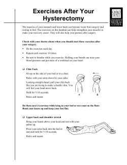

© Copyright 2026