
Original Article Adult Attachment Predicts Maternal Brain
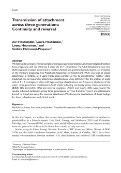
Original Article
Neuropsychopharmacology (2009) 34, 2655–2666; doi:10.1038/npp.2009.103;
published online 26 August 2009
Adult Attachment Predicts Maternal Brain
and Oxytocin Response to Infant Cues
Lane Strathearn1,2,3, Peter Fonagy4,5, Janet Amico6,7 and P Read Montague2,5
1.
2.
3.
4.
5.
6.
7.
1
Department of Pediatrics, The Meyer Center for Developmental Pediatrics,
Baylor College of Medicine, Texas Children's Hospital, Houston, TX, USA
2
Department of Neuroscience, Human Neuroimaging Laboratory, One Baylor
Plaza, Baylor College of Medicine, Houston, TX, USA
3
School of Medicine, The University of Queensland, Queensland, Australia
4
Research Department of Clinical, Educational and Health Psychology,
University College London, Gower St, London, UK
5
Menninger Department of Psychiatry and Behavioral Sciences, One Baylor
Plaza, Baylor College of Medicine, Houston, TX, USA
6
Department of Medicine, University of Pittsburgh School of Medicine,
University of Pittsburgh, Pittsburgh, PA, USA
7
Department of Pharmaceutical Sciences, University of Pittsburgh School of
Pharmacy, University of Pittsburgh, Pittsburgh, PA, USA
Correspondence: Dr L Strathearn, The Meyer Center for Developmental Pediatrics,
Baylor College of Medicine, Texas Children's Hospital, One Baylor Plaza S-104,
Houston, TX 77030, USA. Tel: +1 713 798 3822, Fax: +1 713 798 4488, E-mail:
[email protected]
Received 15 November 2008; Revised 15 July 2009; Accepted 16 July 2009;
Published online 26 August 2009.
Top of page
Abstract
Infant cues, such as smiling or crying facial expressions, are powerful motivators of
human maternal behavior, activating dopamine-associated brain reward circuits.
Oxytocin, a neurohormone of attachment, promotes maternal care in animals,
although its role in human maternal behavior is unclear. We examined 30 first-time
new mothers to test whether differences in attachment, based on the Adult
Attachment Interview, were related to brain reward and peripheral oxytocin
response to infant cues. On viewing their own infant's smiling and crying faces
during functional MRI scanning, mothers with secure attachment showed greater
activation of brain reward regions, including the ventral striatum, and the oxytocinassociated hypothalamus/pituitary region. Peripheral oxytocin response to infant
contact at 7 months was also significantly higher in secure mothers, and was
positively correlated with brain activation in both regions. Insecure/dismissing
mothers showed greater insular activation in response to their own infant's sad
faces. These results suggest that individual differences in maternal attachment may
be linked with development of the dopaminergic and oxytocinergic neuroendocrine
systems.
Keywords:
attachment, mother–infant relations, dopamine, oxytocin, functional MRI, insula
Top of page
INTRODUCTION
The attachment relationship between infants and their caregivers is critical for
human development, ensuring infant survival and optimal social, emotional, and
cognitive development (Insel and Young, 2001; Sroufe et al, 2005). The
relationship between a mother and her infant is particularly salient, with evidence
that the biological processes of pregnancy, parturition, and lactation may all
contribute to the establishment of the mother–infant bond (Strathearn et al, 2009;
Kinsley et al, 2008).
In both human and animal research, significant differences in early maternal
caregiving have been observed—ranging from sensitive and responsive infant care
to maternally perpetrated abuse or neglect (Strathearn et al, 2009; Sroufe et al,
2005), with corresponding differences in infant health and developmental outcomes
(Sroufe et al, 2005; Strathearn et al, 2001; Thompson, 2008; Francis et al, 1999;
Weaver et al, 2004). Understanding the neurobiological processes underlying these
differences in maternal behavior may help us to identify more effective treatment
and preventative strategies.
The neurobiology of attachment behavior has been studied extensively in animal
models (Insel and Young, 2001; Swain et al, 2007), and more recently in humans
using functional magnetic resonance imaging (fMRI) (Lorberbaum et al, 2002;
Bartels and Zeki, 2004; Swain et al, 2007; Strathearn et al, 2008). Although there
is likely to be a complex interaction of multiple neuroendocrine systems, two
specific systems have been shown to consistently play a role in promoting and
maintaining maternal behavior: (1) the dopaminergic reward processing system
(Champagne et al, 2004; Strathearn et al, 2008; Ferris et al, 2005) and (2) the
oxytocinergic system (Bartels and Zeki, 2004; Champagne et al, 2001; Levine et al,
2007). Oxytocin, a neuromodulatory hormone produced in the hypothalamus, has
well-described central actions associated with the onset of maternal behavior, as
well as peripheral actions in stimulating uterine contraction during labor and milk
ejection during lactation. It is released in response to stimuli such as infant
suckling, somatosensory touch, or even the sight or sound of a nursing mother's
infant (Lucas et al, 1980; McNeilly et al, 1983; Johnston and Amico, 1986; UvnasMoberg et al, 1993). Oxytocin release into the peripheral circulation occurs within
seconds of stimulation and its half-life has been estimated to be 6–7 min
(Vankrieken et al, 1983; Robinson and Verbalis, 2003). In randomized, placebocontrolled trials, intranasal oxytocin produces a broad range of social effects,
including enhanced social memory, improved eye gaze when viewing faces,
increased recognition and memory of facial expressions and identity, and increased
manifestations of trust (Domes et al, 2007; Savaskan et al, 2008; Baumgartner et
al, 2008; Kosfeld et al, 2005; Guastella et al, 2008b; Guastella et al, 2008a).
Oxytocin receptors are located in the ventral striatum, a key dopaminergic brain
region, and receptor binding is linked functionally to maternal behavior in the rat
(Olazabal and Young, 2006a). Thus, oxytocin may link social cues, such as infant
facial expressions, with dopamine-associated reinforcement pathways.
The extent to which these biological systems explain differences in the quality of
human attachment between mothers and infants is yet to be explored (Strathearn,
2006). In this study, we aimed to measure the differences in maternal brain reward
activation and peripheral oxytocin release in response to infant cues, based on the
mother's adult attachment classification. We hypothesized that mothers with secure
patterns of adult attachment would show an increased brain response to their own
infant's face in mesocorticolimbic reward regions, including the midbrain ventral
tegmental area, the ventral striatum, and the medial prefrontal cortex (mPFC), and
that this would be true on viewing both happy and sad infant face cues. We also
hypothesized that 'secure' mothers would show an enhanced peripheral oxytocin
response on interacting with their infants, which would correlate with maternal
brain responses.
Top of page
MATERIALS AND METHODS
Study Setting and Participants
In this cohort study, we recruited first-time pregnant women during the third
trimester of pregnancy and monitored them for 14 months postnatally. Recruitment
occurred in Houston, Texas, between August 2004 and April 2006 and was through
prenatal clinic visits and advertisements on billboards, in magazines, and on the
internet. We excluded potential subjects who were on psychotropic medications,
were using cigarettes during pregnancy, were left-handed, or had any
contraindication to MRI scanning. Research was approved by the Institutional
Review Board at Baylor College of Medicine, and all subjects provided written
informed consent.
Study Design
Visit 1: Pregnancy
(Figure 1) During this visit, each enrolled woman participated in a modified version
of the Adult Attachment Interview (AAI) (Crittenden, 2004; George et al, 1996), a
semi-structured 1½ -2 h-long interview involving specified questions and follow-up
inquiries relating to childhood relationships with attachment figures, usually
parents. The modified version was chosen because of its theoretical links with
patterns of information processing in the brain (Strathearn, 2006; Crittenden,
2008). Each digitally recorded interview was transcribed (with personally identifying
details altered to preserve anonymity), and coded blindly to classify each woman's
adult attachment pattern, which was not revealed until study completion.
Figure 1.
Study timeline and data collected at each of four study visits. AAI,
adult attachment interview; PDQ, personality disorder questionnaire 4+; BDI, beck
depression inventory; PANAS, positive and negative affect schedule; ATQ, adult
temperament questionnaire—short form; IBQ, infant behavior questionnaire—
revised; PSI, parenting stress index; WTAR, Wechsler test of adult reading.
Full figure and legend (137K)
During this visit, we also collected sociodemographic data, and screening
information for depression (Beck Depression Inventory, BDI) (Beck et al, 1996) and
personality disorders (Personality Disorder Questionnaire 4+, PDQ) (see
Supplementary Table 1). We repeated the BDI on each postnatal visit, and
calculated a mean postnatal score.
Visit 2: Videotaping and oxytocin sampling
Approximately 7 months after delivery, each mother and infant attended a session
at the Human Neuroimaging Laboratory. We requested that mothers abstain from
caffeine and tobacco for 2–3 h before the visit. After separating from their infants,
the mothers had an intravenous cannula inserted, and 20 min later had blood
drawn for baseline measurements of serum oxytocin, free cortisol, epinephrine, and
norepinephrine. We also measured serum estradiol, progesterone, and
human chorionic gonadotropin levels to exclude a current pregnancy
and to assess menstrual status. During this separation period, we videotaped each
infant to obtain still images for use in the subsequent fMRI visit. Smiling, neutral,
and crying faces were elicited in a standardized setting, as described elsewhere
(Strathearn et al, 2008). The mother and infant were then reunited for a 5-min
'free-play' period in which they physically interacted on the floor, after which
another blood sample was drawn. They then participated in a 6-min modified 'stillface' procedure (Koos and Gergely, 2001), during which mother and infant could
hear and see each other through a mirror, but not interact physically. We then
obtained a third blood sample after the mother left the room, followed by a final
blood drawn after 20 min of separation. Before and after the interaction period,
each mother rated their current feelings using the Positive and Negative Affect
Schedule (PANAS) (Watson et al, 1988), a 5-point rating of 20 affect states, such
as 'interested', 'excited', 'irritable', and 'nervous'.
Each mother also completed a 120-item self-report questionnaire, the Parenting
Stress Index (PSI) (Abidin, 1995), designed to help identify potentially
dysfunctional parent–child relations. We assessed adult and infant temperament
using the self-report Adult Temperament Questionnaire—Short form (ATQ) and the
Infant Behavior Questionnaire—Revised (IBQ) (Gartstein and Rothbart, 2003). The
mothers also reported their breastfeeding status, which was repeated at Visit 3.
Visit 3: Scanning
At
11 months after delivery, a minimum of 3 months after the
videotaping session, each mother underwent fMRI scanning while viewing 60
unique infant face images, 30 of her own infant and 30 of the matched unknown
infant face. There were 6 face categories, each containing 10 images, namely, ownhappy (OH), own-neutral (ON), own-sad (OS), unknown-happy (UH), unknownneutral (UN), and unknown-sad (US). Each mother viewed randomly presented
baby face images for 2 s each within a rapid event-related fMRI design, with a
random inter-stimulus interval of 2, 4, or 6 s (Figure 2). Visual images were
generated using a computer-controlled LCD projector, and presented to the mother
on an overhead mirror display.
Figure 2.
Baby face presentation paradigm in functional MRI experiment.
Infant face images were presented for 2 s, followed by a variable 2–6 s period of a
plain black screen. Six stimulus types were presented in random order: own-happy
(OH), own-neutral (ON), own-sad (OS), unknown-happy (UH), unknown-neutral
(UN), unknown-sad (US). Reproduced with permission from Pediatrics, Vol. 122,
Pages 40–51, Copyright © 2008 by the AAP.
Full figure and legend (190K)
Visit 4: Child follow-up
Finally, at 14 months of age we performed a general assessment of child
development using the Screening Test of the Bayley Scales of Infant and Toddler
Development (Bayley, 2006).
Variables and Statistical Methods
Predictor variable—adult attachment
We determined each mother's adult attachment classification using the AAI (George
et al, 1996; Fonagy et al, 1991; Crittenden, 2004), which categorizes the mother's
capacity to form secure attachment relationships on the basis of a narrative of her
own attachment experience. Over the past 25 years, over 200 studies have
reported over 10 000 AAIs (van IJzendoorn and Bakermans-Kranenburg, 2009).
From both cross-sectional and prospective longitudinal studies, adult attachment
has been shown to reliably predict maternal behavior patterns, the development of
infant attachment (van IJzendoorn, 1995), and infant social and emotional
development (Sroufe et al, 2005). We chose to measure attachment during
pregnancy using a longitudinal design to preclude the possibility that the infant's
temperament or mother–infant interaction patterns might influence the way the
mother discusses her own attachment experiences.
The coding is based on the subject's coherence and consistency in describing
attachment-related experiences and their effects on current functioning
(Crittenden, 2004). The three basic styles, which parallel Ainsworth's original
classification of attachment in infancy (Ainsworth and Bell, 1970) include Type A
'Insecure/Dismissing', Type B 'Secure' and Type C 'Insecure/Preoccupied'.
Individuals with Type B attachment styles tend to provide balanced descriptions of
childhood experiences, using both temporal/causal order and affect to describe both
positive and negative events and feeling states. Individuals with Type A attachment
describe events or feelings in more cognitive terms, avoiding or inhibiting displays
of negative affect. In contrast, Type C individuals exaggerate affective responses,
with omitted or distorted cognitive processing (Crittenden, 2008). Fifty percentage
of the transcripts were double coded to ensure reliability, with an 87% agreement
with regard to a four-group classification (
were resolved through conferencing between coders.
=0.78). Discrepancies
Potential confounding variables
We measured a variety of socioeconomic and behavioral factors to compare the
characteristics of women in the two attachment groups (see Supplementary Table
1). Continuous measures were evaluated using t-tests or the Mann–Whitney U-test
for nonparametric data (as determined from histogram analysis). We compared
categorical variables using the
2
test, or Fisher exact test when
numbers were insufficient. We used the Kendall's
-b test for ordinal
or ranked nonparametric variables. We compared PANAS ratings of the mothers'
affect before and after contact with their infants between groups using a repeated
measure analysis of variance (ANOVA).
Serial measurements of cortisol, norepinephrine, and epinephrine were also
compared between attachment groups using linear mixed modeling. Analyses were
performed using SPSS (version 15.0) and P<0.05 (two-tailed) was considered
statistically significant.
Outcome variables
Oxytocin response: We used linear mixed modeling to assess the effects of
attachment group, 'mother–infant interaction' time point, breastfeeding status, and
all two-way interactions, on oxytocin response. Residual plots were used to confirm
normality of distribution. Cases with missing data points were excluded (one Type B
and two Type A subjects). The difference in mean oxytocin concentration between
attachment groups, at each time point, was compared using a z-test (with
Bonferroni correction for multiple comparisons;
0.0125 was considered statistically significant). The mean oxytocin concentration
from the two 'mother–infant interaction' time points (which were highly correlated:
rS=0.77, P<0.001) (see Figure 1) was recomputed as a percentage change from
the first baseline measure, to provide a single index for correlation with fMRI data.
To determine the correlation between 'percentage change in oxytocin' and fMRI
blood-oxygen-level-dependent (BOLD) activation measured 4 months later (z-
transformed
weights), we calculated a Spearman correlation
coefficient. We used a Bonferroni correction to adjust the
multiple comparisons with the
level for
weights for the 6 types of infant face
(OH, OS, UN, etc). An
<0.008 was considered statistically
significant.
We measured oxytocin concentrations using a sensitive and specific liquid phase
radioimmunoassay, in which oxytocin antiserum does not cross-react with arginine
vasopressin or other oxytocin-like peptides (Amico et al, 1985). The lower limit for
detectability of the assay is 0.5 pg/ml; inter- and intra-assay coefficients of
variation are <10%.
Functional MRI brain response: We prepared 30 standardized face images from
each infant (10 happy, 10 neutral, and 10 sad) for use in the fMRI scanning
paradigm, along with 30 images from an 'unknown' baby, which were matched on
age, race, and independently coded degree of affect (Figure 2) (Strathearn et al,
2008). To ensure that the degree of infant facial affect did not vary between
attachment groups, all faces were recoded by three blinded raters using the 9-point
Self-Assessment Manikin (Bradley and Lang, 1994) (ICC=0.90). Using a mixed
model three-way ANOVA, we saw no main effects for attachment group (F2,28=1.9,
NS) or order of presentation (Wilk's
=0.502, F9,20=2.0, NS).
Similarly, none of the interactions with attachment security were significant.
Imaging was performed using a 3 Tesla Siemens Allegra head-only MRI scanner.
High-resolution T1-weighted structural images (192 slices, in plane resolution 256
256; field of view [FOV] 245 mm; slice thickness 1 mm) were first
acquired, followed by whole-brain functional runs of around 185 scans (gradient
recalled echo planar imaging; 37 slices; repetition time 2000 msec; echo time
25 msec; flip angle, 90°; 64
64 matrix [in plane resolution]; FOV
220 mm; slice thickness 3 mm; positioned at 30 degrees in the axial plane to the
anterior commissure/posterior commissure line). Imaging data for each subject
were preprocessed in BrainVoyager QX, version 1.7.9 and analyzed in version
1.9.10, as described earlier (Strathearn et al, 2008). Coregistration of functional
and anatomical data for individual subjects confirmed that the functional data did
include the hypothalamus/pituitary region (see representative image of
coregistration in Supplementary Figure 1).
A BrainVoyager protocol file was created for each functional run, representing the
timing of each stimulus event. Each predictor was then convolved using a double-
hemodynamic response function. Using the general linear model,
effects for the whole group (n=30) were evaluated using a random effects
between-subjects analysis. After specifying a particular contrast in stimulus types
(eg, OH>UH or OS>US), a group t-map was generated onto a template threedimensional anatomical image. An activation map threshold was determined using a
false discovery rate (FDR) of 5% to control for multiple comparisons, and a cluster
threshold of four voxels. Smaller cluster thresholds were also examined in the
striatum (three voxels) and brainstem (one voxel) to reveal activation of smaller
nuclei. Anatomical regions were identified using the automated 'Talairach Daemon'
(Lancaster et al, 2000), and confirmed manually using a human brain atlas (Mai et
al, 2004).
Next, we compared activation patterns between attachment groups using a twofactor random effects ANOVA model, with fixed effects analyses for (1) 'infant face
category' as a repeated measure within-factor variable and (2) 'attachment group'
as a between-factor variable. Whole brain differences in activation were assessed
using a threshold of q(FDR) <0.05. Mean
weights were calculated
and compared between the two attachment groups, in a priori regions of interest
(midbrain, striatum, prefrontal cortex) and the hypothalamus, using the t-test and
the Mann–Whitney U-test for nonparametric data.
Top of page
RESULTS
Description of Subjects
Of 112 women recruited during pregnancy, 61 met eligibility criteria and were
enrolled in the study, with 44 participating in fMRI scanning approximately 1 year
later. Ten women were unable to be scanned (nine due to a current pregnancy and
one because of a history of seizures) and seven had withdrawn from the study or
were lost to follow-up. Of the 44 scanned women, 15 were classified as having
insecure/dismissing attachment (Type A). A further 16 women demonstrated secure
patterns of attachment, without unresolved trauma or loss (Type B). A small group
(n=4) were classified as insecure/preoccupied (Type C), and the remaining nine
women had combined or atypical patterns. We specifically compared women from
the two predominant attachment groups—Types A and B, and to ensure equal
numbers in each group, one Type B mother was excluded.
The 30 women who were enrolled into the study were generally from middle to high
socioeconomic backgrounds (based on the Four-Factor Index of Social Status [AB
Hollingshead, PhD, working paper, 1985]: mean score 51.4
9.4 at
the time of enrollment). Eighty percent had completed a college or graduate degree
and 70% were married. The median WTAR-predicted IQ for the group was 112
(range, 81–120). Sixty percent identified themselves as non-Hispanic White, onequarter were Hispanic and one-tenth African American.
Subjects within the two attachment groups did not differ in age, race, education,
socioeconomic status, marital status, or predicted IQ (see Materials and methods
and Supplementary Table 1). Both groups were also comparable in screening
measures of personality disorder risk and parenting stress (at the 7-month visit)
and depression (measured at each study visit). There were no significant
differences seen in temperament subscales of either the mother or child, the
mothers' ratings of emotions before and after mother–infant interaction (based on
the PANAS during Visit 2) (Watson et al, 1988) or in scales of infant development
(measured during Visit 4). We also found no significant difference in breastfeeding
status at Visits 2 or 3, although Type B mothers tended to breastfeed longer and
Type A mothers were significantly more likely to be separated from their child for
longer periods of time each week (P=0.03).
Oxytocin Response to Mother–Infant Interaction (Visit 2)
During the 7-month postpartum visit, Type B mothers showed a significantly higher
peripheral oxytocin response after periods of mother–infant interaction (Figure 3a;
time point by attachment group interaction effect adjusted for breastfeeding at this
visit, F=2.9, P=0.04). Although there were no differences between attachment
groups in the two baseline measurements, after the 5-min 'free-play' interaction
Type B mothers had significantly higher oxytocin levels (P=0.01). This difference
persisted into an additional mirror-based interaction period, although it was no
longer statistically significant (P=0.07). There were no significant differences in
serum-free cortisol, epinephrine or norepinephrine, or in baseline serum estradiol or
progesterone.
Figure 3.
Peripheral oxytocin and related brain activation in response to infant
cues. (a) Mothers with Type B (secure) attachment patterns show a greater
peripheral oxytocin response during an episode of physical interaction with their
infant (mean
SEM; Bonferroni corrected comparison at free-play
time point, P=0.01). The first baseline sample was collected 20 min after mother–
infant separation; the second immediately after a 5-min 'free-play' involving direct
physical contact between the mother and infant. The third sample was after a
modified still-face procedure, in which the mother was in direct visual and auditory
contact with her infant (through a mirror) but was physically separated by a screen
divider. The final sample was collected after a further 20-min period of complete
mother–infant separation. (b) Compared with Type A mothers, Type B mothers
show greater activation of the hypothalamus/pituitary region in response to own vs
unknown infant face images (all affect groups combined) (mean
SEM, t=4.2, P=0.0003). The whole brain analysis threshold was
q(FDR)<0.05; P<0.002. Structural brain image created from average of all
subjects. Inset of magnified hypothalamic/pituitary region (single subject image to
improve anatomical clarity). (c) Peripheral oxytocin response correlates with
activation of hypothalamus/pituitary region in response to neutral own-infant face
cues (rS=0.60, P=0.001). A single outlying value was omitted from the graph, but
not the statistical calculations.
Full figure and legend (194K)
Whole Group Analysis of Maternal Brain Responses (Visit 3)
On the whole brain analysis, when mothers viewed their own infant's happy faces,
compared with UH faces (OH>UH), key dopamine-associated reward processing
regions were activated, overlapping earlier reported regions (Strathearn et al,
2008), and including the substantia nigra, dorsal putamen, and thalamic nuclei. In
addition, activation was seen in various regions of the striatum, caudate nuclei,
insular cortex, superior temporal gyrus, and pre- and postcentral gyri (P<0.05, FDR
corrected). As in the prior study, no significant activation was seen on contrasting
own vs unknown sad (OS>US) or neutral (ON>UN) infant faces, or in contrasting
face affect groups, testing 'own' and 'unknown' faces separately or combined (eg
OH>ON, US>UN, H>S). After combining all affect groups together and contrasting
own vs unknown faces, an activation pattern overlapping our earlier study results
(Strathearn et al, 2008) was seen, including both mesocorticolimbic (ventral
tegmental area and ventral striatum) and nigrostriatal pathway (substantia nigra
and dorsal striatum) activation, but not the prefrontal or anterior cingulate cortex.
Attachment Group Comparisons
We next compared own vs unknown (O>U) infant face responses between the two
attachment groups after combining all affect groups, to look specifically for
hypothesized differences in activation of dopamine-associated brain reward regions
(in the midbrain, striatum, and forebrain) and the hypothalamus. Type B mothers
showed significantly more activation in the lateral prefrontal cortex bilaterally, the
left mPFC and the hypothalamus/pituitary region (O>U; P<0.05, FDR corrected)
(Table 1; Figure 3b; Supplementary Figure 1). In the hypothalamus/pituitary
region, where oxytocin is produced and released peripherally, Type B mothers had
a greater response to own-infant faces than did Type A mothers (median
values 1.54 vs -2.09; Mann–Whitney U-test, z=-2.10, P<0.05).
Furthermore, among Type B mothers, the response was greater for their own infant
compared with unknown infant faces (median
values 1.54 vs -2.50;
z=-2.10, P<0.05) (Supplementary Figure 2). On further fMRI analysis of the three
individual affect groups (happy, neutral, and sad), only neutral faces (ON>UN)
produced a similar activation pattern between attachment groups within the
hypothalamic/pituitary region (P<0.05, FDR corrected). The activation signal in
response to ON infant faces correlated significantly with the mother's peripheral
oxytocin response on interaction with her infant (z-transformed
weights and percentage change in oxytocin; rS=0.60, P=0.001) (Figure 3c). When
attachment groups were compared in this correlation analysis, no differences in line
slope (P=0.80) or position (P=0.12) were detected. No correlation was seen
between oxytocin response and brain activation in the mPFC, or when viewing
unknown infant faces.
Table 1 - Areas of Significant Activation Within the Prefrontal Cortex,
Striatum, and Midbrain, when Comparing Type A and Type B Attachment
Groups.
Full table
In post hoc analyses, we then directly compared own-infant faces between
attachment groups, in each affect state separately (eg OH in Type A vs Type B),
without the inclusion of unknown infant face comparisons. From the hypothesized
regions of interest, for the happy face contrast, Type B mothers showed
significantly greater activation in the ventral striatum, as well as the orbitofrontal
cortex and mPFC bilaterally (Table 1). An equal but opposite BOLD response was
seen in Type A mothers in the ventral striatum (Figure 4a). In the mPFC, Type B
mothers had a much larger increase in mean
values compared with
Type A mothers. In contrast, Type A mothers showed significantly more activation
in the dorsolateral prefrontal cortex (dlPFC) bilaterally.
Figure 4.
Brain responses to happy and sad own-infant faces, contrasting
mothers with Type A (insecure/dismissing) and B (secure) attachment
classifications (mean
values
SEM) (a) Type B
mothers show greater activation of the ventral striatum (VS; t=3.1, P<0.005) and
medial prefrontal cortex (mPFC; t=3.0, P<0.01) in response to own-happy infant
faces. (b) Type B mothers show greater activation of the right ventral striatum
(t=3.0, P<0.01) in response to own-sad infant faces. Type A mothers show greater
activation of the right anterior insula (t=-3.9, P<0.0005).
Full figure and legend (239K)
In response to own infant sad faces, the right ventral striatum was also more active
in Type B mothers (though at a more anterior position than seen in the happy face
contrast) (Table 1; Figure 4b). Type A mothers again showed more activation of the
dlPFC in response to OS faces, as well as a much stronger activation signal in the
anterior insula bilaterally, compared with Type B mothers (Figure 4b). Activation in
the right ventral striatum in response to ON infant faces was also highly correlated
with peripheral oxytocin response (rS=0.57, P=0.002; Figure 5). Unknown infant
faces produced no such correlation. None of the contrasts, for happy or sad faces,
showed significant differences across attachment groups in activation of midbrain
regions.
Figure 5.
Peripheral oxytocin response after episodes of mother–infant
interaction correlates with activation in the right ventral striatum (area shown in
Figure 4b) in response to neutral own-infant face cues (rS=0.57, P=0.002).
Percentage oxytocin change calculated from the first baseline measurement and a
mean of the second and third samples, which were taken during episodes of
mother–infant interaction.
Full figure and legend (69K)
Overall, mothers with Type B attachment tended to show greater left hemisphere
activation, whereas Type A had predominantly right hemisphere activation,
especially for happy and sad infant faces (Table 1).
Top of page
DISCUSSION
This study demonstrates group differences in maternal brain and oxytocin response
to infant cues, based on adult attachment patterns measured before the birth of the
mother's first child. As hypothesized, mothers with secure vs insecure/dismissing
attachment showed increased activation of mesocorticolimbic reward brain regions,
on viewing their own infant's smiling face. Furthermore, they showed an increased
peripheral oxytocin response while interacting with their infants, which was
positively correlated with activation of oxytocinergic and dopamine-associated
reward processing regions of the brain (hypothalamus/pituitary and ventral
striatum). Finally, striking differences in brain activation were seen in response to
their own infant's sad facial affect. Securely attached mothers continued to show
greater activation in reward processing regions, whereas 'insecure/dismissing'
mothers showed increased activation of the anterior insula, a region associated with
feelings of unfairness, pain, and disgust (see review, Montague and Lohrenz, 2007).
The lack of 'reward' activation in mothers with insecure/dismissing attachment is
consistent with a recent study of brain responses to smiling adult faces and positive
task feedback (Vrticka et al, 2008), where ventral striatum activation was inversely
correlated with dismissing attachment scores. In linking attachment security with
ventral striatal activation, our findings suggest that for securely attached mothers,
infant cues (whether positive or negative in affect) may act as an important signal
of 'incentive salience' (Berridge, 2007), reinforcing, and motivating responsive
maternal care.
Striatal activation and de-activation has also been modeled to represent deviations
from expectation, with regard to the timing and magnitude of predicted reward
(Montague et al, 1996; Schultz et al, 1997; Daw and Doya, 2006). Specifically, an
unexpected reward signal predicts an increase in dopaminergic activity and in
measurable neural response at the level of the striatum, whereas the omission of
an expected reward at a specific time predicts a decrease in dopamine-related
response. Although the prediction error model has not been tested directly with
regard to subjective feelings, our results suggest that insecure/dismissing mothers
may interpret their own infant's face (regardless of affect) as representing an
omitted reward. This is consistent with the theoretical and observed nature of
dismissing adult attachment, in which close interpersonal relationships are
perceived as being less intrinsically rewarding (Cassidy and Shaver, 1999).
Furthermore, mothers with insecure/dismissing attachment styles showed greater
activation of dlPFC and anterior insula in response to their own infant's sad face,
suggesting cognitive control over a negative affective response (Greene et al, 2004;
Sanfey et al, 2003). In line with our current understanding that activation of the
anterior insula may signal 'norm violations' (Montague and Lohrenz, 2007),
insecure/dismissing mothers may cognitively appraise their infant's sad affect as a
violation of an 'expected' affect state. This may lead to avoidance or rejection of
negative infant cues (Sanfey et al, 2003), rather than the 'approach' responses
seen in Type B secure mothers. While the ventral striatal activation seen in Type B
mothers is associated with anticipated gain, right anterior insula activation is seen
in anticipation of loss (Knutson et al, 2007). These results are consistent with an
earlier published model of the cortical organization of the attachment system
(Strathearn, 2006; Crittenden, 2008), which postulates that individuals with
insecure/dismissing attachment are biased toward cognitive information processing,
and tend to inhibit negative affective responses. Although anterior insula activation
has also been linked with empathic responses to a loved one's feeling of physical
pain (Singer et al, 2004), dismissing individuals score much lower on a scale of
emotional empathy (Sonnby-Borgstrom and Jonsson, 2004), making this
interpretation less likely.
Oxytocin has long been implicated as an important neuromodulatory hormone
involved in maternal behavior (Insel, 1992; Insel and Young, 2001). Synthesized in
the paraventricular nucleus of the hypothalamus, there are oxytocinergic
projections to the posterior pituitary gland where it is released into the blood
stream. In addition, oxytocin neurons project centrally to regions important in the
manifestation of social and maternal behaviors (Numan, 2006). There is some
evidence to suggest that oxytocin neurons in the hypothalamus may directly project
to the ventral striatum, facilitating dopamine release (Liu and Wang, 2003; Ross et
al, 2009) and thus linking social and maternally related cues to reward processing
and behavioral reinforcement (Insel, 2003). Rodent studies have demonstrated that
oxytocin receptor binding in the nucleus accumbens (a nucleus of the ventral
striatum) facilitates the onset of maternal behavior (Olazabal and Young, 2006a;
Olazabal and Young, 2006b).
Although there has been some controversy surrounding the relationship between
peripheral and central oxytocin production (McGregor et al, 2008), these results,
while tentative, are consistent with the idea that differences in peripheral oxytocin
response may reflect central oxytocin production and contribute to individual
differences in maternal caregiving behavior. Other studies have shown reduced
peripheral oxytocin responses in cocaine addicted mothers (Light et al, 2004) and
in pregnant women with lower maternal–fetal attachment scores (Levine et al,
2007). Furthermore, reduced peripheral oxytocin levels have been seen in
orphanage–adopted children with histories of early neglect, who display severe
impairments in social reciprocity (Fries et al, 2005). The observation that oxytocin
levels are higher in securely attached mothers after interaction with their infants
suggests the importance of this neuropeptide in mediating attachment and social
behaviors, as seen in human randomized placebo-controlled trials of intranasal
oxytocin (Baumgartner et al, 2008; Guastella et al, 2008b), as well as in rodent
studies (Insel and Young, 2001; Champagne et al, 2001; Insel, 1992; Liu and
Wang, 2003). In our study, the correlation of interaction-elicited peripheral
oxytocin with the activation of reward regions in the brain suggests that oxytocin
may be one mechanism by which socially relevant cues activate dopaminergic
pathways and thus reinforce behavior. Mothers with secure attachment patterns
when interacting with their infants may produce more oxytocin, which increases the
experience of reward and in turn may contribute to the mother's ability to provide
consistent, nurturant care. However, caution is warranted in interpreting these
findings. We have no independent measure of (1) the effect of oxytocin secretion
on the estimated
values in specified brain regions, nor (2) whether
oxytocin is actually released during this behavioral condition. In fact, peripheral
oxytocin measurements during real-time mother–infant interaction were collected 4
months before fMRI scanning, providing no opportunity to examine simultaneous
correlations. Nevertheless, the correlation between oxytocin and hemodynamic
response separated over time suggests that the oxytocin response may reflect an
enduring trait difference associated with attachment security.
Numerous previous investigations have shown that mothers with insecure
attachment patterns are less likely to establish secure relationships with their
children, and that their children tend to have greater difficulties regulating affect,
forming peer relationships and establishing secure attachment relationships
themselves (Sroufe et al, 2005; van IJzendoorn, 1995). Although the
transgenerational transmission of attachment has been frequently observed, its
mechanism is still poorly understood (van IJzendoorn, 1995). This study may help
shed light on this question, with evidence that secure attachment is associated with
more intense maternal reward activation to infant facial expressions, whereas
insecure/dismissing mothers show greater insula response to negative infant cues.
Additional research is needed to confirm these findings in larger cohorts of mothers,
including mothers with insecure/preoccupied attachment. A randomized-controlled
trial of intranasal oxytocin may also help to clarify any causal relationship between
oxytocin response and maternal brain activation.
In conclusion, this study is the first to examine the neuroendocrine basis of human
mother–infant attachment. As such, it may help us to better understand the
transmission of attachment patterns across generations and how secure maternal
attachment may confer the observed developmental advantages in infants and
children (Sroufe et al, 2005; van IJzendoorn, 1995).
See related commentary by Rilling on page 2621.
Top of page
Notes
DISCLOSURE
The authors declare no conflict of interest.
Top of page
References
1. Abidin RR (1995). Parenting Stress Index, Professional Manual. Psychological
Assessment Resources: Lutz, FL.
2. Ainsworth MD, Bell SM (1970). Attachment, exploration, and separation:
illustrated by the behavior of one-year-olds in a strange situation. Child Dev
41: 49–67. | Article | PubMed | ChemPort |
3. Amico JA, Ervin MG, Leake RD, Fisher DA, Finn FM, Robinson AG (1985). A
novel oxytocin-like and vasotocin-like peptide in human plasma after
administration of estrogen. J Clin Endocrinol Metab 60: 5–
12. | Article | PubMed | ChemPort |
4. Bartels A, Zeki S (2004). The neural correlates of maternal and romantic
love. Neuroimage 21: 1155–1166. | Article | PubMed | ISI
5. Baumgartner T, Heinrichs M, Vonlanthen A, Fischbacher U, Fehr E (2008).
Oxytocin shapes the neural circuitry of trust and trust adaptation in humans.
Neuron 58: 639–650. | Article | PubMed | ChemPort |
6. Bayley N (2006). Bayley Scales of Infant and Toddler Development.
Harcourt Assessment: San Antonio, TX.
7. Beck AT, Steer RA, Brown GK (1996). Manual for the Beck Depression
Inventory-II. Psychological Corporation: San Antonio, TX.
8. Berridge KC (2007). The debate over dopamine's role in reward: the case for
incentive salience. Psychopharmacology (Berl) 191: 391–
431. | Article | PubMed | ChemPort |
9. Bradley MM, Lang PJ (1994). Measuring emotion: the self-assessment
manikin and the semantic differential. J Behav Ther Exp Psychiatry 25: 49–
59. | Article | PubMed | ISI | ChemPort |
10. Cassidy J, Shaver PR (1999). Handbook of Attachment: Theory, Research,
and Clinical Applications. The Guilford Press: New York.
11. Champagne F, Diorio J, Sharma S, Meaney MJ (2001). Naturally occurring
variations in maternal behavior in the rat are associated with differences in
estrogen-inducible central oxytocin receptors. Proc Natl Acad Sci USA 98:
12736–12741. | Article | PubMed | ChemPort |
12. Champagne FA, Chretien P, Stevenson CW, Zhang TY, Gratton A, Meaney MJ
(2004). Variations in nucleus accumbens dopamine associated with
individual differences in maternal behavior in the rat. J Neurosci 24: 4113–
4123. | Article | PubMed | ISI | ChemPort |
13. Crittenden PM (2008). Raising Parents. attachment, Parenting and Child
Safety. Willan Publishing: Devon, UK.
14. Crittenden PM (2004). Patterns of attachment in adulthood: a dynamicmaturational approach to analyzing The Adult Attachment Interview.
(Unpublished manuscript).
15. Daw ND, Doya K (2006). The computational neurobiology of learning and
reward. Curr Opin Neurobiol 16: 199–
204. | Article | PubMed | ISI | ChemPort |
16. Domes G, Heinrichs M, Michel A, Berger C, Herpertz SC (2007). Oxytocin
improves 'mind-reading' in humans. Biol Psychiatry 61: 731–
733. | Article | PubMed | ISI | ChemPort |
17. Ferris CF, Kulkarni P, Sullivan Jr JM, Harder JA, Messenger TL, Febo M
(2005). Pup suckling is more rewarding than cocaine: evidence from
functional magnetic resonance imaging and three-dimensional computational
analysis. J Neurosci 25: 149–156. | Article | PubMed | ChemPort |
18. Fonagy P, Steele H, Steele M (1991). Maternal representations of
attachment during pregnancy predict the organization of infant-mother
attachment at one year of age. Child Dev 62: 891–
905. | Article | PubMed | ChemPort |
19. Francis D, Diorio J, Liu D, Meaney MJ (1999). Nongenomic transmission
across generations of maternal behavior and stress responses in the rat.
Science 286: 1155–1158. | Article | PubMed | ISI | ChemPort |
20. Fries ABW, Ziegler TE, Kurian JR, Jacoris S, Pollak SD (2005). Early
experience in humans is associated with changes in neuropeptides critical for
regulating social behavior. PNAS 102: 17237–
17240. | Article | PubMed | ChemPort |
21. Gartstein MA, Rothbart MK (2003). Studying infant temperament via the
Revised Infant Behavior Questionnaire. Infant Behav Develop 26: 64–
86. | Article
22. George C, Kaplin N, Main M (1996). Adult Attachment Interview (third
edition). (Unpublished manuscript).
23. Greene JD, Nystrom LE, Engell AD, Darley JM, Cohen JD (2004). The neural
bases of cognitive conflict and control in moral judgment. Neuron 44: 389–
400. | Article | PubMed | ISI | ChemPort |
24. Guastella AJ, Mitchell PB, Dadds MR (2008a). Oxytocin increases gaze to the
eye region of human faces. Biol Psychiatry 63: 3–
5. | Article | PubMed | ChemPort |
25. Guastella AJ, Mitchell PB, Mathews F (2008b). Oxytocin enhances the
encoding of positive social memories in humans. Biol Psychiatry 64: 256–
258. | Article | ChemPort |
26. Insel TR (1992). Oxytocin—a neuropeptide for affiliation: evidence from
behavioral, receptor autoradiographic, and comparative studies.
Psychoneuroendocrinology 17: 3–35. | Article | PubMed | ISI | ChemPort |
27. Insel TR, Young LJ (2001). The neurobiology of attachment. Nat Rev
Neurosci 2: 129–136. | Article | PubMed | ISI | ChemPort |
28. Insel TR (2003). Is social attachment an addictive disorder? Physiol Behav
79: 351–357. | Article | PubMed | ISI | ChemPort |
29. Johnston JM, Amico JA (1986). A prospective longitudinal study of the
release of oxytocin and prolactin in response to infant suckling in long term
lactation. J Clin Endocrinol Metab 62: 653–
657. | Article | PubMed | ChemPort |
30. Kinsley CH, Bardi M, Karelina K, Rima B, Christon L, Friedenberg J et al.
(2008). Motherhood induces and maintains behavioral and neural plasticity
across the lifespan in the rat. Arch Sex Behav 37: 43–56. | Article | PubMed
31. Knutson B, Rick S, Wimmer GE, Prelec D, Loewenstein G (2007). Neural
predictors of purchases. Neuron 53: 147–
156. | Article | PubMed | ISI | ChemPort |
32. Koos O, Gergely G (2001). A contingency-based approach to the etiology of
'disorganized' attachment: the 'flickering switch' hypothesis. Bull Menninger
Clin 65: 397–410. | Article | PubMed | ChemPort |
33. Kosfeld M, Heinrichs M, Zak PJ, Fischbacher U, Fehr E (2005). Oxytocin
increases trust in humans. Nature 435: 673–
676. | Article | PubMed | ISI | ChemPort |
34. Lancaster JL, Woldorff MG, Parsons LM, Liotti M, Freitas CS, Rainey L et al.
(2000). Automated Talairach atlas labels for functional brain mapping. Hum
Brain Mapp 10: 120–131. | Article | PubMed | ISI | ChemPort |
35. Levine A, Zagoory-Sharon O, Feldman R, Weller A (2007). Oxytocin during
pregnancy and early postpartum: individual patterns and maternal-fetal
attachment. Peptides 28: 1162–1169. | Article | PubMed | ChemPort |
36. Light KC, Grewen KM, Amico JA, Boccia M, Brownley KA, Johns JM (2004).
Deficits in plasma oxytocin responses and increased negative affect, stress,
and blood pressure in mothers with cocaine exposure during pregnancy.
Addict Behav 29: 1541–1564. | Article | PubMed
37. Liu Y, Wang ZX (2003). Nucleus accumbens oxytocin and dopamine interact
to regulate pair bond formation in female prairie voles. Neuroscience 121:
537–544. | Article | PubMed | ISI | ChemPort |
38. Lorberbaum JP, Newman JD, Horwitz AR, Dubno JR, Lydiard RB, Hamner MB
et al. (2002). A potential role for thalamocingulate circuitry in human
maternal behavior. Biol Psychiatry 51: 431–445. | Article | PubMed
39. Lucas A, Drewett RB, Mitchell MD (1980). Breast-feeding and plasma
oxytocin concentrations. Br Med J 281: 834–
835. | Article | PubMed | ChemPort |
40. Mai JK, Assheuer J, Paxinos G (2004). Atlas of the Human Brain. Elsevier:
San Diego, CA.
41. McGregor IS, Callaghan PD, Hunt GE (2008). From ultrasocial to antisocial:
a role for oxytocin in the acute reinforcing effects and long-term adverse
consequences of drug use? Br J Pharmacol 154: 358–
368. | Article | PubMed | ChemPort |
42. McNeilly AS, Robinson IC, Houston MJ, Howie PW (1983). Release of
oxytocin and prolactin in response to suckling. Br Med J (Clin Res Ed) 286:
257–259. | Article | PubMed | ChemPort |
43. Montague PR, Dayan P, Sejnowski TJ (1996). A framework for
mesencephalic dopamine systems based on predictive Hebbian learning. J
Neurosci 16: 1936–1947. | PubMed | ISI | ChemPort |
44. Montague PR, Lohrenz T (2007). To detect and correct: norm violations and
their enforcement. Neuron 56: 14–18. | Article | PubMed | ChemPort |
45. Numan M (2006). Hypothalamic neural circuits regulating maternal
responsiveness toward infants. Behav Cogn Neurosci Rev 5: 163–
190. | Article | PubMed
46. Olazabal DE, Young LJ (2006a). Oxytocin receptors in the nucleus
accumbens facilitate 'spontaneous' maternal behavior in adult female prairie
voles. Neuroscience 141: 559–568. | Article | ChemPort |
47. Olazabal DE, Young LJ (2006b). Species and individual differences in
juvenile female alloparental care are associated with oxytocin receptor
density in the striatum and the lateral septum. Hormones Behav 49: 681–
687. | Article | ChemPort |
48. Robinson AG, Verbalis JG (2003). The Posterior Pituitary Gland. In: Larson
PR, Kronenberg HM, Melmed S, Polonsky KS (eds). Williams Textbook of
Endocrinology. WB Saunders: Philadelphia, pp 281–330.
49. Ross HE, Cole CD, Smith Y, Neumann ID, Landgraf R, Murphy AZ et al.
(2009). Characterization of the oxytocin system regulating affiliative
behavior in female prairie voles. Neuroscience 162: 892–
903. | Article | PubMed | ChemPort |
50. Sanfey AG, Rilling JK, Aronson JA, Nystrom LE, Cohen JD (2003). The neural
basis of economic decision-making in the ultimatum game. Science 300:
1755–1758. | Article | PubMed | ISI | ChemPort |
51. Savaskan E, Ehrhardt R, Schulz A, Walter M, Schachinger H (2008). Postlearning intranasal oxytocin modulates human memory for facial identity.
Psychoneuroendocrinology 33: 368–374. | Article | PubMed | ChemPort |
52. Schultz W, Dayan P, Montague PR (1997). A neural substrate of prediction
and reward. Science 275: 1593–1599. | Article | PubMed | ISI | ChemPort |
53. Singer T, Seymour B, O'Doherty J, Kaube H, Dolan RJ, Frith CD (2004).
Empathy for pain involves the affective but not sensory components of pain.
Science 303: 1157–1162. | Article | PubMed | ISI | ChemPort |
54. Sonnby-Borgstrom M, Jonsson P (2004). Dismissing-avoidant pattern of
attachment and mimicry reactions at different levels of information
processing. Scand J Psychol 45: 103–113. | Article | PubMed
55. Sroufe LA, Egeland B, Carlson E, Collin WA (2005). The development of the
person: the Minnesota study of risk and adaptation from birth to adulthood.
Guilford: New York.
56. Strathearn L (2006). Exploring the neurobiology of attachment. In: Mayes
LC, Fonagy P, Target M (eds). Developmental Science and Psychoanalysis:
Integration and Innovation. Karnac Press: London, pp 117–130.
57. Strathearn L, Abdullah M, Najman JM, O'Callaghan M (2009). Does
breastfeeding protect against substantiated child abuse and neglect? A 15year cohort study. Pediatrics 123: 483–493. | Article | PubMed
58. Strathearn L, Gray PH, O'Callaghan M, Wood DO (2001). Childhood neglect
and cognitive development in extremely low birth weight infants: a
prospective study. Pediatrics 108: 142–151. | Article | PubMed | ChemPort |
59. Strathearn L, Li J, Fonagy P, Montague PR (2008). What's in a smile?
Maternal brain responses to infant facial cues. Pediatrics 122: 40–
51. | Article | PubMed
60. Swain JE, Lorberbaum JP, Kose S, Strathearn L (2007). Brain basis of early
parent-infant interactions: physiology, and in vivo functional neuroimaging
studies. J Child Psychol Psychiat 48: 262–287. | Article | PubMed
61. Thompson RA (2008). Early attachment and later development: familiar
questions, new answers. In: Cassidy J, Shaver PR (eds). Handbook of
Attachment. Guilford Press: New York.
62. Uvnas-Moberg K, Bruzelius G, Alster P, Lundeberg T (1993). The
antinociceptive effect of non-noxious sensory stimulation is mediated partly
through oxytocinergic mechanisms. Acta Physiol Scand 149: 199–
204. | Article | PubMed | ChemPort |
63. van IJzendoorn MH (1995). Adult attachment representations, parental
responsiveness, and infant attachment: a meta-analysis on the predictive
validity of the Adult Attachment Interview. Psychol Bull 117: 387–
403. | Article | PubMed | ChemPort |
64. van IJzendoorn MH, Bakermans-Kranenburg MJ (2009). The first 10 000
Adult Attachment Interviews: distributions of adult attachment
representations in clinical and non-clinical groups. Attach Hum Dev 11: 223–
263. | Article | PubMed
65. Vankrieken L, Godart A, Thomas K (1983). Oxytocin determination by
radioimmunoassay. Gynecol Obstet Invest 16: 180–
185. | PubMed | ChemPort |
66. Vrticka P, Andersson F, Grandjean D, Sander D, Vuilleumier P (2008).
Individual attachment style modulates human amygdala and striatum
activation during social appraisal. PLoS ONE 3:
e2868. | Article | PubMed | ChemPort |
67. Watson D, Clark LA, Tellegen A (1988). Development and validation of brief
measures of positive and negative affect: the PANAS scales. J Pers Soc
Psychol 54: 1063–1070. | Article | PubMed | ISI | ChemPort |
68. Weaver ICG, Cervoni N, Champagne FA, D'Alessio AC, Sharma S, Seckl JR et
al. (2004). Epigenetic programming by maternal behavior. Nat Neurosci 7:
847–854. | Article | PubMed | ISI | ChemPort |
Top of page
Acknowledgments
This research was supported by National Institute of Child Health and Human
Development (K23 HD43097), General Clinical Research Center (MO1 RR00188),
Baylor Child Health Research Center: Pediatrics Mentored Research Program (K12
HD41648) (L Strathearn); Kane Family Foundation, National Institute of
Neurological Disorders and Stroke (NS 045790), National Institute of Drug Abuse
(DA 11723) (PR Montague); and a Child and Family Center Program Grant from the
Menninger Foundation (P Fonagy). We thank H Cai for technical assistance in
performing the radioimmunoassays of oxytocin, O Smith for statistical advice, and
technical staff at the Human Neuroimaging Laboratory for assistance with
conducting the experiments.
Supplementary Information accompanies the paper on the
Neuropsychopharmacology website (http://www.nature.com/npp)
Top of page
MORE ARTICLES LIKE THIS
These links to content published by NPG are automatically generated
REVIEWS
The neurobiology of attachment
Nature Reviews Neuroscience Perspective (01 Feb 2001)
RESEARCH
Determination of Growth Inhibitory Concentrations of Tetracycline for Bone in
Organ Culture
Nature Letters to Editor (14 Oct 1967)
Opiate inhibition of peptide release from the neurohumoral terminals of
hypothalamic neurones
Nature Letter (13 Dec 1979)

© Copyright 2026