
Article PDF - The Korean Journal of Parasitology
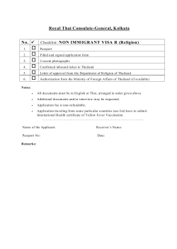
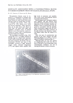
ISSN (Print) 0023-4001
ISSN (Online) 1738-0006
Korean J Parasitol Vol. 53, No. 2: 201-207, April 2015
http://dx.doi.org/10.3347/kjp.2015.53.2.201
▣ ORIGINAL ARTICLE
Developmental and Phylogenetic Characteristics of
Stellantchasmus falcatus (Trematoda: Heterophyidae)
from Thailand
Pralongyut Sripalwit1,2, Chalobol Wongsawad1,*, Thapana Chontananarth3, Somboon Anuntalabhochai4,
Pheravut Wongsawad1, Jong-Yil Chai5
Department of Biology, Faculty of Science, Chiang Mai University, Chiang Mai 50200, Thailand; 2Biology Program, School of Science, University of
Phayao, Muang, Phayao 56000, Thailand; 3Department of Biology, Faculty of Science, Srinakharinwirot University, Bangkok 10110, Thailand;
4
Biotechnology Unit, Biology Department, Science Faculty, Phayao University, Phayao 56000, Thailand; 5Department of Parasitology and Tropical
Medicine, Seoul National University College of Medicine, Seoul 110-799, Korea
1
Abstract: This study aimed to investigate the infection status, worm development, and phylogenetic characteristics of
the intestinal trematode, Stellantchasmus falcatus. The metacercariae of S. falcatus were detected only in the half-beak
(Dermogenus pusillus) out of the 4 fish species examined. Their prevalence was 90.0%, and the intensity of infection was
919 metacercariae on average. Worms were recovered from 33 (97.1%) of 34 chicks that were experimentally infected
with 200 S. falcatus metacercariae each, and the average recovery rate was 43.0%. The body size and inner organs of S.
falcatus quickly increased in the experimental chicks over days 1-2 post-infection (PI). In addition, ITS2 sequence data of
this parasite were analyzed to examine the phylogenetic relationships with other trematodes using the UPGMA method.
The results indicated that the ITS2 sequence data recorded from trematodes in the family Heterophyidae appeared to be
monophyletic. This study concluded that D. pusillus serves as a compatible second intermediate host of S. falcatus in
Thailand and that S. falcatus can develop rapidly in the experimental chicks. Data collected from this study can help to
close the gap in knowledge regarding the epidemiology, biology, and phylogenetic characteristics of S. falcatus in Thailand.
Key words: Stellantchasmus falcatus, worm development, ITS2, phylogenetic analysis, prevalence
INTRODUCTION
high infection rate has been reported, and the rate of infection
has been found to be high in the northern region of Thailand.
Even though the symptomatic features of human S. falcatus infection have not been clearly determined, the first clinical reports have revealed that S. falcatus can be found at autopsy and
many embryonated eggs remained in the blood vessels of the
cardiac muscle of the host [7]. Hence, it should be reminded
that S. falcatus has the potential to retain its clinical importance as a great foodborne zoonotic trematode.
Nowadays, molecular approaches are the most efficient and
accurate tools used to understand the relationships that exist
in an organism and for screening the genetic variations that
occur among many populations of parasites. The high annealing temperature-randomly amplified polymorphic DNA
(HAT-RAPD) PCR technique had been applied to study the
phylogenetic relationships of heterophyid trematodes [8,9].
However, currently, this technique is not commonly used because the HAT-RAPD PCR technique does not involve a DNA
profile and exhibits a lower level of stability than other meth-
Several kinds of mammals, i.e., humans, rats, cats, and dogs,
as well as chicks, have been reported as definitive hosts of this
fluke. This trematode is known to be the possible cause of
clinical problems in some Asian countries [1,2]. In addition,
metacercariae ( = the infective stage) are mainly found in
brackish water or marine fish, which serve as the second intermediate host, especially the group of mullets known as Mugil
spp. and Liza spp. [3,4]. However, several previous studies
have reported that S. falcatus metacercariae can also be found
in freshwater fish, such as the half-beak (Dermogenys pusillus)
[5] and climbing perch (Anabas tertudinaus) [6], for which a
• Received 11 April 2014, revised 20 February 2015, accepted 5 March 2015.
* Corresponding author ([email protected])
© 2015, Korean Society for Parasitology and Tropical Medicine
This is an Open Access article distributed under the terms of the Creative Commons
Attribution Non-Commercial License (http://creativecommons.org/licenses/by-nc/3.0)
which permits unrestricted non-commercial use, distribution, and reproduction in any
medium, provided the original work is properly cited.
201
202 Korean J Parasitol Vol. 53, No. 2: 201-207, April 2015
ods. The internal transcribed spacer 2 (ITS2) of nuclear rDNA
is far more variable in the sequence, and because of the relatively rapid rate at which new mutants are fixed, these regions
may reveal closely distinguished related species that otherwise
would show little genetic divergence [10]. Therefore, phylogenetic analyses of ITS2 rDNA sequences have successfully been
used to resolve evolutionary relationships among closely related species. Differing sequences of a given region are often assumed to be homogenized within a population of the same
species by concerting evolution [11].
PCR based-methods targeting ITS2 have been applied for
phylogenetic studies of various trematodes, such as Haplorchis
taichui and H. pumilio [12,13], Clonorchis sinensis [14], Fasciola
hepatica [15,16], and trematodes in the family Paramphistomidae [17]. Recently, the phylogenetic analysis of this gene was
performed by more appropriate methods than the HAT-RAPD
procedure applied in previous reports [5,8,9,18,19]. The purpose of this study was to investigate the prevalence of S. falcatus infection in the half-beaks (D. pussilus) and the worm recovery in experimentally infected chicks. In addition, phylogenetic relationships of S. falcatus with other heterophyid trematodes were analyzed for additional information to the recent
reports [8,9].
MATERIALS AND METHODS
Investigation of metacercarial infections in the fish host
Thirty individual specimens of 4 species of freshwater fish,
including D. pusillus, Henicorhynchus siamensis, Puntius brevis,
and Cyclocheilichthys armatus, were subjected to analyses by
surveying the metacercarial infections of heterophyid trematodes. Each individual fish was digested using an acid pepsin
solution (conc. HCl 1 ml: pepsin 1 g: 0.85% sodium chloride
solution 99 ml) for 3 hr at 37˚C. The digested material was
then rinsed with 0.85% sodium chloride solution and examined for metacercariae under a light microscope. Some metacercarial specimens were excysted and fixed in 4% formalin
under the pressure of a cover slip. Specimens were then stained
with hematoxylin, dehydrated in alcohol series, cleared with
xylene, and mounted in Permount®. The metacercariae were
confirmed in terms of species, based on Pearson and Ow-Yang
[2] and Chai and Sohn [3].
Experimental infection and worm recovery
The half-beaks (D. pusillus) were collected from Chiang Mai
province, Thailand and were examined and isolated for the
metacercarial stage of S. falcatus infection. The fish were digested by 1% acid pepsin solution at 37˚C for 2 hr. The digested
material was passed through graded sieves, rinsed in 0.85% saline solution, and then collected for metacercariae under a
light microscope. Two hundred metacercariae of S. falcatus
were orally infected to 34 chicks, Gallus gallus domesticus. The
exposed hosts were killed daily and their small intestines,
which were roughly divided into the duodenum, jejunum,
and ileum, were opened in 0.85% saline solution. Worms
were collected in a modified Baermann’s apparatus, and then
examined and observed under a stereomicroscope. The specimens of S. falcatus were fixed in 4% formalin under the cover
slip pressure, stained with hematoxylin, dehydrated in alcohol
series, cleared with xylene, and mounted in permount. The
specimens were measured and drawn under a compound microscope for morphological studies.
Parasite preparation for phylogenetic analysis
The metacercariae of S. falcatus, H. taichui, and C. formosanus
were collected from different fish hosts; D. pusillus, H. siamensis, and P. brevis, respectively. Meanwhile, the adult specimens
of Haplorchoides sp. were gathered from the intestines of the
Yellow cat fish, Hemibragus filamentus, and the adult stages of
other trematodes, namely the giant liver fluke, Fasciola gigantica
and the rumen fluke, Orthocoelium streptocoelium, were recovered from cows (Bos taurus).
Total genomic DNA extraction
The genomic DNA of all parasites was extracted and purified
from the adult worms using a DNeasy Tissue Kit (Vivantis,
Oceanside, California, USA) according to the instructions of the
manufacturer. All extracted genomic DNA were diluted to a
working concentration of 30 ng/µl and stored at -20˚C until used.
Amplification of ITS2 region
The PCR amplification of partial ITS2 fragments was done
using a pair of primers as has been described in the previous
report [20]. This consisted of (ITS3) 5´GCA TCG ATG AAG
AACGCA GC 3´as a forward primer and (ITS4) 5´TCC TCCGCT TAT TGA TAT GC 3´as a reverse primer. The reaction was
carried out at a final volume of 20 µl containing 1× PCR buffer, 2 mM MgCl2, 10 µM of each dNTP, 1 µM of each primer,
and 1 U of Vivantis Tag DNA polymerase. The analysis was
then performed in a MyCyclerTM Thermocycler (BioRAD, Her-
Sripalwit et al.: Characteristics of Stellantchasmus falcatus from Thailand 203
cules, California, USA). Recommended PCR protocols were as
follows; 1 cycle of 95˚C for 2 min, 35 cycles of 94˚C for 45 sec,
50˚C for 45 sec, 72˚C for 1 min, and 1 cycle of final extension
at 72˚C for 7 min. Specific PCR products were separated on
1.4% TBE agarose gel electrophoresis, stained with ethidium
bromide, and photographed using a Kodak digital camera Gel
Logic 100 and direct sequenced by the dideoxy terminator
method (First Base Company). The sequence was checked using the BLAST program in the NCBI (National Center for Biotechnology Information, Bethesda, Maryland, USA) database,
to confirm the PCR target. The eletropherograms of each sequence were examined for sequence accuracy using a Sequence
Scanner version 1.0 and Bioedit version 7.1. All sequences
were aligned automatically using Clustal X version 2.0. The
phylogenetic relationships of 11 trematodes were constructed
using MEGA version 5.0. All molecular data were analyzed by
UPGMA (Unweighted Pair Group Method with Arithmetic
Mean) method (Table 1). The reliability of the internal
branches was assessed using the bootstrap method per 1,000
replicates.
Table 1. List of materials and sequences of ITS2 used for phylogenetic analysis Species
Accession no./ references
KJ630831
KJ630832
KJ630833
KJ630834
KJ630835
KJ630836
EU826640a
AB470525a
KF658459a
AY584735a
Haplorchis taichui
Haplorchoides sp.
Stellantchasmus falcatus
Orthocoelium sterptocoelium
Fasciola hepatica
Centrocestus formosanus
Haplorchis taichui
Metagonimus yokogawai
Centrocestus sp.
Opisthorchis viverrini
The sequence data were from GenBank database.
a
RESULTS
Infection status of metacercariae in fish hosts
The prevalence of infection in 4 heterophyid trematodes; S.
falcatus, H. taichui, Haplorchoides sp., and C. formosanus, were
recorded individually from their fish hosts which revealed the
prevalence of 90.0%, 73.3%, 33.3%, and 33.3%, respectively,
while only D. pusillus was infected by S. falcatus with the highest value of intensity (919 metacercariae/fish) (Table 2).
Worm recovery and development of worms
Worms were recovered in 33 (97.1%) of 34 chicks that were
experimentally infected with 200 S. falcatus metacercariae
each. The average worm recovery rate was 43.0%. Measurements of worms recovered from the chick are summarized in
Fig. 1. The body length and width were remarkably increased
from days 1-2 post-infection (PI), and then gradually increased
until day 14 PI (Fig. 1A). The oral sucker, pharynx, esophagus,
and ventrogenital sucker also showed a similar developmental
pattern to the body size over days 1-2 PI, before stabilizing at a
plateaued level (Fig. 1B). The left testis was a little bit larger
than the right one in terms of both length and width. The size
of 2 testes was dramatically increased from day 1 to day 2 PI,
and gradually increased until day 11 PI. The ovary and the expulsor developed at an equivalent level, as was also observed
in the testes (Fig. 1C). Uterine eggs were recognized in 87.5%
of the specimens at day 2 PI. Eggs in the uterine duct were
constant in size during days 2-14 PI, with a length of 20.0-22.5
μm and a width of 7.5-10.5 μm.
On day 1 PI, the worms differed morphologically from the
metacercariae particularly in terms of their body size. The body
measured 240.0-375.0 μm in length and 130.0-160.0 μm in
width. The ovary was 30.0-37.50 μm in length and 27.5-45.0
Table 2. Summary of fish species, prevalence, and intensities of heterophyid trematodes examined in each fish species Fish species
Dermogenys pusillus
Henicorhynchus
siamensis
Puntius brevis
Cyclocheilichthys
armatus
No. of fish infected with each heterophyid species/No. of fish examined
S. falcatus
H. taichui
Haplorchoides sp.
C. formosanus
27/30
919.0 (28-3,414)a
0
0
0
-
0
17/30
37.0 (2-129)a
0
05/30
29.0 (23-187)a
0
8/30
16.5 (3-44)a
0
2/30
79.1a
0
0
10/30
213.5 (2-2,492)
0
-
The intensity values represent the mean no. of metacercariae per fish (range).
a
Mean
90.0%
919
83.3%
26.8
33.3%
213.5
23.3%
54.1
204 Korean J Parasitol Vol. 53, No. 2: 201-207, April 2015
(μm)
Body (length)
B
Body (width)
700
600
500
400
300
200
100
0
0246810
12
14
16
Oral sucker
120
60
40
20
0
0246810
12
14
16
Days post-infection
D
Testis (right)
OS
200 (μm)
P
200 (μm)
Length (μm)
140
120
100
80
60
40
20
0
Ventrogenital sucker
Ovary
Expulsor
Testis (left)
Esophagus
80
Days post-infection
C
Pharynx
100
Length (μm)
A
0246810
12
14
16
Days post-infection
E
VS
GP
MVG
EP
W
SV
I
O
V
T
EB
Fig. 1. Morphologic changes of Stellantchasmus falcatus recovered from experimental chicks (A-C) and illustrations demonstrating the
permanent slide capture and line-drawing (D).
76
Haplorchis taichui KJ630832
100
Haplorchoides sp. KJ630832
Haplorchis taichui EU826640
Opisthorchis viverini AY584735
84
Metagonimus yokogawai AB470525
Centrocestus sp. KF658459
100 Centrocestus formosanus KJ630836
Stellantchasmus falcatus KJ630833
Orthocoelium steptpcoelium KJ630834
100
Fasciola gigantica KJ630835
0.20 0.15 0.100.050.00
Fig. 2. The rooted phylogeny from partial ITS2 sequences of heterophyid trematodes based on the UPGMA method. Bootstrap values
were computed independently for 1,000 resembling.
μm in width. The right testis was found to be 42.5-90.0 μm
long and 20.0-42.5 μm wide, while the left testis was 37.5-70.0
μm long and 22.5-30.0 μm wide. On day 2 PI, mature worms
were observed. The body measured 360.0-510.0 μm in length
and 130.0-210.0 μm in width. The ovary enlarged to 30.0-62.5
μm long and 27.5-50.0 μm wide. The right testis was 55.087.5 μm long and 32.5-52.5 μm wide, while the left testis was
47.5-80.0 μm long and 30.0-52.5 μm wide. The expulsor was
oval and relatively large, with a length of 32.5-82.5 μm and a
width of 25.0-37.5 μm. The ventrogenital sucker was slightly
submedian to the right side and contained a ventral sucker.
The ventral sucker armed with 2 dense lateral groups of small
spines on the lip. The ovary was submedian and located slightly to the right side between the ventral sucker and right testis
and was 30.0-62.5 μm long and 27.5-50.0 μm wide. The vitellaria extended dorsally into the ovariotesticular zone (Fig. 1D).
Sripalwit et al.: Characteristics of Stellantchasmus falcatus from Thailand 205
Phylogenetic analysis
The partial sequence data of the ITS2 region were used to
understand their phylogenetic relationships. The length of this
fragment was 410-590 bp. The trematodes in the family Heterophyidae appeared to be in a monophyletic clade. The heterophyids were separated into 2 sister groups including S. falcatus group and the other heterophyid group (Fig. 2).
DISCUSSION
In this study, we reported the metacercarial infection status
of 4 heterophyid flukes, i.e., S. falcatus, H. taichui, Haplorchoides
sp., and C. formosanus, which were collected individually from
their fish hosts to be 90.0%, 86.7%, 33.3%, and 23.0%, respectively. Our findings corresponded well with previous reports that have confirmed a high prevalence of infection in S.
falcatus and H. taichui, particularly in the northern part of
Thailand [21-23]. Various reports have detected the presence of
intestinal trematodes in the family Heterophyidae, such as, H.
taichui, H. pumilio, S. falcatus, C. formosanus, and Procerovum
sp., which were found to be wildly distributed and had a high
prevalence and intensity. Previous surveys have also revealed
that the metacercariae, which were mostly found in Mae Taeng
District of Chiang Mai Province, belonged to the family Heterophyidae [21-23]. Our findings corresponded well with
those of Radomyos et al. [24] who reported that certain heterophyid species were widely distributed in certain regions in
the north of Thailand. Epidemiological surveys from 19972013 have shown an increase in trematode infections in the
northern and central parts of Thailand. The surveyed region
has been reported as an endemic area of heterophyid trematodes with high prevalences in a variety of intermediate and
definitive hosts. For instance, Wongsawad et al. [6] investigated
on trematode infections in freshwater fish from Mae Sa
stream, Chiang Mai Province, where 4 species of heterophyid
trematodes were found, i.e., Haplorchis spp., Haplorchoides sp.,
C. formosanus, and S. falcatus [6].
From the present results, it is strongly suggested that the
half-beaks (D. pusillus) in northern Thailand typically serve as
the highly compatible secondary host for infection with S. falcatus. This result corresponded well with a previous report [25].
In contrast, the metacercarial stage of S. falcatus has been detected also in marine or brackish water fish [3,4]. However,
further studies must be done to investigate S. falcatus metacercariae in fish species from a larger area in order to confirm that
this fish species (D. pussilus) serves as the most popular second
intermediate host of this parasite.
Based on the results of experimental infection, chicks
seemed to be a susceptible host for S. falcatus infection revealing much higher rates of infection and worm recovery than
those observed in mice (Mus musculus) (13.6%) and rats (Rattus
norvegicus) (20.2%) [26]. These results are supported by the far
higher worm recovery that was recorded in mice (13.6%) and
chicks (33.8%). In our study, parasites were observed to have
invaded the jejunum during days 1-6 PI and later they were
typically observed in the ileum. A similar pattern was observed
in H. taichui and H. pumilio which were found to have invaded
the upper part of the small intestines during the first period of
infection, but when they matured the parasites were more
common in the middle and lower parts of the intestinal tract
because the flukes became gradually excreted into the lumen
of the intestinal canal along with mucus and other exudates.
The flukes then reattached themselves further down the tract
[27]. However, host-parasite relationships in heterophyid infections have never been studied in detail. Since human infections with S. falcatus and other heterophyid trematodes are expected to increase in areas where fresh and brackish water fish
are popularly eaten raw, studies on host-parasite relationships
are considered highly useful.
According to the phylogenetic analysis, the partial sequences
of ITS2 of some heterophyid trematodes are declared to be
known now. However, only a few studies have focused on the
relationship that exists among the ITS2 of S. falcatus in Thailand [28]. Our sequence data of ITS2 agreed well with the sequence data reported in other recent reports [28].
The UPGMA trees showed the monophyletic relationships
of heterophyid trematodes. Heterophyid trematodes were separate in a same group except S. falcatus which was separated as
another group with a high bootstrap value. This agrees with
the reports on phylogenetic relationships based on mitochondrial cytochrome c oxidase 1 (mtCOI) sequence data, which
summarized that only S. falcatus has a ventrogenital complex
[29], whereas the results of a phylogenetic study of S. falcatus
using the HAT-RAPD method showed a group of S. falcatus together with other heterophyid trematodes [8].
ITS2 has been used to study the systematic analysis of some
heterophyid trematodes, i.e., Haplorchis [12,13] and Metagonimus [30]. From our results, it is believed that the ITS2 locus
could be used as a barcode for authenticating animal species,
as well as a complementary locus to other barcoding genes for
206 Korean J Parasitol Vol. 53, No. 2: 201-207, April 2015
the purpose of identifying all organisms, including heterophyid trematodes. Several regions that have become known for a
relevant presence of nuclear ribosomal DNA (nu rDNA) were
selected and used for identification of various stages as well as
for a study of the life cycle of trematodes (cercarial, metacercarial, and adult stages) in freshwater fish and snails because
of the high levels of accuracy, sensitivity, and rapidity of this
method. ITS2 has been revealed as a sensitive marker at the
species level for trematodes. The PCR based-method targeting
ITS2 has been applied for use in the phylogenetic study of various trematodes, such as H. taichui and H. pumilio [12,13], C.
sinensis [14], F. hepatica [15], and trematodes in the family Paramphistomidae [17]. Using the sequence data of the ITS2 region, the species-level identification could be achieved, and
the ITS2 analysis could actually provide a phylogenetic study
of these trematodes.
This study can conclude that D. pusillus fish serves as a second intermediate host for S. falcatus with the highest rate of
prevalence and rapid development being found in the experimental host, chicks (G. gallus domesticus). Finally, the present
study provided valuable and beneficial information that can
be used to demonstrate the epidemiological situation, as well
as to examine the development and phylogenetic relationships
of S. falcatus with regard to the biological control and monitoring of this parasitic disease.
ACKNOWLEDGMENTS
The financial support from the Thailand Research Fund
through the Royal Golden Jubilee Ph.D. Program (grant no.
PHD/0192/2543 and BGJ4580020) to student's initials and
adviser's initials is acknowledged. We also thank the Biology
Department, Faculty of Science, the Applied Parasitology Research Laboratory, Institute for Science and Technology Research, and Economic Plants Genome Research and Service
Center, Chiang Mai University, Thailand. We thank them for
providing the necessary facilities.
CONFLICT OF INTEREST
We declare that we have no conflict of interest related to this
study.
REFERENCES
1.Pearson JC. A revision of the subfamily Haplorchinae Looss,
1899 (Trematoda: Heterophyidae). 1. The Haplorchis group. Parasitology 1964; 54: 601-676.
2.Pearson JC, Ow-Yang CK. New species of Haplorchis from Southeast Asia, together with keys to the Haplorchis-group of heterophyid trematodes of the region. Southeast Asian J Trop Med
Public Health 1982; 13: 35-60.
3.Chai JY, Sohn WM. Identification of Stellantchasmus falcatus
metacercariae encysted in mullets in Korea. Korean J Parasitol
1988; 26: 65-68.
4.Shin EH, Guk SM, Kim HJ, Lee HY, Chai JY. Trends in parasitic
diseases in the Republic of Korea. Trends Parasitol 2008; 24:
143-150.
5.Sripalwit P, Wongsawad C, Wongsawad P, Anuntalabhochai S.
High annealing temperature-random amplified polymorphic
DNA (HAT-RAPD) analysis of three paramphistome flukes from
Thailand. Exp Parasitol 2007; 115: 98-102.
6.Wongsawad C, Rojtinnakorn J, Wongsawad P, Rojanapaibul A.
Helminths of vertebrates from Mae Sa Stream, Chiang Mai,
Thailand. Southeast Asian J Trop Med Public Health 2004; 35
(Suppl 1): 140-146.
7.Kliks M, Tantachumrun T. Heterophyid (trematoda) parasites of
cats in north Thailand, with notes on a human case found at
necropsy. Southeast Asian J Trop Med Public Health 1974; 5:
547-555.
8.Wongsawad C, Wongsawad P. Molecular markers for identification of Stellantchasmas falcatus and a phylogenetic study using
the HAT-RAPD method. Korean J Parasitol 2010; 48: 303-307.
9.Wongsawad C. Development of HAT-RAPD marker for detection of Stellantchasmus falcatus infection. Southeast Asian J Trop
Med Public Health 2011; 42: 46-52.
10.Mas-Coma S, Bargues MD. Populations, hybrids and the systematic concepts of species and subspecies in Chagas disease triatomine vectors inferred from nuclear ribosomal and mitochondrial DNA. Acta Trop 2009; 110: 112-136.
11.Darissa OM, Iraki NM. Molecular identification of six Steinernema isolates and characterization of their internal transcribed
spacers regions. Jordan J Biol Sci 2014; 7: 31-34.
12.Van Van K, Dalsgaard A, Blair D, Le TH. Haplorchis pumilio and H.
taichui in Vietnam discriminated using ITS-2 DNA sequence
data from adults and larvae. Exp Parasitol 2009; 123: 146-151.
13.Skov J, Kania PW, Dalsgaard A, Jørgensen TR, Buchmann K. Life
cycle stages of heterophyid trematodes in Vietnamese freshwater
fishes traced by molecular and morphometric methods. Vet Parasitol 2009; 160: 66-75.
14.Müller B, Schmidt J, Mehlhorn H. Sensitive and species-specific
detection of Clonorchis sinensis by PCR in infected snails and
fishes. Parasitol Res 2007; 100: 911-914.
15.Erensoy A, Kuk S, Ozden M. Genetic identification of Fasciola hepatica by ITS-2 sequence of nuclear ribosomal DNA in Turkey.
Sripalwit et al.: Characteristics of Stellantchasmus falcatus from Thailand 207
Parasitol Res 2009; 105: 407-412.
16.Wongsawad C, Wongsawad P. Anuntalaphochai S, Chai CJ, Sukontason K. Occurrence and molecular identification of liver
and minute intestinal flukes metacercariae in freshwater fish
from Fang-Mae Ai Agricultural Basin, Chiang Mai Province,
Thailand. Asian Biomed 2013; 7: 97-104.
17.Loffy WM, Brant SV, Ashmawy KI, Devkota R, Mkoji GM, Loker
ES. A molecular approach for identification of paramphistomes
from Africa and Asia. Vet Parasitol 2010; 174: 234-240.
18.Chuboon S, Wongsawad C. Molecular identification of larval
trematode in intermediate hosts from Chiang Mai, Thailand.
Southeast Asian J Trop Med Public Health 2009; 40: 1216-1220.
19.Wongsawad P, Wongsawad C. Development of PCR-based diagnosis of minute intestinal fluke, Haplorchis taichui. Southeast
Asian J Trop Med Public Health 2009; 40: 919-923
20.Barber KE, Mkoji GM, Loker ES. PCR-RFLP analysis of the ITS2
region to identify Schistosoma haematobium and S. bovis from Kenya. Am J Trop Med Hyg 2000; 62: 434-440.
21.Boonchot K, Wongsawad C. A survey of helminths in cyprinoid
fish from the Mae Ngad Somboonchon Reservoir, Chiang Mai
Province, Thailand. Southeast Asian J Trop Med Public Health
2005; 36: 103-107.
22.Kumchoo K, Wongsawad C, Chai JY, Vanittanakom P, Rojanapaibul A. High prevalence of Haplorchis taichui metacercariae in cyprinoid fish from Chiang Mai Province, Thailand. Southeast
Asian J Trop Med Public Health 2005; 36: 451-455.
23.Nithikathkul C, Wongsawad C. Prevalence of Haplorchis taichui
and Haplorchoides sp. metacercariae in freshwater fish from water
reservoirs, Chiang Mai, Thailand. Korean J Parasitol 2008; 46:
109-112.
24.Thu ND, Dalsgaard A, Loan LTT, Murrell KD. Survey for zoonotic liver and intestinal trematode metacercariae in culture and
wild fish in An Giang Province, Vietnam. Korean J Parasitol
2007; 45: 45-54
25.Pubua J, Wongsawad C. Redescription of the trematode metacercariae from the mullet (Liza subviridis) and half-beak (Dermogenus pusillus). Southeast Asian J Trop Med Public Health 2007;
38 (Suppl 1): 106-109.
26.Saenphet S, Wongsawad C, Saenphet K, Chai JY. Susceptibility
of rodents to Stellanchasmus falcatus infection. Southeast Asian J
Trop Med Public Health 2003; 32 (Suppl 2); 123-127.
27.Africa CM, de Leon W, Garcia EY. Visceral complications in intestinal heterophyidiasis of man. Acta Med Philipina (Monogr
Ser) 1940; 1: 1-132.
28.Chontananarth T, Wongsawad C. Prevalence of Haplorchis taichui
in field-collected snails: a molecular approach. Korean J Parasitol 2010; 48: 343-346.
29.Chontananarth T, Wongsawad C, Chomdej S, Krailas D, Chai JY.
Molecular phylogeny of trematodes in family Heterophyidae
based on mitochondrial cytochrome c oxidase subunit I
(mCOI). Asian Pac J Trop Med 2014; 7: 446-450.
30.Lee SU, Huh S, Sohn WM, Chai JY. Sequence comparisons of
28S ribosomal DNA and mitochondrial cytochrome c oxidase
subunit I of Metagonimus yokogawai, M. takahashii and M. miyatai. Korean J Parasitol 2004; 42: 129-135.


© Copyright 2026