
40749 Antenatal Diagnosis for Anomalies of the Corpus Callosum Sam Armstrong Bell
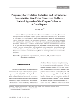
40749
Antenatal Diagnosis for Anomalies of the Corpus Callosum
Sam Armstrong Bell1, Edward O’Mahoney2, Michelle Fink3,4, Nicole Woodrow4, Karen Reidy1,2, Ricardo
Palma Dias1,2,4
1 Fetal Medicine Unit and Pregnancy Research Centre, Department of Perinatal Medicine, The Royal
Women's Hospital, Melbourne, Australia
2 Department of Obstetrics & Gynaecology, The University of Melbourne, Melbourne, Australia
3 Medical Imaging Department, Royal Children's Hospital, Melbourne, Australia
4 Pauline Gandel Imaging, The Royal Women's Hospital, Melbourne, Australia
Objective
To assess the diagnostic accuracy of ultrasound and fetal MRI in the diagnosis of anomalies of the corpus
callosum.
Methods
Hospital ultrasound and MRI databases were searched for cases of suspected callosal anomalies
between 2003 and 2012. Subsequent ultrasound scans, fetal MRI, postnatal imaging, postmortem
investigations and birth records were reviewed. Fetuses with major additional extracranial anomalies,
abnormal karyotype and multiple pregnancies were excluded. Callosal anomalies were classified into
isolated or complex based on the presence or absence of accompanying intracranial findings.
Results
47 callosal anomalies were diagnosed during the study period. 66% were detected on a second
trimester morphology ultrasound, 26% from a later ultrasound, and 9% from MRI performed for the
investigation of ventriculomegaly. Of the 43 cases suspected from ultrasound, 77% had a subsequent
MRI. In 27% of cases where callosal anomalies were suspected on ultrasound, fetal MRI revealed one or
more additional diagnoses. Two thirds of these were anomalies of cortical development. MRI changed
the classification to complex anomaly in 21% of cases thought to be isolated from ultrasound. 20% of
cases thought to be isolated antenatally (following ultrasound +/- MRI) were diagnosed with additional
anomalies after birth.
Conclusions
In cases of callosal anomaly suspected on ultrasound, MRI provides greater certainty and the potential
to identify significant additional anomalies. The additional information may alter or clarify prognosis and
help parents to better understand the pathology, allowing for informed decisions about the pregnancy
to be made. However, despite extensive antenatal investigation, some cases may still be diagnosed with
additional anomalies after birth and parents should be aware of such limitations. Availability of local
data as compiled in this study will be of value for future parents facing this challenging clinical scenario.
41333
Prenatal diagnosis of sirenomelia by combining new fetal skeletal rendering, three-dimensional helical
computer tomography and magnetic resonance imaging
CHEN-Xinlin*, LIU Rong YANG Xiao-hong, CHEN Pei-wen, ZHAO Sheng , XIAO Mei,
Department of Ultrasound, Hubei Maternal and Child Health Hospital,Wuhan 430070,P.R.CHINA
Objective
To evaluate the value of prenatal diagnosis of skeletal abnormalities in sirenomelia by combining twodimensional ultrasound, new fetal skeletal rendering, three-dimensional helical computer tomography
and magnetic resonance imaging.
Methods Between September 2010 and February 2012, a prospective study was conducted in our
hospital. Fetal skeletal rendering, three-dimensional helical computer tomography (3D-HCT) and
magnetic resonance imaging (MRI) were performed after two-dimensional ultrasound (2D-US) in seven
cases of sirenomelia. Diagnostic skeletal detailed findings with each of the three techniques were
compared with postnatal radiological findings and 3D-HCT. All cases performed postnatal autopsy, five
cases performed chorionic villus and/or cord blood sampling.
Results
Six cases are singleton and one is conjoined twins. The abnormalities associated with seven siremomelia
cases included different degrees of fusion of the lower extremities, bilateral renal agenesis, absent
bladder, absent external genitalia and single umbilical cord. Five cases are associated with
Oligohydramnios, five cases with partial absent ribs, seven cases with spine anomalies and five cases
with cardiac anomalies. Karyotype and postnatal autopsy show that three cases are male and two are
female.
Conculsion
MRI can help to diagnose sirenomelia, it’s less useful to skeletal abnormalities. Although 3D-HCT is a
gold standard in diagnosis of skeletal abnormalities, the fetuses can’t be X-rayed, so it is restricted. The
new fetal skeletal rendering seem to be useful complementary method to 2D-US, and can quickly
provide skeletal imaging just as 3D-HCT, and may improve accuracy of the prenatal diagnosis of skeletal
abnormalities in sirenomelia.
[Key words] sirenomelia, two-dimensional ultrasound, fetal skeletal rendering, three-dimensional
helical computer tomography, magnetic resonance imaging
41421
Feasibility of comprehensive hemodynamic assessment in the normal late gestation human fetus by
phase contrast MR and T2 mapping
Liqun Sun1,2, Mashael Alrujaib1,3, Joshua van Amerom1,4, Christopher K. Macgowan1,4, Mike Seed1
1 Hospital for Sick Children, Canada
2 University of Toronto, Canada
3 Department of Diagnostic Imaging, Canada
4 Department of Medical Biophysics and Medical Imaging, Canada
Objective
Ovine studies have defined oxygen transport in the fetal circulation. We sought to reproduce these
findings in the human fetus using a combination of phase contrast (PC) magnetic resonance (MR) with
metric optimised gating (MOG) and MR oximetry with T2 mapping.
Methods
The major vessels of 15 late gestation normal fetuses (mean GA: 37 weeks SD 1.2 weeks ) were studied
during late gestation with PC MR with MOG according to our previously published technique[1] and a
new T2 mapping algorithm[2]. Vessel T2s were converted to vessel oxygen saturation according to
previous work[3]. Using umbilical vein (UV) and descending aorta T2 mapping and UV PC flow, we
calculated fetal oxygen delivery (DO2) and consumption (VO2). We investigated the relationship
between the distribution of flow in the fetal circulation and vessel T2s, DO2 and VO2 using Pearson’s
correlation coefficient.
Results
Mean flows and T2s with SDs are shown in Table 1. Fetal DO2 was proportional to UV flow (r = 0.91),
while fetal VO2 correlated with UV flow (r = 0.74) and fetal DO2 (r = 0.80). PBF was related to the UV (r =
0.65) and MPA T2 (r = 0.58). We found an inverse relationship between PBF and FO flow (r = -0.75).
Table 1- Flow, T2 and oxygen saturation in the major fetal vessels
Mean
Flow
(ml/min/kg)
Mean
Flow
(% CVO)
Mean T2 (ms)
Mean SaO2[3]
UV
AAO
MPA
SVC
DAO
DA
PBF
FO
129±39
210±41
250±36
135±33
254±41
189±46
78±39
145±57
28±10
45±6
53±6
29±8
54±10
41±10
17±8
31±10
199±21
127±18
107±17
0.79±0.06
0.60±0.05
0.52±0.06
91±17
0.46±0.07
108±16
0.53±0.06
UV- umbilical vein, AAo- ascending aorta, MPA- main pulmonary artery, SVC- superior vena cava, DAo- descending aorta, DA- ductus arteriosus, PBFpulmonary blood flow, FO- foramen ovale, CVO- combined ventricular output, SaO2- oxygen saturation.
PBF vs UV T2
VO2 vs DO 2
PBF vs FO
UV T2 (ms)
30
DO2
25
20
250
200
r = 0.6463
p = 0.0092
150
15
r = 0.8042
p = 0.0003
100
0
10
0
5
10
15
50
100
150
PBF LPA+RPA Flow (ml/min/kg)
VO2
200
FO LVO-PBF Flow (ml/min/kg)
300
300
200
100
r = -0.7486
p = 0.0013
0
0
50
100
150
200
PBF LPA+RPA Flow (ml/min/kg)
Figure -1 The relationship between DO2 and VO2 (left) ; PBF LPA+ RPA and FO LVO - PBF (middle); PBF LPA+ RPA and UV T2 (right). LVO = left ventricular output
Conclusion
This approach represents the first attempt to non-invasively characterize oxygen delivery and
consumption in the human fetal circulation, made possible by combining MR oximetry and PC MRI. The
results are in keeping with previous invasive measurements in human fetuses[4] and animal
experiments[5] showing that fetal DO2 is matched by VO2, with both related to placental blood flow.
Our demonstration of streaming of oxygenated blood across the FO and the relationship between PBF
and MPA and UV T2 are is in keeping with the known hypoxic pulmonary vasoconstriction mechanism
operating in the human fetal lung.
References
[1] Seed et al. JCMR. 2012;14:79.
[2] Giri et al. MRM. 2012;68:1570-1578.
[3] Wright et al. MRI. 1991;1:275-283.
[4] Rudolph. Wiley Blackwell. 2009.
[5] Rurak et al. Am J Physiol. 1990;258:1116-1122.
41429
In utero brain growth of foetuses with congenital heart disease: Case control study using 3D SSFP
Mashael Alrujaib1,2, Liqun Sun1,2, Christopher Macgowan1,2, Joshua van Amerom1,2, Mike Seed1,2
1 Hospital for Sick Children, Canada
2 University of Toronto, Canada
Introduction
Ultrasound and MRI studies have indicated there is an association between congenital heart disease
(CHD) and abnormal brain development in utero [1,2,3]. We used a three dimensional steady state free
precession (3D SSFP) sequence [4] to assess brain growth during late gestation in fetuses with complex
CHD compared with normals.
Methods
45 fetuses with CHD and 25 normal fetuses were studied with MRI on a 1.5T system (Siemens Avanto) at
mean gestational age of 36 weeks. 3D SSFP data were acquired during a maternal breath hold and
segmented to yield fetal and brain volumetry which was converted to estimated fetal and brain weights
(EFW and EBW) based on published formulas [5,6]. EBW for normal and CHD fetuses were compared
with gestational age adjusted autopsy data [7] and a Z-score calculated for each brain (Z=1 SD) (ref 7).
EBWs were also indexed to fetal weight and a student t-test used to compare the CHD and normal
groups.
Results
We found no significant difference between mean EBW Z-score (-0.35, SD 0.93) or EBW/EFW in CHD
fetuses (10.5, 1.1) compared with EBW Z-score; (-0.07, 0.92) and EBW/ EFW (10.6, 1.3) in normal fetuses
(p = 0.23 and 0.71 respectively). Three fetuses with CHD had EBW Z-score >2SDs below the mean, while
all of the normal fetuses were within 2 SDs of the mean.
Conclusions
Contrary to one similarly sized previous study [2] we found no significant difference between EBW or
EBW/EFW in fetuses with CHD compared with controls. The reason for this is not entirely clear but may
be in part due to the difference in technique. In keeping with previous studies, we did find examples of
fetuses with EBWs below the 5th centile [2,8].
Figure. Estimated fetal brain weight (EBW) Z-scores in the congenital heart disease (CHD) group vs.
controls.
References
[1] Miller et al. N Engl J Med. 2007 Nov 8;357(19):1928-38.
[2] Limperopoulos et al. Circulation. 2010 Jan 5;121(1):26-33.
[3] Meise et al. Ultrasound Obstet Gynecol. 2001 May;17(5):398-402.
[4] J. Anquez et al. Conf Proc IEEE Eng Med Biol Soc. 2007;2007:771-4
[5] Seed et al. J Cardiovasc Magn Reson. 2012; 14:79.
[6] Baker et al. Lancet. 1994; 343:644–645.
[7] Guihard-Costa et al. Fetal Diagnosis and Therapy (Karger), vol. 10, n°4, 75 pp.
[8] Glauser et al. Pediatrics. 1990 Jun;85(6):984-90.
42921
Fetal "Black Bone" MRI, a new sequence in the fetal MRI imaging armamentarium
Ashley Robinson1, Susan Blaser1, Andrei Vladimirov1, Debra Drossman1
1 Hospital For Sick Children, Canada
Objectives
Recent advances in the adult musculoskeletal literature have led to the development of the “black
bone” MRI sequence. This sequence was developed in response to growing concerns regarding the
harmful effects of radiation.
Fetal CT is now being increasingly used for assessment of fetal skeletal abnormalities. However there is
also concern regarding harmful effects particularly as the fetus is considered to be more radiosensitive
due to rapid growth, cell division and organogenesis.
Although the poor detail of bone on MRI has been a significant limitation of MRI in assessment of bony
structures, Echo-Planar Imaging has recently been used to assess human long bone development. Fetal
MRI therefore has the potential to reduce or avoid radiation exposure from fetal CT.
Methods
Susceptibility Weighted Imaging is a technique developed by Siemens. The technique gives high
contrast between bone and soft tissues, but low contrast between different soft tissues, thus the low
signal bone is easily distinguished from the surrounding soft tissues. Thus the SWI sequence can be
used for “black bone” imaging in the fetus.
Results
We have used the SWI sequence to assess the skeleton, in particular the skull and axial skeleton,
particularly in the assessment of spinal abnormalities, most commonly in myelomeningocele.
Conclusion
Fetal “black bone” MRI is a new technique for evaluation of the fetus, particularly in cases of spinal
pathology, and also demonstrates potential use in the evaluation of craniofacial pathologies related to
fetal syndromes, for evaluation of fetal skeletal dysplasias, and for fetal MR necropsy.
43339
Managing fetuses at high risk of retinoblastoma: lesion detection on screening MRI
Amber Bristowe1, Sandra Staffieri1, Michelle Fink1,
1 The Royal Children's Hospital Melbourne, Australia
Objective
To describe the positive magnetic resonance imaging (MRI) findings in a 35 week old fetus with familial
retinoblastoma (RB) and report the use of antenatal ultrasound and MRI screening in the management
of fetuses at high risk of RB.
Methods
A retrospective review of the antenatal course and immediate post natal findings in all children
considered at high risk of RB who had antenatal imaging with both ultrasound (US) and MRI at our
institution over a 5 year period.
Results
Five patients met the inclusion criteria. No lesions were identified on US in any patients. Fetal MRI
identified bilateral posterior pole lesions in one patient at 35 weeks gestation. Of the 4 remaining
patients, 3 developed lesions by 5 weeks of age. Only 1 fetus was delivered early following detection of
RB.
Conclusion
We present the first reported case of RB detected in a high risk fetus on screening MRI at 35 weeks
gestation. Protocols for screening this population using both imaging modalities are suggested. The
benefit of improved visual outcomes is to be balanced with potential morbidity from early delivery.
43375
The diagnostic value of antenatal Magnetic Resonance imaging in cases suspected to have Placental
Adhesive Disorders (PAD).
Nadia Rahaim, Elspeth Whitby
University of Sheffield, United Kingdom
Objectives
To evaluate the impact of antenatal diagnosis of PADs on pregnancy outcome, assess modalities used
for diagnosis and the value of the individual MRI criteria.
Methods
Retrospective analysis of patient data in period between February 2010- 2013 has. 43 cases recruited
and 2 excluded because of unknown outcome leaving 41 for statistical analysis.
Results
7/41 cases had PAD and only one case missed in antenatal diagnosis. Risk factors analysis showed that
Odds ratio of H/O caesarean section (P=0.59), placenta praevia (P= 0.72), is high though not statistically
significant for invasion. Blood loss was significantly higher in invaded compared to non-invaded cases
(p<0.001). Women with an invasive placenta had significantly more blood transfused (p<0.001). Median
days in hospital was significantly longer in invaded group compared to that of non invaded (p<0.001).
MRI was better than ultrasound having both higher sensitivity (86% vs 43%) and specificity (81% vs
79%). The most useful sign was heterogeneity being both highly sensitive (86%) and specific (91%) for
placenta invasion. Median blood loss was higher in women with multiple bands (3000 l) compared to
those with single bands (600 l).
Conclusion
Antenatal diagnosis although aided in surgery planning, favourable pregnancy outcome has not been
achieved yet. MRI proved to have better diagnostic sensitivity than that of US and it was successful in
defining depth of invasion in the majority of cases indicating its importance in recruitment of the specific
expertise required. Multiple dark bands seem to be a useful predictor of blood loss in PAD.
43435
Use of ultrasonography and magnetic resonance imaging in the diagnosis of placenta membranacea
CHEN Xin-lin, YANG Xiao-hong, ZHAO Sheng.
Department of Ultrasonography, Hubei Maternal & Children’s Hospital, Wuhan 430070, China
Objective
To analyze the contribution of ultrasonography and magnetic resonance imaging in the evaluation of
placenta membranacea.
Methods
This was a prospective study involving 2 fetuses suspected of having placenta membranacea on
ultrasound examination. MRI was used to analyze the location of the placenta, and to distinguish the
normal placenta and placenta membranacea in a twin pregnancy. All the results were compared with
pathology results.
Results
A 25-year-old woman and a 24-year-old woman were referred to our unit for abnormal placenta, at 24
weeks and 3 days and 25 weeks and 5 days, respectively. The latter was a twin pregnancy. No obvious
abnormality were detected in all the fetuses, however, placenta abnormalities were detected in the first
fetus and one of the latter fetuses. Displayed by ultrasonography, the abnormal placenta nearly
occupied the whole uterine cavity, and showed diffuse low-level internal echoes inside the placenta.
Displayed by MRI, the abnormal placentas showed hyperintense T2-weighted signal and flowing void
effect consistent with vascular branches. In the first case, a few normal placenta were seen. In the latter
case, normal placenta were not seen in the fetus with placenta membranacea. Postpartum histologic
examination revealed chorionic villi directly attached to the fetal membranes in these two cases,
consistent with the diagnosis of placenta membranacea.
Conclusion
Ultrasonography could be used to display the 2D image and color Doppler image of the placenta
membranacea in real time, and the hemodynamics change are very important in the prognosis analysis.
MRI are good complementary tool to ultrasonography for identifying the outline of placenta, especially
the spatial relationship between normal placenta and placenta membranacea in twin pregnancy.
43447
MRI of the Fetal Cerebellum and Posterior Fossa- Spectrum of Abnormalities
Sherelle Laifer-Narin
Columbia University, United States of America
Objective
Ultrasound is the imaging modality of choice for screening the pregnant patient and performing a
structural survey. However, fetal MRI has greatly improved analysis and diagnosis of fetal cerebral and
cerebellar anatomy and pathology. The spectrum of cerebellar abnormalities will be reviewed.
Methods
Review of fetal MRI database was performed, cases involving cerebellar pathology were identified. In all
cases, multiplanar MRI was performed on a 1.5 Tesla system. Single shot fast spin echo T2, gradient
echo, T1, and diffusion weighted sequences were obtained.
Results
Cerebellar abnormalities can be divided into disorders of development, presenting with either a large
posterior fossa, or with a normal or small posterior fossa, and destructive disorders. Disorders
presenting with a large posterior fossa include the Dandy-Walker malformation, mega cistern magna,
posterior fossa arachnoid cyst, and Blake’s pouch cyst. Disorders presenting with a normal or small
posterior fossa include the Dandy-Walker variant, cerebellar hypoplasia/agenesis, and
rhombencephalosynapsis. Destructive disorders include cerebellar hemorrhage and infarct.
Conclusion
A detailed ultrasound examination of the fetus will detect abnormalities involving the posterior fossa
and cerebellum. Precise evaluation and delineation of cerebellar abnormalities can be accomplished
with the additional use of MRI. By providing a more accurate determination of pathology, MRI can
facilitate genetic counseling to patients with fetuses with abnormal appearing posterior
fossa/cerebellum on ultrasound.
43451
Added Value of Fetal MRI in the Diagnosis of CNS Anomalies
Sherelle Laifer-Narin
Columbia University, United States of America
Objective
Ultrasound is the imaging modality of choice for screening the pregnant patient and performing a
structural survey. However, MRI has been utilized as a complementary tool in the imaging workup for
over 20 years. The indication to perform an MRI is determined by a detailed ultrasound examination.
Methods
Multiplanar MRI was performed on a 1.5 Tesla system. Single shot fast spin echo T2, gradient echo, T1,
and diffusion weighted sequences were obtained. The results of antenatal ultrasound and in utero
magnetic resonance were compared.
Results
Common indications for fetal CNS MRI include but are not limited to ventriculomegaly, agenesis of the
corpus callosum, Dandy-Walker malformation, arachnoid cyst, holoprosencephaly, neural tube defect,
and possible intracranial mass or abnormal fluid collection. MRI has been useful for evaluating isolated
sonographic findings, normal variants, well defined sonographic cerebral lesions, cerebral lesions in
fetuses at high risk for intracranial pathology, and complex fetal anomalies with multiple findings.
Additionally, it has facilitated determining prognosis of well defined pathology, assisted in the decision
to continue or terminate pregnancy, and has aided in genetic counseling for future pregnancies.
Conclusion
Many studies have shown that MRI is beneficial in the evaluation of fetuses with CNS anomalies.
Diagnosis is often changed, occasionally from a poor prognosis to a more favorable one, when a normal
variant is diagnosed. Timing/mode of delivery is adjusted based on MRI findings. MRI is an integral
component in evaluation of CNS anomalies, aiding and often altering antenatal management.
43455
Fetal MRI of Chest Masses
Sherelle Laifer-Narin
Columbia University, United States of America
Objective
Fetal chest anomalies have a broad differential diagnosis. Some can be difficult to discern on
ultrasound. MRI provides excellent depiction, allowing for a more accurate diagnosis.
Methods
Multiplanar MRI was performed on a 1.5 Tesla system. Single shot fast spin echo T2, gradient echo, T1,
and diffusion weighted sequences were obtained. The results of antenatal ultrasound and in utero
magnetic resonance were compared.
Results
Review of fetal MRI database was performed. A multitude of chest anomalies were identified. These
included congenital diaphragmatic hernia (right, left, and bilateral), congenital pulmonary airway
malformation, bronchopulmonary sequestration, diaphragmatic eventration, lung agenesis, congenital
lobar emphysema, and hybrid lesions with components of both congenital pulmonary airway
malformation and sequestration.
Conclusion
MRI is complementary to ultrasound in discerning various anomalies and can be problem solving. The
ability to differentiate between mass (cystic or solid), hyperinflated lung, and bowel enhances diagnostic
accuracy. The differentiation between lesions is important for patient management and directing
appropriate prenatal counseling and postnatal treatment planning.
43459
Placental Pathology: Spectrum of MRI Findings
Sherelle Laifer-Narin
Columbia University, United States of America
Objective
Obstetric hemorrhage is one of the leading causes of maternal morbidity and mortality, with placental
abnormalities as one of the leading causes. There is increasing use of MRI for evaluation of possible placental
pathology. Knowledge of placental pathology and its appearance on both ultrasound and MRI is crucial in
managing the high risk obstetric patient.
Methods
Multiplanar MRI was performed on a 1.5 Tesla system. Single shot fast spin echo T2, gradient echo, T1, and
diffusion weighted sequences were obtained.
Results
Retrospective review of fetal MRI database was performed. Indications for referral for MRI evaluation included
prior history of cesarean section, history of bleeding during pregnancy, and abnormal appearance of the placenta
on ultrasound. Common placental abnormalities identified included placental abruption, placenta previa,
abnormal placentation (placenta accreta, increta, percreta), and retained placenta.
Conclusion
MRI is complementary to ultrasound in discerning placental pathology and can be problem solving. MRI should
be performed when ultrasound is non-diagnostic, when visualization of a normal placenta is difficult in the obese
patient, in high risk patients who present with bleeding, even without definite signs of accrete on ultrasound, and
when there is suspicion of placental pathology with a posteriorly located placenta. MRI can guide the clinicians as
to conservative or expectant management versus surgical management and can impact the need for
multidisciplinary involvement in suspected emergent, high risk cases.
43463
The role of Fetal MRI in difficult cases
Elspeth Whitby
University of Sheffield, United Kingdom
Fetal MRI has gradually established itself as a useful adjuvant imaging modality in cases where the
Ultrasound is inconclusive. This means that the more complicated cases are referred for fetal MRI and
may not be resolved with the additional imaging modality. The role here is to add information for the
management of the pregnancy and the safe delivery of the fetus rather than provide a definitive
diagnosis.
Poor images of the spine on the 20-week ultrasound and again at 24 weeks. Bony spur seen in the
thoracic spine at 24 weeks plus, cervical spine still unclear and no clear view of the spinal cord.
Fetal MRI demonstrated a thoracic diastomatomyelia and wide spinal canal throughout. In addition the
foramen magnum was very wide and the cervical spine was not clearly seen. The neck appeared to be
absent with elevation of the shoulders and absent muscles on both the axial and sagittal views and a
mass of soft tissue anteriorly under the chin. The diagnosis was still uncertain but the main concern was
method of delivery and protection of the neck to avoid damage to the spinal cord and brain stem.
A further MRI showed some cervical vertebra but no additional details.
The baby was delivered by caesarean section and the neck stabilised. Post natal MRI was identical to the
antenatal imaging. X-rays confirmed absent posterior aspect of the cervical spine.
Fetal MRI may not provide the diagnosis but aids management and delivery.
43467
Prenatal detection of impaired corpus callosum growth using two-dimensional neurosonography in
growth-restricted fetuses: Potential indicator of fetal brain remodeling in-utero.
Nuruddin Mohammed1, Najveen Ali1, Rozina Nuruddin2, Iqbal Azam2
1Department of Obstetrics and Gynaecology, Division of Women and Child Health, Aga Khan University,
Karachi, Pakistan
2Section of Epidemiology and Biostatistics, Department of Community Health Sciences, Aga Khan
University, Karachi, Pakistan
Objectives
Fetal corpus callosum (CC) is a sensitive indicator for brain development and maturation. It provides
inter-hemispheric-communication of sensory, motor and higher-order information. We compared the
growth of CC in appropriate-for-gestational-age (AGA) and growth-restricted-fetuses (GRFs) using twodimensional-neuroimaging.
Methods
42 pregnant women in their third-trimester (25-37 weeks) were enrolled from October- December 2013.
They had singleton fetuses without structural or chromosomal abnormalities or medical complications.
CC length was measured across outer-outer and inner-inner diameters along with its area in mid-sagittal
plane using trans-abdominal approach. Mean of three measurements recorded in millimetres was
included in the analysis.
Results
There were 31 AGA fetuses and 11 GRFs based on their estimated-fetal-weight. Mid-sagittal view was
successfully obtained in all except for 4 AGA fetuses (90%). Mean maternal age, mean gestational age
(GA) and mean CC area did not differ between the groups (p-value > 0.05). Mean outer-outer and innerinner diameters of CC were significantly lower for GRFs [37.12 (S.D. = 4.6) and 31.27 (S.D. = 4)]
compared to AGA fetuses [41.2 (S.D. = 3.4) and 35.8 (S.D. = 2.4)] (p-values: 0.006 and 0.001),
respectively. However, both the diameters showed a positive correlation with GA in AGA and GRFs.
Conclusions
GRFs show a diminished growth of CC suggesting possible fetal brain remodelling as an adaptation to
compromised intra-uterine environment. Further studies with larger sample size and with inclusion of
additional neural-biomarkers are needed to validate our findings and to evaluate the effect of reduced
fetal CC growth on cognitive and motor development during early childhood.
43475
Thirteen-Year Review of the Prenatal Diagnosis of Congenital Cystic Adenomatoid Malformations at a
Single Institution
Renuka Sekar, Vanessa Watson
Royal Brisbane and Women's Hospital, Australia
Objective
Congenital cystic adenomatoid malformations (CCAM) are hamartomatous lung lesions often detected
on second trimester ultrasound. They frequently regress in size after 30 weeks gestation and fetal
hydrops is a poor prognostic indicator. Over the past 15 years fetal MRI has been increasingly used for
diagnosis. This study aimed to audit prenatal surveillance of CCAM and evaluate the use of fetal MRI.
Method
A retrospective review was performed of cases with a prenatal diagnosis of fetal CCAM since 2001 at a
major perinatal referral centre in Queensland.
Results
57 cases with suspected CCAM were managed at this institution from 2001-2013. The average gestation
at referral was 22 weeks. 44% of lesions were classified as microcystic, 23% macrocystic, 14% mixed, 1%
uniform and 18% were not classified. The average gestation at peak size was 27.3 weeks. Mediastinal
shift occurred in 54% and polyhydramnios in 10%. Sonographic regression was observed in 67% of cases
(occurred after 31 weeks in only 3 cases). Hydrops occurred in 5 cases of which 2 pregnancies were
terminated, 2 survived to delivery at 34 and 39 weeks and 1 suffered intra-uterine fetal death at 30
weeks. MRI was performed for 40 cases with presumed CCAM confirmed in 39. The average number of
examinations (USS or MRI) was 5.8 per pregnancy. Median gestation at delivery was 39 weeks.
Discussion
This study contributes to the literature regarding the natural disease progression of prenatal CCAM and
optimization of surveillance. MRI is useful in diagnosis and informing patient counseling.
43479
Placental T2* in normal pregnancy and in two cases of Fetal Growth restriction.
Marianne Sinding1, Anne Sørensen1, David Peters2, Eva Hoseth2, Carsten Simonsen2, Astrid Petersen2,
Ole Bjarne Christiansen2, Niels Uldbjerg2
1 Aalborg University Hospital, Denmark
2 Aahus University Hospital, Denmark
Objective
To investigate placental T2* in uncomplicated pregnancy and in two cases of fetal growth restriction
(FGR) due to placental insufficiency.
Methods
T2* was estimated in 24 uncomplicated pregnancies between gestational week 24 and 40 and in two
extreme cases of FGR. In FGR cases ultrasound Doppler examination demonstrated redistribution of
fetal blood flow indicating light fetal hypoxia (case 1, gestational week 30+3) and severe hypoxia and
acidosis (case 2, gestational week 24+5). T2* measurements were performed using a gradient recalled
echo sequence with multiple readouts at 16 different echo times. T2* value was calculated using a nonlinear fitting algorithm. The linear correlation between T2* measurements and gestational age was
estimated by Pearsons correlation coefficient.
Results (figure 1)
In normal pregnancies the mean T2* was 81.3±28.1 ms and a negative linear correlation between T2*
and gestational age was found (R2 =0.68, p<0.001) with a decline of 4.8 ms per week. In the FGR cases
T2* was 29.6 ms and 27.0 ms respectively.
Conclusion
In normal pregnancies T2* decreases with gestational age. This finding reflects a combination of: (1) A
morphological maturation of the developing placenta as previously demonstrated by placental T2
measurements; and (2) A decrease in placental oxygenation as pregnancy advances, which is a result of
an increasing metabolic demand of the fetoplacental unit. In the FGR cases the T2* was reduced
suggesting abnormal placental morphology and reduced placental oxygenation. Placental T2*
measurement has the potential to become a non-invasive test of placental morphology and oxygenation
in FGR pregnancy.
Figure 1: Placental T2*: Blue dots: normal pregnancies, green dot: case 1, red dot: case 2.
Placental T2*
160
140
T2* (ms)
120
100
80
60
40
20
0
20
25
30
35
40
Gestational age
43483
Placental BOLD MRI: Oxygen test in two cases of Fetal growth restriction.
Sørensen A, Peters D, Sinding M, Hoseth E, Simonsen C, Petersen A, Christiansen OB, Uldbjerg N
Aahus University Hospital, Denmark
Objective
Fetal growth restriction (FGR) due to placental insufficiency is associated with an increased risk of fetal
morbidity and mortality, and fetal prognosis is closely related to placental function. In two cases of FGR
placental function was estimated by placental BOLD (Blood Oxygen Level Dependent) MRI during
oxygen test, as the hyperoxic increase in placental BOLD signal reflects the placental oxygen transport.
Fetal wellbeing was estimated by ultrasound Doppler flow examination: Case 1: Moderate hypoxia. Case
2: severe hypoxia and acidosis. Fetal characteristics and outcome are presented in Table 1.
Method
Dynamic T2* weighted placental BOLD MRI was performed during maternal hyperoxia, and the
hyperoxic increase in placental BOLD signal was estimated. Scan protocol: TE=50, TR=8000, Flip: 90°. The
two cases were compared to eight normal controls.
Results
In the BOLD image the placentas of the FGR cases appeared darker than normal (Figure 1). The oxygen
test: Case 1 demonstrated a strong placental BOLD response above normal and Case 2 demonstrated no
increase in placental BOLD signal, Figure 2.
Conclusion
Placental BOLD MRI demonstrated a clear visual difference between the normal and the FGR placenta,
as the latter appeared darker in the BOLD image. Furthermore the oxygen test was different in the two
FGR cases. The placental non-response of Case 2 indicates a very poor placental oxygen transport, which
explains the adverse neonatal outcome in this case. Placental BOLD MRI has the potential to become a
non-invasive test of placental function, and thereby a predictor of fetal outcome in cases of FGR.
Figure 1
A
C
B
Legend: (A) Case1: Gest. week 30+3. (B) Case2: Gestational week 24+5. (C)Control: Gestational week
24+5. Arrow marks the placenta.
Figure 2
40%
Hyperoxi
35%
30%
25%
ΔBOLD
20%
15%
10%
5%
0%
-5%
-10%
0
2
4
6
Time (minutes)
8
10
The hyperoxic placental BOLD response in a group of normal controls (n=8) mean ±SD (Blue), case1
(Green), and case2 (Red).
Table 1
Characteristic:
case 1
case 2
Gestational week
30+3
24+5
Estimated fetal weight (UL)
956 g (-43%)
361 (-51%)
Fetal karyotype (amniocentesis)
Normal
Normal
Fetal malformation (ultrasound)
None
None
Biophysical profile
Normal
Abnormal (Oligohydramnious)
Cerebro placental ratio
0.89
0.40
Ductus venosus flow
Normal
Abnormal (Pulsatility
Index:1.63)
Outcome
Acute caesarian section in
gestational week 31 because of nonreassuring fetal heart rate pattern.
The neonate had an uneventful
admission to neonatal care unit and
development is normal at one year.
Still birth in gestational week 26.
Because of the small size of the fetus
acute delivery on fetal indication
was never an option in this case
43487
Imaging review of fetal neck masses on ultrasound and MRI with a suggested algorithm to aid
differential diagnosis
Elspeth Whitby1, Michael Weston2, Sarah Fleming3
1 University of Sheffield, Great Britain
2 Leeds teaching Hopitals, Great Britain
3 Peterborough Hospital, Great Britain
When antenatal ultrasound detects an abnormality in the fetal neck, magnetic resonance imaging (MRI)
is often utilised to confirm the findings and evaluate it further. MRI not only has a role in aiding
differential diagnosis, but with its excellent tissue plane differentiation capabilities, helps to illustrate
structures involved. This illustration of different tissue planes, means that MRI also has a role in
establishing airway patency, alongside ultrasound colour/power Doppler which can demonstrate
nasopharyngeal flow. Where airway compromise is confirmed an ex utero intrapartum treatment (EXIT)
procedure can be performed, where the fetus is partially delivered and the airway secured prior to
umbilical cord clamping.
Neck masses are broadly categorised into anterior and posterior. Posterior neck masses rarely affect
the airway but do have other clinical and prognostic implications for the fetus. The anterior neck
masses need to be differentiated as not only can they compromise the airway, but can also have major
clinical implications for the fetus. This imaging review presents images of the different types of neck
masses on ultrasound and MRI, and a suggested algorithm to aid their differential diagnosis.
43527
Fetal Ventriculomegaly diagnosed at the 18 week ultrasound anomaly scan. Management?
John Smoleniec1, Nira Boruk2, Alan Adno2
1 Sydney South West Local Health District, Australia
2 SSWLHD, Australia
Objective
Optimal tertiary hospital management!?
Methods
Presentation of 6 cases of CNS ventriculomegaly. 5 within the last calendar year where a
multidisciplinary fetal neurologic team approach was added to the usual investigation of CNS anomalies.
Multidisciplinary team included fetal medicine specialists, radiologists – fetal MRI, clinical geneticists,
neonatologists, paediatric “neurology request”. Investigations included tertiary unit neurosonography in
all cases; fetal brain MRI, post-mortem and genetic array.
Results
Investigations which contributed to the diagnoses: fetal neurosonography in all cases ; - MRI in 3* cases;
postmortem(PM)-3 cases*;(* 1 case both fetal MRI and PM);. Genetic deletion 1 case.
Diagnoses
3 cases kinked brainstem;2 cases Lissencephaly :-Type II & cobblestone; 1 case progressive unilateral
V/megaly MRI at 30 weeks prognostic range of interpretation variable; 1 case 8MB genetic deletion
(array).
Outcome
5 terminations of pregnancies and 1 live birth.
Conclusions
Advances in paediatric & fetal neuro-imaging,-genetics, -pathology expertese in relatively few centres
throughout the world has raised the standard of prenatal diagnosis significantly. This is associated with
significant management challenges for tertiary Fetal Medicine Units & associated disciplines. These
include making a timely diagnosis, providing objective prognosis & managing patient FEAR and the
consequences thereof. The management, diagnoses & outcomes of the cases presented highlight the
challenges facing a single tertiary centre.
43931
Choroidal fissure cysts in the fetus: imaging findings and outcomes
Karen Atkin, Alison Thomas, Michelle Fink
The Royal Children's Hospital, Melbourne, Australia
Objectives
Choroidal fissure cysts (CFC), usually considered an incidental finding on brain imaging, are infrequently
reported in the antenatal literature. We illustrate the spectrum of appearances on fetal MRI and
correlate with clinical outcome.
Methods
Our fetal MRI database was interrogated to identify all cases of antenatally detected CFC at our
institution. The antenatal and postnatal imaging and the clinical records of each case was reviewed to
determine clinical outcome.
Results
Six patients with CFC were diagnosed on fetal MRI following detection of an intracranial cyst on routine
antenatal ultrasound. Gestational age at time of MRI ranged from 24-34 weeks. Cyst size ranged from
20 mm to 62 mm in maximal dimension. Associated findings included temporal lobe atrophy,
hippocampal compression, callosal dysgenesis and ventriculomegaly. Five cases resulted in live births;
with a range of clinical outcomes from no neurological sequelae, to seizures, to hydrocephalus with
recurrent surgical intervention and related complications. In the sixth case the parents opted for
termination of pregnancy due to progressive antenatal expansion of the cyst and associated anomalies
including expansion of the hemicranium and callosal dysgenesis.
Conclusions
CFC detected in fetuses are usually larger than those seen incidentally in children and adults, and can be
associated with other structural brain anomalies and post natal neurological morbidity. Therefore they
cannot be regarded as benign entities. Increasing diagnostic accuracy of antenatal ultrasound and fetal
MRI is likely to increase the frequency of detection of CFC. There is currently little published data on
clinical outcome and standardised management. Caution is therefore advised in counselling of these
patients due to the broad range of possible clinical outcomes.
43935
Klippel Feil syndrome with diastematomyelia and inner ear dysplasia: unexpected diagnosis on fetal
MRI
Karen Atkin, Michelle Fink
The Royal Children's Hospital, Melbourne, Australia
Objectives
Klippel Feil syndrome (KFS) is characterised clinically by a triad of short neck, low posterior hairline and
limited neck movement; and radiologically by a congenital defect in the formation or segmentation of
the cervical spine. It is associated with multiple developmental abnormalities, and is almost exclusively
a post natal diagnosis. We present a case of KFS with associated cervical diastematomyelia and inner
ear dysplasia demonstrated on fetal MRI, performed for an unrelated indication.
Methods
The fetal MRI and postnatal imaging of a known case of Klippel Feil is reviewed.
Results
Fetal MRI performed at 33 weeks gestation for suspected congenital diaphragmatic hernia showed
minor bilateral diaphragmatic eventrations. In addition, the fetal neck was short and extended with a
split cervical cord and the inner ears were dysplastic. Postnatally the infant had a webbed neck and
hearing deficiency, with complex cervical segmentation fusion anomalies, cervical diastematomyelia and
dysplastic inner ear structures on imaging.
Conclusions
KFS is associated with defects in many organ systems including the inner ear and spinal cord. We report
an unsuspected case with diagnostic features on fetal MRI. This illustrates that although MRI is often
used as a problem solving tool to answer specific clinical questions prompted by abnormal ultrasound
findings, careful inspection of the entire study can lead to an unexpected diagnosis.
44447
Prenatal diagnosis of small bowel obstruction with ultrasonography and MR
ZHAO Sheng, CHEN Xin-lin, YANG Xiao-hong..
Department of Ultrasonography, Hubei Maternal & Children’s Hospital, Wuhan 430070, China
Objective
To evaluate the value of ultrasonography and magnetic resonance imaging in the diagnosis of fetal small
bowel obsrtuction.
Methods
14 fetus with “doubble bubble” sign or dilated small bowel were included in this study. MRI was used to
analyze the location of the obstruction and the visibility of the colon and rectum. The final diagnosis was
based on postnatal or fetal pathological examination.
Results
With ultrasonography, 6 fetus showed classic “doubble bubble” sign, and the other 8 fetus showed vary
degrees of dilated small bowel. 10 of them were associated with polyhydramnions, while 4 of them
were associated with normal amniotic fluid volume. With MR, the “doubble bubble” sign were
confirmed in 7 fetus. Besides, MR precisely determined the site of occlusion, such as superior part,
descending part, horizontal part or ascending part. The postatretic bowel were also clearly displayed by
MR.
Conclusions
MRI are good complementary tools to prenatal ultrasonography for identifying small bowel obstruction.
MRI could assess the location of the obstruction and the postatretic bowel.
Key words - Fetus; Duodenal atresia; Jejunal atresia; Ileal atresia; Ultrasonography.
44503
Prenatal ultrasound and MRI findings of temporal and occipital lobe dysplasia in a twin with
achondroplasia
Denise Pugash
University of British Columbia, Canada
Thanatophoric dysplasia, hypochondroplasia and achondroplasia are all caused by FGFR3 (fibroblast
growth factor receptor 3) mutations. Neuropathological findings of temporal lobe dysplasia are found in
thanatophoric dysplasia and recently, temporal and occipital lobe abnormalities have been described in
brain imaging studies of children with hypochondroplasia. We describe twins discordant for
achondroplasia, in one of whom the prenatal diagnosis was based on ultrasound and fetal MRI
documentation of temporal and occipital lobe abnormalities characteristic of hypochondroplasia in
addition to the finding of short long bones. Despite the intracranial findings suggestive of
hypochondroplasia, achondroplasia was confirmed on postnatal clinical and genetic testing. These
intracranial abnormalities have not been previously described in a fetus with achondroplasia.
44519
Dysgenesis of corpus callosum in surviving fetus of monochorionic twins with one fetal demise :
findings of neurosonography and fetal MRI
Yan Zhu Wang
Chung Hua Christian Hospital, Taiwan
Objective
To demonstrate brain lesions of surviving twins in monochorionic twins with one intrauterine fetal
demise with neurosonography and fetal MRI.
Method
A 32-year-old woman, G2P0A1, without past history, was pregnant with monochorionic twins; selective
IUGR had been noted since early second trimester. Amniocentesis reported 46,XX. Single intrauterine
fetal demise was found at 22+5 weeks of gestation and was transferred to our hospital at 24+5 weeks of
gestation.
Result
Transabdominal sonography of the surviving fetus showed dysgenesis of the corpus callosum.
Transvaginal neurosonography revealed multiple tiny cysts over cortical area of parietal lobes and
paraventricular white matter area, highly suspected clastic lesions resulting from hypoxic-ischemic
insult; polymicrogyria was also suspected due to abnormal sulci of cortex. Fetal MRI confirmed
multicystic encephalomalacia affecting the cingulate gyri with destruction of the body and splenium of
the corpus callosum. The couple decided to terminate the pregnancy after feticide. Autopsy was
declined. In our case, prenatal neurosonography is useful for assessment of brain lesions. Fetal MRI
confirmed our neurosonographic findings, expect polymicrogyria.
Conclusion
For monochorionic twins, single intrauterine death is associated with high risk of long-term neurological
sequelae for the surviving fetus. Both fetal neurosonography and MRI were useful to exclude cerebral
lesions in surviving fetus of monochorionic twins with one intrauterine fetal demise. The use of
transvaginal sonography gives great information for fetus with vertex presentation. The experiences of
sonographist and radiologist are crucial for accurate detection of hypoxic-ischemic brain lesions.
44551
MR Assessment of Blood Flow in the Microvasculature of Placenta using Diffusion Weighted Imaging
Loredana S Truica1, Lilia Mesina1, Andre Gruslin2, Ian Wishaw1, Ian Cameron3
1 Canadian Center for Behavioral Neuroscience, Canada
2 Ottawa Hospital General-Campus, Canada
3 Ottawa Health Research Institute, Canada
Introduction
The fetal-placental hemodynamics have not been well investigated with MRI, due primarily to motion
artifacts. Few studies have shown that placental blood flow could be assessed with diffusion weighting
imaging (DWI); however, there is a need for improved acquisition and analysis tools.
Objective
Measure capillary blood flow in human placenta in vivo using MRI without the use of extrinsic-contrast
agents.
Methods
8 pregnant women were imaged on a 1.5 T Siemens scanner using phased array coils. Study had IRB
approval; subjects were considered to have normal placentas. Entire placenta was imaged using a
respiratory-triggered DW_SS_EPI sequence. Parallel-imaging and navigator respiratory-triggered
techniques were used to minimize motion artifacts (imaging time 124 s, 13 b-values).
Analysis
In placenta vascular compartment is large, thus diffusion decay is bi-exponential; a component
corresponds to diffusing water, the other to water in the microvasculature. This is the Intravoxel
S/S
= (1 − f ) exp(−bD) + f exp(−bD*)
0
Incoherent Motion (IVIM) model:
where parameters are S(DW signal),
S0(b=0 signal), and blood flow related D and D*(diffusion and pseudo-diffusion) coefficients and fperfusion fraction. ROI-analysis as well as pixel-by-pixel parametric maps were performed.
Results
IVIM parameters were not dependent on gestational age. Whole placenta values were:
D=(1.761±0.215)x10-3mm2/s, D*=(30.30±11.65)x10-3mm2/s, and f=(39.46±6.36 %). D* variation is
directly related to the velocity of circulating fetal blood and the length of randomly-oriented capillary
segments. Conclusion: Parametric maps were consistent with placental structure and fetal/maternal
hemodynamics. Such maps could identify tissue differences and give important insight into placental
transport. This technique could become instrumental in the assessment and management of abnormal
pregnancies.
44779
Does MRI have an impact on clinical decision making for CNS anomalies initially defined by
ultrasound?
Trang Nguyen*, Nguyen Ha*
* Department of Diagnostic Imaging, Tudu Maternity Hospital Ho Chi Minh City, Vietnam
Objectives
In Vietnam, most pregnant women are offered ultrasound scans at 12, 22 and 32 weeks. If an anomaly
is diagnosed, then women are referred to Tudu hospital for further assessment or advice about
prognosis. We were the first centre in Vietnam that has been able to offer fetal MRI as an additional
method of imaging assessment, with a service starting in April 2010. We present data on the impact of
this technology on the diagnosis of fetal CNS abnormalities.
Methods
This is a retrospective review of women who had both US and MR imaging of potential CNS anomalies
over a three year period (April 2010 - April 2013). 621 pregnant women were imaged for indications
including ventriculomegaly (n=273; 43.9%), abnormalities of the posterior fossa (n= 128; 20.6 %),
abnormalities of the corpus callosum (n=81; 13.1%) and others (n = 139, 22,4%)
Results
The findings at MRI were the same, showed minor differences / further information or changed in
diagnosis and clinical management in 354 (57%), 96 (15.5%) and 171 (27.5%) patients respectively. The
original ultrasound diagnosis was most likely correct and complete for fetuses with isolated
microcephaly, ventriculomegaly, an arachnoid cyst or mega cisterna magna. The US diagnosis was least
likely correct / complete for callosal anomalies, posterior fossa anomalies, liscencephaly or intracranial
haemorrhage.
Conclusions
MRI is a valuable tool, improving the diagnosis and management of fetal CNS abnormalities in a
significant proportion (43%) of cases. MRI appears to be particularly valuable for accurate diagnosis of
abnormalities of the corpus callosum and posterior fossa.
45791
DCDA Twins with T21 and ovarian metastatic signet ring adenocarcinoma: MRI used in the diagnosis
of an unusual cause of preterm labour.
Wendy Carseldine1, 2, Andrew Carlin2, Rachel O’Sullivan3, Ken Jaaback3, Felicity Park1, 2.
1 Women and Babies, Royal Prince Alfred Hospital, Sydney, NSW, Australia
2 Obstetrics and Gynaecology, John Hunter Hospital, Newcastle, NSW, Australia
3 HNE Centre for Gynaecological Cancer, John Hunter Hospital, Newcastle, NSW, Australia
A 33 year old primip with a spontaneous DCDA pregnancy presented at 25+6 weeks gestation with
threatened preterm labour, hirsutism, acne and clitoromegaly. Ultrasound revealed IUGR in Twin 1,
both twins had hypoplastic nasal bones and increased liquor volume (DVP 7/11cm). Incidentally a
maternal large LUQ solid cystic mass with ascites was noted. Amnioreduction of both sacs was
performed, karyotype 47XY +21 was found in both twins. MRI examination further defined the ovarian
pathology: Left ovary (22 x 13 x 17 cm) solid mass with multiple peripheral cysts, right ovary (7 x 5 x 8
cm) multicystic. Raised tumour markers (βHCG, DHEA-S, Testosterone, SHBG, FAI, αFP and inhibin) were
noted. Medical termination of pregnancy was undertaken (with Ethics Approval), followed by ovarian
biopsy, debulking/staging laparotomy and endosocpy, confirming the diagnosis of metastatic signet ring
adenocarcinoma of likely GI origin. The patient is now nine months post-surgery, having completed
eight cycles of FOLFOX chemotherapy (Oxaliplatin with fluorouracil (5FU) and folinic acid).
Up to 2% of pregnancies are complicated by adnexal masses but only approximately 10% of these are
malignant. The vast majority of these are primarily ovarian in origin (epithelial or germ cell). As in the
non-pregnant population, a small proportion of ovarian malignancies are metastatic, the commonest
primary sources are breast and gastric cancers. Interpretation of tumour markers during pregnancy is
difficult, as many demonstrate a physiological rise. Metastatic signet-ring cell adenomucinous
carcinomas (Krukenberg tumours) are most commonly found in pre-menopausal women, and have been
reported in the perinatal period.


© Copyright 2026