
NDP 12 - National Plant Biosecurity Diagnostic Network
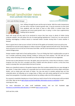
Diagnostic protocol for European stone fruit yellows phytoplasma “Candidatus Phytoplasma prunorum” PEST STATUS Not present in Australia PROTOCOL NUMBER NDP 12 VERSION NUMBER V1.2 PROTOCOL STATUS Endorsed ISSUE DATE November 2011 REVIEW DATE 2016 ISSUED BY SPHDS Prepared for the Subcommittee on Plant Health Diagnostic Standards (SPHDS) This version of the National Diagnostic Protocol (NDP) for European stone fruit yellows phytoplasma “Candidatus Phytoplasma prunorum” is current as at the date contained in the version control box on the front of this document. NDPs are updated every 5 years or before this time if required (i.e. when new techniques become available). The most current version of this document is available from the SPHDS website: http://plantbiosecuritydiagnostics.net.au/resource-hub/priority-pest-diagnosticresources/ 1 Contents 1. Introduction.............................................................................. 3 1.1 Hosts ................................................................................... 3 1.1.1. 1.2 Alternative host plants ...................................................... 3 Symptoms ............................................................................. 3 2. Taxonomic information ................................................................ 4 3. Detection ................................................................................. 4 3.1 4. The signs or symptoms associated with infection ............................... 5 Identification .......................................................................... 13 4.1 Recommended phytoplasma detection method ................................ 13 4.1.1. DNA extraction procedure using the QIAGEN DNeasy® Plant mini kit (Green et al. 1999) ..................................................................... 13 4.1.2. 4.2 PCR............................................................................. 16 Interpretation of results ........................................................... 19 5. Contact points for further information ........................................... 20 6. Acknowledgements ................................................................... 20 7. References ............................................................................. 21 8. Appendices ............................................................................. 25 8.1 Appendix 1: Nucleic acid cleanup................................................ 25 8.2 Appendix 2: Alternative extraction methods ................................... 26 8.2.1. Phytoplasma enrichment extraction method (Kirkpatrick et al. 1987 and modified by Ahrens and Seemüller, 1992)..................................... 26 8.2.2. Quick nucleic acid extraction methods for phytoplasmas in plants (Maixner et al. 1995) .................................................................. 28 8.3 Appendix 3: Phytoplasmas ......................................................... 30 NDP European stone fruit yellows 2 1. INTRODUCTION The species name Candidatus phytoplasma prunorum was proposed for ESFY phytoplasma in 2004 (Seemuller and Schneider, 2004). It is closely related to both Candidatus phytoplasma mali (apple proliferation phytoplasma) and Candidatus phytoplasma pyri (pear decline phytoplasma) and the three phytoplasmas share 98.599% sequence similarity across the 16S gene. All three phytoplasmas belong to the 16SrX (apple proliferation) phytoplasma group (Lee et al. 1998). However significant genetic differences are observed in other genes amongst the three phytoplasmas and they have distinct biological and epidemiological properties including host range and vectors. 1.1 Hosts Ca. P. prunorum is associated with European stone fruit yellows (ESFY) disease, which includes diseases of apricot (Prunus armeniaca), Japanese (flowering) cherry (P. serrulata), black cherry (P. mahaleb), peach (P. persica), Japanese plum (P. salicina), European plum (P.domestica), cherry (myrobalan) plum (P. cerasifera) and almond (P. dulcis syn. P. amygdalus Batsch) (Lorenz et al. 1994; Seemüller and Foster 1995; Marcone et al. 1996; Sertkaya et al. 2005). Rootstocks can be infected by the phytoplasma including P. marianna, P. domestica, P. cerasifera, P. domestica x P. cerasifera, P. salicina x P. spinosa, and P. persica x P. cerasifera (Jarausch et al. 1998). The severity of symptom expression in Prunus sp. is dependent on the species and the variety (Jarausch et al. 2000). 1.1.1. Alternative host plants Alternative hosts for ESFY include Hackberry (Celtis australis), Ash (Fraxinus excelsior), Dog rose (Rosa canina), Wild cherry (Prunus avium) and Blackthorn (Prunus spinosa). Non-Prunus species may be symptomless. These hosts are important in the epidemiology of the phytoplasma as they act as a source of phytoplasma inoculum for orchards and may also host insect vectors. Blackthorn is a preferred host of the vector. 1.2 Symptoms General symptoms of ESFY disease in various stone fruit species include early flowering and shooting through winter and this early break in dormancy can increase the susceptibility of affected trees to frost, causing damage to the phloem. During the growing season leaves can be chlorotic and roll. Leaves can drop prematurely. Affected shoots are often stunted and bear smaller, deformed leaves. Shoots may die back. Fruit on affected branches develop poorly and may fall prematurely. Fruit yield can be reduced. Only a few branches are affected during the early stage of disease but as the disease progresses the whole tree may become affected. Many stone fruit tree species or varieties show decline (Nemeth, 1986; Seemüller and Foster 1995). Specific symptoms in apricot (apricot chlorotic leafroll disease) include upward curling of leaves, which are chlorotic and early reddening, Sudden dieback can occur during the growing season. Small, wilted fruit and dried leaves may also persist during the autumn. In plum (plum leptonencrosis disease) the leaf margins roll upward and leaves may be chlorotic and /or smaller. In peach the midribs and NDP European stone fruit yellows 3 lateral veins of the leaves can become enlarged and corky tissue develops along the veins. The leaves become red and roll upward. In cherry the first symptom observed is slight chlorosis of leaves in summer. Flowers are malformed and fruit set is poor in the following year. Rosetting of leaves occurs on affected shoots and young shoots remain unlignified. (Nemeth, 1986; Seemüller and Foster 1995). In almond leaf rolling, reddening of the shoot bark and leaves, and sparse foliation may be observed. Although symptoms are indicative of infection by Ca. P. prunorum, phytoplasma infection should be confirmed through diagnostic testing. Ca. P. prunorum can be detected using a PCR based on the 16S rRNA gene with universal primers for all known phytoplasmas and identified by RFLP analysis or sequencing (Seemüller et al. 1998). Specific primers may also be useful in identifying infection by Ca. P. prunorum. Ca. P. prunorum can be detected in aerial parts of trees during dormancy (Jarausch et al. 1998) and the possibility exists that this phytoplasma might be transmitted through vegetative propagation of dormant budwood. 2. TAXONOMIC INFORMATION The taxonomic classification of the phytoplasma associated with European stone fruit yellows is: Bacteria; Firmicutes; Mollicutes; Acholeplasmatales; Acholeplasmataceae; Candidatus Phytoplasma; 16SrX (Apple proliferation group). Candidatus Phytoplasma prunorum Ca. P. prunorum is closely related to Ca. P. mali (apple proliferation phytoplasma) and Ca. P pyri (pear decline phytoplasma) and the three species share 98.6–99.1% sequence similarity. However, each species is transmitted by different vectors and has different hosts or induces different symptoms in the same host. Ca. P prunorum has several common names and acronyms depending on the disease and host and these include: European stone fruit yellows (ESFY) mycoplasma-like organism, European stone fruit yellows phytoplasma, Apricot chlorotic leaf roll (ACLR) phytoplasma, plum leptonecrosis (PLN) phytoplasma 3. DETECTION European stone fruit yellows disease can be identified by the presence of symptoms, however diagnosis should be confirmed through PCR detection and sequencing of the 16S rRNA gene of Ca. P. prunorum, particularly as other phytoplasmas may cause similar symptoms. Most symptoms, particularly if they are observed on their own, may be caused by other biotic and abiotic factors. Symptomless infections can occur and if this is suspected it is important to thoroughly sample different phloem tissue from different shoots and branches of the one plant for phytoplasma isolation. NDP European stone fruit yellows 4 Ca. P. prunorum is phloem-limited, however it may infect the phloem tissue of all parts of a tree, including roots trunk, branches and shoots. Phytoplasmas can be unevenly distributed and in uneven titre throughout woody hosts, and symptomatic tissue is optimal for phytoplasma detection (Berges et al. 2000; Christensen et al. 2004; Constable et al. 2003; Necas and Krska, 2006). The location and titre of phytoplasmas may be affected by seasonal changes and therefore the timing of sample collection for phytoplasma detection is important (Jarausch et al. 1999). In Europe the best time and tissue type for Ca. P. prunorum detection is June (early summer) for phloem samples from woody shoots and September (summer) from petiole samples (Necas et al. 2008). Ca. P. prunorum can persist and be detected in the phloem of aerial parts of trees during the dormant season (Seemuller et al. 1998). Vascular tissue from symptomatic plant material provides the best opportunity to detect phytoplasmas in stone fruit trees. Leaf petioles, mid veins from symptomatic leaves and bark scrapings from shoots and branches can be used from actively growing plant hosts. If the plant is dormant, buds and bark scrapings from branches, trunk and roots can be used, although these are likely to be less reliable. If using bark scrapings from woody material remove the dead outer bark layer, to reveal the green inner vascular tissue. 3.1 The signs or symptoms associated with infection ESFY disease may be suspected if the following symptoms described for each stone fruit species are observed. Trees flower and shoot in winter • Chlorosis of the leaves later in the growing season. • Premature leaf drop • Stunted shoots bearing smaller, deformed leaves • Die back of shoots • Necrosis of the phloem. • Fruit on affected branches develop poorly and may fall prematurely • Yield is reduced Many stone fruit tree species or varieties show decline (Nemeth, 1986; Seemüller and Foster 1995). During the early stage of disease often only a few branches are affected but the whole tree may become affected as the disease progresses. Symptom expression can differ in severity amongst different cultivars of the one stone fruit species. Specific symptoms in apricot (apricot chlorotic leafroll) include (Figures 1-4): • upward curling of leaves • chlorotic leaves • early reddening • Sudden dieback can occur during the growing season. • Small, wilted fruit and dried leaves may also persist during the autumn. NDP European stone fruit yellows 5 Specific symptoms in peach include (Figure 5-9): • midribs and lateral veins of the leaves can become enlarged and corky tissue develops along the veins (Figure 9) • leaves become red and roll upward (Figure 7 and 8). Specific symptoms in Japanese plum (plum leptonencrosis) include: • leaf margins roll upward • leaves may be chlorotic • small leaves • necrosis of the vascular tissue (Figure 10) Specific symptoms in cherry include: • slight chlorosis of leaves in summer • flowers are malformed and fruit set is poor in the following year. • rosetting of leaves occurs on affected shoots • young shoots remain unlignified (Nemeth, 1986; Seemüller and Foster 1995). Specific symptoms in almond include: • leaf rolling • reddening of the shoot bark and leaves • sparse foliation may be observed. NDP European stone fruit yellows 6 Figure 1. Apricot tree partially affected by ESFY disease, symptoms include chlorosis and rolling of leaves on the affected braches at the front. (Source: F. Constable) NDP European stone fruit yellows 7 Figure 2. ESFY affected apricot tree (front) with severe decline compared to an unaffected tree (rear) (Source: F. Constable). NDP European stone fruit yellows 8 Figure 3. Severe chlorosis and leaf rolling of an ESFY affected apricot tree (Source: F. Constable). Figure 4. Rolling and chlorosis of leaves on an ESFY affected apricot tree (Source: F. Constable). NDP European stone fruit yellows 9 Figure 5. Peach tree partially affected by ESFY disease (front) showing chlorosis and leaf rolling compared with an unaffected part of the same tree (back) (Source: F. Constable). Figure 6. Peach tree affected by ESFY disease exhibiting decline, sparse foliation, chlorosis and smaller leaves (Source: F. Constable). NDP European stone fruit yellows 10 Figure 7. Chlorosis, some reddening and rolling of peach leaves on a shoot affected by ESFY disease (Source: F. Constable). Figure 8. Rolling and reddening of peach leaves on a shoot affected by ESFY disease (Source: F. Constable). NDP European stone fruit yellows 11 Figure 9. Development of corky tissue along a lateral vein of a peach leaf affected by ESFY disease (Image courtesy of Dr B. Schneider Julius Kuehn Institute, Federal Research Centre for Cultivated Plants, Institute for Plant Protection in Fruit Crops and Viticulture, Dossenheim, Germany). Figure 10. Necrosis of the vascular tissue of an ESFY affected Prunus tree (Image courtesy of Dr B. Schneider Julius Kuehn Institute, Federal Research Centre for Cultivated Plants, Institute for Plant Protection in Fruit Crops and Viticulture, Dossenheim, Germany). NDP European stone fruit yellows 12 4. IDENTIFICATION The most reliable method for confirmation of Ca. P. prunorum is polymerase chain reaction (PCR), which is used to detect the DNA of the phytoplasma. The efficiency of this test is dependent on appropriate sampling of plant tissue and reliable nucleic acid extraction methods. 4.1 Recommended phytoplasma detection method 1. Extract total DNA using the method described by Green et al. (1999), which uses a CTAB extraction buffer and the DNeasy® Plant Mini Kit (Qiagen Cat. No. 69104) 2. Perform a housekeeping PCR with the rP1/fD2 primers. The rP1/fD2 primers amplify the 16S rRNA gene from most prokaryotes as well as from chloroplasts. If this test is negative then there is no DNA present or there are DNA polymerase inhibitors co-extracted with the nucleic acid. In this situation, try cleaning the nucleic acid (Appendix 1) or repeat the extraction using a different procedure (Appendix 2). 3. Perform PCR using the following procedure: Use a nested PCR on the purified DNA using the universal phytoplasma primer pair, P1/P7 for the first-stage PCR followed by the R16F2n/R16R2 primer pair for the second-stage PCR (Table 4). 4. Analyse the PCR products by agarose gel electrophoresis. 5. To determine phytoplasma identity, direct sequence the nested PCR product. If direct sequencing is problematic, the PCR product can be cloned and then sequenced using standard cloning and sequencing procedures. Sequence data can be analysed using the Basic Local Alignment Search Tool (BLAST) available at: http://blast.ncbi.nlm.nih.gov/Blast.cgi. If sequencing facilities are unavailable, a single PCR using the Ca. P prunorum specific primers ECA1 and ECA2 can be used to determine the identity of the phytoplasma that was detected. However this is a single PCR and may not detect phytoplasmas associated with low tire infections. A nested PCR using PCR product for the first stage (P1/P7) PCR product and 16SrX group specific primers (Table 4) can be used to identify the phytoplasma to the group level, however this will not determine which 16SrX phytoplasma species is present. 4.1.1. DNA extraction procedure using the QIAGEN DNeasy® Plant mini kit (Green et al. 1999) Materials and equipment 1. QIAGEN DNeasy® Plant mini kit 2. 1.5 ml centrifuge tubes 3. 20-200 µl and 200-1000 µl pipettes 4. 20-200 µl and 200-1000 µl sterile filter pipette tips 5. Autoclave 6. Balance NDP European stone fruit yellows 13 7. Bench top centrifuge 8. Distilled water 9. Ice machine 10. Freezer 11. Sterile mortars and pestles or “Homex” grinder (Bioreba) and grinding bags (Agdia or Bioreba) or hammer and grinding bags (Agdia or Bioreba) If using mortar and pestles, ensure they are thoroughly cleaned prior to use to prevent cross-contamination from previous extractions. To clean thoroughly, soak mortars and pestles in 2% bleach for 1 hour. Rinse with tap water then soak in 0.2 M HCl or 0.4 M NaOH for 1 hour. Rinse thoroughly with distilled water. 12. Scalpel handle 13. Sterile scalpel blades 14. Vortex 15. Water bath or heating block at 55-65°C 16. Latex or nitrile gloves 17. Buffers: • CTAB grinding buffer (Table 1) • Absolute ethanol The 2% cetylmethylammonium bromide (CTAB) buffer (Table 1) is required for all extraction procedures: Table 1. 2.5% cetyltrimethylammonium bromide (CTAB) buffer for DNA purification Reagent Final Amount needed for 1 concentration L CTAB (cetyltrimethylammonium bromide bromide) 2.5% 25 g Sodium chloride 1.4 M 56 g 1 M Tris-HCl, pH 8.0 (sterile) 100 mM 100 ml 0.5 M EDTA, pH8.0 (sterile) 20 mM 40 ml 1% 10 g Polyvinylpyrrolidone (PVP-40) Make up to volume with sterile distilled water. Store at room temperature. Just before use, add 0.2% 2-mercaptoethanol (v/v) to the required volume of buffer. If a fume hood is unavailable β – mercaptoethanol can be omitted but the quality of the extract from some plant species may be affected. NDP European stone fruit yellows 14 Method • • • • • • • • • • Grind 0.5 g of plant tissue in 5 ml of CTAB extraction buffer (room temperature) containing 0.2% β – mercaptoethanol. Transfer 500 µl of extract to a 1.5 ml microfuge tube and add 4 µl of RNase A (Supplied with the DNeasy kit), cap tube and incubate at 65°C for 25-35 min, mixing gently several times. Add 130 µl of QIAGEN buffer AP2 to extract. Invert 3 times to mix and place on ice for 5 minutes. Apply lysate onto a Qiashredder column and centrifuge at 20,000 x g (14,000 rpm or maximum speed) for 2 minutes. Transfer 450 µl of flowthrough from QIAshredder™ column to a 1.5 ml centrifuge tube containing 675 µl QIAGEN buffer AP3/E. Mix by pipetting. Transfer 650 µl of extract onto a DNeasy column and spin at 6,000 × g (8000 rpm) for 1 minute Discard flow-through and add the rest of the sample to the column and spin at 10000 rpm for 1 minute Place DNeasy column in a new 2 ml collection tube and add 500 µl of QIAGEN buffer AW (wash buffer) and spin at 10000 rpm for one minute. Discard flowthrough and add another 500 µl of QIAGEN buffer AW and spin at maximum speed for 2 minutes. Discard flowthrough and collection tube. Ensure that the base of the column is dry (blot on tissue if it is not) and place in an appropriately labeled microfuge tube. Add 100 µl of pre-warmed 65°C AE buffer directly to the filter (don’t apply down the side of the tube) and spin at 10000 rpm for 1 minute. Discard column and store DNA in Freezer. The reliability of the PCR test is affected by phytoplasma titre in the plant host (Marzachì et al. 2004) and low titres can lead to false negative results. If a phytoplasma infection is suspected but phytoplasmas have not been detected using the extraction procedure of Green et al. (1999) it may be useful to use a phytoplasma enrichment procedure (Appendix 2) to improve detection from symptomless material or from material collected outside the optimum time frame for detection. NDP European stone fruit yellows 15 4.1.2. PCR Laboratory requirements To reduce the risk of contamination and possible false positive results, particularly when nested PCR is used for phytoplasma detection, it is desirable to set up PCR reactions in a different lab to where nucleic acid extractions have been done. It is also desirable to handle PCR reagent stocks and to set up PCR reactions in a clean room or bio-safety cabinet with dedicated pipettes, PCR tubes and tips that have not been exposed to nucleic acid extracts. Use a separate pipette for the addition of nucleic acids to the PCR reactions. Do not add nucleic acid to reactions in the same clean room or bio-safety cabinet in which PCR stocks are handled. PCR materials and equipment 1. PCR reagents of choice 2. Primers (Table 2) 3. PCR grade water 4. 0-2 µl, 2-20 µl, 20-200 µl and 200-1000 µl pipettes 5. 0-2 µl, 2-20 µl, 20-200 µl and 200-1000 µl sterile filter pipette tips 6. 1.5 ml centrifuge tubes to store reagents 7. PCR tubes (volume depends on thermocycler) 8. Bench top centrifuge – with adapters for small tubes 9. Freezer 10. Ice machine 11. Latex or nitrile gloves 12. Thermocycler 13. DNA molecular weight marker NDP European stone fruit yellows 16 Table 2. PCR primers used for phytoplasma detection and internal control primers PCR test† Primer name (direction) Primer sequence (5´-3´) P1 (forward) AAGAGTTTGATCCTGGCTCAGGATT P7 (reverse) CGTCCTTCATCGGCTCTT R16F2n (forward) GAAACGACTGCTAAGACTGG R16R2 (reverse) TGACGGGCGGTGTGTACAAACCCCG fO1 (forward) CGGAAACTTTTAGTTTCAGT rO1(reverse) AAGTGCCCAACTAAATGAT ECA1 AATAATCAAGAACAAGAAGT ECA2 GTTTATAAAAATTAATGACTC FD2 AGAGTTTGATCATGGCTCAG RP1 ACG GTT ACC TTG TTA CGA CTT Tm Product size (bp) Reference Phytoplasmas Universal phytoplasma – single or nested first stage PCR Universal phytoplasma – single PCR or nested second stage PCR 16SrX group specific single PCR or nested PCR with P1/P7 primers used for the first PCR* Ca. P prunorum specific single PCR 55oC 1,784 Deng and Hiruki (1991) Schneider et al. (1995) 55oC 1,248 Lee et al. (1993) 55oC 1071 Lorenz et al. (1995) 55ºC 237bp Jarausch et al. (1998) Internal control 16S bacterial and plant chromosomal approx. 55ºC 1400-1500 bp. Weisberg et al. (1991) † Both the R16F2n/R16R2 and 16SrX group specific primer pairs can be used in single PCR for X-disease phytoplasma detection, however single PCR is less sensitive than nested PCR. * If sequencing facilities are unavailable these can be used to indicate if the phytoplasma is likely to belong to the X-disease (16SrIII group) phytoplasmas. These primers do not identify the phytoplasm to species or strain level. NDP European stone fruit yellows 17 Polymerase Chain Reaction The housekeeping PCR, using the components and concentrations listed in Table 3 below, is done prior to conducting the phytoplasma PCR, to determine if the nucleic extract is of sufficient quality for phytoplasma detection. The cycling times are listed in Table 6. Run the PCR products on a gel as described below. The house keeping PCR is successful if a product of the expected size is observed, indicating the presence of quality DNA in the nucleic acid extract. If no product is observed the nucleic acid extract should be cleaned up or the sample should be re-extracted and a housekeeping PCR conducted on these extracts. If the housekeeping PCR is successful the universal phytoplasma PCR reactions can be done. For universal phytoplasma detection the primers and the expected size of the PCR product are listed in Table 2. The recommended primers are universal and were developed to amplify all known phytoplasmas. For nested PCR, the first-stage PCR products, generated by the P1 and P7 primers are diluted 1:25 (v/v) in water prior to re-amplification using the second-stage PCR primers using the R16F2n and R16R2 primers. If a positive result is obtained the PCR product should be sequenced to determine the identity of the organism that is detected. If sequencing facilities are unavailable a nested PCR can be done using the first-stage PCR products, generated by the P1 and P7 primers in a second-stage PCR primers using the 16SrX group specific primers (Table 2) to determine phytoplasma identity. It is also possible to determine if the phytoplasma is Ca. P. prunorum by conducting a single PCR using the specific primers ECA1 and ECA2 (Table 2). When establishing the test initially, it is advised that a negative control (DNA extracted from healthy plant tissue) is included. Controls Positive control: DNA of known good quality (internal control PCR) DNA extracted from any phytoplasma-infected tissue (phytoplasma PCR) No template control: Sterile distilled water Table 3. Conventional PCR reaction master mix Reagent Sterile (RNase, DNase free) water 10 × reaction buffer 50 mM MgCl 2 10 mM dNTP mixture 10 µM Forward primer 10 µM Reverse primer Volume per reaction 18.05 µl 2.5 µl 0.75 µl 0.5 µl 1 µl 1 µl 5 units/µl Platinum Taq DNA polymerase (Invitrogen 10966026) DNA template or control* 0.2 µl Total reaction volume 25 µl NDP European stone fruit yellows 1 µl 18 Pipette 24 µl of reaction mix into each tube then add 1 µl of DNA template. *Up to 5µl DNA template may be added, reducing water accordingly, as target DNA may be in low concentration. Non-acetylated molecular biology grade bovine serum albumin (BSA) can be added to the master mix at 0.5mg/ml to reduce the effect of inhibitors on the PCR. Table 4. PCR cycling conditions Phytoplasma universal and 16SrIII group primers Housekeeping primers Step Temperature Time No. of cycles Temperature Time No. of cycles Initial denaturation 94oC 2 min 1 94oC 2 min 1 Denaturation Annealing 94oC 55ºC 35 94oC 55 oC 72oC 1 min 1 min 1 min 30 s 35 Elongation 45 s 45 s 1 min 30 s Final elongation 72oC 10 min 1 72oC 72oC 10 min 1 Electrophoresis Electrophorese PCR products (5-10 µl) on a 1% agarose gel containing ethidium bromide or SybR-Safe and visualise using an UV transilluminator (ethidium bromide staining) or blue light box (SybR-Safe staining). Use a DNA molecular weight marker to determine the size of the products. Table 2 lists the expected PCR product size for each primer pair. 4.2 Interpretation of results Failure of the samples to amplify with the housekeeping primers suggests that the DNA extraction has failed, compounds inhibitory to PCR are present in the DNA extract or the DNA has degraded. The phytoplasma universal and specific PCR tests will only be considered valid if: (a) the positive control produces the correct size product as indicated in Table 2; and (b) No bands are produced in the negative control (if used) and the no template control. Confirmation of the specific phytoplasma species infecting the tree can only be determined through sequence analysis. As sequence similarity of 97.5% or above indicates that the phytoplasma detected is most likely to be a strain of Ca. P. prunorum. NDP European stone fruit yellows 19 5. CONTACT POINTS FOR FURTHER INFORMATION Dr. Fiona Constable Department of Primary Industries - Knoxfield Private Bag 15 Ferntree Gully Delivery Centre Victoria 3156 AUSTRALIA Ph + 61 3 92109222 6. ACKNOWLEDGEMENTS This protocol was written and complied by Dr. Fiona Constable, Department of Primary Industries – Knoxfield, Private Bag 15 , Ferntree Gully Delivery Centre, Victoria 3156, AUSTRALIA, Ph + 61 3 92109222 Many thanks to the following researchers for providing information, advice and images for this protocol: Wolfgang Jarausch and Barbara Jarausch AlPlanta, Nuestadt and der Weinstrasse, Germany, Bernd Schneider. Julius Kuehn Institute, Federal Research Centre for Cultivated Plants, Institute for Plant Protection in Fruit Crops and Viticulture, Dossenheim, Germany Much of this phytoplasma detection protocol is based on the advice and experience of various researchers who brought together and developed a protocol for the International Sanitary and Phytosanitary Measures (ISPM) for phytoplasma detection for the International Plant Protection Convention (IPPC). The researchers included: • Fiona Constable DPI, Victoria Australia • Wilhelm Jelkmann' Institute for Plant Protection in Fruit Crops and Viticulture Dossenheim Germany, • Lia Liefting, MAF, New Zealand • Phil Jones, Rothamsted Research Harpenden Hertfordshire • Esther Torres, Laboratori de Sanitat Vegetal, Departament d'Agricultura Ramaderia i Pesca, Barcelona • Jacobus Verhoeven' Plant Protection Service, Wageningen, Netherlands This protocol was reviewed by New Zealand Ministry of Agriculture and Forestry, MAF Biosecurity. NDP European stone fruit yellows 20 7. REFERENCES Berges R, Rott M and Seemüller E. 2000. Range of phytoplasma concentration in various hosts as determined by competitive polymerase chain reaction, Phytopathology 90, 1145–1152. Christensen NM, Nicolaisen M, Hansen M, and Schulz A. 2004. Distribution of phytoplasmas in infected plants as revealed by Real-Time PCR and bioimaging, Molecular Plant Microbe Interactions 17 , 1175–1184. Constable FE, Gibb KS and Symons RH, 2003. Seasonal distribution of phytoplasmas in Australian grapevines, Plant Pathology 52, 267–276. Deng SJ and Hiruki C 1991 Amplification of 16S rRNA genes from culturable and non-culturable Mollicutes. Journal of Microbiological Methods 14, 5361 Green MJ, Thompson DA and MacKenzie DJ. 1999. Easy and efficient DNA extraction from woody plants for the detection of phytoplasmas by polymerase chain reaction. Plant Disease 83, 482-485. The IRPCM Phytoplasma/Spiroplasma Working Team – Phytoplasma taxonomy group (2004) ‘Candidatus Phytoplasma’, a taxon for the wall-less, nonhelical prokaryotes that colonize plant phloem and insects International Journal of Systematic and Evolutionary Microbiology 54, 1243–1255 Jarausch W, Lancas M and Dosba F. 1999. Seasonal colonization pattern of European stone fruit yellows phytoplasmas in different prunus species detected by specific PCR, Journal of Phytopathology 147, 47–54. Jarausch W, Lansac M, Saillard C, Broquaire JM and Dosba F. 1998 PCR assay for specific detection of European stone fruit yellows phytoplasmas and its use for epidemiological studies in France. European Journal of Plant Pathology. 104, 17-27. Jarausch, W., Eyquard, J.P., Lansac, M., Mohns, M. and Dosba, F. 2000. Susceptibility and tolerance of new French Prunus domestica cultivars to European stone fruit yellows phytoplasmas. Journal of Phytopathology 148, 489-493. Lee I-M, Hammond RW, Davis RE and Gundersen, DE. 1993. Universal amplification and analysis of pathogen 16S rDNA for classification and identification of mycoplasma-like organisms. Phytopathology 83, 834-842 Lee I-M, Gundersen DE, Hammond RW and Davis RE, 1994. Use of mycoplasma like organism (MLO) group-specific oligonucleotide primers for nested-PCR assays to detect mixed-MLO infections in a single host plant. Phytopathology 84, 559-566 Lee I-M, Gundersen-Rindal DE, Davis RE and Bartoszyk IM, 1998. Revised classification scheme of phytoplasmas based on RFLP analyses of 16S rRNA and ribosomal protein gene sequences. International Journal of Systematic Bacteriology 48, 1153-1169. Lorenz KH, Dosba F, Poggi Pollini C, Llacer G, Seemüller E. 1994. Phytoplasma diseases of Prunus species in Europe are caused by NDP European stone fruit yellows 21 genetically similar organisms. Zeitschrift für Pflanzenkrankheiten und Pflanzenschutz 101, 567-575. Lorenz, K-H, Schneider, B., Ahrens, U. and Seemuller, E. 1995. Detection of the apple proliferation and pear decline phytoplasmas of ribosomal and non ribosomal DNA. Phytopathology 85, 771-776. Marcone C, Ragazzino A, Seemüller E. 1996. European stone fruit yellows phytoplasma as the cause of peach vein enlargement and other decline diseases of stone fruits in southern Italy. Journal of Phytopathology 144, 559-564. Necas T and Krska B. 2006. Selection of woody indicators and the optimum plant material and sampling time for phytoplasma ESFY detection. Acta Horticulturae 717, 101-105. Necas T; Maskova V and Krska B. 2008. The possibility of ESFY phytoplasma transmission: through flowers and seeds. Acta Horticulturae. 781, 443447. Nemeth M. 1986. Virus, Mycoplasma and Rickettsia Diseases of Fruit Trees, Martinus Nijhoff Publishers, Dordrecht, The Netherlands Seemüller E, Foster JA, 1995. European stone fruit yellows. In: Ogawa JM, Zehr EI, Bird GW, Ritchie DF, Uriu K, Uyemoto JK, eds. Compendium of stone fruit diseases. St. Paul, MN, USA: American Phytopathological Society, 59-60. Seemüller E, Schneider B. 2004. Taxonomic description of ‘Candidatus Phytoplasma mali’ sp. nov., ‘Candidatus Phytoplasma pyri’ sp. nov. and ‘Candidatus Phytoplasma prunorum’ sp. nov., the causal agents of apple proliferation, pear decline and European stone fruit yellows, respectively. International Journal of Systematic and Evolutionary Microbiology 54, 1217-1226. Seemüller E, Marcone E, Lauer U, Ragozzino A, Göschl M, 1998. Current status of molecular classification of the phytoplasmas. Journal of Plant Pathology 80, 3-26. Sertkaya G, Martini M, Ermacora P, Musetti R and OslerR. 2005 Detection and characterization of phytoplasmas in diseased stone fruits and pear by PCR-RFLP analysis in Turkey. Phytoparasitica 33, 380-390. Weisberg, WG, Barns, S, Pelletier, DA and Lane DJ. 1991. 16S ribosomal DNA amplification for phylogenetic study. Journal of Bacteriology 173, 697703 7.1 Useful references Carraro L, Osler R, Loi N, Ermacora P, Refatti E. 1998. Transmission of European stone fruit yellows phytoplasma by Cacopsylla pruni. Journal of Plant Pathology 80, 233-239. NDP European stone fruit yellows 22 Carraro L, Osler R, Loi N, Ermacora P and Refatti E. 2001. Fruit tree phytoplasma diseases diffused in nature by psyllids. Acta Horticulturae. 550, 345-350. Carraro L; Ferrini F; Ermacora P and Loi N. 2004a. Transmission of European stone fruit yellows phytoplasma to Prunus species by using vector and graft transmission. Acta Horticulturae. 657, 449-453. Carraro L; Ferrini F; Labonne G, Ermacora P and Loi, N. 2004b. Seasonal infectivity of Cacopsylla pruni, vector of European stone fruit yellows phytoplasma. Annals of Applied Biology. 144, 191-195. Conci C, Rapisarda C and Tamanini L. 1992. Annotated catalogue of Italian Psylloideae. Attidell Accademia Roveretana degli Agiati II-B (ser. VII), Rovereto, Italy, pp. 104-107. Firraro G, Smart GD and Kirkpatrick BC, 1996. Physical map of the Western X-Disease phytoplasma chromosome. Journal of Bacteriology 178, 39853988. Gibb KS, Padovan AC and Mogen BD, 1995. Studies on sweet potato littleleaf phytoplasma detected in sweet potato and other plant species growing in Northern Australia. Phytopathology 85, 169-174. Kirkpatrick BC. 1991. Mycoplasma like organisms – plant and invertebrate pathogens, Chapter 229, p. 4050-4067. In: Balows A, Truper HG, Dworkin M, Harder W and Schleifer KH (eds.)The prokaryotes Volume IV, 2nd Ed.. Springer-Verlag, New York. Kollar A and Seemüller E. 1989. Base composition of the DNA of mycoplasma-like organisms associated with various plant diseases. Journal of Phytopathology 127, 177-186. Lim PO and Sears BB. 1989. 16S rRNA sequence indicates that plantpathogenic mycoplasma like organisms are evolutionarily distinct from animal mycoplasmas. Journal of Bacteriology 171, 5901–5906. McCoy RE. 1984. Mycoplasma-like organisms of Plants and invertebrates. In: Krieg NR and Holt JG (eds). Bergey’s manual of systematic bacteriology volume 1 pp792 – 793,. William and Wilkins, Baltimore/London. Marcone C, Ragozzino A, Cousin MT, Berges R and Seemuller E. 1999. Phytoplasma diseases of trees and shrubs of urban areas in Europe. Acta Horticulturae. 496, 69-75; Marzachi C. 2004. Molecular diagnosis of phytoplasmas Phytopathologia Mediterranea. 43, 228-231. Neimark H and Kirkpatrick BC. 1993. Isolation and characterization of fulllength chromosomes from non-culturable plant-pathogenic Mycoplasmalike organisms. Molecular Microbiology 7, 21-28. Oshima K, Shiomi T, Kuboyama T, Sawayanagi T, Nishigawa H, Kakizawa S, Miyata S, Ugaki M and Namba S. 2001. Isolation and characterisation of derivative lines of the onion yellows phytoplasma that do not cause stunting or phloem hyperplasia. Phytopathology 91, 1024-1029. NDP European stone fruit yellows 23 Ossiannilsson F, 1992. The Psylloidea (Homoptera) of Fennoscandia and Denmark. Fauna Entomologica Scandinava, Vol. 26. Leiden, the Netherlands: E. J. Brill. Poggi Pollini C, Zelger R, Wolf M, Bissani R, Giunchedi L, 2002. Indagine sulla presenza di psillidi infetti dal fitoplasma degli scopazzi del melo (AP = apple proliferation) in provincia di Bolzano. ATTI Giornate Fitopatologiche 2, 607–12. Padovan AC, Firrao G, Schneider B and Gibb KS. 2000. Chromosome mapping of the sweet potato little leaf phytoplasma reveals genome heterogeneity within the phytoplasmas. Microbiology Reading 146, 893-902. Razin S and Freundt EA, 1984. The mycoplasmas. In Bergey’s Manual of Systematic Bacteriology, Volume 1. Pp740-792. Noel R. Krieg and John G. Holt Eds. William and Wilkins, Baltimore/London. Sears BB, Lim P-O, Holland N, Kirkpatrick BC and Klomparens KL. 1989. Isolation and characterization of DNA from a mycoplasmalike organism. Molecular Plant Microbe Interactions 2, 175-180. Schneider B, Seemüller E, Smart CD and Kirkpatrick BC, 1995. Phylogenetic classification of plant pathogenic Mycoplasma like organisms or phytoplasmas. In: Razin S and TullyJG (eds). Molecular and Diagnostic Procedures in Mycoplasmology, Vol. 1, pp369-380, Academic press, San Diego. Tedeschi R, Bosco D, Alma A, 2002. Population dynamics of Cacopsylla melanoneura (Homoptera: Psyllidae), a vector of apple proliferation in northwestern Italy. Journal of Economic Entomology 95, 544–51. Schaub L, Monneron A, 2003. Phénologie de Cacopsylla pruni vecteur de l'enroulement chlorotique de l'arbricotier. Revue Suisse de Viticulture, Arboriculturae, Horticulturae 35, 123–6. Thebaud G., 2005.Etude du développement spatio-temporel d'une maladie transmise par vecteur en intégrant modélisation statistique et expérimentation: cas de l'ESFY (European stone fruit yellows). 176 p., PhD Thesis, SupAgro, Montpellier, France. Zreik L, Carle P, Bove JM and Garnier M. 1995. Characterization of the mycoplasma like organism associated with witches'-broom disease of lime and proposition of a Candidatus taxon for the organism, "Candidatus Phytoplasma aurantifolia". International Journal of Systematic Bacteriology 45, 449-453. NDP European stone fruit yellows 24 8. APPENDICES 8.1 Appendix 1: Nucleic acid cleanup Materials and equipment 1. 1.5 ml centrifuge tubes 2. 20-200 µl and 200-1000 µl pipettes 3. 20-200 µl and 200-1000 µl sterile filter pipette tips 4. Autoclave 5. Balance 6. Bench top centrifuge 7. Distilled water 8. Freezer 9. Vortex 10. Latex or nitrile gloves 11. Reference: 12. Buffers/solutions: • Chloroform:iso-amyl alcohol (24:1 v/v) • Ice-cold isopropanol • 70% (v/v) ethanol • Sterile distilled water • TE buffer (10 mM Tris-HCl, 1 mM EDTA, pH 7.5 or 8.0) Method 1. Add an additional 100-200 µl of sterile water or TE to the nucleic extract to assist ease of handling. 2. Add an equal volume of chloroform:isoamyl alcohol (24:1) and mix thoroughly by vortexing. Centrifuge in a microfuge at room temperature for 15 minutes at 13000 rpm. 3. Transfer the epiphase into a new 1.5ml microcentrifuge tube and add an equal volume of isopropanol (stored at -20°C). Mix immediately by inversion. Centrifuge for 15 minutes at 13000rpm. 4. Discard the supernatant and wash the pellet once with 70% ethanol. 5. Air dry the pellet and resuspend in 20-50 µl of water. Alternatively the DNA may be purified through a MicroSpin™ S-300 HR column (GE Healthcare Cat. No 27-5130-01) according to the manufacturer’s instructions. NDP European stone fruit yellows 25 8.2 Appendix 2: Alternative extraction methods 8.2.1. Phytoplasma enrichment extraction method (Kirkpatrick et al. 1987 and modified by Ahrens and Seemüller, 1992) Ahrens U and Seemüller E. 1992. Detection of DNA of plant pathogenic mycoplasma-like organisms by a polymerase chain reaction that amplifies a sequence of the16S rRNA gene. Phytopathology 82, 828-832 Kirkpatrick BC, Stenger DC, Morris TJ and Purcell AH. 1987. Cloning and detection of DNA from a nonculturable plant pathogenic mycoplamsalike organism. Science 238, 197-199 Materials and equipment 1. 2 ml centrifuge tubes 2. 20-200 µl and 200-1000 µl pipettes 3. 20-200 µl and 200-1000 µl sterile filter pipette tips 4. Autoclave 5. Balance 6. Bench top centrifuge 7. Distilled water 8. Ice 9. Freezer 10. Sterile mortars and pestles or “Homex” grinder (Bioreba) and grinding bags (Agdia or Bioreba) or hammer and grinding bags (Agdia or Bioreba) 11. Scalpel handle 12. Sterile scalpel blades 13. Vortex 14. Water bath or heating block at 55-65°C 15. Latex or nitrile gloves 16. Buffers: • Phytoplasma isolation buffer - The potassium (Table 5) and sodium (Table 6) isolation buffers are interchangeable. To make the isolation buffer use sterile distilled water or filter sterilise. The phytoplasma isolation buffer can be stored in 50 ml aliquots at -20°C and defrosted for use. Just before use add 0.15% [w/v] bovine serum albumin and 1 mM ascorbic acid. Make up 100 mM stocks of ascorbic acid (0.176 g/ml water) and store in 500 µl aliquots at -20°C for up to two weeks. Just before using the grinding buffer, add ascorbic acid at 500 µl/50ml phytoplasma isolation buffer. Adjust pH to 7.6 after adding ascorbic acid and BSA. NDP European stone fruit yellows 26 • CTAB grinding buffer (Table 1) • Chloroform:iso-amyl alcohol (24:1 v/v) • 70% (v/v) ethanol • Sterile distilled water • Ice-cold isopropanol Table 5. Potassium phosphate phytoplasma isolation buffer Reagent K 2 HPO 4 -3H 2 O Final concentratio n 0.1 M Amount needed for 1 L 21.7 g KH 2 PO 4 0.03 M 4.1 g Sucrose 10% 100 g Polyvinylpyrrolidone (PVP-40) 2% 20 g EDTA, pH 7.6 10 mM 20 ml of a 0.5 M solution Table 6. Sodium phosphate phytoplasma isolation buffer Reagent Na 2 HPO 4 Final concentratio n 0.1 M Amount needed for 1 L 14.2 g NaH 2 PO 4 0.03 M 3.6 g Sucrose 10% 100 g Polyvinylpyrrolidone (PVP-40) 2% 20 g EDTA, pH 7.6 10 mM 20 ml of a 0.5 M solution Method 1. Grind 0.3 g leaf petioles and mid-veins or buds and bark scrapings in 3 ml (1/10; w/v) in ice-cold isolation buffer 2. Transfer 1.5-2 ml of the ground sample to a cold 2 ml microcentrifuge tube and centrifuge at 4ºC for 5 min at 4,500 rpm. 3. Transfer supernatant into a new 2 ml microcentrifuge tube and centrifuge at 4ºC for 15 min at 13,000 rpm. 4. Discard the supernatant. 5. Resuspend the pellet in 750 µl hot (55-65°C) CTAB buffer. 6. Incubate at 55-65°C for 30 min with intermittent shaking then cool on ice for 30 seconds. NDP European stone fruit yellows 27 7. Add 750 µl chloroform:isoamyl alcohol (24:1 v/v), vortex thoroughly and centrifuge at 4°C or at room temperature for 4 min at 13,000 rpm. 8. Carefully remove upper aqueous layer into a new 1.5 ml microcentrifuge tube. 9. Add 1 volume ice-cold isopropanol, vortex thoroughly and incubate on ice for 4 min. Centrifuge at 4°C or at room temperature for 10 min at 13,000 rpm. Discard supernatant. 10. Wash DNA pellet with 500 µl ice-cold 70% (v/v) ethanol, centrifuge at 4oC or at room temperature for 10 min at 13,000 rpm. 11. Dry DNA pellet in a DNA concentrator or air-dry. 12. Resuspend in 20 µl sterile distilled water. Incubating the tubes at 55oC for 10 min can aid DNA resuspension. 13. Store DNA at -20ºC for short term storage or -80ºC for long term storage. 8.2.2. Quick nucleic acid extraction methods for phytoplasmas in plants (Maixner et al. 1995) Maixner M, Ahrens U and Seemüller E. 1995. Detection of the German grapevine yellows (Vergilbungskrankheit) MLO in grapevine, alternative hosts and a vector by a specific PCR procedure. European Journal of Plant Pathology 101, 241-250. Materials and equipment 1. 2 ml centrifuge tubes 2. 20-200 µl and 200-1000 µl pipettes 3. 20-200 µl and 200-1000 µl sterile filter pipette tips 4. Autoclave 5. Balance 6. Bench top centrifuge 7. Distilled water 8. Freezer 9. Sterile mortars and pestles or “Homex” grinder (Bioreba) and grinding bags (Agdia or Bioreba) or hammer and grinding bags (Agdia or Bioreba) 10. Scalpel handle 11. Sterile scalpel blades 12. Vortex 13. Water bath or heating block at 55-65°C 14. Latex or nitrile gloves 15. Buffers: NDP European stone fruit yellows 28 • • • • • CTAB buffer (Table 2) Chloroform:iso-amyl alcohol (24:1 v/v) – these two solutions are interchangeable 70% (v/v) ethanol Sterile distilled water Ice-cold isopropanol Method Perform all operations on ice unless otherwise specified. 1. Grind 0.5 g of plant material in 5 ml of CTAB extraction buffer containing 0.2% β – mercaptoethanol. 2. Transfer 500 µl of extract to a 1.5 ml microfuge tube, close the tube and incubate at 55-65°C for 25-35 min, mixing gently several times. 3. Add 0.8-1 ml of chloroform:isoamyl alcohol (24:1 v/v) and mix thoroughly but gently. Centrifuge in a microfuge at room temperature for 5 minutes at 13000 rpm. 4. Transfer the epiphase into a new 2 ml centrifuge tube and add an equal volume of isopropanol (stored at -20°C). Mix immediately. Centrifuge for 5 minute at 13000 rpm. Discard the supernatant and wash the pellet twice with 70% ethanol. 5. Dry the pellet under vacuum or air dry and resuspend in 50 µl of water. NDP European stone fruit yellows 29 8.3 Appendix 3: Phytoplasmas Phytoplasmas are obligate intracellular parasites, principally restricted to the phloem cells of infected plant hosts or the salivary glands of their insect vectors (McCoy, 1984). Phytoplasmas have not been successfully cultured in vitro (Kirkpatrick, 1991). Phytoplasmas were originally referred to as mycoplasma–like organisms (MLO) since morphologically and ultrastructurally they resemble animal mycoplasmas (Mycoplasma sp.), which belong in the class Mollicutes (common name: mollicutes) of the kingdom Prokaryotae. Like mycoplasmas, phytoplasmas lack a rigid cell wall, have a double membrane and are pleiomorphic. Phytoplasmas are susceptible to antibiotics such as oxy-tetracycline but they are resistant to penicillin because they lack a cell wall (Razin and Freundt 1984). The genome sizes of phytoplasmas are amongst the smallest known for cellular organisms and range between 530 kilobases (kb) to 1350kb (Firrao et al. 1996; Gibb et al. 1995; Marcone et al. 1999; Neimark and Kirkpatrick 1993; Oshima et al. 2001; Padovan et al. 2000; Zriek et al. 1995). Like other members of the mollicutes, phytoplasma genomes have a low mole percent guanine plus cytosine content (mol % G+C) value compared to other organisms (Kollar and Seemüller, 1989; Sears et al. 1989). Analysis of the 16S ribosomal RNA (rRNA) gene of phytoplasmas showed that these organisms were distinguishable from mycoplasmas and more closely related to acholeplasmas (Lim and Sears 1989). In 1992 at the 9th Congress of the International Organization of Mycoplasmology, the Phytoplasma Working Team of the International Research Project for Comparative Mycoplasmology (IRPCM) assigned these simple bacteria the trivial name ‘phytoplasma’ to acknowledge that they formed a large, distinct, monophyletic group within the class Mollicutes. Most phytoplasmas were originally named for the symptoms with which they were associated, e.g. European stone fruit yellows (ESFY) phytoplasma. In 2004 the IRPCM Phytoplasma/Spiroplasma Working Team – Phytoplasma Taxonomy Group published guidelines for the description of a ‘Candidatus Phytoplasma’ genus. The ‘Candidatus’ status is used for phytoplasmas because they cannot be cultured nor characterised using many traditional methods for the classification of bacteria, which are based on morphological, biochemical and physiological properties, antigenicity and pathogenicity. The species name Candidatus phytoplasma prunorum was proposed for ESFY phytoplasma in 2004 (Seemuller and Schneider, 2004). It is closely related to both Candidatus phytoplasma mali (apple proliferation phytoplasma) and Candidatus phytoplasma pyri (pear decline phytoplasma) and the three phytoplasmas share 98.5-99% sequence similarity across the 16S gene. All three phytoplasmas belong to the 16SrX (apple proliferation) phytoplasma group (Lee et al. 1998). However significant genetic differences are observed in other genes amongst the three phytoplasmas and they have distinct biological and epidemiological properties including host range and vectors. NDP European stone fruit yellows 30


© Copyright 2026