
Sexually Transmitted Infections: UK National Screening and Testing Guidelines
Sexually Transmitted Infections: UK National Screening and Testing Guidelines August 2006 Screening Guideline Steering Group Jonathan Ross (co-chair) Cathy Ison (co-chair) Caroline Carder David Lewis Danielle Mercey Hugh Young Screening Guidelines Steering Committee commissioned by Clinical Effectiveness Group Contents Introduction Summary Tables Testing guidelines for individual sexually transmitted infections: gonorrhoea chlamydia syphilis bacterial vaginosis chancroid donovanosis lymphogranuloma venereum trichomoniasis candida herpes hepatitis A, B and C genital warts HIV page 3 5 16 26 33 40 47 52 57 63 68 76 85 97 101 2 Introduction The Bacterial Special Interest Group (BSIG) of the British Association for Sexual Health and HIV was commissioned by the Clinical Effectiveness Group (CEG) to write screening and testing guidelines for use in UK genitourinary (GU) medicine clinics. The aims of these guidelines are to: • provide advice on what tests for sexually transmitted diseases are most appropriate in a UK GU clinic setting (excluding HIV infected patients) • provide a basis for audit • support clinics when bidding for additional resources to meet national standards Although designed for use by GU clinics the recommendations may also provide information and guidance for other healthcare settings wishing to optimise the diagnosis of sexually transmitted infections. In compiling the guideline advice has been taken from a variety of different experts in the UK. The grade of evidence for each recommendation is given and it is evident that in many cases there is a lack of clinical trial data which has led to the use of appropriate expert opinion. There is therefore a clear need for future research programmes to assess the efficacy of different approaches for sexually transmitted infections (STI) screening and testing. The levels of evidence and recommendations have been graded as shown below. Levels of evidence Ia evidence obtained from meta-analysis of randomised controlled trials Ib evidence obtained from at least one randomised controlled trial IIa evidence obtained from at least one well designed controlled study without randomisation IIb evidence obtained from at least one other type of well designed quasi-experimental study III evidence obtained from well designed non-experimental descriptive studies IV evidence obtained from expert committee reports or opinions and/or clinical experience of respected authorities Grading of recommendation A evidence at level Ia or Ib B evidence at level IIa, IIb or III C evidence at level IV The structure of the guideline is as follows: • Summary tables – which make recommendations for the testing of individual sexually transmitted infections with regard to the site that should be tested and the most appropriate test that should be used, both in asymptomatic and symptomatic men and women presenting to a UK GU medicine clinic. • Testing guidelines for individual sexually transmitted infections – for each individual infection more detailed information is provided regarding the recommended tests, recommended site for testing, factors which might alter the tests or sites recommended (sexual history, risk group, etc), frequency of repeat testing in asymptomatic patients and recommendation for test of cure. 3 The guidelines have been developed following the methodological framework of the Appraisal of Guidelines Research and Evaluation instrument (AGREE – adapted as described in Int J STD and AIDS 2004 15. 297 – 298, 299 – 305). The key features are as follows: 1. Scope and purpose: the overall aim of the guidelines, target population and target users are as described above. 2. Stakeholder involvement. The extent to which the guideline represents the views of intended users has been addressed primarily by the authorship coming from the multidisciplinary membership of the BSIG. As practising clinicians the authors were able to draw on their experience of applying the tests to symptomatic and asymptomatic patients but it was not feasible to obtain formal input from representative patients. 3. Rigour of development. For each guideline the strategy used to search for evidence is outlined. The process used to formulate the recommendations varies with the authorship, which is listed in each case. After drafting, other health care professionals and professional bodies in genitourinary (GU) medicine were asked to comment, the draft guidelines posted on the BASHH website for 3 months, and all comments reviewed before final publication. 4. Presentation. A standard format was set by the BSIG editors and has been followed throughout. 5. Applicability. The authors were asked to comment on the organisational and the cost implications of applying each guideline and have identified issues that may be problematic for routine GU medicine departments and laboratories. The cost of specific tests are not included as these vary according to individual contracts. Each guideline suggests standards for audit. 6. Editorial Independence. Each of the guidelines has a statement about potential conflicts of interest. As with previous guidelines it is intended that the recommendations will be updated as new evidence becomes available. Those wishing to contribute to this process should contact either Jonathan Ross ([email protected]) or Cathy Ison ([email protected]). Screening Guideline Steering Group Jonathan Ross (co-chair) Cathy Ison (co-chair) Caroline Carder David Lewis Danielle Mercey Hugh Young BASHH Clinical Effectiveness Group Keith Radcliffe (chair) Jan Welch Imtyaz Ahmed-Yusuf Mark FitzGerald Guy Rooney David Daniels 4 Summary Tables 5 The following tables summarise the guidance on screening and testing for sexually transmitted infections (STIs) in patients attending genitourinary medicine clinics in the UK. These provide an overview of the most appropriate investigations to use to detect STIs but further detail and clarification is provided in the subsequent sections covering individual infections. 6 Recommended Tests for Asymptomatic Patients Test(s) of choice in asymptomatic heterosexual men. Site or Specimen Gonorrhoea Chlamydia Nonspecific urethritis Syphilis HIV Urethra culture NAAT NR NR NR Rectum NR NR NR NR NR Oropharynx NR NR NR NR NR Urine NAAT (if urethral specimen not available) NAAT NR NR NR Blood NR NR NR EIA or TPPA or cardiolipin test plus TPHA EIA NR: Not recommended NAAT: nucleic acid amplification test Screening tests in asymptomatic heterosexual men are not recommended for the following infections except where indicated in Testing guidelines for individual sexually transmitted infections: candida trichomoniasis bacterial vaginosis chancroid donovanosis hepatitis A, B and C herpes simplex lymphogranuloma venereum genital warts (visual inspection only) 7 Test(s) of choice in asymptomatic men who have sex with men (MSM) Site or Specimen Gonorrhoea Chlamydia Nonspecific urethritis Syphilis Urethra culture NAAT NR NR NR NR Rectum* culture** NAAT (in NR some situations***) NR NR NR Oropharynx* culture** NR NR NR NR NR Urine NAAT (if urethral specimen not available) NAAT NR NR NR NR Blood NR NR NR EIA for HBsAg and anti-HBcAb and antiHBsAb EIA NR: Not recommended EIA or TPPA or cardiolipin test plus TPHA NAAT: nucleic acid amplification test Hepatitis B HIV * Samples only appropriate if indicated by sexual history ** If samples are taken from this site then culture should be used but NAAT may be considered if culture is not available *** NAATs are increasingly being used but remain unlicensed. Screening using NAATs should be offered in men who are contacts of LGV and guidance for more widespread rectal screening for Chlamydia in MSM is still under review. The site of testing may vary according to sexual history (see Testing guidelines for individual sexually transmitted infections for specific details). Screening tests in asymptomatic MSM are not recommended for the following infections except where indicated in Testing guidelines for individual sexually transmitted infections: candida trichomoniasis bacterial vaginosis chancroid donovanosis hepatitis A and C herpes simplex lymphogranuloma venereum genital warts (visual inspection only) 8 Test(s) of choice in asymptomatic women Site or Specimen Gonorrhoea Chlamydia Syphilis HIV Urethra NR NR NR NR Cervix culture NAAT NR NR Vagina -self-taken tampons or swabs -vulval-introital -posterior fornix NR | | | NAAT | | NR NR Rectum NR NR NR NR Oropharynx NR NR NR NR Urine NR NAAT (if urethral specimen not available) NR NR Blood NR NR NR: Not recommended EIA or TPPA or cardiolipin test plus TPHA NAAT: nucleic acid amplification test EIA The site of testing may vary according to sexual history or whether the woman has had a hysterectomy (see Testing guidelines for individual sexually transmitted infections for specific details). Screening tests in asymptomatic women are not recommended for the following infections except where indicated in Testing guidelines for individual sexually transmitted infections: candida trichomoniasis bacterial vaginosis chancroid donovanosis hepatitis A, B and C herpes simplex lymphogranuloma venereum genital warts (visual inspection only) 9 Recommended Tests for Patients presenting with Genital Discharge Test(s) of choice for genital discharge in heterosexual men and men who have sex with men Site or Specimen Gonorrhoea Chlamydia NSU Candida Trichomonas Urethra microscopy plus culture NAAT microscopy NR culture** Rectum* culture NR NR NR Oropharynx* *** tissue culture *** tissue culture culture NR NR NR **** Urine NAAT NAAT NR NR culture** * samples only appropriate if indicated by sexual history or local symptoms/signs ** only if symptoms/signs persist after excluding or treating gonorrhoea, chlamydia and Mycoplasma genitalium infection *** NAAT can be considered if culture not available. **** if urethral specimen not available. NR: Not recommended NAAT: nucleic acid amplification test 10 Test(s) of choice for genital discharge in women Site or Specimen Gonorrhoea Chlamydia Candida Trichomonas Bacterial vaginosis Urethra microscopy plus culture NR NR NR NR Cervix microscopy plus culture NAAT NR NR NR culture or latex agglutination microscopy Vagina - self-taken tampons or swabs - vulval-introital - wall smear - posterior fornix microscopy culture | | NAAT (not | validated) | | | NAAT (not | validated) | +/- microscopy** Rectum* culture tissue culture NR NR NR Oropharynx* culture tissue culture NR NR NR NAAT (if NR NR cervical/ vaginal specimen not available) * samples only appropriate if indicated by sexual history or local symptoms/signs ** microscopy provides an immediate diagnosis, but culture is more sensitive NR: Not recommended NAAT: nucleic acid amplification test Urine NR NR 11 Recommended Tests for Patients presenting with Genital Ulceration Test(s) of choice for genital ulceration in men or women Site or Specimen Syphilis Herpes Chancroid* Ulcer Microscopy (dark ground) or NAAT (if available) NAAT (culture only if NAAT unavailable) Culture or NAAT Microscopy (if available) Microscopy (immunofluorescence with an anti-C. trachomatis conjugate ) Culture NAAT (not validated) Biopsy NR NR NR Microscopy Microscopy Culture NAAT (not validated) Lymph nodes, aspirate or pus Microscopy (dark ground) NR Culture or NAAT Microscopy (if available) Microscopy Culture NAAT (not validated) NR NR Other sites Oral fluid Skin lesions Condylomata Rectum Blood | | | NAAT | (if available) | Donovanosis* LGV* NR Microscopy Culture NAAT (not validated) *** EIA (IgM & HSV IgG by NR NR Complement fixation IgG) and TPPA type-specific Whole inclusion fluorescence Micro-immunofluorescence and cardiolipin EIA, Immunoblot test or Western blot** * samples only appropriate if indicated by sexual history or local symptoms/signs; ** in selected cases if virus detection is negative. Repeat serology required to demonstrate IgG seroconversion; *** NAAT not validated, but may use as part of HPA algorithm (see text) NR: Not recommended NAAT: nucleic acid amplification test Additional notes: Non specific urethritis (NSU)1 NSU is diagnosed on the basis of identifying 5 or more polymorphs per high power field (x 1000) on a gram stained urethral smear, averaged over 5 fields containing the greatest concentration of polymorphs. Alternatively, or additionally, the diagnosis can be made from a first pass urine specimen by identifying 10 or more polymorphs per high power field. The specimen may be collected using a 5mm plastic loop or cotton-tipped swab. The sensitivity of the tests is affected by the time period since last passing urine. The optimum time for testing is not known but 4 hours is conventional. Symptomatic patients who have a negative urethral smear test should be retested after holding their urine overnight. The Clinical Effectiveness Group of BASHH recommends that a Gram-stained urethral smear should not routinely be performed in male patients who do not have symptoms of urethral discharge or dysuria on questioning by a health care worker In some men with NSU Mycoplasma genitalium is probably an important pathogen but commercial test kits are not currently available for its detection. Pelvic Inflammatory Disease (PID)2 • PID may be symptomatic or asymptomatic. Even when present, clinical symptoms and signs lack sensitivity and specificity (the positive predictive value of a clinical diagnosis is 65-90% compared to laparoscopic diagnosis)2 • Testing for gonorrhoea and chlamydia in the lower genital tract is recommended since a positive result supports the diagnosis of PID. The absence of infection at this site does not exclude PID however. • An elevated ESR or C reactive protein also supports the diagnosis. • Laparoscopy may strongly support a diagnosis of PID but is not justified routinely on the basis of cost, the potential difficulty in identifying mild intra-tubal inflammation or endometritis and high rates of intra- and inter-observer variation in diagnosing PID • Endometrial biopsy and ultrasound scanning may also be helpful when there is diagnostic difficulty but there is insufficient evidence to support their routine use at present. The presence of histological endometritis is not necessarily associated with higher rates of infertility, chronic pelvic pain nor recurrent PID. • The absence of endocervical or vaginal pus cells has a good negative predictive value (95%) for a diagnosis of PID but their presence is non-specific (poor positive predictive value – 17%). Because of the serious long term sequelae of PID and the low risk associated with antibiotic use, a low threshold for making a clinical diagnosis of PID is appropriate i.e. any sexually active woman with lower abdominal pain plus either adnexal tenderness or cervical motion tenderness. Window Period The minimum time gap between exposure to a sexually transmitted infection and its successful detection will vary depending on a number of factors, including: 13 • • • the organism the size of inoculum the type of test utilised The evidence base for specific recommendations on how long to wait before testing for different STIs is limited. In general: • for serological testing (e.g. HIV, syphilis, hepatitis), an interval of 3-6 months is required with the interval reflecting the timing of potential exposure to infection (level IIb) • for bacterial STIs, many clinicians would wait 3-7 days before testing (level IV) Recent Antibiotic Use Patients taking antibiotics to which the organism being tested is likely to be sensitive, should have testing deferred. The optimal time for testing in this situation is not known but will depend on: • the possibility of re-exposure to infection • the half life of the antibiotic • the sensitivity of the organism to the antibiotic In general, testing may be considered 3-7 days after completing the antibiotic course (level IV). Repeat Screening The recommended interval between repeat screening in asymptomatic patients will depend on the sexual history including: • frequency of sexual contact • number and concurrency of sexual partners • use of barrier contraception • history of previous STIs • the prevalence of the specific infection in the community References 1. Clinical Effectiveness Group. UK National Guidelines on Sexually Transmitted Infections 2002 – Non Specific Urethritis. http://www.bashh.org/guidelines/NGU%2009%2001c.pdf [accessed 29.4.04] 2. Clinical Effectiveness Group. UK National Guidelines on Sexually Transmitted Infections 2002 - Pelvic Inflammatory Disease. http://www.bashh.org/guidelines/Pid%2006%2001.pdf [accessed 29.4.04] 14 Testing guidelines for individual sexually transmitted infections 15 Sexually Transmitted Infections Screening and Testing Guidelines for GU Medicine Clinics in United Kingdom 2005 Gonorrhoea Rationale for screening Neisseria gonorrhoeae is a highly infectious, bacterial sexually transmitted pathogen that is frequently identified and treated in GU Medicine clinics in the UK. In heterosexuals, its prevalence is associated with age (<25 years), black ethnicity and socio-economic deprivation. Population prevalence estimates from the HPA suggest that it may be more prevalent in men who have sex with men than in heterosexual men. Infection is frequently asymptomatic at the endocervix and urethra in women, and usually (>90%) asymptomatic in the rectum and oro-pharynx in both men and women 1. It is associated with significant morbidity. Testing for Neisseria gonorrhoeae is a core component of screening for sexually transmitted infection within GU Medicine clinics. Tests • Microscopy for intracellular Gram-negative diplococci. Microscopical examination of Gram-stained smears of urethral discharge in men or endocervical discharge can be used as a near patient test to provide an immediate presumptive diagnosis of gonorrhoea (level of evidence II, recommendation grade B). In men, microscopy of urethral smears has a sensitivity of >95% in symptomatic patients, lower in asymptomatic patients (50-75%) 1-4. Microscopy of endocervical smears in women has a sensitivity of between 30-50%. Specificity is high when screened by trained personnel, >99% 2,3,4. Microscopy is not suitable for pharyngeal or rectal specimens where many other bacteria are present including Gram negative cocci belonging to other genera 4,5. • Isolation of Neisseria gonorrhoeae. • Specimens collected from an appropriate site should be cultured onto an enriched medium, usually GC agar base or Columbia agar, supplemented with lysed or chocolatised horse blood or a non-blood based supplement such as IsoVitaleX (Becton-Dickinson) or Vitox (Oxoid) (evidence II, recommendation B). If a single medium is used this should contain antimicrobial agents as selective agents to suppress the normal flora and allow the growth of N. gonorrhoeae (GC audit)6 (evidence II, recommendation B). Antibiotic cocktails, available commercially, contain vancomycin or linomycin (to inhibit Gram positive organisms), colistin and trimethoprim (to inhibit other Gram negative organisms) and nystatin or amphotericin (to inhibit Candida spp.). Lincomycin is sometimes preferred over vancomycin because env mutants with increased susceptibility to vancomycin do not grow. However, lincomycin is less inhibitory than vancomycin and overgrowth of normal flora can occur particularly with rectal or pharyngeal specimens. Trimethoprim sensitive strains can also occur. Choice of selective agents is 16 dependent on the sites being screened. If resources are available culture on a non-selective medium in addition is ideal (recommendation C). The primary isolation medium should be incubated in a CO2 enriched environment for 48 hours before discarded as negative. • Direct plating of the specimen and use of transport swabs both give acceptable results 4,6 (evidence level IV). Culture plates inoculated directly should be kept at 37oC, in the presence of 5-7% carbon dioxide if possible, before and after transfer to the laboratory. Transport swabs should be stored in the refrigerator at +4oC and transported to the laboratory as soon as possible, preferably within 48 hours (Evidence level IV). • All colonies isolated on specialised media for Neisseria that are oxidase positive Gram negative cocci should be further identified using biochemical or immunological tests (recommendation C). With confirmation, culture has a specificity of 100% and PPV of 100%. • Culture for N. gonorrhoeae can be used with specimens from all sites and provides a viable organism for antimicrobial susceptibility testing. Culture has been reported to have a sensitivity for urethral and endocervical infection between 85-95% where conditions for culture are optimal. However, in settings where optimisation of culture is difficult the sensitivity of culture may be lower, particularly in comparison to nucleic amplification methods.7-13 Methods for confirmation of N. gonorrhoeae vary greatly. • Nucleic Acid Hybridisation or Amplification tests (NAATs) • Tests that probe or amplify specific nucleic acid sequences have the ability to detect small amounts of nucleic acid and can detect non-viable organisms. These tests can be used with non-invasive samples such as urine or self-taken swabs. Although NAATs offer high sensitivity (95%) for endocervical and urethral samples they are currently not recommended for screening in GU Medicine clinics where samples are directly taken from mucosal surfaces because they do not provide a viable organism for susceptibility testing and PPV is < 100% 14 (recommendation C). No molecular test to detect all known mechanisms of antibiotic resistance currently exists. • The nucleic acid hybridisation test available is Gen-Probe, Pace 2 and Pace 2C, which has a sensitivity comparable to culture estimated to be 92.1% for endocervical and 96.4% for urethral specimens.15 The specificity of Pace 2 appears to be 99% using discrepant analysis 15 • Three nucleic acid amplification tests (NAATs) are commercially available, COBAS AMPLICOR (Roche), BD ProbeTec-SDA (Becton Dickinsen) and GenProbe APTIMA Combo 2 (Biomerieux). The sensitivity of these tests is high (>90%) in comparison to culture (50-60%) for all specimens (endocervical swabs, self taken vaginal swabs, tampons, urethral swabs and male urines), except for female urines, where the sensitivity has been found to be lower (30-60%).10-15 The absolute values in the comparison of the sensitivities, between NAATs and 17 culture, differ between studies and reflect inconsistencies in the definition used for a true positive and differences in collection and transport of specimens which may reduce the sensitivity of culture. • All positive nucleic acid tests should be considered presumptive evidence of infection within a GU Medicine clinic setting. Where the prevalence of gonorrhoea is low, PPV may be < 80% and culture confirmation of a positive NAAT result is recommended 9,14 (recommendation C). • Nucleic acid tests have had limited evaluation on rectal and oropharyngeal samples16-18 but may have increased sensitivity (>90%) compared to cultures(<60%) taken from these sites. They are not currently licensed or recommended for testing at these sites (recommendation C). Recommendation Factors determining the choice of screening test for Neisseria gonorrhoeae include test sensitivity, ability to assess antimicrobial susceptibility, ease of specimen collection, cost, biological site tested, tolerance of possible non-culture false positive results, specimen transport and laboratory capability. Within genitourinary medicine clinics, culture remains the preferred test for routine use on invasively collected samples (recommendation C). NAATs are the recommended tests for urine and noninvasively collected samples (evidence II, recommendation B). The use of NAATs on endocervical and urethral specimens may offer advantages in terms of sensitivity and specimen transport but denies the opportunity for continuing surveillance of antimicrobial resistance. Sites for Testing • All mucosal sites associated with symptoms (discharge and/or pain) should be tested for Neisseria gonorrhoeae (recommendation C). • There is little evidence to guide testing protocols with respect to which sites to test when screening asymptomatic individuals. In women, the sensitivity of a single endocervical culture is 85 to 95% in detecting infection with N. gonorrhoeae. The urethra is the only site of infection in 6% of infected women 1,19-20 . There has been no recent evaluation of the additional contribution of routinely taking rectal and pharyngeal specimens when screening women, although these sites should be sampled when there is a history of direct exposure1 (recommendation C). • Microscopy of Gram-stained endocervical and urethral smears has low (4060%) sensitivity in screening asymptomatic patients 1,19. It is time-consuming and has considerable resource implications for a clinic. It is relevant in patients with symptoms or signs and when screening high-risk individuals who are unlikely to reattend for follow-up. Its routine utility in screening asymptomatic individuals warrants further evaluation 19. 18 • Samples may be taken by loop or cotton-tipped swab for culture. Samples for nucleic acid tests should be taken and transported as specified by the manufacturer of the test used. Endocervix Samples taken from the endocervix during speculum examination are suitable for microscopy, culture and nucleic acid tests. Vaginal lubricants should be avoided since some gels are toxic to Neisseria gonorrhoeae 21(evidence II, recommendation B). Urethra Samples directly taken from the urethra are suitable for microscopy, culture and nucleic acid tests. As with microscopy, NAATs are less sensitive using urethral specimens in men with asymptomatic infection than with symptomatic infection,22,23. For sampling, a loop or cotton-tipped swab is introduced 1-2cm into the urethral orifice. A higher sensitivity for microscopy is reported for urethral samples taken with a plastic loop compared to those taken with a cotton-tipped swab 19(evidence III). Rectum Rectal samples are suitable for culture (sensitivity not well-defined). However the sensitivity of microscopy is low20 because of the large numbers of other bacteria present in the rectum and is not recommended on anorectal swabs (recommendation C), although may be useful if smears are obtained following insertion of a proctoscope on symptomatic patients1,24 (evidence level III, recommendation C). Nucleic acid tests are susceptible to false positive reactions due to contamination/cross-reaction and are not well evaluated at this site. Anorectal samples from patients without symptoms may be obtained by blindly passing a moist swab 2 to 4 cm into the anal canal, using lateral pressure to try and avoid any faecal mass1,25 (evidence III, recommendation B). Swabs with heavy faecal contamination should be discarded. In symptomatic patients, anorectal specimens should be obtained under direct vision following insertion of a proctoscope. Oropharynx Pharyngeal samples are suitable for culture (although sensitivity not defined). Nucleic acid tests are not well evaluated at this site and cross-reactions with other species are possible1,26. Specimens are obtained wiping a swab over the posterior pharynx, tonsils and tonsillar crypts. Urine The first 15 to 30 mls of urine is collected after the patient has held urine for at least an hour. Urine samples should be tested using a NAAT. The sensitivity of testing urine using a NAAT to identify gonococcal infection in women is lower than testing an endocervical specimen 9,23, (evidence III). Vagina Patient taken vaginal swabs or tampon specimens from the vagina are suitable for testing using a NAAT. Such samples offer a sensitive alternative for screening women who decline speculum examination or be would deterred from screening by the need for such an examination 8 (evidence III). 19 Neisseria gonorrhoeae may infect the vaginal mucosa of prepubertal girls. Vaginal samples should be cultured in these circumstances in view of the implications of the diagnosis and to provide diagnostic certainty (recommendation C) Bartholin’s duct When a Bartholin’s abscess is present, purulent material expressed from the duct may be cultured and stained for microscopy. Ophthalmic and systemic sites. Ophthalmic samples are suitable for culture. Conjunctival samples are obtained by wiping a swab over the inner lower eye lid. All patients must be referred to an ophthalmologist (recommendation C) Proving infection in patients with suspected disseminated infection is sometimes difficult. Culture of blood and joint aspirate may confirm the diagnosis. Genital and pharyngeal samples should also be taken and have a higher yield in identifying the presence of N. gonorrhoeae 1 (evidence level III). Screening in specific patient groups Infection of mucosal surfaces with Neisseria gonorrhoeae may be, and often is, asymptomatic. Screening procedures/protocols are influenced by sexual history. A wider number of sites may need to be tested in symptomatic compared with asymptomatic individuals to include the symptomatic sites. A history of condom use for intercourse is generally not an indication to omit screening for gonorrhoea. • Heterosexual women A single endocervical test (culture) will detect 85 to 95% of women infected with N. gonorrhoeae 19,20. The urethra is the sole site of infection in 6% of infected women 19,20. There is no contempory data on how frequently the rectum and /or pharynx are the sole site of infection; historically this has been low 20. Repeat testing gives a small increase in the diagnostic yield in women 27 . An endocervical test (culture or nucleic acid) should be regarded as a core screening test for Neisseria gonorrhoeae in asymptomatic women receiving a speculum examination in GU Medicine clinics 28 (recommendation C). A urethral culture may be combined with a cervical culture on the same plate where direct plating is practised to increase sensitivity. Testing non-invasively collected samples (urine and vaginal or vulval samples) should currently be reserved for women not undergoing speculum examination (recommendation C). Non-invasive samples should be tested by a NAAT. Rectal and pharyngeal tests should be taken when directed by sexual history or symptoms. (recommendation C) • Heterosexual men Urethral swab or first catch urine test. Microscopy of a urethral smear may allow immediate presumptive diagnosis, but all men should receive a sensitive direct identification test (recommendation C). • Men who have sex with men 20 Tests should be taken from all sites (urethra, rectum and oropharynx) potentially exposed to infection as directed by the sexual history (recommendation C). Rectal infection may be acquired by transmission from the oropharynx in the absence of penetrative anal intercourse 29. • Women who have had a hysterectomy Urethral swab for culture offers a better yield than high vaginal culture 30. • ‘Young’ men and women Testing in post-pubertal young men and women follows that in adults. Young people may be intimidated by the prospect of invasive tests and may prefer non-invasive options when available, notably urine testing. • Pregnancy Screening tests as for heterosexual women. • Sex workers Test all sites potentially exposed to infection as indicated by sexually history. Testing should generally proceed at sites apparently protected by consistent condom use (recommendation C). • Sexual assault Culture is the recommended method for detecting Neisseria gonorrhoeae at all sites following sexual assault in adults because of 100% specificity (recommendation C). Tests should include all sites potentially exposed to infection. • Sexual contacts of individuals with gonococcal infection Consider including rectal test in addition to endocervical and urethral tests in female contacts (recommendation C). Consider pharyngeal test in cases of oropharyngeal contact. Test of Cure Patients should be assessed after treatment. A test of cure is not routinely necessary when infection has been treated with a recommended directly observed therapy, symptoms have resolved and there is no risk of reinfection. If the patient is symptomatic, received a suboptimal treatment, a potentially resistant strain is identified on culture or there is a possibility of reinfection, test of cure with culture is advised. Pregnancy does not impair treatment efficacy. Efficacy of treatment at eradicating pharyngeal infection is lower for some antimicrobials than their efficacy at ano-genital sites 31. Test of cure is recommended following treatment for pharyngeal infection (recommendation C). Frequency of screening in asymptomatic patients Advice on frequency of screening in the absence of symptoms is dependent on individual risk for infection and is determined by pragmatism rather than prospective studies. Young people with a history of gonorrhoea may be at higher risk of repeat infection; encouragement for repeat screening may be prudent although screening intervals have not been defined 32,33. 21 Auditable Outcome Measures. All men presenting with symptoms or signs suggestive of urethritis (urethral discharge/dysuria) should be tested for gonorrhoea. All women with symptoms suggestive of pelvic inflammation should be tested for gonorrhoea. All sexually active women aged ≤ 25years with recent onset symptoms of vaginal discharge should be tested for gonorrhoea. All sexually active men and women aged ≤ 25years requesting screening for sexually transmitted infection should be offered a test for gonorrhoea. Test of cure should be performed in no more than 25% of patients treated for gonorrhoea in the genital tract. Test of cure should be offered to all patients with pharyngeal gonorrhoea. The sensitivity of microscopy, when performed, should exceed 90% for urethral samples in symptomatic men and exceed 40% for endocervical samples in symptomatic women. Patients tested for gonorrhoea should receive written information about sexually transmitted infections and their prevention (≥ 80%) Rigour of development This guideline was obtained by searching the PubMed database 1970 to October 2004 using the terms gonorrhoea and diagnosis. All entries in English language considered. The 2005 National guideline on the management of gonorrhoea in adults, the European guideline for the management of gonorrhoea and the Centers for Disease Control & Prevention recommendations for screening tests to detect Chlamydia trachomatis and Neisseria gonorrhoeae infections – 2002 were also consulted. This guideline was developed without patient or public involvement. Authors and Centre Catherine Ison, Director, Sexually Transmitted Bacteria Reference Laboratory, Centre for Infections, Health Protection Agency Centre for Infections, Colindale, London, UK Eva Jungmann, Consultant Physician in Genitourinary Medicine, Mortimer Market Centre, London. 22 Chris Bignell, Consultant Physician in Genitourinary Medicine, Nottingham City Hospital, Nottingham, UK. Conflicts of interest CI – None; EJ – None; CJB – None Acknowledgements The guideline was commissioned and edited by the Clinical Effectiveness Group of BASHH. Membership of the CEG: Chairman Keith Radcliffe; Imtyaz Ahmed-Jushuf; David Daniels; Mark FitzGerald; Guy Rooney; Jan Welch. References 1. Hook EW III, Handsfield HH. Gonococcal infections in the adult. In Holmes KK, Sparling PF, et al eds. Sexually Transmitted Diseases 3rd ed. New York, NY. McGraw Hill 1999;451-66 2. Sherrad J, Barlow D. Gonorrhoea in men: clinical and diagnostic aspects. Genitourin. Med. 1996;72:422-6. 3. Ison CA. Laboratory methods in genitourinary medicine:methods of diagnosing gonorrhoea. Genitourin Med. 1990;66:453-9 4. Jephcott AE. Microbiological diagnosis of gonorrhoea. Genitourin Med. 1997;73:245-52 5. Knapp JS, Koumans EH. Neisseria and Branhamella. In Murray PR et al eds. Manual of Clinical Microbiology. 1999;586-603. 6. Fitzgerald M, Bedford C. National standards for the management of gonorrhoea. Int J STD & AIDS 1996;7:298-300 7. Van Dyck E, Ieven M, Pattyn S, Van Damme L, Laga M. Detection of Chlamydia trachomatis and Neisseria gonorrhoeae by enzyme immunoassay, culture and three nucleic acid amplification tests. J Clin Microbiol. 2001;39:1751-56 8. Knox J, Tabrizi SN, Miller P, Petoumenos K, Law M, Chen S, Garland SM. Evaluation of self-collected samples for Chlamydia trachomatis, Neisseria gonorrhoeae, and Trichomonas vaginalis by polymerase chain reaction among women living in remote areas. Sex Transm Dis 2002,29:697-654 23 9. Van Doornum GJ, Schouls LM, Pijl A, Cairo I, Buimer M, Bruisten S. Comparison between the LCx Probe system and the COBAS AMPLICOR system for the detection of Chlamydia trachomatis and Neisseria gonorrhoeae infections in patients attending a clinic for sexually transmitted diseases in Amsterdam, The Netherlands. J Clin Microbiol. 2001;39:829-835 10. Palladino S, Pearman JW, Kay ID, Smith DW, Harnett GB, Woods M, Marshall L, McCloskey J. Diagnosis of Chlamydia trachomatis and Neisseria gonorrhoeae, genitourinary infections in males by the Anplicor PCR assay of urines. Diagn. Microbiol Infect Dis. 1999;33:141-6 11. Akduman D, Ehret JM, Messina K, Ragsdale S, Judson FN. Evaluation of a strand displacement amplification (BD Probtec-SDA) for detection of Neisseria gonorrhoeae in urine specimens. J Clin Microbiol. 2002;40:281-283 12. Moncada J, Schachter J, Hook EW III, Ferrero D, Gaydos C, Quinn TC, Willis D, Weissfield A, Martin DH. The effect of urine testing in evaluations of the sensitivity of the Gen-Probe APTIMA Combo 2 assay on endocervical swabs for Chalymdia trachomatis and Neisseria gonorrhoeae. Sex Transm Dis 2004;31:273-277 13. Golden MR, Hughes JP, Cles LE, Crouse K, Gudgel K, Hu J, Swenson PD, Stamm WE, Handsfield HH. Positive predictive value of Gen-Probe APTIMA Combo 2 testing for Neisseria gonorrhoeae in a population of women with low prevalence of N. gonorrhoeae infection. Clin Infect Dis. 2004;39:1387-90 14. Katz AR, Effler PV, Ohye RG, et al. False-positive gonorrhoea test results with a nucleic acid amplification test: the impact of low prevalence on positive predictive value. Clin Infect Dis 2004; 38: 814-819. 15. Koumans EH, Johnson RE, Knapp JS, St. Louis ME. Laboratory testing for Neisseria gonorrhoeae by recently introduced nonculture tests: a performance review with clinical and public health considerations. Clin Infect Dis 1998;27:1171-80 16. Page-Shafer K, Graves A, Kent C, Balls JE, Zapitz VM, Klausner JD. Increased sensitivity of DNA amplification testing for the detection of pharyngeal gonorrhoea in men who have sex with men. Clin Infect Dis. 2002;34:173-176 17. Young H, Anderson J, Moyes A, McMillan A. Non-cultural detection of rectal and pharyngeal gonorrhoea by the en-Probe PACE-2 assay. Genitourin Med 1997;73:59-62 18. Young H, Manavi K, McMillam A. Evaluation of ligase chain reaction for the non-cultural detection of rectal and pharyngeal gonorrhoea in men who have sex with men. Sex Transm Infect 2003; 79: 484-486. 19. Ghanem M, Radcliffe K, Allan P. The role of urethral samples in the diagnosis of gonorrhoea in women. Int T STD & AIDS 2004;15:45-47. 24 20. Barlow D, Phillips I. Gonorrhoea in women. Lancet 1978; i: 761-764. 21. Singh B, Cutler JC. The effect of vaginal lubricants on Neisseria gonorrhoeae. Am J Obstet Gynecol. 1976 ;126 :365-9 22. Martin DH, Cammarata C, Van der Pol B, et al. Multicenter evaluation of AMPLICOR and automatedCOBAS AMPLICOR CT/NG tests for Neisseria gonorrhoeae. J Clin Microbiol 2000; 38:3544-3549. 23. Van Der Pol B, Ferrero DV, Buck-Barrington L, et al. Multicenter evaluatio of the BDProbeTec ET system for detection of Chlamydia trachomatis and Neisseria gonrrhoeae in urine specimens, female endocervical swabs and male urethral swabs. J Clin Microbiol 2001; 39: 1008-1016. 24. Deheragoda P. Diagnosis of rectal gonorrhoea by blind anorectal swabs compared with direct vision swabs taken via a proctoscope. Br J Vener Dis 1977; 53: 311-313 . 25. Kolator B, Rodin P. Comparison of anal and rectal swabs in the diagnosis of anorectal gonorrhoea in women. Br J Vener Dis 1979;55:186-187. 26. Palmer HM, Mallinson H, Wood RL, Herring AJ. Evaluation of the specificities of five DNA amplification methods for detection of Neisseria gonorrhoeae. J Clin Microbiol. 2003;41:835-837. 27. Harry TC, Rashid S, Saravanamuttu KM, Shrestha TL, Roberts SAM. Is one diagnostic testing of multiple sites enough to detect gonorrhoea infection in women? Int J STD % AIDS 1997; 8: 64-65. 28. Center for Disease Control and Prevention. Screening tests to detect Chlamydia trachomatis and Neisseria gonorrhoeae infections – 2002. MMWR 2002;51:No. RR-15 29. McMillan A, Young H, Moyes A. Rectal gonorrhoea in homosexual men: source of infection. Int J STD & AIDS 2000; 11:284-287. 30. Judson FN, Ruder MA. Effect of hysterectomy on genital infections. Br J Vener Dis 1979; 55:434-438. 31. Bignell CJ. Antibiotic treatment for gonorrhoea – clinical evidence for choice. Genitourin Med 1996; 72: 315-320. 32. Fortenberry J D, Brizendine E J, Katz B P, Wools KK, Blythe MJ, Orr DP. Subsequent Sexually transmitted infections among adolescent women with genital infection due to Chlamydis trachomatis, Neisseria gonorrhoeae, or Trichomonas vaginalis. Sex Transm Dis 1999;26(1):26-32. 33. Miller JM, Maupin RT, Mestad RE, Nsuami M. Initial and repeated screening for gonorrhoea during pregnancy. Sex Trans Dis 2003; 30: 728-730. 25 Sexually Transmitted Infections Screening and Testing Guidelines: Chlamydia trachomatis Available Tests. Nucleic acid amplification tests (NAATs) The role of the nucleic acid amplification technology in the routine diagnosis of C. trachomatis infections is evolving rapidly. Three commercial assays are now available for routine use: • • • Polymerase chain reaction (PCR; Roche Diagnostics) Strand displacement amplification (SDA; Becton Dickinson) Transcription mediated amplification (TMA; GenProbe) Although these commercial assays differ in their target sequence and their method of amplification, it is their ability to produce a positive signal from theoretically a single copy of the target DNA or RNA (see pack inserts from the kit manufacturers) that has lead to the reported increased sensitivity of NAATs 1. Similar to other nonculture tests, NAATs do not require viable organisms. With the advent of molecular diagnostic technology, it is now appreciated that no single test provides 100% sensitivity and specificity. Currently, NAATs are proving to be the best tests on the market. There is no room for complacency, however, as further work is required to eliminate test problems, such as inhibitors, contamination2, reproducibility3 and hormonal factors4, that have played a part in lowering sensitivity. Confirming positive NAATs by another technique. Only another NAAT is sensitive enough to confirm a positive result 5. This approach needs further evaluation, as it is rare that individual laboratories will be able to offer more than one NAAT platform. Equivocal results Re-test the original sample (according to manufacturer’s instructions). Inhibition. Inhibitors can be identified from all sites, in particular first-void urine. An internal amplification control to identify inhibition should be used and is available using some of the commercial kits. The Gen-Probe TMA test has a stage in the extraction process which the manufacturer claims removes the majority of inhibitors and therefore no inhibitory control is needed (see individual manufacturer’s instructions). Pooling samples This is possible and improves cost efficiency but is not licensed. Optimal pool sizes will vary according to the prevalence in the population being tested. 26 Tissue culture (TC) The traditional method of diagnosing C. trachomatis was by cell culture. However, few laboratories in the UK still offer this service. Cell culture procedures are expensive, labour intensive and time consuming. Although chlamydiae are bacteria, they cannot be cultivated in non-living or cell free media. Tissue culture techniques vary among laboratories. With no standardised protocol it is difficult to compare interlaboratory performance. Cell culture detects only viable organisms, and hence, as with any other bacterial investigation the specimen collection and transport to the laboratory has to be optimal, irrespective of which laboratory method is to be used. Even under ideal conditions the sensitivity is probably no more than 75% 6, although specificity should be 100% if a C. trachomatis-MOMP-specific stain is used 7. Direct fluorescent antibody (DFA) Specimen material is obtained with a swab or brush, which is then rolled over the specimen well of a slide. Once air dried and fixed the specimen can be stained using either a MOMP or LPS fluorescein-labelled monoclonal antibody that binds to C. trachomatis elementary bodies. Stained elementary bodies can then be identified using a fluorescence microscope. This technique is ideally suited for small numbers. It can give a quick turnaround time but its sensitivity and specificity are dependent on the expertise of the laboratory. DFA detects both viable and non-viable organisms. This is the only test allowing simultaneous assessment of specimen adequacy. Enzyme Immuno assay (EIA) There are many commercially available EIA tests on the market for detecting C. trachomatis infection. They detect chlamydial LPS with a monoclonal or polyclonal antibody that has been labelled with an enzyme. The enzyme converts a colourless substrate into a coloured product, which is detected by a spectrophotometer. As the EIA detects LPS, there is a potential that cross reaction occurs with other microorganisms causing a false positive reaction, hence it is vital that confirmation either by DFA or blocking antibody test is performed. Sensitivity has been shown to be lower than for NAATs 6. “Point of care”/ Serological tests/ Leukocyte esterase tests As they stand at present, are not advised for diagnosis of genital C. trachomatis in the GUM setting (Grade C recommendation). 27 Recommendations Because of the superior sensitivity and good specificity of NAATS these are the tests of choice for urethral, cervical and first catch urine specimens (Grade A recommendation). Sites for Testing Guidance on how to take samples can be made by following the pack inserts from the different manufacturer’s kits. First catch urine (FCU) - Grade C recommendations First 15-50 mls of urine passed anytime of the day. Patient must not have urinated for at least one hour (maybe 2 hours for some kits). Follow manufacturer’s instructions. FCU both male and female licensed for most NAATs, although less sensitive than from urethral or endocervical specimens. Male urine licensed for some EIAs, shown to be sensitive with symptomatic, relatively insensitive for asymptomatic males. Female urine unsuitable for EIAs. Urine suitable but not ideal for DFA, needs expertise. Urine unsuitable for tissue culture techniques. Cervical, (Cx) Cervical samples are suitable for all tests. Taken under speculum examination, the swab inserted into the os using the manufacturers swab collection packs and rotated two or more times for 15-30 seconds (Grade C recommendation). Urethral, (Ur) Both male and female urethral samples are suitable for all tests. For men the swab is inserted into the urethra 2-4 cm and rotated one or more times (Grade C recommendation). Pharynx, (Ph) Pharyngeal samples licensed for tissue culture technique (Grade A recommendation). DFA is licensed for pharyngeal swab specimens but not suitable for large throughput use (Grade C recommendation). Not licensed for most EIAs. NAAT not licensed but increasing work on validation means that for any centre without access to culture this is the test of choice (Grade C recommendation). Rectal, (Re) (obtained via proctoscopy) Rectal samples validated for tissue culture technique (Grade A recommendation). 28 DFA is licensed for rectal swab specimens but not suitable for large throughput use (Grade C recommendation). Not licensed for EIA testing owing to the cross reaction with other organisms leading to false positive EIA results. Routinely available NAATs for C. trachomatis will detect all serovars including LGV serovars and are licensed for genital specimens. There are no licensed NAATs for the detection of C. trachomatis in rectal specimens but data is available supporting the validity of these tests for use with rectal specimens and therefore for centres without access to culture this is the test of choice (Level of Evidence III, Grade of recommendation B). Vulval-vaginal, (VV) Not licensed for use with NAATs, but demonstrated by a number of workers to produce equivalent sensitivity to cervical testing. Table 1. Summary of recommended tests for use with different sites of samples. Sites Test NAAT ELISA DFA TC Key: 1 2 3 4 5 FCU 1 4 2 5 Cx 1 2 2 2 Ur 1 2 2 2 Ph 3 5 2 1 Re 3 5 2 1 VV 3 5 5 5 Test of choice Acceptable, but not first choice Not licensed, although encouraging work being performed Only for use in asymptomatic males Not recommended All recommendations are at level B unless stated otherwise. Screening in the following patient groups: Owing to the frequently asymptomatic nature of genital C. trachomatis there is no difference in the screening guidelines for those showing symptoms to those who do not. Frequency of repeat testing in an asymptomatic patient. This is in part being addressed by the DoH Chlamydia Screening Programme. Reexposure to a possible source of chlamydia should lead to re-screening if the patient re-presents. 29 Heterosexual women. Cervical or vulval-vaginal (clinician or self taken) or first catch urine (Grade A recommendation) Heterosexual men. Urethral or first catch urine (Grade A recommendation). Homosexual men. Urethral or first catch urine (Grade A recommendation) Young women. Offer non-invasive tests if speculum examination is declined Vulval-vaginal (clinician or self-taken) or first catch urine (Grade A recommendation) Young men. Offer non-invasive testing if urethral specimen is declined First catch urine (Grade A recommendation) Pregnant women As for heterosexual women. See notes below on TOC. Contacts No different advice Sex workers No different advice Sexual Assault Victims Culture was the recommended method for detecting Chlamydia trachomatis at all exposed sites following sexual assault in adults because of 100% specificity (Grade C recommendation). This guideline recommends that a NAAT be taken from all exposed sites in addition to a chlamydial culture (if culture is available) owing to the low sensitivity of culture and lack of availablilty. Test of cure (TOC) Test of cure is not routinely recommended if standard treatment has been given, there is confirmation that the patient has adhered to therapy and there is no risk of reinfection. However, if these criteria cannot be met or if the patient is pregnant a TOC is advised. This should be taken using the same technique as used for the initial testing. Ideally, a minimum of 3-5 weeks post treatment is required 9 as NAATs will demonstrate residual DNA/RNA even after successful treatment of the organism (Grade A recommendation). Applicability/Resource Requirements The availability of different microbiology tests may vary and use of optimal tests as outlined in this guideline may have resource implications. 30 Audit Standard 95% of testing for chlamydia performed using a test of choice or acceptable test (Table 1). Search criteria A Medline search using the terms Chlamydia trachomatis, diagnosis and genital, from 1996 to Jan 2004 was conducted and the most relevant references are included. Authors Caroline Carder University College London Hospitals Trust Danielle Mercey UCL Paul Benn Camden PCT Conflict of Interest CC has been funded to attend conferences by various diagnostic companies. DM – none declared PB – none declared 31 References. 1. Watson EJ, Templeton A, Russell I,; Paavonen J, Mardh P-A,; Stary A, Pedersen BS. The accuracy and efficiency of screening tests for Chlamydia trachomatis: a systematic review. J Med Microbiol 2002;51:1021-1031. 2. Clad A, Naudascher I, Flecken U, Freidank HM, Petersen EE. Evidence of labile inhibitors in the detection of Chlamydia trachomatis in cervical specimens by polymerase chain reaction. Eur J Clin Microbiol Infect Dis 1996;115:744-7. 3. Peterson EM, Darrow V, Blanding J, Aarnaes S, Maza LM. Reproducibility problems with the Amplicor PCR Chlamydia trachomatis test. J Clin Microbiol 1997;135:957-9. 4. Crowley T, Horner P, Hughes A, Berry J, Paul I, Caul O. Hormonal factors and the laboratory detection of Chlamydia trachomatis in women, implications for screening. Int J STD & AIDS 1997;8:25-31. 5. Health Protection Agency. Chlamydia Infection - Testing by Nucleic Acid Amplification Tests (NAATs) - minimum testing algorithm. National Standard Method VSOP 37 Issue 1 2004;Available from: URL: http://www.hpastandardmethods.org.uk/documents/vsop/pdf/vsop37.pdf 6. Robinson AJ, Ridgway GL. Modern diagnosis and management of genital Chlamydia trachomatis infection. Br J Hosp Med 1996;55:388-93. 7. MMWR CDC. Screening Tests to Detect Chlamydia trachomatis and Neisseria gonorrhoeae Infections – 2002. http://www.cdc.gov/std/labguidelines. 8. Schachter J, McCormack WM, Chernesky MA et al, Vaginal swabs are appropriate specimens for diagnosis of genital tract infection with Chlamydia trachomatis. J Clin Microbiol; 2003; 41: 3784-3789. 9. Gaydos CA, Crotchfelt KA, Howell MR, Kralian S, Hauptman P, Quinn TC. Molecular amplification assays to detect chlamydial infections in urine specimens from high school female students and to monitor the persistence of chlamydial DNA after therapy. J. Infect Dis 1998:177;417-24. 32 Name of Infection Syphilis Syphilis, caused by infection with Treponema pallidum subsp. pallidum, is a mucocutaneous sexually transmitted infection (STI) with high infectivity in the early infectious stages. It may also be passed transplacentally from the 9th week of gestation onwards. The primary stage, with an incubation period of 9-90 days, usually consists of a painless single ulcer at the site of inoculation which may be accompanied by regional lymphadenopathy. The secondary stage, with an incubation period of 6 weeks to 6 months, has many clinical manifestations including rash, mouth ulcers, condylomata lata, patchy alopecia, generalised lymphadenopathy, meningitis and hepatitis. The later, non-infectious, tertiary stage manifestations of syphilis are now rarely seen in the United Kingdom and include gummatous syphilis, neurosyphilis and cardiovascular syphilis. Latent syphilis may be divided into early (less than 2 years’ duration) and late (more than 2 years’ duration). Screening is recommended for all asymptomatic patients attending a UK GU clinic. There are no controlled studies to support this statement but the recent increase in infectious syphilis in the UK and other European countries1 supports screening as part of good clinical practice. Apart from the public health benefit of detecting infectious syphilis, screening will detect non-infectious stages of syphilis, which will benefit the individual patient. Patients with syphilitic lesions will require further investigation as outlined below. Recommended Tests i) Serological screening tests: • Treponema pallidum enzyme immunoassay (EIA). Level of evidence IIb; Grade of recommendation B. There are a number of different EIA’s to detect anti-treponemal antibodies and very few have been subject to peer review evaluation so it is important to establish satisfactory performance of any EIA used; this applies to all types of serological test. • EIA’s that detect both IgG and IgM are recommended as they tend to be more sensitive in primary infection2,3. Level of evidence IIb; Grade of recommendation B. • The Treponema pallidum particle assay (TPPA) is recommended in preference to the Treponema pallidum haemagglutination assay (TPHA)4. Level of evidence IV; Grade of recommendation C. • Screening with either EIA alone (Level of evidence IIb; Grade of recommendation B) or the TPPA alone (Level of evidence IV; Grade of recommendation C) is recommended4 (the TPPA is more sensitive than the TPHA in primary infection5). 33 • The TPHA can be used in combination with a cardiolipin antigen/reagin test such as VDRL or RPR to maximize the detection of primary infection on screening. (Level of evidence III; Grade of recommendation B) ii) Additional and confirmatory serological tests: (Level of evidence IV; Grade of recommendation C) • An EIA IgM test should be performed in addition to routine screening tests in all cases of genital ulceration as well as in those who are known contacts of syphilis (see below)4. Note: the rationale for this is that IgM becomes detectable in the serum 2-3 weeks after infection and IgG 4-5 weeks after infection. Therefore there will be a window of 1-2 weeks when routine screening tests may be negative. • A quantitative TPPA should be used to confirm a positive EIA 2,4 • An EIA should be used to confirm a positive TPPA 2,4 • An additional test such as immunoblotting based on recombinant antigens 6 or the fluorescent antibody absorbed (FTA-abs) test2 can be used in the case of a discrepancy between the EIA and TPPA • An EIA for anti-treponemal IgM should be performed on all sera reactive in one or more of the screening tests4 • Quantitative VDRL/RPR tests should be performed before therapy4 Note: in patients who have previously been treated for syphilis a fourfold increase in VDRL/RPR titre and/or a change in the EIA IgM from negative to positive (confirmed on a second specimen) suggests re-infection or relapse. iii) Direct detection of T. pallidum in 1o and 2o syphilis ● Dark ground/darkfield (DGM) microscopy of lesion exudate or lymph nodes should be performed by experienced clinicians.7 Level of evidence IV; Grade of recommendation C. Because of interference from commensal spirochaetes that are found in the normal flora of the genital and rectal mucosae, DGM is considered to be less reliable in examining rectal and non-penile genital lesions. DGM is not suitable for examining oral lesions. Note: To obtain lesion exudates from a presumptive syphilitic chancre for DGM, the ulcer should be cleaned with sterile saline using a gauze swab. Any crust on the ulcer surface should first be removed. The ulcer should then be squeezed for sufficient time to produce sufficient serous fluid to be collected by a loop or other suitable instrument and placed on a glass microscope slide. The exudate should have a coverslip placed over it and DGM performed within 10 minutes in order to look for the characteristic morphology and motility of T. pallidum organisms. Other sites from which exudative material can be examined include skin lesions (after removal of the epithelial surface) and condylomata lata. Material from 34 enlarged lymph nodes can be aspirated using a sterile 23 gauge needle and syringe filled with 0.2 ml of sterile saline. • If the initial examination is negative DGM should be repeated daily for at least three days: antibiotics should be withheld during this period – local saline lavage may be used to reduce local sepsis. Level of evidence IV; Grade of recommendation C • Testing of material submitted on dry swabs by the polymerase chain reaction (PCR) is recommended for oral or other lesions where contamination with commensal treponemes is likely7,8. Level of evidence IV; Grade of recommendation C • PCR is also useful in the diagnosis of primary syphilis and is available via local laboraties sending samples to the Sexually Transmitted Bacteria Reference Laboratory (STBRL) at the Health Protection Agency ([email protected])4. Level of evidence IV; Grade of recommendation C Recommended Sites for Testing • • • Clotted blood (all patients) Ulcer material (primary syphilis) Lesion material (secondary syphilis) Factors which alter tests recommended or sites tested. Genital or extra-genital lesions (including oral) that could be due to primary syphilis or a history of sexual contact with a patient known to have syphilis are the only factors which would influence the recommended tests or sites tested . In these circumstances an anti-treponemal IgM EIA should be performed in addition to the routine tests (see above). Other aspects of sexual history (e.g. oral sex, unprotected sex with multiple partners, past history of STD, sexual assault) will not alter tests or sites but factors such as unprotected oral, vaginal or anal sex with multiple partners and sexual assault may influence the frequency of repeat testing (see below - Recommendation for frequency of repeat testing in an asymptomatic patient). Risk Groups • MSM (no alteration to standard recommendation) • sex workers (no alteration to standard recommendation) • ‘young’ (under 25) patients (no alteration to standard recommendation) 35 Other • • • pregnant women (no alteration to standard recommendation) women with history of hysterectomy (no alteration to standard recommendation) patients who are known contacts of the infection need a request for an anti-treponemal IgM EIA on the blood specimen submitted for standard screening7. Recommendation for Frequency of Repeat Testing in an Asymptomatic Patient (Level of evidence IV; Grade of recommendation C in each case) • The frequency of repeat testing depends on the sexual history, particularly type of sexual exposure and number of sexual partners. • A ‘high risk’ exposure would include unprotected oral, anal or vaginal intercourse with a ‘high risk’ partner, e.g. partner with suspected or proven syphilis, homosexual male with multiple partners, anonymous partner(s) in saunas and other venues, commercial sex worker, partner just arrived from or living in a country where the prevalence of syphilis is known to be high. • No further testing is recommended if the patient had a single ‘low risk’ episode more than six weeks previously (this is a pragmatic approach but is based on the scientific premise that the average pre-patent period is three weeks and IgG production starts around the fourth week of infection). • A repeat screening test is recommended three months after exposure if the patient had a single ‘high risk’ exposure less than six weeks prior to attending the clinic. • Routine screening as well as specific EIA-IgM tests should be repeated at six weeks and three months for patients who: a) have had multiple ‘high risk’ exposures b) have DGM negative ulcerative lesions that could be due to primary syphilis c) are contacts of a suspected or proven case of syphilis, regardless of whether they have received epidemiological treatment for syphilis • Patients with ‘high risk’ exposures should be informed about the symptoms of primary or secondary syphilis and encouraged to return immediately if these develop before the next serological screening visit. Recommendation for Test of Cure 36 • Quantitative VDRL/RPR tests are recommended (Level of evidence III; Grade of recommendation B) and should be performed with the same antigen (Manufacturer) and in the same laboratory4 (Level of evidence IV; Grade of recommendation C). • VDRL/RPR tests should be performed monthly for three months and at 6 and 12 months for early (infectious) syphilis7,9 Level of evidence IV; Grade of recommendation C • VDRL/RPR tests should be performed every six months until negative/serofast for late (non-infectious) syphilis9,10. Level of evidence IV; Grade of recommendation C • HIV positive patients should have repeat treponemal serology performed yearly, or more frequently if at risk of re-infection with syphilis through their sexual activity (see above – recommendations for frequency of repeat testing). Level of evidence IV; Grade of recommendation C • Lumbar punctures are not normally taken in early syphilis. If lumbar puncture is taken in accordance with appropriate guidelines7,10 then the CSF should be tested on a 6 monthly basis until the cell count is normal. Level of evidence IV; Grade of recommendation C Stakeholder Involvement PHLS Syphilis Forum No patient involvement has been undertaken Conflict of Interest DAL and HY have no conflicts of interest Rigour of Development This guideline was obtained by searching the Medline database from 1965 up until August 2002 using the MeSH headings “syphilis, Treponema pallidum, serodiagnosis”. The recommendations of the PHLS Syphilis Forum2,4, the UK national guidelines for the management of syphilis7,10, the European guidelines for the management of 37 syphilis9 and the CDC STI treatment guidelines of 200213 were used as a source for expert consensus. A key review paper (Young H. Syphilis: new diagnostic directions. Int. J. STD & AIDS 1992;3:391-413) was also consulted. Applicability/Resource Requirements The guideline recommends the use of EIA IgM serological tests and PCR testing in certain situations. As these tests are not routinely available, this will impact on laboratory staff as samples, particularly for PCR, will need to be sent away to specialist or reference laboratories capable of performing these tests. Staff in GUM clinics will need to be trained in DGM to increase the sensitivity and the specificity of this test in routine clinical practice. Auditable Outcome Measures a) At least 90% of patients at ‘high risk’ of syphilis should be re-screened on at least one occasion within three months of their first serological test b) At least 80% of patients treated for syphilis should have a repeat VDRL/RPR within 3 months of treatment and at least 70% should return for a second VDRL/RPR around 6 months. Comparison with the pre-treatment titre should show the following: • In primary and secondary syphilis, VDRL/RPR titres should decrease fourfold by 3-6 months and eightfold by 6-12 months11,12. • In early latent syphilis, VDRL/RPR titres should decrease fourfold by 12 months11,12. • A fourfold increase in VDRL/RPR titre (confirmed on a second specimen) suggests re-infection or relapse4. References 1. Nicoll A, Hamers FH. Are trends in HIV, gonorrhoea, and syphilis worsening in Western Europe? BMJ 2002;324;1324-1327. 2. Egglestone SI, Turner AJL on behalf of the Syphilis Forum. Serological diagnosis of syphilis. Communicable Diseases and Public Health2000;3:158-162. 3. Schmidt BL, Edjlalipour M, Luger A. Comparative evaluation of nine different enzyme-linked immunosorbent assays for determination of antibodies against 38 Treponema pallidum in patients with primary syphilis. Journal of Clinical Microbiology 2000;38:1279-1282. 4. Turner AJL, Egglestone SI, Young H, on behalf of the Syphilis Forum. Diagnosis of syphilis: guidelines for the laboratory diagnosis in co-existing HIV infection, neurosyphilis, antenatal screening and congenital syphilis – In final stages of preparation; will be submitted for publication in July. 5. Kobayashi S, Yamaya S-I, Sugahara T, Matuhsi T. Microcapsule agglutination tests for Treponema pallidum antibodies: a new serodiagnostic test for syphilis. British Journal of Venereal Diseases 1983;59:1-7. 6. Ebel A, Vanneste L, Cardinaels M, Sablon E, Samson I, De Bosschere K, Hulstaert F, Zrein M. Validation of the INNO-LIA syphilis kit as a confirmatory assay for Treponema pallidum antibodies. Journal of Clinical Microbiology 2000;38:215-219. 7. UK Guidelines for the management of early syphilis 2002. 8. Orle KA, Gates CA,Martin DH,Body BA,Weiss JB. Simultaneous PCR detection of Haemophilius ducreyi, Treponema pallidum, and Herpes Simplex virus types 1 and 2 from genital ulcers. Journal of Clinical Microbiology 1996;34:49-54. 9. Goh BT, vanVoorst Vader PC. European guidelines for the management of syphilis. International Journal of STD&AIDS 2001;12 (Supplement3):14-26. 10. UK Guidelines for the management of late syphilis 2002. 11. Brown ST, Zaidi A, Larsen SA, Reynolds GH. Serological response to syphilis treatment. A new analysis of old data. JAMA 1985;253:1296-1299. 12. Romanowski B, Sutherland R, Fick GH,Mooney D, Love EJ. Serologic response to treatment of infectious syphilis. Annals of Internal Medicine 1991;114:1005-1009 13. Centers for Disease Control and Prevention. Sexually transmitted diseases treatment guidelines 2002. MMWR 2002; 51 (No RR-6):18-25. Author(s) and Centre David A. Lewis, Department of Genitourinary Medicine, Guy’s and St. Thomas’ NHS Trust, London Hugh Young, STD/Chlamydia Laboratory, Department of Laboratory Medicine (Microbiology), Edinburgh Royal Infirmary, Edinburgh. 39 Sexually Transmitted Infections Screening and Testing Guidelines Bacterial Vaginosis Introduction: Why test for bacterial vaginosis? Bacterial Vaginosis (BV) is a very common condition causing distressing vaginal symptoms, primarily a malodourous discharge. A high proportion of women with BV are asymptomatic1. The aetiology is unknown but BV is associated with a change in vaginal ecology, resulting in overgrowth of certain bacteria such as Gardnerella vaginalis, and anaerobes, replacing the lactobacillus-dominated flora of the normal vagina. The true prevalence is unknown, being reported as 10-20% in sexually active women2 and higher in women attending specialised clinics for sexually transmitted infections or for termination of pregnancy.3 The bacteria associated with BV can be treated but recurrence is common.4 BV has been associated with serious health outcomes including adverse pregnancy outcomes such as preterm delivery and low birth-weight babies 5,6 as well as an increased risk of pelvic inflammatory disease (PID)7,8 and post-abortal sepsis.9 BV has also been linked to increased rates of HIV acquisition.10 Although early trials of antibiotic therapy of BV in pregnancy gave variable results 11-14, more recent studies have shown a positive effect of treatment 15, 16 . However, there is no follow-up data available on the effect of treating BV on the subsequent risk of developing PID. There is limited data available on the benefits of treating BV in women undergoing first-trimester abortions 17,18. Recommended tests. A variety of tests, which reflect the changes in vaginal ecology, have been used to diagnose BV. Isolation of the bacteria associated with this condition, such as G. vaginalis, has a poor specificity (these bacteria being present in a proportion of normal women, albeit in smaller numbers) and is discouraged. Other tests that detect the biochemical changes associated with BV are more useful for studies on pathogenesis rather than for clinical diagnosis and include the detection of sialidase and proline aminopeptidase. Two diagnostic methods for BV have been used extensively in genitourinary medicine clinics and remain the tests of choice. Both require interpretation within the given clinical scenario. 1. Amsel’s Criteria The use of composite criteria was proposed by Amsel et al 19 in 1984 and reflected the clinical entity as first described by Gardner & Dukes20 in 1955. The presence of three or more of the criteria is considered consistent with BV and this has been used as the gold standard for many years. • • • Typical appearance of discharge at vaginal examination Vaginal discharge pH > 4.5 Positive ‘whiff test’ following the addition of potassium hydroxide to a sample of discharge 40 • Clue-cells on dark-ground microscopy of a saline wet mount preparation The criteria are simple to perform, particularly in a clinic setting and require minimal material with the exception of a microscope. However, the disadvantages are that the patient must undergo a vaginal examination and the recognition of the vaginal discharge and the fishy ‘smell’ has a subjective endpoint. In the majority of UK clinics the ‘whiff’ test is no longer performed because of the caustic nature of the potassium hydroxide, hence invalidating the method, which is dependent on measurement of all four criteria to achieve a high sensitivity for the diagnosis of BV21. 2. Appearance of Gram-stained vaginal smear The grading or scoring of Gram-stained smears offers an alternative to use of the composite criteria; it has the advantage of a more objective endpoint, and allows for a common approach that can be audited. A microscope is required. The original method, as described by Spiegel22 divided patients into two groups, with or without BV (normal), but subsequent methods have included an intermediate category, believed to be a transition between normal and BV. The disadvantage is that multiple methods have been described and are in use resulting in a lack of consistency in diagnosis and reporting. The method described by Nugent et al 23 which is widely used particularly for research studies requires counting of bacteria; this is time consuming and not feasible in a busy GUM clinic. A number of simplified schemes have been described 24, 25 but the grading of vaginal flora described by Ison and Hay 26 allows a method of assessment that gives a good correlation with Amsel’s criteria for the diagnosis of BV and correlates well with other scoring methods 27. This latter method has been endorsed by the Bacterial Special Interest Group of BASHH. Recommended diagnostic test: Appearance of Gram-stained smear according to modified Ison-Hay scoring system Modified Ison-Hay suggests five grades of flora Grade 0 epithelial cells with no bacteria Grade 1 normal vaginal flora (lactobacillus morphotypes alone) Grade II reduced numbers of lactobacillus morphotypes with a mixed bacterial flora Grade III mixed bacterial flora only, few or absent lactobacillus morphotypes. Grade IV: Gram positive cocci only Grades 0,I and IV are found in women without BV Grade II is intermediate and not found in women with BV as defined by Amsel’s criteria. Grade III is consistent with BV as diagnosed by Amsel’s criteria. Thus, only Grade III flora is indicative of BV. There is some evidence to suggest that Grade II flora responds to oral, but not vaginal clindamycin in pregnant women 16, 28. There is insufficient evidence on the clinical significance of grades 0, II and IV in the non-pregnant population and their response to standard treatment regimens for BV 41 Diagnostic methods for BV Amsel’ s criteria Gram-stain Ison/Hay Convenient to perform Microscope required yes yes yes yes Caustic material Required yes no Reproducible no yes Screening should take place in the following patient groups: • Women presenting with vaginal discharge, an offensive odour or any genital symptom. Grade of recommendation A, evidence level (1a) • Women found to have a copious discharge at examination. Grade of recommendation A • Pregnant women with a history of previous pre-term labour may be offered screening. 29 Grade of recommendation A, evidence level (Ia) • To date there is insufficient evidence to support routine screening of asymptomatic pregnant women29, Grade of recommendation A, evidence level (1a) • There is some evidence to support screening and treating BV prior to termination of pregnancy to reduce subsequent endometritis and PID Grade of recommendation B, evidence level (1b) • There is a complete lack of evidence to inform any decision on screening asymptomatic non-pregnant women as regards PID outcomes. Grade of recommendation C, evidence level IV Sites for testing Vaginal wall smear following the insertion of a speculum is recommended Pre-pubertal women and those declining speculum examination • posterior vaginal wall sample -blind 42 Pregnant women • vaginal wall smear Sex workers • no different advice Men • not applicable Recommendation for test of cure There is no available evidence to support or refute need for a test of cure Grade of recommendation C, evidence level IV Audit standard To compare the scoring of Gram-stained vaginal smears by routine readers against a collection of smears pre-scored by an expert panel. 90% concordance would be an acceptable standard. Stakeholder involvement Conflict of interest FK –none, CI-none, HN- none, CE-none, None Rigour of Development A Medline search was conducted from 1966-January 2005 using the terms bacterial vaginosis, diagnostics, pregnancy, screening and treatment as key words. A Medline search was also conducted 1988- December 2004 using bacterial vaginosis, pelvic inflammatory disease and treatment. The Cochrane data base was also searched using the terms bacterial vaginosis, diagnosis and preganacy The Centres for Disease Control and Prevention guidelines for bacterial vaginosis and the draft revised guidelines for the management of bacterial vaginosis were also consulted. Resource Requirements: laboratory with staining facility and appropriate microscopes, staff training and updates, quality assurance programme for reading Gram-stained slides. References 1. Klebanoff MA, Schwebke JR, Zhang J, Nansel TR, Yu KF, Andrews WW. Vulvovaginal symptoms in women with bacterial vaginosis. Obstet Gynecol 2004;104 (267-72) 2. Hay PE, Taylor-Robinson D, and Lamont RF. Diagnosis of bacterial vaginosis in a gynaecology clinic, Br J Obstet Gynaecol. 1992;99:63-6 43 3. Blackwell AL, Thomas PD et al. Health gains from screening for infections of the lower genital tract in women attending for termination of pregnancy. Lancet 1993;342:206-210 4. Hay PE. Therapy of bacterial vaginosis. J Antimocrob Chemother. 1998;41:69 5. Martius J, Krohn MA, Hillier SL, Stamm WE, Holmes KK, Eschenbach DA. Relationships of vaginal Lactobacillus species, cervical Chlamydia trachomatis and bacterial vaginosis to pre-term birth. Obstet Gynecol 1998;71:89-95 6. Hillier SL, Nugent RP, Eschenbach DA et al. Association between bacterial vaginosis and preterm delivery of a low birth-weight infant. New Eng J Med 1995;333:1737-42 7. Sweet RL, Draper DL, Schachter J, James J, Hadley WK, Brooks GF. Microbiology and pathogenesis of acute salpingitis as determined by laparoscopy: what is the appropriate site to sample? Am J Obstet Gynecol 1980;138:985-9 8. Soper DE, Brockwell NJ, Dalton HP, Johnson D. Observations concerning the microbial aetiology of acute salpingitis. Am J Obstet Gynecol 1994;170:100814 9. Larsson P-G, Bergman B, Forsum U, Platz-Christensen J-J, Pahlson C. Mobiluncus and clue cells as predictors of PID after first trimester abortion. Acta Obstet Gynecol Scand 1989;68:217-20 10. 10 Taha TE, Hoover DR, Dallabetta GA et al. Bacterial vaginosis and disturbances of vaginal flora: association with increased acquisition of HIV. AIDS 1998;12:1699-706 11. Morales WJ, Schorr S, Albritton J. Effect of metronidazole in patients with preterm birth in preceding pregnancy and bacterial vaginosis: a placebo controlled, double blind study. Am J Obstet Gynecol 1994; 171: 345-49 12. Hauth JC, Goldenberg RL, Andrews WW, DuBard MB, Copper RL. Reduced incidence of preterm delivery with metronidazole and erythromycin in women with bacterial vaginosis. NEJM 1995; 333: 1732-36 13. McDonald HM, O’Loughlin JA, Vigneswaran R, Jolley PT, Harvey JA, Bof A, McDonald PJ. Impact of metronidazole therapy on preterm birth in women with bacterial vaginosis flora (Gardnerella vaginalis): a randomised, placebo controlled trial. British Journal of Obstetrics and Gynaecology 1997; 104: 1391-97 14. Carey JC, Klebanoff MA, Hauth JC, Hillier SL, Thom EA, Ernest JM, Heine RP, Nugent RP, Fischer ML, Leveno KL, Wapner R, Varner M. 44 Metronidazole to prevent preterm delivery in pregnant women with asymptomatic bacterial vaginosis. NEJM 2000; 342: 534-40 15. Lamont RF, Jones BM, Mandal D, Hay PE, Sheehan M. The efficacy of vaginal clindamycin for the treatment of abnormal genital tract flora in pregnancy. Infect Dis Obstet Gynecol 2003;11:181-9 16. Ugwumadu A, Manyonda I, Reid F, Hay P. Effect of early oral clindamycin on late miscarriage and preterm delivery in asymptomatic women with abnormal vaginal flora and bacterial vaginosis: a randomised controlled trial. Lancet. 2003.361, 983-8. 17. Larrson PG, Platz-Christensen JJ, Thejls H, Forsum U, Pahlson C. Incidence of pelvic inflammatory disease in women with bacterial vaginosis after treatment with metronidazole: a double-blind, randomised study. Am J Obstet Gynecol 1992;166:100-3 18. Crowley T, Low N, Turner A, Harvey I, Bidgood K, Horner P. Antibiotic prophylaxis to prevent post-abortal upper genital tract infection in women with bacterial vaginosis: randomised controlled trial. BJOG 2001;108:396-402 19. Amsel R, Totten PA, Spiegel CA, Chen KC, Eschenbach D, Holmes KK. Nonspecific vaginitis. Diagnostic criteria and microbial and epidemiologic associations. Am J Med. 1983;74:14-22 20. Gardner HL, Dukes CD. Haemophilus vaginalis vaginitis. A newly defined specific infection previously classified “non-specific” vaginitis. Am J Obstet Gynecol. 1955;69:962-976. 21. Keane FE, Maw R, Pritchard C, Ison CA. Methods employed by genitourinary medicine clinics in the United Kingdom to diagnose bacterial vaginosis. Sex Transm Infect. 2005 Apr;81(2):155-7. 22. Spiegel CA, Amsel R, Holmes KK. Diagnosis of bacterial vaginosis by direct Gram stain of vaginal fluid. J Clin Microbiol. 1983;18:170-1 23. Nugent RP, Krohn MA et al. Reliability of diagnosing bacterial vaginosis is improved by a standardised method of Gram stain. J Clin Microbiol. 1991;29:297-301 24. Hay PE, Lamont RF et al. Abnormal bacterial colonisation of the lower genital tract and subsequent preterm delivery and late miscarriage. Br Med J 1994;308:295-8 25. Thomason JL, Gelbart SM, Anderson RJ, Walt AK, Osypowski PJ, Broekhuizen FF. Statistical evaluation of diagnostic criteria for bacterial vaginosis. Am J Obstet Gynecol. 1990;162:155-160 45 26. Ison CA, Hay PE. Validation of a simplified grading of Gram stained vaginal smears for use in genitourinary medicine clinics. Sex Transm Infect 2002;78:413-5. 27. Forsum U, Jakobsson T et al. An international study of the interobserver variation between interpretations of vaginal smear criteria of bacterial vaginosis. APIMS 2002;110:811-8 28. Taylor-Robinson D, Morgan DJ, Sheehan M , Rosenstein IJ, Lamont RF. Relation between Gram-stain and clinical criteria for diagnosing bacterial vaginosis with special reference to Gram grade II grade evaluation. Int J STD AIDS. 2003;14:6-10 29. McDonald H, Brocklehurst P, ParsonS J. Antibiotics for treating bacterial vgainosis in pregnancy. The Cochrane Database of Systematic Reviews. 2005, issue 1.Art No.:CD000262.DOI:10.1002/14651858.CD000262.pub2 Authors Dr Frances Keane, Department of Genito-urinary Medicine, Royal Cornwall Hospital, Truro, Professor Cathy Ison, Health Protection Agency, Colindale, London. Dr Heather Noble, Ambrose King Centre, Barts and the London NHS Trust, London, Dr Claudia Estcourt Ambrose King Centre, Barts and the London NHS Trust. 46 Sexually Transmitted Infections Screening and Testing Guidelines Chancroid Name of Infection Chancroid Chancroid, caused by infection with Haemophilus ducreyi, is characterised by anogenital ulceration and lymphadenitis with progression to bubo formation. The incubation period for this disease is short, around 3-10 days, and the initial lesion is a papule which may progress to form an ulcer through an intermediate pustular stage. It is a disease of resource-poor settings and may be considered as a tropical sexually transmitted infection. It is rare in the UK and cases are almost always acquired overseas. Testing, wherever possible, is recommended in all cases of ano-genital ulceration acquired overseas in areas of the world where chancroid is prevalent including Africa, Asia, Latin America, parts of the USA and the Caribbean. The importance of asymptomatic carriage of H. ducreyi is unclear and appropriate studies have yet to be performed.1,2 Recommended tests i) Isolation of causative agent, Haemophilus ducreyi: • Culture of material obtained from the undermined edge of the ano-genital ulcer, after removing superficial pus with a cotton-tipped swab, that is plated directly onto culture medium and incubated at 33oC, in high humidity with 5% carbon dioxide for a minimum of 48-72 hours. Transport media have been described but they have not been widely evaluated and in one study have shown little advantage over direct plating.3 Pus aspirate from inguinal buboes can also be cultured in the same way but the yield is lower than with ulcer-derived material. • Different strains of Haemophilus ducreyi appear to grow preferentially on some culture media and so the use of more than one type of culture medium (described below) is recommended to give the greatest number of positives (sensitivity varies between 33% in low prevalence populations to 80%, in high prevalence populations4 Evidence level IIa, B). Addition of a selective agent, 3mg/l vancomycin, is recommended5 (Evidence level III, B) • Culture media include: • GC agar supplemented with 1% haemoglobin, 5% foetal calf serum, 1% IsoVitaleX and 3mg/l vancomycin6. Mueller-Hinton agar supplemented with 5% chocolatised horse blood, 1% IsoVitaleX and 3mg/l vancomycin6. GC agar supplemented with 1% haemoglobin, 0.2% activated charcoal, 1% IsoVitaleX and 3mg/l vancomycin7. • • 47 ii) Direct detection of H. ducreyi by nucleic acid amplification: • There are no commercial tests available but there are a number of laboratories which have described in house tests, some of which also amplify T. pallidum and HSV8,9. Molecular detection for H. ducreyi is available via local laboratories sending specimens to the Sexually Transmitted Bacteria Reference Laboratory (STBRL) at the Health Protection Agency ([email protected]). Evidence level IIb, B. iii) Microscopy: • Detection of sheets of Gram-negative cocco-bacilli has a low sensitivity and is not recommended as a diagnostic test9. Evidence level IV, C. iv) Serology The detection of antibody to H. ducreyi as a marker of chancroid has been useful for epidemiological studies but has no role in direct patient management.10,11 (Evidence level III, B) Recommended sites for Testing • • Ano-genital ulcer material Bubo pus Factors which alter tests recommended or sites tested Recent travel by an index patient with genital ulceration (or his/her sexual partner) to a part of the world where chancroid is endemic suggests that H. ducreyi infection should be considered as a cause of genital ulceration. The presence of a bubo may require pus to be aspirated in addition to taking a sample of the ulcer material. The inability of the local laboratory to offer a diagnostic facility for H. ducreyi infection may make it impossible for the clinician to undertake a diagnostic test for chancroid. Due to the infrequency of requests the laboratory diagnosis for chancroid is often unavailable. In low prevalence populations, such as the UK, culture media is often produced in response to a typical clinical presentation, which has made it very difficult to maintain good quality control. There is no quality assurance programme for culture for H. dureyi in the UK. Risk Groups 48 • • Men who have sex with men (no alteration to standard recommendation) Sex workers (no alteration to standard recommendation) Other groups • ‘young’ patients (no alteration to standard recommendation) • pregnant women (no alteration to standard recommendation) • women with a history of hysterectomy (no alteration to standard recommendation) Recommendation for Frequency of Repeat Testing in an Asymptomatic Patient • • Testing should only be performed in the presence of an ano-genital ulcer or a bubo in an individual at risk of acquiring chancroid Screening asymptomatic patients is not recommended Recommendation for Test of Cure • • A test of cure for chancroid is not recommended If ulceration persists after therapy for chancroid, patients should have a repeat chancroid culture performed to determine if a strain of H. ducreyi resistant to the prescribed antimicrobial is present Potential conflicts of interest DL and CAI have no potential conflicts of interest. Rigour of development This guideline was obtained by searching the Medline database from 1980 up until November 2002 using the MeSH headings “chancroid, Haemophilus ducreyi, diagnosis” The UK National Guidelines for the management of chancroid.12 CDC STI guidelines of 2002 were used as a source for expert consensus13 European guideline for the management of tropical genito-ulcerative diseases14 Key review papers have been referenced15,16. 49 Applicability This guidelines recommends the use of culture media and nucleic acid amplification technologies to diagnose H. ducreyi infection. However, these tests may not be routinely available in many laboratories. Staff in GUM clinics should liase closely with their laboratory staff to ensure that every effort is made to diagnose chancroid effectively. Auditable Outcome Measures a) H. ducreyi should be isolated from genital ulcer swabs in 40% of clinically diagnosed chancroid cases. References 1. Hawkes S, West B, Whittle H, Wilson S, Mabey D. Asymptomatic carriage of H. ducreyi confirmed by polymerase chain reaction. Genitourin. Med. 1995;71:224227 2. Lewis DA. Chancroid: From clinical practice to basic science. AIDS Patient Care and STDs 2000;14:19-36 3. Dangor Y, Radebe F, Ballard RC. Transport media for Haemophilus ducreyi. Sex Transm. Dis 1993;20:5-9 4. Trees DL, Morse SA. Chancroid and Haemophilus ducreyi: an update. Clin Microbiol Reviews 1995;8:357-75 5. Hammond GW, Lian C-J, Wilt JC, Ronald AR. Comparison of specimen collection and laboratory techniques for isolation of Haemophilus ducreyi. J Clin Microbiol. 1978;7:39-43 6. Dangor Y, Miller D, Koornhof HJ, Ballard RC. A simple medium for the primary isolation of Haemophilus ducreyi. Eur J Clin Microbiol Infect Dis. 1992;11:93034 7. Lockett AE, Dance DAB, Mabey DCW, Drasar BS. Serum-free media for isolation of Haemophilus ducreyi. Lancet 1991;338;326 8. Orle KA, Gates CA, Martin DH, Body BA, Weiss JB. Simultaneous PCR detection of Haemophilus ducreyi, Treponema pallidum and herpes simplex virus types 1 and 2 from genital ulcers. J Clin Microbiol. 1996;34:49-54 50 9. Morse SA, Trees DL, Htun Y et al. Comparison of clinical diagnosis of genital ulcer disease in Lesotho: Association with human immunodeficiency virus infection. J Infect Dis. 1997;175:583-589 10. Alfa MJ, Olsen N, Degagne P et al. Humoral response of humans to lipooligosaccharide and outer membrane proteins of Haemophilus ducreyi. J Infection Dis 1993;167:1206-1210 11. Museyi K, Van Dyck E, Vervoort T et al. Use of an enzyme immunosorbant assay to detect serum IgG antibodies to Haemophilus ducreyi. J Infect Dis. 1988;157:1039-43 12. Mayaud P. National guideline for the management of chancroid. Sex Transm Infe 199;75(suppl. 1) S43-5 13. CDC sexually transmitted diseases treatment guidelines 2002. http://www.cdc.gov/mmwr/PDF/rr/rr5106.pdf 14. Roest RW, van der Meijden WI. European guideline for the management of tropical genito-ulcerative diseases. Int J STD & AIDS 2001;12(Suppl. 3):78-83 15. Lewis DA. Diagnostic tests for chancroid. Sex Transm Infect. 2000 Apr;76:13741 16. Albritton WL. Biology of Haemophilus ducreyi. Microbiol Rev. 1989;53:377-89 Author(s) and Centre Catherine A. Ison, Sexually Transmitted Bacteria Reference Laboratory, Health Protection Agency Centre for Infections, Colindale, London. David A. Lewis, Sexually Transmitted Infections Reference Centre, National Institute for Communicable Diseases, Johannesburg, South Africa 51 Sexually Transmitted Infections Screening and Testing Guidelines Name of infection Donovanosis (granuloma inguinale) Donovanosis or granuloma inguinale is caused by infection with Klebsiella granulomatis, formerly known as Donovania granulomatis and Calymmatobacterium granulomatis, and recently re-named following comparative DNA sequencing studies1. Alternative phylogenetic analyses have argued in favour of retaining the previous species name, Calymmatobacterium2.The infection produces ulceration at the primary site of inoculation which is usually genital but may be oral, anal or at other extragenital locations. Prominent local lymphadenopathy usually ensues often leading to further ulcerative lesions in the skin overlying the nodes involved. In the absence of treatment the disease may spread locally and cause lymphoedema and genital mutilation. Rare cases of systemic spread have been reported. Transmission to infants during birth has been reported. The disease is rarely reported in the UK and cases seen are likely to have lived in one of the main endemic areas which are currently in India, Papua New Guinea, among Australian aboriginals, Brazil and South Africa. Screening is recommended only for patients presenting with unusual forms of ulceration where other diagnoses have been ruled out and a suggestive travel history is obtained. Screening of asymptomatic patients attending UK GU clinics is not indicated. Contacts of known cases should undergo careful examination. Recommended tests for suspected clinical cases of donovanosis 1. Examination of stained smears for Donovan bodies. Level of Evidence: IV. Grade of recommendation C. This method was that originally described by Donovan in 19053 and has been the most widely used since then. Donovan bodies show up well with Giemsa, Wright’s and Leishman stains. Rapi-diff is a useful quick version of the Giemsa stain4. This approach to diagnosis has been recommended consistently as a simple and reliable method. Specimen collection5: surface debris from purulent ulcers should be removed gently with a cotton swab, after this the lesion may be pressed directly on to a glass slide, or material collected by rolling a swab over the lesion and then on to a slide6. The slide should be air-dried and either stained immediately or, where this is not possibly, fixed in 95% ethanol for 5 minutes and stained later. This approach to diagnosis works well in patients whose lesions have plentiful Donovan bodies. Additional methods listed below are more suitable for cases with low numbers of Donovan bodies. 2. Biopsy. Level of Evidence: IV. Grade of recommendation C. Biopsy may be considered for smear negative lesions, large lesions with easily removed friable tissue, any lesion where malignancy is suspected and less common lesions of the mouth, anus, cervix and uterus. Examination of biopsy material is more time-consuming and may involve greater discomfort for the patient. Good results may be obtained by taking up to three 3-5mm punch or snip biopsies7 and placing them in 10% formalin/saline solution. Smears for more rapid diagnosis may be made 52 by smearing the inferior surface of one of the biopsy specimens on to a glass slide, avoiding re-spreading of any area and stopping when the specimen becomes dry. Biopsy tissue may be examined with the stains recommended for smears and also with silver stains or slow Giemsa8. 3. Culture (not currently available in UK). Level of Evidence: IIa. Grade of recommendation B. Successful culture has been reported in human peripheral blood mononuclear cells9 and in Hep-2 cells10. So far these techniques have only been successfuly utilized by two research laboratories outside the UK (Darwin and Durban). Pre-treatment of specimens with antibiotics such as vancomycin and metronidazole is necessary to remove contaminants. 4. PCR (not currently available in the UK) Level of evidence: IIa. Grade of recommendation B. A PCR test has been developed in Australia11,12 and is used on a small scale in the Australian eradication programme. Testing facilities are located in Queensland and Perth. Recommended sites for testing • • Base or edge of ulcerated lesions. Regional lymph nodes if enlarged or ulcerated especially if ulcer gives negative results. Factors which alter tests recommended or sites tested Culture and PCR only available in special centres. Use of biopsy depends whether smear diagnosis is achievable and whether biopsy is acceptable to the patient. Sites tested depend on clinical presentation. Risk Groups: • Gay men (no alteration to standard recommendation) • Sex workers (no alteration to standard recommendation) • Young patients (no alteration to standard recommendation) Other: • Pregnant women (no alteration to standard recommendation) • Women with a history of hysterectomy (no alteration to standard recommendation) • Patients who are known contacts of the infection (no alteration to standard recommendation) Recommendation for Frequency of Repeat Testing in an Asymptomatic Patient • Not applicable 53 Recommendation for test of cure • Clinical assessment without sampling is sufficient. Stakeholder involvement MSSVD Bacterial Special Interest Group. Prior to submission this guideline was circulated to Nigel O’Farrell and Francis Bowden, two leading international experts with knowledge of donovanosis. Their comments were noted and incorporated into the current document. Rigour of development Search for evidence The Medline database and Cochrane libarary were searched up to July 2002, using the MESH heading granuloma inguinale and free text searches using “donovanosis” , “granuloma inguinale”, “calymmatobacterium” and “klebsiella granulomatis”. The author obtained and read all published papers dealing with diagnosis of donovanosis for a review published in 199113. Other sources of information used were the STI Guidelines for the UK, Europe, USA (CDC) and WHO, “Donovanosis control or eradication? A situation review of donovanosis in Aboriginal and Torres Strait Islander populations in Australia” by Penny Miller, published by Office for Aboriginal and Torres Strait Islander Health, GPO 9848 (MDP 17), Canberra ACT 2601 and recent articles in press or in preparation sent to the author for comment or peer review. Criteria for including/excluding evidence All articles retrieved by the above search strategy that deal with diagnosis have been consulted as the total number is relatively small and manageable. No systematic reviews have been published in this area. Methods used to formulate recommendations Research on donovanosis has been conducted by only 2 specialists in the UK (the author, JR and Dr Nigel O’Farrell) who have both agreed the recommendations in this guideline. Advice has also been obtained from Francis Bowden a leading Australian expert. Health benefits, side effects and risks of recommendations Obtaining material for smear examination of Donovan bodies carries no hazards and involves minimal discomfort to patients and allows confirmation of the diagnosis and planning suitable treatment. Where biopsy is undertaken use of local anaesthetic may reduce discomfort. The use of punch biopsies is a standard dermatological procedure for diagnosis of skin diseases and carries the following potential hazards: Local bleeding and bruising in the surrounding tissues Pain associated with the surgery or the healing process Excessive scarring at the surgery site Allergic reaction to the numbing medicine or the surgical instruments 54 Local infection in the surrounding tissues Damage to structures beneath the skin such as an artery or nerve Rare, unusual reactions, including possible death following any surgical procedure Applicability The recommendations given above do not call for any changes in the current organization of care. Auditable outcome measures All cases of donovanosis should be subjected to clinicopathological review. Target 100%. Source: National Guideline for the management of donovanosis (granuloma inguinale). Clinical Effectiveness Group (Association of Genitourinary Medicine and the Medical Society for the Study of Venereal Diseases). Sex Transm Inf 1999;75(Suppl 1):S38-39. Authors and Centre: John Richens, Department of Sexually Transmitted Diseases, University College London Potential conflicts of interest: None References 1. Carter JS, Bowden FJ, Bastian I, Myers GM, Sriprakash KS, Kemp DJ. Phylogenetic evidence for reclassification of Calymmatobacterium granulomatis as Klebsiella granulomatis comb. nov. Int J Systematic Bacteriol 1999;49:1695-1700. 2. Kharsany AB, Hoosen AA, Kiepiela P, Kirby R, Stum AW. Phylogenetic analysis of Calymmatobacterium granulomatis based on 16S rRNA gene sequences. J Med Microbiol 1999;48: 841-7 3. Donovan, C. Ulcerating granuloma of the pudenda. Indian Medical Gazette 1905;40:414. 4. O’Farrell N, Hoosen AA, Coetzee K, Van den Ende J. A rapid stain for the diagnosis of granuloma inguinale. Genitourin Med 1990;66:200-1. 5. Cannefax GR. The technic of the tissue spread method for demonstrating Donovan bodies. J Vener Dis Inform 1948;29:210-204 6. O’Farrell N. Donovanosis. Sex Transm Inf 2002;78:452-7 7. Bowden FJ. Donovanosis. In: Holmes KK, Morse S. Atlas of STDs and AIDS. In press. 8. Sehgal, VN, Jain MK. Tissue section donovan bodies – identification through slow Giemsa (overnight) technique. Dermatologica 1987;174:228-231. 9. Kharsany AB, Hoosen AA, Kiepiela P, Naicker T, Sturm AW. Growth and cultural characteristics of Calymmatobacterium granulomatis – the aetiological agent of granuloma inguinale (Donovanosis). J Med Microbiol 1997;46:597-85 10. Carter J, Hutton S, Sriprakash KS et al. Culture of the causative organism of donovanosis (Calymmatobacterium granulomatis) in HEp-2 cells. J Clin Microbiol 1997;35:2915-7 11. Carter J, Bowden FJ, Sriprakash KS, Kemp DJ. Diagnostic polymerase chain reaction for donovanosis. Clin Infect Dis 1999;28:1169-9 55 12. Carter JS, Kemp DJ. A colorimetric detection system for Calymmatobacterium granulomatis. Sex Trans Inf 2000;76:134-6. 13. Richens J. The diagnosis and treatment of donovanosis (granuloma inguinale). Genitourin Med 1991;67:441-452 56 Sexually Transmitted Infections Screening and Testing Guidelines Name of Infection Lymphogranuloma venereum (LGV) LGV is caused by the invasive L1, L2 and L3 serovars of Chlamydia trachomatis. In contrast to serovars A-C that cause ocular infections and the more common D-K serovars of C. trachomatis associated with genital infections, the L1-3 strains cause considerable disturbance in the local lymph nodes creating the characteristic clinical picture of painful swelling in the inguinal lymph glands. Recent whole genome sequence comparisons of oculo-genital and LGV strains have thus far failed to identify novel virulence factors that would account for the pathology of LGV. Classical LGV Historically patients were unlikely to acquire the disease in the UK and most cases were diagnosed in those who had travelled to Asia, Africa, South America or the Caribbean. Clinical LGV infection has three stages. The first stage arises at the site of inoculation and is usually a small ulcer somewhere on the external genitalia. This stage is transient and frequently passes unnoticed. If rectal transmission occurs, the first manifestation may be an acute proctitis, and this has been the most common presenting symptom of recent cases in the UK. Classically most patients present at the second stage, when the regional lymph nodes involved become firm, swollen and painful, although this has been uncommon in UK acquired infections. Fever and malaise commonly accompany local symptoms. The primary ulcerative lesions often resolve before or during this stage but proctitis is likely to persist. Late stage disease results from lymphatic damage during the second stage and is characterized by lymphoedema and sometimes secondary ulceration. Scarring, strictures and fistulae involving the inguinal glands, genitalia, anus and rectum may develop. Diseases most readily confused with LGV are chancroid, donovanosis, tuberculosis, cat scratch disease, plague, lymphoma, irritable bowel syndrome and Crohns disease. Recent LGV in the UK Most recent UK cases differ significantly from the classical presentation above: • • • Following initial outbreaks in western Europe1-6, LGV infections that have been acquired in the United Kingdom have now been identified7-9 and acquisition from abroad is unusual. Acute proctitis is the key presenting complaint, with constipation, tenesmus and rectal discharge. Lymphadenopathy is rare. Widespread screening is currently not recommended; the need to test for LGV will arise in the following patients: • Patients presenting with an acute proctitis who have been at high risk. 57 • • • Patients presenting with inguinal buboes (inflammatory lymph node swellings in the inguinal-femoral lymph gland group), and a suggestive travel history. Patients with manifestations of late stage disease Sexual contacts of confirmed cases of LGV infection Recommended Tests The laboratory diagnosis is dependent on the detection of C. trachomatis specific DNA followed by genotyping to identify serovars L1, L2 or L3. • The method of choice for the laboratory diagnosis of LGV is the detection of C. trachomatis specific DNA belonging to an LGV serovar, L1, L2 or L3. • The first step is the detection of C. trachomatis using a nucleic acid amplification test (NAAT). Routinely available NAATs for C. trachomatis will detect all serovars including LGV serovars and are licensed for genital specimens. However, rectal specimens need to be tested in most patients recently identified. There are no licensed NAATs for the detection of C. trachomatis in rectal specimens but data is available supporting the validity of these tests for use with rectal specimens (Level of Evidence III, Grade of recommendation B). • Confirmation of the presence of LGV specific DNA can then be obtained by direct detection of LGV specific DNA using real-time PCR10 . Alternatively genotyping can be performed by amplifying the omp1 gene followed by restriction endonuclease digestion to identify specific serovars11. An additional RFLP method is based on the digest of the CrP gene which differentiates between L1312. (Level of Evidence III, Grade of recommendation B). • The Health Protection Agency has published an algorithm for the detection of LGV, which recommends that any NAAT positive for C. trachomatis from men who have sex with men presenting with proctitis should be sent to the Sexually Transmitted Bacteria Reference Laboratory (STBRL) for confirmation. At STBRL, the C. trachomatis status of the specimen will be confirmed using an ‘in house’ real-time PCR with independent primers specific to all unknown C. trachomatis strains. Specimens positive for C. trachomatis will be screened using RT-PCR to detect LGV serovars directly including L1, L2 and L310. Any LGV positive samples will be genotyped to determine the LGV serovar11. (Level of Evidence III, Grade of recommendation B). • Typing for epidemiological purposes using DNA sequencing of the omp1 gene should only be performed at a reference laboratory. • Culture is the most specific test but very few laboratories have culture facilities and sensitivity can be prejudiced by the toxic nature of bubo aspirates (13, Level of Evidence: IV. Grade of recommendation C). 58 • Serology may be useful if direct detection has been unsuccessful. A high titre in a patient with symptoms is highly suggestive of LGV. However, a low titre cannot exclude LGV and a high titre in the absence of symptoms cannot confirm LGV. The two methods most used have been complement fixation (CF) and microimmunofluorescence-IgG (MIF); single point titres of > or = to 1/64 (14, Level of Evidence: IV. Grade of recommendation C.) and 1/256 (15) respectively are considered positive. The whole inclusion fluorescence test (16) has also been used (17). Where MIF is used, it is important that a L serovar is included as an antigen. • There are now many commercial immunoassays on the market for C. trachomatis serology but their use for LGV diagnosis has not been reported. Many of these kits use undisclosed peptide antigens that may not include LGV serovar sequences and thus are not recommended. Recommended Sites for Testing • • • • • • • • Ulcer material (if ulcer is present) Lymph node aspirate (may require injection and re-aspiration of saline) Lymph node biopsy (if investigation by other means is unsuccessful) Rectal swabs (if proctitis is present) Urine Urethral swab Rectal biopsy tissue. Clotted blood (for serology) Factors which alter tests recommended or sites tested Sites for testing will be determined by the clinical presentation. Clinicians should consult with their microbiology laboratory colleagues to alert them regarding unusual specimens and to inform them that specialist tests will be required. Sexual History • Travel to, and sexual exposure in, an LGV endemic country by the index patient or his/her partner (no alteration to standard recommendation). Risk Groups • MSM with high risk behaviour, in particular attendance at sex parties, anonymous sex, fisting and use of enemas (no alteration to standard recommendation). • Patients who are known contacts of the infection (no alteration to standard recommendation) 59 Recommendation for Frequency of Repeat Testing in an Asymptomatic Patient DNA amplification tests: Repeat testing four weeks after exposure only in individuals with known or strongly suspected exposure to LGV if the initial test has been done within three weeks of exposure and epidemiological treatment has been declined. Serology: Repeat testing is only required if symptoms suggestive of LGV develop following the initial test. Recommendation for test of cure and follow up Test of cure is necessary and should be provided 3-5 weeks after treatment. For those very few patients who may have extensive lesions or fistulas as a result of late treatment, surgical intervention may be required. Stakeholder Involvement The rare nature of this disease precluded patient consultation. Rigour of Development The main evidence for the development of this guideline was obtained by searching ‘Medline’ using the term ‘lymphogranuloma venereum’. The Cochrane Library was also searched (no records). In addition, standard text books were consulted as was the 2002 CDC STI treatment guidelines. Applicability This guideline recommends the use of DNA amplification tests that may not be available in all microbiology laboratories. The identification of LGV strain infection by omp-1 sequence analysis will incur additional costs for primers and sequencing reactions. It will also need to be performed by a Clinical/Biomedical Scientist skilled in PCR and amplicon purification. The serological tests recommended are available only in a limited number of laboratories. Auditable Outcome Measures All cases of LGV should be subjected to clinicopathological review and reported to the Health Protection Agency. Target 100%, subject to annual audit. References 1. van de Laar MJ, Gotz H, de Zwart o, et al. Lymphogranuloma Venereum Among Men Who Have Sex with Men --- Netherlands, 2003--2004. MMWR 60 Morb Mortal Wkly Rep 2004; 53(42): 985-8. (http://www.cdc.gov/mmwr/preview/mmwrhtml/mm5342a2.htm) 2. Vandenbruaene M. Uitbraak van lymphogranuloma venereum in Antwerpen en Rotterdam. Epidemiologisch Bulletin van de Vlaamse Gemeenschap 2004; 47(1): 4-6. http://www.wvc.vlaanderen.be/epibul/47/lymphogranuloma.htm 3. Herida M, Sednaoui P, Couturier E, Neau D, Clerc M, Scieux C, et al. Rectal lymphogranuloma venereum, France [letter]. Emerg Infect Dis [serial on the Internet]. February 2005 (http://www.cdc.gov/ncidod/EID/vol11no03/040621.htm) 4. Berglund T, Herrmann B. Utbrott av Lymfogranuloma venereum (LGV) i Europa. EPI-aktuellt 2004: 3(25): 17/06/2004. http://www.smittskyddsinstitutet.se/SMItemplates/BigArticle____3942.aspx# LGV 5. Robert Koch-Institut. Zum gehäuften Auftreten von Lymphogranuloma venereum in Hamburg im Jahr 2003. Epidemiologisches Bulletin 2004: 25: 18/06/2004. http://www.rki.de/INFEKT/EPIBULL/2004/25_04.PDF 6. Mayans MV, Colomo BS, Ossewaarde J M. First case of LGV confirmed in Barcelona. Eurosurveillance Weekly 2005; 10(5):03/02/2005. 7. Simms I, Macdonald N, Ison C, Martin I, Alexander S, Lowndes C, et al. Enhanced surveillance of LGV starts in England. Eurosurveillance Weekly 2004; 8(41):07/10/2004. (http://www.eurosurveillance.org/ew/2004/041007.asp) 8. Macdonald N, Ison C, Martin I, Alexander S, Lowndes C et al. Initial results of enhanced surveillance for lymphogranuloma venereum in England. Eurosurveillance Weekly 2005; 10(4): 27/01/2005. (http://www.eurosurveillance.org/ew/2005/050127.asp#5) 9. McMillan A. Lymphogranuloma venereum proctitis in Edinburgh. HPS Weekly Report 2005; 39(05/06): 16/02/2005. (http://www.show.scot.nhs.uk/scieh/PDF/pdf2005/0506.pdf) 10. Morre S, Spaargaren J, Fennema JS, de Vries HJ. Molecular diagnosis of lymphogranuloma venereum: PCR-based restriction fragment length polymorphism and real-time PCR. J Clin Microbiol. 2005 Oct;43(10):5412-3. 61 11. Lan J, Walboomers JM, Roosendaal R, van Doornum GJ, MacLaren DM, Meijer CJ, van den Brule AJ. Direct detection and genotyping of Chlamydia trachomatis in cervical scrapes by using polymerase chain reaction and restriction fragment length polymorphism analysis. J Clin Microbiol. 1993 May;31(5):1060-5. 12. Sturm PD, Moodley P, Govender K, Bohlken L, Vanmali T, Sturm AW. Molecular diagnosis of lymphogranuloma venereum in patients with genital ulcer disease. J Clin Microbiol. 2005 Jun;43(6):2973-5. 13. Van Dyck E, Piot P. Laboratory techniques in the investigation of chancroid, lymphogranuloma venereum and Donovanosis. Genitourin Med. 1992 Apr;68(2):130-3. 14. Kimberly A. Workowski, William C. Levine. Sexually Transmitted Diseases Treatment Guidelines - 2002 MMWR 2002 51:1-80 15. Bauwens JE, Orlander H, Gomez MP, Lampe M, Morse S, Stamm WE, Cone R, Ashley, Swenson P, Holmes KK. Epidemic Lymphogranuloma venereum during epidemics of crack cocaine use and HIV infection in the Bahamas. Sex Transm Dis. 2002 May;29(5):253-9. 16. Richmond SJ, Caul EO. Fluorescent antibody studies in chlamydial infections. J Clin Microbiol. 1975 Apr;1(4):345-52. 17. Kellock DJ, Barlow R, Suvarna SK, Green S, Eley A, Rogstad KE. Lymphogranuloma venereum: biopsy, serology, and molecular biology. Genitourin Med. 1997 Oct;73(5):399-401. Authors Alan Herring, formerly Head of the PHLS Genitourinary Infections Reference Laboratory, Bristol. John Richens, Department of Sexually Transmitted Diseases, University College, London. LGV incident group, Health Protection Agency. The authors wish to thank Professor David Mabey of the London School of Tropical Medicine and Hygiene for his comments on this guideline. Conflict of Interest None Sexually Transmitted Infections Screening and Testing Guidelines 62 Trichomonas vaginalis infection Trichomonas vaginalis is a sexually transmissible protozoal parasite. It is the commonest curable STI; WHO estimate that about 170 million new cases occur annually (1). It is a common cause of vaginal discharge in women, in whom it may also cause vulval irritation and inflammation, dysuria and inflammation of the exocervix. It has been associated with dysuria and urethral discharge in men; but asymptomatic infection also occurs in both sexes. T. vaginalis infection is associated with low socio-economic status, and is more prevalent in developing than in developed countries (2,3). Opinions vary concerning whether or not T. vaginalis can be transmitted by non-sexual contact (4,5). A morphologically similar organism, Pentatrichomonas hominis, is a commensal of the human large intestine, but conventional wisdom has it that this organism does not multiply in the human reproductive tract. Recommended tests Microscopy of a wet mount preparation is the most commonly used diagnostic test for T. vaginalis infection. Characteristic motile flagellated protozoa are readily seen. Microscopy for T. vaginalis should be performed as soon as possible after the sample is taken as motility diminishes with time. Wet mount microscopy is approximately 70% sensitive compared to culture in women, and significantly less sensitive in men (6,7,8). At present, culture techniques are still regarded as the most sensitive and specific; they provide the "gold standard" against which other methods are judged. Level of evidence: III, B Culture media vary in efficiency but Diamond's TYM medium (9) (sometimes with minor modifications) is amongst the best (10,11). Most tubes will be positive within 48 h but should be kept for 7 to 10 days before being finally discarded. A very convenient, but expensive, way of culturing specimens is the InPouch® system which appears to be at least as sensitive as conventional tubed media (12,13). Level of evidence: III, B. A latex agglutination test which detects T. vaginalis antigen was described some years ago. This rapid and simple bedside test, which does not require electricity or special equipment, has been reported to have sensitivities of 95% and 98.8 % and specificities of 99% and 92.1% compared to culture for the diagnosis of T. vaginalis infection in women (14,15). This diagnostic test is available in kit form (TVlatex; Kalon Biological Ltd, Ash Vale, GU12 5QJ, UK). Level of evidence: III, B More recently, several protocols have been described for the detection of T. vaginalis DNA in clinical samples using the polymerase chain reaction (PCR) (16,17,18,19). Some of these assays appear to be more sensitive than culture although, as with PCR assays for Chlamydia trachomatis infection when they were first introduced, it is not immediately apparent whether samples positive by PCR and negative by culture represent false negatives by culture, or false positives by PCR. No PCR assay for T. vaginalis is currently on the market in the UK. Level of evidence: III, B Who should be tested? 63 Until recently T. vaginalis has not been considered an important pathogen since, unlike other STIs, it was not believed to cause serious sequelae. Its importance is now being reassessed in the light of recent evidence that it is associated with adverse pregnancy outcome and facilitates the sexual transmission of HIV infection (20,21,22). However further research is needed to confirm these associations and to prove that the association is causal. Moreover recent trials have found that treatment of TV infection in pregnancy does not improve pregnancy outcome, and may be harmful (23, 24, 25). Screening of asymptomatic individuals for T. vaginalis infection is therefore not currently recommended. Level of evidence: I II, A Women attending clinics with a complaint of vaginal discharge should be tested for T. vaginalis infection. Level of evidence: III, B. It is generally recommended that sexual partners of infected women should be treated epidemiologically (26, 27, 28, 29). Level of evidence: 1b, A. Testing of male partners could in theory lead to further contact tracing in those who test positive. Level of evidence: IV, C Men with urethral symptoms which persist after infection with Neisseria gonorrhoeae, Chlamydia trachomatis and Mycoplasma genitalium have been excluded or treated should be tested for T. vaginalis infection (30, 31). Level of evidence: III, B. Test of cure is only recommended in those whose symptoms persist after treatment. Level of evidence: IV, C. Recommended sites for testing In women, a swab should be taken from the posterior fornix at the time of speculum examination. Level of evidence: III, B. Self-administered vaginal swabs have been used in many recent studies, and are likely to give equivalent results (32). Level of evidence: III, B. First catch urine specimens, with or without centrifugation, have also been tested in women, but the sensitivity is less than that achieved with vaginal swabs. Level of evidence: III, B. In men, urethral swabs or first catch urine (FCU) samples are recommended. The sensitivity of FCU can be improved by testing a cell pellet after centrifugation. Sensitivity can be improved by testing both a swab and a FCU (33,34). Level of evidence: III, B. Swabs from the sub-preputial space may also be tested, but this method of specimen collection has not been well validated. Level of evidence: IV, C. Factors which alter tests recommended or sites tested Nil Applicability/Resource Requirements The ‘wet prep’ microscopy has little associated cost. Kalon latex agglutination costs approximately £1 and the in-pouch culture approximately £2. Audit standard Women attending clinics with a complaint of vaginal discharge should be tested for T. vaginalis infection using a recommended test – target 95%. 64 Search strategy A PubMed search of the English language literature was conducted up to December 2004, using the key words Trichomonas vaginalis and trichomoniasis. Personal libraries and the abstracts of recent meetings of the International Society for STD Research were also scrutinised. Conflict of Interest None declared. Authors and Centres David Mabey, John Ackers, Yaw Adu-Sarkodie Department of Infectious & Tropical Diseases, London School of Hygiene & Tropical Medicine References 1. Gerbase AC, Rowley JT, Heyman DL et al. Global prevalence and incidence estimates of selected curable STDs. Sex Transm Infect 1998;74(Suppl 1):s12s16 2. Cotch MF, Pastorek JG, Nugent PR et al. Demographic and behavioural predictors of Trichomonas vaginalis infection among pregnant women. Obstet Gynecol 1991;78:1087-1092 3. Buvé A, Weis HA, Laga M et al. The epidemiology of trichomoniasis in women in four African cities. AIDS 2001;15(Suppl 4): s89-s96 4. Catterall RD, Nicol CS. Is trichomonal infestation a venereal disease? Br Med J 1960; 1:1177-1179 5. Adu-Sarkodie Y . Trichomonas vaginalis transmission in a family. Genitourin Med 1995;71:199-200 6. Gelbart S, Thomason J, Osypowski P et al. Comparison of Diamond's modified medium and Kupferberg for the detection of Trichomonas vaginalis. J Clin Microbiol 1989; 27:1095-1096 7. Levi MH, Torres J, Pina C et al. Comparison of the InPouch TV culture system and Diamond's modified medium for detection of Trichomonas vaginalis. J Clin Microbiol 1997;35(12):3308-3310 8. Krieger JN, Tam MR, Stevens CE et al. Diagnosis of trichomoniasis: comparison of conventional wet mount examination with cytological studies, cultures and monoclonal antibody staining of direct specimens. J Am Med Assoc 1988;259:1223-1227 9. Diamond, L.S. The establishment of various trichomonads of animals and man in axenic cultures. J.Parasitol 1957;488-490 10. Schmid, G.P., Matheny, L.C., Zaidi, A.A. et al. Evaluation of six media for the growth of Trichomonas vaginalis from vaginal secretions. J.Clin.Microbiol 1989;27:1230-1233. 11. Gelbart, S.M., Thomason, J.L., Osypowski et al. Growth of Trichomonas vaginalis in commercial culture media. J.Clin.Microbiol 1990;28, 962-964. 12. Borchardt, K.A., Smith, R.F. An evaluation of an InPouch TV culture method for diagnosing Trichomonas vaginalis infection. Genitourin.Med 1991;67: 149-152. 65 13. Borchardt, K.A., Zhang, M.Z et al. A comparison of the sensitivity of the InPouch TV, Diamond's, and Trichosel media for detection of Trichomonas vaginalis. Genitourin Med 1997;73, 297-298. 14. Carney JA, Unadakt P, Yule A et al. New rapid latex agglutination test for diagnosing Trichomonas vaginalis infection. J Clin Pathol 1988;41:806-808 15. Adu-Sarkodie Y, Opoku BK, Danso KA, et al. Comparison of latex agglutination, wet preparation, and culture for the detection of Trichomonas vaginalis. Sex Transm Infect 2004;80:201-203 16. Mayta H, Gilman RH, Calderon MM et al. 18S ribosomal DNA-based PCR for diagnosis of Trichomonas vaginalis. J Clin Microbiol 2000;38:2683-2687 17. Madico G, Quinn TC, Rompalo A, Mckee KT, Gaydos C. Diagnosis of Trichomonas vaginalis infection by PCR using vaginal swabs. J Clin Microbiol 1998;36:3205-3210 18. Shaio M-F, Lin P-R, Liu J-Y. Colorimetric one tube nested PCR for detection of Trichomonas vaginalis in vaginal discharges. J Clin Microbiol 1997;35:132-138 19. Crucitti T, Van Dyck E, Tehe A et al. Comparison of culture and different PCR assays for detection of Trichomonas vaginalis in self collected swab specimens. Sex Transm Infect 2003;79:393-398 20. Cotch MF, Pastorek J, Nugent RP et al. Trichomonas vaginalis associated with low birth weight and preterm labour. Sex Trans Dis 1997;24:353-360 21. Laga M, Manoka A, Kivuvu M et al. Non ulcerative sexually transmitted diseases as risk factors for HIV-1 transmission in women: results from a cohort study. AIDS 1993;7:95-102 22. Hobbs MM, Kazembe P, Reed AW et al. Trichomonas vaginalis as a cause of urethritis in Malawian men. Sex Trans Dis 1999;26(7),381-387 23. Klebanoff MA, Carey JC, Hauth JC et al. Failure of metronidazole to prevent preterm delivery among pregnant women with asymptomatic Trichomonas vaginalis infection. N Eng J Med 2001; 345: 487-93. 24. Andrews WW, Sibai BM, Thom EA et al. Randomised controlled trial of metronidazole plus erythromycin to prevent spontaneous preterm delivery in fetal firbronectin-positive women. Obstet Gynecol 2003; 101: 847-55. 25. Kigozi GG, Brahmbhatt H, Wabwire-Mangen F et al. Treatment of Trichomonas in pregnancy and adverse outcomes of pregnancy: A sub analysis of a randomized trial in Rakai, Uganda. Am J Obstet Gynecol 2003; 189: 1398-1400. 26. Lyng J, Christensen J. A double blind study of treatment with a single dose tinidazole of partners to females with trichomoniasis. Acta Obstet Gynecol Scand 1981;60:199-201 27. Dykers JR. Single dose metronidazole treatment for trichomonal vaginitis – patient and consort. N Eng J Med 1975; 293; 23-24 28. British Association for Sexual Health. 2002 National Guidelines on the management of Trichomonas vaginalis. www.bashh.org 29. Centers for Disease Control and prevention. Sexually transmitted diseases treatment guidelines 2002. MMWR 2002;51: 44-5. 30. Krieger JN, Verdon M, Siegel N et al. Natural history of urogenital trichomoniasis in men. J Urol 1993;149:1455-1458 31. Holmes KK, Handsfield HH, Wang SS et al. Etiology of non gonococcal urethritis. N Eng J Med 1975;292:1199-1205 66 32. Tabrizi SN, Paterson B, Fairley CK, Bowden FJ, Garland SM. A self administered technique for the detection of sexually transmitted diseases in remote communities. J Infect Dis 1997;176:289-292 33. Krieger JN, Verdon M, Siegel N et al. Risk assessment and laboratory diagnosis of Trichomoniasis in men. J Infect Dis 1992; 166: 1362-6. 34. Saxena SB, Jenkins RR. Prevalence of Trichomonas vaginalis in men at high risk for sexually transmitted diseases. Sex Trans Dis 1991;18: 138-42. 67 Sexually Transmitted Infections Screening and Testing Guidelines Vulvovaginal candidiasis (VVC) Introduction VVC is a syndrome rather than an infection and diagnosis of VVC does not rely on laboratory or clinical criteria alone but a combination of the two. The disease spectrum ranges from "innocent bystander" where symptoms are wrongly attributed to co-incidental isolation of Candida to complicated disease where VVC is severe, persistent or recurrent or there is an underlying host abnormality 1. Vulvovaginal Candidiasis (VVC) Who to test and treat? Screening is not required for asymptomatic women (Evidence level IV, recommendation C) 2;3. Episodic VVC Episodic VVC includes normal women with mild-moderate symptoms and no history of persistent or recurrent symptoms1 (Evidence level IV, recommendation C). Symptoms suggestive of episodic VVC include external dysuria, vulval pruritus, swelling or redness. Signs include vulval oedema, fissures, excoriation, or thick curdy discharge. The vaginal pH is usually normal 4-9(Evidence level III, recommendation B). • Testing is recommended for episodic VVC whenever possible (Evidence level III, recommendation B)4-9. • Treatment is clearly indicated for symptomatic women who are microscopy positive and/or those who are culture positive4-9 (Evidence level III, recommendation B). • Treatment on the basis of symptoms alone is common clinical practice but results in the over-treatment of a large number of women4-9 (Evidence level III, recommendation B). Complicated VVC This includes; severe episodic VVC, persistent non-C. albicans infection, recurrent VVC and those with underlying host abnormality e.g. pregnancy, HIV infection and diabetes 1 (Evidence level IV, recommendation C). As well as microbiological testing women with chronic symptoms need a careful history and examination. Particular attention needs to be paid to alternative diagnoses, most commonly vulval eczema/dermatitis. Possibilities otherwise include other causes of vaginal discharge e.g.recurrent Bacterial vaginosis and also recurrent 68 herpes, vulval vestibulitis syndrome and other vulvar dermatoses10 (Evidence level III, recommendation B). More than one condition may occur and this may vary with time e.g. the patient may cycle between bacterial vaginosis and VVC11. A general examination of the skin can sometimes be very helpful (Evidence level IV, recommendation C). Recommended tests Except in research settings samples are almost universally taken with a cotton tipped swab from the vaginal wall. Possible uncomplicated VVC In the context of specialist services offering a comprehensive sexual health service routine microscopy and culture is the standard of care for symptomatic women4-9 (Evidence level III, recommendation B). A vaginal swab taken from the anterior fornix12 (Evidence level III, recommendation B). • • • Gram or wet film examination4-9 (Evidence level III, recommendation B) Directly plated to solid fungal media. Speciation to albicans/non albicans is strongly preferred 3;13-15 (Evidence level III, recommendation B). Vaginal pH is not useful in the diagnosis of VVC which can coincide with BV11(Evidence level IV, recommendation C). Blind16(Evidence level III, recommendation B) or self taken swabs (Evidence level IV, recommendation C) may be useful if directly taken swabs are not easily taken and if examination is not deemed necessary. Complicated disease Tests for individual episodes as above. • Speciation to albicans/non albicans is essential and should be performed to species level if a non-albicans species is isolated on more than one occasion 3;13-15 (Evidence level III, recommendation B). • Self taken swabs are useful in obtaining culture evidence of recurrent/persistent VVC. These can taken when the patient is symptomatic before treatment and can be combined with a symptom diary as part of the assessment process (Evidence level IV, recommendation C). Recommended sites for testing • If a speculum is being passed then a cotton tipped swab should be used to take a sample from the anterior fornix12 (Evidence level III, recommendation B). 69 • If speculum is not being passed then blind 16 (Evidence level III, recommendation B) or self taken swabs may be used (Evidence level IV, recommendation C) Processing of samples Microscopy should be of either a Gram stained or Wet mount preparation4-9 (Evidence level III, recommendation B). Culture should be from a directly plated solid fungal media (Evidence level III, recommendation B). Chromogenic agar if available enables easy identification of species and mixed species infection and is preferred for investigation for complicated VVC17 (Evidence level III, recommendation B). Liquid culture media are not recommended as they do not allow semi-quantitation. Other methods of testing for Candida such as latex agglutination have not made their way into routine clinical practice18-20. PCR is currently of use only as a research tool21-23. Antifungal sensitivities There is no proven utility of antifungal sensitivity testing for complicated VVC24 (Evidence level III, recommendation B). It is possibly indicated for women with: • • a chronic immunological abnormality25 (Evidence level III, recommendation B) repeated isolation of a non-albicans yeast 26;27(Evidence level IV, recommendation C). Reporting of results Microscopy should be reported as fungal pseudohyphae and/or blastospores present or absent 4-9 (Evidence level III, recommendation B). Cultures should be reported as3;13-15 (Evidence level III, recommendation B): • Negative • Light growth <10 colonies per plate • Moderate growth 10-99 colonies per plate • Heavy growth > 100 colonies per plate. Interpretation of results In interpreting results the possibility of Candida being an "innocent bystander" needs to be considered i.e. that symptoms from another condition are wrongly attributed to coincidental asymptomatic isolation of Candida1 (Evidence level IV, recommendation C). 70 Isolation of Candida is common in asymptomatic women2;3. Treatment is not indicated in the absence of symptoms (Evidence level III, recommendation B). Symptoms correlate with hyphal burden, and the presence of pseudohyphae and/or blastospores on light microscopy implies a relatively high fungal burden 3;13-15. Microscopy is therefore relatively specific but insensitive in the diagnosis of VVC 4-9 28;29 (Evidence level III, recommendation B). In contrast culture is sensitive but not specific. Symptoms are not clearly associated with colony counts of <10 colonies/plate (Evidence level III, recommendation B). Severity of individual episodes is based on clinical and not laboratory data. Severe disease may however require more intensive treatment30 (Evidence level Ib, recommendation A). Non-albicans species, most commonly C. glabrata, are isolated in 5-10% of episodic VVC but cannot be distinguished from C. albicans on clinical criteria10;26;31(Evidence level III, recommendation B). They are inherently relatively azole resistant and may not respond well to conventional courses of antifungal treatment 10;26 (Evidence level III, recommendation B). Recurrent VVC is defined as four or more attacks of VVC in a year1 (Evidence level IV, recommendation C). It is usually due to C. albicans. Although there is evidence of persistence of infection between attacks using PCR (so called vaginal relapse) culture is negative between attacks. A diagnosis of recurrent VVC therefore requires either positive microscopy or a moderate/heavy growth of C.albicans, when symptomatic, on at least two occasions with treatment and at least partial resolution of symptoms in between (Evidence level IV, recommendation C) Persistent VVC is usually due to non-C. albicans yeast1. Risk factors include underlying host abnormality and being peri-menopausal. Diagnosis of persistent/chronic non-albicans infection requires isolation of the same species of yeast on at least two concurrent samples and treatment on the first occasion (Evidence level IV, recommendation C). Recommendation for Test of cure. Tests of cure are only indicated after the treatment of persistent non-albicans infection (Evidence level IV, recommendation C). Proof of cure requires at least two negative cultures at least a week after treatment and with an interval of at least a week between cultures (Evidence level IV, recommendation C). Stakeholder Involvement No stakeholders were involved in developing the guideline. Rigour of development The Cochrane database was searched for articles on exp Candidiasis, Vulvovaginal. Medline (1966-Jan 2003) was searched using exp Candidiasis, Vulvovaginal/di [Diagnosis] and exp Candidiasis, Vulvovaginal (1990-Jan 2003). The resulting 71 articles were handsearched and sorted. Further references were obtained from these articles. References were also obtained from Candida and Candidosis, A review and bibliography by Odds 2. This book contains an extensive bibliography for papers predating 1988. Applicability The diagnosis of VVC is syndromic. Diagnostic criteria may therefore vary with the clinical setting. These guidelines are specifically written for women of reproductive age presenting to departments of Genito-urinary medicine or Sexual Health. They are written on the assumption that on-site facilities are available for microscopy with direct inoculation of culture media and incubation of microbiological samples. In other settings the effects of transportation and the use of transport media have not been investigated but it is likely that germination and growth will occur32 thereby increasing the sensitivity and reducing specificity. If transport media are used then slides for microscopy should be prepared before inoculation. Auditable Outcome Measures Auditable measures • • • Proportion of symptomatic culture positive women (moderate or heavy growth) who are microscopy positive. Target: 50% Proportion of women with complicated VVC who have speciation performed. Target 100% Proportion of women dispensed anti-fungals with negative culture results. Target less than 30% Conflict of interest An Vanthuyne has no conflicts of interest. David J. White has received a research grant from Astra Zeneca Authors Dr David J. White An Vanthuyne Hawthorn house Birmingham Heartlands hospital Birmingham B9 5SS [email protected] 0121-424-3365 72 Reference List (1) Sobel JD, Faro S, Force RW, Foxman B, Ledger WJ, Nyirjesy PR et al. Vulvovaginal candidiasis: epidemiologic, diagnostic, and therapeutic considerations. American Journal of Obstetrics & Gynecology 1998; 178(2):203-211. (2) Odds FC. Candida and Candidosis; A review and bibliography. Second ed. London: Bailliere Tindall; 1988. (3) Priestley CJ, Jones BM, Dhar J, Goodwin L. What is normal vaginal flora? Genitourinary Medicine 1997; 73(1):23-28. (4) Zdolsek B, Hellberg D, Froman G, Nilsson S, Mardh PA. Culture and wet smear microscopy in the diagnosis of low-symptomatic vulvovaginal candidosis. European Journal of Obstetrics, Gynecology, & Reproductive Biology 1995; 58(1):47-51. (5) Sonnex C, Lefort W. Microscopic features of vaginal candidiasis and their relation to symptomatology. Sexually Transmitted Infections 1999; 75(6):417419. (6) Schaaf VM, Perez-Stable EJ, Borchardt K. The limited value of symptoms and signs in the diagnosis of vaginal infections. Archives of Internal Medicine 1990; 150(9):1929-1933. (7) Eckert LO, Hawes SE, Stevens CE, Koutsky LA, Eschenbach DA, Holmes KK. Vulvovaginal candidiasis: clinical manifestations, risk factors, management algorithm. Obstetrics & Gynecology 1998; 92(5):757-765. (8) Bergman JJ, Berg AO, Schneeweiss R, Heidrich FE. Clinical comparison of microscopic and culture techniques in the diagnosis of Candida vaginitis. Journal of Family Practice 1984; 18(4):549-552. (9) Abbott J. Clinical and microscopic diagnosis of vaginal yeast infection: a prospective analysis. Annals of Emergency Medicine 1995; 25(5):587-591. (10) Geiger AM, Foxman B, Sobel JD. Chronic vulvovaginal candidiasis: characteristics of women with Candida albicans, C glabrata and no candida. Genitourinary Medicine 1995; 71(5):304-307. (11) Hay PE, Ugwumadu A, Chowns J. Sex, thrush and bacterial vaginosis. International Journal of STD & AIDS 1997; 8(10):603-608. (12) Emmerson J, Gunputrao A, Hawkswell J, Dexter A, Sykes R, Searle S et al. Sampling for vaginal candidosis: how good is it? International Journal of STD & AIDS 1994; 5(5):356-358. (13) Hopwood V, Crowley T, Horrocks CT, Milne JD, Taylor PK, Warnock DW. Vaginal candidosis: relation between yeast counts and symptoms and clinical signs in non-pregnant women. Genitourinary Medicine 1988; 64(5):331-334. 73 (14) Odds FC, Webster CE, Riley VC, Fisk PG. Epidemiology of vaginal Candida infection: significance of numbers of vaginal yeasts and their biotypes. European Journal of Obstetrics, Gynecology, & Reproductive Biology 1987; 25(1):53-66. (15) Odds FC, Webster CE, Mayuranathan P, Simmons PD. Candida concentrations in the vagina and their association with signs and symptoms of vaginal candidosis. Journal of Medical & Veterinary Mycology 1988; 26(5):277-283. (16) Blake DR, Duggan A, Quinn T, Zenilman J, Joffe A. Evaluation of vaginal infections in adolescent women: can it be done without a speculum? Pediatrics 1998; 102(4 Pt 1):939-944. (17) Houang ET, Chu KC, Koehler AP, Cheng AF. Use of CHROMagar Candida for genital specimens in the diagnostic laboratory. Journal of Clinical Pathology 1997; 50(7):563-565. (18) Brown HL, Fuller DA, Davis TE, Schwebke JR, Hillier SL. Evaluation of the Affirm Ambient Temperature Transport System for the detection and identification of Trichomonas vaginalis, Gardnerella vaginalis, and Candida species from vaginal fluid specimens. Journal of Clinical Microbiology 2001; 39(9):3197-3199. (19) Evans EG, Lacey CJ, Carney JA. Criteria for the diagnosis of vaginal candidosis: evaluation of a new latex agglutination test. European Journal of Obstetrics, Gynecology, & Reproductive Biology 1986; 22(5-6):365-371. (20) Lewis DH. Use of slide latex agglutination test for rapid diagnosis of vaginal candidosis. Genitourinary Medicine 1988; 64(2):136. (21) Giraldo P, von Nowaskonski A, Gomes FA, Linhares I, Neves NA, Witkin SS. Vaginal colonization by Candida in asymptomatic women with and without a history of recurrent vulvovaginal candidiasis. Obstetrics & Gynecology 2000; 95(3):413-416. (22) Weissenbacher S, Witkin SS, Tolbert V, Giraldo P, Linhares I, Haas A et al. Value of Candida polymerase chain reaction and vaginal cytokine analysis for the differential diagnosis of women with recurrent vulvovaginitis. Infectious Diseases in Obstetrics & Gynecology 2000; 8(5-6):244-247. (23) El-Din SS, Reynolds MT, Ashbee HR, Barton RC, Evans EG. An investigation into the pathogenesis of vulvo-vaginal candidosis. Sexually Transmitted Infections 2001; 77(3):179-183. (24) Sobel JD, Zervos M, Reed BD, Hooton T, Soper D, Nyirjesy P et al. Fluconazole susceptibility of vaginal isolates obtained from women with complicated Candida vaginitis: clinical implications. Antimicrobial Agents & Chemotherapy 2003; 47(1):34-38. (25) Vazquez JA, Sobel JD, Peng G, Steele-Moore L, Schuman P, Holloway W et al. Evolution of vaginal Candida species recovered from human 74 immunodeficiency virus-infected women receiving fluconazole prophylaxis: the emergence of Candida glabrata? Terry Beirn Community Programs for Clinical Research in AIDS (CPCRA). Clinical Infectious Diseases 1999; 28(5):1025-1031. (26) Nyirjesy P, Seeney SM, Grody MH, Jordan CA, Buckley HR. Chronic fungal vaginitis: the value of cultures. American Journal of Obstetrics & Gynecology 1995; 173(3 Pt 1):820-823. (27) Otero L, Fleites A, Mendez FJ, Palacio V, Vazquez F. Susceptibility of Candida species isolated from female prostitutes with vulvovaginitis to antifungal agents and boric acid. European Journal of Clinical Microbiology & Infectious Diseases 1999; 18(1):59-61. (28) Schaaf VM, Perez-Stable EJ, Borchardt K. The limited value of symptoms and signs in the diagnosis of vaginal infections. Archives of Internal Medicine 1990; 150(9):1929-1933. (29) Sonnex C, Lefort W. Microscopic features of vaginal candidiasis and their relation to symptomatology. Sexually Transmitted Infections 1999; 75(6):417419. (30) Sobel JD, Kapernick PS, Zervos M, Reed BD, Hooton T, Soper D et al. Treatment of complicated Candida vaginitis: comparison of single and sequential doses of fluconazole. American Journal of Obstetrics & Gynecology 2001; 185(2):363-369. (31) Geiger AM, Foxman B, Sobel JD. Chronic vulvovaginal candidiasis: characteristics of women with Candida albicans, C glabrata and no candida. Genitourinary Medicine 1995; 71(5):304-307. (32) Cibley LJ, Cibley LJ, Baldwin D. Diagnosing candidiasis. A new, costeffective technique. Journal of Reproductive Medicine 1998; 43(11):925-928. 75 Sexually Transmitted Infections Screening and Testing Guidelines Name of Infection Genital herpes Genital herpes (GH) is the fourth most common sexually transmitted infection diagnosed at genitourinary (GU) clinics in the UK.1 There are two herpes simplex virus (HSV) types; HSV-2 is almost entirely associated with genital disease whereas HSV-1 is associated with both oropharyngeal and genital disease. In some,2-7 although not all,8 areas of the UK HSV-1 accounts for >50% of first-episodes of GH. Differentiating between HSV types yields important prognostic information. Genital infection with HSV-1 shows a milder natural history than infection with HSV-2 and both symptomatic recurrences and sub-clinical shedding are less frequent.9-16 GH is classified as primary, when an HSV seronegative person acquires HSV-1 or HSV-2; initial non-primary, when a person with antibody against one virus type acquires the opposite type; and recurrent. Primary and initial infections are often asymptomatic or unrecognised, but can become symptomatic at any time.9,12,14 Thus a first-episode of GH may represent a recently acquired or a long-lasting infection. Most asymptomatic individuals with HSV-2 subsequently develop symptomatic disease.14 GH is a life-long infection that can cause substantial morbidity to those infected and have serious consequences, including neonatal herpes and increased risk for HIV acquisition and transmission.17 As clinical signs and symptoms are often subtle, most infections are unrecognized and undiagnosed.18,19 Infected persons shed the virus intermittently, regardless of whether lesions are clinically apparent.15 Recommended Tests Screening of asymptomatic GU attendees by either HSV antibody testing (C IV)20-24 or HSV detection in genital specimens (B IIa)18,20 is not recommended at present, although this area is under active review. i) HSV antibody testing: • Testing for HSV type-specific antibodies can be used to diagnose HSV infection in asymptomatic persons.18,20 • HSV-2 antibodies are indicative of GH. HSV-1 antibodies do not differentiate between genital and oropharyngeal infection.18 • Arguments in favour of serological screening include: a) HSV-2 infection rates are as high as or higher than those of other STIs for which screening is in place.18,25 b) Persons with asymptomatic or undiagnosed infection may transmit HSV to sexual partners or neonates.20,26,27 c) Behavioural changes, condom use and suppressive antiviral therapy reduce the risk of HSV transmission.28-30 76 d) Vaccines may soon become available to protect HSV seronegative persons from infection and disease.31 e) HSV-2 seropositive persons who engage in high-risk sexual behaviour can be counselled about the increased risk of HIV acquisition (A Ia).17 • Arguments against screening include: a) The specificity and sensitivity of current antibody assays are <100%.32,33 b) False-positive results generate unnecessary psychological morbidity. c) False-positive and false-negative results lead to inappropriate counselling. d) Counselling of HSV-2 seronegative HSV-1 seropositive persons is problematic, given the large proportion of GH due to HSV-1.2-7 • Assays should be used that detect antibodies against the antigenically unique glycoproteins gG1 and gG2 (B III).18,32,33 • Western blot (WB) is the diagnostic gold-standard. It is >97% sensitive and >98% specific, but is labour-intensive and not commercially available.18 • Several commercial assays have become available.33,34 Well validated inhouse assays have also been developed.35 Among commercial assays, the HerpeSelect-1 and HerpeSelect-2 ELISA IgG, and HerpeSelect 1 and 2 Immunoblot IgG (Focus Technology, California, US) have been approved by the American Federal Drug Administration. In sexually active adults, sensitivity and specificity of ELISA relative to WB are 91% and 92% for HSV-1 and 96% and 97% for HSV-2. Immunoblot sensitivity and specificity are 99% and 95% for HSV-1 and 97% and 98% for HSV-2.36 • HSV seroprevalence rates in the local population and the presence or absence of risk factors for GH influence the positive predictive value of HSV typespecific antibody assays. Local epidemiological data and patient demographic characteristics should guide testing and result interpretation (B III).24,32 • In patients with a low likelihood of GH, a positive HSV-2 result should be confirmed in a repeat sample or by using a different assay (B III).32 • Type-specific antibody can take months to develop and false-negative results may occur early after infection.32 In first episode disease the diagnostic use of type-specific antibody testing will require follow-up samples after 3 months to demonstrate seroconversion. ii) Direct detection of HSV in genital lesions • • • Methods should be used that directly demonstrate HSV in swabs or scrapings from a lesion (A Ia).20,37,38 Cytological examination (Tzanck and Papanicolaou smears) has modest diagnostic specificity and sensitivity and should not be relied upon for diagnosis (A Ib).9,38 HSV isolation in cell culture is the diagnostic gold standard and the current routine diagnostic method in the UK39. Isolates can be typed and tested for antiviral susceptibility. Virus culture is slow, labour-intensive and expensive. Specificity is virtually 100%, but levels of virus shedding, quality of specimens, and transport conditions influence sensitivity.9,40-42 First-episode ulcers more often yield the virus than recurrent lesions (82% versus 43%).9 Average sensitivity is 52-93% for vesicles, 41-72% for ulcers and 19-27% for crusted lesions.9,40 77 • • • • • • Delayed sample processing and lack of specimen refrigeration after collection and during transport significantly reduce the yield of virus culture41. HSV DNA detection by polymerase chain reaction (PCR) increases HSV detection rates by 11-71% compared with virus culture.37,40-48 HSV PCR is widely available in UK virology laboratories for testing of cerebrospinal fluid in patients with neurological disease39. There have been at least 14 large studies comparing virus culture with PCR for the detection of HSV in muco-cutaneous swabs, together comprising data from over 3500 patients. These studies demonstrated that the relative sensitivity of virus culture averaged 70% and ranged between 25% and 89%. PCR should be implemented, after local validation, as the preferred diagnostic method for GH (A Ib).37,40-48 Unlike virus culture, PCR-based methods do not rely on virus growth and may allow less stringent conditions for sample storage and transport. Real-time PCR assays allow detection and typing of HSV in a single reaction tube, with faster turn-around-times (potentially 2 hours) and lower risk of contamination than traditional PCR assays.42 The RealArt™HSV 1/2 PCR kit (Artus, Germany) is commercially available for use in real-time assays. Viral antigen can be detected by direct immunofluorescence assay (IFA) using fluorescein-labelled monoclonal antibodies on smears, or by enzyme immunoassay (EIA) on swabs. IFA shows lower sensitivity (74%) and specificity (85%) than virus culture and cannot be recommended (A Ia).49 Commercially available EIAs (e.g. HerpChek, PerkinElmer, Belgium) show ≥ 95% specificity and 62-100% sensitivity relative to virus culture.43-45,50-54 Sensitivity may be higher than virus culture for typical presentations and late specimens, but lower for cervical or urethral swabs and recurrent episodes.43-45,5054 HerpChek does not differentiate between HSV types. Recommended sites for testing • • Clotted blood (if serology indicated) Lesion material (if lesion is present) Factors which alter tests recommended or sites tested • • • Genital lesions that could be due to HSV (direct detection) Serological screening should be considered in persons with a history of recurrent genital symptoms of unknown aetiology when direct virus detection methods (e.g., virus culture or PCR testing of genital specimens) have been repeatedly negative (BIII).18,21-24 Patients who are known contacts: serological screening should be considered for sexual partners of persons with GH, where there is a concern about transmission. Some couples may find that their HSV status is concordant. Discordant couples can identify strategies to prevent transmission (B III).20-24,32 Risk Groups 78 • • • Gay men: no alteration to standard recommendation Sex workers: no alteration to standard recommendation Young patients: HSV-2 antibody tests should not be used in children <14 years of age due a high false-positive rate (B III).32 Other • Pregnant women: Routine screening of pregnant women, and their partners, to identify those already infected and those at risk of infection remains controversial.55 The identification of serologically discordant couples may offer the opportunity to counsel seronegative women about strategies to prevent infection during pregnancy (B III).20,21,56-58 Screening of pregnant women is recommended where there is a history of genital herpes in the partner (B III).56-58 • Women with a history of hysterectomy: no alteration to standard recommendation Recommendation for Frequency of Repeat Testing • In HSV-2 seropositive persons with a low likelihood of infection, a positive HSV2 result should be confirmed in a repeat sample or by using a different assay. • Repeat testing of HSV seronegative women with seropositive male partners may be helpful in pregnancy. • Decision about repeat testing should be guided by the patient’s history of potential exposure. • In patients with a suspected recent infection who test HSV antibody negative early after presentation, repeat serological testing is recommended after three months as seroconversion may be delayed.32 • Repeat direct testing for HSV in genital specimens is not indicated in the presence of typical recurrent HSV lesions as long as viral detection and typing were successfully accomplished during a previous episode. Recommendation for a Test of Cure Not recommended Stakeholder Involvement BASHH Herpes Special Interest group Dr Simon Barton Dr David Brown Dr Frances Cowen Dr Susan Drake Dr Anna Maria Geretti Dr John Green Dr James Hickling Dr George Kinghorn Dr Patricia Munday Ms Marian Nicholson Dr Raj Patel Dr Anne Scoular Dr Derek Timmins 79 Dr Mark Whitaker Dr Paul Woolley Rigour of Development MeSH: "Herpes-genitalis-diagnosis," "Herpes-simplex-diagnosis," "Sensitivity," "Specificity" (1983 to April 2004). Further evidence was obtained from the International Herpes Management Forum guidelines59 and the 2002 Center for Disease Control STI treatment guidelines60. Applicability HSV type-specific antibody assays may not be available in all laboratories. Auditable Outcome Measures • HSV antibody tests that do not discriminate between virus types should not be used for the diagnosis of GH. Target 100% • In HSV-2 seropositive persons with a low likelihood of infection, a positive HSV2 result should be confirmed in a repeat sample or by using a different assay. Target 100% References 1. http://www.hpa.org.uk/infections/topics_az/hiv_and_sti/stiherpes/epidemiology/epidemiology.htm (last accessed April 2004). 2. Woolley PD, Kudesia G. Incidence of herpes simplex virus type-1 and type-2 from patients with primary (first-attack) genital herpes in Sheffield. Int J STD AIDS 1990;1:184-6. 3. Ross JD, Smith IW, Elton RA. The epidemiology of herpes simplex types 1 and 2 infection of the genital tract in Edinburgh 1978-1992. Genitourin Med 1993;69:381-3. 4. Tayal SC, Pattman RS. High prevalence of herpes simplex virus type 1 in female anogenital herpes simplex in Newcastle upon Tyne 1983-92. Int J STD AIDS 1994;5:359-61. 5. Slomka MJ, Emery L, Munday PE, Moulsdale M, Brown DW. A comparison of PCR with virus isolation and direct antigen detection for diagnosis and typing of genital herpes. J Med Virol 1998;55:177-83. 6. Thompson C. Genital herpes simplex typing in genitourinary medicine: 19951999. Int J STD AIDS 2000;11:501-2. 7. Scoular A, Norrie J, Gillespie G, Mir N, Carman WF. Longitudinal study of genital infection by herpes simplex virus type 1 in Western Scotland over 15 years. BMJ 2002;324:1366-7. 8. Ramaswamy M, McDonald C, Sabin C, Tenant-Flowers M, Smith M, Geretti AM. The epidemiology of genital infection with herpes simplex virus type 1 (HSV-1) and type 2 (HSV-2) in genitourinary medicine attendees in inner London. Sex Transm Infect 2005; 81: 306 - 308. 9. Corey L, Holmes KK. Genital herpes simplex virus infections: current concepts in diagnosis, therapy, and prevention. Ann Intern Med 1983; 98:973-83. 80 10. Lafferty WE, Coombs RW, Benedetti J, Critchlow C, Corey L. Recurrences after oral and genital herpes simplex virus infection. Influence of site of infection and viral type. N Engl J Med. 1987;316:1444-9. 11. Benedetti J, Corey L, Ashley R. Recurrence rates in genital herpes after symptomatic first-episode infection. Ann Intern Med 1994; 121:847-54. 12. Langenberg AG, Corey L, Ashley RL, Leong WP, Straus SE. A prospective study of new infections with herpes simplex virus type 1 and type 2. Chiron HSV Vaccine Study Group. N Engl J Med 1999;341:1432-8. 13. Benedetti JK, Zeh J, Corey L. Clinical reactivation of genital herpes simplex virus infection decreases in frequency over time. Ann Intern Med 1999; 131:14-20. 14. Wald A, Zeh J, Selke S, et al. Reactivation of genital herpes simplex virus type 2 infection in asymptomatic seropositive persons. N Engl J Med 2000; 342: 844-50. 15. Koelle DM, Wald A. Herpes simplex virus: the importance of asymptomatic shedding. J Antimicrob Chemother 2000; 45 (Suppl T3):1-8. 16. Engelberg R, Carrell D, Krantz E, Corey L, Wald A. Natural history of genital herpes simplex virus type 1 infection. Sex Transm Dis 2003; 30:174-7. 17. Wald A, Link K. Risk of human immunodeficiency virus infection in herpes simplex virus type 2-seropositive persons: a meta-analysis. J Infect Dis 2002;185:45-52. 18. Ashley RL, Wald A. Genital herpes: review of the epidemic and potential use of type-specific serology. Clin Microbiol Rev 1999;12:1-8. 19. Cowan FM, Johnson AM, Ashley R, Corey L, Mindel A. Relationship between antibodies to herpes simplex virus (HSV) and symptoms of HSV infection. J Infect Dis 1996;174:470-5. 20. Wald A. Testing for genital herpes: how, who, and why. Curr Clin Top Infect Dis 2002;22:166-80. 21. Munday PE, Vuddamalay J, Slomka MJ, et al. Role of type specific herpes simplex virus serology in the diagnosis and management of genital herpes. Sex Transm Infect 1998; 74:175-8. 22. Kinghorn GR. Type-specific serological testing for herpes simplex infection. Int J STD AIDS 1998;9:497-500. 23. Malkin JE. Herpes simplex virus. Who should be tested. Herpes 2002;9:31. 24. Copas AJ, Cowan FM, Cunningham AL, Mindel A. An evidence based approach to testing for antibody to herpes simplex virus type 2. Sex Transm Infect 2002;78:430-4. 25. Smith JS, Robinson NJ. Age-specific prevalence of infection with herpes simplex virus types 2 and 1: a global review. J Infect Dis. 2002; 186 Suppl 1:S3-28. 26. Mertz GJ, Schmidt O, Jourden JL, et al. Frequency of acquisition of first-episode genital infection with herpes simplex virus from symptomatic and asymptomatic source contacts. Sex Transm Dis 1985;12:33-9. 27. Kimberlin DW. Advances in the treatment of neonatal herpes simplex infections. Rev Med Virol 2001;11:157-63. 28. Newton EA, Kuder JM. A model of the transmission and control of genital herpes. Sex Transm Dis 2000;27:363-70. 29. Corey L. Challenges in genital herpes simplex virus management. J Infect Dis. 2002;186 Suppl 1:S29-33. 30. Corey L, Wald A, Patel R, et al. Once daily valaciclovir to reduce the risk of transmission of genital herpes. N Engl J Med. 2004;350:11-20. 31. Stanberry LR, Spruance SL, Cunningham AL, et al. Glycoprotein-D-adjuvant vaccine to prevent genital herpes. N Engl J Med 2002;347:1652-61. 81 32. Ashley RL. Performance and use of HSV type-specific serology test kits. Herpes 2002;9:38-45. 33. Morrow RA, Friedrich D, Krantz E. Performance of the Focus and Kalon enzymelinked immunosorbent assays for antibodies to herpes simplex virus type 2 glycoprotein G in culture-documented cases of genital herpes. J Clin Microbiol 2003; 41:5212-4. 34. Van Dyck E, Buve A, Weiss HA, et al. Performance of commercially available enzyme immunoassays for detection of antibodies against herpes simples virus type 2 in African populations. J Clin Microbiol 2004;42:2961-2965. 35. Gopal R, Gibbs T, Slomka MJ, et al. A monoclonal blocking EIA for herpes simplex virus type 2 antibody: validation for seroepidemiological studies in Africa. J Virol Methods 2000; 87:71-80. 36. http://www.herpeselect.com (accessed December 2002). 37. Scoular A. Using the evidence base on genital herpes: optimising the use of diagnostic tests and information provision. Sex Transm Infect 2002;78:160-5. 38. Koutsky LA, Stevens CE, Holmes KK, Ashley RL, Kiviat NB, Critchlow CW, Corey L. Underdiagnosis of genital herpes by current clinical and viral-isolation procedures. N Engl J Med 1992;326:1533-9. 39. Geretti AM, Brown WD. National survey of diagnostic services for genital herpes. Sex Transm Infect (In press). 40. Scoular A, Gillespie G, Carman WF. Polymerase chain reaction for diagnosis of genital herpes in a genitourinary medicine clinic. Sex Transm Infect 2002; 78:215. 41. Wald A, Huang ML, Carrell D, Selke S, Corey L. Polymerase chain reaction for detection of herpes simplex virus (HSV) DNA on mucosal surfaces: comparison with HSV isolation in cell culture. J Infect Dis 2003; 188:1345-51. 42. Ramaswamy M, McDonald C, Smith M, Thomas D, Maxwell S, Tenant-Flowers M, Geretti AM. Diagnosis of genital herpes by real-time PCR in routine clinical practice. Sex Transm Infect 2004;80:406-410. 43. Slomka MJ, Emery L, Munday PE, Moulsdale M, Brown DW. A comparison of PCR with virus isolation and direct antigen detection for diagnosis and typing of genital herpes. J Med Virol 1998;55:177-83. 44. Koenig M, Reynolds KS, Aldous W, Hickman M. Comparison of Light-Cycler PCR, enzyme immunoassay, and tissue culture for detection of herpes simplex virus. Diagn Microbiol Infect Dis 2001;40:107-10. 45. Burrows J, Nitsche A, Bayly B, Walker E, Higgins G, Kok T. Detection and subtyping of Herpes simplex virus in clinical samples by LightCycler PCR, enzyme immunoassay and cell culture. BMC Microbiol 2002;2:12. 46. Aldea C, Alvarez CP, Folgueira L, Delgado R, Otero JR. Rapid detection of herpes simplex virus DNA in genital ulcers by real-time PCR using SYBR green I dye as the detection signal. J Clin Microbiol 2002; 40:1060-2. 47. van Doornum GJ, Guldemeester J, Osterhaus AD, Niesters HG. Diagnosing herpesvirus infections by real-time amplification and rapid culture. J Clin Microbiol 2003;41:576-80. 48. Schmutzhard J, Merete Riedel H, Zweygberg Wirgart B, Grillner L. Detection of herpes simplex virus type 1, herpes simplex virus type 2 and varicella-zoster virus in skin lesions. Comparison of real-time PCR, nested PCR and virus isolation. J Clin Virol 2004; 29:120-6. 82 49. Lafferty WE, Krofft S, Remington M, et al. Diagnosis of herpes simplex virus by direct immunofluorescence and viral isolation from samples of external genital lesions in a high-prevalence population. J Clin Microbiol 1987;25:323-6. 50. Baker DA, Pavan-Langston D, Gonik B, et al. Multicenter clinical evaluation of the Du Pont Herpchek HSV ELISA, a new rapid diagnostic test for the direct detection of herpes simplex virus. Adv Exp Med Biol 1990;263:71–76. 51. Gleaves CA, Rice DH, Lee CF. Evaluation of an enzyme immunoassay for the detection of herpes simplex virus (HSV) antigen from clinical specimens in viral transport media. J Virol Methods 1990;28:133-9. 52. Kudesia G, Van Hegan A, Wake S, Van Hegan RJ, Kinghorn GR. Comparison of cell culture with an amplified enzyme immunoassay for diagnosing genital herpes simplex infection. J Clin Pathol 1991;44:778-80. 53. Sillis M. Clinical evaluation of enzyme immunoassay in rapid diagnosis of herpes simplex infections. J Clin Pathol 1992;45:165-7. 54. Cone RW, Swenson PD, Hobson AC, Remington M, Corey L. Herpes simplex virus detection from genital lesions: a comparative study using antigen detection (HerpChek) and culture. J Clin Microbiol 1993;31:1774-6. 55. Rouse DJ, Stringer JSA. An appraisal of screening for maternal type-specific herpes simplex antibodies to prevent neonatal herpes. Am J Obstet Gynecol 2000;183:400-6. 56. Brown ZA, Selke S, Zeh J, et al. The acquisition of herpes simplex virus during pregnancy. N Engl J Med 1997;337:509-515. 57. Kinghorn GR. Debate: the argument for. Should all pregnant women be offered type-specific serological screening for HSV infection? Herpes 2002;9:46-7. 58. Brown ZA, Wald A, Morrow RA, Selke S, Zeh J, Corey L. Effect of serologic status and cesarean delivery on transmission rates of herpes simplex virus from mother to infant. JAMA 2003;289:203-9. 59. http://www.ihmf.org/guidelines/summary7.asp#Genital HSV (accessed Dec 2002). 60. Centers for Disease Control and Prevention. Sexually transmitted diseases treatment guidelines 2002. MMWR 2002;51:12-17. 83 Authors and Centre Anna Maria Geretti Dept of Virology, Royal Free Hospital, London Gratefully acknowledging the help of : David Brown Virus Reference Division, Central Public Health Laboratory, Colindale, London Susan Drake Dept of Sexual Medicine, Birmingham Heartlands Hospital, Birmingham George Kinghorn Dept of Genitourinary Medicine, Royal Hallamshire Hospital, Sheffield Patricia Munday Watford Sexual Health Centre, Watford General Hospital, Watford Prior to submission this guideline was distributed to all members of The Herpes Simplex Advisory Panel. Their comments were noted and incorporated into the current document. Conflict of interest The Herpes Simplex Advisory Panel is a special interest group of the MSSVD, currently sponsored by an educational grant from GlaxoSmithKline. Members have undertaken research and been funded to attend meetings by GlaxoSmithKline 84 Sexually Transmitted Infections Screening and Testing Guidelines Name of Infections Hepatitis A, B and C. [1] These three viruses cause acute infection of the liver that may manifest as an acute icteric illness or be detected incidentally as raised transaminase levels. Most cases are diagnosed only in retrospect on serological screening. Hepatitis B and C can persist as chronic infections (> 6 months). • • Hepatitis A virus (HAV) is transmitted faeco-orally [2,3]. There is evidence for sexual transmission between homosexual men with several outbreaks reported. The specific risk factors are not well defined but probably relate to oro-anal or digital-rectal contact [1,4,5], particularly in settings such as public saunas and dark rooms. Acute icteric hepatitis appears after an incubation period of 15-45 days, symptoms last for about six weeks and it is only rarely fatal. Most infections are asymptomatic (but severity increases with age). Infectivity lasts from approximately two weeks before the onset of jaundice to one week after [6]. • Diagnostic tests for HAV are recommended in anyone presenting with an acute illness or raised transaminase levels, suggesting acute hepatitis and in contacts of known cases (sexual, household or other close contact) [II]. • Screening of asymptomatic STD clinic attendees is recommended to ascertain their immune status only if they meet the criteria for hepatitis A vaccination (see National Guideline on Management of the Viral Hepatitides A, B & C) which includes homosexual men in regions where an outbreak of hepatitis A has been reported, injecting drug users, and patients with chronic hepatitis B or C, or other causes of chronic liver disease [III][1,6]. Hepatitis B virus (HBV) infection is transmitted vertically (mother to child), parenterally and sexually [7-14]. There is a much lower risk to household contacts of acute cases and high infectivity carriers. Of those seen in STD clinic, those at greatest risk of infection are homosexual mean and injecting drug users [7-15]. Acute hepatitis B has an incubation period of 40 - 160 days with symptoms lasting up to 12 weeks. Fulminant hepatitis occurs in about 1% and may be fatal [6]. A high proportion of infected adults are asymptomatic [6,16]. About 5-10% of immunocompetent and up to 40% of immunocompromised patients develop chronic infection. Symptomatic acute infection very rarely leads to chronicity. Infectivity lasts from approximately two weeks before the onset of jaundice until the loss of infection markers. Cirrhosis or liver cancer may develop in up to 20% of chronic carriers over 10-50 years [16,17]. Tests for HBV markers are indicated for diagnostic purposes and for screening. Screening serves the dual purpose of identifying those who are currently infected, and those who are immune by natural 85 infection (and by elimination those who are still susceptible and should receive vaccine). • • Diagnostic tests for HBV are recommended in anyone presenting with suspected acute hepatitis and in those with symptoms or signs of chronic liver disease, or abnormal LFTs consistent with acute or chronic hepatitis [II]. • Screening of asymptomatic STD clinic attendees is recommended if they fall into one of the groups at increased risk of hepatitis B and who should be given vaccine if still susceptible. The testing strategy used should identify both those who are already immune to infection and those who are currently infected (most will be chronic carriers). Those who should be screened include homosexual men or their contacts, sex workers or their contacts, intravenous drug users or their contacts, recipients of blood/blood products, needlestick recipients, sexual assault victims, HIVpositive people and sexual partners of HBsAg-positive people [II] [7-15], and people from areas where Hepatitis B is endemic. • Screening of patients who have been born, raised or otherwise resident in endemic countries and travellers who have had sexual contacts in endemic countries, is also recommended to identify those who are currently infected and may be at risk of transmitting infection to others (those who are still susceptible should be given vaccine only if they are at future risk of infection) [II]. Hepatitis C virus (HCV) is transmitted parenterally although there is a low rate of sexual and vertical transmission, which is more likely to occur within the setting of HIV/HCV co-infection [14,18-22]. Acute icteric hepatitis is rare (about 10% of infections). The majority (60-70%) develop chronic infection. As with HBV infection, cirrhosis and liver cancer ensue in 20% or more over the next 10-50 years [23,24]. • • Diagnostic tests for HCV are recommended in anyone presenting with suspected acute hepatitis, and in those with symptoms or signs of chronic liver disease, or abnormal LFTs consistent with acute or chronic hepatitis [II]. Screening of asymptomatic STD clinic attendees is recommended if they fall into one of the groups at increased risk which includes intravenous drug users, recipients of blood/blood products, needlestick recipients, HIV-positive people and sexual partners of HCV-positive people [II] [ 14,18-24]. Recommended Tests NOTE: For simplicity the following recommendations refer to tests, such as ELISA or DNA amplification which are all, unless otherwise stated, conducted on blood samples. Most commercial serological assays for hepatitis virus infections can be 86 used with either serum or plasma. Local protocols should be agreed with relevant laboratory departments. i] Hepatitis A • To diagnose suspected acute hepatitis: ELISA for anti-HAV IgM ( detectable at or before the onset of symptoms and persists for up to six months ) [II] [2527] • To determine if immune to infection: ELISA for anti-HAV (total antibody standard tests detect both IgM and IgG antibody) [II] [25] • Sensitivities and specificities approach 100% [II] [27-29] • Assays for salivary samples exist but are not generally available for routine use. They have a sensitivity of about 80% for IgA [II] [30]. ii] Hepatitis B • To diagnose suspected acute hepatitis: ELISA for hepatitis B surface antigen (HBsAg) and IgM anti-HBc antibody. If HBsAg-positive, proceed to hepatitis B ‘e’ antigen (HBeAg) and antibody (HBeAb) [II] [28, 31-34] • Screening in asymptomatic patients may include tests for HBsAg, anti-HBc and anti-HBs on all samples, or may follow a sequential testing algorithm [II]. (The flow charts show algorithms starting with anti-HBc or HBsAg) [28, 3134]. • Testing for anti-HBs alone prior to vaccination may also be considered, but must be followed by serological investigation of any patient who remains antiHBs-negative post-vaccine, because they may already be HBsAg-positive. Testing for anti-HBc antibody and anti-HBs prior to vaccination may also be considered [II]. 87 Flow chart for hepatitis B screening using anti-HBc as the primary screening test ____________________________Anti-HBc____________________________ ↓ ↓ Negative Positive ↓ ↓ No previous exposure to hepatitis B. Test for HBsAg ↓ (Do anti-HBs test if history of previous vaccination to confirm immunity) ___________________________ ↓ ↓ ↓ Positive Negative Acute hepatitis B or chronic Patient hepatitis B carrier: naturally test for IgM anti-HBc immune to and HBeAg/HBeAb hepatitis B (+/- HBV-DNA) Flow chart for hepatitis B screening using HBsAg as the primary screening test _________________________HBsAg_________________________________ ↓ ↓ Negative Positive ↓ ↓ anti-HBc_________________________________ • Acute hepatitis B or ↓ ↓ chronic hepatitis B ↓ ↓ carrier: test for IgM ↓ ↓ anti- HBc, HBeAg/ Negative Positive ↓ ↓ No previous exposure to hepatitis B. Patient naturally Do anti-HBs test if history of previous immune to vaccination hepatitis B HBeAb (+/-HBV-DNA) Assays for anti-HBc and HBsAg in saliva samples have been used for surveillance and research purposes but are not currently available commercially for diagnostic use [35] [II]. 88 iii] Hepatitis C • To diagnose suspected acute hepatitis C: serum anti-HCV by second or third generation ELISA or other immunoassays (e.g. chemiluminescence) [II]. • Different strategies exist to confirm a positive result. These include a recombinant immunoblot assay (RIBA), using another ELISA, or proceeding directly to an assay for HCV-RNA [II] [36-45]. Seroconversion for HCV antibody may take 3 months so antibody tests may give negative results when a patient presents with acute hepatitis [II]. Detection of HCV-RNA by reversetranscriptase polymerase chain reaction (RT-PCR) or another genome amplification assay will establish or exclude the diagnosis at this time [II] [3942]. HCV-RNA can be detected as early as two weeks after infection. An HCV-antigen ELISA can be used to diagnose acute infection in HCV-antibody negative cases, but is not as sensitive as genome detection [II]. [46]. • HCV-RNA detection should be repeated 6 months after acute hepatitis C to confirm whether the infection has become chronic [II]. • Screening in asymptomatic patients: As for acute infection but test all patients with detectable HCV-antibody for HCV-RNA, to confirm persistent viral replication [II]. Antibody-negative patients do not require further testing unless recent infection is suspected, or there is a strong suspicion of infection in an immunocompromised patient in whom persistent infection has occasionally been reported without detectable antibody [III]. 89 Flow chart for hepatitis C testing using antibody assay ELISA-II/III or equivalent ↓ ↓ ↓ ↓ ↓ Positive ↓ ↓ ↓_________________________________Confirmation e.g RIBA ↓ ↓ ←←Negative Positive ↓ ↓ ↓ ↓ If in window period ↓ ↓ or indeterminate result ↓ ↓ ↓ ↓ ↓ ↓ ↓ ↓ ____________ HCV-RNA____________ HCV- RNA____________ ↓ ↓ ↓ ↓ ↓ ↓ ↓ ↓ ↓ ↓ ↓ Negative ↓→ Not infected. Positive Negative Confirms current Infection Past infection ( advise repeat test to confirm ) Recommended Samples for Testing • Serum or plasma Factors which alter tests recommended (see flow charts above) i. Hepatitis A: Some clinics do not test for anti-HAV in patients who are being considered for vaccination. This may be more cost-effective depending upon the age and risk group, but the additional cost may be small if, for example, HAV testing is carried out at the same time as HBV screening [III]. ii. Hepatitis B: Serum HBV-DNA may be detectable in patients with anti-HBc but without detectable HBsAg [33]. In patients with abnormal LFTs other causes should be excluded before attributing liver disease to HBV infection in such cases [II]. Some patients have detectable anti-HBc but neither anti-HBs nor HBsAg are detectable. These patients should considered to be immune [II]. iii. Hepatitis C: In patients with abnormal liver function tests serum HCV-RNA may be the only test that is positive during 90 acute HCV infection, or rarely in immunosuppressed patients (see above) [II] [36,39,46]. Sexual History: • No Change Risk Groups • • • • Homosexual men - no change Sex workers - no change Young patients - no change Other Groups: o Pregnant women - no change o Patients who are known contacts – tests as for suspected acute hepatitis Recommendations for frequency of repeat testing in an asymptomatic patient • The frequency of testing depends on the history of sexual exposure and number of sexual partners. However, in the case of hepatitis A and B, once the patient has completed a course of vaccination no further repeat testing is required. • For those at continuing risk and who have not received a course of vaccination, the following is recommended: o Hepatitis A: No routine repeat screening [IV] If a previously non-immune homosexual man gives a history of contact with a known case of hepatitis A, post-exposure prophylaxis with vaccine (and possibly immunoglobulin if over 50yo, immunocompromised, or with co-existing liver disease) should be offered as soon as possible [II]. Prophylaxis needs to be given within 1 – 2 weeks of exposure, although immunoglobulin may be of additional value for up to 2 – 3 weeks [II] [47-49]. Screening for anti-HAV should be offered at the same time as prophylaxis with further tests if indicated clinically [IV]. o Hepatitis B: If a previously non-immune person gives a history of unprotected anal or vaginal sex with a known case of infectious hepatitis B, post-exposure prophylaxis with vaccine should be offered as soon as possible (if less than six weeks post exposure) [II] [50,51] and screening repeated and again at three months post-exposure. Hepatitis B specific immunoglobulin should only be given if within 72 hours of first exposure [II][28,31-34]. Otherwise repeat screening at yearly intervals if risk behaviour continues [IV][31,32]. o Hepatitis C: 91 The rate of seroconversion after unprotected vaginal or anal sex is about two percent per year if neither partner is HIV-positive but the risk rises to over ten percent if there is HIV infection in either partner [II] [20,21,52,53]. Repeat screening should be offered to contacts with an HCV-infected partner who continue to be exposed to infection. The optimum frequency has not been defined but may be every 6-12 months [IV]. [23,24,36,37]. Repeat screening of others considered to be at risk, as listed above may be offered. No frequency of screening has been defined, but annual testing may be considered [IV]. There is value in screening at 6 and 12 weeks using an HCVRNA assay after a high-risk incident (e.g. parenteral exposure from an HCV-positive source) to detect acute infection early, when therapy may reduce the risk of ensuing chronic infection, at least in HIV-uninfected patients [54-56] [II]. Antibody tests should be repeated at 3, 6 and 12 months [III]. o Patients with high-risk exposures to any of these viruses should be informed about the symptoms of acute hepatitis and encouraged to seek advice immediately if these develop. Recommendation for Test of Cure • Not relevant for these infections. • Patients with newly diagnosed infection due to HBV or HCV should have serological markers of infection (HBsAg or HCV-RNA) measured three and six months later to establish whether the infection has become chronic [16,17,31, 57] [II]. • Serological follow-up after antiviral therapy is beyond the scope of this guideline. Authors Dr Richard Gilson, University College London and Camden Primary Care Trust, The Mortimer Market Centre, London WC1E 6AU Dr M Gary Brook, Patrick Clements Clinic, Central Middlesex Hospital, London NW10 7NS. Stakeholder Involvement • British Liver Trust. • SHASTD Rigour of Development • Literature search For each type of hepatitis, a Medline search was performed for the years 1966 2003 (June) for hepatitis types A and B and 1990-2003 (June) for hepatitis C. From the MeSH terms “hepatitis A”, “hepatitis B”, and “hepatitis C”, the following sub-headings were used: Diagnosis, Epidemiology, Etiology, Prevention and Control, Transmission, Virology. Textword searches for “hepatitis A”, hepatitis B”, and “hepatitis C” were combined, as appropriate, with textword searches for “ complication$”, “diagnosis”, “prevention”, “transmission”, “HIV” 92 • Cross references to published guidelines The following published guidelines were reviewed and cross-referenced with the recommendations made in this guideline. Brook MG. European guideline for the management of Hepatitis B and C virus infections. Int J STD AIDS 2001;12(suppl 3):48-57 Brook MG. National guideline for the management of the viral hepatitides A, B and C. (BASHH Clinical Effectiveness Group, July 2002) www.mssvd.org.uk/CEG/ceguidelines.htm Cramp M, Rosenberg W. Guidance on the treatment of hepatitis C incorporating the use of pegylated interferons. (British Society of Gastroenterology 2003) http://www.bsg.org.uk/clinical_prac/guidelines/pegylated.htm) Applicability The guideline includes the routine use of HCV-RNA testing which is not available in all microbiology or virology laboratories, however all centres have access to these tests through reference laboratories. Auditable Outcome Measures • At least 90% of asymptomatic patients in any of the risk groups listed above for screening should have a HAV test or receive hepatitis A vaccine. • At least 90% of patients in any of the at-risk groups listed should have a HBV and/or HCV test as appropriate. • At least 90% of patients with symptoms suggesting acute hepatitis should have anti-HAV-IgM, HBsAg, anti-HBc-IgM and anti-HCV tests. • At least 90% of patients with a positive test result for HBsAg or HCV-RNA should have the test repeated. Conflict of Interest RG has received support from Gilead Sciences, Roche Products and Schering-Plough to attend conferences, and has received departmental support for research from Gilead Sciences. MGB- None. References [1] Brook MG. Clinical Effectiveness Group. National guideline for the management of the viral hepatitides A, B and C. July 2002 www.mssvd.org.uk/CEG/ceguidelines.htm [2] Maguire HC, Handford S, Perry KR et al. A collaborative case-control study of sporadic hepatitis A in England. CDR Review 1995;5:R33-40 [3] Shapiro CN, Margolis HS. Worldwide epidemiology of hepatitis A virus infection. J Hepatol 1993;18(suppl 2):S11-4 93 [4] Reintjes R, Bosman A, de Zwart O et al. Outbreak of hepatitis A in Rotterdam associated with visits to ‘darkrooms’ in homosexual bars. Comm Dis Pub Health 1999;2:43-6 [5] Ferson MJ, Young LC, Stokes ML. Changing epidemiology of hepatitis A in the 1990s in Sydney, Australia. Epidemiol Infect 1998;121:631-6 [6] McIntyre N. Clinical presentation of acute viral hepatitis. Brit Med Bull 1990;46:533-47 [7] Ward H, Day S, Weber J. Risky business: health and safety in the sex industry over a 9 year period. Sex Trans Infect 1999;75:340-3 [8] Hart GJ, Dawson J, Fitzpatrick RM et al. Risk behaviour, anti-HIV and antihepatitis B core prevalence in clinic and non-clinic samples of homosexual men in England, 1991-1992. AIDS 1993;7:863-9 [9] Gilson RJ, de Ruiter A, Waite J et al. Hepatitis B virus infection in patients attending a genitourinary medicine clinic: risk factors and vaccination coverage. Sex Trans Inf 1998;74:110-5 [10] Struve J, Giesecke J, Lindh G et al. Heterosexual contact as a major route for transmission of acute hepatitis B amongst adults. J Infect. 1990;20:111-21 [11] Balogun MA, Ramsay ME, Fairley CK, Collins M, Heptonstall J. Acute hepatitis B infection in England and Wales: 1985-96. Epidemiol Infect 1999;122:125-31 [12] Walsh B, Maguire H, Carrington D. Outbreak of hepatitis B in a acupuncture clinic. Comm Dis Pub Health 1999;2:137-40 [13] Kiire CF. The epidemiology and prophylaxis of hepatitis B in sub-Saharan Africa: a view from tropical and sub-tropical Africa. Gut 1996;38(suppl 2):S5-12 [14] Lamden KH, Kennedy N, Beeching NJ et al. Hepatitis B and hepatitis C virus infections: risk factors among drug users in Northwest England. J Infect 1998;37:2609 [15] Cramp ME, Grundy HC, Perinpanayagam RM et al. Seroprevalence of hepatitis B and C virus in two institutions caring for mentally handicapped adults. J Roy Soc Med 1996;89:401-2 [16] Hyams KC. Risks of chronicity following acute hepatitis B virus infection: a review. Clin Infect Dis 1995;20:992-1000 [17] Hoofnagle JH. Chronic hepatitis B. N Engl J Med 1990;323:337-9 [18] Kaldor JM, Archer GT, Buring ML et al. Risk factors for hepatitis C virus infection in blood donors: a case-control study. Med J Aust 1992;157:227-30 [19] Alter M. Epidemiology of hepatitis C. Hepatology 1997;26 (suppl 1):62S-65S [20] Tedder RS, Gilson RJC, Briggs M et al. Hepatitis C virus: evidence for sexual transmission. Brit Med J 1991;302:1299-1302 [21] Bodsworth NJ, Cunningham P, Kaldor J et al. Hepatitis C virus infection in a large cohort of homosexually active men: independent associations with HIV-1 infection and injecting drug use but not sexual behaviour. Genitourin Med 1996;72:118-22 [22] Papaevangelou V, Pollack H, Rochford G et al. Increased transmission of vertical hepatitis C virus (HCV) infection to human immunodeficiency virus (HIV)-infected infants of HIV- and HCV-coinfected women. J Infect Dis 1998;178:1047-52 [23] Hoofnagle J. Hepatitis C: the clinical spectrum of disease. Hepatology 1997;26(suppl 1):15S-20S [24] Seeff LB. Natural history of hepatitis C. Hepatology 1997;26(suppl 1):21S-28S [25] McPherson RA. Laboratory diagnosis of human hepatitis viruses. J Clin Lab Anal. 1994;8:369-77 94 [26] Liaw YF, Yang CY, Chu CM et al. Appearance and persistence of hepatitis A IgM antibody in acute clinical hepatitis A observed in an outbreak. Infection 1986;14:156-8 [27] Stapleton JT. Host immune response to hepatitis A virus. J Infect Dis 1995;171(suppl 1):S9-14 [28] Kotwal GJ. Approaches to the diagnosis of hepatitis viruses. Molec Biotechnol 2000;16:271-89 [29] LaBrecque FD, LaBrecque DR, Klinzman D et al. Recombinant hepatitis A virus antigen: improved production and utility in diagnostic immunoassays. J Clin Microbiol. 1998;36:2014-8 [30] Oba IT, Spina AM, Saraceni CP et al. Detection of hepatitis A antibodies by ELISA using saliva as clinical samples. Rev Inst Med Trop Sao Paulo 2000;42:197200 [31] Gitlin N. Hepatitis B: diagnosis, prevention and treatment. Clin Chem 1997;43:1500-6 [32] Allain JP, Hewitt PE, Tedder RS, Williamson LM. Evidence that anti-HBc but not HBV DNA testing may prevent some HBV transmission by transfusion. Br J Haematol 1999;107:186-95 [33] Gutierrez C, Leon G, Loureiro CL et al. Hepatitis B virus DNA in blood samples positive for antibodies to core antigen and negative for surface antigen. Clin Diag Lab Immunol 1999;6:768-70 [34] el-Dalil A, Radcliffe KW, Bailey J et al. A survey on hepatitis B vaccination policies in genitourinary medicine in the UK and Ireland. Genitourin Med 1995;71:251-3 [35] Nokes DJ, Enguselassie F, Nigatu W et al. Has oral fluid the potential to replace serum for the evaluation of population immunity levels? A study of measles, rubella and hepatitis B in rural Ethiopia. Bull WHO 2001;79:588-95 [36] Gretch DR. Diagnostic tests for hepatitis C. Hepatology 1997;(suppl 1):43S-47S [37] Lok ASF, Gunaratnam NT. Diagnosis of hepatitis C. Hepatology 1997;26(suppl 1):48S-56S [38] Jusot JF, Colin C. Cost-effectivenes analysis of strategies for hepatitis C screening in French blood recipients. Eur J Pub Health 2001;11:373-9 [39] Dore GJ, Kaldor JM, McCaughan W. Systematic review of role of polymerase chain reaction in defining infectiousness in people infected with hepatitis C virus. Brit Med J 1997;315:333-7 [40] Villano SA, Vlahov D, Nelson KE, Cohn S, Thomas DL. Persistence of viremia and the importance of long-term follow-up after acute hepatitis C infection. Hepatology 1999;29:908-14 [41] Young KC, Chang TT, Hsiao WC et al. A reverse-transcription competitive PCR assay based on chemiluminescence hybridisation for detection and quantification of hepatitis C virus RNA. J Virol Methods 2002;103:27-39 [42] Ross RS, Viazov S, Sarr S et al. Quantification of hepatitis C virus RNA by third generation branched DNA-based signal amplification assay. J Virol Methods. 2002;101:159-68 [43] Abdel-Hamid M, El-Daly M, El-Kafrawy S et al. Comparison of second- and third-generation enzyme immunoassays for detecting antibodies to hepatitis C virus. J Clin Microbiol 2002;40:1656-9 [44] Polywka S, Schroter M, Feucht HH et al. Relevance of reactivity in commercially available hepatitis C virus antibody assays. J Clin Microbiol 2001;39:1665-8 95 [45] Colin C, Lanoir D, Touzet S ete al. Sensitivity and specificity of third-generation hepatitis C virus antibody detection assays: an analysis of literature. J Vir Hep. 2001;8:87-95 [46] Icardi G, Ansaldi F, Bruzzone BM et al. Novel approach to reduce the hepatitis C virus (HCV) window period: clinical evaluation of a new enzyme-linked immunosorbant assay for HCV core antigen. J Clin Microbiol. 2001;39:3110-4 [47] Winokur PL, Stapleton JT. Immunoglobulin prophylaxis for hepatitis A. Clin Infect Dis 1992;14:580-6 [48] Irwin DJ, Millership S. Control of a community hepatitis A outbreak using hepatitis A vaccine. Comm Dis Pub Helath 1999;2:184-7 [49] Mele A. Efficacy of hepatitis A vaccine in prevention of secondary hepatitis A infection: a randomized trial. Lancet 1999;353;1136-9 [50] Anon. Specific immunoglobulin in the prevention of hepatitis B. Lancet 1975;ii:1132-4 [51] Francis DP, Hadler SC, Thompson SE et al. The prevention of hepatitis B with vaccine. Report of the Centers for disease Control multi-center trial among homosexual men. Ann Int Med 1982;97:362-6 [52] Guadagnino V, Stroffolini T, Foca A et al. Hepatitis C virus infection in a family setting. Eur J Epidemiol 1998;14:229-32 [53] Satoglu N, Tasova Y, Butgut R, Dundar IH. Sexual and non-sexual intrafamilial spread of hepatitis C virus: intrafamilial transmission of HCV. Eur J Epidemiol 1998;14:225-8 [54] Vogel W, Graziadei I, Datz C et al. High-dose interferon-alpha 2b treatment prevents chronicity in acute hepatitis C: a pilot study. Dig Dis Sci 1996;41(suppl 12):81S-85S [55] Oketani M, Higashi T, Yamasaki M et al. Complete response to twice-a-day interferon-beta with standard interferon-alpha therapy in acute hepatitis C after a needle-stick. J Clin Gastroenterol 1999;28:49-51 [56] Takagi H, Uehara M, Kakizaki S et al. Accidental transmission of HCV and treatment with interferon. J Gastroenterol Hepatol 1998;13:238-43 [57] Villano SA, Vlahov D, Nelson KE, Cohn S, Thomas DL. Persistence of viremia and the importance of long-term follow-up after acute hepatitis C infection. Hepatology 1999;29:908-14 96 Sexually Transmitted Infections Screening and Testing Guidelines Name of Infection Anogenital Warts Anogenital warts are caused by the human papilloma virus. There have been over 90 HPV types sequenced. The common types causing genital warts are type 6 and 11. These are usually referred to as low-risk HPV types indicative of their low or absent oncogenic potential.1 Both male and female patients independent of sexual orientation attending a genito-urinary medicine clinic should have the anogenital skin examined under good light as part of a routine assessment. The presence of exophytic warts should be noted. Speculum examination of female patients is a routine component of female genito-urinary examination and the presence of vaginal or cervical warts should be noted. Anogenital warts are essentially a cosmetic problem but often cause patients considerable psychological and psychosexual distress. They are therefore usually highly motivated to have warts detected and removed. Recommended Tests • Visual examination which may be aided by a magnifying glass is the only recommended test for routine diagnosis. There is no place for HPV typing in routine clinical practice.2 (IV, C) • If there is doubt as to the diagnosis, biopsy under local anaesthetic for histology is justifiable. Biopsy is indicated if there is a concern that a lesion may be dysplastic and may need a different management strategy to genital warts. (IV, C) • The acetic-acid test, i.e. soaking the skin under examination with 5% acetic acid and examination for “aceto-white” lesions is occasionally justifiable for lesions that may be dysplastic or may not be warts or for targeting biopsy. This test should be aided by the use of a colposcope. There is a high false positive rate with the “aceto-white” test3 and it should not be used for screening purposes. (IIb, B) • Cervical cytology test is not recommended for women under 25 years of age and is not indicated for women who have kept their normal smear intervals.4 (IV, C) • Women with exophytic warts on the cervix should have colposcopic directed biopsy to exclude high grade CIN prior to treatment.5 (III, B) Recommended Sites for Testing Examination of anogenital skin and speculum examination of the vagina and cervix. 97 Factors which alter tests recommended or sites tested Proctoscopy is not recommended except if the patient has symptoms such as bleeding from the anus or irritation. Warts identified in the anal canal during proctoscopy for other reasons should be discussed with the patient as to whether they wish them to be treated. Examination of the oral cavity is indicated if the patient feels they may have warty lesions at that site. Risk Groups • • • • gay men (no alteration to standard recommendation) sex workers (no alteration to standard recommendation) young patients (no alteration to standard recommendation) HIV positive gay men. There is a high prevalence of anal intraepithelial neoplasia (AIN) in this group, and an increased incidence of anal carcinoma.6 It can be difficult to differentiate warty AIN from ordinary warts, and surgical biopsy is recommended in cases of doubt. A carcinoma would tend to present with a palpable lump, which to the patient might feel very similar to a wart. Patients presenting with lumps in the anal canal should be advised that further investigation may be indicated. • • Pregnant women (no alteration to standard recommendation) Women with history of hysterectomy (no alteration to standard recommendation) Patients who are known contacts of the infection and are not found to have any exophytic genital warts should be advised as to self examination of the genitals and advised to return for advice if they detect lesions. They should be advised that most persons developing warts as a result of recent contact do so within several months.7 Other • Recommendation for Frequency of Repeat Testing in an Asymptomatic Patient • As noted above, patients should self-refer if lesions appear. • Some patients may be reassured by a follow up examination in 3 months’ time. Recommendation for Test of Cure • Visual examination for clearance of warts is the only appropriate test of cure. 98 Stakeholder Involvement MSSVD Human Papilloma Virus Special Interest Group (Raymond Maw, Chris Sonnex, Paul Fox) No patient involvement has been undertaken Potential Conflicts of Interest Dr Moore has acted as a Consultant to 3M, Perstorp and Stiefel. Dr Sonnex has conducted clinical trials for 3M and Stiefel. Rigour of Development This guideline was obtained by searching the Medline database from 1965 until August 2002 using the MeSH headings “genital warts, anogenital warts, diagnosis, guidelines”. The recommendations of the UK National Guidelines for the management of anogenital warts, the European course on HPV associated pathology: Guidelines for Primary Care Physicians for the diagnosis and management of anogenital warts and the CDC STI treatment guidelines of 2002. Applicability Personnel involved in the management of patients in genito-urinary medicine clinics should be trained in identification of anogenital warts. Auditable Outcome Measures All patients attending for genito-urinary examination should have a documented adequate visual examination of the anogenital region. 99 References 1. Koutsky L. Epidemiology of genital human papillomavirus infection. Am J Med 1997;102:3-8. 2. Centers for Disease Control and Prevention. Guidelines for treatment of genital warts. MMWR 1998;47(RR-1):88-98. 3. Wikstrom A, Hedblad M-A, Johansson B, et al. The acetic acid test in evaluation of subclinical genital papillomavirus infection: a comparative study on penoscopy, histopathology, virology and scanning electron microscopy findings. Genitourin Med 1992;68:90-9. 4. Duncan I, ed. Guidelines of clinical practice and programme management. 2nd ed. London: NHS Cervical Screening Programme Publication No 8, December 1997. 5. Murphy M, Fairley I, Wilson J. Exophytic cervical warts – an indication for colposcopy? Genitourin Med 1993;69:81-2. 6. Critchlow CW, Surawicz CM, Holmes KK et al. Prospective study of high grade anal squamous intraepithelial neoplasia in a cohort of homosexual men: influence of HIV infection, immunosuppression and human papillomavirus infection. AIDS 1995; 11: 1255-62. Melbye M, Coté TR, Kessler L et al. High incidence of anal cancer among AIDS patients. Lancet 1994; 343: 636-9. 7. Oriel JD. Natural history of genital warts. Br J Vener Dis 1971;47:1-13. Author: Dr. Raymond Maw on behalf of the HPV Special Interest Group of BASHH 100 Sexually Transmitted Infections Screening and Testing Guidelines Name of Infection: Human Immunodeficiency Virus (HIV) Screening All patients attending the GUM clinic should be offered an HIV test, according to the National Strategy for Sexual Health and HIV, as part of the initial screening for sexually transmitted infections (1). This does not mean that testing is restricted to new patients only and all re-presenting HIV negative patients should be offered and encouraged to have serological testing for HIV and syphilis following possible reexposure. Screening of symptomatic and asymptomatic patients attending GUM clinics for HIV is indicated for the following reasons: the benefits of early self-knowledge of HIV infection in controlling the spread of HIV infection are now recognised (2); there is also enough evidence through cohort studies that show that many people will reduce sexual and needle sharing risk behaviour after a diagnosis of HIV infection (3-10) and similarly, those who are unaware of their HIV status, do not change their high risk behaviours (6, 11-13); highly active anti-retroviral treatment (HAART) is an important contributor in reducing transmission due to the reduction in HIV burden and therefore infectivity in those individuals who are diagnosed early and treated (14); there is also consensus that it is best to start HAART before the onset of severe immunosuppression (15). Screening of asymptomatic at risk groups is most effective if it is coupled with a personalised prevention counselling service. The screening service should provide information regarding the transmission, prevention, and the meaning of HIV test results (16). This information should form part of a leaflet that everybody should receive. Additional information should be offered to those declining testing as lack of perceived risk has been found to be the main reason for test refusal (17). Confidentiality of patients must be ensured and informed consent must be obtained beforehand according to the DOH Guidelines for Pretest Discussion (18). Recommended Tests Only Conformité Européenne (CE) marked tests should be used for diagnostic purposes. There are a number of different HIV antibody tests available in the UK and all have similar sensitivities (99.78% – 100%) and specificities (99.5% - 99.93%) when they are performed according to the manufacturers specifications (19). Most laboratories use enzyme immunoassays (EIA) for screening although some of the rapid types of tests are also used for same day test results. A Clinical Pathology Accreditation (CPA) accredited laboratory should perform these tests and the specific test choice will be dictated by local circumstances. The screening assay should be able to detect both anti-HIV-1 and anti-HIV-2 antibodies (third generation test) and preferably p24ag 101 (fourth generation test)(20). Initial repeated screen positive tests should be referred to a specialist laboratory for confirmatory testing. Interpreting Test Results When interpreting test results the requesting physician should always remember that no diagnostic test is 100%, and although the tests have sensitivities and specificities close to 100%, false positive and false negative tests can still occur. Because the prevalence of HIV in the UK is very low, as a general rule low false positive screening tests (negative on confirmatory tests) tend to occur, whilst false negative tests (unless a person is in the window period) are extremely rare. Negative HIV test results Patients whose specimens test non-reactive (negative) on the initial HIV screening assay should be regarded as non-infected unless the patient presents with symptoms of primary HIV infection (PHI) when it should be repeated after a week. (Grade of recommendation C, evidence level (IV)) If a recent exposure to an infected partner or partner of unknown HIV status has occurred within the previous three months, the patient may still be in the window period where HIV antibodies have not yet been produced, but p24 antigen (detected as part of the fourth generation or “combo” tests) and/or HIV RNA may test positive (16)(21). Repeat testing after at least 3 months has lapsed since the exposure (see frequency of repeat testing later) should be performed. (Grade of recommendation C, evidence level (IV)) HIV seroconversion is detected in about 50% of cases about one month after exposure using third generation tests (22) and three to four weeks after exposure using fourth generation tests (23). Cases of prolonged or no seroconversion have rarely been reported (24-25). These initial reports were all tested with older generation antibody tests and many of these long window period cases tested HIV RNA negative on retesting, suggesting infection was caused by a re-exposure at a later date. It is therefore important to stress that the majority of the population will seroconvert within 3 months, however repeated reexposure is common and that can seemingly prolong the seroconversion period. In cases where post exposure prophylaxis (PEP) was given it will still be recommended that a 6 month follow-up period should be allowed to exclude the majority of seroconversions (21) simply because of the lack of literature to prove otherwise and due to the fact that antiretrovirals my reduce replication and prolong antibody response. (Grade of recommendation C, evidence level (IV)) If a patient presents with clinical symptoms suggestive of HIV infection or AIDS and the HIV screening tests are repeatedly negative, then referral of the specimen to a specialist testing unit is recommended. (Grade of recommendation C, evidence level (IV)) 102 Positive HIV test results The approach in England and Wales is to employ at least two confirmatory HIV antibody tests following the initial reactive screening assay (20). The third confirmatory assay may or may not be a highly specific test such as a line immunoassay (LIA). This approach is recommended by the World Health Organisation (26) and the underlying principal has been thoroughly substantiated (2729). It is important that the referral confirmatory laboratory distinguish between HIV-1 and HIV-2 infections. A positive diagnosis of HIV-2 can be made by means of a line immunoassay, Western blot (WB) or rapid test devices that incorporate separate typespecific reaction spots (20). The GUM clinic should be aware if the referral laboratory is not able to distinguish between HIV-1 and HIV-2 infections, since the viral load assays and treatment need to be tailored for people with HIV-2 infections. Patients who are HIV positive and at risk of HIV-2 infections, such as those from Portugal or West Africa, should have their blood specimens sent to a laboratory that can make the distinction. A second specimen for confirmation of HIV seropositivity always should be tested to exclude mislabelling and misidentification of the patient (20). (Grade of recommendation C, evidence level (IV)) Indeterminate and unconfirmed HIV test results The occurrence of false positive or non-specific reactions in the screening assays is not that uncommon, since most of the HIV screening is done in populations with a low prevalence (<1%). The usual scenario is that of a low positive signal (repeated twice) in a screening assay while the second and a third assay are negative. At this stage, if primary HIV infection is not suspected, patients should not be told that they are HIV positive but rather that a false positive reaction is most likely. A repeat blood sample should be sent to the laboratory for exclusion of seroconversion. In the interim period, the patient should refrain from unprotected sex that might put their partners at risk of infection. Most patients who are truly infected with HIV-1 will develop a confirmed HIV antibody positive profile within one month (30-32). However, evolving signals in the EIAs or evolution to specific HIV antigens in the WB/ LIA develop quickly in cases of seroconversion and therefore an anxious patient can be reassured of a non-specific reaction after a repeat sample taken at least one week after the first sample if there is non-evolving serology. Once again, it is important to ensure that another follow-up blood is tested at least 3 months after the last exposure to exclude infections in the window period. (Grade of recommendation C, evidence level (IV)) In the cases where a test initially weakly reactive becomes strongly reactive in all of the confirmatory assays seroconversion can be diagnosed. At this stage, it is also common to detect p24 antigen that needs to be neutralised to increase specificity. At this stage, it should be decided whether to enrol the patient into the MRC seroconversion cohort or other available treatment studies. 103 Nucleic acid testing for HIV-1 RNA (viral load assay) or HIV-1 DNA can help to distinguish non-specific reactions from seroconversion. A low level HIV viral load result may well be falsely positive in the situation of possible seroconversion. The caveat is that HIV-1 viral load assays are not validated for HIV diagnosis and it is best performed on a follow-up EDTA blood sample. GUM clinics that make use of same day testing should ensure that the patient is made aware of the fact that a delay in providing a test result on the same day does not, per definition, mean that the result is positive and that it happens not uncommonly. Recommended Specimens for Testing Blood (EDTA or clotted) is sent to the laboratory for anti-HIV-1 and 2 testing. Other body fluids, such as urine, oral fluid and finger-stick blood, although routinely used in the other countries including the USA, have mainly been used for seroepidemiological studies in the UK. Rapid tests in order to provide a same day result service should preferably be performed in a local accredited laboratory and not on site in a GUM clinic. Factors which alter tests recommended or sites tested Due to a restraint of resources, a GUM clinic may not be able to comply with the Department of Health’s sexual health directive to test all patients attending the clinic. In these circumstances priority should be given to the following risk groups: 1. 2. 3. 4. 5. 6. Patients whose symptoms are compatible with acute retroviral illness or immunosuppression; Patients who practice unsafe sex, i.e. unprotected anal/vaginal sex with multiple partners, past/current history of STD, sexual assault; Patients who are known contacts of HIV infected patients; IVDUs who share “equipment”; Patients who come from countries with a high HIV prevalence; Patients who travel abroad with exposure to high risk activity. Recommendation for Frequency of Repeat Testing in an Asymptomatic Patient (Grade of recommendation C, evidence level (IV)) A positive test should be followed up by a repeat HIV test to exclude the possibility of a specimen mix-up. A negative test cannot exclude a recent infection if the exposure was less than 3 months ago (see interpretation of tests). The timing and frequency of retesting has not yet been firmly established (16). The following factors should be taken into consideration when recommending followup testing: 104 1. 2. 3. 4. 5. Timing of last potential exposure. If it is thought that a recent possible exposure has happened, then a patient with a negative test should undergo a repeat test in at least three months’ time; Probability of HIV infection given type of exposure. Patients who have had a definite HIV exposure and in those cases where post exposure prophylaxis was given, need follow up at three and six months (33); Ongoing high-risk behaviour. One of the aims of counselling is to modify high risk behaviour, but if there is continuation then frequent testing would be advocated; Patients who are very anxious might be retested sooner following a indeterminate test result (i.e. after one week) – see under indeterminate results; When a patient presents again to a GUM clinic then per definition they should be treated as a new patient and be retested for HIV. Recommendation for Test of Cure There is no test of cure, but all HIV antibody positive patients should be referred on to a specialist HIV treatment and care centre for further HIV-1 viral load testing and management. It is important to make sure that the referral laboratory stores all HIV viral load plasma indefinitely for future retrospective resistance testing should the need arise. Stakeholder Involvement No stakeholders were involved in the drawing up of these guidelines. Rigour of Development The guidelines are based on all available scientific sources and where evidence is lacking, opinion of “best practices” by specialists in the field was used. Two main documents were consulted, CDC’s “Revised Guidelines for HIV counselling, testing” (Nov 2001) and “Towards error free HIV diagnosis: guidelines on laboratory practice” produced by the HPA HIV Laboratory Diagnostic Forum. Publications from the CDC, HPA and DOH were searched by means of their respective Internet search engines for keywords “HIV +/- guideline +/- testing”. Likewise a Medline search was undertaken (November 2003) with the search criteria: “HIV + testing + guidelines” and the titles of the first 200 “hits” were reviewed of which 27 articles were selected for abstract review. Special mention on the 3 month follow-up post sexual exposure should be made. The CDC’s guidelines states that following a sexual exposure a six month follow-up period should be allowed to exclude HIV infection. The HPA guidelines do state that at least six months needs to pass following a needle stick injury to exclude infection, a period also accepted in these guidelines. However, following sexual exposure, the HPA guidelines are not clear whether the recommendation of “testing immediately after the exposure and then: at one to two months, at three to four months and six months” only pertains to needle stick injuries or also to sexual exposures. 105 As mentioned in these guidelines, the six months waiting period is based on some pivotal old studies, namely that of Busch (1995), Simmonds (1988) and Horsburgh (1989) that used “known” exposure dates to calculate seroconversion periods. Of the three studies, Busch seems to be the most reliable and from a subsequent review of their and other data a conclusion was drawn that states that seroconversion in a third generation assay would in about 50% of cases occur one month after exposure and four to eight days earlier using a fourth generation assay. The drawback from the other studies were that they were performed when less sensitive (first and second generation) tests were used, it was not taken into account that most people will only seroconvert following repeated sexual exposures and retesting initial PCR test positive samples did not confirm the results. This can be explained by the fact that initial PCR reactions were crude and gave many false positive reactions which meant that the infected patients most probably got infected at a much later stage when they were re-exposed to HIV. At the Birmingham HPA laboratory, we have employed an “at least” 3 month followup period after the last sexual exposure for a few years and we have not had any known patients seroconverting beyond this time period. Dr Philip Mortimer, ExDirector Sexually Transmitted & Blood Borne Virus Laboratory, HPA is also not aware of any seroconversion beyond 3 month exposure cases and he is of the opinion that the three month follow-up period is perfectly reasonable following a sexual contact (personal communication). Selecting the phrase “at least” 3 months follow-up also does not go against the DOH guidelines for pre-test discussion (18) that states: “ If thought a recent possible exposure, a patient could be in the window period they should be advised to undergo a repeat test in three to six month’s time”. Applicability Auditable Outcome Measures • All HIV positive laboratory diagnoses should be recorded and patients contact traced. • Each new patient seen should be offered a HIV antibody test with appropriate pre-test discussion, unless they have already been diagnosed as being infected with HIV • At least 60% of all patients who tested HIV negative following a high risk exposure but where at least 3 months since the exposure has not yet passed when tested should be retested. References 1. The national strategy for sexual health and HIV. Sexual health and HIV Strategy Integrated Steering Group United Kingdom. London: PHLS, 2001. 106 2. 3. 4. 5. 6. 7. 8. 9. 10. 11. 12. 13. 14. 15. 16. 17. 18. Valdiserri RO, Holtgrave DR, West GR. Promoting early HIV diagnosis and entry into care. AIDS 1999;13:2317-30. Rietmeijer CA, Kane MS, Simons PZ, et al. Increasing the use of bleach and condoms among injecting drug users in Denver: outcomes of a targeted, community-level HIV prevention program. AIDS 1996;10:291-8. Rhodes F, Malotte CK. HIV risk interventions for active drug users. In: S.Oskamp, S.Thompson, eds. Understanding HIV risk behavior: safer sex and drug use. Thousand Oaks, CA: Sage Publications, 1996:297-36. Gibson DR, Lovelle-Drache J, Young M, Hudes ES, Sorensen JL. Effectiveness of brief counseling in reducing HIV risk behavior in injecting drug users: final results of randomized trials of counseling with and without HIV testing. AIDS and Behavior 1999;3:3-12. Doll LS, O'Malley PM, Pershing AL, Darrow WW, Hessol NA, Lifson AR. High-risk sexual behavior and knowledge of HIV antibody status in the San Francisco City Clinic Cohort. Health Psychol 1990;9:253-65. Cleary PD, Van Devanter N, Rogers TF, et al. Behavior changes after notification of HIV infection. Am J Pub Health 1991;81:1586-90. Fox R, Odaka NJ, Brookmeyer R, Polk BF. Effect of HIV antibody disclosure on subsequent sexual activity in homosexual men. AIDS 1987;1:241-6. van Griensven GJP, de Vroome EMM, Tielman RAP, et al. Effect of human immunodeficiency virus (HIV) antibody knowledge on high-risk sexual behavior with steady and nonsteady sexual partners among homosexual men. Am J Epidemiol 1989;129:596-603. Coates TJ, Morin SF, McKusick L. Behavioral consequences of AIDS antibody testing among gay men [Letter]. JAMA 1987;258:1889. Wenger NS, Kusseling FS, Beck K, Shapiro MF. Sexual behavior of individuals infected with the human immunodeficiency virus: the need for intervention. Arch Intern Med 1994;154:1849-54. Desenclos J-C, Papaevangelou G, Ancelle-Park R, for the European Community Study Group on HIV in Injecting Drug Users. Knowledge of HIV serostatus and preventive behaviour among European injecting drug users. AIDS 1993;7:1371-7. Dawson J, Fitzpatrick R, McLean J, Hart G, Boulton M. The HIV test and sexual behavior in a sample of homosexually active men. Soc Sci Med 1991;32:683-8. Quinn TC, Wawer MJ, Sewankambo N, et al. Viral load and heterosexual transmission of human immunodeficiency virus type 1. N Eng J Med 2000;342:921-9. BHIVA Writing Committee. British HIV Association (BHIVA) guidelines for the treatment of HIV-infected adults with antiretroviral therapy. July 2003; www.bhiva.org/guidelines/2003/hiv/index.html CDC. Revised guidelines for HIV counseling, testing and referral. MMWR 2001;50(RR19):1-58. Wickramasinghe TK, Rogstad KE. Which patients attending genitourinary medicine clinics have HIV tests? Int J STD AIDS (England) 2002; 13(12):843-6. Guidelines for pretest discussion on HIV testing. Department of Health, March 1996. 107 19. 20. 21. 22. 23. 24. 25. 26. 27. 28. 29. 30. 31. 32. 33. Perry KR, Kenny C, Parry JV, Mortimer PP. bioMerieux VIDAS HIV DUO. Medical Devices Agency evaluation report 1999; MDA/99/69 Parry JV, Mortimer PP, Perry KR, Pillay D, Zuckerman M. Towards error free HIV diagnosis: guidelines on laboratory practice. Commun Dis Public Health 2003; 6(4): 334-50. Lindback S, Thorstensson R, Karlsson AC, von Sydow M, Flamholc L, Blaxhult A, Sonnerborg A, Biberfeld G, Gaines H. Diagnosis of primary HIV-1 infection and duration of follow-up after HIV exposure. Karolinska Institute Primary HIV Infection Study Group. AIDS. 2000 Oct 20;14(15):2333-9. Busch MP, Lee LL, Statten GA et al. Time course of detection of viral and serological markers preceding human immunodeficiency virus type 1 seroconversion: implications for screening of blood and tissue donors. Transfusion 1995; 35:91-97. Busch MP, Statten GA. Time course of viremia and antibody seroconversion following human immunodeficiency virus exposure. American Journal of Medicine 1997; 102:117-124. Horsburgh CR Jr, Jason J, Longini I, et al. Duration of human immunodeficiency virus infection before detection of antibody. Lancet 1989;2:637--40. Reimer L, Mottice S, Schable C, Sullivan P, Nakashima A, Rayfield M, Den R, Brokopp C. Absence of detectable antibody in a patient infected with human immunodeficiency virus. Clin Infect Dis. 1997 Jul;25(1):98100. Joint United Nations Programme on HIV/AIDS (UNAIDS) – WHO. Revised recommendations for the selection and use of HIV antibody tests. Weekly Epidemiological Record 1997; 73: 81-88. Groen G van der, Kerckhoven I van, Vercauteren G et al. Simplified and less expensive confirmatory testing. Bull WHO 1992; 69:747-52. Nkengasong J, Kerckhoven I van, Vercauteren G, Piot P, Groen G van der. Alternative confirmatory strategies for anti-HIV antibody detection. J Virol Meth 1992; 36: 159-70. Urassa WK, Bredberg-Raden U, Mbena Aphraim et al. Alternative confirmatory strategies in HIV-1 antibody testing. JAIDS 1992; 5:170-6. Celum CL, Coombs RW, Lafferty W, et al. Indeterminate human immunodeficiency virus type 1 Western blots: seroconversion risk, specificity of supplemental tests, and an algorithm for evaluation. J Infect Dis 1991;164:656--64. Jackson JB, MacDonald KL, Cadwell J, et al. Absence of HIV infection in blood donors with indeterminate Western blot tests for antibody to HIV-1. N Eng J Med 1990;322:217--22. Dock NL, Kleinman SH, Rayfield MA, Schable CA, Williams AE, Dodd RY. Human immunodeficiency virus infection and indeterminate Western blot patterns: prospective studies in a low prevalence population. Arch Intern Med 1991;151:525--30. HIV post exposure prophylaxis - Guidance from the UK Chief Medical Officers’ Expert Advisory Group on AIDS. UK Health Departments ( July 2000) 108 Author(s) and Centre Dr EJ Smit, Birmingham: I do not have any personal interests or conflict of interest to be declared 109

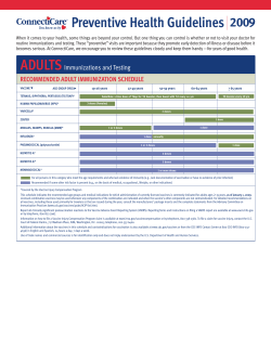


© Copyright 2026