
Potential hepatic stem cells reside in EpCAM cells of normal +
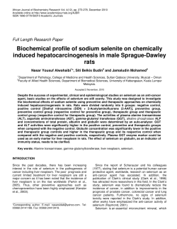
DEVELOPMENT AND DISEASE
RESEARCH ARTICLE 1951
Development 136, 1951-1960 (2009) doi:10.1242/dev.031369
Potential hepatic stem cells reside in EpCAM+ cells of normal
and injured mouse liver
Mayuko Okabe1,*, Yuko Tsukahara1,*, Minoru Tanaka1,*,†, Kaori Suzuki1, Shigeru Saito1, Yoshiko Kamiya1,
Tohru Tsujimura2, Koji Nakamura3 and Atsushi Miyajima1
Hepatic oval cells are considered to be facultative hepatic stem cells (HSCs) that differentiate into hepatocytes and cholangiocytes in
severely injured liver. Hepatic oval cells have also been implicated in tumorigenesis. However, their nature and origin remain
elusive. To isolate and characterize mouse oval cells, we searched for cell surface molecules expressed on oval cells and analyzed
their nature at the single-cell level by flow cytometric analysis and in the in vitro colony formation assay. We demonstrate that
epithelial cell adhesion molecule (EpCAM) is expressed in both mouse normal cholangiocytes and oval cells, whereas its related
protein, TROP2, is expressed exclusively in oval cells, establishing TROP2 as a novel marker to distinguish oval cells from normal
cholangiocytes. EpCAM+ cells isolated from injured liver proliferate to form colonies in vitro, and the clonally expanded cells
differentiate into hepatocytes and cholangiocytes, suggesting that the oval cell fraction contains potential HSCs. Interestingly, such
cells with HSC characteristics exist among EpCAM+ cells of normal liver. Intriguingly, comparison of the colony formation of EpCAM+
cells in normal and injured liver reveals little difference in the number of potential HSCs, strongly suggesting that most proliferating
mouse oval cells represent transit-amplifying cells rather than HSCs.
INTRODUCTION
Most of the metabolic functions in the liver are carried out by
hepatocytes that form hepatic cords, whereas cholangiocytes form
bile ducts that drain bile produced by hepatocytes. During
development, hepatocytes and cholangiocytes, two types of hepatic
epithelial cells, derive from hepatoblasts emerging from the foregut
endoderm (Notenboom et al., 2003; Zaret, 2000). Hepatoblasts are
highly proliferative and express hepatocytic proteins such as
albumin (ALB) and alpha-fetoprotein (AFP), an immature
hepatocyte marker. As the liver develops, hepatoblasts propagate
and those close to the portal mesenchyme differentiate into
cholangiocytes, while the rest become mature hepatocytes
(Lemaigre, 2003). Therefore, hepatoblasts are thought to be hepatic
stem/progenitor cells in the fetus.
By contrast, it is highly controversial whether adult mammalian
liver contains hepatic stem cells (HSCs). Adult liver has a
remarkable potential to regenerate from severe parenchymal loss,
even though hepatocytes and cholangiocytes are mitotically dormant
under normal conditions. Hepatocytes have a remarkable potential
to self-replicate (Fausto, 2004; Michalopoulos and DeFrances,
1997) and are capable of at least 80 doublings by serial
transplantation (Overturf et al., 1997), allowing the liver to
regenerate. However, liver injury that limits this pathway
accompanies the proliferation of a potential stem/progenitor cell
compartment at the interface of the biliary tree and hepatic cords,
which is known as the ductular reaction (Alison et al., 1996;
Roskams et al., 2004; Theise et al., 1999). These undifferentiated
1
Laboratory of Cell Growth and Differentiation, Institute of Molecular and Cellular
Biosciences, The University of Tokyo, Tokyo 113-0032, Japan. 2Department of
Pathology, Hyogo College of Medicine, Nishinomiya, Hyogo 663-8501, Japan.
3
LivTech, Miyamae-ku, Kawasaki, Kanagawa 216-0001, Japan.
*These authors contributed equally to this work
Author for correspondence (e-mail: [email protected])
†
Accepted 31 March 2009
epithelial cells are often referred to as ‘oval cells’ because of their
ovoid nucleus (Farber, 1956). Upon activation, oval cells expand
into liver parenchyma from the portal area. Oval cells express both
ALB and cytokeratin 19 (CK19; KRT19 – Mouse Genome
Informatics), which are hepatocytic and cholangiocytic markers,
respectively, and are believed to differentiate into hepatocytic and
biliary lineages, similar to hepatoblasts in embryonic liver. Thus,
oval cells are thought to be facultative stem/progenitor cells in adult
liver. Although oval cells have been most extensively studied in
rodents, similar cells have been found in connection with various
human liver diseases and are implicated in tumorigenesis (Fausto,
2004; Lee et al., 2006). Whether oval cells constitute HSCs has been
debated in numerous reports involving various rodent injury models
using chemical reagents, including carcinogenic agents. The 2acetylaminofluorene (2-AAF)/partial hepatectomy (PH) model, in
which hepatocyte proliferation is blocked by 2-AFF prior to PH, has
been extensively used to characterize oval cells in rats (Evarts et al.,
1987; Laishes and Rolfe, 1981). However, the same procedure does
not generate oval cells in mice and alternatives such as the use of
a choline-deficient ethionine-supplemented diet (Akhurst et al.,
2001) and a 3,5-diethoxycarbonyl-1,4-dihydro-collidine (DDC)containing diet (Preisegger et al., 1999; Wang et al., 2003) have been
developed. Although the proliferating epithelial cells that are present
in the periportal region upon injury caused by various insults are
referred to as oval cells, it remains unclear whether oval cells
generated via different protocols in different species have the same
characteristics. A major problem in characterizing oval cells is the
lack of appropriate cell surface markers to identify and isolate the
oval cell compartment. Therefore, despite a large number of studies,
the exact nature of oval cells – including their origin, stemness and
bi-directional differentiation – is still poorly understood. Because of
the difficulty in performing clonal analysis of HSCs, it has also been
unclear whether HSCs exist in normal liver.
The aim of this study was to identify cell surface molecules on
mouse oval cells and to analyze their nature at the clonal level. To
this end, we utilized the 2-AAF/PH rat model and the DDC diet
DEVELOPMENT
KEY WORDS: Hepatic stem cell, Oval cell, EpCAM, TROP2 (TACSTD2), Hepatocyte, Cholangiocyte, Liver injury
1952 RESEARCH ARTICLE
Development 136 (11)
mouse model to generate oval cells and found that epithelial cell
adhesion molecule (EpCAM) and the related molecule, TROP2
(TACSTD2), were upregulated in these livers. EpCAM was
expressed in normal cholangiocytes and also in oval cells in the liver
of mice fed the DDC diet (DDC liver). By contrast, TROP2 was
expressed in almost all EpCAM+ cells in DDC liver, but not in
normal liver, indicating that TROP2 is a novel marker to distinguish
between normal cholangiocytes and oval cells. Furthermore, we
isolated EpCAM+ cells from DDC liver and demonstrated that
clonally expanded cells were able to differentiate into hepatocytes
and cholangiocytes. Finally, we provide evidence for the presence
of potential HSCs in EpCAM+ cells of normal liver and compare
their characteristics before and after oval cell activation.
was recovered and redigested with basic perfusion solution containing 0.5
g/l collagenase type IV, 0.5 g/l pronase (Roche Diagnostics) and 50 mg/l
DNase I (Sigma) by stirring for 20 minutes at 37°C. This digested liver was
also passed through a 70-μm cell strainer and the flow-through fraction was
combined with the first cell suspension. After centrifugation at 700 rpm
(100 g) for 2 minutes, the supernatant was transferred to a new tube and the
centrifugation repeated until no cell pellet was visible. The final supernatant
was centrifuged at 1200 rpm (300 g) for 5 minutes and the precipitated cells
were used as non-parenchymal cells (NPCs) for FCM analysis. Aliquots of
cells were blocked with anti-FcR antibody, co-stained with fluorescein- and
biotin-conjugated antibodies, washed, incubated with allophycocyaminconjugated streptavidin (Invitrogen), and analyzed by FACSCalibur
(Becton Dickinson). Dead cells were excluded by propidium iodide
staining.
MATERIALS AND METHODS
Antibodies and immunohistochemistry (IHC)
Total RNA was prepared from the non-parenchymal fraction of rat liver
treated with 2-AAF/PH at 7 days after PH as described previously (Tanimizu
et al., 2004b). Total RNA was amplified using the MessageAmp aRNA
Amplification Kit (Ambion), and used to construct a cDNA library in the
pMXs-SST vector using the SuperScript Choice System (Invitrogen,
Carlsbad, CA, USA). The cDNA library contained 4.3⫻106 independent
clones. The signal sequence trap method was performed as described
previously (Kojima and Kitamura, 1999).
The anti-EpCAM monoclonal antibody was generated by immunization of
a rat with a Ba/F3 cell transfectant overexpressing EpCAM cDNA. The
establishment of hybridoma clones was performed as described previously
(Hara et al., 1999). The anti-EpCAM monoclonal antibody was biotinylated
using Amersham ECL Protein Biotinylation Module (GE Healthcare, UK)
or fluorescein-conjugated using the Fluorescein Labeling Kit-NH2 (Dojindo
Molecular Technologies), and then used for all FCM analyses. The rabbit
anti-mouse CK19 polyclonal antibody was raised against the C-terminal
peptide, HYNNLPTPKAI, and the serum was used for IHC as previously
described (Tanimizu et al., 2003). The biotin-conjugated anti-mouse TROP2
antibody was purchased from R&D Systems. The anti-human Ki67 antibody
was purchased from Becton Dickinson. Frozen sections (8 μm) of livers
were prepared using a HM505E cryostat (Microm International) after
fixation with 4% paraformaldehyde, and incubated with each antibody,
followed by a biotin- or fluorescein-conjugated secondary antibody. The
signals were visualized by fluorescence microscopy.
Preparation of liver cells and flow cytometry (FCM)
RNA extraction and reverse transcription PCR (RT-PCR)
A single-cell suspension from DDC and normal livers was obtained by a
modified two-step collagenase perfusion method as described previously
(Seglen, 1976). In short, livers were perfused with liver perfusion medium
(Invitrogen) at a flow rate of 3 ml/minute for 5 minutes. Then, the liver was
perfused with basic perfusion solution (136 mM NaCl, 5.4 mM KCl, 5 mM
CaCl2, 0.5 mM NaH2PO3 2H2O, 0.42 mM Na2HPO3 12H2O, 10 mM
HEPES pH 7.5, 5 mM glucose and 4.2 mM NaHCO3) containing 0.5 g/l
collagenase type IV (Sigma, St Louis, MO, USA) at a flow rate of 3
ml/minute for 8 minutes. The digested liver was transferred to a glass dish
and chopped into small pieces using a surgical knife in D-PBS (Dulbecco’s
phosphate-buffered saline). Cells dispersed by pipetting were passed
through a 70-μm cell strainer and the flow-through fraction was used for
the next step as the first cell suspension. The undigested clot on the strainer
Total RNA was extracted from each cell preparation using Trizol reagent
(Invitrogen). Total RNA (1 μg) and random hexamer primers were used to
synthesize cDNA using the First-Strand cDNA Synthesis Kit (Amersham
Pharmacia Biotech). The samples were denatured at 94°C for 5 minutes,
then subjected to 25-40 cycles of denaturation at 94°C for 30 seconds,
annealing at 52-57°C for 30 seconds, and extension at 72°C for 1 minute,
with the final extension at 72°C for 5 minutes. PCR primers for mouse genes
are shown in Table 1. The quantitative real-time RT-PCR was performed
using a LightCycler ST300 (Roche) and the following primers (5⬘ to 3⬘): rat
Epcam, TCTACAAGGAAGAGATCAGCAAAA and TGTGTATCTCACCCATCTCCTTT; rat Trop2, GACCAAATGTGTTGGCCTGT and
GTCACAGCTGGGAGGAAAAT; rat Hprt, GACCGGTTCTGTCATGTCG and ACCTGGTTCATCATCACTAATCAC.
Animals
C57BL/6 mice (Japan SLC, Hamamatsu, Japan) at 8-12 weeks were used
for all experiments. All experiments with animals were performed according
to institutional guidelines. The diet containing 0.1% DDC was purchased
from CLEA, Japan. Mouse oval cells were activated by feeding with the diet
containing 0.1% DDC.
Identification of cDNA encoding a membrane protein
Table 1. Oligonucleotides used in RT-PCR
Epcam
Ck19
Ck7
Alb
Afp
Gapdh
Hprt
G6Pase
Tat
Cps
integrin β4
Ggt1
mucin 1
Trop2
claudin 4
Cd44
Slc10a1
Tdo2
Forward (5⬘ to 3⬘)
CGGCTCAGAGAGACTGTGTC
GTCCTACAGATTGACAATG
GGGATGACCTCCGCAACACC
CATGACACCATGCCTGCTGAT
CTGGAGTGTCTGCAGGATGG
GGAGCGAGACCCCACTAA
GACTGAAAGACTTGCTCGAG
AACCCATTGTGAGGCCAGAGG
TGCATCCTCCTGAAGACATG
ACTGAGAGATGCTGACCCTA
GACCTATGAAGAAGGTGCTC
GCTGAGCTGATTGAGCATCCG
GAGCGCCAGCCTTGAGTTTG
CTGACCTAGACTCCGAGCTG
GACTTTGACCCCTGCAGAGG
CAGAGGCGACTAGATCCCTC
AGATCAAGGCTCACTTCTGG
ACAATGAAGAAGACAGAGC
Reverse (5⬘ to 3⬘)
GATCCAGTAGGTCCTCACGC
CACGCTCTGGATCTGTGACAG
CTCCAGCAGCTTGCGGTAGG
GCCTTTCCACCAGGGATCCAC
CCACAGCCGGACCATTTCTC
GTGTAGCCCAAGATGCCC
CCAGCAAGCTTGCAACCTTAACAA
TACTCATTACACTAGTTGGTC
CTTCTCTCTGGTGTAGCTCT
CCTGGAAATTGGTGAGGAGA
GGCTCAGATGCGTGCCATAG
GGTTGATGAAGTTGGGCGAGC
GGAGGCACTACTGTGGACTG
CCAACCCATCTGGTCTGAGG
GGCCACAGGCTGTTATGAGC
GAGTCACAGTGCGGGAACTC
AGAAGTCCTTCTGCAAGCTG
TGTAGTCTCCTCCAAAGTTA
DEVELOPMENT
Gene
Hepatic stem cells in adult liver
RESEARCH ARTICLE 1953
Culture of EpCAM+ cells and colony formation assay
NPCs were prepared as described above. EpCAM+ cells were sorted by
FACSVantage SE (Becton Dickinson). The cells were suspended in the
standard medium (Williams’ medium E containing 10% FBS, 10 mM
nicotinamide, 2 mM L-glutamine, 0.2 mM ascorbic acid, 20 mM HEPES pH
7.5, 1 mM sodium pyruvate, 17.6 mM NaHCO3, 14 mM glucose, 100 nM
dexamethasone, 1⫻ ITS (insulin, transferrin, selenium X) and 50 mg/ml
gentamicin) and seeded on a type-I collagen-coated dish. human EGF,
human recombinant HGF and mouse IL6 were added to the culture to a final
concentration of 10 ng/ml each. After the establishment of cell lines, IL6 was
excluded from the culture medium because it was confirmed to have no
apparent effect. For colony formation assays, EpCAM+ cells were sorted by
a two-step selection (see Fig. S1 in the supplementary material) and plated
at 1⫻104 cells per 35-mm dish. The isolated cells were cultured for 9 days
and then the number and size of colonies were counted.
Differentiation into the hepatocytic lineage in vitro
In vitro differentiation into the cholangiocytic lineage
Cellmatrix Type I-A (Nitta Gelatin) was used for three-dimensional culture
to induce cholangiocytic differentiation according to the manufacturer’s
instructions. In short, 0.3% Cellmatrix Type I-A, a 5⫻ DMEM containing
250 mg/ml gentamicin, and the reconstitution buffer (0.05 M NaOH, 200
mM HEPES pH 7.5, 262 mM NaHCO3) were mixed at a ratio of 7:2:1. This
mixture was mixed with an equal volume of 5⫻104 cells suspended in
standard culture medium without dexamethasone and nicotinamide but with
human recombinant HGF (20 ng/ml). The cell suspension was poured into
a 6-well plate and left at 37°C to form a gel. Then, the culture medium was
gently laid onto the gel.
RESULTS
Screening for cell surface markers of oval cells
To identify the cell surface molecules expressed on hepatic oval
cells, we utilized the signal sequence trap (SST) method, which can
efficiently isolate genes encoding a protein with a signal sequence
(Kojima and Kitamura, 1999; Watanabe et al., 2007). As there are
several protocols for generating oval cells in rats and mice, the
characteristics of oval cells might not be the uniform. We therefore
searched for cell surface molecules expressed on oval cells in the
two species using different protocols (see Fig. S2 in the
supplementary material). To this end, we first constructed an SST
cDNA library from non-parenchymal cells (NPCs) of rat liver
subjected to 2-AAF/PH treatment and identified 54 membrane
proteins (see Table S1 in the supplementary material). First, we
compared the expression of their counterpart genes for mouse
between normal and DDC liver by RT-PCR, and found that Epcam,
Trop2, mucin 1, claudin 4, Cd44, integrin β3, Lyve1, gp130 (Il6st)
and Fxyd5 were significantly upregulated in DDC liver relative to
normal liver. Chronic liver injury caused by DDC diet induces oval
cells in mice, whereas PH and carbon tetrachloride-induced acute
hepatitis do not. Next, the expression of the candidate genes was
compared by northern blotting among normal liver, DDC liver and
models of acute hepatitis, resulting in the identification of six of the
nine genes that were specifically upregulated in the DDC liver, but
not in the other livers (Fig. 1A). The remaining three genes, Lyve1,
gp130 and Fxyd5, were upregulated in an acute hepatitis model,
suggesting that they might be involved in inflammation (data not
shown). Because the expression of EpCAM, mucin 1, CD44 and
Fig. 1. Expression profiles of candidate genes in normal and
injured mouse and rat liver. (A) Northern blot analysis of candidate
genes in mouse liver. The expression of these genes was selectively
upregulated in DDC liver, but not in injured liver without oval cell
activation. (B) Quantitative RT-PCR of Epcam and Trop2 in rat liver.
Whereas Epcam was expressed in normal rat liver (cont.) and upregulated
in 2-AAF/partial hepatectomy (PH)-treated liver, Trop2 was not expressed
in normal liver but was expressed in 2-AAF/PH-treated liver. N, adult
mouse normal liver; O, DDC liver (6 weeks); P, liver 48 hours after 70%
PH; C, liver 24 hours after carbon tetrachloride administration.
claudin 4 has been reported to be upregulated in the rat oval cell
fraction (Yovchev et al., 2007), the characteristics of mouse oval
cells in the DDC model appear similar to those of rat oval cells in
the 2-AAF/PH model. In this study, we have focused on two
structurally related type-I membrane proteins, EpCAM and TROP2.
EpCAM is known to be expressed in many types of normal
epithelial cell as well as in tumor cells (Armstrong and Eck, 2003;
Went et al., 2004). In the liver, EpCAM is expressed on
cholangiocytes but not on hepatocytes (de Boer et al., 1999;
Momburg et al., 1987). Consistent with previous studies (Yovchev
et al., 2007; Yovchev et al., 2008), real-time PCR showed that
Epcam was expressed in normal rat liver and its expression was
upregulated by 2-AAF/PH (Fig. 1B). As for mouse oval cells,
Gleiberman et al. reported that EpCAM is expressed in oval cells
upon carbon tetrachloride-induced liver injury (Gleiberman et al.,
2005). However, the expression status of EpCAM in DDC liver and
the nature of isolated EpCAM+ cells have remained unknown. By
contrast, Trop2 was expressed in both rat and mouse injured liver,
but not in normal liver (Fig. 1). TROP2 is a member of the EpCAM
family and exhibits nearly 50% homology with EpCAM. TROP2
has been shown to be expressed in various tumors, whereas its
expression in the liver was not known. We further examined the
expression of EpCAM and TROP2 on mouse oval cells in DDC
liver and investigated the nature of isolated EpCAM+ cells.
Expression of EpCAM in mouse hepatic oval cells
As shown in Fig. 2A, after 4 weeks of DDC diet feeding, the mouse
liver turned black because of hepatic porphyria resulting from the
inhibition of the heme biosynthetic pathway (Fonia et al., 1996).
Hematoxylin and Eosin (H&E) staining was performed in adult
normal liver and DDC liver. The DDC liver clearly showed numerous
small cells with a large nucleus around the portal veins (Fig. 2B).
Immunohistochemistry (IHC) for CK19, a marker for oval cells and
DEVELOPMENT
Clonally expanded cells (3⫻105 per well) were cultured in the standard
culture medium in a 6-well plate. After 2 days, 20 ng/ml Oncostatin M
(OSM) and 1% DMSO were added into the confluent culture. After 5 days,
the medium was changed to standard culture medium containing 20 ng/ml
OSM, 1% DMSO and 17% Matrigel (growth factor reduced). After 3 and 5
days, the cultured cells were used for RNA preparation and periodic acidSchiff (PAS) staining as described (Kamiya et al., 1999).
1954 RESEARCH ARTICLE
Development 136 (11)
Fig. 2. DDC diet causes hepatic injury
and oval cell activation. (A) The liver
turned black after mice were fed a DDC
diet. (B) H&E staining of a frozen section of
mouse normal liver (top) and 4 weeks after
DDC feeding (middle and bottom).
Numerous small cells appeared around the
portal veins in the DDC liver (arrows). The
brown clots represent the deposition of
iron hemes (arrowheads).
(C) Immunohistochemistry (IHC) with antiCK19 antibody showed that these
numerous small cells included CK19expressing oval cells (arrows) in DDC liver.
PV, portal vein. Scale bars: 100 μm
TROP2 is a novel marker for mouse oval cells
Because TROP2 expression was specifically upregulated in both the
mouse and rat injury models with oval cell activation (Fig. 1),
TROP2 was anticipated to be expressed in oval cells. To reveal
TROP2-expressing cells in normal and injured liver, we performed
IHC with anti-EpCAM and anti-TROP2 antibodies. In contrast to
EpCAM, TROP2 was not expressed in normal mouse liver (Fig.
4A). However, numerous TROP2+ cells appeared around the portal
area in DDC liver (Fig. 4B). Double immunostaining of TROP2 and
EpCAM clearly showed that most of the EpCAM+ cells coexpressed TROP2 in DDC liver, suggesting that TROP2 is a novel
marker for oval cells (Fig. 4B). Although we could not distinguish
the original bile duct in DDC liver, almost all EpCAM+ cells
expressed TROP2. FCM of the NPCs prepared from DDC liver also
showed that the expression level of TROP2 and the population of
TROP2+ cells among EpCAM+ cells gradually increased upon
ingestion of the DDC diet (Fig. 4C). Consistent with the result of
IHC, whereas TROP2 was not present in cholangiocytes expressing
EpCAM at day 0, almost all EpCAM+ cells became TROP2+ in the
DDC-fed mice and EpCAM+ TROP2– cells were hardly detected
after 4 weeks, suggesting that cholangiocytes themselves might also
begin to express TROP2 by oval cell activation in the DDC model.
Alternatively, it is also possible that TROP2+ oval cells differentiate
into mature cholangiocytes and replace the pre-existing
cholangiocytes damaged by DDC administration.
Characterization of mouse oval cells
To reveal the characteristics of mouse oval cells, the gene expression
profile of freshly isolated EpCAM+ cells from DDC liver was
examined by RT-PCR. As previously reported in rat oval cells,
mouse EpCAM+ cells also expressed both cholangiocytic markers
[Ck19, Ck7 (Krt7) and Ggt1] and a hepatocytic marker (Alb),
whereas the other NPCs did not (Fig. 5A). Consistent with the
previous report that AFP expression was rarely detected in mouse
oval cells (Jelnes et al., 2007), AFP was not detected in mouse
EpCAM+ cells (data not shown). Rat oval cells were reported to
express c-KIT, CD34 and THY1 (Petersen et al., 1998). It was also
reported that CD133 (PROM1) is expressed in both mouse and rat
oval cells (Rountree et al., 2007; Suzuki et al., 2008; Yovchev et al.,
2008). Taking advantage of FCM using the anti-EpCAM antibody,
we investigated the expression of these oval cell markers in
EpCAM+ cells before and after DDC feeding (Fig. 5B) and found
DEVELOPMENT
cholangiocytes, demonstrated that these cells included oval cells as
well as other CK19-negative cells, such as inflammatory cells and
fibroblasts (Fig. 2C). Since EpCAM expression was upregulated in
DDC liver, it was examined by IHC using sections of normal liver and
of liver from mice fed DDC for 1 or 4 weeks. EpCAM+ bile ducts
were located adjacent to the portal vein in normal liver as reported
previously (de Boer et al., 1999; Hreha et al., 1999; Joplin et al., 1990),
whereas there were many EpCAM+ cells forming ductular structures
away from the portal vein in DDC liver (Fig. 3A). IHC with both antiEpCAM and anti-CK19 antibodies demonstrated that all CK19+ cells
expressed EpCAM in DDC liver (Fig. 3B). Thus, all oval cells
expressing CK19 also expressed EpCAM. To further investigate the
EpCAM+ cells, we generated rat monoclonal antibodies against
mouse EpCAM that were applicable for flow cytometry (FCM). FCM
of NPCs prepared from DDC liver showed that neither CD45
(PTPRC) nor PECAM was expressed on EpCAM+ cells, indicating
that EpCAM+ cells are not hematopoietic or endothelial cells (Fig.
3C). Furthermore, the isolated EpCAM+ cells were individually
examined by immunostaining after cytospin. Almost all the sorted
cells were immunostained with anti-CK19 antibody as well as A6
antibody, a mouse oval cell marker (Engelhardt et al., 1993) (Fig. 3D).
Hepatic oval cells are known to be highly proliferative. To investigate
their proliferation in vivo, the isolated EpCAM+ cells were stained
with anti-Ki67 antibody (Fig. 3E). Whereas the percentage of Ki67+
cells in the isolated EpCAM+ cells was ~1% in normal liver, it was
12.2% after 1 week on the DDC diet and increased to 17.4% after 4
weeks (Fig. 3F). These results strongly suggested that EpCAM is
expressed in proliferating oval cells and that anti-EpCAM antibody is
useful for isolating oval cells from mice fed with DDC.
Hepatic stem cells in adult liver
RESEARCH ARTICLE 1955
Fig. 3. EpCAM is a cell surface marker
for mouse oval cells. (A) IHC of frozen
liver sections with anti-EpCAM antibody
after DDC feeding. EpCAM was expressed
in cholangiocytes around the portal vein
of normal mouse liver (0 w). Feeding DDC
caused the proliferation of EpCAM+ cells
(1 and 4 weeks). (B) IHC of frozen liver
sections with anti-EpCAM and anti-CK19
antibodies after 4 weeks of DDC feeding.
(C) Flow cytometry (FCM) of nonparenchymal cells (NPCs) prepared from
the liver of mice fed DDC for 4 weeks
with anti-EpCAM antibody and either
CD45 or PECAM antibody. EpCAM+ cells
were negative for CD45 (hematopoietic
marker) and PECAM (endothelial marker).
(D) Immunostaining of EpCAM+ cells with
anti-A6 and anti-CK19 antibodies by
cytospin. EpCAM+ cells expressed both
molecules. (E) Immunostaining of
EpCAM+ cells sorted from normal and
DDC liver with anti-Ki67 antibody by
cytospin. Many EpCAM+ cells from DDC
liver were stained with Ki67 (arrowheads).
(F) The percentage of Ki67+ cells among
EpCAM+ cells after DDC treatment. The
data are derived from five different fields
of view. Error bars, s.d. PV, portal vein.
Scale bars: 100 μm.
Primary culture of EpCAM+ cells from DDC liver
It has been thought that oval cells are proliferative in vivo and
possess the potential to differentiate into hepatocytic and
cholangiocytic lineages. Therefore, oval cells are considered
facultative stem/progenitor cells. To assess such characteristics,
we cultured the EpCAM+ cells isolated from DDC liver in vitro.
The EpCAM+ cells purified with a cell sorter were seeded onto
type-I collagen in the presence of EGF, HGF and IL6. After
plating, adherent cells began to proliferate (Fig. 6A) and covered
the entire dish after 1 month. The in vitro proliferating EpCAM+
cells were apparently homogeneous and exhibited epithelial cell
morphology even after serial passages (Fig. 6B). They could
grow and survive for more than 6 months, maintaining a
homogenous morphology. These results suggested that EpCAM+
cells derived from DDC liver include cells with high proliferative
potential.
Fig. 4. Expression of EpCAM and TROP2
in normal and injured mouse liver.
(A,B) IHC of frozen sections of normal liver
(A) and the liver of mice fed DDC for 5
weeks (B) with anti-EpCAM and anti-TROP2
antibodies. TROP2 was expressed in oval
cells but not in normal cholangiocytes.
(C) FCM of NPCs with anti-EpCAM and antiTROP2 antibodies after DDC feeding. TROP2
begins to be expressed in EpCAM+ cells as
DDC feeding proceeds. PV, portal vein. Scale
bars: 100 μm.
DEVELOPMENT
that c-KIT, CD34 and THY1 were not expressed in EpCAM+ cells
regardless of injury. The expression pattern of CD133 was similar
to that of EpCAM, indicating that both molecules are originally
expressed in normal cholangiocytes. By contrast, TROP2 was
exclusively expressed in oval cells of DDC liver. These results
indicate that TROP2 is induced by oval cell activation and that it is
a novel marker for oval cells in mice.
1956 RESEARCH ARTICLE
Development 136 (11)
Fig. 5. Characterization of freshly
isolated EpCAM+ cells. (A) RT-PCR of
freshly isolated EpCAM+ and EpCAM–
cells from the liver of mice fed DDC for 4
weeks. NPCs from DDC liver were divided
into EpCAM+ and EpCAM– cells by
FACSVantage, then RT-PCR was
performed. (B) FCM of EpCAM+ cells from
normal and DDC livers with known oval
cell markers. EpCAM+ cells surrounded by
bold lines were reanalyzed with other
antibodies (TROP2, CD133, CD34, c-KIT,
THY1) as shown.
Differentiation of clonally expanded EpCAM+ cells
into the hepatocytic lineage in vitro
To address whether the established clones can differentiate into
hepatocytic and cholangiocytic lineages, we used two clones,
HSCE1 and HSCE2, and examined their differentiation potential.
To induce hepatocytic differentiation and maturation, we utilized an
in vitro culture system reported previously (Kamiya et al., 1999;
Kamiya et al., 2002). Oncostatin M (OSM) is a powerful inducer of
the differentiation of fetal hepatocytes and the addition of
Engelbreth-Holm-Swarm (EHS) gel enhances hepatocyte
maturation. Dimethyl sulfoxide (DMSO) is also known to maintain
the differentiation of hepatocytes in culture (Isom et al., 1985; Sakai
et al., 2002). By combining these methods, the expression of
metabolic enzymes and transporters was monitored in two clones
(Fig. 7A). Because both clones exhibited a similar profile, only the
results of HSEC1 are shown. The expression of G6Pase, Cps, Tat,
tryptophan-2,3-dioxygenase (Tdo2) and solute carrier family 10
(sodium/bile acid cotransporter family), member 1 (Slc10a1) was
markedly induced by the addition of OSM, DMSO and EHS gel
(Fig. 7B). By contrast, the expression of Ck19 and of Afp, a marker
of immature fetal hepatocytes, was downregulated. In addition, the
PAS reaction showed that there were many clusters of hepatocytic
Fig. 6. EpCAM+ cells derived from DDC liver have high
proliferative potential. (A,B) In vitro culture of EpCAM+ cells. Freshly
isolated EpCAM+ cells from DDC liver were seeded on type-I collagencoated dishes in the presence of HGF, EGF and IL6. The morphology of
the cells after 5 days of culture (A) and after several passages (B) is
shown. (C) RT-PCR of EpCAM+ cells after 30 days of culture. Afp was
strongly expressed in the cultured cells. (D) Immunostaining of the
cultured cells with anti-CK19 and anti-ALB antibodies.
DEVELOPMENT
Characterization of the cell lines established from
EpCAM+ cells
As in freshly isolated EpCAM+ cells, CK19 and ALB were
expressed in the established cell lines after 30 days of culture, as
evidenced by RT-PCR and immunostaining (Fig. 6C,D). Afp, a
marker of fetal liver progenitor cells, was expressed in the cultured
cells (Fig. 6C), whereas it was not expressed in freshly isolated
mouse oval cells, suggesting that more immature cells might be
selected from EpCAM+ cells of NPCs in DDC liver. Alternatively,
these proliferating cells might acquire a more hepatoblast-like
character when cultured in vitro as previously reported (Schmelzer
et al., 2007). In accordance with the latter idea, Epcam expression
gradually decreased in culture (Fig. 6C). At the onset of mouse liver
development, EpCAM is highly expressed in delta-like 1 homolog
(DLK1)-positive hepatoblasts; however, its expression in
hepatoblasts is dramatically reduced as liver development proceeds
(our unpublished data). The reduction of EpCAM expression in
cultured cells observed in this study is in line with the possibility that
EpCAM+ cells might shift from an HSC-character to a more
hepatoblast-like character.
To investigate the bipotency of cultured hepatic stem-like cells
derived from EpCAM+ cells (HSCEs) at the clonal level, we
randomly picked several clones and finally established 12
independent cell lines (HSCE1-12). All the expanded clones
expressed both hepatocytic (Alb) and cholangiocytic (Ck7, Ck19)
genes (see Table S2 in the supplementary material). The
expression of c-Met, which is known to be expressed in oval cells,
was also detected in all clones. Whereas tyrosine aminotransferase
(Tat), a marker of perinatal hepatocytes, was weakly expressed in
some of the clones, glucose-6-phosphatase (G6Pase; G6pc),
another marker of perinatal hepatocytes, and carbamoyl phosphate
synthetase (Cps), an adult hepatocyte marker, were rarely
expressed (2/12 and 0/12, respectively). Afp, an immature
hepatocyte marker, was expressed in most of the clones (10/12).
These results indicated that HSCEs maintain immature
characteristics.
Hepatic stem cells in adult liver
RESEARCH ARTICLE 1957
Fig. 7. Clonally expanded HSCEs can differentiate into both
hepatocytes and cholangiocytes. (A) Experimental design for the
differentiation of hepatic stem-like cells derived from EpCAM+ cells
(HSCEs) into hepatocytic cells. (B) RT-PCR of clone HSCE1 after
hepatocytic differentiation. The addition of OSM, DMSO and EHS gel
strongly induced the expression of hepatocytic genes and
downregulated that of hepatoblastic and cholangiocytic genes. (C) PAS
staining of HSCE1. The addition of OSM, DMSO and EHS gel strongly
induced the accumulation of glycogen. (D) Morphological changes of
HSCE1 after cholangiocytic differentiation. Tubules and branching
morphology were clearly observed after 11 days of culture. (E) RT-PCR
of HSCE1 after cholangiocytic differentiation. The expression of
cholangiocytic marker genes was markedly upregulated in HSCE1.
Differentiation of clonally expanded EpCAM+ cells
into the cholangiocytic lineage in vitro
To evaluate the potential for differentiation into cholangiocytes, we
utilized a three-dimensional collagen gel culture that is effective for
the formation of tubules (Nishikawa et al., 1996; Tanimizu et al.,
2004a). A cell suspension in collagen type-I gel was plated onto a
basal layer of a collagen type-I gel and then culture medium
containing HGF, an inducer of tubulogenesis, was loaded on top.
After 11 days of culture, the formation of tubules was observed, and
after 18 days of culture branching structures were clearly evident
(Fig. 7D). In addition, RT-PCR revealed that the expression of
cholangiocytic marker genes was upregulated in the cultured cells
(Fig. 7E), indicating that HSCEs differentiated into the
cholangiocytic lineage. Thus, EpCAM was expressed on oval cells
in DDC liver and the clonally expanded cells from sorted EpCAM+
cells exhibited characteristics of HSCs, i.e. unlimited proliferation
and bi-directional differentiation. However, it remained unclear
whether the emergence of such potential HSCs was an event
restricted to liver injury and whether they were derived from oval
cells.
EpCAM+ cells in normal liver include potential
HSCs
Because EpCAM+ cells are present in normal mouse liver, we
investigated whether potential HSCs are present among EpCAM+
cells of normal liver. Interestingly, the colony formation assay
revealed that sorted EpCAM+ cells from normal liver formed both
Fig. 8. Comparison between EpCAM+ cells isolated from normal
and injured liver. (A) Colony formation assay of EpCAM+ cells from
normal liver. Representative morphology of a small colony (top) and a
large colony (bottom). Large colonies composed of more than 100 cells
after 9 days of culture proliferate exponentially. (B) Unlimited cell
proliferation of the established clones (#1-#4) in in vitro culture.
(C) Comparison of cell surface markers between HSCEs from normal
(blue line) and injured (red line) liver by FCM. Control IgG is in gray. The
expression profiles of cell surface markers were similar in both HSCEs.
(D) The number of EpCAM+ cells per normal (n=4) or injured (n=6) liver.
The number was estimated from the percentage of EpCAM+ cells after
immunomagnetic bead selection (see Fig. S2 in the supplementary
material). There was a significant increase in EpCAM+ cells in DDC liver
(*P<0.01). (E) Colony formation assay of EpCAM+ cells from normal
and injured liver of mice fed DDC for 4 weeks. The data are derived
from four independent experiments. Error bars, s.d.
small and large colonies (Fig. 8A). These large colonies, which were
composed of more than 100 cells, continued to proliferate, and these
clones propagated continuously and exhibited bipotency similar to
the HSCE clones derived from DDC liver (Fig. 8B and data not
shown). In addition, HSCEs derived from normal and DDC liver
exhibited a similar expression pattern of cell surface markers,
strongly suggesting that they are closely related (Fig. 8C). By
contrast, EpCAM– cells from normal or DDC liver did not give rise
to any colonies, even when ten times more cells were plated than for
EpCAM+ (data not shown). If oval cells themselves have
characteristics of potential HSCs then oval cell activation by the
DDC diet should increase the number of potential HSCs. To test this
possibility, we compared the number of EpCAM+ cells and potential
HSCs between normal and DDC liver (see Fig. S1 in the
supplementary material). As shown in Fig. 8D, the number of
DEVELOPMENT
cells accumulating glycogen (Fig. 7C). These results strongly
suggested that clonally expanded HSCEs differentiate into the
hepatocytic lineage.
EpCAM+ cells recovered from a DDC liver was approximately
twice that from a normal liver. Interestingly, the numbers of large
colonies corresponding to potential HSCs were very similar for
normal and injured liver (Fig. 8E). In addition, adult potential HSCs
are a very small population of EpCAM+ cells in normal and injured
liver. Although we cannot exclude the possibility that potential
HSCs might increase slightly (not more than 2-fold), it is unlikely
that the activation of oval cells by liver injury significantly increases
the number of potential HSCs.
DISCUSSION
Oval cells have been considered adult liver stem/progenitor cells
(Fausto, 2004; Oertel and Shafritz, 2008). However, previous studies
were mostly histochemical, or involved biochemical and molecular
biological characterization using cell fractions prepared by density
gradient techniques. FCM is a powerful means of characterizing a
particular type of cell, as successfully demonstrated by the
identification of a very rare population of hematopoietic stem cells
using a combination of cell surface markers (Osawa et al., 1996). In
fetal liver, we and others reported the isolation and characterization
of hepatoblasts by FCM using several markers (Kubota and Reid,
2000; Suzuki et al., 2002; Tanimizu et al., 2003). Although definitive
proof of stemness requires a clonal analysis of freshly isolated oval
cells, a lack of specific markers has hampered the precise
identification and prospective isolation of oval cells from NPCs by
FCM. To address this issue, we first searched for cell surface
molecules expressed on oval cells and showed that anti-EpCAM
antibody is useful for isolating oval cells from the liver of DDC-fed
mice by cell sorting. Clonal analyses provide strong evidence that
the EpCAM+ cells from DDC liver contain adult potential HSCs that
possess the capacity for unlimited proliferation and bi-directional
differentiation.
Hepatic stem/progenitor cells have been considered to
contribute to the regeneration of damaged liver when the
proliferation of hepatocytes is restricted, and cell lines with HSC
characteristics have been established from chemically damaged
and/or genetically dysfunctional liver (Braun et al., 1987; Dumble
et al., 2002; Sugiyama et al., 1997; Suzuki et al., 2008; TirnitzParker et al., 2007; Yin et al., 1999). However, it remained unclear
whether such cells are present in normal adult liver. Cell lines with
HSC characteristics have been established from normal mouse
(Fougere-Deschatrette et al., 2006), human (Herrera et al., 2006)
and rat (Sahin et al., 2008) liver. However, these cell lines were
derived from a hepatocyte-enriched cell population by
centrifugation using a Percoll gradient or from unfractionated
cells, leaving their origin ambiguous. Recently, Schmelzer et al.
reported that EpCAM+ cells from postnatal human donors include
HSCs (Schmelzer et al., 2007). In this report, we demonstrate that
EpCAM+ cells isolated from normal or DDC mouse liver by FCM
include potential HSCs. Interestingly, only a limited population of
EpCAM+ cells formed large colonies and the number of potential
HSCs was not significantly increased in DDC liver compared with
normal liver. Thus, our results strongly suggest that most mouse
oval cells do not possess self-renewal potential and are likely to
represent transit-amplifying cells that differentiate into mature
hepatic cells.
Cholangiocytes proliferate under various pathological conditions.
Cholangiocytes proliferate from pre-existing ducts in portal areas
after PH or bile duct ligation in rats. Oval cell proliferation or
ductular hyperplasia is induced by a number of chemicals, including
2-AAF, DDC (Preisegger et al., 1999) and by a choline-deficient
ethionine-supplemented diet (Tian et al., 1997), and the proliferating
Development 136 (11)
cells form disorganized tubular structures that sprout into liver
lobules. Selective damage of the periportal zone reduces such
proliferation, supporting the notion that oval cells derive from the
periportal region, in particular from the canals of Hering that connect
the bile canaliculus and the biliary tree (Paku et al., 2001). However,
the origin of oval cells is controversial and bone marrow stem cells
were suggested to be a source (Petersen et al., 1999; Sell, 2001),
although several reports refute this possibility (Menthena et al.,
2004; Wang et al., 2003). Oval cells and cholangiocytes are not
clearly distinguishable at the molecular level. In fact, oval cells and
normal cholangiocytes are known to express many intracellular and
membrane proteins in common, including EpCAM and CD133.
However, it is also reported that some genes are predominantly
expressed in rat or mouse oval cells. The expression of THY1, c-KIT
and CD34 in oval cells has been controversial and these are also
found in hematopoietic and mesenchymal cells (Dezso et al., 2007;
Yovchev et al., 2008). By contrast, TROP2 is not expressed at all in
normal liver and only in the oval cells of injured liver, indicating that
TROP2 is a useful marker for oval cells.
Intriguingly, after oval cell activation, most of the EpCAM+ cells
express TROP2, and the original normal cholangiocytes, i.e. the
EpCAM+ TROP2– cells, are hardly detected by FCM and
immunostaining. These results raise the possibility that not only
oval cells but also activated cholangiocytes might begin to express
TROP2 after liver injury and partly contribute to oval cell
proliferation in this mouse model (Fig. 9). Alternatively, oval cells
that are activated to proliferate during DDC administration and that
express both EpCAM and TROP2 might continue to express both
of these markers at least transiently after they differentiate into
mature cholangiocytes. Further investigation into the expression
profile of TROP2 in cholangiocytes would provide a clue as to the
origin of oval cells. Moreover, the function of EpCAM and TROP2
in oval cell activation is an interesting issue that remains to be
addressed.
Fig. 9. Model of the hepatic stem/progenitor cell system in vivo
and in vitro. Potential HSCs exist in normal liver as EpCAM+ cells. They
can proliferate unlimitedly and differentiate into both hepatocytes and
cholangiocytes in vitro. Upon liver injury without oval cell activation,
hepatocytes proliferate and contribute to liver regeneration. Upon liver
injury with oval cell activation, EpCAM+ TROP2+ cells appear around
portal veins to regenerate the liver. The oval cells might be partly
derived from EpCAM+ cholangiocytes. Most oval cells lacked the
potential to self-renew in the in vitro colony formation assay.
DEVELOPMENT
1958 RESEARCH ARTICLE
Oval cells are believed to be involved in the regeneration of liver
following injury. In addition, they are also considered as a cellular
precursor for hepatocellular carcinoma (HCC) (Knight et al., 2005).
Most of the experimental procedures used to induce oval cell
proliferation in the liver ultimately lead to tumorigenesis. However,
the connection between oval cells and tumorigenesis remains
unclear. EpCAM is expressed on many normal as well as neoplastic
epithelial cells and tumor-initiating cells (Al-Hajj et al., 2003;
Armstrong and Eck, 2003; Momburg et al., 1987). Most recently,
Maetzel et al. reported that EpCAM is a potent signal transducer and
that its cleaved intracellular domain utilizes components of the Wnt
pathway to induce cell proliferation (Maetzel et al., 2009). Such
signaling via EpCAM might be involved in oval cell proliferation.
Conversely, Epcam is itself a Wnt/β-catenin signaling target gene in
HCC cell lines (Yamashita et al., 2007). Consistently, the
involvement of the Wnt/β-catenin pathway in the oval cell response
has been reported (Apte et al., 2008; Hu et al., 2007; Yang et al.,
2008). We recently reported that Wnt7a, Wnt7b and Wnt10a are
upregulated in DDC liver and that the Wnt/β-catenin pathway is
likely to be involved in oval cell activation in vivo (Itoh et al., 2009).
Therefore, the correlation between tumorigenesis and the expression
of EpCAM in oval cells is plausible. Because EpCAM is widely
expressed in normal epithelial cells, including biliary epithelial cells,
the expression and signaling of EpCAM should be tightly regulated
at the steady state. However, the regulation of EpCAM signaling in
normal and malignant stem-like cells remains unknown.
It is also reported that TROP2 is expressed on cells of some normal
tissues as well as on cancer cells (El Sewedy et al., 1998; Huang et
al., 2005; Ohmachi et al., 2006). Our finding that TROP2, a member
of the EpCAM family, is upregulated in oval cells raises the
possibility that TROP2 might modulate and/or enhance the
intracellular signaling of EpCAM to promote proliferation and
migration into liver parenchyma. Alternatively, TROP2 itself might
transduce intracellular signaling in a manner similar to EpCAM. In
fact, ectopic expression of TROP2 in NIH3T3 cells is sufficient to
promote both anchorage-independent growth and tumorigenesis
(Wang et al., 2008). Most recently, Goldstein et al. reported that
TROP2 identifies a subpopulation of murine and human prostate
basal cells with stem cell characteristics (Goldstein et al., 2008),
suggesting an association between TROP2 and stem/progenitor cells.
Although chronic liver injury induces the proliferation of oval
cells in some rodent models and in human disease, the underlying
relationship between oval cells and HSCs and their developmental
mechanisms require further investigation. We showed the presence
of potential HSCs in normal liver, as well as in injured liver, by
isolating EpCAM+ cells and demonstrated the expression of TROP2
in oval cells. Further investigation into the interaction between
EpCAM and TROP2 in oval cells will help us to understand the
mechanisms of growth and differentiation in cancer-initiating cells
as well as in normal stem/progenitor cells.
Anti-A6 antibody was kindly provided by Dr Snorri Thorgeirsson and Dr
Valentina Factor. This work was supported in part by a grant for Core Research
for Evolutionary Science and Technology from the Japan Science and
Technology Agency (JST) and a research grant from the Ministry of Education,
Sports, Science and Technology (Japan).
Supplementary material
Supplementary material available online at
http://dev.biologists.org/cgi/content/full/136/11/1951/DC1
References
Akhurst, B., Croager, E. J., Farley-Roche, C. A., Ong, J. K., Dumble, M. L.,
Knight, B. and Yeoh, G. C. (2001). A modified choline-deficient, ethionine-
RESEARCH ARTICLE 1959
supplemented diet protocol effectively induces oval cells in mouse liver.
Hepatology 34, 519-522.
Al-Hajj, M., Wicha, M. S., Benito-Hernandez, A., Morrison, S. J. and Clarke,
M. F. (2003). Prospective identification of tumorigenic breast cancer cells. Proc.
Natl. Acad. Sci. USA 100, 3983-3988.
Alison, M. R., Golding, M., Sarraf, C. E., Edwards, R. J. and Lalani, E. N.
(1996). Liver damage in the rat induces hepatocyte stem cells from biliary
epithelial cells. Gastroenterology 110, 1182-1190.
Apte, U., Thompson, M. D., Cui, S., Liu, B., Cieply, B. and Monga, S. P. (2008).
Wnt/beta-catenin signaling mediates oval cell response in rodents. Hepatology
47, 288-295.
Armstrong, A. and Eck, S. L. (2003). EpCAM: A new therapeutic target for an
old cancer antigen. Cancer Biol. Ther. 2, 320-326.
Braun, L., Goyette, M., Yaswen, P., Thompson, N. L. and Fausto, N. (1987).
Growth in culture and tumorigenicity after transfection with the ras oncogene
of liver epithelial cells from carcinogen-treated rats. Cancer Res. 47, 4116-4124.
de Boer, C. J., van Krieken, J. H., Janssen-van Rhijn, C. M. and Litvinov, S. V.
(1999). Expression of Ep-CAM in normal, regenerating, metaplastic, and
neoplastic liver. J. Pathol. 188, 201-206.
Dezso, K., Jelnes, P., Laszlo, V., Baghy, K., Bodor, C., Paku, S., Tygstrup, N.,
Bisgaard, H. C. and Nagy, P. (2007). Thy-1 is expressed in hepatic
myofibroblasts and not oval cells in stem cell-mediated liver regeneration. Am. J.
Pathol. 171, 1529-1537.
Dumble, M. L., Croager, E. J., Yeoh, G. C. and Quail, E. A. (2002). Generation
and characterization of p53 null transformed hepatic progenitor cells: oval cells
give rise to hepatocellular carcinoma. Carcinogenesis 23, 435-445.
El Sewedy, T., Fornaro, M. and Alberti, S. (1998). Cloning of the murine TROP2
gene: conservation of a PIP2-binding sequence in the cytoplasmic domain of
TROP-2. Int. J. Cancer 75, 324-330.
Engelhardt, N. V., Factor, V. M., Medvinsky, A. L., Baranov, V. N., Lazareva,
M. N. and Poltoranina, V. S. (1993). Common antigen of oval and biliary
epithelial cells (A6) is a differentiation marker of epithelial and erythroid cell
lineages in early development of the mouse. Differentiation 55, 19-26.
Evarts, R. P., Nagy, P., Marsden, E. and Thorgeirsson, S. S. (1987). A precursorproduct relationship exists between oval cells and hepatocytes in rat liver.
Carcinogenesis 8, 1737-1740.
Farber, E. (1956). Similarities in the sequence of early histological changes induced
in the liver of the rat by ethionine, 2-acetylamino-fluorene, and 3⬘-methyl-4dimethylaminoazobenzene. Cancer Res. 16, 142-148.
Fausto, N. (2004). Liver regeneration and repair: hepatocytes, progenitor cells, and
stem cells. Hepatology 39, 1477-1487.
Fonia, O., Weizman, R., Coleman, R., Kaganovskaya, E. and Gavish, M.
(1996). PK 11195 aggravates 3,5-diethoxycarbonyl-1,4-dihydrocollidine-induced
hepatic porphyria in rats. Hepatology 24, 697-701.
Fougere-Deschatrette, C., Imaizumi-Scherrer, T., Strick-Marchand, H.,
Morosan, S., Charneau, P., Kremsdorf, D., Faust, D. M. and Weiss, M. C.
(2006). Plasticity of hepatic cell differentiation: bipotential adult mouse liver
clonal cell lines competent to differentiate in vitro and in vivo. Stem Cells 24,
2098-2109.
Gleiberman, A. S., Encinas, J. M., Mignone, J. L., Michurina, T., Rosenfeld,
M. G. and Enikolopov, G. (2005). Expression of nestin-green fluorescent
protein transgene marks oval cells in the adult liver. Dev. Dyn. 234, 413-421.
Goldstein, A. S., Lawson, D. A., Cheng, D., Sun, W., Garraway, I. P. and
Witte, O. N. (2008). Trop2 identifies a subpopulation of murine and human
prostate basal cells with stem cell characteristics. Proc. Natl. Acad. Sci. USA 105,
20882-20887.
Hara, T., Nakano, Y., Tanaka, M., Tamura, K., Sekiguchi, T., Minehata, K.,
Copeland, N. G., Jenkins, N. A., Okabe, M., Kogo, H. et al. (1999).
Identification of podocalyxin-like protein 1 as a novel cell surface marker for
hemangioblasts in the murine aorta-gonad-mesonephros region. Immunity 11,
567-578.
Herrera, M. B., Bruno, S., Buttiglieri, S., Tetta, C., Gatti, S., Deregibus, M. C.,
Bussolati, B. and Camussi, G. (2006). Isolation and characterization of a stem
cell population from adult human liver. Stem Cells 24, 2840-2850.
Hreha, G., Jefferson, D. M., Yu, C. H., Grubman, S. A., Alsabeh, R., Geller, S.
A. and Vierling, J. M. (1999). Immortalized intrahepatic mouse biliary epithelial
cells: immunologic characterization and immunogenicity. Hepatology 30, 358371.
Hu, M., Kurobe, M., Jeong, Y. J., Fuerer, C., Ghole, S., Nusse, R. and
Sylvester, K. G. (2007). Wnt/beta-catenin signaling in murine hepatic transit
amplifying progenitor cells. Gastroenterology 133, 1579-1591.
Huang, H., Groth, J., Sossey-Alaoui, K., Hawthorn, L., Beall, S. and Geradts,
J. (2005). Aberrant expression of novel and previously described cell membrane
markers in human breast cancer cell lines and tumors. Clin. Cancer Res. 11,
4357-4364.
Isom, H. C., Secott, T., Georgoff, I., Woodworth, C. and Mummaw, J. (1985).
Maintenance of differentiated rat hepatocytes in primary culture. Proc. Natl.
Acad. Sci. USA 82, 3252-3256.
DEVELOPMENT
Hepatic stem cells in adult liver
Itoh, T., Kamiya, Y., Okabe, M., Tanaka, M. and Miyajima, A. (2009). Inducible
expression of Wnt genes during adult hepatic stem/progenitor cell response.
FEBS Lett. 583, 777-781.
Jelnes, P., Santoni-Rugiu, E., Rasmussen, M., Friis, S. L., Nielsen, J. H.,
Tygstrup, N. and Bisgaard, H. C. (2007). Remarkable heterogeneity displayed
by oval cells in rat and mouse models of stem cell-mediated liver regeneration.
Hepatology 45, 1462-1470.
Joplin, R., Strain, A. J. and Neuberger, J. M. (1990). Biliary epithelial cells from
the liver of patients with primary biliary cirrhosis: isolation, characterization, and
short-term culture. J. Pathol. 162, 255-260.
Kamiya, A., Kinoshita, T., Ito, Y., Matsui, T., Morikawa, Y., Senba, E.,
Nakashima, K., Taga, T., Yoshida, K., Kishimoto, T. et al. (1999). Fetal liver
development requires a paracrine action of oncostatin M through the gp130
signal transducer. EMBO J. 18, 2127-2136.
Kamiya, A., Kojima, N., Kinoshita, T., Sakai, Y. and Miyaijma, A. (2002).
Maturation of fetal hepatocytes in vitro by extracellular matrices and oncostatin
M: induction of tryptophan oxygenase. Hepatology 35, 1351-1359.
Knight, B., Matthews, V. B., Olynyk, J. K. and Yeoh, G. C. (2005). Jekyll and
Hyde: evolving perspectives on the function and potential of the adult liver
progenitor (oval) cell. BioEssays 27, 1192-1202.
Kojima, T. and Kitamura, T. (1999). A signal sequence trap based on a
constitutively active cytokine receptor. Nat. Biotechnol. 17, 487-490.
Kubota, H. and Reid, L. M. (2000). Clonogenic hepatoblasts, common precursors
for hepatocytic and biliary lineages, are lacking classical major histocompatibility
complex class I antigen. Proc. Natl. Acad. Sci. USA 97, 12132-12137.
Laishes, B. A. and Rolfe, P. B. (1981). Search for endogenous liver colonyforming units in F344 rats given a two-thirds hepatectomy during short-term
feeding of 2-acetylaminofluorene. Cancer Res. 41, 1731-1741.
Lee, J. S., Heo, J., Libbrecht, L., Chu, I. S., Kaposi-Novak, P., Calvisi, D. F.,
Mikaelyan, A., Roberts, L. R., Demetris, A. J., Sun, Z. et al. (2006). A novel
prognostic subtype of human hepatocellular carcinoma derived from hepatic
progenitor cells. Nat. Med. 12, 410-416.
Lemaigre, F. P. (2003). Development of the biliary tract. Mech. Dev. 120, 81-87.
Maetzel, D., Denzel, S., Mack, B., Canis, M., Went, P., Benk, M., Kieu, C.,
Papior, P., Baeuerle, P. A., Munz, M. et al. (2009). Nuclear signalling by
tumour-associated antigen EpCAM. Nat. Cell Biol. 11, 162-171.
Menthena, A., Deb, N., Oertel, M., Grozdanov, P. N., Sandhu, J., Shah, S.,
Guha, C., Shafritz, D. A. and Dabeva, M. D. (2004). Bone marrow
progenitors are not the source of expanding oval cells in injured liver. Stem Cells
22, 1049-1061.
Michalopoulos, G. K. and DeFrances, M. C. (1997). Liver regeneration. Science
276, 60-66.
Momburg, F., Moldenhauer, G., Hammerling, G. J. and Moller, P. (1987).
Immunohistochemical study of the expression of a Mr 34,000 human
epithelium-specific surface glycoprotein in normal and malignant tissues. Cancer
Res. 47, 2883-2891.
Nishikawa, Y., Tokusashi, Y., Kadohama, T., Nishimori, H. and Ogawa, K.
(1996). Hepatocytic cells form bile duct-like structures within a threedimensional collagen gel matrix. Exp. Cell Res. 223, 357-371.
Notenboom, R. G., van den Bergh Weerman, M. A., Dingemans, K. P.,
Vermeulen, J. L., van den Eijnde, S., Reutelingsperger, C. P., Hut, H.,
Willemsen, R., Offerhaus, G. J. and Lamers, W. H. (2003). Timing and
sequence of differentiation of embryonic rat hepatocytes along the biliary
epithelial lineage. Hepatology 38, 683-691.
Oertel, M. and Shafritz, D. A. (2008). Stem cells, cell transplantation and liver
repopulation. Biochim. Biophys. Acta 1782, 61-74.
Ohmachi, T., Tanaka, F., Mimori, K., Inoue, H., Yanaga, K. and Mori, M.
(2006). Clinical significance of TROP2 expression in colorectal cancer. Clin.
Cancer Res. 12, 3057-3063.
Osawa, M., Hanada, K., Hamada, H. and Nakauchi, H. (1996). Long-term
lymphohematopoietic reconstitution by a single CD34-low/negative
hematopoietic stem cell. Science 273, 242-245.
Overturf, K., al-Dhalimy, M., Ou, C. N., Finegold, M. and Grompe, M. (1997).
Serial transplantation reveals the stem-cell-like regenerative potential of adult
mouse hepatocytes. Am. J. Pathol. 151, 1273-1280.
Paku, S., Schnur, J., Nagy, P. and Thorgeirsson, S. S. (2001). Origin and
structural evolution of the early proliferating oval cells in rat liver. Am. J. Pathol.
158, 1313-1323.
Petersen, B. E., Goff, J. P., Greenberger, J. S. and Michalopoulos, G. K.
(1998). Hepatic oval cells express the hematopoietic stem cell marker Thy-1 in
the rat. Hepatology 27, 433-445.
Petersen, B. E., Bowen, W. C., Patrene, K. D., Mars, W. M., Sullivan, A. K.,
Murase, N., Boggs, S. S., Greenberger, J. S. and Goff, J. P. (1999). Bone
marrow as a potential source of hepatic oval cells. Science 284, 1168-1170.
Preisegger, K. H., Factor, V. M., Fuchsbichler, A., Stumptner, C., Denk, H. and
Thorgeirsson, S. S. (1999). Atypical ductular proliferation and its inhibition by
transforming growth factor beta1 in the 3,5-diethoxycarbonyl-1,4-dihydrocollidine
mouse model for chronic alcoholic liver disease. Lab. Invest. 79, 103-109.
Roskams, T. A., Theise, N. D., Balabaud, C., Bhagat, G., Bhathal, P. S.,
Bioulac-Sage, P., Brunt, E. M., Crawford, J. M., Crosby, H. A., Desmet, V. et
Development 136 (11)
al. (2004). Nomenclature of the finer branches of the biliary tree: canals,
ductules, and ductular reactions in human livers. Hepatology 39, 1739-1745.
Rountree, C. B., Barsky, L., Ge, S., Zhu, J., Senadheera, S. and Crooks, G. M.
(2007). A CD133-expressing murine liver oval cell population with bilineage
potential. Stem Cells 25, 2419-2429.
Sahin, M. B., Schwartz, R. E., Buckley, S. M., Heremans, Y., Chase, L., Hu, W.
S. and Verfaillie, C. M. (2008). Isolation and characterization of a novel
population of progenitor cells from unmanipulated rat liver. Liver Transpl. 14,
333-345.
Sakai, Y., Jiang, J., Kojima, N., Kinoshita, T. and Miyajima, A. (2002).
Enhanced in vitro maturation of fetal mouse liver cells with oncostatin M,
nicotinamide, and dimethyl sulfoxide. Cell Transplant. 11, 435-441.
Schmelzer, E., Zhang, L., Bruce, A., Wauthier, E., Ludlow, J., Yao, H. L., Moss,
N., Melhem, A., McClelland, R., Turner, W. et al. (2007). Human hepatic
stem cells from fetal and postnatal donors. J. Exp. Med. 204, 1973-1987.
Seglen, P. O. (1976). Preparation of isolated rat liver cells. Methods Cell Biol. 13,
29-83.
Sell, S. (2001). Heterogeneity and plasticity of hepatocyte lineage cells.
Hepatology 33, 738-750.
Sugiyama, K., Kato, N., Mizutani, T., Ikeda, M., Tanaka, T. and Shimotohno,
K. (1997). Genetic analysis of the hepatitis C virus (HCV) genome from HCVinfected human T cells. J. Gen. Virol. 78, 329-336.
Suzuki, A., Zheng, Y. W., Kaneko, S., Onodera, M., Fukao, K., Nakauchi, H.
and Taniguchi, H. (2002). Clonal identification and characterization of selfrenewing pluripotent stem cells in the developing liver. J. Cell Biol. 156, 173-184.
Suzuki, A., Sekiya, S., Onishi, M., Oshima, N., Kiyonari, H., Nakauchi, H. and
Taniguchi, H. (2008). Flow cytometric isolation and clonal identification of selfrenewing bipotent hepatic progenitor cells in adult mouse liver. Hepatology 48,
1964-1978.
Tanimizu, N., Nishikawa, M., Saito, H., Tsujimura, T. and Miyajima, A. (2003).
Isolation of hepatoblasts based on the expression of Dlk/Pref-1. J. Cell Sci. 116,
1775-1786.
Tanimizu, N., Saito, H., Mostov, K. and Miyajima, A. (2004a). Long-term
culture of hepatic progenitors derived from mouse Dlk+ hepatoblasts. J. Cell Sci.
117, 6425-6434.
Tanimizu, N., Tsujimura, T., Takahide, K., Kodama, T., Nakamura, K. and
Miyajima, A. (2004b). Expression of Dlk/Pref-1 defines a subpopulation in the
oval cell compartment of rat liver. Gene Expr. Patterns 5, 209-218.
Theise, N. D., Saxena, R., Portmann, B. C., Thung, S. N., Yee, H., Chiriboga,
L., Kumar, A. and Crawford, J. M. (1999). The canals of Hering and hepatic
stem cells in humans. Hepatology 30, 1425-1433.
Tian, Y. W., Smith, P. G. and Yeoh, G. C. (1997). The oval-shaped cell as a
candidate for a liver stem cell in embryonic, neonatal and precancerous liver:
identification based on morphology and immunohistochemical staining for
albumin and pyruvate kinase isoenzyme expression. Histochem. Cell Biol. 107,
243-250.
Tirnitz-Parker, J. E., Tonkin, J. N., Knight, B., Olynyk, J. K. and Yeoh, G. C.
(2007). Isolation, culture and immortalisation of hepatic oval cells from adult
mice fed a choline-deficient, ethionine-supplemented diet. Int. J. Biochem. Cell
Biol. 39, 2226-2239.
Wang, J., Day, R., Dong, Y., Weintraub, S. J. and Michel, L. (2008).
Identification of Trop-2 as an oncogene and an attractive therapeutic target in
colon cancers. Mol. Cancer Ther. 7, 280-285.
Wang, X., Foster, M., Al-Dhalimy, M., Lagasse, E., Finegold, M. and Grompe,
M. (2003). The origin and liver repopulating capacity of murine oval cells. Proc.
Natl. Acad. Sci. USA 100 (Suppl. 1), 11881-11888.
Watanabe, N., Tanaka, M., Suzuki, K., Kumanogoh, A., Kikutani, H. and
Miyajima, A. (2007). Tim2 is expressed in mouse fetal hepatocytes and
regulates their differentiation. Hepatology 45, 1240-1249.
Went, P. T., Lugli, A., Meier, S., Bundi, M., Mirlacher, M., Sauter, G. and
Dirnhofer, S. (2004). Frequent EpCam protein expression in human carcinomas.
Hum. Pathol. 35, 122-128.
Yamashita, T., Budhu, A., Forgues, M. and Wang, X. W. (2007). Activation of
hepatic stem cell marker EpCAM by Wnt-beta-catenin signaling in hepatocellular
carcinoma. Cancer Res. 67, 10831-10839.
Yang, W., Yan, H. X., Chen, L., Liu, Q., He, Y. Q., Yu, L. X., Zhang, S. H.,
Huang, D. D., Tang, L., Kong, X. N. et al. (2008). Wnt/beta-catenin signaling
contributes to activation of normal and tumorigenic liver progenitor cells. Cancer
Res. 68, 4287-4295.
Yin, L., Lynch, D. and Sell, S. (1999). Participation of different cell types in the
restitutive response of the rat liver to periportal injury induced by allyl alcohol. J.
Hepatol. 31, 497-507.
Yovchev, M. I., Grozdanov, P. N., Joseph, B., Gupta, S. and Dabeva, M. D.
(2007). Novel hepatic progenitor cell surface markers in the adult rat liver.
Hepatology 45, 139-149.
Yovchev, M. I., Grozdanov, P. N., Zhou, H., Racherla, H., Guha, C. and
Dabeva, M. D. (2008). Identification of adult hepatic progenitor cells capable of
repopulating injured rat liver. Hepatology 47, 636-647.
Zaret, K. S. (2000). Liver specification and early morphogenesis. Mech. Dev. 92,
83-88.
DEVELOPMENT
1960 RESEARCH ARTICLE


© Copyright 2026