
Repetitive concussions in adolescent athletes
REVIEW ARTICLE
published: 02 April 2015
doi: 10.3389/fneur.2015.00069
Repetitive concussions in adolescent athletes – translating
clinical and experimental research into perspectives on
rehabilitation strategies
Bridgette D. Semple 1,2 , Sangmi Lee 1 , Raha Sadjadi 1 , Nora Fritz 3 , Jaclyn Carlson 1 , Carrie Griep 4 ,
Vanessa Ho 4 , Patrice Jang 4 , Annick Lamb 4 , Beth Popolizio 4 , Sonia Saini 4 , Jeffrey J. Bazarian 5 ,
Mayumi L. Prins 6 , Donna M. Ferriero 7,8 , D. Michele Basso 9 and Linda J. Noble-Haeusslein 1,10 *
1
Department of Neurological Surgery, University of California San Francisco, San Francisco, CA, USA
Department of Medicine, Royal Melbourne Hospital, University of Melbourne, Parkville, VIC, Australia
3
Kennedy Krieger Institute, John Hopkins University, Baltimore, MD, USA
4
San Francisco State University Graduate Program in Physical Therapy, University of California San Francisco, San Francisco, CA, USA
5
School of Medicine and Dentistry, University of Rochester Medical Center, Rochester, NY, USA
6
Department of Neurosurgery, University of California Los Angeles David Geffen School of Medicine, Los Angeles, CA, USA
7
Department of Pediatrics, University of California San Francisco, San Francisco, CA, USA
8
Department of Neurology, University of California San Francisco, San Francisco, CA, USA
9
School of Health and Rehabilitation Sciences, Ohio State University, Columbus, OH, USA
10
Department of Physical Therapy and Rehabilitation Sciences, University of California San Francisco, San Francisco, CA, USA
2
Edited by:
Cameron Bass, Duke University, USA
Reviewed by:
Vassilis E. Koliatsos, Johns Hopkins
University School of Medicine, USA
Tessa Hart, Moss Rehabilitation
Research Institute, USA
Jason Luck, Duke University, USA
*Correspondence:
Linda J. Noble-Haeusslein,
Department of Neurological Surgery,
University of California San Francisco,
513 Parnassus Avenue, Room
HSE-860 San Francisco, CA
94143-0112, USA
e-mail: [email protected]
Sports-related concussions are particularly common during adolescence, a time when even
mild brain injuries may disrupt ongoing brain maturation and result in long-term complications. A recent focus on the consequences of repetitive concussions among professional
athletes has prompted the development of several new experimental models in rodents,
as well as the revision of guidelines for best management of sports concussions. Here,
we consider the utility of rodent models to understand the functional consequences and
pathobiology of concussions in the developing brain, identifying the unique behavioral and
pathological signatures of concussive brain injuries. The impact of repetitive concussions
on behavioral consequences and injury progression is also addressed. In particular, we
focus on the epidemiological, clinical, and experimental evidence underlying current recommendations for physical and cognitive rest after concussion, and highlight key areas in
which further research is needed. Lastly, we consider how best to promote recovery after
injury, recognizing that optimally timed, activity-based rehabilitative strategies may hold
promise for the adolescent athlete who has sustained single or repetitive concussions.
The purpose of this review is to inform the clinical research community as it strives to
develop and optimize evidence-based guidelines for the concussed adolescent, in terms
of both acute and long-term management.
Keywords: concussion, adolescent, athletes, experimental models, behavior, pathology, rehabilitation, exercise
INTRODUCTION
Sports-related concussions are common during adolescence, a
time when approximately half of all high-school students participate in sports in the United States (US) (1). Concussions account
for 5–13% of all reported sports injuries in high-school-aged athletes (1–3). In high-school football, an estimated 5.6% of players
sustain a concussion in a given season (4), although the actual
incidence may be closer to 15% due to under-reporting (5).
There is increasing evidence that the adolescent, defined by the
World Health Organization as between the ages of 10 and 19 years,
may have poorer outcomes after concussions compared to older
athletes (6–8). Importantly, repetitive concussions to the brain
during maturation may result in both acute and long-term complications (9). Moreover, even subtle cognitive deficits may have profound consequences on both academic and athletic performance
and social integration. There has been considerable attention to
identifying risk factors for concussions for professional athletes in
www.frontiersin.org
contact sports, and a burgeoning interest in injuries sustained at a
younger age. Most recently, the Institute of Medicine (IOM) and
National Research Council released a report titled “Sports-Related
Concussions in Youth: Improving the Science, Changing the Culture,” which considers risk factors, screening, detection, treatment
and management recommendations, and long-term consequences
of repetitive concussions in the developing brain (10). In addition,
an updated consensus report from the Fourth International Conference of Concussion in Sports (11) and evidence-based guidelines for the management of sports concussions have recently been
published (12). These reports provide an important foundation
for understanding the consequences of concussion and acceptable
practices for medical management.
Here, we provide an overview of adolescent concussions from
a clinical perspective, address the utility of rodent models in
understanding the pathobiology and behavioral consequences of
concussions, and discuss how repetitive concussions may alter
April 2015 | Volume 6 | Article 69 | 1
Semple et al.
injury progression and neurobehavioral outcomes. In addition,
we consider how best to optimize recovery, recognizing that
optimally timed, exercise-based rehabilitative strategies may hold
promise for the adolescent who has sustained single or repetitive
concussions.
DEFINITION AND DIAGNOSIS
In this review, we define concussion as a subset of traumatic
brain injury (TBI), in line with the consensus report from the
Fourth International Conference of Concussion in Sports (11). A
concussion, or mild TBI, is a complex process triggered by a biomechanical insult to the brain, which is typically not associated
with the presence of early structural damage on standard neuroimaging (13, 14). Suspected diagnosis is primarily based upon
reported symptoms, which may be evaluated together with neuroimaging to rule out a more severe injury, as evidenced by skull
fracture or intracranial hemorrhage (15). Symptoms may include
headache, fatigue, nausea, dizziness, difficulty concentrating, sleep
disturbance, sensitivity to light and/or noise, balance problems,
irritability, anxiety, and depression (1, 16). Among high school
and college athletes, the most common symptom is headache (1,
17). A brief loss of consciousness (LOC) has previously been considered a hallmark of concussion (18); however, this symptom is
no longer considered a defining feature as it presents in <10% of
all concussions (4, 11, 17).
Most of the neurocognitive assessments available to assist in
identification of a suspected concussion have been developed for
adults (12). For children and adolescents over 12 years of age,
the Sports Concussion Assessment Tool version 3 (SCAT3) was
recently adapted from SCAT2, and the new Child-SCAT3 has
been tailored for concussed children aged 5–12 years (11). Based
upon data from experimental models (see Section “Pathology
after Experimental Concussions at Adulthood”), single and repetitive concussions may result in acute neuropathology including
axonal injury, microbleeds, and glial activation, all of which may be
sources of biomarkers. Biomarkers with high sensitivity may thus
prove useful to both detect pathology, establish baseline physiology, and track recovery after concussions (10, 19). As ongoing
studies attempt to evaluate the potential benefit of rehabilitative interventions based largely upon the subjective self-reporting
of symptoms [e.g., see Ref. (20)], biomarkers may provide a
more objective measure. Further, in the context of individualized
medicine, biomarkers may ultimately predict those who are at
greater risk than others for developing long-term problems when
exposed to repetitive concussions. State-of-the-art neuroimaging
techniques such as single photon positron emission tomography,
diffusion tensor imaging, functional magnetic resonance imaging
(fMRI), nuclear magnetic resonance spectroscopy (NMRS), and
high-definition fiber tracking also show promise for future utility
in the assessment and detection of sports-related concussions with
increased sensitivity (21–25).
The severity of concussion symptoms can vary and may take
hours to days to manifest, particularly in children. Fortunately,
the effects of a single concussion are typically transient, with
acute neurological dysfunction resolving within 7–10 days in most
adult athletes, and within 1 month in most younger athletes (11,
26). However, a subset of patients shows sub-acute or chronic
Frontiers in Neurology | Neurotrauma
Concussions in adolescent athletes
symptoms including sleep dysregulation, cognitive deficits, and
emotional disturbances (27, 28). Such persistent post-concussive
symptoms affect between 2 and 11% of concussed patients more
than 1 month after injury (27). Among children and adolescents,
a range of 11–29% of patients report persistent symptoms up
to 1 month after mild traumatic brain injuries, including single
and repetitive concussions (28–32). A recent prospective cohort
study of 335 young concussed athletes (8–23 years old) exhibited
symptoms for an average of 43 ± 53 days (33). Emerging evidence
that both metabolic dysfunction (34–36) and disruption of functional connectivity (23, 37) persist for several weeks to months
after repetitive concussive insults, suggests that recovery to normal brain health may be prolonged even after the resolution of
clinical symptoms.
EPIDEMIOLOGY AND ETIOLOGY
Youths typically engage in organized sports with increased frequency relative to adults, and thus sustain the majority of
the sports-related concussions (38, 39). In fact, of the ~23,000
football-related, non-fatal TBIs resulting in emergency department visits annually in the US, almost 90% occur in adolescents
aged 5–18 years (40). Among 15–24 year olds, sports-related head
impacts are second only to road trauma as the most common cause
of TBI (41). In contrast, concussions sustained during childhood,
prior to 10 years of age, more often result from non-sports-related
falls in the home, school, or playground (42). Over the past decade,
the incidence of concussions reported by high-school students has
increased by ~15% annually (43). It is unclear, however, whether
this figure reflects a true increase in events, or a rise in reporting
prompted by greater awareness.
Within the adolescent population, concussions occur most
commonly in full-contact sports (e.g., football, soccer, ice hockey,
and lacrosse) compared to non-contact sports (e.g., volleyball and
swimming) (1, 44). Participation in football is responsible for the
majority of sports-related concussions in the US (45, 46), likely
due in part to its large number of participants, particularly at the
high-school level (1, 2, 47). The most common injury mechanisms
are player–player contact (70%) and falls (1). Concussion rates are
typically higher during competition than in practice (48, 49).
By the start of high school, 53% of student athletes have
reported a history of concussion (6). However, the actual incidence
of sports-related concussions during adolescence may, in fact, be
higher than reported. In a confidential survey across 20 schools in
Wisconsin, 15% of high-school football players sustained a concussion during the previous football season; however, only 44%
of these athletes reported their injury (5). Under-reporting may
result from a lack of recognition by the athlete, coaches, trainers,
or other medical personnel; not thinking the injury was serious
enough to warrant a report; or fear of removal from participation (5, 50). On the contrary, over-reporting may also occur when
athletes are asked to reflect on past events.
Gender differences in both the incidence and outcomes of concussions have also been reported. Comparison between the sexes is
complicated by different numbers of males and females who play
specific sports, as well as potential differences in baseline verbal
and visual neuropsychological test measures during adolescence
(51). However, reviews have repeatedly suggested that high school
April 2015 | Volume 6 | Article 69 | 2
Semple et al.
Concussions in adolescent athletes
and college-aged female athletes are at higher risk of concussion,
sustaining more injuries than males (3, 52). Further, females report
a greater number and severity of symptoms, and show poorer
outcomes, requiring a longer duration to recover (14, 52–56). In
high-school sports played by both sexes (e.g., soccer, baseball, and
basketball), concussions represented a greater proportion of total
injuries in girls (3). The mechanisms underlying this gender bias
are unclear, and may in part relate to biological differences that
are compounded by cultural distinctions in how concussions are
reported and managed (57).
more poorly on serial neuropsychological tests at 3, 5, and 7 days
after injury (6), and took longer to return to neurocognitive and
symptom-free baselines compared to older athletes (8). Average
times to normalization of neurocognitive abilities in high-school
students have been reported as 10–14 days, compared to 5–7 and
3–5 days in collegiate and professional athletes, respectively (78,
79).Together, these studies suggest that adolescents may be particularly susceptible to the consequences of concussive injury,
although there remains a paucity of studies evaluating the recovery
patterns of participants under the age of 15 years (14).
REPEATED CONCUSSIONS IN SPORTS
EXPERIMENTAL MODELS OF SINGLE AND REPEAT
CONCUSSIVE BRAIN INJURY IN RODENTS
While there is growing awareness of the adverse consequences
of repeated concussive injuries, the exact mechanisms underlying these injuries remain unclear, as are the factors that (i.e. ’as
are the factors that contribute...’) contribute to persistent symptomatology in some patients. Animal models, mimicking aspects of
repetitive concussions, have served as a platform for monitoring
both acute and long-term behavioral outcomes after injury, and
for understanding the associated cellular and molecular mechanisms. In this section, we briefly review the published literature
regarding experimental models of repetitive concussions in adult
rodents and the resulting behavioral outcomes, and consider the
few studies that have examined injury at an adolescent age.
The risk of a subsequent concussion is elevated in athletes with a
history of previous concussions (44, 58, 59). Among high-school
football players in the US, between 12 and 32% of those sustaining a concussion report a prior sports-related concussion either
in the same season or the one prior (1, 4, 60, 61). In collegiate
football athletes, between 6 and 36% have reported multiple concussions (6, 62).The brain is hypothesized to be more susceptible
to subsequent injury after a concussion, such that sustaining a
second impact while recovering from the first may result in prolonged and/or more severe symptoms (10, 17, 63). High school and
amateur athletes who sustained two or more concussions exhibit
greater impairment on neuropsychological and memory tests than
athletes with a history of only a single concussion (60, 64, 65). Similarly, a recent multi-center study of over 600 high-school athletes
found significantly higher ratings of cognitive, physical, and sleeprelated concussion symptoms in athletes with a history of two or
more concussions, compared to athletes with one or no previous
concussions (66).
There is emerging evidence that repetitive concussions in
both adults and adolescents may yield persistent neurocognitive
changes (14, 65). Repetitive concussions have been implicated as
a contributing factor to neurodegenerative conditions including
chronic traumatic encephalopathy (67, 68), post-traumatic stress
disorder (69), substance abuse (70), anxiety, and depression (69,
71). However, there remains a paucity of prospective or longitudinal studies examining repetitive concussions and long-term
neurodegeneration. As such, there is insufficient information to
determine if there is a causal relationship (72, 73). Together with
evidence that concussed high-school athletes typically present with
more severe cognitive deficits compared to concussed adults (6,
74, 75), the potential for long-term consequences after cumulative
injuries highlights the need for a greater understanding of how the
adolescent brain responds to concussions.
AGE-RELATED EFFECTS OF CONCUSSIONS
It is now widely accepted that the human brain continues to mature
throughout childhood and adolescence, with considerable structural and functional changes ongoing well into the third decade of
life (76, 77). However, despite the prevalence of sports-related concussions among adolescent-aged athletes, only a few studies have
explored whether the adolescent brain shows age-dependent vulnerability to concussive insults; or, conversely, enhanced capacity
for plasticity and recovery.
When comparing high school and collegiate athletes who have
experienced sports-related concussions, the former performed
www.frontiersin.org
INJURY PARAMETERS
Animal models of concussion typically involve the application of
an external force to the intact skull without causing skull fracture, hemorrhage, or overt tissue damage acutely. Weight drop
(WD) and controlled cortical impact (CCI) are two commonly
used models of TBI, which have been adapted to study concussions in rodents (80, 81). In the WD model, a weight is dropped
through a tube positioned over the head of an anesthetized animal,
to deliver the impact either directly to the head/skull or onto a surface covering the skull (i.e., a metal helmet or gauze) (Table 1). For
the modified CCI model, an electronically driven piston impacts
the intact skull (Table 2). The head may or may not be stabilized
at the time of impact, and models that allow for movement of
the head typically produce a diffuse pattern of injury (82). Only
models that involve minimally invasive experimental procedures
(e.g., no craniotomy) are examined in this review. Experimental
concussions vary considerably across several key parameters, such
as the weight of the impactor and height from which the weight
is dropped for WD models, and the impact velocity, depth, dwell
time, and tip diameter for modified CCI models. Regardless of
the model employed, the number of impacts, and the time interval between successive impacts in repetitive concussive models are
additional determinants of outcome. The high diversity of parameters, as illustrated in Tables 1 and 2, renders direct comparison
of findings difficult across different laboratories.
To date, few laboratories have developed models of concussivelike brain injuries in juvenile animals (98, 113) and of these only
one (98) has examined the consequences of repetitive insults. Here,
a modified CCI model has been utilized to generate concussions in
male rats at postnatal day 35 (p35), estimated to approximate an
adolescent-aged animal based upon behavioral and biochemical
April 2015 | Volume 6 | Article 69 | 3
Semple et al.
Concussions in adolescent athletes
Table 1 | Key parameters of weight drop (WD) models of concussive injury.
Reference Species and age
Sex
Weight used
Impact
Skull exposed;
location
head immobile
Drop height
# Impacts
Interval
(83)
Mouse, adult
M
50, 100, or 150 g Dorsal head
No; no
40 or 60 cm
0, 1, 4
24 h
(84)
Mouse, adult
M
21 g
Dorsal head
No; yes
35 cm
3
24 h
(85)
Rat, adult
M
450 g
Midline
Yes; no
500, 750, or
0, 1, 2, 3
1.5, 3, 5, or 10 h
24 h
100 cm
(86)
Mouse, adult
M
95 g + steel cap
(87)
Mouse, adult
(88)
Mouse, adult
M
95 g
(89)
Mouse, adult
M
54 g
M + F 54 g
Midline
No; no
100 cm
1, 5, 10
Dorsal head
No; no
97 or 107 cm
0, 1, 3, 5, 10 24 h, 1 week, or 1 month
Left parietal
Yes; yes
~10 cm
0, 2
24 h
Dorsal head
No; no
71 cm
0, 1, 5, 7
1 or 2 days; 1, 2, or 4 weeks
Yes; N/A
15 cm
0, 1, 2
3 or 20 days
No; no
100 cm
1, 5, 10
24 h
(90)
Mouse, adult
M
36.73 g
Left parietal
(82)
Mouse, p28
M
95 g + steel cap
Midline
(91)
Mouse, adult
M
54 g
Dorsal head
No; no
71 cm
0, 7
24–48 h
(92)
Mouse, p35–42
M
40 g
Dorsal head
Yes; no
100 cm
1, 4, or 12
1–4 days (1–3/day)
N/A, not stated in paper; M, male; F, female; p, postnatal day.
Gray shading highlights studies which have examined adolescent-aged rodents.
Table 2 | Key parameters of modified controlled cortical impact (CCI) models of concussive injury.
Reference Species
(93–95)
Mouse, adult
Sex
M
Tip
Impact
Skull exposed;
width material
Tip
location
head immobile dwell time
6 mm
Left parietal
Yes; yes
4.8–5.6 m/s, 1 mm, n/a 0, 1, 2
Rubber-
Velocity, depth,
# Impacts Interval
24 h
coated
(96)
Mouse, adult
M
9 mm
Silicone
Left parietal
Yes; yes
4.8–5 m/s, 3 mm, n/a
1, 2
3, 5, or 7 days
(97)
Mouse, adult
M
9 mm
Silicone
Left parietal
Yes; yes
4.8–5 m/s, 2 mm, n/a
16
20 min (4 in 1 day), once
per week for 4 weeks
(98, 100)
Rat, p35
M
5 mm
N/A
Left parietal
No; no
Head displaced 8 mm
1, 2
1 or 3 days
1, 2
8 days
at 36 psi
(101)
Rat, adult
M
N/A
Silicon
N/A
Yes; yes
5 m/s, 3 mm, n/a
(102)
Mouse, adult
M
9 mm
Rubber
Left parietal
Yes; yes
5 m/s, 3.3 mm, 100 ms 1, 2
24 h
(103–105)
Mouse, adult
N/A
9 mm
Rubber
Left parietal
Yes; yes
n/a, 3.3 mm, n/a
0, 2
24 h
(106, 107)
Mouse, adult M + F N/A
Metal
Midline
Yes; yes
5 m/s, 1 mm, 200 ms
1, 5
48 h
(108)
Mouse, adult
M
5 mm
Metal
Midline
Yes; no
5 m/s, 1 mm, n/a
1, 4
24 h
(109)
Mouse, adult
M
9 mm
Rubber
Left parietal
No; no
4 m/s, 3 mm, 200 ms
0, 1, 3
24 h
(110)
Mouse, adult
M
6 mm
Rubber
Left parietal
No; no
5 m/s, 1 cm, 100 ms
0, 1, 42
2 h (6 daily hits) for
(111, 112)
Rat, p35
M
5 mm
N/A
Left parietal
No; no
Head displaced 8 mm
0, 4
7 days
1 day
at 36 psi
N/A, not stated in paper; M, male; p, postnatal day.
Studies from the same laboratory using similar or identical parameters are grouped together.
Gray shading highlights studies which have examined adolescent-aged rodents.
features at this age (98). In this model, the head is allowed to move
freely in the direction of impact, resulting in apnea and delayed
righting and toe pinch responses in the absence of a skull fracture.
Two or four repeated injuries have also been characterized, with
an inter-injury interval of 24 h (98, 100). A second juvenile model,
developed by Mychasiuk and colleagues by adapting the Wayne
Frontiers in Neurology | Neurotrauma
State method (82, 86), has thus far focused on a single concussivelike insult. In this model, p30 rats are positioned onto a piece of
aluminum foil – upon impact to the head, the animal undergoes
a 180° horizontal rotation to fall freely onto a cushioned surface.
These rats also exhibit a delayed righting response immediately
after injury (86, 113).
April 2015 | Volume 6 | Article 69 | 4
Semple et al.
BEHAVIORAL CONSEQUENCES OF EXPERIMENTAL CONCUSSIONS AT
ADULTHOOD
Studies of the behavioral consequences of concussions in rodents
typically focus on cognitive, emotional, and/or sensorimotor functions – processes that may be disrupted or altered following a concussion in humans (15, 16). A commonly used test for cognition
in rodents is the Morris water maze (MWM), which specifically
evaluates spatial learning and memory [for a detailed description, see Ref. (114)]. In adult animals, these cognitive functions
are generally unperturbed by a single concussive injury (93, 101,
113). In contrast, repetitive concussions may produce deficits in
spatial learning and memory, which manifest as early as 24 h
after injury and may persist for several weeks to months (84,
87, 91, 102, 108). Further, greater cognitive deficits have been
reported after a shorter time interval between repetitive concussions (up to 7 days apart) (87, 89, 96). Non-spatial cognitive deficits
resulting from concussions have also been demonstrated by the
novel object recognition (NOR) test, which evaluates recognition
memory (88, 115).
Several studies have also reported concussion-induced balance
problems along with numerous other sensorimotor deficits, in
experimental models of single and repeated concussions. The
accelerating rotarod is the most commonly used tool to evaluate sensorimotor function in rodents (116), and in adult animals, transient motor deficits have been reported within the first
week post-injury after repetitive concussive insults, with recovery by several weeks to months later (88, 106, 109). Together,
these findings point toward transient motor dysfunction following concussive brain injury, compared to cognitive changes,
which may persist for weeks to months after repeated injuries
(Table 3). As with cognitive dysfunction, inter-impact interval appears to be a determinant of sensorimotor impairments,
whereby shorter intervals lead to greater duration and severity of
motor impairments (96).
BEHAVIORAL CONSEQUENCES OF EXPERIMENTAL CONCUSSIONS AT
ADOLESCENCE
Cognitive deficits, including impaired recognition memory evident as a decreased preference for a novel object, are visible by
24 h after either a single or two repetitive concussions in male
p35 rats (98), but by 3 days, deficits persisted only in those with
repeated concussions (100). In contrast, these rats showed normal
activity and anxiety levels, performing similarly to sham animals
in the open field test at 24 h after surgery (98). However, 10 daily
concussions to adolescent (p28) mice impaired balance and coordination on a rotarod at 24 h after injury, which recovered when
re-tested 1 month later (82), consistent with evidence that sensorimotor deficits also tend to be transient in nature after repetitive
concussions in adults (86).
Given that adolescence is a time of pubertal hormonal changes,
which influence brain development, it is possible that concussions may interfere with this maturation process. Indeed, four
repetitive concussions (24 h intervals) to p35 male rats results
in reduced testosterone production both acutely (24 h) and subchronically (2–4 weeks), with recovery to age-appropriate levels
by 2–3 months after injury (112). These mice also exhibited a
delayed onset of puberty, erectile dysfunction, and abnormalities
www.frontiersin.org
Concussions in adolescent athletes
in reproductive behaviors including social interest and copulatory
interactions. The authors link these observations with hypopituitarism, and provide further evidence that repetitive injuries
depress growth hormone and insulin-like growth hormone levels at 1–4 weeks after injury, which was associated with reduced
weight gain in these animals (111). In light of ongoing controversy on whether or not to test for or treat hormonal deficiencies
after TBI in children, these findings suggest that even a transient
window of hormonal imbalance due to repetitive concussions at
adolescence may result in long-lasting changes to critical adult
behaviors.
IDENTIFYING THE UNIQUE PATHOLOGICAL “FINGERPRINTS”
OF CONCUSSIVE BRAIN INJURIES
We are only just beginning to fully appreciate how concussions
may influence the structural and functional integrity of the brain.
Here, we briefly review the salient neuropathological features of
single and repeated concussive impacts based upon evidence from
experimental models in adult and adolescent-aged animals.
PATHOLOGY AFTER EXPERIMENTAL CONCUSSIONS AT ADULTHOOD
In animal models of concussions, acute and chronic pathology is
often not readily apparent by traditional methods of detection. A
single concussive injury is most often characterized by the lack of
skull fracture and absence of acute intra-parenchymal hemorrhage
or cerebral contusion (117, 118), and tissue integrity typically
appears intact (93, 106–108), even after two repeated concussions
(90, 102). Several studies have detected neuronal loss only after
repetitive injuries, suggestive of an additive or cumulative effect
(93, 108). Evidence of pathology in experimental models is difficult
to compare due to differences in injury severity, methodologies
and detection methods, and selected time points for analysis. Of
note, cognitive deficits are often reported after repetitive concussions to adult rodents in the absence of cell death, suggesting
that a lower threshold of injury may be needed to induce functional deficits compared to that required for neuropathological
changes (83, 87).
Axonal injury may be detected after even a single concussive insult in the adult rodent brain, often in the absence of
overt cell death (106, 108, 118). Widespread silver staining and
immuno-reactivity for beta amyloid precursor protein (β-APP),
a commonly used indicator of impaired axonal transport, provides evidence of axonal injury as early as 24 h after a single
concussion in the adult rodent brain (84, 96, 106, 119), which
may persist for up to 2 months (93, 108). Patterns of axonal injury
are consistently increased by repetitive brain insults (90, 93, 96,
102, 108). Diffusion tensor imaging in adult mice after repetitive concussions has corroborated these findings of white matter
damage (103).
Enhanced glial activation, most commonly detected by
immuno-labeling with glial fibrillary acidic protein (GFAP) and
ionized calcium binding adaptor molecule-1 (Iba-1), has been
associated with regions of axonal injury after even single concussive injury in the adult rodent brain (90, 108, 109, 118). This
inflammatory response is exacerbated after repeated insults (90,
102–104, 106, 108), and may persist for several months following the final impact (91, 106). However, as is the case with more
April 2015 | Volume 6 | Article 69 | 5
Semple et al.
Concussions in adolescent athletes
Table 3 | Chronic behavioral deficits detected after repeated concussive injuries in rodents.
Time point
# Impacts
(months)
1
Persistence
disorders
Absence
1 and 7 days
WD
MWM
2
3 days
WD
BM
(87)
(90)
42
2 h (6/day × 7 days)
CCI
MWM
(110)
(102)
1 day
CCI
MWM
WD
MWM
(89)
3
1 day
CCI
RAWM
(109)
(108)
3
4
1 day
CCI
APACL
5
1 and 7 days
WD
MWM
(89)
42
2 h (6/day × 7 days)
CCI
MWM
(110)
3
1 day
CCI
FC
(109)
5
1 day
WD
MWM
(89)
5
1 day
WD
MWM
(87)
1
5
30 days
WD
MWM
(87)
2
2
1 day
CCI
MWM
(93)
4
2
1 day
CCI
MWM
(94)
16
20 min (4/day, 1 day/week for 4 week)
CCI
MWM
(97)
5
14 days and 30 days
WD
MWM
(89)
3
1 day
CCI
YM
(109)
12
5
7 days
WD
MWM
(87)
2
2
1 day
CCI
RPT, RR
(93)
2
1 day
CCI
CN, RPT
(93)
5
1 day
WD
OF
(86)
5 or 10
1 day
WD
RR
(82)
3
1 day
CCI
SHM, RR
(109)
4
2
1 day
CCI
CN
(94)
6
16
20 min (4/day, 1 day/week for 4 week)
CCI
CN
(97)
42
2 h (6/day × 7 days)
CCI
EPM, FST, TST, DSI
(110)
1 day
WD
ST, NSFT
(82)
1
anxiety, and sleep
5
1 day
6
Persistence
Reference
2
Absence
Depressive-like symptoms,
Test
5
12
Absence
model
2
Cognitive deficits
Sensorimotor deficits
Injury
between impacts
1.75
6
Persistence
Frequency/interval
1.5
1
5 or 10
6
42
2 h (6/day × 7 days)
CCI
EPM
(110)
1.5
3
1 day
CCI
EZM
(109)
Studies, which conducted behavioral testing at or later than 1 month after CCI or WD injuries, are tabulated, and outcomes categorized as cognitive, sensorimotor/activity, or other (anxiety, depression, and sleep disorders).Together, these studies suggest that cognitive deficits may persist long-term after repeated concussions,
compared to sensorimotor dysfunction, which is typically resolved by this time.
APACL, active place avoidance and conflict learning; a test for spatial learning, memory, and cognitive flexibility; BM, Barnes maze, a test for spatial learning and
memory, and memory retention; CN, composite neuroscore, a test for motor function; DSI, Data Sciences International EEG and EMG recordings; EPM, elevated
plus maze, a test for anxiety; EZM, elevated zero maze, a test for anxiety; FC, fear conditioning, a test for contextual associative learning; FST, forced swim test;
MWM, Morris water maze, a test for spatial learning, memory, and memory retention; NOR, novel object recognition, a test for recognition memory; NSFT, novelty
suppressed feeding test, a test for anxiety; OF, open field, a test for general activity motor function; RAWM, radial arm water maze, a test for spatial learning, memory,
and memory retention; RPT, rotating pole test, for motor function and coordination; RR, rotarod, a test for sensorimotor function and learning; SHM, smart home-cage
monitoring, which tests for activity and exploratory behavior; ST, splash test, for depressive-like behavior; TST, tail-suspension test, for depressive-like behavior; YM,
y-maze, a test for working memory.
severe CNS injury, the precise role of inflammation in neurodegeneration versus repair remains unclear (120). Recent experiments
aimed to determine the contribution of CD11b+ microglia to secondary axonal injury processes, by inducing two repetitive injuries
(24 h apart) in CD11b-thymidine kinase transgenic mice. Surprisingly, the resulting microglial depletion had little effect on the
degree of axonal injury by several complementary markers (105).
Thus, the relationship between cerebral inflammation and axonal
degeneration after concussions remains unclear.
Several studies have also demonstrated an increase in total
or phosphorylated Tau protein after repeated impacts at both
Frontiers in Neurology | Neurotrauma
sub-acute (30 days) and chronic time points (3–6 months) postinjury (86, 109). However, a considerable number of studies
have failed to detect such changes long-term (89, 92, 97, 106),
such that the link between repetitive concussions and chronic
neuropathology remains inconclusive to date.
PATHOLOGY AFTER EXPERIMENTAL CONCUSSIONS AT ADOLESCENCE
Given the unique properties of the brain across development, it is
possible that pathogenesis may be influenced by age at the time of
concussion. Moreover, repeated concussions across an extended
developmental timeline may likewise exhibit distinct pathological
April 2015 | Volume 6 | Article 69 | 6
Semple et al.
changes. However, to date, very limited data are available regarding injuries to the adolescent brain. One study has examined acute
pathological markers after two repetitive impacts in the adolescent (p35) rat, reporting no evidence of neuronal degeneration by
FluoroJade staining (98). However, an increase in β-APP-labeled
axons was evident in the ipsilateral white matter of adolescent rats
after repeated (but not single) concussions, alongside an increase
in GFAP-positive astrocytes at the gray–white matter junction.
Further work is needed to confirm these findings across both an
acute and chronic time course, and to determine the threshold
(e.g., the number and timing of repetitive concussions) at which
neuronal and axonal integrity becomes compromised. Improving our understanding of changes in white matter integrity and
connectivity following concussions at adolescence is crucial, particularly in light of recent evidence of white matter abnormalities
in high school and college-aged athletes, even in the absence of a
clinically diagnosed concussion (23, 121).
IMPROVING ANIMAL MODELS OF CONCUSSIVE INJURIES TO THE
DEVELOPING BRAIN
While adult rodent models of concussions are at the forefront of
investigation, there is opportunity to expand this effort to the adolescent brain. Assessment of age-dependent vulnerability to concussive brain injury remains in its infancy. Functional and pathological outcomes, across both an acute and chronic time course
in young animals, may reveal potential vulnerability or resilience
during childhood and adolescence. To improve translation, such
approaches are predicated upon gaining a better understanding of
the respective time courses of rodent versus human brain development and associated behaviors. Current modeling of repetitive
concussions to the adolescent brain does not fully encapsulate
all populations at risk of experiencing repetitive concussions. For
example, in light of epidemiological evidence suggesting poorer
outcomes in females after concussions (52, 75) adequate representation of both sexes in animal models is warranted and may in fact
identify important sex-specific responses to injury.
Further, there is much to be gained by a critical examination
of the current adult models of concussion and their relevance to
the concussed human brain. For example, many models involve
restriction of head movement via positioning in a fixed stereotaxic frame, contrary to emerging evidence suggesting that head
rotation, resulting in acceleration and deceleration forces are necessary components of a concussion (82, 122). Several groups have
now modified traditional models to incorporate an element of
movement to the head and/or body, or developed new devices
to generate clinically relevant brain injuries (123–125). Despite
such advances, there remains a lack of consensus regarding the
appropriate interval between repetitive injuries to provide the
most relevant model of the human condition. Further, it is unclear
how loading weights in rodent models compare to injury forces
seen in concussed patients, and whether we are approximating
an appropriate injury severity in line with what is seen clinically.
For example, LOC is a feature of <10% of concussions (4, 17);
yet, the surrogate marker in rodents, of prolonged righting time
immediately following the impact, remains a salient characteristic of many animal models (80). As findings from experimental
studies may inform clinical management decisions, for example,
www.frontiersin.org
Concussions in adolescent athletes
in determining the optimal timing for returning to activity, it
is important to consider these limitations and support ongoing efforts to overcome these challenges and improve animal
modeling.
CLINICAL MANAGEMENT OF CONCUSSIONS – EVIDENCE OR
INTUITION?
While experimental models serve as a foundation for understanding the pathobiology of concussions in children and adults, the
questions asked by basic researchers may also be guided by clinical uncertainties regarding optimal medical management. Here,
we provide a brief overview of current clinical management, and
demonstrate how animal modeling may offer insight into critical
questions posed by the medical community.
Clinical management of concussion has predominantly been
dictated by expert-derived position statements. These position
statements are routinely revisited and released, and are considered a key reference source for medical doctors, athletic trainers,
and neuropsychologists, as well as other health-related professionals involved in the treatment of concussed young people (126).
Such guidelines have persisted for nearly two decades, and are
historically based upon the judgment and clinical intuition of
field leaders rather than empirical evidence of physiological data,
or an understanding of injury mechanisms (127, 128). Updated
concussion guidelines have recently been published by the American Medical Society for Sports Medicine (129), the Concussion in
Sports Zurich Consensus Working Group (11), and the American
Academy of Neurology (12).
However, there remains a lack of strong empirical evidence
to support these guidelines of concussion management, and recommendations continue to be at least partly based upon expert
opinion and educated speculation, particularly when considering
young athletes. As a consequence, controversy remains regarding
what constitutes the “optimal management” of a concussion. The
purpose of concussion management is to promote recovery, reduce
the risk of recurrent injuries, and minimize the occurrence of
long-term symptoms (12). Guidelines emphasize the importance
of education for the athlete and their family; support neurocognitive and balance testing using reliable and validated assessments;
and advocate for both physical and cognitive rest after concussion. All current guidelines recommend that a concussed athlete
be removed from play immediately, and it is generally accepted
that an athlete must not return to play (RTP) until concussive
symptoms have resolved (11, 130).
RETURN TO PLAY GUIDELINES
Much of the debate over post-concussive management in both
children and adults is focused on RTP. A RTP schedule is typically
based upon two yet-to-be-proven concepts: first, that sustaining
a single concussion may render the individual more vulnerable to a subsequent injury; and second, that repeated injuries
within a short time window may result in cumulative structural
and functional consequences. Although not yet definitive, both of
these concepts are gaining support from accumulating laboratory,
clinical, and epidemiological research.
Return to play decision making by healthcare providers should
depend upon close monitoring of symptoms over time, as well as
April 2015 | Volume 6 | Article 69 | 7
Semple et al.
Concussions in adolescent athletes
reference to standardized checklists, age-matched normative data,
or pre-injury baseline testing. The RTP guidelines presented in
the Zurich Concussion in Sports Consensus statement are widely
accepted as the current standard of care (11), and consist of a
six-step pathway (Table 4). Return to work or study should follow a similar time course (131). It is worth noting, however,
that poor neurocognitive performance may persist in concussed
college-aged athletes even after concussion-specific symptoms
have resolved, cautioning against the exclusive use of symptom
resolution for RTP decision making (132).
Athletes at highest risk for sustaining a concussion are those
with a history of concussive injuries, and this risk is greatest during the first 10 days post-injury (12, 47). Therefore, RTP decisions
initially after injury may impact an athletes’ likelihood of sustaining a repeated head insult. RTP protocols are further complicated
for patients who experience repetitive concussive injuries; in these
cases, the RTP time course is typically extended to err on the side
of caution. RTP protocols have been widely adopted by individual sporting organizations such as the National Football League, as
well as state legislation. However, whether implementation of RTP
guidelines improves symptom resolution or prevents subsequent
concussions has not yet been established. Well-designed studies are
required to determine whether current RTP recommendations are
the most appropriate (134, 135). Further, the precise definition
of being “asymptomatic” for RTP has been questioned, particularly when dependent upon self-reporting of symptoms by young
athletes, and in light of reports of post-concussive symptoms in
non-concussed populations (136, 137).
RTP IN ADOLESCENT ATHLETES
Due to both the paucity of data specifically regarding injury at
adolescence and the high degree of diversity between different
experimental models, we do not yet have a good understanding of
how repeated brain insults may affect the developing brain (47). As
such, there are no universally accepted, evidence-based guidelines
that have been validated for safe RTP following sport-related concussion in children or adolescents (134, 138, 139). It is generally
recommended that child and adolescent athletes never return to
physical activity on the same day of injury, and only return when
clinically symptom-free or cleared to RTP by a licensed health
care professional trained in the evaluation and management of
concussions (11, 39, 133).
Typically, a more conservative approach is recommended in
children and adolescents compared to adults (130, 140). Of note,
a recent study suggested that a minimum of 14 days is required
following symptom resolution and normalization of neuropsychological tests before RTP in young concussed athletes, compared
to the 7-day protocol typically used for adults (141), based on
reports of prolonged neuropsychological impairments in children
(6, 7, 79). As with adults, some have advocated for individualized RTP protocols for child athletes, which take into account
their unique, sports-specific injury profile, and developmental
stage (39, 133).
A unique, under-studied consideration of concussions during
childhood and adolescence is the timing of returning to school
work or “return-to-learn” (140). As with RTP, a step-wise procedure has been recommended for concussed adolescents returning to school, involving sequential stages of cognitive challenges
interspersed with rest periods, which are closely monitored for
symptom exacerbation (142, 143) (Table 5). Additionally, the optimal timing for resumption of learning to drive, a newly acquired,
cognitively demanding task for older adolescents, should also be
carefully evaluated.
JUSTIFICATION FOR REST: METABOLIC AND NEUROCHEMICAL
DYSFUNCTION AFTER CONCUSSION
One of the key pathophysiological phenomena characterizing TBI
in rodent models is the adverse “neurometabolic cascade,” indicating alterations in cerebral metabolism and blood flow, which
may result in an energy crisis (99, 120, 144, 145). Although more
commonly associated with moderate-to-severe TBI, changes in
cerebral metabolism have also been implicated in the pathophysiology of concussions in both animal models and humans. Such
findings are important for identifying a window of vulnerability
to repetitive insults, and may prove critical to the establishment of
evidence-based guidelines for optimal concussion management.
Table 4 | Gradual return to play (RTP) protocol for young athletes.
Stage
Functional exercise
Objective
1
No activity
Complete physical and cognitive rest
Recovery
2
Light aerobic exercise
Walking, swimming, or stationary cycling keeping intensity <70%
Increase heart rate
maximum permitted heart rate. No resistance training
3
Sport-specific exercise
Skating drills in ice hockey, running drills in soccer. No head impact
Add movement
activities
4
Non-contact training drills
Progression to more complex training; e.g., passing drills in
Exercise, coordination, and cognitive load
football and ice hockey. May start progressive resistance training
5
Full-contact practice
Following medical clearance participate in normal training activities
Restore confidence and assess functional skills
6
Return to play
Normal game play
Full return to pre-concussion activities
Adapted from May et al. (133) and Halstead and Walter (39). Each stage requires a minimum of 24 h duration, and athletes are recommended to remain asymptomatic
before progression to the next step (11, 79). Once all steps have been completed without symptom exacerbation, the individual may be cleared for full return to
athletics participation.
Frontiers in Neurology | Neurotrauma
April 2015 | Volume 6 | Article 69 | 8
Semple et al.
Concussions in adolescent athletes
Table 5 | Gradual return-to-learn (RTL) protocol for young athletes.
1
2
3
4
5
6
Stage
Functional exercise
Objective
No activity
Complete cognitive rest
Recovery
Minor cognitive activity
Short periods (5–15 min) of cognitive
Gradual, closely monitored increase in sub-symptom
at home
activity (homework)
threshold activities
Increase cognitive stamina; self-paced activity
Moderate cognitive
Longer periods (20–30 min) of
activity at home
cognitive activity (homework)
Partial school re-entry
Part day of school attendance, plus 1–2
Re-entry into school with accommodations to maintain
cumulative hours of homework
cognitive load below symptom threshold
Gradual reintegration to
Gradual increase to full day of school
Increase cognitive stamina; gradual decrease of
school
attendance
accommodations
Full cognitive workload
Catch up on essential missed work;
Full return to school; recommended to commence RTP
resumed
re-introduce testing and assessments
protocol
Adapted from Master et al. (131, 142).
Evidence from adult animals suggests that the interval between
repetitive concussions may determine the extent of metabolic
depression – for example, concussions delivered 3 days apart produced a prolonged reduction in key energy metabolites compared
to a single insult, or two injuries delivered 5 days apart (146,
147). These data indicate a temporal window of impaired cellular
metabolism including mitochondrial dysfunction and oxidative
stress. It has been postulated that transient disruptions of cerebral metabolism by concussions may have greater ramifications in
the context of the developing brain, where neurochemical alterations may result in disrupted brain plasticity (144, 145). There
are known differences in baseline cerebral blood flow and glucose
uptake between pediatric and adult populations (10, 148–150),
and rodent studies have indicated that even a mild injury can
generate hypotension in young rats (144). This is exemplified in
studies by Prins and colleagues, who reported a 20% reduction
in glucose metabolic rate by 24 h after a single concussion to
p35 rats, which was restored to normal levels by 3 days. When
a second injury was introduced during this period of metabolic
depression, glucose metabolism was reduced even further (~35%)
and remained depressed for up to 5 days after the final injury
(100). These experimental data demonstrate a critical window of
vulnerability to glucose hypo-metabolism after concussive injury,
when a second concussion may exacerbate the first. This evidence,
together with clinical findings, also suggest that changes in glucose metabolism may resolve more rapidly in younger animals,
indicating an age-dependent difference in the duration of metabolic depression post-injury, and emphasizing the need for further
age-specific studies (145).
A series of clinical studies have detected similar neurometabolic dysfunction after concussions in adult humans, which are
typically longer lasting compared to rodents (144). Using proton magnetic resonance spectroscopy, repeated concussions were
associated with mitochondrial malfunction and a prolonged time
to normalization of energy metabolites to 15 days (151). Interestingly, the time course of metabolic recovery after even a single
concussion extended past the resolution of post-concussive symptoms, prompting concern that being “symptom-free” may not
www.frontiersin.org
necessarily imply a return to normal brain homeostasis (34, 36). In
children and adolescents, a significant depression of cerebral blood
flow was identified in concussed individuals aged 11–15 years,
which persisted beyond 30 days after injury in the absence of
neuronal or axonal disruption by MRI (35). Corroborating this
finding, reduced cerebral blood flow was also demonstrated in
a small cohort of concussed patients aged 5–17 years, as well
as regional changes in neuronal metabolites including N -acetyl
aspartate (152).
IS REST THE BEST MEDICINE? THE CLINICAL EVIDENCE
Current guidelines for concussion management in sports advocate for physical and cognitive rest after injury (72, 129), which is
argued to be the best immediate treatment for concussion (153).
Physical rest refers to the absence of athletic activities, as well as
a reduction in daily life activities that require exertion, such as
trips and social visits in or out of the home (12, 39, 154). Cognitive rest involves minimizing activities that require high levels
of concentration and attention, including reading, board games,
television, text messaging, computer, and video games (153, 154).
For younger athletes, this definition should extend to include
homework and academic workloads (131). The assumption is that
symptom exacerbation due to physical or cognitive activity is a sign
that brain activity is triggering neurometabolic dysfunction (131).
However, empirical evidence attributing efficacy to post-injury
rest is limited, and high-quality studies are needed to evaluate this
commonplace intervention. Second, the definition of what exactly
constitutes “rest” varies considerably between studies, and both
physical and cognitive rest are often evaluated together. Further,
the optimal timing of reintegration into activity following a period
of rest has not been established. Of those studies that have aimed
to test the efficacy of rest after concussion, it is commonly difficult
to distinguish between a change in status attributed to rest compared to the normal trajectory of recovery over time. Key studies
examining the potential effects of rest after concussion are summarized in Table 6. Most recently, a cohort of concussed adolescents
were randomized to either 5 days of strict rest or standard care
(1–2 days of rest followed by graded RTP) (20). The groups did
April 2015 | Volume 6 | Article 69 | 9
Semple et al.
Concussions in adolescent athletes
Table 6 | Clinical evidence regarding rest after concussions.
Reference
(155)
Cohort
Intervention
Key findings
Caveats
54 Patients with “mild
±complete bed rest for
• Both groups showed improvement over time
• Cohort consists of more
TBI” in emergency
6 days
• Complete rest did not alter the recovery rate
severe injuries than
room
(156)
95 High-school athletes
sports-related concussions
Self-reported “activity
intensity scale”
(157)
(158)
• Engagement in highest intensity of activities
• Potential for recall bias
exhibited greatest impairments in visual memory
when self-reporting activity
and reaction speeds
levels
635 High school and
Prospective; “symptom-
college-aged athletes
free waiting period” or
recovery, neuropsychological, or balance testing,
not prior to RTP
or risk of a repeat injury
184 Athletes
Cohort study;
(8–26 years old)
±recommended
• No effect of post-injury rest on symptom
• Lack of randomization
• Longer duration of symptoms in individuals who
were recommended rest
may have had a more
cognitive rest
(159)
49 High school and
≥1 week physical and
college-aged athletes
cognitive rest, followed
by graded reintegration
of activity
(33)
(160)
335, 8–23 years olds
Prospective;
reporting to a
self-reported cognitive
concussion clinic
intensity survey
13 Adolescents (subset
Prescribed 1 week of
of 2012 study with
“comprehensive rest”
• Individuals prescribed rest
severe injury
• All demonstrated involved neurocognitive
• Lack of blinding,
performance and reduced symptoms
randomization or a control
• Longer period of rest was associated with more
group
improved cognitive processing speeds
• Highest cognitive activity post-injury associated
with a longer duration of symptoms
• Potential for recall bias
when self-reporting activity
levels
• Improved symptoms in 62% of athletes
• Lack of randomization or a
control group
persistent symptoms)
• Potential contribution of
spontaneous recovery and
education
(20)
99, 11–22 years olds
Prospective; randomized
presenting to a
to strict rest for 5 days or
pediatric ED
usual care (1–2 days rest
then step-wise RTP)
• No difference in neurocognitive or balance
outcomes at 3 or 10 days
• Group assigned strict rest reported more
• Potential for recall bias
when reporting symptoms
symptoms and slower resolution at 10 days
not differ in terms of neurocognitive or balance outcomes; however, the “strict rest” group reported more symptoms and slower
symptom resolution across a 10 days period.
Given the limited studies in this arena, and their inconclusive
nature, the IOM’s recent report titled “Sports-Related Concussions in Youth” stated that only weak evidence exists in support
of physical and cognitive rest for adolescents following concussion (10). Similarly, the 2013 AAN guidelines, which specifically
focused on formulating evidence-based recommendations, noted
that “no conclusions can be drawn regarding the effect of postconcussive activity level on the recovery from sports-related concussion or the likelihood of developing chronic post-concussion
complications”(12).
ACTIVITY-BASED REHABILITATION – INCORPORATING
EXERCISE IN CONCUSSION MANAGEMENT?
With guidelines for RTP after mild TBI now legislated in state
law across the US, it is imperative that effective, evidence-based
treatment strategies are established. While management guidelines
typically call for physical and cognitive rest to avoid aggravation of
Frontiers in Neurology | Neurotrauma
• Lack of blinding
symptoms, a paucity of evidence exists to support the presumed
benefits of rest. On the contrary, physical and cognitive training
are the foundation of rehabilitation for more severe TBI (161),
raising the question of whether mild activity might not only be
tolerated after concussion but may also promote better recovery
for patients with persistent symptoms (162–164).
DELAYED ONSET EXERCISE PROMOTES RECOVERY AFTER CNS INJURY
IN EXPERIMENTAL MODELS
The benefits of physical exercise have been studied in injured or
diseased CNS. For example, exercise training in rodents after CNS
injury promotes neural plasticity by increasing neurotransmitters,
neurotrophic factors, and neurogenesis (165–169). In both animal models and patients with neurodegenerative conditions such
as multiple sclerosis and Huntington’s disease, cognitive stimulation also induces trophic factors and may provide neuroprotection,
benefiting long-term cognition and quality of life (170–172). In
particular, brain derived neurotrophic factor (BDNF) is highly
responsive to activity, increasing in response to wheel running,
environmental enrichment, and task-specific practice following
April 2015 | Volume 6 | Article 69 | 10
Semple et al.
brain injury (166, 173). BDNF appears to be important for synaptic
plasticity, and likely plays a role in improved motor and cognitive
function by re-establishing dense synaptic networks after various
CNS injuries (165, 169).
As demonstrated in animal models of adult TBI, and reviewed
elsewhere (174), the effects of exercise after moderate-to-severe
injuries vary depending upon the timing, intensity, and voluntary
nature of the activity. In general, sub-acute or chronic exercise
yields neuroprotection (166, 168, 175, 176), whereas implementation of exercise during the early post-injury period is largely
detrimental to cognitive performance (166, 177, 178). Together,
these findings are in accordance with the hypothesis that early
onset exercise introduces an increase in metabolic demand at a
time when the brain is energetically compromised, with detrimental consequences on functional recovery (178). However, the
onset of carefully monitored exercise interventions during the subacute or chronic phases of CNS injury may promote mechanisms
of recovery. Of note, studies to date have focused exclusively on
moderate-to-severe TBI in adult animals – thus, the effect of exercise after single or repetitive concussive brain injuries has yet to be
studied in the adult or the adolescent brain.
ARE THERE RISKS ASSOCIATED WITH “REST” AFTER CONCUSSION?
The risk of causing damage with early exercise following trauma
is certainly valid, based upon evidence from experimental models
of CNS injury that exercise initiated early or during periods of
high inflammation results in detrimental cellular and behavioral
consequences (166, 168, 179–181). Using rest to promote recovery from CNS injury would thus seem to be a conservative and
safe approach; however, imposing rest after concussion in a previously active athlete may result in the withdrawal of growth and
trophic factors at the site of injury. When rats that were habitual
runners are deprived of exercise, brain expression of BNDF drops
well below levels in normally sedentary rats (182). Translating
this finding to brain-injured humans, the imposition of rest may
create a situation where BDNF expression is critically low after
injury, resulting in an environment conducive to greater neural
damage and impaired neuroplasticity. Moreover, regular exercise
in non-injured populations has been shown to minimize depression, which can be a serious consequence of even mild TBI (183,
184). Thus, it is possible that the withdrawal of physical activity
post-injury may compound or contribute to the manifestation of
injury-induced depression (163). Further research is needed to
evaluate the effects of activity-based rehabilitation on psychiatric
outcomes after concussive brain injuries.
ACTIVITY-BASED REHABILITATION AFTER REPETITIVE CONCUSSIONS
Both physical and cognitive rehabilitation are considered integral
to promoting recovery after moderate-to-severe TBI (163, 185).
Vestibular rehabilitation has proven beneficial for patients presenting with persistent dizziness and balance deficits, by focusing
on restoring the vestibular system’s ability to adequately detect
motion of the head, and in turn maintain postural control and
stability in patients (186, 187). Some evidence also supports the
utility of vestibular rehabilitation in patients younger than 18 years
(188, 189), particularly for persistent symptoms. In concussion,
preliminary studies have suggested that physical exercise is both
www.frontiersin.org
Concussions in adolescent athletes
safe and has positive effects on brain recovery during the sub-acute
post-injury phase in children and adolescents (190, 191). In both
of these studies, exercise training was carefully monitored to ensure
it remained submaximal (below the threshold for symptom exacerbation), avoiding the postulated negative outcomes associated
with excessive exertion.
In line with this concept, evidence has emerged suggesting
that low-to-moderate activity may provide benefit in adolescent
patients with symptoms after concussion. Majerske and colleagues
found that, although high levels of physical activity were associated with poor outcomes, moderate levels of exertion (defined
as “light activity at home” and “school activity”) were in fact
associated with better outcomes compared to those who rested
completely or participated in only cognitive activity (156). In a
separate study, although the highest level of cognitive activity was
associated with a longer duration of symptoms after concussion,
patients who engaged in all other levels of cognitive activity (from
“complete cognitive rest ” up to “significant cognitive activity, but
reduced from that you would usually do”) had a similar trajectory
of symptom duration (33). Together, these studies suggest that
the complete abstinence of physical and cognitive activity may be
unnecessary and in fact sub-optimal for recovery after concussions. Further, moderate levels of activity may in fact promote
recovery of persistent symptoms.
Although there are no randomized controlled trials to guide
the resumption of activity after concussions, the graded incorporation of exercise and cognitive activities may be of use in
improving outcomes after concussions, and future studies to investigate the timing and efficacy of such interventions are warranted
(163). One possible approach would be to combine light physical and cognitive activity during the sub-acute to chronic time
period with the objectives of expediting recovery, addressing cognitive impairments, and attenuating long-term sequelae after TBI.
Such an approach would warrant caution, as exercise in the setting of uncontrolled or worsening symptoms may contribute to
a neurotoxic environment that interferes with recovery and/or
impedes brain development. Recent clinical evidence suggests that
performing dual-tasks, typically motor and cognitive activities
simultaneously, is well-tolerated by patients (192) and improves
both functional and cognitive performance after both mild and
severe TBI (161, 193). When treating patients with concussive
symptoms, physical therapists may consider introducing activities that have reported good tolerance after severe TBI, such as
combining light physical tasks (e.g., walking) with cognitive tasks
(e.g., memory tasks).
CONCLUSION
The past decade has seen a flourishing increase in attention to the
consequences of sports-related concussions, and substantial international efforts to develop guidelines for concussion management.
However, evidence supporting such guidelines remains scarce, and
further research in both the clinical and experimental arenas is
needed to ensure that recommendations align with the best possible outcomes. Importantly, it is becoming evident that concussions
in young athletes may differ from those in adult populations
in terms of incidence, symptom duration, and risk of recurrent
injury, implicating developmentally related susceptibility to mild
April 2015 | Volume 6 | Article 69 | 11
Semple et al.
brain injuries during this crucial stage of ongoing brain maturation. Thus, guidelines developed for adult athletes may not
necessarily translate to best clinical practice in children and adolescence. Much work remains to better understand the impact of
repetitive concussions in youth, and animal models can provide
the opportunity to delineate both the biomechanical and pathophysiological mechanisms, which underlie the manifestation of
post-concussive symptoms. However, there remains a concerning
disparity between experimental models and the clinical scenario,
which may be addressed by improving the alignment between
experimental and clinical studies in this field in terms of subject
choice, injury mechanisms, and clinically relevant outcome measures. Lastly, the paucity of empirical evidence supporting physical
and cognitive rest after concussive brain injuries has been identified, particularly in the adolescent. While caution is warranted
to ensure that activity is delivered within symptom tolerance,
carefully timed and monitored mild-to-moderate exercise may
offer an untapped potential to enhance recovery after repetitive
concussions in adolescent athletes.
AUTHOR CONTRIBUTIONS
BS, LN, JB, and DF for design and synthesis; SL for pathology; BS,
RS, JC, and MP for experimental models and behavior; BS, NF,
CG, VH, DB, and LN for rehabilitation; BS, VH, PJ, AL, BP, SS, and
MP epidemiology, etiology, and current guidelines.
ACKNOWLEDGMENTS
The authors would like to thank Shannon and Dennis Wong for
their generous financial support. The authors’ work is also supported by the National Institutes of Health grants NS050159 (LN),
NS077767 (LN) and NS074882 (DB), and a CJ Martin Overseas
Biomedical Postdoctoral Fellowship from the National Health and
Medical Research Council of Australia (BS).
REFERENCES
1. Marar M, Mcilvain NM, Fields SK, Comstock RD. Epidemiology of concussions among United States high school athletes in 20 sports. Am J Sports Med
(2012) 40:747–55. doi:10.1177/0363546511435626
2. Powell JW, Barber-Foss KD. Traumatic brain injury in high school athletes.
JAMA (1999) 282:958–63. doi:10.1001/jama.282.10.958
3. Gessel LM, Fields SK, Collins CL, Dick RW, Comstock RD. Concussions
among United States high school and collegiate athletes. J Athl Train (2007)
42:495–503.
4. Guskiewicz K, Weaver N, Padua D, Garrett WE Jr. Epidemiology of concussion in collegiate and high school football platers. Am J Sports Med (2000)
28:643–50.
5. McCrea M, Hammake T, Olsen G, Leo P, Guskiewicz K. Unreported concussion
in high school football players: implications for prevention. Clin J Sport Med
(2004) 14:13–7. doi:10.1097/00042752-200401000-00003
6. Field M, Collins MW, Lovell MR, Maroon J. Does age play a role in recovery
from sports-related concussion? A comparison of high school and collegiate
athletes. J Pediatr (2003) 142:546–53. doi:10.1067/mpd.2003.190
7. Baillargeon A, Lassonde M, Leclerc S, Ellemberg D. Neuropsychological and
neurophysiological assessment of sport concussion in children, adolescents
and adults. Brain Inj (2012) 26:211–20. doi:10.3109/02699052.2012.654590
8. Zuckerman SL, Lee YM, Odom MJ, Solomon GS, Forbes JA, Sills AK.
Recovery from sports-related concussion: days to return to neurocognitive
baseline in adolescents versus young adults. Surg Neurol Int (2012) 3:130.
doi:10.4103/2152-7806.102945
9. McCrory P, Collie A, Anderson V, Davis G. Can we manage sport related concussion in children the same as in adults? Br J Sports Med (2004) 38:516–9.
doi:10.1136/bjsm.2004.014811
Frontiers in Neurology | Neurotrauma
Concussions in adolescent athletes
10. Graham R, Rivara FP, Ford MA, Spicer CM. Sports-related concussions in
youth: improving the science, changing the culture. Institute of Medicine and
National Research Council. Washington, DC: The National Academies Press
(2014).
11. McCrory P, Meeuwisse W, Aubry M, Cantu B, Dvorak J, Echemendia RJ, et al.
Consensus statement on concussion in sport-the 4th international conference
on concussion in sport held in Zurich. Clin J Sport Med (2013) 23:89–117.
doi:10.1097/JSM.0b013e31828b67cf
12. Giza CC, Kutcher JS, Ashwal S, Barth J, Getchius TS, Gioia GA, et al.
Summary of evidence-based guideline update: evaluation and management
of concussion in sports: report of the guideline development subcommittee of the American academy of neurology. Neurology (2013) 80:2250–7.
doi:10.1212/WNL.0b013e31828d57dd
13. Kirkwood MW, Yeates KO, Taylor HG, Randolph C, Mccrea M, Anderson VA.
Management of pediatric mild traumatic brain injury: a neuropsychological
review from injury through recovery. Clin Neuropsychol (2008) 22:769–800.
doi:10.1080/13854040701543700
14. King D, Brughelli M, Hume P, Gissane C. Assessment, management and
knowledge of sport-related concussion: systematic review. Sports Med (2014)
44(4):449–71. doi:10.1007/s40279-013-0134-x
15. Prigatano GP, Gale SD. The current state of postconcussive syndrome. Curr
Opin Psychiatry (2011) 24:243–50. doi:10.1097/YCO.0b013e328344698b
16. Lovell MR, Pardini JE, Welling J, Collins MW, Bakal J, Lazar N, et al. Functional
brain abnormalities are related to clinical recovery and time to return-to-play
in athletes. Neurosurgery (2007) 61:352–9. doi:10.1227/01.NEU.0000279985.
94168.7F
17. Guskiewicz KM, Mccrea M, Marshall SW, Cantu RC, Randolph C, Barr W,
et al. Cumulative effects associated with recurrent concussion in collegiate
football players: the NCAA concussion study. JAMA (2003) 290:2549–55.
doi:10.1001/jama.290.19.2549
18. McCrory PR, Berkovic SF. Concussion: the history of clinical and pathophysiological concepts and misconceptions. Neurology (2001) 57:2283–9.
doi:10.1212/WNL.57.12.2283
19. Davis GA, Iverson GL, Guskiewicz KM, Ptito A, Johnston KM. Contributions
of neuroimaging, balance testing, electrophysiology and blood markers to the
assessment of sport-related concussion. Br J Sports Med (2009) 43:i36–45.
doi:10.1136/bjsm.2009.058123
20. Thomas DG, Apps JN, Hoffmann RG, Mccrea M, Hammeke T. Benefits of strict
rest after acute concussion: a randomized controlled trial. Pediatrics (2015)
135:213–22. doi:10.1542/peds.2014-0966
21. Gonzalez PG, Walker MT. Imaging modalities in mild traumatic brain
injury and sports concussion. PM R (2011) 3:S413–24. doi:10.1016/j.pmrj.
2011.08.536
22. Slobounov S, Gay M, Johnson B, Zhang H. Concussion in athletics: ongoing
clinical and brain imaging research controversies. Brain Imaging Behav (2012)
6:224–43. doi:10.1007/s11682-012-9167-2
23. Bazarian JJ, Zhu T, Zhong J, Janigro D, Rozen E, Roberts A, et al. Persistent,
long-term cerebral white matter changes after sports-related repetitive head
impacts. PLoS One (2014) 9(4):e94734. doi:10.1371/journal.pone.0094734
24. Shin SS, Pathak S, Presson N, Bird W, Wagener L, Schneider W, et al. Detection
of white matter injury in concussion using high-definition fiber tractography.
Prog Neurol Surg (2014) 28:86–93. doi:10.1159/000358767
25. Slobounov S, Bazarian J, Bigler E, Cantu R, Hallett M, Harbaugh R, et al.
Sports-related concussion: ongoing debate. Br J Sports Med (2014) 48:75–6.
doi:10.1136/bjsports-2013-092362
26. Zuckerman SL, Lee YM, Odom MJ, Solomon GS, Forbes JA, Sills AK. Recovery from sports-related concussion: days to return to neurocognitive baseline in adolescents versus young adults. Surg Neurol Int (2012) 3:2152–7806.
doi:10.4103/2152-7806.102945
27. Blume H, Hawash K. Subacute concussion-related symptoms and postconcussion syndrome in pediatrics. Curr Opin Pediatr (2012) 24:724–30.
doi:10.1097/MOP.0b013e328359e4cc
28. Makdissi M, Cantu RC, Johnston KM, Mccrory P, Meeuwisse WH. The difficult concussion patient: what is the best approach to investigation and management of persistent (>10 days) postconcussive symptoms? Br J Neurosurg
(2013) 47:308–13. doi:10.1136/bjsports-2013-092255
29. Barlow KM, Crawford S, Stevenson A, Sandhu SS, Belanger F, Dewey D. Epidemiology of postconcussion syndrome in pediatric mild traumatic brain
injury. Pediatrics (2010) 126:e374–81. doi:10.1542/peds.2009-0925
April 2015 | Volume 6 | Article 69 | 12
Semple et al.
30. Lau BC, Kontos AP, Collins MW, Mucha A, Lovell MR. Which on-field
signs/symptoms predict protracted recovery from sport-related concussion
among high school football players? Am J Sports Med (2011) 39:2311–8.
doi:10.1177/0363546511410655
31. Eisenberg MA, Andrea J, Meehan W, Mannix R. Time interval between concussions and symptom duration. Pediatrics (2013) 132:8–17. doi:10.1542/peds.
2013-0432
32. Eisenberg MA, Meehan WP III, Mannix R. Duration and course of postconcussive symptoms. Pediatrics (2014) 133:999–1006. doi:10.1542/peds.
2014-0158
33. Brown NJ, Mannix RC, O’Brien MJ, Gostine D, Collins MW, Meehan WP III.
Effect of cognitive activity level on duration of post-concussion symptoms.
Pediatrics (2014) 133(2):e299–304. doi:10.1542/peds.2013-2125
34. Vagnozzi R, Signoretti S, Cristofori L, Alessandrini F, Floris R, Isgrò E, et al.
Assessment of metabolic brain damage and recovery following mild traumatic
brain injury: a multicentre, proton magnetic resonance spectroscopic study in
concussed patients. Brain (2010) 133:3232–42. doi:10.1093/brain/awq200
35. Maugans TA, Farley C, Altaye M, Leach J, Cecil KM. Pediatric sportsrelated concussion produces cerebral blood flow alterations. Pediatrics (2012)
129:28–37. doi:10.1542/peds.2011-2083
36. Vagnozzi R, Signoretti S, Floris R, Marziali S, Manara M, Amorini AM,
et al. Decrease in N-acetylaspartate following concussion may be coupled to
decrease in creatine. J Head Trauma Rehabil (2013) 28:284–92. doi:10.1097/
HTR.0b013e3182795045
37. Gardner A, Kay-Lambkin F, Stanwell P, Donnelly J, Williams WH, Hiles A,
et al. A systematic review of diffusion tensor imaging findings in sports-related
concussion. J Neurotrauma (2012) 29:2521–38. doi:10.1089/neu.2012.2628
38. Browne GJ, Lam LT. Concussive head injury in children and adolescents related
to sport and other leisure physical activities. Br J Sports Med (2006) 40:163–8.
doi:10.1136/bjsm.2005.021220
39. Halstead ME, Walter KD. American academy of pediatrics. Clinical report –
sport-related concussion in children and adolescents. Pediatrics (2010)
126:597–615. doi:10.1542/peds.2010-2005
40. Gilchrist J, Thomas KE, Wald M, Langlois J. Nonfatal traumatic brain injuries
from sports and recreation activities – United States, 2001-2005. MMWR Morb
Mortal Wkly Rep (2007) 56:733–7.
41. Sosin D, Sniezek J, Thurman D. Incidence of mild and moderate brain
injury in the United States, 1991. Brain Inj (1996) 10:47–54. doi:10.1080/
026990596124719
42. Willer B, Dumas J, Hutson A, Letty J. A population based investigation of head
injuries and symptoms of concussion of children and adolescents in schools.
Inj Prev (2004) 10:144–8. doi:10.1136/ip.2003.005017
43. Lincoln AE, Caswell SV, Almquist JL, Dunn RE, Norris JB, Hinton RY. Trends in
concussion incidence in high school sports. Am J Sports Med (2011) 39:958–63.
doi:10.1177/0363546510392326
44. Schulz MR, Marshall SW, Mueller FO, Yang J, Weaver NL, Kalsbeek WD, et al.
Incidence and risk factors for concussion in high school athletes, North Carolina, 1996-1999. Am J Epidemiol (2004) 160:937–44. doi:10.1093/aje/kwh304
45. Delaney JS. Head injuries presenting to emergency departments in the United
States from 1990 to 1999 for ice hockey, soccer, and football. Clin J Sport Med
(2004) 14:80–7. doi:10.1097/00042752-200403000-00005
46. Patel DR, Reddy V. Sport-related concussion in adolescents. Pediatr Clin North
Am (2010) 57:649–70. doi:10.1016/j.pcl.2010.03.007
47. Kirkwood MW, Yeates KO, Wilson PE. Pediatric sport-related concussion: a
review of the clinical management of an oft-neglected population. Pediatrics
(2006) 117:1359–71. doi:10.1542/peds.2005-0994
48. Rechel JA, Yard EE, Comstock RD. An epidemiologic comparison of high
school sports injuries sustained in practice and competition. J Athl Train (2008)
43:197–204. doi:10.4085/1062-6050-43.2.197
49. Daneshvar DH, Nowinski CJ, Mckee AC, Cantu RC. The epidemiology of sportrelated concussion. Clin Sports Med (2011) 30:1–17. doi:10.1016/j.csm.2010.
08.006
50. Meehan WPR, Bachur RG. Sport-related concussion. Pediatrics (2009)
123:114–23. doi:10.1542/peds.2008-0309
51. Covassin T, Swanik CB, Sachs M, Kendrick Z, Schatz P, Zillmer E, et al. Sex
differences in baseline neuropsychological function and concussion symptoms
of collegiate athletes. Br J Sports Med (2006) 40:923–7. doi:10.1136/bjsm.2006.
029496
www.frontiersin.org
Concussions in adolescent athletes
52. Dick RW. Is there a gender difference in concussion incidence and outcomes?
Br J Sports Med (2009) 43:058172. doi:10.1136/bjsm.2009.058172
53. Colvin AC, Mullen J, Lovell MR, West RV, Collins MW, Groh M. The role of
concussion history and gender in recovery from soccer-related concussion. Am
J Sports Med (2009) 37:1699–704. doi:10.1177/0363546509332497
54. Bazarian JJ, Blyth B, Mookerjee S, He H, Mcdermott MP. Sex differences in
outcome after mild traumatic brain injury. J Neurotrauma (2010) 27:527–39.
doi:10.1089/neu.2009.1068
55. Berz K, Divine J, Foss KB, Heyl R, Ford KR, Myer GD. Sex-specific differences
in the severity of symptoms and recovery rate following sports-related concussion in young athletes. Phys Sportsmed (2013) 41:58–63. doi:10.3810/psm.
2013.05.2015
56. Zuckerman SL, Apple RP, Odom MJ, Lee YM, Solomon GS, Sills AK. Effect of
sex on symptoms and return to baseline in sport-related concussion. J Neurosurg Pediatr (2014) 13:72–81. doi:10.3171/2013.9.PEDS13257
57. Lovell MR, Fazio V. Concussion management in the child and adolescent athlete. Curr Sports Med Rep (2008) 7:12–5. doi:10.1097/01.CSMR.0000308671.
45558.e2
58. Zemper ED. Two-year prospective study of relative risk of a second cerebral
concussion. Am J Phys Med Rehabil (2003) 82:653–9. doi:10.1097/01.PHM.
0000083666.74494.BA
59. Elbin RJ, Covassin T, Henry L, Whalen DJ, Wedge J, Kontos AP. Sports-related
concussion: ‘How many is too many?’. Transl Stroke Res (2013) 4:425–31.
doi:10.1007/s12975-012-0237-y
60. Collins MW, Lovell MR, Iverson GL, Cantu RC, Maroon JC, Field M. Cumulative effects of concussion in high school athletes. Neurosurgery (2002)
51:1175–9. doi:10.1097/00006123-200211000-00011
61. Moser RS, Schatz P, Jordan BD. Prolonged effects of concussion in high school
athletes. Neurosurgery (2005) 57:300–6. doi:10.1227/01.NEU.0000166663.
98616.E4
62. Slobounov S, Slobounov E, Sebastianelli W, Cao C, Newell K. Differential rate
of recovery in athletes after first and second concussion episodes. Neurosurgery
(2007) 61:338–44. doi:10.1227/01.NEU.0000280001.03578.FF
63. Grindel SH, Lovell MR, Collins MW. The assessment of sport-related concussion: the evidence behind neuropsychological testing and management. Clin
J Sport Med (2001) 11:134–43. doi:10.1097/00042752-200107000-00003
64. Collins MW, Grindel SH, Lovell MR, Dede DE, Moser DJ, Phalin BR, et al. Relationship between concussion and neuropsychological performance in college
football players. JAMA (1999) 282:964–70. doi:10.1001/jama.282.10.964
65. De Beaumont L, Henry LC, Gosselin N. Long-term functional alterations in
sports concussion. Neurosurg Focus (2012) 33:1–7.
66. Schatz P, Moser RS, Covassin T, Karpf R. Early indicators of enduring symptoms in high school athletes with multiple previous concussions. Neurosurgery
(2011) 68:1562–7. doi:10.1227/NEU.0b013e31820e382e
67. Jordan BD. The clinical spectrum of sport-related traumatic brain injury. Nat
Rev Neurol (2013) 9:222–30. doi:10.1038/nrneurol.2013.33
68. Lucke-Wold BP, Turner RC, Logsdon AF, Bailes JE, Huber JD, Rosen CL. Linking traumatic brain injury to chronic traumatic encephalopathy: identification
of potential mechanisms leading to neurofibrillary tangle development. J Neurotrauma (2014) 31:1129–38. doi:10.1089/neu.2013.3303
69. Peskind ER, Brody D, Cernak I, Mckee A, Ruff RL. Military- and sports-related
mild traumatic brain injury: clinical presentation, management, and long-term
consequences. J Clin Psychiatry (2013) 74:180–8. doi:10.4088/JCP.12011co1c
70. Reeves RR, Panguluri RL. Neuropsychiatric complications of traumatic brain
injury. J Psychosoc Nurs Ment Health Serv (2011) 49:42–50. doi:10.3928/
02793695-20110201-03
71. Teasdale TW, Engberg AW. Suicide after traumatic brain injury: a population study. J Neurol Neurosurg Psychiatry (2001) 71:436–40. doi:10.1136/jnnp.
71.4.436
72. McCrory P, Meeuwisse WH, Kutcher JS, Jordan BD, Gardner A. What is the
evidence for chronic concussion-related changes in retired athletes: behavioural, pathological and clinical outcomes? Br J Sports Med (2013) 47:327–30.
doi:10.1136/bjsports-2013-092248
73. Gardner A, Iverson GL, Mccrory P. Chronic traumatic encephalopathy in sport:
a systematic review. Br J Sports Med (2014) 48:84–90. doi:10.1136/bjsports2013-092646
74. Pellman EJ, Lovell MR, Viano DC, Casson IR. Concussion in professional
football: recovery of NFL and high school athletes assessed by computerized
April 2015 | Volume 6 | Article 69 | 13
Semple et al.
75.
76.
77.
78.
79.
80.
81.
82.
83.
84.
85.
86.
87.
88.
89.
90.
91.
92.
93.
94.
neuropsychological testing – part 12. Neurosurgery (2006) 58:263–74. doi:10.
1227/01.NEU.0000200272.56192.62
Covassin T, Elbin RJ, Harris W, Parker T, Kontos A. The role of age and
sex in symptoms, neurocognitive performance, and postural stability in athletes after concussion. Am J Sports Med (2012) 40:1303–12. doi:10.1177/
0363546512444554
Giedd JN, Blumenthal J, Jeffries NO, Castellanos FX, Liu H, Zijdenbos A, et al.
Brain development during childhood and adolescence: a longitudinal MRI
study. Nat Neurosci (1999) 2:861–3. doi:10.1038/13158
Lenroot RK, Giedd JN. Brain development in children and adolescents: insights
from anatomical magnetic resonance imaging. Neurosci Biobehav Rev (2006)
30:718–29. doi:10.1016/j.neubiorev.2006.06.001
McClincy MP, Lovell MR, Pardini J, Collins MW, Spore MK. Recovery from
sports concussion in high school and collegiate athletes. Brain Inj (2006)
20:33–9. doi:10.1080/02699050500309817
Karlin AM. Concussion in the pediatric and adolescent population: “Different
population, different concerns”. PM R (2011) 3:S369–79. doi:10.1016/j.pmrj.
2011.07.015
Dewitt DS, Rperez-Polo R, Hulsebosch CE, Dash PK, Robertson CS. Challenges in the development of rodent models of mild traumatic brain injury.
J Neurotrauma (2013) 30:688–701. doi:10.1089/neu.2012.2349
Xiong Y, Mahmood A, Chopp M. Animal models of traumatic brain injury.
Nat Rev Neurosci (2013) 14:128–42. doi:10.1038/nrn3407
Angoa-Pérez M, Kane MJ, Briggs DI, Herrera-Mundo N, Viano DC, Kuhn
DM. Animal models of sports-related head injury: bridging the gap between
pre-clinical research and clinical reality. J Neurochem (2014) 129:916–31.
doi:10.1111/jnc.12690
DeFord SM, Wilson MS, Rice AC, Clausen T, Rice LK, Barabnova A, et al.
Repeated mild brain injuries result in cognitive impairment in B6C3F1 mice.
J Neurotrauma (2002) 19:427–38. doi:10.1089/08977150252932389
Creeley CE, Wozniak DF, Bayly PV, Olney JW, Lewis LM. Multiple episodes of
mild traumatic brain injury result in impaired cognitive performance in mice.
Acad Emerg Med (2004) 11:809–19. doi:10.1111/j.1553-2712.2004.tb00761.x
Fujita M, Wei EP, Povlishock JT. Intensity- and interval-specific repetitive traumatic brain injury can evoke both axonal and microvascular damage. J Neurotrauma (2012) 29:2172–80. doi:10.1089/neu.2012.2357
Kane MJ, Angoa-Pérez M, Briggs DI, Viano DC, Kreipke CW, Kuhn DM. A
mouse model of human repetitive mild traumatic brain injury. J Neurosci
Methods (2012) 203:41–9. doi:10.1016/j.jneumeth.2011.09.003
Meehan WPR, Zhang J, Mannix R, Whalen MJ. Increasing recovery time
between injuries improves cognitive outcome after repetitive mild concussive brain injuries in mice. Neurosurgery (2012) 71:885–91. doi:10.1227/NEU.
0b013e318265a439
Namjoshi DR, Martin G, Donkin J, Wilkinson A, Stukas S, Fan J, et al. The
liver X receptor agonist GW3965 improves recovery from mild repetitive traumatic brain injury in mice partly through apolipoprotein E. PLoS One (2013)
8:e53529. doi:10.1371/journal.pone.0053529
Mannix R, Meehan WP, Mandeville J, Grant PE, Gray T, Berglass J, et al. Clinical
correlates in an experimental model of repetitive mild brain injury. Ann Neurol
(2013) 74:65–75. doi:10.1002/ana.23858
Weil ZM, Gaier KR, Karelina K. Injury timing alters metabolic, inflammatory
and functional outcomes following repeated mild traumatic brain injury. Neurobiol Dis (2014) 28:180–6. doi:10.1016/j.nbd.2014.06.016
Mannix R, Berglass J, Berkner J, Moleus P, Qiu J, Andrews N, et al. Chronic
gliosis and behavioral deficits in mice following repetitive mild traumatic
brain injury. J Neurosurg (2014) 121(6):1342–50. doi:10.3171/2014.7.
JNS14272
Xu L, Nguyen JV, Lehar M, Menon A, Rha E, Arena J, et al. Repetitive mild traumatic brain injury with impact acceleration in the mouse: multifocal axonopathy, neuroinflammation, and neurodegeneration in the visual system. Exp Neurol (2014). doi:10.1016/j.expneurol.2014.11.004
Laurer HL, Bareyre FM, Lee VMYC, Trojanowski JQ, Longhi L, Hoover R, et al.
Mild head injury increasing the brain’s vulnerability to a second concussive
impact. J Neurosurg (2001) 95:859–70. doi:10.3171/jns.2001.95.5.0859
Uryu K, Laurer H, McIntosh T, Praticò D, Martinez D, Leight S, et al. Repetitive mild brain trauma accelerates Abeta deposition, lipid peroxidation, and
cognitive impairment in a transgenic mouse model of Alzheimer amyloidosis.
J Neurosci (2002) 22:446–54.
Frontiers in Neurology | Neurotrauma
Concussions in adolescent athletes
95. Conte V, Uryu K, Fujimoto S, Yao Y, Rokach J, Longhi L, et al. Vitamin E reduces
amyloidosis and improves cognitive function in Tg2576 mice following repetitive concussive brain injury. J Neurochem (2004) 90:758–64.
96. Longhi L, Saatman KE, Fujimoto S, Raghupathi R, Meaney DF, Davis J,
et al. Temporal window of vunlnerability to repetitive experimental concussive brain injury. Neurosurgery (2005) 56:364–74. doi:10.1227/01.NEU.
0000149008.73513.44
97. Yoshiyama Y, Urgu K, Higuchi M, Longhi L, Hoover R, Fujimoto S, et al.
Enhanced neurofibrillary tangle formation, cerebral atrophy, and cognitive
deficits induced by repetitive mild brain injury in a transgenic tauopathy mouse
model. J Neurotrauma (2005) 22:1134–41. doi:10.1089/neu.2005.22.1134
98. Prins ML, Hales A, Reger M, Giza CC, Hovda DA. Repeat traumatic brain
injury in the juvenile rat is associated with increased axonal injury and cognitive impairments. Dev Neurosci (2010) 32:510–8. doi:10.1159/000316800
99. Prins M, Greco T, Alexander D, Giza CC. The pathophysiology of traumatic
brain injury at a glance. Dis Model Mech (2013) 6:1307–15. doi:10.1242/dmm.
011585
100. Prins ML, Alexander D, Giza CC, Hovda DA. Repeated mild traumatic brain
injury: mechanisms of cerebral vulnerability. J Neurotrauma (2013) 30:30–8.
doi:10.1089/neu.2012.2399
101. Gurkanlar D, Coven I, Erdem R, Ozen O, Kosdak S, Yilmaz C, et al. The effect of
repetitious concussions on cognitive functions in rats. Turk Neurosurg (2010)
20:442–8. doi:10.5137/1019-5149.JTN.3176-10.1
102. Shitaka Y, Tran HT, Bennett RE, Sanchez L, Levy MA, Dikranian K, et al. Repetitive closed-skull traumatic brain injury in mice causes persistent multifocal axonal injury and microglial reactivity. J Neuropathol Exp Neurol (2011)
70:551–67. doi:10.1097/NEN.0b013e31821f891f
103. Bennett RE, Mac Donald CL, Brody DL. Diffusion tensor imaging detects
axonal injury in a mouse model of repetitive closed-skull traumatic brain
injury. Neurosci Lett (2012) 513:160–5. doi:10.1016/j.neulet.2012.02.024
104. Klemenhagen KC, O’Brien SP, Brody DL. Repetitive concussive traumatic
brain injury interacts with post-injury foot shock stress to worsen social and
depression-like behavior in mice. PLoS One (2013) 8(9):e74510. doi:10.1371/
journal.pone.0074510
105. Bennett RE, Brody DL. Acute reduction of microglia does not alter axonal
injury in a mouse model of repetitive concussive traumatic brain injury. J Neurotrauma (2014) 16:16. doi:10.1089/neu.2013.3320
106. Mouzon B, Chaytow H, Crynen G, Bachmeier C, Stewart J, Mullan M, et al.
Repetitive mild traumatic brain injury in a mouse model produces learning and
memory deficits accompanied by histological changes. J Neurotrauma (2012)
29:2761–73. doi:10.1089/neu.2012.2498
107. Ojo JO, Mouzon B, Greenberg MB, Bachmeier C, Mullan M, Crawford F.
Repetitive mild traumatic brain injury augments tau pathology and glial
activation in aged hTau mice. J Neuropathol Exp Neurol (2013) 72:137–51.
doi:10.1097/NEN.0b013e3182814cdf
108. Hylin MJ, Osri SA, Rozas NS, Hill JL, Zhao J, Redell JB, et al. Repeated mild
closed head injury impairs short-term visuospatial memory and complex
learning. J Neurotrauma (2013) 30(9):716–26. doi:10.1089/neu.2012.2717
109. Luo J, Nguyen A, Villeda S, Zhang H, Ding Z, Lindsey D, et al. Long-term
cognitive impairments and pathological alterations in a mouse model of repetitive mild traumatic brain injury. Front Neurol (2014) 5:12. doi:10.3389/fneur.
2014.00012
110. Petraglia AL, Plog BA, Dayawansa S, Dashnaw ML, Czerniecka K, Walker CT,
et al. The pathophysiology underlying repetitive mild traumatic brain injury
in a novel mouse model of chronic traumatic encephalopathy. Surg Neurol Int
(2014) 5:184. doi:10.4103/2152-7806.147566
111. Greco T, Hovda D, Prins M. The effects of repeat traumatic brain injury on the
pituitary in adolescent rats. J Neurotrauma (2013) 30:1983–90. doi:10.1089/
neu.2013.2990
112. Greco T, Hovda D, Prins ML. Adolescent TBI-induced hypopituitarism causes
sexual dysfunction in adult male rats. Dev Neurobiol (2015) 75(2):193–202.
doi:10.1002/dneu.22218
113. Mychasiuk R, Farran A, Esser MJ. Assessment of an experimental rodent
model of pediatric mild traumatic brain injury. J Neurotrauma (2014) 31:1–9.
doi:10.1089/neu.2013.3132
114. Vorhees CV, Williams MT. Morris water maze: procedures for assessing spatial and related forms of learning and memory. Nat Protoc (2006) 1:848–58.
doi:10.1038/nprot.2006.116
April 2015 | Volume 6 | Article 69 | 14
Semple et al.
115. Bevins RA, Besheer J. Object recognition in rats and mice: a one-trial nonmatching-to-sample learning task to study ‘recognition memory’. Nat Protoc
(2006) 1:1306–11. doi:10.1038/nprot.2006.205
116. Hamm RJ, Pike BR, O’Dell DM, Lyeth BG, Jenkins LW. The rotarod test: an
evaluation of its effectiveness in assessing motor deficits following traumatic
brain injury. J Neurotrauma (1994) 11:187–96. doi:10.1089/neu.1994.11.187
117. Tashlykov V, Katz Y, Gazit V, Zohar O, Schreiber S, Pick CG. Apoptotic changes
in the cortex and hippocampus following minimal brain trauma in mice. Brain
Res (2007) 26:197–205. doi:10.1016/j.brainres.2006.10.032
118. Khuman J, Meehan WP III, Zhu X, Qiu J, Hoffmann U, Zhang J, et al. Tumor
necrosis factor alpha and Fas receptor contribute to cognitive deficits independent of cell death after concussive traumatic brain injury in mice. J Cereb Blood
Flow Metab (2011) 31:778–89. doi:10.1038/jcbfm.2010.172
119. Creed JA, Dileonardi AM, Fox DP, Tessler AR, Raghupathi R. Concussive
brain trauma in the mouse results in acute cognitive deficits and sustained
impairment of axonal function. J Neurotrauma (2011) 28:547–63. doi:10.1089/
neu.2010.1729
120. Patterson ZR, Holahan MR. Understanding the neuroinflammatory response
following concussion to develop treatment strategies. Front Cell Neurosci
(2012) 6:58. doi:10.3389/fncel.2012.00058
121. Abbas K, Shenk TE, Poole VN, Breedlove EL, Leverenz LJ, Nauman EA, et al.
Alteration of default mode network in high school football athletes due to
repetitive sub-concussive mTBI – a resting state fMRI study. Brain Connect
(2014) 22:22. doi:10.1089/brain.2014.0279
122. Viano DC, Hamberger A, Bolouri H, Saljo A. Evaluation of three animal models
for concussion and serious brain injury. Ann Biomed Eng (2012) 40:213–26.
doi:10.1007/s10439-011-0386-2
123. Hamberger A, Viano DC, Saljo A, Bolouri H. Concussion in professional
football: morphology of brain injuries in the NFL concussion model –
part 16. Neurosurgery (2009) 64:1174–82. doi:10.1227/01.NEU.0000316855.
40986.2A
124. Viano DC, Hamberger A, Bolouri H, Saljo A. Concussion in professional football: animal model of brain injury – part 15. Neurosurgery (2009) 64:1162–73.
doi:10.1227/01.NEU.0000345863.99099.C7
125. Ren Z, Iliff JJ, Yang L, Yang J, Chen X, Chen MJ, et al. ‘Hit & Run’ model of
closed-skull traumatic brain injury (TBI) reveals complex patterns of posttraumatic AQP4 dysregulation. J Cereb Blood Flow Metab (2013) 33:834–45.
doi:10.1038/jcbfm.2013.30
126. Slobounov S, Bazarian J, Bigler E, Cantu R, Hallett M, Harbaugh R, et al.
Sports-related concussion: ongoing debate. Br J Sports Med (2014) 48:75–6.
doi:10.1136/bjsports-2013-092362
127. Cantu RC. Return to play guidelines after a head injury. Clin Sports Med (1998)
17:45–60. doi:10.1016/S0278-5919(05)70068-5
128. Aubry M, Cantu R, Dvorak J, Graf-Baumann T, Johnston KM, Kelly J, et al.
Summary and agreement statement of the 1st international symposium on
concussion in sport, Vienna 2001. Clin J Sport Med (2002) 12:6–11.
129. Harmon KG, Drezner JA, Gammons M, Guskiewicz KM, Halstead M, Herring
SA, et al. American medical society for sports medicine position statement:
concussion in sport. Br J Sports Med (2013) 47:15–26. doi:10.1136/bjsports2012-091941
130. Schnadower D, Vazquez H, Lee J, Dayan P, Roskind CG. Controversies in the
evaluation and management of minor blunt head trauma in children. Curr
Opin Pediatr (2007) 19:258–64. doi:10.1097/MOP.0b013e3281084e85
131. Master CL, Gioia GA, Leddy JJ, Grady MF. Importance of ‘return-to-learn’ in
pediatric and adolescent concussion. Pediatr Ann (2012) 41:e180–5. doi:10.
3928/00904481-20120827-09
132. Broglio SP, Macciocchi SN, Ferrara MS. Neurocognitive performance of concussed athletes when symptom free. J Athl Train (2007) 42:504–8.
133. May KH, Marshall DL, Burns TG, Popoli DM, Polikandriotis JA. Pediatric
sports specific return to play guidelines following concussion. Int J Sports Phys
Ther (2014) 9:242–55.
134. Johnson LS. Return to play guidelines cannot solve the football-related concussion problem. J Sch Health (2012) 82:180–5. doi:10.1111/j.1746-1561.2011.
00684.x
135. Cancelliere C, Hincapie CA, Keightley M, Godbolt AK, Cote P, Kristman
VL, et al. Systematic review of prognosis and return to play after sport concussion: results of the international collaboration on mild traumatic brain
injury prognosis. Arch Phys Med Rehabil (2014) 95:035. doi:10.1016/j.apmr.
2013.06.035
www.frontiersin.org
Concussions in adolescent athletes
136. Gouvier WD, Uddo-Crane M, Brown LM. Base rates of post-concussional
symptoms. Arch Clin Neuropsychol (1988) 3:273–8. doi:10.1016/08876177(88)90019-4
137. Alla S, Sullivan SJ, Mccrory P. Defining asymptomatic status following sports
concussion: fact or fallacy? Br J Sports Med (2012) 46:562–9. doi:10.1136/bjsm.
2010.081299
138. Purcell L. What are the most appropriate return-to-play guidelines for concussed child athletes? Br J Sports Med (2009) 43:i51–5. doi:10.1136/bjsm.2009.
058214
139. West TA, Marion DW. Current recommendations for the diagnosis and treatment of concussion in sport: a comparison of three new guidelines. J Neurotrauma (2014) 31:159–68. doi:10.1089/neu.2013.3031
140. Sady MD, Vaughan CG, Gioia GA. School and the concussed youth – recommendations for concussion education and management. Phys Med Rehabil Clin
N Am (2012) 22:701–19. doi:10.1016/j.pmr.2011.08.008
141. Khurana VG, Kaye AH. An overview of concussion in sport. J Clin Neurosci
(2012) 19:1–11. doi:10.1016/j.jocn.2011.08.002
142. Master CL, Gioia GA, Leddy JJ, Grady MF. Importance of ‘return-to-learn’ in
pediatric and adolescent concussion. Pediatr Ann (2012) 41:1–6. doi:10.3928/
00904481-20120827-09
143. Zemek R, Duval S, Dematteo C, Al E. Guidelines for Diagnosing and Managing
Pediatric Concussions. Toronto, ON: Neurotrauma Foundation (2014).
144. Giza CC, Hovda DA. The neurometabolic cascade of concussion. J Athl Train
(2001) 36:228–35.
145. Choe MC, Babikian T, Difiori J, Hovda DA, Giza CC. A pediatric perspective on concussion pathophysiology. Curr Opin Pediatr (2012) 24:689–95.
doi:10.1097/MOP.0b013e32835a1a44
146. Tavazzi B, Vagnozzi R, Signoretti S, Amorini AM, Belli A, Cimatti M, et al.
Temporal window of metabolic brain vulnerability to concussions: oxidative
and nitrosative stresses – part II. Neurosurgery (2007) 61:390–5. doi:10.1227/
01.NEU.0000255525.34956.3F
147. Vagnozzi R, Tavazzi B, Signoretti S, Amorini AM, Belli A, Cimatti M, et al. Temporal window of metabolic brain vulnerability to concussions: mitochondrialrelated impairment – part I. Neurosurgery (2007) 61:379–88. doi:10.1227/01.
NEU.0000280002.41696.D8
148. Vavilala MS, Newell DW, Junger E, Douville CM, Aaslid R, Rivara FP, et al.
Dynamic cerebral autoregulation in healthy adolescents. Acta Anaesthesiol
Scand (2002) 46:393–7. doi:10.1034/j.1399-6576.2002.460411.x
149. Tontisirin N, Armstead W, Waitayawinyu P, Moore A, Udomphorn Y, Zimmerman JJ, et al. Change in cerebral autoregulation as a function of time in children after severe traumatic brain injury: a case series. Childs Nerv Syst (2007)
23:1163–9. doi:10.1007/s00381-007-0339-0
150. Udomphorn Y, Armstead WM, Vavilala MS. Cerebral blood flow and autoregulation after pediatric traumatic brain injury. Pediatr Neurol (2008) 38:225–34.
doi:10.1016/j.pediatrneurol.2007.09.012
151. Vagnozzi R, Signoretti S, Tavazzi B, Floris R, Ludovici A, Marziali S, et al.
Temporal window of metabolic brain vulnerability to concussion: a pilot 1Hmagnetic resonance spectroscopic study in concussed athletes – part III. Neurosurgery (2008) 62:1286–95. doi:10.1227/01.neu.0000333300.34189.74
152. Bartnik-Olson BL, Holshouser B, Wang H, Grube M, Tong K, Wong V, et al.
Impaired neurovascular unit function contributes to persistent symptoms
after concussion: a pilot study. J Neurotrauma (2014) 15:15. doi:10.1089/neu.
2013.3213
153. Moser RS, Schatz P. A case for mental and physical rest in youth sports
concussion: it’s never too late. Front Neurol (2012) 11:171. doi:10.3389/fneur.
2012.00171
154. Meehan WPR. Medical therapies for concussion. Clin Sports Med (2011)
30:115–24. doi:10.1016/j.csm.2010.08.003
155. de Kruijk JR, Leffers P, Meerhoff S, Rutten J, Twijnstra A. Effectiveness of
bed rest after mild traumatic brain injury: a randomised trial of no versus six days of bed rest. J Neurol Neurosurg Psychiatry (2002) 73:167–72.
doi:10.1136/jnnp.73.2.167
156. Majerske CW, Mihalik JP, Ren D, Collins MW, Reddy CC, Lovell MR, et al. Concussion in sports: postconcussive activity levels, symptoms, and neurocognitive
performance. J Athl Train (2008) 43:265–74. doi:10.4085/1062-6050-43.3.265
157. McCrea M, Guskiewicz K, Randolph C, Barr WB, Hammeke TA, Marshall
SW, et al. Effects of a symptom-free waiting period on clinical outcome and
risk of reinjury after sport-related concussion. Neurosurgery (2009) 65:876–82.
doi:10.1227/01.NEU.0000350155.89800.00
April 2015 | Volume 6 | Article 69 | 15
Semple et al.
158. Gibson S, Meehan WPR. The effect of cognitive rest on duration of sportrelated concussion symptoms. Med Sci Sports Exerc (2010) 42:752. doi:10.3109/
02699052.2013.775494
159. Moser RS, Glatts C, Schatz P. Efficacy of immediate and delayed cognitive
and physical rest for treatment of sports-related concussion. J Pediatr (2012)
161:922–6. doi:10.1016/j.jpeds.2012.04.012
160. Moser RS, Schatz P, Glenn M, Kollias KE, Iverson GL. Examining prescribed
rest as treatment for adolescents who are slow to recover from concussion.
Brain Inj (2015) 29:58–63.
161. Fritz NE, Basso DM. Dual-task training for balance and mobility in a person
with severe traumatic brain injury: a case study. J Neurol Phys Ther (2013)
37:37–43. doi:10.1097/NPT.0b013e318282a20d
162. Iverson GL, Gaetz M, Lovell MR, Collins MW. Cumulative effects of
concussion in amateur athletes. Brain Inj (2004) 18:433–43. doi:10.1080/
02699050310001617352
163. Fogelman D, Zafonte R. Exercise to enhance neurocognitive function after traumatic brain injury. PM R (2012) 4:908–13. doi:10.1016/j.pmrj.2012.09.028
164. Boriboon K. Concussion management in football: don’t shake it off. PT Motion
(2013) 5:18–25.
165. Kleim JA, Jones TA, Schallert T. Motor enrichment and the induction of
plasticity before or after brain injury. Neurochem Res (2003) 28:1757–69.
doi:10.1023/A:1026025408742
166. Griesbach GS, Hovda DA, Molteni R, Wu A, Gomez-Pinilla F. Voluntary
exercise following traumatic brain injury: brain-derived neurotrophic factor upregulation and recovery of function. Neuroscience (2004) 125:129–39.
doi:10.1016/j.neuroscience.2004.01.030
167. Hutchinson KJ, Gomez-Pinilla F, Crowe MJ, Ying Z, Basso DM. Three exercise
paradigms differentially improve sensory recovery after spinal cord contusion
in rats. Brain (2004) 127:1403–14. doi:10.1093/brain/awh160
168. Griesbach GS, Gomez-Pinilla F, Hovda DA. Time window for voluntary exercise-induced increases in hippocampal neuroplasticity molecules
after traumatic brain injury is severity dependent. J Neurotrauma (2007)
24:1161–71. doi:10.1089/neu.2006.0255
169. Vivar C, Potter MC, Van Praag H. All about running: synaptic plasticity, growth
factors and adult hippocampal neurogenesis. Curr Top Behav Neurosci (2013)
15:189–210. doi:10.1007/7854_2012_220
170. Prakash RS, Snook EM, Erickson KI, Colcombe SJ, Voss MW, Motl RW,
et al. Cardiorespiratory fitness: a predictor of cortical plasticity in multiple
sclerosis. Neuroimage (2007) 34:1238–44. doi:10.1016/j.neuroimage.2006.10.
003
171. van Dellen A, Cordery PM, Spires TL, Blakemore C, Hannan AJ. Wheel running from a juvenile age delays onset of specific motor deficits but does not
alter protein aggregate density in a mouse model of Huntington’s disease. BMC
Neurosci (2008) 9:34. doi:10.1186/1471-2202-9-34
172. Wood L, Burke E, Morrison A. Individual cognitive behavioural therapy for
psychosis (CBTp): a systematic review of qualitative literature. Behav Cogn
Psychother (2013) 5:1–13. doi:10.1017/S1352465813000970
173. MacLellan CL, Keough MB, Granter-Button S, Chernenko GA, Butt S, Corbett
D. A critical threshold of rehabilitation involving brain-derived neurotrophic
factor is required for poststroke recovery. Neurorehabil Neural Repair (2011)
25:740–8. doi:10.1177/1545968311407517
174. Griesbach GS. Exercise after traumatic brain injury: is it a double-edged sword?
PM R (2011) 3:S64–72. doi:10.1016/j.pmrj.2011.02.008
175. Griesbach GS, Hovda DA, Gomez-Pinilla F. Exercise-induced improvement in
cognitive performance after traumatic brain injury in rats is dependent on
BDNF activation. Brain Res (2009) 8:105–15. doi:10.1016/j.brainres.2009.06.
045
176. Piao CS, Stoica BA, Wu J, Sabirzhanov B, Zhao Z, Cabatbat R, et al. Late exercise
reduces neuroinflammation and cognitive dysfunction after traumatic brain
injury. Neurobiol Dis (2013) 54:252–63. doi:10.1016/j.nbd.2012.12.017
177. Griesbach GS, Gomez-Pinilla F, Hovda DA. The upregulation of plasticityrelated proteins following TBI is disrupted with acute voluntary exercise. Brain
Res (2004) 6:154–62. doi:10.1016/j.brainres.2004.04.079
178. Crane AT, Fink KD, Smith JS. The effects of acute voluntary wheel running on
recovery of function following medial frontal cortical contusions in rats. Restor
Neurol Neurosci (2012) 30:325–33. doi:10.3233/RNN-2012-120232
179. Kozlowski DA, James DC, Schallert T. Use-dependent exaggeration of neuronal injury after unilateral sensorimotor cortex lesions. J Neurosci (1996)
16:4776–86.
Frontiers in Neurology | Neurotrauma
Concussions in adolescent athletes
180. Humm JL, Kozlowski DA, James DC, Gotts JE, Schallert T. Usedependent exacerbation of brain damage occurs during an early post-lesion
vulnerable period. Brain Res (1998) 783:286–92. doi:10.1016/S0006-8993(97)
01356-5
181. Hansen CN, Fisher LC, Deibert RJ, Jakeman LB, Zhang H, Noble-Haeusslein
L, et al. Elevated MMP-9 in the lumbar cord early after thoracic spinal cord
injury impedes motor relearning in mice. J Neurosci (2013) 33:13101–11.
doi:10.1523/JNEUROSCI.1576-13.2013
182. Widenfalk J, Olson L, Thoren P. Deprived of habitual running, rats downregulate BDNF and TrkB messages in the brain. Neurosci Res (1999) 34:125–32.
doi:10.1016/S0168-0102(99)00051-6
183. Danielsson L, Noras AM, Waern M, Carlsson J. Exercise in the treatment of
major depression: a systematic review grading the quality of evidence. Physiother Theory Pract (2013) 29:573–85. doi:10.3109/09593985.2013.774452
184. Chrisman SP, Richardson LP. Prevalence of diagnosed depression in adolescents with history of concussion. J Adolesc Health (2014) 54:582–6. doi:10.
1016/j.jadohealth.2013.10.006
185. Devine JM, Zafonte RD. Physical exercise and cognitive recovery in acquired
brain injury: a review of the literature. PM R (2009) 1:560–75. doi:10.1016/j.
pmrj.2009.03.015
186. Han BI, Song HS, Kim JS. Vestibular rehabilitation therapy: review of indications, mechanisms, and key exercises. J Clin Neurol (2011) 7:184–96.
doi:10.3988/jcn.2011.7.4.184
187. Aligene K, Lin E. Vestibular and balance treatment of the concussed athlete.
NeuroRehabilitation (2013) 32:543–53. doi:10.3233/NRE-130876
188. Alsalaheen BA, Mucha A, Morris LO, Whitney SL, Furman JM, CamioloReddy CE, et al. Vestibular rehabilitation for dizziness and balance disorders after concussion. J Neurol Phys Ther (2010) 34:87–93. doi:10.1097/NPT.
0b013e3181dde568
189. Schneider KJ, Meeuwisse WH, Nettel-Aguirre A, Barlow K, Boyd L, Kang J, et al.
Cervicovestibular rehabilitation in sport-related concussion: a randomised
controlled trial. Br J Sports Med (2014) 48(17):1294–8. doi:10.1136/bjsports2013-093267
190. Gagnon I, Galli C, Friedman D, Grilli L, Iverson GL. Active rehabilitation for
children who are slow to recover following sport-related concussion. Brain Inj
(2009) 23:956–64. doi:10.3109/02699050903373477
191. Leddy JJ, Kozlowski DA, Donnelly JP, Pendergast DR, Epstein LH, Willer B.
A preliminary study of subsymptom threshold exercise training for refractory
post-concussion syndrome. Clin J Sport Med (2010) 20:21–7. doi:10.1097/JSM.
0b013e3181c6c22c
192. Ingriselli JM, Register-Mihalik JK, Schmidt JD, Mihalik JP, Goerger BM,
Guskiewicz KM. Outcomes, utility, and feasibility of single task and dual task
intervention programs: preliminary implications for post-concussion rehabilitation. J Sci Med Sport (2014) 17:580–5. doi:10.1016/j.jsams.2013.11.006
193. Rabago CA, Wilken JM. Application of a mild traumatic brain injury rehabilitation program in a virtual realty environment: a case study. J Neurol Phys Ther
(2011) 35:185–93. doi:10.1097/NPT.0b013e318235d7e6
Conflict of Interest Statement: The Review Editor Vassilis E. Koliatsos declares
that, despite being affiliated to the same institution as author Nora Fritz, the review
process was handled objectively and no conflict of interest exists. The authors declare
that the research was conducted in the absence of any commercial or financial
relationships that could be construed as a potential conflict of interest.
Received: 04 November 2014; accepted: 13 March 2015; published online: 02 April 2015.
Citation: Semple BD, Lee S, Sadjadi R, Fritz N, Carlson J, Griep C, Ho V, Jang P,
Lamb A, Popolizio B, Saini S, Bazarian JJ, Prins ML, Ferriero DM, Basso DM and
Noble-Haeusslein LJ (2015) Repetitive concussions in adolescent athletes – translating
clinical and experimental research into perspectives on rehabilitation strategies. Front.
Neurol. 6:69. doi: 10.3389/fneur.2015.00069
This article was submitted to Neurotrauma, a section of the journal Frontiers in
Neurology.
Copyright © 2015 Semple, Lee, Sadjadi, Fritz, Carlson, Griep, Ho, Jang , Lamb,
Popolizio, Saini, Bazarian, Prins, Ferriero, Basso and Noble-Haeusslein. This is an
open-access article distributed under the terms of the Creative Commons Attribution
License (CC BY). The use, distribution or reproduction in other forums is permitted,
provided the original author(s) or licensor are credited and that the original publication in this journal is cited, in accordance with accepted academic practice. No use,
distribution or reproduction is permitted which does not comply with these terms.
April 2015 | Volume 6 | Article 69 | 16
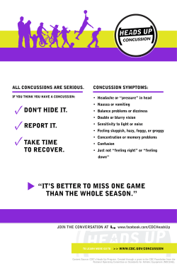
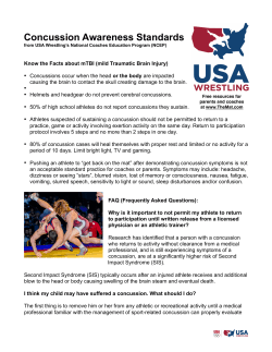


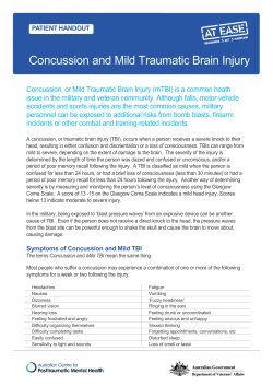
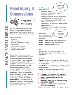
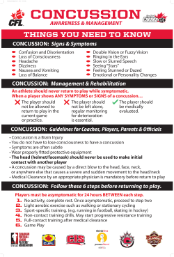

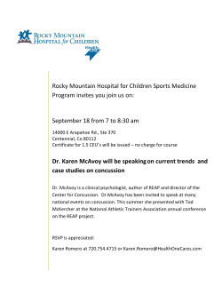
© Copyright 2026