
Disease of Aquatic Organisms 77:255
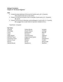
Vol. 77: 255–258, 2007
doi: 10.3354/dao01852
DISEASES OF AQUATIC ORGANISMS
Dis Aquat Org
Published October 15
NOTE
Myxobolus sp. can cause in vivo myoliquefaction in
the host Paralichthys orbignyanus (Osteichthyes,
Paralichthydae)
J. C. Eiras1,*, Joaber P. Júnior2, L. A. Sampaio3, R. Robaldo3, P. C. Abreu4
1
Departamento de Zoologia e Antropologia, Faculdade de Ciências, and CIIMAR, Universidade do Porto, 4099-002 Porto,
Portugal
2
Fundação Universidade Federal do Rio Grande, Departamento de Ciências Morfobiológicas, Laboratório de
Ictioparasitologia, Av. Itália, 96201-900 Rio Grande, RS, Brasil
3
Fundação Universidade Federal do Rio Grande, Departamento de Oceanografia, Laboratório de Maricultura, Av. Itália,
96201-900 Rio Grande, RS, Brasil
4
Fundação Universidade Federal do Rio Grande, Departamento de Oceanografia, Av. Itália, 96201-900 Rio Grande,
RS, Brasil
ABSTRACT: We found intense myoliquefaction in vivo among specimens of wild and farmed South
American flatfish Paralichthys orbignyanus Valenciennes, 1839. The soft flesh condition was attributed to Myxobolus sp. since the fish were not infected by myxosporeans which are usually associated
with this condition (Kudoa spp.), and other causes (bacteria, fungi) were not found. The histopathology of the infection is described.
KEY WORDS: Myxobolus sp. · Paralichthys orbignyanus · Myxozoa · Myoliquefaction
Resale or republication not permitted without written consent of the publisher
Several Myxosporea species can cause post-mortem
myoliquefactive necrosis in a number of fish species.
The condition may be associated with Unicapsula seriolae, U. muscularis and Hexacapsula neothunni, and it
occurs mainly when the flesh is warmed during cooking (Lester 1982). However, post-mortem myoliquefaction is most frequently caused by Kudoa spp. Moran et
al. (1999) listed 44 species of Kudoa, and more species
have been described subsequently. The following species have been reported as causes of enzymatic muscle
degradation after host death: K. clupeidae, K. cruciformum, K. funduli, K. hystolitica, K. mirabilis, K. musculoliquefaciens, K. paniformis, K. peruvianus, K.
rosenbuschi and K. thyrsites (Moran et al. 1999). K.
lateolabracis causes the same effect (Yokoyama et al.
2004), and Kudoa sp. causes post-morten myoliquefaction of the North Pacific giant octopus Paroctopus
dofleini (Yokoyama & Masuda 2001).
Post-mortem myoliquefaction can be a limiting factor
in the exploitation of wild species (Kabata & Whitaker
1981, 1985) and for marine fish culture (Langdon 1991,
Alvarez-Pellitero & Sitjà-Bobadilla 1993, Barja & Toranzo 1993, St-Hillaire et al. 1997, Moran et al. 1999,
Kent et al. 2001, Dawson-Coates et al. 2003, Yokoyama
et al. 2004). It causes economic loss in the fish canning
industry (Gilman & Eiras 1998).
Here, we report myoliquefaction, apparently caused
by Myxobolus sp., in live specimens of farmed and
wild flatfish Paralichthys orbignyanus Valenciennes,
1839. This flatfish is found in the southwestern Atlantic
from Rio de Janeiro, Brazil, to at least Mar de la Plata,
Argentina. It has important economic value.
Extensive and generalized myoliquefaction was first
observed in 2 specimens at the Marine Aquaculture
Station of the University of Rio Grande, Brazil. The fish
were 1.5 yr old and had been reared from eggs at the
*Email: [email protected]
© Inter-Research 2007 · www.int-res.com
256
Dis Aquat Org 77: 255–258, 2007
Station; they were kept in 1 m diameter
round tanks, and fed with trash fish
including Paralonchurus brasiliensis,
Odonthesthes argentinensis, Micropogonias furnieri, Trachinotus marginatus,
Brevortia pectinata and Mugil sp. juveniles. The 2 afflicted fish had breeding
difficulties and their movements were
restricted. They had zones of soft flesh
and some degree of myoliquefaction
(determined by touch) while other fish of
the species were apparently normal. The
degree of myoliquefation increased over
time, with fish becoming soft over their
entire bodies. Both specimens died. No
further observations were made on these
individuals. In another tank, 2 dead,
completely liquefied specimens were
discovered, and only the hard body
structures remained intact.
Fig. 1. Paralichthys orbignyanus infected by Myxobolus sp. Muscle fibres
A second observation was made
partially filled with Myxobolus sp. plasmodia. Mallory’s triple stain
recently in wild moribund specimens net
collected at Rio Grande. The 2 live specimens (20 to 30 cm long) had body
injuries, with a soft, milky cream of liquefied muscle flowing from the injuries.
When the fish were handled, most muscle was transformed into milky cream
and specimens died shortly after removal from the net. Tissue samples were
taken immediately, fixed in formalin,
routinely processed for histology, and
stained with haemalumen and eosin,
toluidene blue, Mallory’s trichrome and
Periodic Acid Schiff reagent. The
remaining pieces of fish were fixed in
formalin and thoroughly examined for
Myxosporea.
Careful observation of the specimens
revealed infection by Myxobolus sp. The
parasites formed plasmodia containing
only mature spores and were located
inside muscle fibres near the fins. The
fibres were either partially filled with the
Fig. 2. Paralichthys orbignyanus infected by Myxobolus sp. Degeneration of
plasmodia (Fig. 1), or all of the fibre conthe myofibrils inside infected muscle fibres. Mallory’s triple stain
tent was replaced by parasite spores. The
spores were more or less elliptical, with
The myofibrils inside the infected fibres showed
equal smooth cell valves about 1–1.5 µm thick. Spores
some degree of degeneration, with partial loss of
were 10.6 (10–11) µm long, 7 µm wide and 6.5 (6–7) µm
myofibrillar detail and striation (Fig. 2). The main
thick. Some spores were rounded and were ~9 µm in
changes in the muscle were observed in regions some
diameter. The polar capsules were equal in size, with a
distance from the plasmodia, where no spores were
rounded anterior and pointed posterior extremity; they
present; both dorsal and ventral muscles appeared
were 4.7 (4–5) µm long and 3 µm wide. The number of
affected. In these areas the muscle cells lost their
coils of the polar filament could not be distinguished
integrity, the myofibrilar detail and striation was
in formalin-fixed spores.
Eiras et al.: Myxobolus sp. causing myoliquefaction
257
continually, but removed by vascular
perfusion in the live fish, or these
enzymes may be released only upon
death of spores or plasmodia. Observations of Langdon (1991) on the infection
of Coryphaena hippurus by K. thyrsites
suggested that to facilitate the release of
the spores the parasite initiated excretion of the liquefactive enzymes in
response to fish death. However, Patashnik et al. (1982) reported accumulation of
the enzymes within the pseudocyst, concluding that diffusion from pseudocysts
after host death causes myoliquefaction.
Proteolytic enzymes are apparently not
associated with the spores, but are produced by non-sporous elements of the
plasmodium (Stehr & Whitaker 1986).
Moran et al. (1999) reviewed literature
on milky flesh caused by Kudoa spp.
Fig. 3. Paralichthys orbignyanus infected by Myxobolus sp. Extensive
Why muscle liquefaction in our samdegeneration and myoliquefaction of muscle. Toluidene blue stain
ples began before host death is unknown. If liquefactive enzymes were
produced continually, then their normal
removed, and hyaline degeneration and myoliquefacremoval or inhibition must have been impaired in
tion were evident (Fig. 3). Neither inflammation nor a
some way. Assuming that proteolytic enzymes were
macrophage response was detected.
localized within the plasmodia and released to other
Careful examination of the specimens showed that
sites as the host cells broke down after the death of the
other myxosporeans (Kudoa spp.) were absent from
host (Patashnik et al. 1982), outward diffusion of the
the fish, and bacteria and fungi were not detected.
enzymes from the plasmodia must have occurred in the
Therefore, it is very likely that the muscle changes and
live fish. What triggered this myoliquefaction in vivo is
myoliquefaction were caused by Myxobolus sp.
unknown, although stress of capture caused by netting
As far as we are aware, this is the first description
the wild specimens may have played a role.
of myoliquefaction caused by Myxobolus sp. PostThe current material was compared with 744 nomimortem myoliquefaction caused by Kudoa has been
nal species of Myxobolus listed by Eiras et al. (2005),
reported for congeners of the fish we examined,
including 30 species parasitic in the muscle of fish, and
including wild Paralichthys adspersus infected by
28 occurring simultaneously in muscle and other
K. thyrsites in Chile (Castro & Burgos 1996), which
organs. However, in spite of sharing features with
has also parasitized farmed P. olivaceus in Japan
some of these species, the absence of fresh spores in
(Yokoyama et al. 2004).
the Myxobolus we examined precludes species identiOccurrence of myoliquefaction in live hosts is
fication.
unusual. Lom & Dyková (1992) reported only one
LITERATURE CITED
record of myoliquefaction in a living fish, i.e. Atherestes evermani from the Bering Sea that was infected
Alvarez-Pellitero P, Sitjà-Bobadilla A (1993) Pathology of
by a Kudoa sp. Later, Davies et al. (1998) observed
Myxosporea in marine fish culture. Dis Aquat Org 17:
229–238
muscle necrosis and milky condition caused by Kudoa
Barja JL, Toranzo A (1993) Myoliquefaction post-morten caused
sp. (superficially resembling K. novae) in live gobies
by the myxosporean Kudoa thyrsites in reared Atlantic
(Pomastochistus microps and Gobius paganellus) from
salmon in Spain. Bull Eur Assoc Fish Pathol 13:86–88
Devon, UK. In this case, rod-shaped bacteria, which
Castro CR, Burgos R (1996) Kudoa thyrsithes (Myxozoa, Mulmay have been secondary invaders, might also have
tivalvulida) causing ‘milky condition’ in the musculature
of Paralichthys adspersus (Neopterigii, Pleuronectiformes,
contributed to tissue necrosis. All other observations of
Paralichthyidae) from Chile. Mem Inst Oswaldo Cruz
myoliquefaction caused by myxosporeans are of post91:163–164
mortem changes in the muscle of the host. Lester
Davies AJ, Andrews T, Upton NDP, Matthews RA (1998)
(1982) maintained that enzymes produced by the paraKudoa as a contributory cause of muscle necrosis in gobsites and responsible for the soft flesh may be excreted
ies. Bull Eur Assoc Fish Pathol 18:62–66
258
Dis Aquat Org 77: 255–258, 2007
Dawson-Coates JA, Chase JC, Funk V, Booy MH and 5 others
(2003) The relationship between flesh quality and numbers of Kudoa thyrsites plasmodia and spores in farmed
Atlantic salmon, Salmo salar L. J Fish Dis 26:451–459
Eiras JC, Molnár K, Lu YS (2005) Synopsis of the species of
Myxobolus Bütschli, 1882 (Myxozoa: Myxosporea:
Myxobolidae). Syst Parasitol 61:1–46
Gilman MM, Eiras JC (1998) Kudoa sp. (Myxosporea,
Multivalvulida) infecting Sardina pilchardus (Walb.,
1792) off the Portuguese coast. Res Rev Parasitol
58:135–137
Kabata Z, Whitaker DJ (1981) Two new species of Kudoa
(Myxosporea: Multivalvulida) parasitic in the flesh of Merluccius productus (Ayres, 1855) (Pisces: Teleostei) in the
Canadian Pacific. Can J Zool 69:2085–2091
Kabata Z, Whitaker DJ (1985) Parasites as a limiting factor in
exploitation of Pacific Whiting, Merluccius productus. Mar
Fish Rev 47:55–59
Kent ML, Andree KB, Bartholomew JL, El-Matbouli M and 12
others (2001) Recent advances in our knowledge of the
Myxozoa. J Eukaryot Microbiol 48:395–413
Langdon JS (1991) Myoliquefaction post-morten (‘milky
flesh’) due to Kudoa thyrsites (Gilchrist) (Myxosporea:
Multivalvulida) in mahi mahi, Coryphaena hippurus L.
J Fish Dis 14:45–54
Lester RJG (1982) Unicapsula seriolae n. sp. (Myxosporea,
Multivalvulida) from Australian yellowtail kingfish Seriola
lalandi. J Protozool 29:584–587
Lom J, Dyková I (1992) Protozoan parasites of fishes. Develop-
ments in aquaculture and fisheries science, Vol 26. Elsevier, Amsterdam
Moran JDW, Whitaker DJ, Kent ML (1999) A review of the
myxosporean genus Kudoa Meglitsch, 1947, and its
impact on the international aquaculture industry and commercial fisheries. Aquaculture 172:163–196
Patashnik M, Groninger HS, Barnett H, Kudo G, Koury B
(1982) Pacific whiting Merluccius productus: I. Abnormal
texture caused by myxosporidium induced proteolysis.
Mar Fish Rev 44:1–12
Stehr C, Whitaker DJ (1986) Host-parasite interaction on
myxosporeans Kudoa paniformis Kabata & Whitaker, 1981
and Kudoa thyrsites (Gilchrist, 1924) in the muscle of
Pacific whiting, Merluccius productus (Ayres): an ultrastructural study. J Fish Dis 9:505–517
St-Hilaire S, Hill M, Kent ML, Whitaker DJ, Ribble C (1997)
A comparative study of muscle texture and intensity of
Kudoa thyrsites infection in farmed-reared Atlantic salmon Salmo salar on the pacific coast of Canada. Dis Aquat
Org 31:221–225
Yokoyama H, Masuda K (2001) Kudoa sp. (Myxozoa) causing
a post-morten myoliquefaction of North-Pacific giant octopus Paroctopus dofleini (Cephalopoda: Octopodidae). Bull
Eur Assoc Fish Pathol 21:266–268
Yokoyama H, Whipps CM, Kent ML, Mizuno K, Kawakami H
(2004) Kudoa thyrsites from Japanese flounder and Kudoa
lateolabracis n. sp. from Chinese sea bass: causative
myxozooans of post-morten myoliquefaction. Fish Pathol
39:79–85
Editorial responsibility: Dieter Steinhagen,
Hannover, Germany
Submitted: June 15, 2007; Accepted: July 30, 2007
Proofs received from author(s): September 17, 2007


© Copyright 2026