
How to describe a cryptic species? Practical challenges of molecular taxonomy
Jörger and Schrödl Frontiers in Zoology 2013, 10:59
http://www.frontiersinzoology.com/content/10/1/59
RESEARCH
Open Access
How to describe a cryptic species? Practical
challenges of molecular taxonomy
Katharina M Jörger1,2* and Michael Schrödl1,2
Abstract
Background: Molecular methods of species delineation are rapidly developing and widely considered as fast
and efficient means to discover species and face the ‘taxonomic impediment’ in times of biodiversity crisis. So
far, however, this form of DNA taxonomy frequently remains incomplete, lacking the final step of formal species
description, thus enhancing rather than reducing impediments in taxonomy. DNA sequence information
contributes valuable diagnostic characters and –at least for cryptic species – could even serve as the backbone
of a taxonomic description. To this end solutions for a number of practical problems must be found, including a
way in which molecular data can be presented to fulfill the formal requirements every description must meet.
Multi-gene barcoding and a combined molecular species delineation approach recently revealed a radiation
of at least 12 more or less cryptic species in the marine meiofaunal slug genus Pontohedyle (Acochlidia,
Heterobranchia). All identified candidate species are well delimited by a consensus across different methods
based on mitochondrial and nuclear markers.
Results: The detailed microanatomical redescription of Pontohedyle verrucosa provided in the present paper does
not reveal reliable characters for diagnosing even the two major clades identified within the genus on molecular
data. We thus characterize three previously valid Pontohedyle species based on four genetic markers
(mitochondrial cytochrome c oxidase subunit I, 16S rRNA, nuclear 28S and 18S rRNA) and formally describe nine
cryptic new species (P. kepii sp. nov., P. joni sp. nov., P. neridae sp. nov., P. liliae sp. nov., P. wiggi sp. nov., P. wenzli
sp. nov., P. peteryalli sp. nov., P. martynovi sp. nov., P. yurihookeri sp. nov.) applying molecular taxonomy, based on
diagnostic nucleotides in DNA sequences of the four markers. Due to the minute size of the animals, entire
specimens were used for extraction, consequently the holotype is a voucher of extracted DNA (‘DNA-type’). We
used the Character Attribute Organization System (CAOS) to determine diagnostic nucleotides, explore the
dependence on input data and data processing, and aim for maximum traceability in our diagnoses for future
research. Challenges, pitfalls and necessary considerations for applied DNA taxonomy are critically evaluated.
Conclusions: To describe cryptic species traditional lines of evidence in taxonomy need to be modified. DNA
sequence information, for example, could even serve as the backbone of a taxonomic description. The present
contribution demonstrates that few adaptations are needed to integrate into traditional taxonomy novel
diagnoses based on molecular data. The taxonomic community is encouraged to join the discussion and develop
a quality standard for molecular taxonomy, ideally in the form of an automated final step in molecular species
delineation procedures.
* Correspondence: [email protected]
1
Mollusca Section, SNSB-Bavarian State Collection of Zoology,
Münchhausenstr 21, 81247 München, Germany
2
Department Biology II, Ludwig-Maximilians-University, Großhaderner Str. 2,
82152 Planegg-Martinsried, Germany
© 2013 Jörger and Schrödl; licensee BioMed Central Ltd. This is an Open Access article distributed under the terms of the
Creative Commons Attribution License (http://creativecommons.org/licenses/by/2.0), which permits unrestricted use,
distribution, and reproduction in any medium, provided the original work is properly cited.
Jörger and Schrödl Frontiers in Zoology 2013, 10:59
http://www.frontiersinzoology.com/content/10/1/59
Background
Species boundaries are frequently hard to delimit based on
morphology only, a fact which has called for integrative
taxonomy, including additional sources of information
such as molecular data, biogeography, behavior and ecology [1,2]. Founding a species description on a variety of
characters from different, independent datasets is generally
regarded as best practice [3]. When species are considered
as independently evolving lineages [4], different lines of
evidence (e.g., from morphology, molecules, ecology or distribution) are additive to each other and no line is necessarily exclusive nor need different lines obligatory be used
in combination [3,5]. Taxonomists are urged to discriminate characters according to their quality and suitability for
species delineation, rather than to just add more and more
data [5]. The specifics of the taxon in question will guide
the way to the respective set(s) of characters that will provide the best backbone for the diagnosis. In cases of
pseudo-cryptic species (among which morphological differences can be detected upon re-examining lineages separated e.g. on molecular data) or of fully cryptic species
(that morphology fails to delimit), the traditional lines of
evidence have to be modified by using, e.g., molecular information to break out of the ‘taxonomic circle’ [6,7].
Cryptic species are a common phenomenon throughout
the metazoan taxa, and can be found in all sorts of habitats
and biogeographic zones [8-10]. Groups characterized by
poor dispersal abilities (e.g., most meiofaunal organisms or
animals inhabiting special regions where direct developers
predominate, such as Antarctica), are especially prone to
cryptic speciation [11,12]. Uncovering these cryptic species
is fundamental for the understanding of evolutionary processes, historical biogeography, ecology, and also to conservation approaches, as distribution ranges that are smaller
than initially assumed mean a higher risk of local extinction [8,10]. The lack of morphological characters to distinguish cryptic species should not lead to considerable parts
of biological diversity remaining unaddressed.
The utility of DNA barcoding and molecular species delineation approaches to uncover cryptic lineages has been
demonstrated by numerous studies (e.g., [11,13-19]). Unfortunately, inconsistencies in terminology associated with the
interface between sequence data and taxonomy have led to
confusion and various criticisms [6,20]. First of all, one
needs to distinguish between species identification via molecular data (DNA barcoding in its strict sense) and species
discovery [6,21,22]. While species identification is a primary
technical application, species delimitation requires means
of molecular species delineation that is either distance, tree
or character based [6,23]. Under ideal circumstances sufficient material is collected from different populations across
the entire distribution area of a putative group of cryptic
species. Using population genetics the distribution of haplotypes can be analyzed and different, genetically isolated
Page 2 of 27
lineages can be detected [24]. Population genetic approaches are, however, not always feasible with animals that
are rare or hard to collect, which might actually be a common phenomenon across faunas of most marine ecosystems [25-28]. Derived from barcoding initiatives, threshold
based species delimitation became the method of choice,
aiming for the detection of a ‘barcoding gap’ between intraand interspecific variation [29-31]. This approach has been
criticized, however, due to its sensitivity to the degree of
sampling, the general arbitrariness of fixed or relative
thresholds, and to frequent overlap between intra- and interspecific variation [6,32,33]. In the recently developed
Automatic Barcode Gap Discovery (ABGD) [34], progress
has been made in avoiding the dependence of a priori
defined species hypotheses in threshold based approaches,
but reservations remain concerning the concept of a
barcoding gap [25]. Several independent delineation tools
exist, e.g. using haplotype networks based on statistical parsimony [35], maximum likelihood approaches applying the
General Mixed Yule-Coalescent model [36,37], or Bayesian
species delineation [38,39]. Empirical research currently
compares the powers of these different tools on real
datasets [25,32,40]. The effect of the inclusion of singletons in analyses is considered as most problematic [25]. At
the present stage of knowledge, independent approaches
allowing cross-validation between the different methods of
molecular species delineation and other sources of information (morphology, biogeography, behavioral traits) seem
the most reliable way of delimiting cryptic species [25].
The second inconsistency in terminology concerns usages of ‘DNA taxonomy’. Originally, DNA taxonomy was
proposed to revolutionize taxonomy by generally founding
descriptions on sequence data and overthrowing the
Linnaean binominal system [41]. Alternatively, it was
suggested as a concept of clustering DNA barcodes into
MOTUs [42]. Since then, however, it has been applied
as an umbrella term for barcoding, molecular species
delineation, and including molecular data in species
descriptions (see e.g., [13,14,20,36,43,44]). In a strict sense,
one cannot speak of molecular taxonomy if the process
of species discovery is not followed by formal species
description (i.e. there are two steps to a taxonomic process:
species discovery (delimitation) and attributing them with
formal diagnoses and names.) Taxonomy remains incomplete if species hypotheses new to science are flagged as
merely putative by provisional rather than fully established
scientific names. For practical reasons and journal requirements, most studies on molecular species delineation
postpone formal descriptions of the discovered species
(e.g., [13,14,25,33,36,40,43-46]), and then rarely carry them
out later. DNA barcoding and molecular species delineation are promoted as fast and efficient ways to face the
‘taxonomic impediment’, i.e. the shortage of time and
personnel capable of working through the undescribed
Jörger and Schrödl Frontiers in Zoology 2013, 10:59
http://www.frontiersinzoology.com/content/10/1/59
species richness in the middle of a biodiversity crisis
[7,47,48]. However, keeping discovered entities formally
unrecognized does not solve the taxonomic challenges
but adds to them by creating parallel worlds populated
by numbered MOTUs, OTUs or candidate species. In
many cases the discovered taxa remain inapplicable to
future research, thus denying the scientific community
this taxonomic service, e.g. for species inventories or
conservation attempts. Without formal description or a
testable hypothesis, i.e. a differential diagnosis, 1) the
discovered species might not be properly documented or
vouchered by specimens deposited at Natural History
Museums; and 2) their reproducibility can be hindered
and confusion caused by different numbering systems. A
deterrent example of the proliferation of informal epithets
circulating as ‘nomina nuda’ (i.e. species which lack formal
diagnoses and deposited vouchers) in the literature is
given by the ‘ten species in one’ Astraptes fulgerator
complex [31,49]. Thus, we consider it as all but indispensable for DNA taxonomy to take the final step and
formalize the successfully discovered molecular lineages.
The transition from species delimitation to species description is the major task to achieve. Nearly ten years after
the original proposal of DNA taxonomy [41], revolutionizing traditional taxonomy has found little acceptance in the
taxonomic community, as most authors agree that there is
no need for overthrowing the Linnaean System. Consequently, the challenge is to integrate DNA sequence information in the current taxonomic system. Several studies
have attempted to include DNA data in taxonomic descriptions, albeit in various non-standardized ways; see the review by Goldstein and DeSalle ([21]; box 3): In some cases,
DNA sequence information is simply added to the taxonomic description (in the form of GenBank numbers or
pure sequence data), without evaluating and reporting diagnostic features [21]. Others rely on sequence information
for the description, either reporting results of species delineation approaches, e.g. raw distance measurements or
model based assumptions, or extracting diagnostic characters from their molecular datasets. There still is a consensus
that species descriptions should be character based [50]
(but see the Discussion below for attempts at model based
taxonomy), and that tree or distance based methods fail to
extract diagnostic characters [6]. Character based approaches, like the Characteristic Attribute Organization System (CAOS), are suggested as an efficient and reliable way
of defining species barcodes based on discrete nucleotide
substitution, and these established diagnostics from DNA
sequences can be used directly for species descriptions as
molecular taxonomic characters [51,52]. Yet, the application
of CAOS or similar tools requires an evaluation of how to
select and present molecular synapomorphies and how to
formalize procedures to create a ‘best practice’ linking DNA
sequence information to existing taxonomy [20].
Page 3 of 27
In the present study, we formally describe the candidate species of minute mesopsammic sea slugs in the
genus Pontohedyle Golikov & Starobogatov (Acochlidia,
Heterobranchia) discovered by Jörger et al. [25]. This
cryptic radiation was uncovered in a global sampling approach with multi-gene and multiple-method molecular
species delineation [25]. The initially identified 12 MOTUs,
nine of which do not correspond to described species, are
considered as species [following 4] resulting from a conservative minimum consensus approach applying different
methods of molecular species delineation [25]. The authors
demonstrated that traditional taxonomic characters (external morphology, spicules and radula features) are insufficient to delineate cryptic Pontohedyle species [25]. To
evaluate the power of more advanced histological and microanatomical data, we first provide a detailed computer
based 3D redescription of the anatomy of Pontohedyle
verrucosa (Challis, 1970) and additional histological semithin sections of P. kepii sp. nov. In the absence of reliable
diagnostic characters from morphology and microanatomy,
we then rely on DNA sequence data as the backbone for
our species descriptions. For the three previously valid
Pontohedyle species we extract diagnostic characters using
the Character Attribute Organization System (CAOS) based
on four standard markers (mitochondrial cytochrome c oxidase subunit I, 16S rRNA, and nuclear 18S rRNA and 28S
rRNA). In addition, nine new species are formally described
on molecular characteristics and evidence from other data
sources. Various approaches to the practical challenges for
molecular driven taxonomy – such as critical consideration
of the quality of the alignment, detection of diagnostic nucleotides and their presentation aiming for maximum traceability in future studies – are tested and critically evaluated.
Results
Evaluation of putative morphological characters
The diversity within Pontohedyle revealed by molecular
data cannot be distinguished externally: the body shows
the typical subdivision into the anterior head-foot complex
and the posterior visceral hump. Bodies are whitishtranslucent, digestive glands are frequently bright green
to olive green. Rhinophores are lacking, labial tentacles are
bow-shaped and tapered towards the ends (see Figures 1
and 2). Monaxone rodlet-like spicules distributed all
over the body and frequently found in an accumulation
between the oral tentacles are characteristic for Pontohedyle.
These spicules can be confirmed for P. wenzli sp. nov., for
P. yurihookeri sp. nov., P. milaschewitchii (Kowalevsky,
1901) and P. brasilensis (Rankin, 1979), and, in contrast to
the original description [53], also in P. verrucosa. No spicules could be detected in P. peteryalli sp. nov. from Ghana.
The absence of spicules is insufficient, however, to delineate
microhedylid species, since their presence can vary under
environmental influence [54].
Jörger and Schrödl Frontiers in Zoology 2013, 10:59
http://www.frontiersinzoology.com/content/10/1/59
Page 4 of 27
Figure 1 External morphology (living specimens) and radula characteristics (SEM micrographs) in Pontohedyle species (part 1).
A) Pontohedyle kepii sp. nov. (Pontohedyle sp. 1 in [25]); B) Pontohedyle joni sp. nov. (Pontohedyle sp. 2 from WA-5 (Belize) in [25]); C) Pontohedyle
liliae sp. nov. (Pontohedyle sp. 4 in [25]), * marks putative 4th cusp on rhachidian tooth. cc = central cusp of rhachidian tooth, llp = left lateral
plate, rlp = right lateral plate, rt = rhachidian tooth.
The radulae of eight species were investigated using SEM
(see Figures 1 and 2). Radulae of P. neridae sp. nov., P.
martynovi sp. nov. and P. yurihookeri sp. nov. were not recovered whole from molecular preparations, and thus were
unavailable for further examination [25]. The radula of P.
wiggi sp. nov. could only be observed under the lightmicroscope, but not successfully transferred to a SEM
stub. All radulae are hook-shaped with a longer dorsal
and a shorter ventral ramus, typical for Acochlidia. Radula
formulas are 38–53 × 1.1.1, lateral plates are curved
rectangular, and the rhachidian tooth is triangular and bears
a central cusp and typically three smaller lateral denticles.
Most radulae bear one pointed denticle centrally on
the anterior margin of each lateral plate and a corresponding notch on the posterior side. Only the radula
of P. kepii sp. nov. and P. verrucosa can be clearly distinguished from the others by the absence of this denticle and the more curved lateral teeth (see Figure 1A
and [25], Figure 1D,E). Uniquely, P. verrucosa bears
five lateral denticles next to the central cusp of the
Jörger and Schrödl Frontiers in Zoology 2013, 10:59
http://www.frontiersinzoology.com/content/10/1/59
Page 5 of 27
Figure 2 External morphology (living specimens) and radula characteristics (SEM micrographs) in Pontohedyle species (part 2).
A) Pontohedyle peteryalli sp. nov. (Pontohedyle sp. 7 in [25]); B) Pontohedyle wenzli sp. nov. (Pontohedyle sp. 6, picture of living animal from WP-1
(holotype), radula from IP-2, see [25]); C) P. brasilensis (living animal from WA-3 (Belize), radula from WA-10 (Brazil), see [25]). cc = central cusp of
rhachidian tooth, llp = left lateral plate, rlp = right lateral plate, rt = rhachidian tooth.
rhachidian tooth [25]; in P. liliae sp. nov. a tiny fourth
denticle borders the central cusp (see * in Figure 1C).
Previous phylogenetic analyses [25] recovered a deep
split into two Pontohedyle clades: the P. milaschewitchii
clade and the P. verrucosa clade. This is supported by
novel analyses in a larger phylogenetic framework and
additionally including a second nuclear marker (18S rRNA)
(own unpublished data). Since no detailed histological
account exists of any representative from the large P.
verrucosa clade, we redescribe P. verrucosa (based on ZSM
Mol-20071833, 20071837 and 20100548), supplementing
the original description with detailed information of
the previously undescribed nervous and reproductive
systems. The central nervous system (cns) of P. verrucosa
lies prepharyngeal and shows an epiathroid condition. It
consists of paired rhinophoral, cerebral, pleural, pedal and
buccal ganglia and three unpaired ganglia on the visceral
nerve cord, tentatively identified as left parietal ganglion,
median fused visceral and subintestinal ganglion and right
fused parietal and supraintestinal ganglion (Figure 3A). An
Jörger and Schrödl Frontiers in Zoology 2013, 10:59
http://www.frontiersinzoology.com/content/10/1/59
osphradial ganglion or gastro-oesophagial ganglia were not
detected. Anterior and lateral to the cerebral ganglia are
masses of accessory ganglia. Due to the retracted condition
of all examined specimens, tissues are highly condensed
and no separation in different complexes of accessory ganglia could be detected. Attached to the pedal ganglia are
large monostatolith statocysts. Oval, unpigmented globules
are located in an antero-ventral position of the cerebral ganglia, interpreted as the remainder of eyes (see Figure 3B).
P. verrucosa is a gonochoristic species. The three sectioned specimens include two males and one female.
The male reproductive system is comprised of gonad,
ampulla, postampullary sperm duct, prostatic vas deferens, ciliated (non-glandular) vas deferens, genital opening and a small ciliated ‘subepidermal’ duct leading to a
second genital opening anterodorsally of the mouth
opening (Figure 3C). The sac-like gonad is relatively
small and bears few irregular distributed spermatozoa.
The large tubular ampulla emerges from the gonad without a detectable preampullary sperm duct; it is loosely
filled with irregularly distributed spermatozoa (Figure 3D).
The ampulla leads into a short, narrow ciliated postampullary duct widening into the large tubular prostatic
vas deferens (staining pink in methylene-blue sections,
Figure 3D). Close to the male genital opening, the duct
loses its glandular appearance and bears cilia. The primary
genital opening is located on the right side of the body
at the visceral hump and close to the transition with
the head-foot complex. Next to the genital opening, the
anterior vas deferens splits off as an inconspicuous
subepithelial ciliated duct that leads anteriorly on the
right side of the head foot complex. It terminates in a
second genital opening between the oral tentacles
anterodorsally from the mouth opening.
The female reproductive system consists of gonad,
nidamental glands and oviduct (Figure 3E) and a genital
opening located on the right side, in the posterior part
of the visceral hump (not visible in Figure 3E, due to
the retracted stage of the individual). The gonad is saclike and bears one large vitellogenic egg (see Figure 3F)
and several developing oocytes. Three histologically differentiated tube-like nidamental glands could be detected
with a supposedly continuous lumen and with an epithelium bearing cilia. From proximal to distal these glands
are identified as albumen gland (cells filled with dark blue
stained granules), membrane gland (pinkish, vacuolated
secretory cells) and winding mucus gland (secretory cells
stained pink-purple). In its proximal part the distal oviduct
shows a similar histology as the mucous gland, but then
loses its glandular appearance. The epithelium of the distal
oviduct bears long, densely arranged cilia.
Additional notable histological features are numerous
dark-blue-stained epidermal gland cells (see e.g., arrowhead in Figure 3D) and refracting fusiform structures in
Page 6 of 27
the digestive gland (see Figure 3B). An additional series
of histological semi-thin sections of Pontohedyle kepii
sp. nov. was sectioned and brief investigation revealed
no variation in the major organization of the organ systems in Pontohedyle as described herein and in previous
studies [55,56].
Remarks on the presentation of molecular characters
Diagnostic characters for each species of Pontohedyle
were extracted using the ‘Characteristic Attribute
Organization System’ (CAOS) [51,57,58]. We define
diagnostic characters as single pure characters, i.e.
unique character states that respectively occur in all investigated specimens in a single Pontohedyle species
but in none of the specimens of its congeners. As additional information single heterogeneous pure characters (i.e., different character states present within the
species but absent from the congeners) are reported
(for further details on the chosen approach see the
Material and methods and Discussion sections). Positions refer to the position of the diagnostic nucleotide
within the respective alignment (see Additional files 1,
2, 3, 4, 5 and 6). Where alignment positions differ from
those in the deposited sequences, positions within the
sequence of the holotype or in another reference sequence are also provided.
Taxonomy of Pontohedyle
Family: Microhedylidae Odhner, 1938 [59]
Genus: Pontohedyle Golikov & Starobogatov, 1972 [60]
Synonymy: Mancohedyle Rankin, 1979; Gastrohedyle
Rankin, 1979; Maraunibina Rankin, 1979
Type species (by subsequent designation): Pontohedyle
milaschewitchii (Kowalevsky, 1901) [61]
Phylogenetic analyses of the genus Pontohedyle [25]
confirmed earlier assumptions, that the three genera
established by Rankin [62] (see above) present junior
synonyms of Pontohedyle.
Morphological characteristics of genus Pontohedyle:
Minute (0.7–6 mm) marine interstitial microhedylacean
acochlid. Body divided into anterior head-foot complex
and posterior visceral hump. In case of disturbance
head-foot complex can be entirely retracted into visceral hump. Body whithish translucent. Foot with short
rounded free posterior end. Head bears one pair of
bow-shaped dorso-ventrally flattened oral tentacles.
Rhinophores lacking. Monaxone, calcareous spicules irregularly distributed over head-foot complex and visceral hump. Radula hook-shaped band (lateral view),
formula 1-1-1, lateral plates curved or with one pointed
denticle, rhachidian tooth triangular with one central
cusp and 2–4 lateral cusps on each side. Nervous
Jörger and Schrödl Frontiers in Zoology 2013, 10:59
http://www.frontiersinzoology.com/content/10/1/59
Page 7 of 27
Figure 3 Microanatomy of P. verrucosa. A) 3D-reconstruction of the central nervous system, frontal view (ZSM Mol 20071832). B) Histological
semi-thin section of the cerebral ganglia showing unpigmented eyes and rhinophoral ganglia. C) 3D-reconstruction of the male reproductive system
in a partially retracted specimen, right lateral view (ZSM Mol 20071833). D) Histological semi-thin section showing prostatic vas deferens and spermfilled ampulla (arrowhead = dark blue stained epidermal gland). E) 3D-reconstruction of the female reproductive system in a completely retracted
specimen, right lateral view (ZSM Mol 20100548). F) Histological semi-thin section showing nidamental glands and gonad with oocyte.
Jörger and Schrödl Frontiers in Zoology 2013, 10:59
http://www.frontiersinzoology.com/content/10/1/59
Page 8 of 27
system with accessory ganglia at cerebral nerves anterior to the cns. Sexes separate, male reproductive system
aphallic, sperm transferred via spermatophores.
Molecular diagnosis of the genus Pontohedyle, based on
the sequences analyzed herein (Table 1) and on sequences
from a set of outgroups including all acochlidian genera
Table 1 DNA sequence data analyzed in the present study to determine diagnostic nucleotides in Pontohedyle
Species
P. milaschewitchii
P. brasilensis
P. verrucosa
Pontohedyle kepii sp. nov.
Pontohedyle joni sp. nov.
Museums number
DNA
voucher
GenBank accession numbers
18S rRNA
28S rRNA
16S rRNA
COI
ZSM Mol 20071381
AB34404214
-
JQ410926
JQ410925
JQ410897
ZSM Mol 20080054
AB34404241
HQ168435
JF828043
HQ168422
-
ZSM Mol 20080055
AB34404239
-
-
JQ410927
-
ZSM Mol 20080925
-
-
-
JQ410928
HQ168459
ZSM Mol 20080953
AB35081832
KC984282
-
JQ410929
JQ410898
SI-CBC20 10KJ01-E03
AB34500510
KC984283
JQ410941
JQ410940
-
SI-CBC20 10KJ01-B07
AB34402082
-
JQ410943
JQ410942
-
SI-CBC20 10KJ01-D07
AB34500513
-
JQ410944
-
-
SI-CBC20 10KJ01-B09
AB34402031
-
JQ410946
JQ410945
JQ410904
SI-CBC20 10KJ01-C09
AB34500576
-
JQ410948
JQ410947
JQ410905
SI-CBC20 10KJ01-A10
AB34402026
-
-
JQ410949
-
SI-CBC20 10KJ02-E01
AB34402030
-
JQ410950
-
-
ZSM Mol 20110723
AB34402034
KC984284
JQ410952
JQ410951
JQ410906
ZSM Mol 20110722
AB34402086
KC984285
JQ410932
JQ410931
JQ410900
ZSM Mol 20090198
AB35081813
KC984286
JQ410936
JQ410935
-
ZSM Mol 20071820
AB34404223
KC984287
JQ410978
JQ410977
JQ410920
ZSM Mol 20080176
AB34404286
-
JQ410980
JQ410979
JQ410921
ZSM Mol 20071135
AB34404221
KC984288
JQ410971
JQ410970
JQ410914
ZSM Mol 20100388
AB34500547
-
-
-
JQ410916
ZSM Mol 20100389
AB34402044
-
JQ410974
-
JQ410917
ZSM Mol 20100390
AB34402070
-
JQ410975
-
JQ410918
ZSM Mol 20100391
AB34500531
KC984289
-
JQ410976
JQ410919
ZSM Mol 20081013
AB35081769
KC984290
JQ410967
JQ410966
JQ410912
ZSM Mol 20090197
AB34858164
KC984291
JQ410934
JQ410933
JQ410901
SI-CBC20 10KJ01-D05
AB34402049
KC984292
-
JQ410937
JQ410902
SI-CBC20 10KJ01-C08
AB34402065
-
JQ410939
JQ410938
JQ410903
Pontohedyle neridae sp.nov.
AM C. 476062.001
AB34500497
-
JQ410986
JQ410985
JQ410922
Pontohedyle liliae sp.nov.
ZSM Mol 20090471
AB35081802
KC984293
JQ410954
JQ410953
-
ZSM Mol 20090472
AB35081838
-
JQ410956
JQ410955
-
ZSM Mol 20100595
AB34402059
-
JQ410960
JQ410959
JQ410908
ZSM Mol 20100596
AB34402001
-
-
JQ410961
JQ410909
ZSM Mol 20100597
AB34500571
-
JQ410963
JQ410962
JQ410910
ZSM Mol 20100603
AB34402020
-
JQ410965
JQ410964
JQ410911
Pontohedyle wiggi sp.nov.
Pontohedyle wenzli sp.nov.
ZSM Mol 20100592
AB34402021
KC984294
JQ410958
JQ410957
JQ410907
AM C. 476051.001
AB34402037
KC984295
JQ410982
JQ410981
-
ZSM Mol 20081014
AB35081827
KC984296
JQ410969
JQ410968
JQ410913
ZSM Mol 20100379
AB34500521
KC984297
JQ410973
JQ410972
JQ410915
Pontohedyle peteryalli sp. nov.
ZSM Mol 20071133
AB34404268
KC984298
-
JQ410930
JQ410899
Pontohedyle martynovi sp. nov.
AM C. 476054.001
AB34402062
-
JQ410984
JQ410983
-
Pontohedyle yurihookeri sp. nov.
ZSM Mol 20080565
AB34402000
KC984299
JQ410987
-
-
Museum numbers (ZSM – Bavarian State Collection of Zoology, SI – Smithsonian Institute, AM - Australian Museum), DNA vouchers (at ZSM) and GenBank
accession numbers. 18S rRNA sequences generated in this study marked with *, all remaining sequences retrieved from GenBank.
Jörger and Schrödl Frontiers in Zoology 2013, 10:59
http://www.frontiersinzoology.com/content/10/1/59
for which data are available [63,64]. Positions refer to
the alignments in Additional files 1 and 2, and to the
reference sequences of P. milaschewitchii, ZSM Mol
20080054 (GenBank HQ168435 and JF828043) from
Croatia, Mediterranean Sea (confirmed to be conspecific
with material collected at the type locality in molecular species delineation approaches [25]). Molecular diagnosis is
given in Table 2.
Table 2 Molecular diagnostic characters of Pontohedyle
Marker
Diagnostic characters with position
in alignment (in reference sequence)
18S rRNA
165 (168), G; 1358 (1365), A; 1360
(1367), T; 1371 (1378), T; 1514 (1521), T
28S rRNA
260, C; 576, T; 622, T
Pontohedyle milaschewitchii (Kowalevsky, 1901) [61]
Hedyle milaschewitchii Kowalevsky, 1901: p. 19–20 [61]
Pontohedyle milaschewitchii (Kowalevsky) – Golikov &
Starobogatov [60]
Mancohedyle milaschewitchii (Kowalevsky) – Rankin
(1979: p. 100) [62]
Pontohedyle milatchevitchi (Kowalevsky) – Vonnemann
et al. (2005: p. 3) [65]; Göbbeler & Klussmann-Kolb
(2011: p. 122) [66].
Type locality: Black Sea, bay of St George monastery
near Sevastopol, Crimean Peninsula, Ukraine.
Type material: To our knowledge no type material
remains. Nevertheless we refrain from designating a
neotype, as there is no taxonomic need, i.e. no possibility of confusion in the species' area of distribution.
Distribution and habitat: Reported from the Black
Sea and numerous collecting sites throughout the Mediterranean e.g. [55,61,67,68]; marine, interstitial, subtidal
1–30 m, coarse sand.
Molecular diagnosis is given in Table 3.
Page 9 of 27
ZSM Mol 20071381 (recollected at the type locality, see
Figure 4) serves as the reference sequence, unless the
sequence could not be successfully amplified. Then
sequences (indicated below) from material from the
Mediterranean serve as reference sequences (conspecifity
was confirmed in a previous molecular species delineation
approach 25]). Diagnostic characters in 18S rRNA were
determined based on ZSM Mol 20080054 (GenBank
HQ168435 = reference sequence) and ZSM Mol 20080953
(GenBank KC984282); in nuclear 28S rRNA based on
ZSM Mol 20071381 (GenBank JQ410926) and ZSM
Mol 20080054 (GenBank JF828043 = reference sequence),
in mitochondrial 16S rRNA based on ZSM Mol 20071381
(GenBank JQ410925), ZSM Mol 20080054 (GenBank
HQ168422), ZSM Mol 20080055 (GenBank JQ410927),
ZSM Mol 20080925 (GenBank JQ410928) and ZSM Mol
20080953 (GenBank JQ410929), in mitochondrial COI
based on ZSM Mol 20071381 (GenBank JQ410827), ZSM
Mol 20080925 (GenBank HQ168459) and ZSM Mol
20080953 (GenBank JQ410898).
Pontohedyle verrucosa (Challis, 1970) [53]
Microhedyle verrucosa Challis, 1970: pp. 37–38 [53]
Pontohedyle verrucosa (Challis) – Wawra
(1987: p. 139) [69]
Maraunibina verrucosa (Challis) – Rankin (1979:
p. 102) [62]
Type locality: Coarse, clean shell sand, a little
above low water at neap tide, near southern end of
Maraunibina Island, Marau Sound, East Guadalcanal,
Solomon Islands.
Type material: According to Challis [53] in the Natural History Museum, London, and the Dominion Museum, Wellington, New Zealand. Own investigations
revealed that the type material of Challis never arrived
at the Natural History Museum, London and visiting
the Museum of New Zealand Te Papa Tongarewa
(former Dominion Museum), we were unable to locate any of her types. Thus, at current stage of
Table 3 Molecular diagnostic characters of Pontohedyle milaschewitchii
Marker
Diagnostic characters with position in
alignment (in reference sequence)
Heterogeneous single
pure positions
18S rRNA
159, C; 164 (165), G
-
28S rRNA
329 (324), T
-
16S rRNA
8, G; 26, A; 145 (146), C; 203 (209), A; 243 (274),
G; 275 (306), T; 290 (321), T; 333 (363), A; 352 (382), T
351 (381), T (G in ZSM Mol 20080953, position 381)
COI
11, C; 25, C; 58, T; 160, C; 272, A; 273,G; 319, T; 352,
G; 371, G; 376, G; 397, A; 451, A; 476, C; 495, G; 496, G; 520, C
-
COI (AA)
4, L; 124, A; 159, L; 165, S
-
Jörger and Schrödl Frontiers in Zoology 2013, 10:59
http://www.frontiersinzoology.com/content/10/1/59
Page 10 of 27
Figure 4 World map showing the sampling sites and type localities of Pontohedyle species (modified after [25]). Type localities with
material included in this study are marked by triangles. Unsampled type localities are resembled by squares. Additional collecting sites are
marked with dots.
knowledge, type material might only remain in her
private collection. We refrain from designating a neotype because we were unable to recollect at the type
locality (see below).
Distribution and habitat: Reported from Indonesia
and the Solomon Islands [25,53]; marine, interstitial,
intertidal, coarse sand.
Sequenced material: In a collecting trip to the
Solomon Islands, we were unfortunately unable to recollect at the type locality (Maraunibina Island, East
Guadalcanal), but successfully recollected in Komimbo
Bay (West Guadalcanal), a locality, from which the describing author noted similar ecological parameters
and recorded several meiofaunal slug species occurring at both sites [53,70] Additional material was
collected at different collecting sites in Indonesia
(see Figure 4).
Molecular diagnosis is given in Table 4.
ZSM Mol 20071820 (from Komimbo Bay, East
Guadalcanal, Solomon Islands) serves as the reference
sequence. Diagnostic characters in nuclear 18S rRNA
were determined based on ZSM Mol 20071820 (GenBank
KC984287), ZSM Mol 20071135 (GenBank KC984288)
and ZSM Mol 20100391 (GenBank KC984289), in nuclear
28S rRNA based on ZSM Mol 20071820 (GenBank
JQ410978), ZSM Mol 20080176 (GenBank JQ410980),
ZSM Mol 20071135 (GenBank JQ410971), ZSM Mol
20100389 (GenBank JQ410974) and ZSM Mol 20100390
(GenBank JQ410975), in mitochondrial 16S rRNA based
on ZSM Mol 20071820 (GenBank JQ410977), ZSM Mol
20080176 (GenBank JQ410979), ZSM Mol 20071135
(GenBank JQ410970) and ZSM Mol 20100391 (GenBank
JQ410976) and in mitochondrial COIbased on ZSM Mol
20071820 (GenBank JQ410920), ZSM Mol 20080176
(GenBank JQ410921), ZSM Mol 20071135 (GenBank
JQ410914), ZSM Mol 20100388 (GenBank JQ410916),
Table 4 Molecular diagnostic characters of Pontohedyle verrucosa
Marker
Diagnostic characters with position in
alignment (in reference sequence)
Heterogeneous single
pure positions
18S rRNA
-
-
28S rRNA
597 (605), T; 604 (612), G
-
16S rRNA
235, deletion; 243 (266), C; 249 (272), T; 330 (352), C
-
COI
118, A; 343, G; 367, C; 421, A; 451, C
541, T (C in ZSM 20080176, position 541)
Jörger and Schrödl Frontiers in Zoology 2013, 10:59
http://www.frontiersinzoology.com/content/10/1/59
ZSM Mol 20100389 (GenBank JQ410917), ZSM Mol
20100390 (GenBank JQ410918) and ZSM Mol 20100391
(GenBank JQ410919).
Pontohedyle brasilensis (Rankin, 1979)
Microhedyle milaschewitchii (Kowalevsky) – sensu
Marcus (1953: pp. 219–220) [71]
Gastrohedyle brasilensis Rankin, 1979: p. 101 [62]
Pontohedyle milaschewitchii (Kowalevsky) – sensu
Jörger et al. (2007) [56], partim: all Western
Atlantic specimens.
Type locality: Shell gravel, intertidal, Vila, Ilhabela,
São Paulo, Brazil.
Type material: No type material remaining in Marcus’
collection (pers. comm. Luiz Simone). We nevertheless
refrain from designating a neotype, since we lack material from the type locality.
Distribution and habitat: Caribbean Sea to southern
Brazil [25,72]; marine, interstitial, intertidal to subtidal,
coarse sand and shell gravel.
Sequenced material: Despite a series of recollecting
attempts at the type locality and its vicinity in the past
five years, we were unable to recollect any specimen of
Pontohedyle in Southern Brazil. Our reference sequence
refers to the southern-most specimen of a Western Atlantic Pontohedyle clade (see Figure 4), herein assigned
to P. brasilensis (see Discussion). Additional material
was collected at different collecting sites in the Caribbean (see Figure 4 for collecting sites and Figure 2C for
photograph of a living specimen and SEM of radula).
Molecular diagnosis is given in Table 5.
Page 11 of 27
Diagnostic characters in nuclear 18S rRNA were determined based on ZSM Mol 20110722 from Pernambuco,
Brazil (GenBank KC984285 = reference sequence), ZSM
Mol 20110723 (GenBank KC984284), SI-CBC2010KJ01E03 (GenBank KC984283), ZSM Mol 20080198 (Gen
Bank KC984286), in nuclear 28S rRNA based on ZSM
Mol 20110722 (GenBank JQ410932); ZSM Mol 20090198
from St. Lucia Caribbean (GenBank JQ410936 = reference
sequence); SI-CBC2010KJ01-E03 (GenBank JQ410941);
SI-CBC2010KJ01-B07 (GenBank JQ410943), SI-CBC2010
KJ01-D07 (GenBank JQ410944); SI-CBC2010KJ01-B09
(GenBank JQ410946), SI-CBC2010KJ01-C09 (GenBank
JQ410948), SI-CBC2010KJ02-E01(GenBank JQ410950),
ZSM Mol 20110723 (GenBank JQ410952); in mitochondrial 16S rRNA based on ZSM Mol 20110722 (GenBank
JQ410931 = reference sequence); ZSM Mol 20090198
(GenBank JQ410935); SI-CBC2010KJ01-E03 (GenBank
JQ410940); SI-CBC2010KJ01-B07 (GenBank JQ410942),
SI-CBC2010KJ01-B09 (GenBank JQ410945), SI-CBC2010
KJ01-C09 (GenBank JQ410947), SI-CBC2010KJ01-A10
(GenBank JQ410949), ZSM Mol 20110723 (GenBank
JQ410951) and in mitochondrial COI based on ZSM
Mol 20110722 (GenBank JQ410900 = reference sequence); SI-CBC2010KJ01-B09 (GenBank JQ410904);
SI-CBC2010KJ01-C09 (GenBank JQ410905); ZSM Mol
20110723 (GenBank JQ410906).
Descriptions of new Pontohedyle species
Pontohedyle kepii sp. nov.
Pontohedyle sp. 1 (MOTU I) in [25]
Types: Holotype: DNA voucher (extracted DNA in
buffer, stored deep frozen at -80°C) ZSM Mol 20081013
Table 5 Molecular diagnostic characters of Pontohedyle brasilensis
Marker
Diagnostic characters with position in alignment
(in reference sequence)
Heterogeneous single pure positions
18S rRNA
164, T; 213 (225), G; 1693 (1706), T
-
28S rRNA
648 (654), A; 653 (659), T; 678, deletion, 679
(684), T; 683 (688), T; 704 (709), C; 801 (806), T
564 (570), T (in SI-CBC2010KJ01-B09 and ZSM 20090198:
A); 793 (798) , C (in SI-CBC2010KJ02-E01: T, position 682)
16S rRNA
1, T; 11, deletion; 18 (17), A ; 80 (81), T; 102 (103), G; 107
(108), T; 131, G; 142, C; 172 (173), C; 182 (184), A; 210 (212),
A; 214, deletion; 288 (306), G; 308 (325), C; 359 (376), C; 369 (386), G
-
COI
4, G; 16, C; 40, C; 44, G; 46, G; 68, G; 97, C; 101, C; 102, C; 167,
G; 169, C; 170, T; 197, A; 202, G; 217, A; 227, G; 228, C; 239, T; 272,
G; 287, A; 295, G; 310, C; 332, T; 351, deletion; 352, deletion; 353,
deletion; 357 (354), A; 358( 355), G; 365 (362), T; 372 (369), T; 387
(384), C; 434 (431), G; 456 (453), G; 457 (454), G; 467 (464), G; 482
(479), T; 483 (480), G; 497(494), C; 499 (496), T; 512 (509), T; 518
(515), A; 529 (526), A; 535 (532), G; 542 (539), T; 543 (540),
C; 566 (563), C; 619 (616), G; 635 (632), G
70, A (in ZSM Mol 20110722, G); 205, T (in ZSM
Mol 20110722, C); 517, T (in ZSM Mol 20110722, C);
COI (AA)
4, I; 15, A; 23, V; 32, T; 34, P; 56, V; 57, L; 66, I; 76, A; 80, L; 91, A; 96,
M; 111, L; 118, E; 119, deletion; 124 (123), F; 129 (128), A; 145 (144),
V; 152 (151), W; 156 (155), A; 161 (160), W; 171 (170), L; 173 (172), I;
176 (175), L; 189 (188), L; 212 (211), V
-
Jörger and Schrödl Frontiers in Zoology 2013, 10:59
http://www.frontiersinzoology.com/content/10/1/59
(DNA bank accession number AB35081769). Paratypes:
two specimens fixed in 96% ethanol were lost during
DNA extraction. Two specimens fixed in glutaraldehyde and embedded in epoxy resin (ZSM 20080877 and
20080977). ZSM 20080877 sectioned at 1 μm. One additional specimen dissolved for radula preparation, SEM
stub with radula available (ZSM Mol 20131101). All
material collected at type locality.
Type locality: S 8°13′59“, E 117°28′32“; Pulau Moyo,
Nusa Tengarra, Indonesia, Flores Sea, Indo Pacific (see
Figure 4).
ZooBank registration: urn:lsid:zoobank.org:act:694022
A2-BE21-4082-8CFD-A66094740A95
Etymology: Named after our good friend and longtime diving companion, Klaus-Peter (‘Kepi’) Schaaf, who
assisted us in collecting sand samples during diving in
Indonesia.
Distribution and habitat: Currently known from type
locality only; marine, interstitial, subtidal 5–6 m, coarse
coral sand.
Description: morphologically with diagnostic characters of the genus Pontohedyle (see Figure 1A). Radula
formula 1-1-1, rhachidian tooth with three lateral cusps,
lateral plate smooth without denticle (Figure 1A).
Molecular diagnosis is given in Table 6.
Table 6 Molecular diagnostic characters of Pontohedyle
kepii sp. nov.
Marker
Diagnostic characters with position in
alignment (in reference sequence)
18S rRNA
199 (182), G; 202 (185), C; 203, deletion; 204, deletion;
206, deletion; 254 (244), T; 707 (697), T; 1355
(1345), A; 1356 (1346), C
28S rRNA
410 (439), T; 419 (448), C; 719 (754), G; 867 (902), C
16S rRNA
11, T; 184 (189), A; 187 (192), C; 239 (267), A; 242,
deletion; 243, deletion; 244, deletion; 294
(324), G; 302 (328), G
COI
49, A; 79, T; 118, C; 148, C; 160, A; 193, G; 292, G;
331, G; 466, T; 494, G; 583, G; 628, A; 638, C
COI (AA)
165, D
Page 12 of 27
Positions of the diagnostic characters refer to the
sequence of the holotype. Diagnostic characters in nuclear 18S rRNA were determined based on GenBank
KC984290, in 28S rRNA based on GenBank JQ410967,
in mitochondrial 16S rRNA based on GenBank
JQ410966, and in mitochondrial COI based on GenBank
JQ410912.
Pontohedyle joni sp. nov.
Pontohedyle sp. 2 (MOTU II) in [25]
Types: Holotype: DNA voucher (extracted DNA in
buffer) ZSM Mol 20090197 (DNA bank accession number AB34858164). Paratype: one specimen fixed in 96%
ethanol, collected with the holotype.
Type locality: N 14°3′34.56”, W 60°58′18.24”; near
Castries, St. Lucia, Central America, Caribbean Sea, West
Atlantic Ocean (see Figure 4).
Additional material: DNA voucher (extracted DNA
in buffer) SI-CBC2010KJ01-D05 (DNAbank at ZSM
AB34402049) and SEM preparation of radula (ZSM
Mol 20131102) from N 16°48′13.44“, W 88°4′36.9“,
and DNA voucher (extracted DNA in buffer) SICBC2010KJ01-C08 (DNAbank AB34402065) from N
16°48′7.62“, W 88°4′36.42“ both Carrie Bow Cay,
Belize, Central America, Caribbean Sea, West Atlantic
Ocean.
ZooBank registration: urn:lsid:zoobank.org:act:73AA
C79D-5A43-40E4-B0D6-0329CAAA2AA0
Etymology: Named after Dr. Jon Norenburg to honor
his efforts and enthusiasm for meiofaunal research and
to thank him for his support for uncovering the largely
unknown Caribbean meiofauna.
Distribution and habitat: Currently known from
the Caribbean Sea (St. Vincent and Belize), type locality subtidal, 2–3 m depth, sand patches between
seagrass, coarse sand. Additional material also subtidal, 14–15 m, sand patches between corals, coarse
sand.
Description: morphologically with diagnostic characters of the genus Pontohedyle. Radula formula 48 × 1-1-1,
rhachidian tooth with 3 lateral cusps, lateral plate with
one pointed denticle (see Figure 1B).
Molecular diagnosis is given in Table 7.
Table 7 Molecular diagnostic characters of Pontohedyle joni sp. nov.
Marker
Diagnostic characters with position in alignment (in reference sequence)
Heterogeneous single pure positions
18S rRNA
207 (215), T; 209 (217), T; 256 (263), A
-
28S rRNA
443 (446), A; 547 (556), T; 868 (873), A
16S rRNA
44 (47), C; 122 (125), T; 141 (144), A; 142 (145), G; 143 (146), G; 146, G; 152 (157),
A; 182 (188), T; 236 (252), A; 259 (284), C
COI
31, A; 85, G; 160, G; 283, G; 298, G; 451, G; 523, C; 526, A; 578, C; 580, T
181 (187), T (in SI-CBC20 10KJ01-C08,
C at position 187)
Jörger and Schrödl Frontiers in Zoology 2013, 10:59
http://www.frontiersinzoology.com/content/10/1/59
The sequences retrieved from the holotype ZSM Mol
20090197 serve as reference sequences. Diagnostic characters in nuclear 18S rRNA were determined based on
ZSM Mol 20090197 (GenBank KC984291) and SICBC2010KJ01-D05 (GenBank KC984292), in nuclear 28S
rRNAbased on ZSM Mol 20090197 (GenBank JQ410934)
and SI-CBC2010KJ01-C08 (GenBank JQ410939), in
mitochondrial 16S rRNA based on ZSM Mol 20090197
(GenBank JQ410933), SI-CBC2010KJ01-D05 (GenBank
JQ410937) and SI-CBC2010KJ01-C08 (GenBank JQ41
0938), and in mitochondrial COI based on ZSM Mol
20090197 (GenBank JQ410901), SI-CBC2010KJ01-D05
(GenBank JQ410902) and SI-CBC2010KJ01-C08 (GenBank
JQ410903).
Pontohedyle neridae sp. nov.
Pontohedyle sp. 3 (MOTU III) in [25]
Types: Holotype: DNA voucher (extracted DNA in
buffer, stored deep frozen at -80°C) AM C. 476062.001
(DNA bank accession number at ZSM AB34500497).
Paratype: one specimen fixed in 5% formalin and embedded in epoxy resin (AM C.476063.001), collected with
the holotype.
Type locality: S 17°32′50.172”, W 149°46′35.4”;
Motu Iti, Moorea, Oceania, Central Pacific Ocean (see
Figure 4).
ZooBank registration: urn:lsid:zoobank.org:act:BE3E
7920-5451-429D-95E4-C8D2F859C7CB
Etymology: Named after our friend and colleague,
Dr. Nerida Wilson, with a big ‘thank you’ for actively sharing with us the fascination for interstitial
Acochlidia.
Distribution and habitat: Known from type locality
only; subtidal 3-4 m, fine to medium coral sand.
Description: Morphologically with diagnostic characters of the genus Pontohedyle. Radula characteristics
unknown.
Molecular diagnosis is given in Table 8.
Table 8 Molecular diagnostic characters of Pontohedyle
neridae sp. nov.
Page 13 of 27
The sequences retrieved from the holotype serve as
reference sequences. Diagnostic characters in nuclear
28S rRNA were determined based onAM C. 476062.001
(GenBank JQ410986), in mitochondrial 16S rRNA based
on AM C. 476062.001 (GenBank JQ410985), and in mitochondrial COI based on AM C. 476062.001 (GenBank
JQ410922).
Pontohedyle liliae sp. nov.
Pontohedyle sp. 4 (MOTU IV) in [25]
Types: Holotype: DNA voucher (extracted DNA in buffer, stored deep frozen at -80°C) ZSM Mol 20090471
(DNA bank accession number AB35081802). Paratypes
(all collected with the holotype): DNA voucher (extracted
DNA in buffer) ZSM Mol 20090472 (DNA bank accession
number AB35081838), one additional specimen used for
radula preparation, SEM stub with radula available (ZSM
Mol 20131103).
Type locality: N 24°11′50“, E 35°38′26“ (approximation from Google Earth), Sha’ab Malahi, Egypt, Africa,
Red Sea (see Figure 4).
ZooBank registration: urn:lsid:zoobank.org:act:2711E
3E5-1D1D-41B0-B919-7D7E690FD525
Etymology: Named after Reinhilde (‘Lili’) Schmid, our
friend and diving companion, who assisted us during
sand collecting in Egypt and shares our fascination for
this world of little creatures.
Distribution and habitat: Known from type locality
only; subtidal 20 m, relatively fine coral sand.
Description: Morphologically with diagnostic characters of the genus Pontohedyle. Radula formula 45 × 1-1-1,
rhachidian tooth with three (to four) lateral cusps,
lateral plate with one pointed denticle (Figure 1C). Eyes
clearly visibly externally, monaxone spicules in accumulation between oral tentacles and irregular all over the
body.
Molecular diagnosis is given in Table 9.
Table 9 Molecular diagnostic characters of Pontohedyle
liliae sp. nov.
Marker
Diagnostic characters with position in alignment
(in reference sequence)
18S rRNA
33, C; 40, C; 54, G; 117, T; 129, T; 146 (147), C; 149 (150), T;
186 (187), C; 214 (223), A; 215 (224), C; 623 (631), T; 663 (673),
T; 677 (687), C; 841 (853), G; 959 (971), G; 1028 (1040), T;
1030 (1042), C; 1348 (1360), A; 1363 (1375), T
Marker
Diagnostic characters with position
in alignment (in reference sequence)
28S rRNA
61 (57), G; 522 (518), A
16S rRNA
11, G; 121 (123), T; 145 (147), T; 147
(149), G; 252 (276), C; 263 (286), T; 330
(352), G; 336 (358), G
28S rRNA
46, C; 151, C; 169, G; 220, A; 277, C;
278, T; 289, T; 391, C; 397, G; 421, C;
479, T; 505, A; 601, C
34 (30), C; 63 (59), C; 536 (532), T; 537 (533), G; 542, deletion;
555 (554), G; 590 (589), T; 642 (641), C; 643 (642), T; 658 (657),
A; 671 (670), C; 696 (695), A; 827, G; 837, C; 902 (904), C
16S rRNA
10, C; 211 (222), C; 246 (277), C; 330 (359), T; 336 (365), C;
357 (386), C
COI
Jörger and Schrödl Frontiers in Zoology 2013, 10:59
http://www.frontiersinzoology.com/content/10/1/59
The sequences retrieved from the holotype (ZSM Mol
20100471) serve as reference sequences. Diagnostic characters in nuclear 18S rRNA were determined based on
ZSM Mol 20100471 (GenBank KC984293), in nuclear 28S
rRNA based on ZSM Mol 20100471 (GenBank JQ410954)
and ZSM Mol 20100472 (GenBank JQ410956), and in
mitochondrial 16S rRNA based on ZSM Mol 20100471
(GenBank JQ410953) and ZSM Mol 20100472 (GenBank
JQ410955).
Pontohedyle wiggi sp. nov.
Page 14 of 27
The sequences retrieved from the holotype (ZSM Mol
20090595) serve as reference sequences. Diagnostic
characters in nuclear 28S rRNA were determined based
on ZSM Mol 20100595 (GenBank: JQ410960), ZSM Mol
20100597 (GenBank: JQ410963), ZSM Mol 20100603
(GenBank: JQ410965), in mitochondrial 16S rRNA based
on ZSM Mol 20100595 (GenBank: JQ410959), ZSM Mol
20100596 (GenBank: JQ410961), ZSM Mol 20100597
(GenBank: JQ410962), ZSM Mol 20100603 (GenBank:
JQ410964), and in mitochondrial COI based on ZSM Mol
20100595 (GenBank: JQ410908), ZSM Mol 20100596
(GenBank: JQ410909), ZSM Mol 20100597 (GenBank:
JQ410910), ZSM Mol 20100603 (GenBank: JQ410911).
Pontohedyle sp. 5 (MOTU V) in [25]
Types: Holotype: DNA voucher (extracted DNA in
buffer) ZSM Mol-20100595 (DNA bank accession number
AB34402059). Paratypes (all collected with the holotype):
DNA voucher (extracted DNA in buffer) ZSM Mol20100596 (DNA bank AB34402001), ZSM Mol 20100597
(DNA bank AB34500571), ZSM Mol 20100603 (DNA
bank AB34402020); one specimen fixed in glutaraldehyde
and embedded in epoxy resin (ZSM Mol 20100598).
Type locality: N 7°36′15“, E 98°22′37“, Ko Raccha Yai,
Phuket, Thailand, Andaman Sea, Indian Ocean (see
Figure 4).
ZooBank registration: urn:lsid:zoobank.org:act:808E5
62E-0E1A-4D79-BB2C-1377B3734F86
Etymology: Named in memory of Ludwig (‘Wigg’)
Demharter, a malacologist friend, passionate diver, ‘fun
researcher’, and for many years a supporter of the ZSM
and the second author's working group.
Distribution and habitat: Known from the type locality only; marine, interstitial between sand grains, relatively fine coral sand, subtidal 6–7 m depth, sandy slope
among patches of corals.
Description: Morphologically with diagnostic characters of the genus Pontohedyle. Radula formula 1-1-1,
lateral plate with one pointed denticle (as in P. milas
chewitchii). Eyes visibly externally, monaxone spicules
present.
Molecular diagnosis is given in Table 10.
Table 10 Molecular diagnostic characters of Pontohedyle
wiggi sp. nov.
Marker
Diagnostic characters with position
in alignment (in reference sequence)
28S rRNA
483 (472), T; 508 (497), T; 536, deletion; 537,
deletion; 538, deletion; 699 (687), A
16S rRNA
180 (188), C; 374 (406), T
COI
127, C; 325, A; 583, C
COI (AA)
29, T
Pontohedyle wenzli sp. nov.
Pontohedyle sp. 6 (MOTU VIII) in [25]
Types: Holotype: DNA voucher (extracted DNA in
buffer) ZSM Mol 20100379 (DNA bank accession number AB34500521).
Type locality: N 1°27′53“, E 125°13′48“, Lembeh Strait,
Sulawesi, Indonesia, Banda Sea, West Pacific Ocean (see
Figure 4).
Additional material DNA voucher (extracted DNA in
buffer) ZSM Mol 20081014 (DNA bank accession number AB35081827) and one specimen used for SEM
preparation of radula (available at ZSM Mol 20131105),
locality S 8°23′58“, E 119°18′56“, Pulau Banta, Nusa
Tengarra, Indonesia Flores Sea, Indo-Pacific. DNA voucher (extracted DNA in buffer) ZSM 20100592 (DNA
bank AB34402021), locality N 7°36′15“, E 98°22′37“,
Ko Raccha Yai, Phuket, Thailand, Andaman Sea, Indian
Ocean. DNA voucher (extracted DNA in buffer) AM C.
476051.001 (DNA bank AB34402037) and one specimen
fixed in 5% formalin and embedded in epoxy resin (AM
C.476050.001), locality S 17°28′33.96”, W 149°49′51.6”, E
of Cook’s Bay Pass, Moorea, Oceania, Central Pacific.
Note: Most species delineation approaches suggested
ZSM 20100592, and some also AM C. 476051.001, as an
independently evolving lineage [25]. Due to the conservative consensus approach, these specimens were included
in the described species. Future analyses might show that
their separation as independent species is warranted.
ZooBank registration: urn:lsid:zoobank.org:act:558E
C548-1FB3-4B00-B248-4424CA7B098C
Etymology: Named after Alexander Wenzl, for his support during the development of this manuscript and his
interest for meiofaunal research.
Distribution and habitat: Known from Indonesia, with
putative distribution across the Indo-Pacific and Central
Pacific; marine, subtidal (3–22 m), interstitial, coarse sand
and shell grid.
Jörger and Schrödl Frontiers in Zoology 2013, 10:59
http://www.frontiersinzoology.com/content/10/1/59
Description: Morphologically with diagnostic characters
of the genus Pontohedyle, eyes clearly visible externally (see
Figure 2B, picture of living holotype). Radula 43 × 1-1-1,
rhachidian tooth with three lateral cusps, lateral plate with
pointed denticle (like in P. milaschewitchii).
Molecular diagnosis is given in Table 11.
Page 15 of 27
Additional material: six specimens in 75% Ethanol collected at Nzema Cape, Ghana, Africa, Gulf of Guinea, East
Atlantic Ocean; conspecifity still needs to be confirmed
via barcoding.
ZooBank registration: urn:lsid:zoobank.org:act:B25E5
0F7-F0D2-4842-B6C3-5A79EA784A0C
Table 11 Molecular diagnostic characters of Pontohedyle wenzli sp. nov.
Marker
Diagnostic characters with position in alignment (in reference sequence)
Heterogeneous single pure positions
18S rRNA
771 (791), T; 772 (792), T
-
28S rRNA
449 (455), C; 539 (545), A
-
16S rRNA
36, G; 41, T; 84 (88), A; 143 (147), A; 144 (148), A; 161 (167), T; 176 (182), A; 194
(201), T; 207 (214), A; 256 (296), C; 258 (298), A; 269 (309), T; 295, deletion; 331
(369), A; 340 (378), A
332 (370), A (ZSM Mol 20081014,
G at position 370)
COI
181, A; 218, G; 219, T; 296, T; 383, C; 430, T; 593, A
-
COI (AA)
73, V; 94, F; 122, A; 198, I
-
The sequences retrieved from the holotype (ZSM Mol
20100379) serve as reference sequences. Diagnostic characters in nuclear 18S rRNA were determined based on
ZSM Mol 20100379 (GenBank KC984297), ZSM Mol
20081014 (GenBank KC984296), ZSM Mol 20100592
(GenBank KC984294), AM C. 476051.001 (GenBank
KC984295), in nuclear 28S rRNA based on ZSM Mol
20100379 (GenBank JQ410973), ZSM Mol 20081014
(GenBank JQ410969), ZSM Mol 20100592 (GenBank
JQ410958), AM C. 476051.001 (GenBank JQ410982), in
mitochondrial 16S rRNA based ZSM Mol 20100379
(GenBank JQ410972), ZSM Mol 20081014 (GenBank
JQ410968), ZSM Mol 20100592 (GenBank JQ410957),
AM C. 476051.001 (GenBank JQ410981), and in mitochondrial COI based on ZSM Mol 20100379 (GenBank
JQ410915), ZSM Mol 20081014 (GenBank JQ410913),
ZSM Mol 20100592 (GenBank JQ410907).
Pontohedyle peteryalli sp. nov.
Etymology: Named for our friend and malacologist,
Peter (‘Pete’) Ryall, who invited us to explore sea slugs
right in front of his MiaMia home.
Distribution and habitat: Currently only known from
the Ghana West Coast around MiaMia, marine, interstitial, subtidal 2-3 m, fine sand.
Description: Morphologically with diagnostic characters of the genus Pontohedyle. Radula 42 × 1-1-1,
rhachidian tooth with three lateral cusps, lateral plate
with pointed denticle (like in P. milaschewitchii), see
Figure 2A.
Molecular diagnosis is given in Table 12.
Table 12 Molecular diagnostic characters of Pontohedyle
peteryalli sp. nov.
Marker
18S rRNA
160, C; 164, C
COI
14, T; 23, A; 48, C; 68, A; 76, C; 81, T; 83, A; 95, T; 101, A;
102, G; 140, A; 141, C; 167, A; 187, C; 209, C; 232, C; 280,
A; 286, C; 293, A; 294, G; 357, C; 358, A; 361, A; 365, A; 373,
A; 433, C; 448, G; 467, A; 468, T; 487, T; 503, T; 504, G; 512,
A; 535, C; 556, C; 574, A; 586, C; 628, C; 634, C
COI (AA)
5, L; 8, I; 16, A; 23, I; 27, V; 28, T; 32, S; 34, S; 47, T; 56, I; 70,
L; 119, T; 156, I; 162, D; 168, C; 171, I
Pontohedyle sp. 7 (MOTU VII) in [25]
Types: Holotype: DNA voucher (extracted DNA in buffer) ZSM Mol 20071133 (DNA bank accession number
AB34404268). Paratypes (all collected with the holotype):
eight specimens preserved in 96% ethanol (ZSM Mol
20070827); four in 75% ethanol (ZSM Mol 20070827),
sixteen specimens fixed in glutaraldehyde, post-fixed in
osmium and embedded in epoxy resin (ZSM Mol
20080453–60; ZSM Mol 20080462–69). SEM stub with
radula available (ZSM Mol 20131104).
Type locality: N 04°47′46”, W 02°10′06”, MiaMia, Ghana,
Africa, Gulf of Guinea, East Atlantic Ocean (see Figure 4).
Diagnostic characters with position in
alignment (in reference sequence)
The sequences retrieved from the holotype (ZSM Mol
20071133) serve as reference sequences. Diagnostic
characters in nuclear 18S rRNA were determined based
on GenBank KC984298, in mitochondrial 16S rRNA
based GenBank JQ410930 and in mitochondrial COI
based on GenBank JQ410899.
Jörger and Schrödl Frontiers in Zoology 2013, 10:59
http://www.frontiersinzoology.com/content/10/1/59
Pontohedyle martynovi sp. nov.
Pontohedyle sp. 8 (MOTU IX) in [25]
Types: Holotype: DNA voucher (extracted DNA in
buffer) AM C. 476054.001 (DNA bank accession number
at ZSM AB34402062). Paratype: one specimen fixed in 5%
formalin embedded in epoxy resin (AM C.476053.001),
collected together with the holotype.
Type locality: S 17°28′17”, W 149°48′42”, E of Cook’s
Bay Pass, Moorea, Oceania, Central Pacific Ocean (see
Figure 4).
ZooBank registration: urn:lsid:zoobank.org:act:9431E
4B8-EAF3-4E29-9993-BCD7C52928C6
Etymology: Named to thank our Russian friend and
taxonomist, Alexander (‘Sasha’) Martynov, for collecting
acochlidians for us in many places, including Pontohe
dyle milaschewitchii at its type locality.
Distribution and habitat: Known from type locality
only; marine, interstitial, subtidal 18–20 m, coarse sand,
shell grid and rubble.
Description: Morphologically with diagnostic characters
of the genus Pontohedyle. Radula characteristics unknown.
Molecular diagnosis is given in Table 13.
Table 13 Molecular diagnostic characters of Pontohedyle
martynovi sp. nov.
Marker
Diagnostic characters with position
in alignment (in reference sequence)
28S rRNA
539 (541), C; 623 (629), A
16S rRNA
8, deletion; 33 (32), T; 130 (131), C; 144, deletion; 151
(155), G; 168 (172), G; 171 (175), A; 218 (232), A; 230, T;
232 (244), G; 235 (258), C; 242 (274), C; 332 (365),
C; 334 (367), G; 353 (386), G; 373 (408), G
The sequences retrieved from the holotype (AM C.
476054.001) serve as reference sequences. Diagnostic characters in nuclear 28S rRNA were determined based on
GenBank JQ410984, and in mitochondrial 16S rRNA
based on GenBank JQ410983.
Pontohedyle yurihookeri sp. nov.
Pontohedyle sp. 9 (MOTU X) in [25]
Types: Holotype: DNA voucher (extracted DNA in buffer) ZSM Mol 20080565 (DNA bank accession number
AB34402000).
Type locality: S 3°58′55”, W 80° 59′10”, Punta Sal,
Peru, South America, East Pacific Ocean (see Figure 4).
ZooBank registration: urn:lsid:zoobank.org:act:9B858
AA5-59FA-4505-AE94-FB2EA27FBEF6
Page 16 of 27
Etymology: Named for our Peruvian friend and marine biologist, Yuri Hooker, who joined us during a
great diving expedition to explore the Peruvian sea
slug fauna.
Distribution and habitat: Known from type locality only; marine, interstitial, subtidal (8 m), coarse
sand.
Description: Morphologically with diagnostic characters of the genus Pontohedyle. Radula characteristics
unkown.
Molecular diagnosis is given in Table 14.
Table 14 Molecular diagnostic characters of Pontohedyle
yurihookeri sp. nov.
Marker
Diagnostic characters with position
in alignment (in reference sequence)
18S rRNA
163 (156), T; 200 (193), A; 213 (225), A; 770 (783),
T; 810 (823), T
28S rRNA
110 (139), A; 398 (427), T; 399 (428), T; 403 (432), T; 409 (438),
A; 410, deletion; 413 (441), G; 436 (464), T; 445, deletion; 446,
deletion; 447 (473), C; 449 (475), A; 451 (477), A; 452 (478),
A; 457 (483), A; 460 (486), T; 477 (503), C; 563 (593), T
The sequences retrieved from the holotype (ZSM Mol
20080565) serve as reference sequences. Diagnostic characters in nuclear 18S rRNA were determined based on
GenBank KC984299, and in nuclear 28S rRNA based on
GenBank JQ410987.
Discussion
Cryptic species challenging traditional taxonomy
Largely due to the development of molecular methods,
research on cryptic species has increased over the past
two decades [8,9], demonstrating their commonness across
Metazoan taxa, though with random or non-random distribution among taxa and biomes still to be investigated
[9,10]. Several recent studies have underlined that there
is a large deficit in alpha taxonomy and that the diversity of marine invertebrates and especially meiofaunal
animals might be much higher than expected, partly
caused by high proportions of cryptic species e.g.,
[11,13,14,25,73-75]. Rather than global, amphi-Oceanic,
circum-tropical or otherwise wide ranging, the distribution areas of the biological meiofaunal species involved
may be regional and their ecology more specialized
[12,25,76]. At an initial stage of molecular and ecological
exploration, cryptic meiofauna is potentially threatened
by global change and cannot effectively be included in
conservation approaches.
In traditional taxonomy, most species descriptions
are based on morphological and anatomical characters.
Morphological species delineation, however, can fail to
Jörger and Schrödl Frontiers in Zoology 2013, 10:59
http://www.frontiersinzoology.com/content/10/1/59
adequately address the diversity of life on Earth by leaving
cryptic species unrevealed. Many taxonomists agree that
the future of taxonomic descriptions should be integrative,
embracing all available data sources (morphology, molecular sequences, biogeography, behavioral traits…) that
can contribute to species delineation [1-3]. Previous authors have argued that ‘integrative taxonomy’ does not
necessarily call for a maximum of different character sets,
but rather requires the taxonomist to select character sets
adequate for species delineation in the particular group
of taxa [3,5]. Thus, there should be no obligation in
taxonomic practice to stick to morphology as the primary source [77], and there are no official requirements
by the International Code of Zoological Nomenclature
to do so [78,79].
The results of Jörger et al. [25] indicate that the members of Pontohedyle slug lineages are so extremely uniform
that conventional taxonomic characters (i.e. external
morphology, radula characteristics, spicules) fail to delineate species. A series of studies have demonstrated
the generally high potential of advanced 3D-microanatomy
for character mining in Acochlidia (e.g., [80-82]). However,
the exclusively mesopsammic microhedylacean Acochlidia
form an exception, as they show reduced complexity in all
organ systems and uniformity that leaves few anatomical
features for species delineation even on higher taxonomic
levels [83]. Based on previous histological comparisons,
Jörger et al. [56] were unable to find any morphological
characters justifying discrimination between the closely related western Atlantic P. brasilensis and its Mediterranean
congener, P. milaschewitchii. Here, we provided a detailed
histological (re-)description using 3D-reconstruction
based on serial semi-thin sections of P. verrucosa, to
evaluate whether advanced 3D-microanatomy provides
distinguishing morphological characters for the two
generally accepted species, P. milaschewitchii and P.
verrucosa, as representatives of the two major Pon
tohedyle clades (see [25], Figure 1). Indeed, we revealed
some putative distinguishing features in the reproductive
and digestive systems (see Table 15). However, the
Page 17 of 27
encountered (minor) morphological differences are problematic to evaluate in the absence of data on ontogenetic and intraspecific variation, and on potential overlap
with interspecific differences. For example, slight differences in the reproductive system could be due to different ontogenetic stages, therefore presently they cannot
be used to discriminate species. Comparatively investigated serial semi-thin sections of Pontohedyle kepii sp.
nov. also confirmed the similarity in all major organ systems reported previously [55,56]. We thus conclude that
in Pontohedyle even advanced microanatomy is inefficient or even inadequate for species diagnoses. Molecular character sets currently offer the only chances for
unambiguous discrimination between the different evolutionary lineages. Proponents of morphology based
alpha taxonomy [84] might argue that we have not
attempted a fully integrative approach since we have
not performed 3D-microanatomy on all proposed new
species, including enough material for intra-specific
comparisons, ultrastructural data on, e.g., cilia, sperm
morphology or specific gland types, to reveal whether
these forms indeed represent cryptic species. However,
in light of the biodiversity crisis and the corresponding
challenges to taxonomy, we consider it as little effective
to dedicate several years of a taxonomist’s life to the
search for morphological characters, when there is little
to expect, while molecular characters enable straightforward species delineation. This is not a plea to speed
up description processes at the expense of accuracy
and quality, or by allowing ignorance of morphology,
but for a change in taxonomic practice to give molecular characters similar weight as morphological ones, in
cases in which this is more informative or practical.
Still debated is the way how the traditional Linnaean
System needs to be adapted to incorporate different
character sets, in the first place the growing amount of
molecular data. Probably the most radical way ignores
the character-based requirements of the International
Code of Zoological Nomenclature [78,79] and proposes
to base descriptions of new species directly on support
Table 15 Putative distinguishing features between P. milaschewitchii and P. verrucosa (intraspecific variation not
evaluated)
P. milaschewitchii (Kowalevsky, 1901)
P. verrucosa (Challis, 1970)
Data source
Jörger et al. 2008 [55]
Present study
Epidermal glands
Predominantly whitish, blue stained only in
one small row
Predominantly whitish and numerous dark blue
stained ones
Nervous system
Eyes pigmented and externally visible
Eyes unpigmented
Reproductive system
Only one cephalic male genital opening detected
Two male genital openings (cephalic and visceral)
Digestive system/ putatively
different feeding habits
Lateral radula teeth with central denticle
Lateral radula teeth without denticle
Lipid-like droplets in digestive gland
Refracting fusiform structures
Jörger and Schrödl Frontiers in Zoology 2013, 10:59
http://www.frontiersinzoology.com/content/10/1/59
values under species delineation models [85,86]. Aside
from the paradigm shift this would bring, far away from
long-standing taxonomic practice, opponents criticize
that unambiguous allocation of newly collected material
is impossible in the absence of definitions and descriptors
and requires repetition of the species delineation approach
applied [50]. As a method of species delineation, coalescent based approaches are objective and grounded
on evolutionary history and population genetics [86,87];
thus it is indeed tempting to use results derived from molecular species delineations approaches directly as species
descriptions (‘model-based species descriptions’ [87]).
This would clearly facilitate descriptions, thus reduce
the taxonomic impediment and the risk of an endless
number of discovered but undescribed candidate species. Every species description should aim for differentiation from previously described species; therefore,
diagnostic characters are usually derived from comparisons
to other, closely related species. Nevertheless, the species
description itself has to be self-explanatory and should not
rely on comparative measurements which are only valid in
comparison to a special set of other species used for a certain analysis, i.e. on a complex construct that may not be
reproducible when new data are added. In contrast to Fujita
& Leaché [87], we believe that each species, i.e. separately
evolving lineage [4], will present – in the current snap-shot
of evolutionary processes – fixed diagnostic characters of
some sort (e.g., from morphology, DNA sequence information, behavioral, karyology…), and we consider it the task of
modern taxonomy to detect the most reliable and efficient
set of characters on which to found species descriptions.
The Characteristic Attribute Organization System
(CAOS) [51,57,58] is a character based method proposed
for uniting species discovery and description [88]. As an
approach to species delineation, we consider it inferior to
coalescent based approaches (e.g., GMYC and BP&P);
CAOS successfully determines putative diagnostic nucleotides, but is not predictive, i.e. lacks objective criteria with
which to delimit a threshold number of distinguishing nucleotides that would indicate a species boundary. One has
to distinguish between diagnosability of entities and the delimitation of species. Diagnostic characters of whatever sort
can be found for all levels in the hierarchical classification,
but there is no objective criterion for determining a number
of characters needed to characterize a (new) species, e.g.
versus a population. Nevertheless, for the purpose of species description, we think that character based approaches
like CAOS are highly valuable and should complement molecular species delineation procedures, thus enabling the
transition from species discovery to description.
Requirements of molecular taxonomy
While calls for replacing the Linnaean system by a DNA
sequence based one [41] have trailed away, we still lack
Page 18 of 27
a common procedure on how to include molecular data
into the Linnaean system [21]. Like any other source of
data, molecular data is not explicitly treated by the International Code of Zoological Nomenclature, there are no
provisions dictating the choice of characters [78,79].
Currently, molecular data are included in species descriptions in various mutually inconsistent ways [21]. If DNA
sequence data are only used as additive to, e.g., morphology based species descriptions or molecular species
delineation approaches to confirm pre-identified entities,
the addition is straightforward and requires no specific
considerations. But if molecular sequence information is
to be used as the partial or even sole content of a species
description, a discussion of the corresponding best practice is needed.
Type material for species based on molecular data
Previous authors highlighted the need for voucher material in molecular studies [89]. Ideally, DNA is extracted
from (a subsample of ) a name-bearing type specimen
(holotype, syntype, lectotype or neotype); if no such specimen is available for molecular studies, an attempt should
be made to collect fresh material at the type locality. If
parts of larger animals belonging to putative new species
are used for DNA extraction, DNA and remaining specimen can both become part of the type material under
nomenclatural rules. However, where the members of
a putatively new species, e.g. of meiofauna, are so
small that molecular extraction from only part of an individual is impossible, taxonomists may be confronted
with the critical decision to either have DNA without a
morphological type specimen or a type without DNA. In
taxonomically unproblematic groups one can add new
material or use paratypes for DNA (or other) analyses,
relying on specimens to be conspecific if they were collected from ‘the same population’, i.e. from a place (and
time) close enough to the type locality to assume gene
flow. But what if, as has been shown for Pontohedyle
slugs [25], there is a possibility of cryptic species occurring sympatrically and at the same time? Would it be
better (A) to sacrifice a (single available) type specimen to
obtain molecular data for species delineation or (B) to save
the type and use a secondary specimen, taking the risk
that the latter might not be conspecific with the former?
In a group like our Pontohedyle slugs in which DNA
sequence data are much more promising for species delineation than morphological approaches, and considering
the wealth of potential DNA sequence characters, we prefer to sacrifice even single specimens to DNA extraction.
In absence of a term referring to vouchers exclusively
consisting of extracted DNA, we term this type material:
‘DNA types’. However, prior to this, researchers should attempt an optimization of microscopical documentation
(for details see [90]) and recovery of hard parts (e.g.
Jörger and Schrödl Frontiers in Zoology 2013, 10:59
http://www.frontiersinzoology.com/content/10/1/59
radulae) from the spin columns used for extraction [91]. In
the case of DNA aliquots serving as type material, natural
history collections are urged to create long term DNA storage facilities [41,42] like the DNA bank network (http://
www.dnabank-network.org/), and should apply the same
caution and requirements (i.e. documentation of collection
details) as for any morphological type.
Risk of two parallel taxonomies?
Old type material often does not allow molecular analyses
[84,92], and searching for fresh material at a type locality
can be unsuccessful. Future technical advances are likely
to enable DNA acquisition from some old type material,
as there has been considerable progress in dealing with
degenerated DNA [93]. Nevertheless, there are the potential risks that two parallel taxonomic systems could
develop, and that the one based on molecular characters
could duplicate, under separate names, some taxa already
established on morphological grounds [77]. Similar concerns have arisen previously when the taxonomy of certain
taxa was based on a character set other than morphology
(e.g. cytotaxonomy based on data from chromosomes)
and the investigation of one character set hindered the
exploration of the other. It clearly remains the duty of
taxonomists to carefully check type material of closely
related taxa before describing new species [77]. To keep
molecule driven taxonomy ‘workable’ [94] and connected
to traditional morphology based taxonomy, authors should
include a brief morphological diagnosis of the (cryptic)
species [77], even in the absence of species-diagnostic
characters, in order to make the species recognizable as
belonging to a certain group of (cryptic) species.
Trouble with names
Any specimen identified from molecular data only can
belong to a previously established species or to one new
to science. If unambiguous identification with a single
existing species name is possible then, of course, the
latter should be used. In our cases in Pontohedyle, we
call those Indo-Pacific specimens collected near the
type locality of P. verrucosa (Challis, 1970) on the Solomon
Islands by this single available name for Indo-Pacific
Pontohedyle. Concerning Atlantic Pontohedyle, the name P.
brasilensis (Rankin, 1979), proposed for Brazilian specimens, was treated as a junior synonym of the older name,
P. milaschewitschii (Kowalevsky, 1901). Since we have
shown that P. milaschewitschii refers to Mediterranean
and Black Sea specimens only [25], we resurrected the
name P. brasilensis for Western Atlantic Pontohedyle, and
now apply it to the only species in of two cryptic ones that
has been collected from Brazil. In doing so we accept the
risk resulting from the fact that these specimens were
collected at some distance from the type locality of
P. brasilensis (see Figure 4), as the latter has not yielded
Page 19 of 27
any Pontohedyle specimens for more than the last
50 years, despite considerable and repeated collecting
efforts, including our own. These assignments of previously established species names left at least nine additional, clearly separate Pontohedyle species for which
available names did not exist. In cases of microscopic animals such as Pontohedyle, molecular taxonomy thus may
benefit from morphology based taxonomy having missed
them in the past.
Species descriptions based on singletons
Species descriptions based on singleton specimens cannot
reflect intraspecific variation, and Dayrat [1] even proposed a guideline to restrict species descriptions to
well-sampled taxa. However, there is no objective way
to determine any sample size at which intraspecific
variation would be covered sufficiently. Moreover, excluding taxa described from singletons would lead to considerably lower, and effectively false, estimates of the
scientifically known biodiversity [5,26-28]. The present
study on Pontohedyle includes five species descriptions
based on DNA sequence information from one individual only. Usually, this is done when such a singleton
presents a combination of characters so discrete that it
is considered highly unlikely to fall within the variational
range of another species [28]. In a complex molecular
species delineation approach Jörger et al. [25] recognized
our five singletons as independently evolving lineages.
Approximations with molecular clock analyses estimate
the diversification of these species from their respective
sister groups to have occurred 54–83 mya (own unpublished data), which indicates significant timespans of
genetic isolation. In light of our general revision of the
genus Pontohedyle, we consider it as less productive to
keep these entities on the formally unrecognized level
of candidate species than to run the risk that our species hypotheses may have to be modified due to future
additional material. Nevertheless, we are well aware of
the fact that taxon sampling and data acquisition (i.e.
incomplete molecular data sets) are not yet ideal for
some of our newly described species (e.g., P.martynovi
sp. nov., P. yurihookeri sp. nov.).
What is a diagnostic character in molecular taxonomy?
In character based taxonomy, descriptions of new taxa
are, or should be, based on diagnostic differences from
previously known taxa. In a phenetic framework (key systematics), similarity based distinction relies on sufficient
sampling and detectable degrees of difference, whereas
phylogenetic taxonomy additionally presumes knowledge
of character homologies and sister group relationships.
In an ideal phylogenetic framework diagnoses are based
on apomorphic (i.e. derived) versus homologous but
plesiomorphic (ancestral) states of a given character. In
Jörger and Schrödl Frontiers in Zoology 2013, 10:59
http://www.frontiersinzoology.com/content/10/1/59
molecular taxonomy, the detection of homologies and
apomorphic conditions among the four character states
(bases) is handicapped by the high chance of convergent
multiple transformations causing homoplasy. Reconstruction of ancestral sequences to support homology
and differentiate between apomorphic and plesiomorphic
character states for each node is possible [95]. However,
unfortunately, robust phylogenetic hypotheses with strong
support values for all sister group relationships are the
exception rather than the rule. Since the evaluation of a
state as apomorphic highly depends on the topology, and
reconstruction of ancestral nucleotides is constrained
by sampling coverage, we suggest more conservative
approaches for cases of unclear phylogenetic relationships, as in our study. We use diagnostic nucleotides as
unique character attributes (which may be apomorphic
or plesiomorphic or convergent) within a certain entity,
i.e. a monophylum with strong support values. This is
clearly a trade off between the number and phylogenetic
significance of diagnostic characters and the degree of
dependence of these characters on a certain topology, as
with increasing size and diversity of the selected entity, the
likelihood of homoplasy also rises [96]. To enhance the
stability of our molecular taxonomic characters we chose
to determine diagnostic characters of each Pontohedyle
species in relation to all its congeners, rather than just to
the respective sister taxon as is the default in CAOS. Equal
character states in non-Pontohedyle outgroups are left
unconsidered, however, due to the larger evolutionary
distances and the correspondingly increased risk of homoplasies. It will be one of the major challenges for
molecule driven taxonomy to select the appropriate
monophylum in which all included taxa are evaluated
against each other. Rach et al. [88] addressed homoplasy within the selected ingroup by applying an 80%
rule to so-called single private characters (see below).
Pontohedyle species recognized here offered enough
single pure diagnostic bases to avoid using single private
characters and some further, more equivocal attributes
provided by CAOS.
The Characteristic Attribute Organization System (CAOS)
[51,57,58] can be used to identify diagnostic nucleotides
for pre-defined taxonomic units [51]. The program offers
discrimination between four types of ‘character attributes’
(CAs): simple (single nucleotide position) vs. compound
(set of character states) and pure vs. private [51]. Pure
CAs are nucleotides present in all members of a clade and
absent from members of other clades; private CAs are
only present in some members of the clade, but absent
from others [51]. We consider only single pure CAs as
eligible for diagnostic characters in DNA taxonomy, i.e.
as supporting new species proposals. In our diagnoses
of the new Pontohedyle species we emphasize those single pure CAs, which in protein coding genes code for a
Page 20 of 27
different amino acid. The probability of single pure
CAs referring to fixed genetic differences increases
exponentially with their number [88]. In our dataset, all
Pontohedyle species have between 12 and 36 single
pure CAs on independently evolving markers, which
supports their treatment as genetically isolated lineages.
Additionally, the CAOS program distinguishes between
homogeneous pure CAs (shared by all members of the
taxon under study, and not present in the outgroups)
and heterogeneous pure CAs (with two or three different
characters present in the taxon but absent from the
outgroups). The latter characters can be treated as
diagnostic, but are problematic as they may refer to
convergently evolved character states. Therefore, we
report them as additional information. In contrast, compound CAs can be unique for certain species, but they
may have evolved from several independent mutation
events. Consequently, compound CAs as an entity have
low probabilities of homology; in analogy to morphoanatomical key systematics, these compound CAs can
serve for re-identification of well-sampled species, but
they are not diagnostic characters in a phylogenetic
sense and thus should be avoided in DNA taxonomy.
CAOS identifies discrete nucleotide substitutions at
every node of a given tree and has been complemented
to find diagnostic bases in a ‘phylogenetic-free context’
[97], referring to the difference between CAs and true
apomorphies. This notion can be misleading, however,
as the results provided by CAOS are one hundred percent topology dependent in only comparing sister pairs
at each node. To overcome this topology dependence,
we ran several analyses placing each species at the root
of the ingroup, which we defined as the most inclusive
secure and taxonomically relevant monophylum, in our
case the genus Pontohedyle (see Material and Methods).
This procedure of a manually iterative, exhaustive intrageneric comparison of base conditions makes the recognized single pure CAs less numerous but more rigorous
than with CAOS default parameters, i.e. by decreasing
the chances of homoplasy and increasing the chances of
single pure CAs representing apomorphies in our wider
taxon comparison.
Towards a ‘best practice’ in molecular taxonomy
Considering stability and traceability in future research,
the presentation of the identified diagnostic nucleotides
is not trivial. Some recent studies just reported the number of differing nucleotides without specifying the position
and character state e.g., [98]. This is equivalent to a
morphological species description that would merely
refer to, e.g., ‘diagnostic differences in the reproductive
system’ without offering any descriptive details. Other
studies present part of an alignment without identifying
positions, and underline putative diagnostic nucleotides
Jörger and Schrödl Frontiers in Zoology 2013, 10:59
http://www.frontiersinzoology.com/content/10/1/59
e.g., [99] without explanation what determined these bases
as diagnostic. This practice leaves it to future researchers
to identify the proposed bases, which is highly time consuming and error-prone, especially when the original
alignment is not deposited in a public database. Reporting
the positions within the alignment is a step towards reproducibility and traceability of molecular diagnostic characters e.g., [94,100-102], but when new material is added
that was generated with different primers or includes insertions or deletions, the critical positions are still difficult
to trace. Yassin et al. [103] included the positions within a
reference genome, which probably provides the greatest
clarity for future research. Unfortunately, for non-model
taxa closely related reference genomes which allow for
unambiguous alignment of even fast evolving markers
are usually unavailable. We thus suggest the following
procedure for reporting positions in an alignment. (1)
Clearly report primers and alignment programs, and
clarify what determined position 1 (e.g., first base after
the primer sequence); (2) deposit alignments in public
databases or as additional material accompanying the
publication's online edition. To make a diagnostic position
in a sequence traceable independently from a specific
alignment, we additionally recommend to (3) report the
corresponding position in a deposited reference sequence
(ideally generated from type material). Technically, the
necessary values are easily retrievable from sequence
editing programs such as Geneious [104]. To evaluate
intraspecific variation, sequences from all specimens
assigned to a certain species were included in our analyses
of diagnostic characters. In new species descriptions
the provided reference sequences should be generated
from type material. In cases where the molecular data
retrieved from the type are, however, incomplete, we
consider it little problematic to additionally include
data from other specimens, if there is justification on
conspecifity (e.g. via other molecular markers). If future
research rejects conspecifity, the respective characters
can be easily excluded from the original description.
We refrain from adopting the term ‘genetype’, however,
as label for sequences data from type material [105], as
it might be easily misunderstood: sequences themselves
are not types but amplified copies of certain parts of
type material.
Since an alignment presents the positional homology
assumptions that are crucial for the determination of
diagnostic nucleotides, we consider the quality of the
alignment as essential for the success of molecular taxonomy. Therefore, we sincerely recommend to critically
compare the output of different alignment programs, as in
the present study. While coding mitochondrial markers
(such as COI) can be checked via reading frames and
translation into amino acids, and are generally less
problematic, non-coding fast evolving markers (e.g. 16S
Page 21 of 27
rRNA) can be difficult to align even among closely related species. Obviously, undetected misalignments can
result in tremendous overestimation of diagnostic characters. For example, a misalignment occurred in the ClustalW
approach to our 28S rRNA dataset, which increased
the number of characters diagnostic for a sister clade
within Pontohedyle wenzli sp. nov. on this marker from 0
to 34 compared to the MUSCLE [106] alignment. And
even without obvious misalignments, the use of different
alignment programs can result in a differing number of
diagnostic nucleotides (e.g. 9 vs. 13 diagnostic nucleotides
in P. milaschewitchii comparing the MUSCLE and
ClustalW alignment). By removing ambiguous parts of
the alignment, one reduces the number of diagnostic
characters considerably (e.g. from 19 to 13 diagnostic
nucleotides on 16S rRNA in P. milaschewitchii when
masking ClustalW alignments with Gblocks [107]). However, those diagnostic characters that remain can be
considered as more stable and reliable for species identification. Based on our comparative analyses, we decided
to choose the most conservative approach (alignment
conducted with MUSCLE [106] and masked with GBlocks
[107]), and based on the above mentioned examples stress
the need to dedicate time to alignment issues when
performing molecular taxonomy.
Several potential sources of error unique to taxonomy
from molecular data have been pointed out [23]. (1) contamination and chimeric sequences, (2) faulty alignments
resulting in comparisons of non-homologous nucleotides,
and (3) the risk of dealing with paralogs. Authors of species descriptions based on molecular data should bear
these pitfalls in mind. The risk of chimeric sequences can
be reduced by carefully conducting BLAST searches [108]
for each amplified fragment; misidentifications of diagnostic characters due to non-homologous alignments can be
avoided by applying the considerations discussed above.
The quality and stability of molecular taxonomic results
considerably increase when several independent loci
support the species delineation. To avoid idiosyncrasies
of individual markers, misidentifications due to sequencing errors, or the pitfalls of paralogs, we strongly recommend not to base molecular species delineation and
subsequent species description on single markers. Otherwise, if subsequent results negate the diagnostic value of
nucleotides on that marker, the species description loses
its entire foundation. Furthermore, the use of single
pure CAs rather than of other types of CAs, and especially
the use of genus-level compared CAs as discussed above,
increases the chances of establishing and diagnosing new
species on apomorphies rather than on homoplasies.
We acknowledge the risk that species descriptions
based on molecular data might contain errors in the
form of incorrectly assumed apomorphies, especially
when working in sparsely sampled groups. Moreover,
Jörger and Schrödl Frontiers in Zoology 2013, 10:59
http://www.frontiersinzoology.com/content/10/1/59
putative molecular apomorphies of described species may
have to be reconsidered as plesiomorphies when new
species with the same characteristics are added, or they
may vanish in intraspecific variation. The more potentially
apomorphic nucleotides are found across independently
evolving markers, the higher the chances that at least
some of them truly refer to unique mutations accumulated
due to the absence of gene exchange. But in all this,
molecular characters do not differ from morphological
or other sets of characters. Species descriptions are
complex hypotheses on several levels: novelty of taxon,
placement within systematic context, and hypothesis of
homology applying descriptive terms [5,109,110]. Species
descriptions based on molecular characters are founded
on the well-established hypothesis that character differences reflect lineage independence [50] and that mutations
accumulate in the absence of gene exchange. It is the task
of the taxonomist to evaluate whether the observed differences in character states can be explained by a historical
process causing lineage divergence [3]. According to rough
time estimations by molecular clock analyses, the radiation
of Pontohedyle species included in the present study took
place 100–25 mya (own unpublished data). Therefore
we are confident that many of the bases recognized as
diagnostic within our sampling truly refer to evolutionary
novelties and unique attributes of species-level entities.
However, even in cases of more recent divergences it
should be possible to detect at least some diagnostic bases.
Regardless of which character set a species description
is based on, species descriptions are hypotheses, which
means that they need to be re-evaluated, i.e. confirmed,
falsified or modified when new data, material or methods
of analysis become available.
Conclusions
This contribution issues a plea to follow up discoveries
of cryptic species by molecular species delineation with
the steps necessary to establish formal scientific names
for these species. This can be achieved by selection of
diagnostic characters, e.g., via the CAOS software. Depending on the robustness of the underlying phylogenetic
hypothesis, taxonomists need to evaluate the optimal
balance between the number of diagnostic bases and
their stability subject to the topology. In general, pure
diagnostic bases rather than private or combined ones
should be selected, and such single pure CAs should be
compared against all the potentially closely related lineages, not only against the direct sister in a predefined
tree entered in CAOS as is the default procedure. We
also wish to highlight the following considerations. 1)
When basing a species description on molecular data
the same rules as in traditional taxonomy should be
applied considering deposition and accessibility of data;
DNA aliquots and additional type material should be
Page 22 of 27
deposited in long term storage facilities, and sequences
in public databases (GenBank). As with morphological
type specimens, special attention should be given to the
storage and availability of molecular types. 2) Due to the
underlying homology assumption, we consider the quality
of the alignment as critical to determining and extracting
diagnostic bases. Thus, we recommend exploring changes
to the alignment and, thus, the identified diagnostic
characters by applying different alignment programs
and masking options. 3) Alignments may change when
new data is added, especially concerning non-coding
markers. For better traceability, we regard it as beneficial
to report not only the alignment position but also refer
to a closely related reference genome (if applicable) and
report the position in a deposited reference sequence
(ideally generated from type material). In its current
stage of development, the extraction of diagnostic characters for molecular taxonomy is not yet ready for inclusion
in automated species delimitation procedures, as it still
requires time-consuming manual steps. However, little
adaptation of existing programs would be needed to
make them serve molecular taxonomy in its entirety, to
overcome the current gap between species discovery
and species description.
Methods
Type localities and collecting sites
The collecting sites of material included in the present
study are shown in Figure 4 (modified after Jörger et al.
[25]). Of the three valid species, we were able to recollect
P. milaschewitchii from its type locality. P. verrucosa
was collected in vicinity of the type locality on Guadalcanal, Solomon Islands. Despite several attempts, we
were unsuccessful in recollecting P. brasilensis at the
type locality (see Discussion for assignment of specimens to this species).
Morphology and microanatomy
Jörger et al. [25] analyzed the radulae of most of the species described above. Unfortunately, for Pontohedyle
neridae sp. nov., P. martynovi sp. nov. and P. yurihookeri
sp. nov. radulae could not be recovered from the specimens used for DNA extraction. The radula of P. wiggi sp.
nov. could only be studied under the light microscope, but
was lost when attempting to transfer it to a SEM-stub.
Phylogenetic analyses by Jörger et al. [25] revealed two
major clades within Pontohedyle. One includes P. milas
chewitchii, for which detailed microanatomical and ultrastructural data is available [55,111]. The other clade is
morphologically poorly characterized, since the original
description of P. verrucosa lacks details on major organ
systems like the reproductive system and the nervous system. For detailed histological comparison of the two major
Pontohedyle clades, glutaraldehyde fixed specimens of P.
Jörger and Schrödl Frontiers in Zoology 2013, 10:59
http://www.frontiersinzoology.com/content/10/1/59
verrucosa (from near the type locality WP-3 and WP-2
see [25]) were post-fixed in buffered 1% osmium tetroxide, decalcified using ascorbic acid and embedded
in Spurr low-viscosity epoxy resin [112] or Epon epoxy
resin (for detailed protocols see [113,114]). Serial semithin sections (1 and 1.5 μm) of three specimens were prepared using a diamond knife (Histo Jumbo, Diatome,
Switzerland) with contact cement on the lower cutting
edge to form ribbons [115]. Ribbons were stained using
methylene-blue azur II [116] and sealed with Araldit resin
under cover slips. Sectioned series are deposited at the
Bavarian State Collection of Zoology, Mollusca section
(ZSM Mol-20071833, 20071837 and 20100548). Additionally, histological series of Pontohedyle kepii sp. nov. were
sectioned as described above.
Digital photographs of each section were taken using
a ProgRes C3 camera (Jenoptik, Germany) mounted on
a Leica DMB-RBE microscope (Leica Microsystems,
Germany). Subsequently, photographs were edited (i.e.,
grey-scale converted, contrast enhanced and reduced in
size) using standard imaging software, then loaded into
AMIRA 5.2 (Visage Imaging Software, Germany) for
3D reconstruction of the major organ systems. Alignment,
labeling of the organ systems and surface rendering
followed in principle the method described by
Ruthensteiner [115].
Acquisition of molecular data
This study aims to characterize the genus Pontohedyle
(Acochlidia, Microhedylacea) based on molecular standard
markers, i.e., nuclear 18S and 28S rRNA and mitochondrial COI and 16S rRNA. We included the three previously valid Pontohedyle species (for taxonomy see [69,83]):
P. milaschewitchii (Kowalewsky, 1901), P. verrucosa
(Challis, 1970) and recently re-established P. brasilensis
(Rankin, 1979) [25]. The nine additional species earlier
identified as candidates in the genus Pontohedyle [25]
are subject to molecular taxonomy. 28S rRNA, 16S
rRNA and COI sequences analyzed by Jörger et al. [25]
were retrieved from GenBank (see Table 1 for accession
numbers). Additionally, we amplified nuclear 18S rRNA
(approx. 1800 bp) for at least one individual per species. 18S rRNA was amplified in three parts using the
primers for euthyneuran gastropods by Vonnemann et al.
[65] and Wollscheid & Wägele [117]: 18A1 (5’ - CCT
ACT TCT GGT TGA TCC TGC CAG T – 3′), 700R
(5′ - CGC GGC TGC TGG CAC CAG AC – 3′), 470 F
(5′ - CAG CAG GCA CGC AAA TTA CCC – 3′),
1500R (5′ - CAT CTA GGG CAT CAC AGA CC – 3′),
1155 F (5′ - CTG AAA CTT AAA GGA ATT GAC GG –
3′), 1800 (5′ - TAA TGA TCC TTC CGC AGG TT – 3′).
Polymerase chain reactions were conducted using Phire
polymerase (New England Biolabs) following this protocol:
98°C 30 sec, 30-35x (98°C 5 sec, 55-65°C 5 sec, 72°C
Page 23 of 27
20-25 sec), 72°C 60 sec. Successful PCR products were
cleaned up with ExoSap IT. Cycle sequencing such as
sequencing reactions was performed by the Genomic
Service Unit (GSU) of the Department of Biology,
Ludwig-Maximilians-University Munich, using Big Dye
3.1 kit and an ABI 3730 capillary sequencer. Sequences
were edited (forward and reverse strands), concatenated
and checked for potential contamination via BLAST
searches [108] against the GenBank database via Geneious
5.5.2 [104].
Detection of diagnostic molecular characters
We used the Characteristic Attribute Organization
System (CAOS) [51,57,58] to detect discrete nucleotide
substitutions on our previously determined candidate
species [25]. The program distinguishes single (single
nucleotide) vs. compound (set of nucleotides) ‘character
attributes’ (CA) [51]. Both, single and compound CAs
can be further divided into pure (present in all members of a clade but absent from all members of another
clade) and private CAs (only present in some members
of the clade, but absent in members of other clades)
[51]. For taxonomic purposes at this stage we consider
only ‘single pure characters’ (sPu) as diagnostic characters for species descriptions (see Discussion). Since
some sister group relationships among Pontohedyle
species are not well supported (see [25], Figure 1), we
chose our diagnostic molecular characters in the sense
of unique within the genus Pontohedyle, rather than
assigning plesiomorphic or apomorphic polarity to character states of one species in relation to its direct sister
species.
As discussed above, the homology assumption presented
in the alignment is crucial for the correct detection of
diagnostic characters. For quality control, we performed
data input into CAOS with alignments derived from three
commonly applied alignment programs and critically compared the resulting differences concerning amounts and
positions of the sPus. Alignments were generated for each
marker individually using MUSCLE [106], Mafft [118,119]
and CLUSTAL W [120]. The COI alignment was checked
manually, supported by translation into amino acids. Due
to difficulties in aligning highly variable parts of rRNA
markers, we removed ambiguous parts of the alignment
with two different masking programs, Aliscore [121]
and GBlocks [107], and compared the respective effects
on character selection. After comparison of the various
results we chose MUSCLE [106] in combination with
GBlocks [107] as the most conservative approach that
results in fewer but more reliable diagnostic characters
than the other approaches.
Alignments were analyzed and converted between different formats using Geneious 5.6 (Biomatters) [104].
We performed a phylogenetic analysis under a maximum-
Jörger and Schrödl Frontiers in Zoology 2013, 10:59
http://www.frontiersinzoology.com/content/10/1/59
likelihood approach with RAxML 7.2.8 on each individual
marker, applying the ‘easy and fast way’ described in the
RAxML 7.0.4 manual to obtain an input tree. For our
present study the phylogenetic hypothesis on sister group
relationships of the different Pontohedyle species, however,
is not relevant: We manipulated the resulting trees in
Mesquite [122], generating a single starting file for CAOS
for each species and for each marker, with each of the analyzed species successively being sister to all remaining
Pontohedyle species. This iterative procedure retrieves
diagnostic characters for the node that compares each
single species to all its congeners.
The single gene alignments which formed the basis for
the selection of diagnostic nucleotides are available in
fasta format as Additional material 3–6. Diagnostic nucleotides are reported with positions in the reference
alignment. Position 1 of each alignment refers to position
1 after the primer region, which was removed in the
alignment. For better traceability, and in the absence of
a closely related reference genome, we additionally
report the positions within a reference sequence for
each species (deposited in GenBank; see Table 1). In
the description of our new species these reference
sequences are retrieved from the holotype. Diagnostic
molecular characters of the genus Pontohedyle in 18S
and 28S rRNA are diagnosed based on alignments
including all available Pontohedyle sequences (Table 1)
and representatives of all other acochlidian genera currently available in public databases (see Additional files
1 and 2 for the original alignments in fasta format).
To meet the requirements by the International
Code of Zoological Nomenclature (ICZN) [78,79], this
article was registered at ZooBank (www.zoobank.org)
under the ZooBank Life Science Identifiers (LSIDs):
urn:lsid:zoobank.org:pub:4AE75E9C-4303-42CB-AED277266C8F6601.
Additional files
Additional file 1: 18S rRNA alignment of Pontohedyle with
outgroups to determine diagnostic nucleotides for the genus
(fasta format). The alignment was generated with MUSCLE [107] and
ambiguous parts of the alignment were masked with Gblocks [108]
(settings for a less stringent selection).
Additional file 2: 28S rRNA alignment of Pontohedyle with
outgroups to determine diagnostic nucleotides for the genus
(fasta format). The alignment was generated with MUSCLE [107] and
ambiguous parts of the alignment were masked with Gblocks [108]
(settings for a less stringent selection).
Additional file 3: 18S rRNA alignment of Pontohedyle (fasta format).
The alignment was generated with MUSCLE [107] and ambiguous parts
of the alignment were masked with Gblocks [108] (settings for a less
stringent selection).
Additional file 4: 28S rRNA alignment of Pontohedyle (fasta format).
The alignment was generated with MUSCLE [107] and ambiguous parts
of the alignment were masked with Gblocks [108] (settings for a less
stringent selection).
Page 24 of 27
Additional file 5: 16S rRNA alignment of Pontohedyle (fasta format).
The alignment was generated with MUSCLE [107] and ambiguous parts
of the alignment were masked with Gblocks [108] (settings for a less
stringent selection).
Additional file 6: COI alignment of Pontohedyle (fasta format). The
alignment was generated with MUSCLE [107].
Competing interests
Both authors declare that they have no competing interests.
Authors' contributions
KMJ generated the morphological and molecular data and drafted the
manuscript. MS planned and supervised the study. The material was
collected jointly and in cooperation with a series of collaborators (see
Acknowledgements). All authors read and approved the final manuscript.
Acknowledgements
This study was financed by a VW foundation PhD scholarship to KMJ. We
want to thank a series of friends and colleagues who have contributed
material and/ or helped with sampling permits: Fontje Kaligis and Gustav
Mamangkey (for material from Indonesia), Yuri Hooker (for support in Peru),
Peter Ryall and Timea Neusser (for support and material from Ghana), Nerida
Wilson and Greg Rouse (for material from Moorea), the organizing team of
the World Congress for Malacology 2010 (for sampling permits in Thailand),
the Red Sea Environmental Center (for support in collecting material in
Egypt), Bastian Brenzinger (for material from Croatia), Sascha Martynov (for
material from the Black Sea), the Dumbarton Agricultural Station (for permits
in St.Vincent), and Jon Norenburg, Katrine Worsaae, Rick Hochberg and other
participants of the Encyclopedia of Life Meiofauna Workshop (for sorting
material in the Caribbean). Field activities were supported by DFG SCHR667/
4,6,9,10 to MS. The GeoBio Center LMU provided diving equipment. Martin
Spies (ZSM) is thanked for valuable discussions and for polishing our English.
We would like to express our gratitude to Philippe Bouchet and an
anonymous referee for valuable comments on the manuscript.
Received: 2 May 2013 Accepted: 3 September 2013
Published: 27 September 2013
References
1. Dayrat B: Towards integrative taxonomy. Biol J Linn Soc 2005, 85(3):407–415.
2. Padial JM, Miralles A, De la Riva I, Vences M: The integrative future of
taxonomy. Front Zool 2010, 7:16.
3. Padial JM, De La Riva I: A response to recent proposals for integrative
taxonomy. Biol J Linn Soc 2010, 101(3):747–756.
4. de Queiroz K: Species concepts and species delimitation. Syst Biol 2007,
56(6):879–886.
5. Valdecasas AG, Williams D, Wheeler QD: 'Integrative taxonomy' then and
now: a response to Dayrat (2005). Biol J Linn Soc 2008, 93(1):211–216.
6. DeSalle R, Egan MG, Siddall M: The unholy trinity: taxonomy, species
delimitation and DNA barcoding. Phil Trans R Soc B 2005,
360(1462):1905–1916.
7. Markmann M, Tautz D: Reverse taxonomy: an approach towards
determining the diversity of meiobenthic organisms based on ribosomal
RNA signature sequences. Phil Trans R Soc B 2005, 360(1462):1917–1924.
8. Bickford D, Lohman DJ, Sodhi NS, Ng PKL, Meier R, Winker K, Ingram KK, Das
I: Cryptic species as a window on diversity and conservation. Trends Ecol
Evol 2007, 22(3):148–155.
9. Pfenninger M, Schwenk K: Cryptic animal species are homogeneously
distributed among taxa and biogeographical regions. BMC Evol Biol
2007, 7:6.
10. Trontelj P, Fiser C: Cryptic species diversity should not be trivialised.
Syst Biodivers 2009, 7(1):1–3.
11. Neusser TP, Jörger KM, Schrödl M: Cryptic species in tropic sands Interactive 3D anatomy, molecular phylogeny and evolution of
meiofaunal Pseudunelidae (Gastropoda, Acochlidia). PLoS ONE 2011,
6(8):e23313.
12. Casu M, Curini-Galletti M: Sibling species in interstitial flatworms: a case
study using Monocelis lineata (Proseriata: Monocelididae). Mar Biol 2004,
145(4):669–679.
Jörger and Schrödl Frontiers in Zoology 2013, 10:59
http://www.frontiersinzoology.com/content/10/1/59
13. Fontaneto D, Iakovenko N, Eyres I, Kaya M, Wyman M, Barraclough TG:
Cryptic diversity in the genus Adineta Hudson & Gosse, 1886 (Rotifera:
Bdelloidea: Adinetidae): a DNA taxonomy approach. Hydrobiologia 2011,
662(1):27–33.
14. Fontaneto D, Kaya M, Herniou EA, Barraclough TG: Extreme levels of
hidden diversity in microscopic animals (Rotifera) revealed by DNA
taxonomy. Mol Phylogenet Evol 2009, 53(1):182–189.
15. Johnson SB, Waren A, Vrijenhoek RC: DNA barcoding of Lepetodrilus
limpets reveals cryptic species. J Shellfish Res 2008, 27(1):43–51.
16. Kieneke A, Arbizu PMM, Fontaneto D: Spatially structured populations
with a low level of cryptic diversity in European marine Gastrotricha.
Mol Ecol 2012, 21(5):1239–1254.
17. Puillandre N, Cruaud C, Kantor YI: Cryptic species in Gemmuloborsonia
(Gastropoda: Conoidea). J Molluscan Stud 2010, 76:11–23.
18. Tulchinsky A, Norenburg J, Turbeville J: Phylogeography of the marine
interstitial nemertean Ototyphlonemertes parmula (Nemertea,
Hoplonemertea) reveals cryptic diversity and high dispersal potential.
Mar Biol 2012, 159(3):661–674.
19. Weigand AM, Jochum A, Slapnik R, Schnitzler J, Zarza E, Klussmann-Kolb A:
Evolution of microgastropods (Ellobioidea, Carychiidae): integrating
taxonomic, phylogenetic and evolutionary hypotheses. BMC Evol Biol
2013, 13(18):1471–2148.
20. Vogler AP, Monaghan MT: Recent advances in DNA taxonomy.
J Zool Syst Evol Res 2006, 45(1):1–10.
21. Goldstein PZ, DeSalle R: Integrating DNA barcode data and taxonomic
practice: Determination, discovery, and description. Bioessays 2011,
33(2):135–147.
22. DeSalle R: Species discovery versus species identification in DNA barcoding
efforts: Response to Rubinoff. Conserv Biol 2006, 20(5):1545–1547.
23. Lipscomb D, Platnick N, Wheeler Q: The intellectual content of taxonomy:
a comment on DNA taxonomy. Trends Ecol Evol 2003, 18(2):65–66.
24. Avise JC: Phylogeography: The history and formation of species. Cambridge:
Harvard University Press; 2000.
25. Jörger KM, Norenburg JL, Wilson NG, Schrödl M: Barcoding against a
paradox? Combined molecular species delineations reveal multiple cryptic
lineages in elusive meiofaunal sea slugs. BMC Evol Biol 2012, 12:245.
26. Albano PG, Sabelli B, Bouchet P: The challenge of small and rare
species in marine biodiversity surveys: microgastropod diversity in a
complex tropical coastal environment. Biodivers Conserv 2011, 20
(13):3223–3237.
27. Bouchet P, Lozouet P, Maestrati P, Heros V: Assessing the magnitude of
species richness in tropical marine environments: exceptionally high
numbers of molluscs at a New Caledonia site. Biol J Linn Soc 2002,
75(4):421–436.
28. Lim GS, Balke M, Meier R: Determining species boundaries in a world full
of rarity: Singletons, species delimitation methods. Syst Biol 2012,
61(1):165–169.
29. Puillandre N, Modica MV, Zhang Y, Sirovich L, Boisselier MC, Cruaud C,
Holford M, Samadi S: Large-scale species delimitation method for
hyperdiverse groups. Mol Ecol 2012, 21(11):2671–2691.
30. Meyer CP, Paulay G: DNA barcoding: Error rates based on comprehensive
sampling. PLoS Biol 2005, 3(12):2229–2238.
31. Hebert PDN, Penton EH, Burns JM, Janzen DH, Hallwachs W: Ten species in
one: DNA barcoding reveals cryptic species in the neotropical skipper
butterfly Astraptes fulgerator. Proc Natl Acad Sci USA 2004,
101(41):14812–14817.
32. Sauer J, Hausdorf B: A comparison of DNA-based methods for delimiting
species in a Cretan land snail radiation reveals shortcomings of
exclusively molecular taxonomy. Cladistics 2012, 28(3):300–316.
33. Meier R, Shiyang K, Vaidya G, Ng PKL: DNA barcoding and taxonomy in
diptera: A tale of high intraspecific variability and low identification
success. Syst Biol 2006, 55(5):715–728.
34. Puillandre N, Lambert A, Brouillet S, Achaz G: ABGD, Automatic Barcode
Gap Discovery for primary species delimitation. Mol Ecol 2012,
21(8):1864–1877.
35. Templeton AR, Crandall KA, Sing CF: A cladistic analysis of phenotypic
associations with haplotypes inferred from restriction endonuclease
mapping and DNA-sequence data III. Cladogram estimation. Genet 1992,
132(2):619–633.
36. Pons J, Barraclough TG, Gomez-Zurita J, Cardoso A, Duran DP, Hazell S,
Kamoun S, Sumlin WD, Vogler AP: Sequence-based species delimitation
Page 25 of 27
37.
38.
39.
40.
41.
42.
43.
44.
45.
46.
47.
48.
49.
50.
51.
52.
53.
54.
55.
56.
57.
58.
for the DNA taxonomy of undescribed insects. Syst Biol 2006,
55:595–609.
Monaghan MT, Wild R, Elliot M, Fujisawa T, Balke M, Inward DJ, Lees DC,
Ranaivosolo R, Eggleton P, Barraclough TG, et al: Accelerated species
inventory on Madagascar using coalescent-based models of species
delineation. Syst Biol 2009, 58(3):298–311.
Yang ZH, Rannala B: Bayesian species delimitation using multilocus
sequence data. Proc Natl Acad Sci U S A 2010, 107(20):9264–9269.
Zhang C, Zhang D-X, Zhu T, Yang Z: Evaluation of a Bayesian coalescent
method of species delimitation. Syst Biol 2011, 60(6):747–761.
Astrin JJ, Stuben PE, Misof B, Wägele JW, Gimnich F, Raupach MJ, Ahrens D:
Exploring diversity in cryptorhynchine weevils (Coleoptera) using
distance-, character- and tree-based species delineation. Mol Phylogenet
Evol 2012, 63(1):1–14.
Tautz D, Arctander P, Minelli A, Thomas RH, Vogler AP: A plea for DNA
taxonomy. Trends Ecol Evol 2003, 18(2):70–74.
Blaxter ML: The promise of a DNA taxonomy. Philos Trans R Soc Lond B Biol
Sci 2004, 359(1444):669–679.
Blaxter M, Elsworth B, Daub J: DNA taxonomy of a neglected animal
phylum: an unexpected diversity of tardigrades. Proc R Soc Lond Ser B Biol
Sci 2004, 271:S189–S192.
Monaghan MT, Balke M, Pons J, Vogler AP: Beyond barcodes: complex
DNA taxonomy of a south Pacific island radiation. Proc R Soc B-Biol Sci
2006, 273(1588):887–893.
Monaghan MT, Balke M, Gregory TR, Vogler AP: DNA-based species
delineation in tropical beetles using mitochondrial and nuclear markers.
Phil Trans R Soc B 2005, 360(1462):1925–1933.
Hendrich L, Pons J, Ribera I, Balke M: Mitochondrial cox1 sequence data
reliably uncover patterns of insect diversity but suffer from high lineageidiosyncratic error rates. PLoS ONE 2010, 5(12):e14448.
Blaxter M, Mann J, Chapman T, Thomas F, Whitton C, Floyd R, Abebe E:
Defining operational taxonomic units using DNA barcode data. Phil Trans
R Soc B 2005, 360(1462):1935–1943.
Creer S, Fonseca VG, Porazinska DL, Giblin-Davis RM, Sung W, Power DM,
Packer M, Carvalho GR, Blaxter ML, Lambshead PJD, et al: Ultrasequencing
of the meiofaunal biosphere: practice, pitfalls and promises. Mol Ecol
2010, 19:4–20.
Brower AVZ: Alleviating the taxonomic impediment of DNA barcoding
and setting a bad precedent: names for ten species of 'Astraptes
fulgerator' (Lepidoptera: Hesperiidae: Eudaminae) with DNA-based
diagnoses. Syst Biodivers 2010, 8(4):485–491.
Bauer AM, Parham JF, Brown RM, Stuart BL, Grismer L, Papenfuss TJ, Böhme
W, Savage JM, Carranza S, Grismer JL, et al: Availability of new Bayesiandelimited gecko names and the importance of character-based species
descriptions. Proc R Soc B Biol Sci 2011, 278(1705):490–492.
Sarkar IN, Planet PJ, Desalle R: CAOS software for use in character-based
DNA barcoding. Mol Ecol Resour 2008, 8(6):1256–1259.
Zou SM, Li Q, Kong LF, Yu H, Zheng XD: Comparing the usefulness of
distance, monophyly and character-based DNA barcoding methods in
species identification: A case study of Neogastropoda. PLoS ONE 2011, 6(10).
Challis DA: Hedylopsis cornuta and Microhedyle verrucosa, two new
Acochlidiacea (Mollusca: Opisthobranchia) from the Solomon Islands
Protectorate. Trans R Soc NZ 1970, 12:29–40.
Eder B, Schrödl M, Jörger KM: Systematics and redescription of the
European meiofaunal slug Microhedyle glandulifera (Kowalevsky, 1901)
(Heterobranchia: Acochlidia): evidence from molecules and morphology.
J Molluscan Stud 2011, 77:388–400.
Jörger KM, Neusser TP, Haszprunar G, Schrödl M: Undersized and
underestimated: 3D-visualization of the Mediterranean interstitial
acochlidian gastropod Pontohedyle milaschewitchii (Kowalevsky, 1901).
Org Divers Evol 2008, 8:194–214.
Jörger KM, Neusser TP, Schrödl M: Re-description of a female Pontohedyle
brasilensis (Rankin, 1979), a junior synonym of the Mediterranean P.
milaschewitchii (Kowalevsky, 1901) (Acochlidia, Gastropoda). Bonn Zool
Beitr 2007, 55(3–4):283–290.
Sarkar IN, Thornton JW, Planet PJ, Figurski DH, Schierwater B, DeSalle R: An
automated phylogenetic key for classifying homeoboxes. Mol Phylogenet
Evol 2002, 24(3):388–399.
Bergmann T, Hadrys H, Breves G, Schierwater B: Character-based DNA
barcoding: a superior tool for species classification. Berl Muench Tieraerztl
Wochenschr 2009, 122(11–12):446–450.
Jörger and Schrödl Frontiers in Zoology 2013, 10:59
http://www.frontiersinzoology.com/content/10/1/59
59. Odhner N: Die Acochlidiaceen, eine eigentümliche OpisthobranchiatenGruppe. Basteria 1938, 3(1):5–11.
60. Golikov AN, Starobogatov YI: Classis gastropod mollusks - Gastropoda
Cuvier, 1797 vol. 3. In Guide on the Black Sea and the Sea of Azove fauna
Free living invertebrates. Edited by Vodyanitsky VA. Kyiv: Naukova dumka;
1972:65–166.
61. Kowalevsky A: Les Hédylidés, étude anatomique. Mem Acad Imp Sci StPetersbourg 1901, 12(8):1–32.
62. Rankin JJ: A freshwater shell-less Mollusc from the Caribbean: structure,
biotics and contribution to a new understanding of the Acochlidioidea.
R Ont Mus Life Sci Contrib 1979, 116:1–123.
63. Jörger KM, Stöger I, Kano Y, Fukuda H, Knebelsberger T, Schrödl M: On the
origin of Acochlidia and other enigmatic euthyneuran gastropods, with
implications for the systematics of Heterobranchia. BMC Evol Biol 2010, 10:323.
64. Neusser TP, Fukuda H, Jörger KM, Kano Y, Schrödl M: Sacoglossa or
Acochlidia? 3D reconstruction, molecular phylogeny and evolution of
Aitengidae (Gastropoda: Heterobranchia). J Molluscan Stud 2011, 77:332–350.
65. Vonnemann V, Schrödl M, Klussmann-Kolb A, Wägele H: Reconstruction of
the phylogeny of the Opisthobranchia (Mollusca: Gastropoda) by
means of 18S and 28S rRNA gene sequences. J Molluscan Stud 2005,
71:113–125.
66. Göbbeler K, Klussmann-Kolb A: Molecular phylogeny of the Euthyneura
(Mollusca, Gastropoda) with special focus on Opisthobranchia as a
framework for reconstruction of evolution of diet. Thalassas 2011,
27(2):121–154.
67. Wawra E: Geschlechtsdimorphismus bei Pontohedyle milaschewitchii
(Kowalevsky, 1901) (Gastropoda: Opisthobranchia). Ann Nathist Mus Wien
Ser B Bot Zool 1986, 87(B):325–329.
68. Wawra E: Mitteilung über eine Aufsammlung interstitieller Gastropoden
aus sublitoralen Sanden der Insel Lesbos (Griechenland). Ann Nathist Mus
Wien Ser B Bot Zool 1974, 78:499–503.
69. Wawra E: Zur Anatomie einiger Acochlidia (Gastropoda, Opisthobranchia) mit
einer vorläufigen Revision des Systems und einem Anhang über Platyhedylidae
(Opisthobranchia, Ascoglossa). Wien: Universität Wien; 1987.
70. Challis DA: An ecological account of the marine interstitial
opisthobranchs of the British Solomon Islands Protectorate. Phil Trans Roy
Soc B 1969, 255:527–539.
71. Marcus E: Über Philinoglossacea und Acochlidiacea. Kiel Meeresforsch 1954,
10(2):215–223.
72. Marcus E: Three brazilian sand-Opisthobranchia. Bol Fac Filos Ci Letr Univ
Sao Paulo, Zool 1953, 164(18):165–203.
73. Curini-Galletti M, Artois T, Delogu V, De Smet WH, Fontaneto D, Jondelius U,
Leasi F, Martínez A, Meyer-Wachsmuth I, Nilsson KS, et al: Patterns of
diversity in soft-bodied meiofauna: Dispersal ability and body size
matter. PLoS ONE 2012, 7(3):e33801.
74. Leasi F, Todaro M: Meiofaunal cryptic species revealed by confocal
microscopy: the case of Xenotrichula intermedia (Gastrotricha). Mar Biol
2009, 156(6):1335–1346.
75. Krug PJ, Vendetti JE, Rodriguez AK, Retana JN, Hirano YM, Trowbridge CD:
Integrative species delimitation in photosynthetic sea slugs reveals
twenty candidate species in three nominal taxa studied for drug
discovery, plastid symbiosis or biological control. Mol Phylogenet Evol
2013. http://dx.doi.org/10.1016/j.ympev.2013.0 (in press).
76. Westheide W, Schmidt H: Cosmopolitan versus cryptic meiofaunal
polychaete species: an approach to a molecular taxonomy. Helgol Mar
Res 2003, 57(1):1–6.
77. Cook LG, Edwards RD, Crisp MD, Hardy NB: Need morphology always be
required for new species descriptions? Invertebr Syst 2010, 24(3):322–326.
78. Int Commission Z: Amendment of Articles 8, 9, 10, 21 and 78 of the
International Code of Zoological Nomenclature to expand and refine
methods of publication. Zootaxa 2012, 3450:1–7.
79. Nomenclature ICoZ: International code of zoological nomenclature. 4th
edition. London: International Trust for zoological Nomenclature; 1999.
80. Brenzinger B, Neusser TP, Glaubrecht M, Haszprunar G, Schrödl M:
Redescription and three-dimensional reconstruction of the limnic
acochlidian gastropod Strubellia paradoxa (Strubell, 1892)
(Gastropoda: Euthyneura) from Ambon, Indonesia. J Nat Hist 2011,
45(3–4):183–209.
81. Neusser TP, Heß M, Schrödl M: Tiny but complex - interactive 3D
visualization of the interstitial acochlidian gastropod Pseudunela cornuta
(Challis, 1970). Front Zool 2009, 6(1):20.
Page 26 of 27
82. Neusser TP, Martynov AV, Schrödl M: Heartless and primitive? 3D
reconstruction of the polar acochlidian gastropod Asperspina murmanica.
Acta Zool (Stock) 2009, 90:228–245.
83. Schrödl M, Neusser TP: Towards a phylogeny and evolution of Acochlidia
(Mollusca: Gastropoda: Opisthobranchia). Zool J Linn Soc 2010, 158:124–154.
84. Schlick-Steiner BC, Seifert B, Stauffer C, Christian E, Crozier RH, Steiner FM:
Without morphology, cryptic species stay in taxonomic crypsis following
discovery. Trends Ecol Evol 2007, 22(8):391–392.
85. Leaché AD, Fujita MK: Bayesian species delimitation in West African forest
geckos (Hemidactylus fasciatus). Proc R Soc B Biol Sci 2010,
277(1697):3071–3077.
86. Fujita MK, Leache AD, Burbrink FT, McGuire JA, Moritz C: Coalescent-based
species delimitation in an integrative taxonomy. Trends Ecol Evol 2012,
27(9):480–488.
87. Fujita MK, Leaché AD: A coalescent perspective on delimiting and
naming species: a reply to Bauer et al. Proc R Soc B Biol Sci 2011,
278(1705):493–495.
88. Rach J, DeSalle R, Sarkar IN, Schierwater B, Hadrys H: Character-based DNA
barcoding allows discrimination of genera, species and populations in
Odonata. Proc R Soc B Biol Sci 2008, 275(1632):237–247.
89. Pleijel F, Jondelius U, Norlinder E, Nygren A, Oxelman B, Schander C,
Sundberg P, Thollesson M: Phylogenies without roots? A plea for the use
of vouchers in molecular phylogenetic studies. Mol Phylogenet Evol 2008,
48(1):369–371.
90. De Ley P, De Ley IT, Morris K, Abebe E, Mundo-Ocampo M, Yoder M, Heras
J, Waumann D, Rocha-Olivares A, Burr AHJ, et al: An integrated approach
to fast and informative morphological vouchering of nematodes for
applications in molecular barcoding. Phil Trans R Soc B 2005,
360(1462):1945–1958.
91. Geiger DL, Thacker CE: Micromolluscs in molecular systematics:
Experiences and best practices. In Micromolluscs: Methodological
Challenges - Exciting Results: Proceedings from the Micromollusc Symposium
of the 16th Unitas Malacologica World Congress of Malacology. Edited by
Geiger DL, Ruthensteiner B. Zoosymposia, Magnolia Press; 2008:39–45.
92. Bouchet P, Strong EE: Historical name-bearing types in marine molluscs:
an impediment to biodiversity studies? In Systema Naturae. Edited by
Polaszek A. London: CRC Press; 2010:63–74.
93. Hajibabaei M, Smith MA, Janzen DH, Rodriguez JJ, Whitfield JB, Hebert PDN:
A minimalist barcode can identify a specimen whose DNA is degraded.
Mol Ecol Notes 2006, 6(4):959–964.
94. Kadereit G, Piirainen M, Lambinon J, Vanderpoorten M: Cryptic taxa should
have names: Reflections in the glasswort genus Salicornia
(Amaranthaceae). Taxon 2012, 61(6):1227–1239.
95. Blanchette M, Diallo AB, Green ED, Miller W, Haussler D: Computational
reconstruction of ancestral DNA sequences. Methods Mol Biol 2008,
422:171–184.
96. Reid BN, Le M, McCord WP, Iverson JB, Georges A, Bergmann T, Amato G,
Desalle R, Naro-Maciel E: Comparing and combining distance-based and
character-based approaches for barcoding turtles. Mol Ecol Resour 2011,
11(6):956–967.
97. Kvist S, Sarkar IN, Erseus C: Genetic variation and phylogeny of the
cosmopolitan marine genus Tubificoides (Annelida: Clitellata: Naididae:
Tubificinae). Mol Phylogenet Evol 2010, 57(2):687–702.
98. Andriaholinirina N, Fausser JL, Roos C, Zinner D, Thalmann U, Rabarivola C,
Ravoarimanana I, Ganzhorn JU, Meier B, Hilgartner R, et al: Molecular
phylogeny and taxonomic revision of the sportive lemurs.
BMC Evol Biol 2006, 6:6.
99. Gittenberger A, Gittenberger E: Cryptic, adaptive radiation of
endoparasitic snails: sibling species of Leptoconchus (Gastropoda:
Coralliophilidae) in corals. Org Divers Evol 2011, 11(1):21–41.
100. Ornelas-Gatdula E, Camacho-García Y, Schrödl M, Padula V, Hooker Y,
Gosliner TM, Valdés Á: Molecular systematics of the ‘Navanax
aenigmaticus’ species complex (Mollusca, Cephalaspidea): coming full
circle. Zool Scr 2012, 41(4):374–385.
101. Geiger MF, Schliewen UK: Gymnocephalus ambriaelacus, a new species of
ruffe from Lake Ammersee, southern Germany (Teleostei, Perciformes,
Percidae). Spixiana 2010, 33(1):119–137.
102. Edgecombe GD, Giribet G: A New Zealand species of the
trans-Tasman centipede order Craterostigmomorpha (Arthropoda :
Chilopoda) corroborated by molecular evidence. Invertebr Syst 2008,
22(1):1–15.
Jörger and Schrödl Frontiers in Zoology 2013, 10:59
http://www.frontiersinzoology.com/content/10/1/59
Page 27 of 27
103. Yassin A, Markow TA, Narechania A, O’Grady PM, DeSalle R: The genus
Drosophila as a model for testing tree- and character-based methods of
species identification using DNA barcoding. Mol Phylogenet Evol 2010,
57(2):509–517.
104. Drummond A, Ashton B, Buxton S, Cheung M, Cooper A, Heled J, Kearse M,
Moir R, Stones-Havas S, Strurrock S, et al: Geneious v5.4, In; 2010. www.
geneious.com.
105. Chakrabarty P: Genetypes: a concept to help integrate molecular
phylogenetics and taxonomy. Zootaxa 2010, 2632:67–68.
106. Edgar RC: MUSCLE: multiple sequence alignment with high accuracy and
high throughput. Nucleic Acids Res 2004, 32(5):1792–1797.
107. Talavera G, Castresana J: Improvement of phylogenies after removing
divergent and ambiguously aligned blocks from protein sequence
alignments. Syst Biol 2007, 56(4):564–577.
108. Altschul SF, Gish W, Miller W, Myers EW, Lipman DJ: Basic local alignment
search tool. J Mol Biol 1990, 215(3):403–410.
109. Haszprunar G: Species delimitations - not 'only descriptive'. Org Divers Evol
2011, 11(3):249–252.
110. Wheeler QD: Taxonomic triage and the poverty of phylogeny. Philos Trans
R Soc Lond B Biol Sci 2004, 359(1444):571–583.
111. Jörger KM, Heß M, Neusser TP, Schrödl M: Sex in the beach:
spermatophores, dermal insemination and 3D sperm ultrastructure of
the aphallic mesopsammic Pontohedyle milaschewitchii (Acochlidia,
Opisthobranchia, Gastropoda). Mar Biol 2009, 156(6):1159–1170.
112. Spurr AR: A low-viscosity epoxy resin embedding medium for electron
microscopy. J Ultrastruc Res 1969, 26:31–43.
113. Neusser TP, Heß M, Haszprunar G, Schrödl M: Computer-based threedimensional reconstruction of the anatomy of Microhedyle remanei
(Marcus, 1953), an interstitial acochlidian gastropod from Bermuda.
J Morphol 2006, 267(2):231–247.
114. Neusser TP, Schrödl M: Tantulum elegans reloaded: a computer-based
3D-visualization of the anatomy of a Caribbean freshwater acochlidian
gastropod. Invertebr Biol 2007, 126(1):18–39.
115. Ruthensteiner B: Soft Part 3D visualization by serial sectioning and
computer reconstruction. Zoosymposia 2008, 1:63–100.
116. Richardson KC, Jarett L, Finke EH: Embedding in epoxy resins for ultrathin
sectioning in electron microscopy. Stain Technol 1960, 35:313–323.
117. Wollscheid E, Wägele H: Initial results on the molecular phylogeny of the
Nudibranchia (Gastropoda, Opisthobranchia) based on 18S rDNA data.
Mol Phylogenet Evol 1999, 13(2):215–226.
118. Katoh K, Kuma K, Toh H, Miyata T: MAFFT version 5: improvement in
accuracy of multiple sequence alignment. Nucleic Acids Res 2005,
33(2):511–518.
119. Katoh K, Misawa K, Kuma K, Miyata T: MAFFT: a novel method for rapid
multiple sequence alignment based on fast Fourier transform.
Nucleic Acids Res 2002, 30(14):3059–3066.
120. Chenna R, Sugawara H, Koike T, Lopez R, Gibson TJ, Higgins DG, Thompson JD:
Multiple sequence alignment with the Clustal series of programs.
Nucleic Acids Res 2003, 31(13):3497–3500.
121. Misof B, Misof K: A Monte Carlo approach successfully identifies
randomness in multiple sequence alignments: A more objective means
of data exclusion. Syst Biol 2009, 58(1):21–34.
122. Maddison DR: Mesquite: a modular system for evolutionary analysis. Version
2.75; 2011. http://mesquiteproject.org.
doi:10.1186/1742-9994-10-59
Cite this article as: Jörger and Schrödl: How to describe a cryptic
species? Practical challenges of molecular taxonomy. Frontiers in Zoology
2013 10:59.
Submit your next manuscript to BioMed Central
and take full advantage of:
• Convenient online submission
• Thorough peer review
• No space constraints or color figure charges
• Immediate publication on acceptance
• Inclusion in PubMed, CAS, Scopus and Google Scholar
• Research which is freely available for redistribution
Submit your manuscript at
www.biomedcentral.com/submit


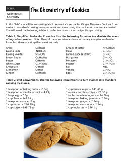

© Copyright 2026