
Cell tracking in cardiac repair: what to image MOLECULAR IMAGING Alessandro Ruggiero
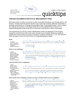
Eur Radiol
DOI 10.1007/s00330-011-2190-7
MOLECULAR IMAGING
Cell tracking in cardiac repair: what to image
and how to image
Alessandro Ruggiero & Daniel L. J. Thorek &
Jamal Guenoun & Gabriel P. Krestin &
Monique R. Bernsen
Received: 4 February 2011 / Revised: 21 April 2011 / Accepted: 9 May 2011
# The Author(s) 2011. This article is published with open access at Springerlink.com
Abstract Stem cell therapies hold the great promise and
interest for cardiac regeneration among scientists, clinicians
and patients. However, advancement and distillation of a
standard treatment regimen are not yet finalised. Into this
breach step recent developments in the imaging biosciences.
Thus far, these technical and protocol refinements have played
a critical role not only in the evaluation of the recovery of
cardiac function but also in providing important insights into
the mechanism of action of stem cells. Molecular imaging, in
its many forms, has rapidly become a necessary tool for the
validation and optimisation of stem cell engrafting strategies
in preclinical studies. These include a suite of radionuclide,
magnetic resonance and optical imaging strategies to evaluate
non-invasively the fate of transplanted cells. In this review, we
highlight the state-of-the-art of the various imaging techniques for cardiac stem cell presenting the strengths and
limitations of each approach, with a particular focus on
clinical applicability.
Keywords Cell tracking . Stem cells . Myocardial
infarction . Heart failure . Molecular imaging
A. Ruggiero (*) : J. Guenoun : G. P. Krestin : M. R. Bernsen
Department of Radiology,
Erasmus MC—University Medical Center,
Dr. Molewaterplein 50,
Rotterdam 3015GE, The Netherlands
e-mail: [email protected]
D. L. J. Thorek
Department of Radiology,
Memorial Sloan-Kettering Cancer Center,
New York, NY, USA
M. R. Bernsen
Department of Nuclear Medicine, Erasmus MC,
Rotterdam, The Netherlands
Introduction
In the last decade a great amount of research and clinical
interest has been directed at stem cells (SC) for their
potential to regenerate otherwise permanently damaged
tissues. Work with these pluripotent cells has begun to be
broadly explored, giving new hope for regenerative
approaches in the therapy of myocardial infarction (MI).
Early success in preclinical studies demonstrated that
stem cell-based therapy holds the potential to limit the
functional degradation of cardiac function after MI [1]. This
instigated clinical translation at a rapid pace (Table 1). Since
the first study in 2002 which showed safety and effectiveness on intracoronary transplantation of autologous SC [2],
several randomised, controlled clinical trials have been
performed. Due to the absence of standardised protocols
(cell number, timing and route of injection, baseline patient
characteristics and techniques of evaluating cardiac function), results have been mixed. However, recent metaanalyses have shown that improvement in ejection fraction
(EF), ventricular dimension and infarct area, despite being
modest, are statistically significant [3–5].
This field clearly benefited from the advancements in
imaging sciences as almost all clinical trials involved the
use of one or more imaging techniques to evaluate the
therapeutic efficacy of stem cell transplantion. Clinically
established techniques allow for the evaluation of myocardial contractility, viability and perfusion, but do not provide
the direct visualisation of transplanted stem cells, therefore
their effective presence and viability can be only presumed.
Ideally, transplanted cells in the infarcted myocardium are
expected to survive engraftment, be self-renewing and
differentiate into cardiac cells (cardiomyocytes, endothelial
cells or smooth muscle cells) forming electromechanical
junctions with adjacent viable tissues. However, the long-
204
Dill T et al. [110]
term improvement appears to be most closely related to
paracrine effects rather than transdifferentiation of the cell
transplant and heart muscle regeneration [6].
Great strides in imaging techniques and technologies have
been made that enable the cellular and molecular imaging of
transplanted stem cells, their short and long term fate and in
some instances their viability and differentiation status.
Stem cells for cardiac repair
BMC, bone marrow stem cells; CPC, circulating progenitor cells; SMB, skeletal myoblast. Pts, number of patients. LVEF, left ventricular ejection fraction
In the BMC group, EF increased significantly by 3.2±1.3 absolute percentage points at 4 months, and this increase was
sustained at 12 months (+3.4±1.3 absolute percentage points vs baseline). In the placebo group, EF was unchanged
(+0.6±1.2 absolute percentage points, at 12 months.
MRI
69
Chen et al. [109]
Intracoronary BMC vs placebo
At 3 months LVEF significantly increased in the BMSC group (67±11%) compared to controls (53±8%) and the same
group before implantation (49±9%). No change in LVEF at 6 months versus 3 months.
F-FDG; Echo
18
No improvement in regional or global LV function at 6 months.
Echo
97
MAGIC [8]
SMB vs placebo injected in and
around the scar
Intracoronary BMSC (bone marrow
mesenchymal stem cells) vs placebo
LVEF improved in the group receiving the highest dose (108 cells) by 6%, 7%, and 7% at months 3, 6, and 12,
respectively.
Echo; 99mTc-SPECT;
18
F-FDG PET
60
Meluzin et al. [108]
Intracoronary BMC (high and
low doser) vs control
At 4 months LV angiography showed significant increase of LVEF (50±10% to 58±10%), and significant decrease of
end-systolic volumes (54±19 ml to 44±20 ml) without differences between the two cell groups. At 12 months MRI
showed reduced infarct size and absence of reactive hypertrophy.
LV angiography; MRI
59
TOPCARE-AMI [16]
Intracoronary BMC vs CPC
At 4 months no effect on LVEF and LV volumes. Reduction of infarct volume (measured by serial contrast-enhanced
MRI) was greater in BMC patients than in controls.
MRI; [11C] acetate PET
67
Janssens et al. [107]
Intracoronary BMC vs placebo
At 6 months global LVEF increase (6.7%). No effects at 18 months and 5 years.
No effect on global left ventricular function at 6 months and 3 years.
Tc-SPECT; echo; MRI
MRI
60
BOOST[105, 106]
Intracoronary BMC vs control
99m
100
ASTAMI [103, 104]
Intracoronary BMC vs control
At 4 months LVEF increased in BMC vs placebo (mean±SD) increase, (5.5±7.3% vs. 3.0±6.5%; P=0.01). At
12 months: death, recurrence of myocardial infarction, rehospitalization for heart failure significantly reduced.
LV angiography
204
REPAIR-AMI [101, 102]
Intracoronary BMC vs placebo
Outcome
Assessment method
Cell type
Pts
Study
Table 1 Selected randomised clinical trials (>50 patients) of stem cell transplantation following myocardial infarction
Eur Radiol
Currently adult stem cells, embryonic stem cells (ESC) and
induced pluripotent stem cells (iPS) can be used to
regenerate heart tissue. Adult stem cells comprise skeletal
myoblasts (SM), mesenchymal stem cells (MSC), bone
marrow- derived stem cells (BMC), endothelial (EPC) and
cardiac progenitor (CPC) cells. SM were the first option
to be used in stem cell transplant as they are available from
an autologous source (therefore lacking ethical or immunogenicity issues), and have been demonstrated to provide
functional benefit after myocardial infarction in animals
[7]. However, in a recent clinical trial no sustained benefit
in the global EF was observed and increased number of
early postoperative arrhythmic events was reported [8].
BMC transplantation has been shown to improve heart
function in animal models [1, 9]. However, others have
identified that most of the cells injected adopted a mature
haematopoietic transformation and only a small number of
cardiomyocytes expressed the genetic markers of the transplanted cells [10, 11]. Mesenchymal stem cells (MSC)
constitute the stromal compartment of bone marrow and,
importantly, are not hematopoietic. These are able to
differentiate into a variety of cell types [12] and improvement
in whole heart function has been described in a swine model
of myocardial infarction [13]. Visceral and subcutaneous
adipose tissue have been shown to contain vascular/adipocyte
progenitor cells and adult multipotent mesenchymal cells
(adipose tissue-derived stromal cells [14]). ASC have been
reported to improve left ventricular function in animal models
of myocardial infarction [15]. The circulating endothelial
progenitor cells (EPC) represent a more accessible source of
autologous SC and have been used in clinical trials [16]. The
existence of a subpopulation of resident cardiac SC (RCSC)
has been reported that is self-renewing, clonogenic and
multipotent, capable of differentiating in myocytes, smooth
muscle and endothelial cells [17]. Promising results have
been reported in preclinical studies [18], and results of phase I
clinical trials, started in 2009, are awaited with interest.
To date, ESC-derived cardiomyocytes [19] and ESCderived endothelial cells [20] have been successfully used
to treat heart disease in animal models. However several
problems are related to their use, including immunological
incompatibility with the host [21], the tendency to form
teratomas [22] and ethical controversies. Several studies
Eur Radiol
have been performed to manipulate the expression of
transcription factors with the goal of transforming somatic
cells (derived from an autologous source, such as keratinocytes and fat stromal cells) into induced pluripotent stem
cells (iPS) [23]. These cells possess the same advantages as
ESC, without the associated immunological and ethical
complications. Cardiomyocytes have been successfully
obtained from iPS in vitro [24] and their transplantation in
animal models of infarction resulted in improved myocardial
function [25].
To summarise, ESC and iPS have the greater potential
for cardiomyogenesis, while the formation of new cardiomyocytes by transdifferentiation of SM and BMC has so far
not been supported with convincing evidence. It should be
noted that several studies have reported moderate improvements in whole cardiac function after transplantation of SM
and BMC [7, 26]. It has been demonstrated that SCs are
responsible for paracrine effects, consisting of the release of
various cytokines or growth factors (eg. VEGF, bFGF) that
increase collateral perfusion and neoangiogenesis and influence the contractile characteristics of chronically failing
myocardium [26].
pre-clinical and clinical imaging techniques have been
leveraged towards this goal; each providing unique advantages and limitations. Figure 1 illustrates the major
paradigms for the labelling of SC for detection by the
various imaging approaches. Table 2 reports the most
important preclinical studies in the field. Table 3 summarises
the most relevant features of each imaging technique.
Imaging of stem cells
The ability to image and monitor the biodistribution,
viability and possibly the differentiation status of implanted
SCs is of massive clinical and research benefit. All of the
Superparamagnetic iron oxide (SPIO) nanoparticles provide
labelled cells with a large magnetic moment and are
detectable by MR imaging devices or benchtop relaxometers. SPIO functions by acting as magnetic inhomogeneities, locally disturbing the magnetic field. This
Fig. 1 Schematic representation of the current technologies available
for stem cell (SC) tracking. Before implantation SC can be passively
loaded with: a superparamagnetic nanoparticles that allow for the MR
detection of labelled cells as areas of signal loss; b radiolabelled PET
or SPECT probes. c Reporter gene approaches consist of the
introduction through viral or non-viral-vectors of a reporter gene driven
by a constitutive or inducible promoter. The reporter gene undergoes
transcription to mRNA, which is translated into a protein that can be: 1)
an enzyme (as HSV1-tk or luciferase), 2) a receptor (as transferrin
receptor or hSSTR [human somatostatin receptor]) 3) a transporter
(hNIS [human sodium iodide symporter]) 4) intracellular iron storage
protein (ferritin). When a complementary reporter probe is administered,
it concentrates or activates only at the site where the reporter gene is
expressed. The level of probe accumulation is proportional to the level
of reporter gene expression and can be monitored to evaluate the
number of cells or the induction of a specific reporter gene
MRI
Magnetic resonance imaging (MRI) is a widely established
technique for the evaluation of cardiac anatomy and
function, often through the addition of paramagnetic
contrast material [27]. Taking advantage of its excellent
spatial (10–100 μm [small animal MRI]); 500–1500 μm
[clinical]) and temporal resolution SC labelled with superparamagnetic and paramagnetic agents can be visualised
[28, 29]. Multispectral non-1H MR imaging (specifically 19F)
has also been exploited to enable tracking of transplanted
cells.
Superparamagnetic iron oxide nanoparticles
EPC
Mitchell et al. [66]
rBMC
hEPC
Chapon et al. [115] rat
rat
rat
Higuchi et al. [99]
Li et al. [116]
In-tropolone
In-oxine
In-oxine
I; hNIS
124
Fluc + D-Luciferin; 18F-FDG PET; Intramyocardial -direct
echocardiography; MRI
SPIO;
Intramyocardial -direct
Intramyocardial -direct
Intramyocardial -direct
SPIO; NIS +124I
F-FHBG
Different retention values were observed at 1 h after injection of cells with normal condition
(17.8%±7.3), arrested heart (75.8%±18.3), adenosine injection (35.4%±5.3) and adenosine
plus fibrin glue (39.3%±11.6).
Viability at day 6 after intramyocardial injection was calculated to be 75%.
In-oxine. At 96 h only
Implanted cells detected up to 7 weeks by bioluminescence. No improvement in cardiac
function assessed by 18F-FDG PET, MRI, echocardiogram and invasive hemodynamic
pressure volume-analysis.
Rapid decrease of 124I uptake after day 3. Signal not detectable at day 7. MRI signal void
remained unchanged throughout the follow-up period. Histology confirmed the presence of
transplanted cells on day 1 but not on day 7, when iron was contained only in resident
macrophages.
MRI detection of SPIO labelled cells grafted in the heart up to 6 weeks, confirmed by
hystology. At 1 week increased 18 F-FDG uptake in BMC implanted heart vs control. No
improvement of heart function.
Increasing 18 F -FHBG uptake up to 4 weeks. Most of the SPIO were contained in
infiltrating macrophages at week 4. Teratoma formation. Increased LVEF. Only <0.5% of the
implanted cell were cardiomyocytes.
Detection up to 6 days after injection and their presence validated by ex vivo imaging and
qPCR.
Intramyocardial -percutaneous 15 days after intramyocardial injection SPECT/CT imaging demonstrated comparable degrees
of retention: 57%±15 for the subepicardial injections and 54%±26 for the subendocardial
injections.
Intramyocardial -direct
Intramyocardial -direct
111
Significant lung activity that obscured the assessment of myocardial cell tracking.
Signal void persisted after 3 weeks in both syngeneic and xenogeneic cell implantation.
Immunohistochemistry identifies the iron containing cells as macrophages.
Intracavitary (left ventriculum) Impairment of cell proliferation and differentiation induced by
1% of radioactivity was detected in the heart.
Intravenous
Intramyocardial -direct
Detection up to 4 weeks by MRI. LVEF identical between the tranplanted group and control.
At 4 weeks after injection, most of the transplanted labelled MSCs did not survive and their
iron content was engulfed by resident macrophages. Injection of labelled or unlabelled
cells attenuate ventricular dilatation and dysfunction after MI.
Intramyocardial –direct
Intramyocardial -direct
No improvement in LEVF. Detection of tranplanted cells up to 16 weeks confirmed by MR
and immunofluorescence.
Intramyocardial –direct
Intramyocardial- percutaneous Gradual loss of intensity of the SPIO label but retection of tranplanted cells (42.4%±15)
at 8 weeks.
Intramyocardial -direct
18
Results
Intramyocardial -percutaneous Detection of tranplanted cells (25.8%) up to 3 weeks.
Delivery
F-FDG
18
SPIO; HSV1-tk+
Tc,
In-tropolone
99m
111
F-FDG
18
111
111
111
SPIO
SPIO
SPIO
GFP-SPIO
SPIO
SPIO
Detection method
BMC (bone marrow derived Stem Cells); MSC mesenchymal stem cells; mESC (mouse embryonic stem cells); hCDC (human cardiac derived stem cells), rCDC (rat cardiac derived stem cells);
HPC (hematopoietic progenitor cells); hEPC, human Endothelial Progenitor Cells; RCSC (resident cardiac stem cells); NIS (sodium iodide symporter); Fluc (firefly luciferase); hNIS (human
sodium-iodide symporter; MI, Myocardial infarction
RCSC
mESC
rat
Qiao et al. [98]
rCDC
rat
Terrovitis et al..
Multimodal
rCDC
Terrovitis et a.. [82] rat
dog
BMC
dog
Blackwood [65]
HPC-CD34
+
rat
Brenner et al.[63]
MSC
swine
Chin et al. [62]
Radionuclide
Terrovitis et al. [45] rat
hCDCrCDC
mESC
Ebert et al. [114]
mice
MSC
Amsalem et al. [43] rat
MSC
swine
BMC
MSC
swine
Species Cell type
Stuckey et al. [113] rat
Kraitchman
et al. [111]
Amado et al. [112]
MRI
Study
Table 2 Selected cell tracking studies of SC
Eur Radiol
FRI: 2–3 mm;
FMT: 1 mm
Fluorescence
Imaging
Cells labeled with
near-infrared probes
(fluorochromes,
Quantum dots, etc.)
Cells transduced to
express luciferase
Cell Manipulation
Advantages
High spatial resolution; high
soft tissue contrast;
functional imaging
High sensitivity,
translational; assessment
viability
High sensitivity, long term
cell tracking
NIR, Near-Infra red imaging; FRI, Fluorescence reflectance imaiging; FMT, Fluorescence molecular tomography
Direct imaging or after
High spatial resolution; high
injection of iron oxides
soft tissue contrast;
(transferrin receptor,
functional imaging; no
ferritin)
probe dilution;
Residence, homing,
After systemic injection
viability, differentiation,
of correspondent
quantification,
radiolabeled probe
( 99mTc, etc.)
Residence, homing,
Direct imaging
migration,
quantification
Residence, homing,
quantification
After systemic injection
of correspondent
radiolabelled probe
(18F-FHBG,
18
F-FEAU, etc.)
Direct imaging
Residence, homing,
differentiation,
quantification,
Low sensitivity; need to
transduce cells; potential
immunogenicity
Relatively low sensitivity;
long scanning times;
probe dilution upon cell
proliferation; persistence
of SPIO after cell death
(macrophage)
Radiation; need to
transduce cells; potential
immunogenicity
Radiation; only short term
cell tracking
Radiation; need to
transduce cells; potential
immunogenicity
High sensitivity, long term
cell tracking; assessment
viability
Direct imaging
Residence, homing,
quantification
Not suitable for clinical
translation; relatively
low spatial resolution
Disadvantages
Easy, high sensitivity, high- Not suitable for clinical
throughput, low cost;
translation; surface
assessment of cell viability
imaging; relatively low
spatial resolution;
requires completely dark
environment
High sensitivity, translational Radiation; only short term
cell tracking
Direct imaging; at NIR
Multiplexed imaging
wavelenghts can image
deep tissue
How to image
Residence, homing,
After systemic injection
viability, differentiation,
of D-Luciferine or
quantification
Coelenterazine
Residence, homing,
quantification
(FMT)
What to image
Cells transduced to
Residence, homing,
express MRI reporter
quantification,
genes β-galactosidase,
migration,
transferrin receptor,
differentiation
ferritin, MagA and
lysine-rich proteins
Cells labeled with Iron
Oxides; Gd or Mn
chelates;
perfluorocarbon (19F)
10−10–10−11 Cells labelled with
99m
Tc-, 111In-labelled
compounds
Cells transduced to
express reporter
genes (hNIS )
10−11–10−12 Cells loaded with
18
F-FDG; 64Cu
labelled compounds
Cells transduced to
express PET reporter
genes (HSV1tk,
HSV1-sr39tk )
10−15–10−17
10−9–10−12
Sensitivity
(mol/L)
0.01–0.1
10−3–10−5
(small animal);
0.5–1.5 (clinical)
0.5-2 (μSPECT);
7–15 (clinical
SPECT)
SPECT
MRI
1-2 (μPET);
6–10
(clinical PET)
PET
Bioluminescence 3–5
Imaging
Spatial resolution
(mm)
Imaging
modality
Table 3 What to image and how to image
Eur Radiol
Eur Radiol
leads to enhanced dephasing of protons, resulting in
decreased signal intensity on T2-weighted and T2*-weighted
images (Figs. 2 and 3). These nanoparticles often consist of a
core of iron oxide (magnetite and/or maghemite) with a
polymeric or polysaccharide coating. They are widely
viewed to be biocompatible, have a limited effect on cell
function and can be synthesized to be biodegradable.
According to their size (diameter), these are classified as
ultrasmall paramagnetic iron oxide (USPIO, <10 nm),
monocrystalline iron oxide particles (MION, or crosslinked CLIO; 10–30 nm), standard superparamagnetic iron
oxide (SPIO; 60–150 nm) and micron-sized iron oxide
particles (MPIO, 0.7–1.6 μm). Of note, ferucarbotran
(Resovist®; Bayer Schering Pharma, Berlin, Germany) and
ferumoxides (Feridex I.V.®, Advanced Magnetic Industries, Cambridge, Maryland, USA; Endorem®, Guerbet,
Gorinchem, the Netherlands) have been approved by the
FDA for contrast enhanced-MRI imaging of liver tumors
[30] and metastatic involvement of lymph nodes [31].
Cell uptake is mediated through the size and electrostatic
charge conditions of the SPIO [32], schematically illustrated in Fig. 1a. Further, loading can be augmented through
the addition of cell penetrating peptides, electroporation or
transfection agents [33].
Studies reported that SPIOs do not affect cell viability,
proliferation, differentiation or migration [34–38]. However, recent work has raised several concerns, such as
decreased MSC migration and colony-formation ability
[39], and interference with cell function [40, 41]. A major
issue beyond potential cellular effects is the question of
contrast specificity to the presence of cells. Namely, the
hypointense signal is maintained at a site regardless of cell
viability and SPIO are present not necessarily within
implanted SC at longer time points [42], but rather in
phagocytosing monocytes following SC death [43]. Recently,
Winter et al. reported the absence of any discrimination
between healthy successfully engrafted SC and dead SC
phagocytosed by macrophages within the heart. In particular,
no differences in signal voids up to more than 40 days were
observed with dead and viable cells recipient with respect to
size, number and localisation [44]. Similarly, it has been
demonstrated that MRI overestimates the SPIO labelled SC
survival after transplantation in the heart [45]. Furthermore,
SPIO-induced hypointensity can sometimes be difficult to
interpret because it may be obscured by the presence of
endogenous blood derivates, such as hemosiderin [46]. The
clinical translation of SPIO for cell tracking is further
reduced now that ferumoxides (Feridex® or Endorem®) are
no longer available in the USA and Europe. However, the
use of iron oxides approaches should not be discouraged
as they provide very high sensitivities. New compounds
with improved tissue clearance properties (therefore
higher specificities) are awaited from material sciences
research.
Fig. 2 Anatomical and functional MR evaluation after transplantion
of adipose-derived stem cell (ASC) and relative controls: cell culture
medium (CCM), and untreated hearts. The CCM-treated and untreated
hearts showed evident thinning in the anterior wall of the left
ventricle. From Wang, L. et al. Am J Physiol Heart Circ Physiol
297: H1020-H1031 2009 [15] (with permission)
Eur Radiol
Fig. 3 Longitudinal BLI and MRI of H9c2 cells after transplantation.
BLI shows a robust distinct heart signal on day 1 (red arrow),
compared to no discernable signal in a representative control rat
having received non-labelled cells (top panel, left). The signal
increases slightly on day 3 but decreases rapidly to near background
levels by day 6. MRI imaging of a representative rat injected with the
same amount of cells labelled with Feridex shows a large hypointense
signal (red arrow) in the anterolateral wall of the myocardium. The
size of the signal decreases slightly over time, and the signal persists
for at least 80 days post cell injection. No signal is observed in control
rat that received non-labelled cells (bottom panel, left) Chen, I. Yet al.
Mol Imaging Biol. 2009 May-Jun;11:178–87. [42] (with permission)
Paramagnetic ions
have recently been used to label and track implanted glioma
cells. Of considerable interest, the feasibility of successfully
tracking two cell populations simultaneously has been
suggested, where one is labelled with Mn-Oxide and the
other with SPIOs [52]. These and other paramagnetic ion
techniques offer the hope that positive contrast approaches
will enable sensitive MR tracking of SC in vivo. Novel
nanotechnology approaches are becoming available for stem
cell tracking such as gadolinium-containing carbon nanocapsules (Gadonanotubes), whose T1 relaxivity is greater
than that of any known material to date (outperforming
clinically available Gd-based contrast agents by 40-fold)
[53]. They will definitely play a role in the future of
imaging sciences as soon as their toxicity profiles,
currently under investigation, have been clarified.
Cell labelling with “positive contrast” such as gadolinium
(Gd) chelates and manganese (Mn) chloride compounds
allow the visualisation of SC as hyperintense signal on
T1-weighted images. Internalization of Gd can be
accomplished by exposure of cells to Gd chelates or
through the use of liposomal formulations [33]. MRI
sensitivity in the detection of Gd-labelled cells is lower
compared to SPIO-labelled cells and is dependent upon
contrast behaviour and relaxivity in the cellular compartment (endosomes) in which they are localised [47, 48].
This is a result of the reduced water accessibility to
chelated ions following intracellular concentration, resulting
in decreased relaxivity. To overcome these issues several
approaches have been considered to drive the endosomal
escape of paramagnetic compounds [49]. Furthermore, safety
issues might be related to the rapid dechelation of
compounds at the low pH of lysosomes and endosomes
raising concerns related to free Gd3+ ions [50].
Sub-millimolar concentrations of Mn chloride (MnCl2)
have been sufficient to enable SC labelling and detection
for both in vitro and in vivo MR, with no detectable toxicity.
Also in the same study the potential of MnCl2 labelling in the
assessment of SC viability by T1 and T2 mapping was
investigated in in vitro studies [51]. Mn-oxide nanoparticles
19
F MR
Similar to the imaging of relaxation of 1H from water, 19F
can be used as the basis of the signal for MR
spectroscopy and image formation. While this technique
is not implemented widely in the clinic, there are unique
advantages to fluorine-MR that make it an attractive
option for SC tracking in myocardial applications. 19F is
not present naturally in soft tissues therefore its signal is
exclusively derived from the exogenous contrast agent
Eur Radiol
applied, be it a perfluorocarbon particle or fluorinated
nucleosides [54]. 19F MRI can be used with existing 1H
imaging hardware since 19F and 1H gyromagnetic ratios
differ by only 6%.
Importantly, 19F signal can be overlaid on 1H-MR
anatomical images for a highly selective, high-resolution
map of cell transplantation. This technique allows for
quantitative determination of the cell population [55]. A
perfluorocarbon particle loaded cell scheme has been used
to show the unequivocal and unique signature for SC,
enabling spatial cell localization via19F- MRI and quantitation via 19F-spectroscopy [56]. Perfluorocarbons have
been extensively studied and used as blood substitutes,
therefore their toxicity profiles are known. 19F cell tracking
has attracted interest, but is still at an early stage of
development. It should be noted that this method does
suffer from the drawback of lower sensitivity requiring
longer imaging times. Efforts are underway to address these
deficits including imaging hardware, imaging sequences,
and label improvement and 19F MR imaging is expected to
play a role in cell tracking in the future [54].
Radionuclide imaging
Imaging of SC has also taken advantage of the high
sensitivity (10-10−10−12 mol/L vs 10−3–10−5 mol/L of MRI)
and quantitative (acute cell retention as a percentage of the
net injected dose per weight, [%ID/g]) characteristics of
radionuclide imaging [57]. However, PET and particularly
SPECT have inferior spatial resolution (1–2 mm) compared
with MRI. Moreover, radionuclide-labelled cells can only
be visualised as long as the radioactivity is still detectable
(e.g. 18F: 110 min; 111In: 2.8 days; 99mTc: 6 h). This sets an
appreciable limitation on the radioisotopes direct labelling value for medium- and long-term SC transplant
monitoring. SPECT has been largely used to investigate
the short-term fate of transplanted cells labelled with
radioactive compounds such as 111In-oxine [58–63], 99mTchexamethylprophylene amine oxine (HMPAO) [64] or 111Intropolone [65, 66]. A persistent limitation for deployment of
SPECT is that in order to generate useful (quantitative)
images within a reasonable time frame, the administration of relatively large doses of radioactivity are
required. This poses the concern of inherent radiation
damage (reduced viability and proliferation). In the case
of 111In, Auger electrons are also emitted leading to
adverse biological effects in very short distances (from the
nm to μm range). Brenner et al., demonstrated that despite
the homing of progenitor cells into the infarction area, cell
labelling with 111In-oxine impairs significantly the viability,
proliferation and differentiation at 48 h after implantation
[63]. Similar results were observed after exposure of murine
haematopoietic progenitor cells at even much lower levels of
radioactivity [67]. The use of other compounds, such as
In-tropolone, inhibited cell proliferation 3 days after
labeling [68]. To abrogate these effects, it has been suggested
that only a fraction of the SC population be labelled [69].
Regardless of the method used, very few studies have
reported the absence of any cell function impairment [58,
62]. These studies underline the need for further in vitro
studies considering different SC, exposed to different activities and importantly following the same labelling protocol.
Positron emission tomography (PET) has been regarded
as having higher sensitivity (2 to 3 orders of magnitude)
and better spatial and temporal resolution than SPECT [70].
18
F-Fluorodeoxyglucose (18F-FDG) has been used for cell
labelling and short term imaging in preclinical [71] and
clinical settings (Fig. 4) [72, 73]. After intracoronary
injection all stem cells showed poor engraftment regardless
of cell type and number of implanted cells [61, 72, 73]. In
all cases intravenous injection of SC did not show
detectable homing of cells to the myocardium [72, 73].
Augmenting the higher sensitivity, the wider availability
of hybrid PET-CT systems allows for a combination of
anatomical non-invasive coronary angiography and cell
tracking. This multimodal imaging capability and clinical
availability are tempered somewhat by the the short half life
of 18F. Isotopes with longer half life, such as 64Cu (12.7 h)
111
Fig. 4 PET/CT images of a patient with hystory of anterior wall
infarction. After percutaneous intervention 18 F-FDG labelled cells
were implanted via intracoronary catheter and images obtained at 2 hrs
after the procedure. Total amount of SC at the injection site was
measured (2.1% of injected dose). From Kang et al. J Nucl Med 2006;
47:1295–1301. [73] (with permission)
Eur Radiol
have been suggested [74]. However, with radionuclide
based techniques pursued so far, only the immediate fate of
transplanted stem cells can be interrogated.
Reporter genes
Reporter gene approaches have significant potential to reveal
insights into the mechanisms and fate of SC therapies. The
reporter gene paradigm requires often the appropriate
combination of reporter transgene and a reporter probe, such
that the reporter gene product has to interact with an imaging
probe (optical, nuclear, magnetic) and when this event occurs
the signal may be detected and quantified with the
corresponding imaging technique (Fig. 1c).
Several advantages of reporter gene approaches have been
described [75]. Namely, this system identifies with exquisite
specificity only viable cells (which actively contain the gene
product) and allows long term tracking of transduced SC
(circumventing issues of probe dilution with cellular proliferation). Reporter genes can be designed as “constitutive”
whose signal is “always turned on” (suitable for the
evaluation of transplantation, migration and proliferation of
stably transduced SC) or “inducible” reporter gene which is
activated and regulated by specific endogenous transcription
factors and promoters [75, 76] providing a non-invasive
readout of information regarding SC differentiation.
The most widely used reporter gene for radiotracer based
imaging is HSV1-tk (Herpes simplex virus type 1 thymidine kinase) and its mutant, the HSV1-sr39tk. Unlike
mammalian TK1, this enzyme efficiently phosphorylates
purine and pyrimidine analogues which results in trapping
and accumulation of these ligands. It has been successfully
used in association with 18F or 124I -2′-deoxy-2′-fluoro-5iodo-1-[β]-D-arabinofuranosyluracil (18F-FIAU and 124IFIAU), 18F 2′-fluoro-5-ethyl-1-[β]-D-arabinofuranosyluracil
(18F-FEAU), and 9-(4-[18F]fluoro-3-hydroxymethylbutyl)
guanine (18F-FHBG) [75, 77].
Wu et al., pioneered the reporter gene approach in the
heart by imaging in vivo transplanted cells (expressing
luciferase or HSV1-sr39tk) up to 2 weeks by 18F-FHBG
PET imaging or BLI [78]. Furthermore, Cao et al., reported
survival and proliferation (through increasing signal up
to 4 weeks) of murine ES stably transduced with a triple
fusion reporter gene, enabling simultaneous PET, bioluminescence and fluorescence imaging [79].
Despite the advantage of signal amplification (through
probe phosphorylation and accumulation within cells) of
HSV-tk based approaches, its immunogenicity might limit
use in humans [80]. To overcome this limitation, the human
mitochondrial thymidine kinase type 2 (hTK2) have been
proposed [81]. An alternative is the sodium iodide symporter
[51] as a PET and SPECT reporter gene used in conjunction
with 124I or 99mTc (pertechnetate), respectively [75, 82].
Here, despite the lack of probe/signal amplification observed
in receptor- and transporter-based techniques (as 124I or
99m
Tc are free to diffuse out of the cells), hNIS is not
immunogenic (since it is expressed in the thyroid, stomach
salivary gland, choroid plexus but not in the heart) and does
not require complex radiosynthesis of the probes. Nevertheless, in reporter gene approach for the imaging of SC-based
cardiac therapy several important issues remain. First, the
non-physiological expression of reporter gene proteins may
perturb the critical SC cellular and therapeutic functions. To
be fully reliable, this system has to guarantee the long term
expression of the reporter gene in the proliferating population. Adenoviral transfection is hampered by episomal gene
expression (the reporter gene is not integrated in the
chromatin, and because they are not replicating, they become
diluted with cell proliferation) and by immunogenicity
(leakiness of immunogenic adenoviral proteins that can lead
to an immune response) [83]. On the other hand, lentiviral
vectors accomplish the integration of the reporter gene in the
host cell chromatin allowing stable expression in dividing
cells [84] and circumventing immunogenicity [85]. Even
when lentiviral vectors are used however, transgene expression can be silenced by DNA methylation especially when
strong promoters, such as CMV, are used to drive the
expression of the reporter gene [86]. The integration of the
reporter gene within the genome has raised concerns about
the risk of mutagenesis and potential oncogenicity [87].
The imaging of differentiation in vivo was recently
investigated by Kammili et al., by employing a novel dualreporter mouse embryonic SC line. Here, enhanced yellow
fluorescent protein (EYFP) was used as a “constitutive
reporter”, and the firefly luciferase reporter as an “inducible
reporter”. This latter gene was under the control of the
cardiac sodium-calcium exchanger 1 (Ncx1) promoter
which showed increased activity upon differentiation of
SC into beating cardiomyocytes [76].
Several candidates have been proposed as MRI reporter
genes such as β-galactosidase, transferrin receptor, ferritin,
MagA and lysine-rich proteins [88, 89]. Recently, SM
engineered to express ferritin have been transplanted in
infarcted heart and detected (as decreased signal up to 25%)
up to 3 weeks [90]. Several studies have been reported with
the application of MR reporters, however, none of these
strategies have led to a significant number of follow-up
studies. This is due to the low sensitivity of MRI for
imaging of gene activity in vivo.
Optical imaging
BLI
In contrast to the immediate clinical impact of magnetic and
nuclear tomographic imaging, optical imaging techniques
Eur Radiol
such as bioluminescence, planar and fluorescence-mediated
tomography have been largely restricted to use in preclinical
models. Bioluminescence imaging is commonly used for cell
tracking in SC transplantation studies [78, 79, 91] (Figs. 3
and 5). SC are transduced with a luciferase gene and
implanted in the recipient animal. Following injection, the
probe (D-Luciferin) is oxidized only in the cells expressing
luciferase in presence of ATP, O2 and Mg2+ resulting in light
photons being emitted (which can be detected by ultrasensitive charge-coupled device [CCD] cameras). BLI has many
advantages: it is highly sensitive, quantitative, simple and
inexpensive. However, the barrier to clinical translation lies
in the inherent limitations imposed by poor tissue penetration
(1–2 cm) (allowing only surface imaging), high rates of
scattering of visible wavelength photons on the human scale
and low resolution (3–5 mm) (which hamper the exact
evaluation of the exact location of the cells) [57].
Fig. 5 Bioluminescence imaging of CD34+ cells expressing the
TGL gene (HSV1-tk, e-GFP, f-luc) and implanted in the heart of a
SCID mouse. Systemically administered luciferin is activated
(oxidized by luciferase) in the injected cells. Here we see follow-
up of implanted cells up to 52 weeks post-implantation. Measurement of emitted light in CD34+ implants is higher than in controls
(PBS injection). From Wang, J. et al. Circ Res 2010;106:1904–1911.
[91] (with permission)
Fluorescence
Direct labelling of SC with fluorescent probes for visualisation
in vitro and in vivo has been fueled by the availability of near
infrared (NIR) probes, as their spectral properties are matched
to lower tissue attenuation in the so-called NIR-window. This
provides greater signal penetration of tissue through
reduced light absorption and tissue scattering. Therefore
they have clinical potential, however limited to nearsurface or intraoperative imaging stem cell tracking.
Near infrared imaging provides high sensitivity as well
as tomographic capabilities and there is no evidence at
Eur Radiol
present that dyes released after cell death are taken up by
macrophages. Intracoronary delivery of MSC labelled with
the NIR dye IR-786 has been successfully tracked in a
swine model of myocardial infarction and sensitivity of
10.000 cells has been reported [92].
Quantum dots (QD) are a class of inorganic, fluorescent
nanoparticles that have been successfully used to label SC.
Biocompatibility of QD at low concentrations has been
demonstrated in vitro in MSC cultures [93] and the
absence of adverse effects on cell viability, proliferation or
differentiation reported [94]. One of the most attractive
qualities of these nanoparticles is their capacity for
multiplexed imaging. The tracking of different cell
populations is concurrently achieved by labelling cells
with different QDs. Multiplex optical imaging of QDlabelled embryonic stem cells have been reported up to
Fig. 6 Co-registration of MRI a and 18 F-FHBG PET b of murine
ESC transduced with HSV1-sr39tk and passively labelled with SPIO.
Images depict the presence and tracking of SC 14 days after
transplantation. This hybrid imaging c approach leverages the
advantages of each technique; the fine anatomical resolution of MR
and the specificity of nuclear imaging. From Qiao et al. Radiology
250:3, 821–829. [98] (with permission)
14 days from injection in mice [94]. Moreover, it has been
shown that single QD-MSC can be detected in histological
sections for at least 8 weeks after delivery [95]. The long-term
effects on SC functionality are still unknown, however
concerns are related to their metallic core include its exposure
or dissolution which may result in toxicity, particularly from
heavy metals such as Pb, Cd and Se [96, 97]
Fig. 7 a MRI (upper row), 124I-PET (middle row), and fusion images
13
N-NH3 (gray scale)/ 124I (colour scale) (bottom row) of rat heart
1 day after injection of EPC labelled with iron (left), NIS only
(middle), or both iron and NIS (right). Signal void of iron-labeled
HEPCs is observed by MRI whereas HEPCs expressing NIS showed
focal 124I accumulation by PET. b Consecutive myocardial sections
showing the presence of transplanted cells: autoradiography for 124I
uptake mediated by NIS reporter (left), X-galactosidase staining for
LacZ gene expression of graft cells (middle), and Prussian blue
staining for iron particle detection (right). c Mean±SD time–activity
curves after 124I administration of transplanted cell and left ventricular
blood measured by PET. From Higuchi T et al. J Nucl Med, 50:1088–
1094. [99] (with permission)
Eur Radiol
Multimodal imaging
The possibility of complementing the sensitivity of radionuclide or optical techniques with the high-resolution
anatomical information from MRI is a key player in the
clinical and research future of SC tracking. To date, the
most interesting approach has been to develop transgenic
cells that carry an optical/nuclear imaging reporter together
with passive labelling with MRI contrast agents before
administration. Qiao et al. assessed the survival and
proliferation of SPIO-labelled murine ESC transduced with
HSV1-sr39tk longitudinally (4 weeks) following injection
into the healthy or infarcted myocardium. ESCs grafted and
underwent proliferation, as shown by increasing uptake of
18
F-FHBG in PET (Fig. 6) and decreasing the size of MR
hypointense areas due to SPIO dilution. Interestingly at
week 4 the majority of SPIO labels (released upon cell
death) were phagocytized and contained in infiltrating
macrophages rather than the ESC. Despite teratoma
formation, a slight increase in left ventricular ejection
fraction in ESC-treated animals was observed, mainly as a
result of paracrine effects, as cardiac differentiation of
implanted ESC was less than 0.5% [98]. In a similar study
human EPC derived from CD34+ mononuclear cells of
umbilical cord blood were transduced with NIS reporter
gene and labelled with SPIOs. Rapid loss of viable grafted
cells was observed, as 124I PET accumulation decreased
below detection limit at 3 days after transplant. However,
MRI signal void resulting from SPIO persisted,
corresponding to retention of SPIOs within macrophages
after graft cells’ death (Fig. 7) [99]. Triple fusion reporter
gene have been widely applied for multimodality fluorescence, bioluminescence and nuclear imaging approaches
[100]. Recently, in a quad-modal optical, PET, CT and MRI
coregistration approach CD34+ cells were transduced with a
triple fusion reporter gene (e-GFP, f-Luc and HSV1-tk).
Bioluminescence imaging revealed that cells persisted in the
heart up to 12 months and MRI studies reported improvement in the left ventricular ejection fraction was preserved
up to 6 months (Fig. 5) [91].
Conclusion
Many of the approaches to image stem cells are promising
but further work is required before a wide clinical
translation becomes reality. Beyond the unresolved safety
and ethical issues, crucial questions: “What is the best route
for cell delivery ?” “What kind and how many cells ?” and
“When to inject ?” remain.
It has become clear that there is no single ‘best method’
in cell tracking. Rather there is an array of high sensitivity,
high spatial resolution and functional techniques that work
best in combination. The persistent trends in molecular
imaging are: to focus on the development of novel MRcompatible probes able to monitor and track with sufficient
sensitivity and specificity the fate of transplanted cells, new
PET/SPECT reporter genes with lessened immunogenicity
and oncogenicity issues, and the application of related
radioprobes with better pharmacokinetic profiles.
Acknowledgements This article has been supported in part by the
ENCITE (funded by the European Community under the 7th
Framework program) and by the NIH R25T CA096945.
Open Access This article is distributed under the terms of the
Creative Commons Attribution Noncommercial License which permits any noncommercial use, distribution, and reproduction in any
medium, provided the original author(s) and source are credited.
References
1. Orlic D, Kajstura J, Chimenti S et al (2001) Bone marrow cells
regenerate infarcted myocardium. Nature 410:701–705.
doi:10.1038/35070587
2. Strauer BE, Brehm M, Zeus T et al (2002) Repair of infarcted
myocardium by autologous intracoronary mononuclear bone
marrow cell transplantation in humans. Circulation 106:1913–
1918
3. Abdel-Latif A, Bolli R, Tleyjeh IM et al (2007) Adult bone
marrow-derived cells for cardiac repair: a systematic review and
meta-analysis. Arch Intern Med 167:989–997. doi:10.1001/
archinte.167.10.989
4. Lipinski MJ, Biondi-Zoccai GG, Abbate A et al (2007) Impact of
intracoronary cell therapy on left ventricular function in the
setting of acute myocardial infarction: a collaborative systematic
review and meta-analysis of controlled clinical trials. J Am Coll
Cardiol 50:1761–1767. doi:10.1016/j.jacc.2007.07.041
5. Martin-Rendon E, Brunskill SJ, Hyde CJ, Stanworth SJ, Mathur
A, Watt SM (2008) Autologous bone marrow stem cells to treat
acute myocardial infarction: a systematic review. Eur Heart J
29:1807–1818. doi:10.1093/eurheartj/ehn220
6. Korf-Klingebiel M, Kempf T, Sauer T et al (2008) Bone marrow
cells are a rich source of growth factors and cytokines:
implications for cell therapy trials after myocardial infarction.
Eur Heart J 29:2851–2858. doi:10.1093/eurheartj/ehn456
7. Taylor DA, Atkins BZ, Hungspreugs P et al (1998) Regenerating
functional myocardium: improved performance after skeletal
myoblast transplantation. Nat Med 4:929–933
8. Menasche P, Alfieri O, Janssens S et al (2008) The Myoblast
Autologous Grafting in Ischemic Cardiomyopathy (MAGIC)
trial: first randomized placebo-controlled study of myoblast
transplantation. Circulation 117:1189–1200. doi:10.1161/
CIRCULATIONAHA.107.734103
9. Yoon YS, Wecker A, Heyd L et al (2005) Clonally expanded
novel multipotent stem cells from human bone marrow regenerate myocardium after myocardial infarction. J Clin Invest
115:326–338. doi:10.1172/JCI22326
10. Balsam LB, Wagers AJ, Christensen JL, Kofidis T, Weissman IL,
Robbins RC (2004) Haematopoietic stem cells adopt mature
haematopoietic fates in ischaemic myocardium. Nature 428:668–
673. doi:10.1038/nature02460
11. Murry CE, Soonpaa MH, Reinecke H et al (2004) Haematopoietic
stem cells do not transdifferentiate into cardiac myocytes in
myocardial infarcts. Nature 428:664–668. doi:10.1038/nature02446
Eur Radiol
12. Ferrari G, Cusella-De Angelis G, Coletta M et al (1998) Muscle
regeneration by bone marrow-derived myogenic progenitors.
Science 279:1528–1530
13. Quevedo HC, Hatzistergos KE, Oskouei BN et al (2009)
Allogeneic mesenchymal stem cells restore cardiac function in
chronic ischemic cardiomyopathy via trilineage differentiating
capacity. Proc Natl Acad Sci USA 106:14022–14027.
doi:10.1073/pnas.0903201106
14. Polascik TJ, Manyak MJ, Haseman MK et al (1999) Comparison
of clinical staging algorithms and 111indium-capromab pendetide immunoscintigraphy in the prediction of lymph node
involvement in high risk prostate carcinoma patients. Cancer
85:1586–1592. doi:10.1002/(SICI)1097-0142(19990401)
85:7<1586::AID-CNCR21>3.0.CO;2-F
15. Wang L, Deng J, Tian W et al (2009) Adipose-derived stem cells
are an effective cell candidate for treatment of heart failure: an
MR imaging study of rat hearts. Am J Physiol Heart Circ Physiol
297:H1020–H1031. doi:10.1152/ajpheart.01082.2008
16. Schachinger V, Assmus B, Britten MB et al (2004) Transplantation of progenitor cells and regeneration enhancement in acute
myocardial infarction: final one-year results of the TOPCAREAMI Trial. J Am Coll Cardiol 44:1690–1699. doi:10.1016/j.
jacc.2004.08.014
17. Beltrami AP, Barlucchi L, Torella D et al (2003) Adult cardiac
stem cells are multipotent and support myocardial regeneration.
Cell 114:763–776
18. Dawn B, Stein AB, Urbanek K et al (2005) Cardiac stem
cells delivered intravascularly traverse the vessel barrier,
regenerate infarcted myocardium, and improve cardiac function. Proc Natl Acad Sci USA 102:3766–3771. doi:10.1073/
pnas.0405957102
19. Laflamme MA, Chen KY, Naumova AVet al (2007) Cardiomyocytes derived from human embryonic stem cells in pro-survival
factors enhance function of infarcted rat hearts. Nat Biotechnol
25:1015–1024. doi:10.1038/nbt1327
20. Li Z, Wu JC, Sheikh AY et al (2007) Differentiation, survival,
and function of embryonic stem cell derived endothelial cells for
ischemic heart disease. Circulation 116(Suppl):I46–I54.
doi:10.1161/CIRCULATIONAHA.106.680561
21. Swijnenburg RJ, Schrepfer S, Cao F et al (2008) In vivo imaging
of embryonic stem cells reveals patterns of survival and immune
rejection following transplantation. Stem Cells Dev 17:1023–
1029. doi:10.1089/scd.2008.0091
22. Nussbaum J, Minami E, Laflamme MA et al (2007) Transplantation of undifferentiated murine embryonic stem cells in the
heart: teratoma formation and immune response. FASEB J
21:1345–1357. doi:10.1096/fj.06-6769com
23. Yu J, Vodyanik MA, Smuga-Otto K et al (2007) Induced
pluripotent stem cell lines derived from human somatic cells.
Science 318:1917–1920. doi:10.1126/science.1151526
24. Zwi L, Caspi O, Arbel G et al (2009) Cardiomyocyte differentiation
of human induced pluripotent stem cells. Circulation 120:1513–
1523. doi:10.1161/CIRCULATIONAHA.109.868885
25. Nelson TJ, Martinez-Fernandez A, Yamada S, Perez-Terzic C, Ikeda
Y, Terzic A (2009) Repair of acute myocardial infarction by human
stemness factors induced pluripotent stem cells. Circulation
120:408–416. doi:10.1161/CIRCULATIONAHA.109.865154
26. Kocher AA, Schuster MD, Szabolcs MJ et al (2001) Neovascularization of ischemic myocardium by human bone-marrow-derived
angioblasts prevents cardiomyocyte apoptosis, reduces remodeling
and improves cardiac function. Nat Med 7:430–436. doi:10.1038/
86498
27. Karamitsos TD, Francis JM, Myerson S, Selvanayagam JB,
Neubauer S (2009) The role of cardiovascular magnetic resonance
imaging in heart failure. J Am Coll Cardiol 54:1407–1424.
doi:10.1016/j.jacc.2009.04.094
28. Chen IY, Wu JC Cardiovascular molecular imaging: focus on
clinical translation. Circulation 123:425–443. doi: 10.1161/
CIRCULATIONAHA.109.916338
29. Kiessling F (2008) Noninvasive cell tracking. Handb Exp Pharmacol (185 Pt 2):305–321. doi: 10.1007/978-3-540-77496-9_13
30. Semelka RC, Helmberger TK (2001) Contrast agents for MR
imaging of the liver. Radiology 218:27–38
31. Harisinghani MG, Barentsz J, Hahn PF et al (2003) Noninvasive
detection of clinically occult lymph-node metastases in prostate
cancer. N Engl J Med 348:2491–2499. doi:10.1056/NEJMoa022749
32. Thorek DLJ, Tsourkas A (2008) Size, charge and concentration
dependent uptake of iron oxide particles by non-phagocytic cells.
Biomaterials 29:3583–3590
33. Bernsen MR, Moelker AD, Wielopolski PA, van Tiel ST, Krestin
GP Labelling of mammalian cells for visualisation by MRI. Eur
Radiol 20:255–274. doi:10.1007/s00330-009-1540-1
34. Arbab AS, Pandit SD, Anderson SA et al (2006) Magnetic
resonance imaging and confocal microscopy studies of
magnetically labeled endothelial progenitor cells trafficking
to sites of tumor angiogenesis. Stem Cells 24:671–678.
doi:10.1634/stemcells.2005-0017
35. Hsiao JK, Chu HH, Wang YH et al (2008) Macrophage
physiological function after superparamagnetic iron oxide labeling.
NMR Biomed 21:820–829. doi:10.1002/nbm.1260
36. Delcroix GJ, Jacquart M, Lemaire L et al (2009) Mesenchymal and
neural stem cells labeled with HEDP-coated SPIO nanoparticles: in
vitro characterization and migration potential in rat brain. Brain Res
1255:18–31. doi:10.1016/j.brainres.2008.12.013
37. Farrell E, Wielopolski P, Pavljasevic P et al (2009) Cell labelling
with superparamagnetic iron oxide has no effect on chondrocyte
behaviour. Osteoarthr Cartil 17:961–967
38. Magnitsky S, Walton RM, Wolfe JH, Poptani H (2008) Magnetic
resonance imaging detects differences in migration between
primary and immortalized neural stem cells. Acad Radiol
15:1269–1281. doi:10.1016/j.acra.2008.05.003
39. Schafer R, Kehlbach R, Muller M et al (2009) Labeling of
human mesenchymal stromal cells with superparamagnetic iron
oxide leads to a decrease in migration capacity and colony
formation ability. Cytotherapy 11:68–78
40. Yang J-X, Tang W-L, Wang X-X (2010) Superparamagnetic
iron oxide nanoparticles may affect endothelial progenitor cell
migration ability and adhesion capacity. Cytotherapy 12:251–
259
41. Kostura L, Kraitchman DL, Mackay AM, Pittenger MF, Bulte
JWM (2004) Feridex labeling of mesenchymal stem cells
inhibits chondrogenesis but not adipogenesis or osteogenesis.
NMR Biomed 17:513–517
42. Chen I, Greve J, Gheysens O et al (2009) Comparison of optical
bioluminescence reporter gene and superparamagnetic iron oxide
MR contrast agent as cell markers for noninvasive imaging of
cardiac cell transplantation. Mol Imaging Biol 11:178–187
43. Amsalem Y, Mardor Y, Feinberg MS, et al (2007) Iron-Oxide
Labeling and Outcome of Transplanted Mesenchymal Stem Cells
in the Infarcted Myocardium. Circulation 116(suppl):I-38-45.
doi: 10.1161/circulationaha.106.680231
44. Winter EM, Hogers B, van der Graaf LM, Gittenberger-de Groot
AC, Poelmann RE, van der Weerd L Cell tracking using iron
oxide fails to distinguish dead from living transplanted cells in
the infarcted heart. Magn Reson Med 63:817–821. doi: 10.1002/
mrm.22094
45. Terrovitis J, Stuber M, Youssef A et al (2008) Magnetic
resonance imaging overestimates ferumoxide-labeled stem cell
survival after transplantation in the heart. Circulation 117:1555–
1562. doi:10.1161/CIRCULATIONAHA.107.732073
46. van den Bos EJ, Baks T, Moelker AD et al (2006) Magnetic
resonance imaging of haemorrhage within reperfused myocardial
Eur Radiol
47.
48.
49.
50.
51.
52.
53.
54.
55.
56.
57.
58.
59.
60.
61.
62.
63.
64.
infarcts: possible interference with iron oxide-labelled cell
tracking? Eur Heart J 27:1620–1626. doi:10.1093/eurheartj/ehl059
Brekke C, Morgan SC, Lowe AS et al (2007) The in vitro effects
of a bimodal contrast agent on cellular functions and relaxometry. NMR Biomed 20:77–89. doi:10.1002/nbm.1077
Aime S, Castelli DD, Crich SG, Gianolio E, Terreno E (2009)
Pushing the sensitivity envelope of lanthanide-based magnetic
resonance imaging (MRI) contrast agents for molecular imaging
applications. Acc Chem Res 42:822–831. doi:10.1021/
ar800192p
Gianolio E, Arena F, Strijkers GJ, Nicolay K, Hogset A, Aime S
Photochemical activation of endosomal escape of MRI-Gd-agents
in tumor cells. Magn Reson Med. doi: 10.1002/mrm.22586
Bulte JW (2009) In vivo MRI cell tracking: clinical studies. AJR
Am J Roentgenol 193:314–325. doi:10.2214/AJR.09.3107
Yamada M, Gurney PT, Chung J et al (2009) Manganeseguided cellular MRI of human embryonic stem cell and
human bone marrow stromal cell viability. Magn Reson Med
62:1047–1054
Gilad AA, Walczak P, McMahon MT et al (2008) MR tracking of
transplanted cells with “positive contrast” using manganese oxide
nanoparticles. Magn Reson Med 60:1–7. doi:10.1002/mrm.21622
Tran LA, Krishnamurthy R, Muthupillai R, et al. Gadonanotubes
as magnetic nanolabels for stem cell detection. Biomaterials
31:9482–9491. doi: 10.1016/j.biomaterials.2010.08.034
Srinivas M, Heerschap A, Ahrens ET, Figdor CG, de Vries IJ
(2010) (19)F MRI for quantitative in vivo cell tracking. Trends
Biotechnol 28:363–370. doi:10.1016/j.tibtech.2010.04.002
Srinivas M, Morel PA, Ernst LA, Laidlaw DH, Ahrens ET (2007)
Fluorine-19 MRI for visualization and quantification of cell
migration in a diabetes model. Magn Reson Med 58:725–734
Partlow KC, Chen J, Brant JA et al (2007) 19F magnetic
resonance imaging for stem/progenitor cell tracking with
multiple unique perfluorocarbon nanobeacons. FASEB J
21:1647–1654. doi:10.1096/fj.06-6505com
Massoud TF, Gambhir SS (2003) Molecular imaging in living
subjects: seeing fundamental biological processes in a new light.
Genes Dev 17:545–580. doi:10.1101/gad.1047403
Aicher A, Brenner W, Zuhayra M et al (2003) Assessment of the
tissue distribution of transplanted human endothelial progenitor
cells by radioactive labeling. Circulation 107:2134–2139.
doi:10.1161/01.CIR.0000062649.63838.C9
Barbash IM, Chouraqui P, Baron J et al (2003) Systemic
delivery of bone marrow-derived mesenchymal stem cells to
the infarcted myocardium: feasibility, cell migration, and
body distribution. Circulation 108:863–868. doi:10.1161/01.
CIR.0000084828.50310.6A
Hou D, Youssef EA, Brinton TJ et al (2005) Radiolabeled cell
distribution after intramyocardial, intracoronary, and interstitial
retrograde coronary venous delivery: implications for current
clinical trials. Circulation 112(9 Suppl):I150–I156. doi:10.1161/
CIRCULATIONAHA.104.526749
Schachinger V, Aicher A, Dobert N et al (2008) Pilot trial on
determinants of progenitor cell recruitment to the infarcted
human myocardium. Circulation 118:1425–1432. doi:10.1161/
CIRCULATIONAHA.108.777102
Chin BB, Nakamoto Y, Bulte JW, Pittenger MF, Wahl R,
Kraitchman DL (2003) 111In oxine labelled mesenchymal stem
cell SPECT after intravenous administration in myocardial
infarction. Nucl Med Commun 24:1149–1154. doi:10.1097/01.
mnm.0000101606.64255.03
Brenner W, Aicher A, Eckey T et al (2004) 111In-labeled CD34+
hematopoietic progenitor cells in a rat myocardial infarction
model. J Nucl Med 45:512–518
Penicka M, Lang O, Widimsky P et al (2007) One-day
kinetics of myocardial engraftment after intracoronary injec-
65.
66.
67.
68.
69.
70.
71.
72.
73.
74.
75.
76.
77.
78.
79.
80.
tion of bone marrow mononuclear cells in patients with acute
and chronic myocardial infarction. Heart 93:837–841.
doi:10.1136/hrt.2006.091934
Blackwood KJ, Lewden B, Wells RG et al (2009) In vivo SPECT
quantification of transplanted cell survival after engraftment
using (111)In-tropolone in infarcted canine myocardium. J Nucl
Med 50:927–935. doi:10.2967/jnumed.108.058966
Mitchell AJ, Sabondjian E, Sykes J, et al. Comparison of initial
cell retention and clearance kinetics after subendocardial or
subepicardial injections of endothelial progenitor cells in a
canine myocardial infarction model. J Nucl Med 51:413–417.
doi: 10.2967/jnumed.109.069732
Nowak B, Weber C, Schober A et al (2007) Indium-111 oxine
labelling affects the cellular integrity of haematopoietic progenitor cells. Eur J Nucl Med Mol Imaging 34:715–721.
doi:10.1007/s00259-006-0275-3
Yoon JK, Park BN, Shim WY, Shin JY, Lee G, Ahn YH In vivo
tracking of 111In-labeled bone marrow mesenchymal stem cells
in acute brain trauma model. Nucl Med Biol 37:381–388. doi:
10.1016/j.nucmedbio.2009.12.001
Gholamrezanezhad A, Mirpour S, Ardekani JM et al (2009)
Cytotoxicity of 111In-oxine on mesenchymal stem cells: a timedependent adverse effect. Nucl Med Commun 30:210–216.
doi:10.1097/MNM.0b013e328318b328
Rahmim A, Zaidi H (2008) PET versus SPECT: strengths,
limitations and challenges. Nucl Med Commun 29:193–207.
doi:10.1097/MNM.0b013e3282f3a515
Doyle B, Kemp BJ, Chareonthaitawee P et al (2007) Dynamic
tracking during intracoronary injection of 18F-FDG-labeled
progenitor cell therapy for acute myocardial infarction. J Nucl
Med 48:1708–1714. doi:10.2967/jnumed.107.042838
Hofmann M, Wollert KC, Meyer GP et al (2005) Monitoring
of bone marrow cell homing into the infarcted human
myocardium. Circulation 111:2198–2202. doi:10.1161/01.
CIR.0000163546.27639.AA
Kang WJ, Kang HJ, Kim HS, Chung JK, Lee MC, Lee DS
(2006) Tissue distribution of 18F-FDG-labeled peripheral
hematopoietic stem cells after intracoronary administration
in patients with myocardial infarction. J Nucl Med 47:1295–
1301
Adonai N, Nguyen KN, Walsh J et al (2002) Ex vivo cell
labeling with 64Cu-pyruvaldehyde-bis(N4-methylthiosemicarbazone) for imaging cell trafficking in mice with positron-emission
tomography. Proc Natl Acad Sci USA 99:3030–3035.
doi:10.1073/pnas.052709599
Serganova I, Mayer-Kukuck P, Huang R, Blasberg R (2008)
Molecular imaging: reporter gene imaging. Handb Exp Pharmacol (185 Pt 2):167–223. doi: 10.1007/978-3-540-77496-9_8
Kammili RK, Taylor DG, Xia J, et al. Generation of novel
reporter stem cells and their application for non-invasive
molecular imaging of cardiac-differentiated stem cells in vivo.
Stem Cells Dev. doi: 10.1089/scd.2009.0308
Ruggiero A, Brader P, Serganova I, et al. Different strategies for
reducing intestinal background radioactivity associated with
imaging HSV1-tk expression using established radionucleoside
probes. Mol Imaging 9:47–58.
Wu JC, Chen IY, Sundaresan G et al (2003) Molecular imaging
of cardiac cell transplantation in living animals using optical
bioluminescence and positron emission tomography. Circulation
108:1302–1305. doi:10.1161/01.CIR.0000091252.20010.6E
Cao F, Lin S, Xie X et al (2006) In vivo visualization of embryonic
stem cell survival, proliferation, and migration after cardiac
delivery. Circulation 113:1005–1014. doi:10.1161/CIRCULATIO
NAHA.105.588954
Berger C, Flowers ME, Warren EH, Riddell SR (2006) Analysis
of transgene-specific immune responses that limit the in vivo
Eur Radiol
81.
82.
83.
84.
85.
86.
87.
88.
89.
90.
91.
92.
93.
94.
95.
96.
97.
persistence of adoptively transferred HSV-TK-modified donor T
cells after allogeneic hematopoietic cell transplantation. Blood
107:2294–2302. doi:10.1182/blood-2005-08-3503
Ponomarev V, Doubrovin M, Shavrin A et al (2007) A humanderived reporter gene for noninvasive imaging in humans:
mitochondrial thymidine kinase type 2. J Nucl Med 48:819–
826. doi:10.2967/jnumed.106.036962
Terrovitis J, Kwok KF, Lautamaki R et al (2008) Ectopic
expression of the sodium-iodide symporter enables imaging of
transplanted cardiac stem cells in vivo by single-photon emission
computed tomography or positron emission tomography. J Am
Coll Cardiol 52:1652–1660. doi:10.1016/j.jacc.2008.06.051
Gray SJ, Samulski RJ (2008) Optimizing gene delivery vectors
for the treatment of heart disease. Expert Opin Biol Ther 8:911–
922. doi:10.1517/14712598.8.7.911
Ma Y, Ramezani A, Lewis R, Hawley RG, Thomson JA (2003)
High-level sustained transgene expression in human embryonic
stem cells using lentiviral vectors. Stem Cells 21:111–117.
doi:10.1634/stemcells.21-1-111
Toelen J, Deroose CM, Gijsbers R et al (2007) Fetal gene
transfer with lentiviral vectors: long-term in vivo follow-up
evaluation in a rat model. Am J Obstet Gynecol 196(352):e351–
e356. doi:10.1016/j.ajog.2007.01.038
Krishnan M, Park JM, Cao F et al (2006) Effects of epigenetic
modulation on reporter gene expression: implications for stem
cell imaging. FASEB J 20:106–108. doi:10.1096/fj.05-4551fje
Mikkers H, Berns A (2003) Retroviral insertional mutagenesis:
tagging cancer pathways. Adv Cancer Res 88:53–99
Zurkiya O, Chan AW, Hu X (2008) MagA is sufficient for
producing magnetic nanoparticles in mammalian cells, making it
an MRI reporter. Magn Reson Med 59:1225–1231. doi:10.1002/
mrm.21606
Gilad AA, McMahon MT, Walczak P et al (2007) Artificial
reporter gene providing MRI contrast based on proton exchange.
Nat Biotechnol 25:217–219. doi:10.1038/nbt1277
Naumova AV, Reinecke H, Yarnykh V, Deem J, Yuan C, Charles
EM Ferritin overexpression for noninvasive magnetic resonance
imaging-based tracking of stem cells transplanted into the heart.
Mol Imaging 9:201–210.
Wang J, Zhang S, Rabinovich B, et al. Human CD34+ cells in
experimental myocardial infarction: long-term survival, sustained functional improvement, and mechanism of action. Circ
Res 106:1904–1911. doi: 10.1161/CIRCRESAHA.110.221762
Ly HQ, Hoshino K, Pomerantseva I et al (2009) In vivo
myocardial distribution of multipotent progenitor cells following intracoronary delivery in a swine model of myocardial
infarction. Eur Heart J 30:2861–2868. doi:10.1093/eurheartj/
ehp322
Muller-Borer BJ, Collins MC, Gunst PR, Cascio WE, Kypson
AP (2007) Quantum dot labeling of mesenchymal stem cells. J
Nanobiotechnology 5:9. doi:10.1186/1477-3155-5-9
Lin S, Xie X, Patel MR et al (2007) Quantum dot imaging for
embryonic stem cells. BMC Biotechnol 7:67. doi:10.1186/14726750-7-67
Rosen AB, Kelly DJ, Schuldt AJ et al (2007) Finding fluorescent
needles in the cardiac haystack: tracking human mesenchymal
stem cells labeled with quantum dots for quantitative in vivo
three-dimensional fluorescence analysis. Stem Cells 25:2128–
2138. doi:10.1634/stemcells.2006-0722
Hardman R (2006) A toxicologic review of quantum dots:
toxicity depends on physicochemical and environmental factors.
Environ Health Perspect 114:165–172
Lovric J, Bazzi HS, Cuie Y, Fortin GR, Winnik FM, Maysinger
D (2005) Differences in subcellular distribution and toxicity of
green and red emitting CdTe quantum dots. J Mol Med 83:377–
385. doi:10.1007/s00109-004-0629-x
98. Qiao H, Zhang H, Zheng Y et al (2009) Embryonic stem cell
grafting in normal and infarcted myocardium: serial assessment
with MR imaging and PET dual detection. Radiology 250:821–
829. doi:10.1148/radiol.2503080205
99. Higuchi T, Anton M, Dumler K et al (2009) Combined reporter
gene PET and iron oxide MRI for monitoring survival and
localization of transplanted cells in the rat heart. J Nucl Med
50:1088–1094. doi:10.2967/jnumed.108.060665
100. Ray P, De A, Min JJ, Tsien RY, Gambhir SS (2004) Imaging trifusion multimodality reporter gene expression in living subjects.
Cancer Res 64:1323–1330
101. Schachinger V, Erbs S, Elsasser A et al (2006) Intracoronary bone
marrow-derived progenitor cells in acute myocardial infarction. N
Engl J Med 355:1210–1221. doi:10.1056/NEJMoa060186
102. Schachinger V, Erbs S, Elsasser A et al (2006) Improved clinical
outcome after intracoronary administration of bone-marrow-derived
progenitor cells in acute myocardial infarction: final 1-year results of
the REPAIR-AMI trial. Eur Heart J 27:2775–2783. doi:10.1093/
eurheartj/ehl388
103. Beitnes JO, Hopp E, Lunde K et al (2009) Long-term results
after intracoronary injection of autologous mononuclear bone
marrow cells in acute myocardial infarction: the ASTAMI
randomised, controlled study. Heart 95:1983–1989.
doi:10.1136/hrt.2009.178913
104. Lunde K, Solheim S, Aakhus S et al (2006) Intracoronary injection
of mononuclear bone marrow cells in acute myocardial infarction.
N Engl J Med 355:1199–1209. doi:10.1056/NEJMoa055706
105. Wollert KC, Meyer GP, Lotz J et al (2004) Intracoronary
autologous bone-marrow cell transfer after myocardial infarction: the BOOST randomised controlled clinical trial. Lancet
364:141–148. doi:10.1016/S0140-6736(04)16626-9
106. Meyer GP, Wollert KC, Lotz J et al (2009) Intracoronary bone
marrow cell transfer after myocardial infarction: 5-year followup from the randomized-controlled BOOST trial. Eur Heart J
30:2978–2984. doi:10.1093/eurheartj/ehp374
107. Janssens S, Dubois C, Bogaert J et al (2006) Autologous bone
marrow-derived stem-cell transfer in patients with ST-segment
elevation myocardial infarction: double-blind, randomised controlled trial. Lancet 367:113–121. doi:10.1016/S0140-6736(05)
67861-0
108. Meluzin J, Janousek S, Mayer J et al (2008) Three-, 6-, and 12month results of autologous transplantation of mononuclear bone
marrow cells in patients with acute myocardial infarction. Int J
Cardiol 128:185–192. doi:10.1016/j.ijcard.2007.04.098
109. Chen SL, Fang WW, Ye F et al (2004) Effect on left ventricular
function of intracoronary transplantation of autologous bone marrow
mesenchymal stem cell in patients with acute myocardial infarction.
Am J Cardiol 94:92–95. doi:10.1016/j.amjcard.2004.03.034
110. Dill T, Schachinger V, Rolf A et al (2009) Intracoronary
administration of bone marrow-derived progenitor cells improves
left ventricular function in patients at risk for adverse remodeling
after acute ST-segment elevation myocardial infarction: results of
the Reinfusion of Enriched Progenitor cells And Infarct
Remodeling in Acute Myocardial Infarction study (REPAIRAMI) cardiac magnetic resonance imaging substudy. Am Heart J
157:541–547. doi:10.1016/j.ahj.2008.11.011
111. Kraitchman DL, Heldman AW, Atalar E et al (2003) In vivo
magnetic resonance imaging of mesenchymal stem cells in
myocardial infarction. Circulation 107:2290–2293. doi:10.1161/
01.CIR.0000070931.62772.4E
112. Amado LC, Saliaris AP, Schuleri KH et al (2005) Cardiac repair
with intramyocardial injection of allogeneic mesenchymal stem
cells after myocardial infarction. Proc Natl Acad Sci USA
102:11474–11479. doi:10.1073/pnas.0504388102
113. Stuckey DJ, Carr CA, Martin-Rendon E et al (2006) Iron
particles for noninvasive monitoring of bone marrow stromal
Eur Radiol
cell engraftment into, and isolation of viable engrafted donor
cells from, the heart. Stem Cells 24:1968–1975. doi:10.1634/
stemcells.2006-0074
114. Ebert SN, Taylor DG, Nguyen HL et al (2007) Noninvasive
tracking of cardiac embryonic stem cells in vivo using magnetic
resonance imaging techniques. Stem Cells 25:2936–2944.
doi:10.1634/stemcells.2007-0216
115. Chapon C, Jackson JS, Aboagye EO, Herlihy AH, Jones WA, Bhakoo
KK (2009) An in vivo multimodal imaging study using MRI and PETof
stem cell transplantation after myocardial infarction in rats. Mol Imaging
Biol 11:31–38. doi:10.1007/s11307-008-0174-z
116. Li Z, Lee A, Huang M et al (2009) Imaging survival and function
of transplanted cardiac resident stem cells. J Am Coll Cardiol
53:1229–1240. doi:10.1016/j.jacc.2008.12.036


© Copyright 2026