
Urachal carcinoma presenting with chronic mucusuria: a case report Ioannis Efthimiou
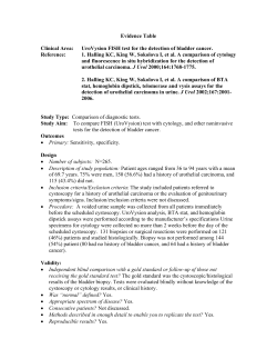
Urachal carcinoma presenting with chronic mucusuria: a case
report
Ioannis Efthimiou*, Mamoulakis Charalampos, Savas Kazoulis,
Sarantos Xirakis, Vernadakis Spiros, Ioannis Christoulakis.
General Hospital of Chania “Agios Georgios”
Chania, Crete, Greece TK 73100
Email addresses:
*Efthimiou Ioannis: [email protected]
Mamoulakis Charalampos: [email protected]
Kazoulis Savas: [email protected]
Xirakis Sarantos: [email protected]
Vernadakis Spiros: [email protected]
Christoulakis Ioannis: [email protected]
*Corresponding
Author:
Ioannis Efthimiou
Platonos 58
Peristeri, Athens
TK 12132 Greece
Phone Numbers: +302105756485
+306974874015
E-mail:[email protected]
Abstract
Urachal adenocarcinoma is a rare tumor and represents 0.17-0.34% of all
bladder tumors. It has an insidious course and variable clinical presentation.
We present a case report of a 58 year old white male with an urachal cyst
who suffered irritative voiding symptoms and long term mucusuria, since
childhood. After surgical removal of the cyst with a partial cystectomy a
mucus adenocarcinoma was diagnosed histologically.
The patient after a negative for metastatic disease screen underwent a
completion radical cystectomy with pelvic lymph node clearance. Clinicians
should have a high degree of suspicion for these rare tumors.
Introduction
Adenocarcinoma of the urachus arises from the urachal remnant. It is a rare
and devastating disease, representing 0.17-0.34% of all bladder tumors[1,
2]. It is believed that arises from malignant transformation of columnar or
glandular metaplastic epithelium. Clinically the distinction of urachal
carcinoma from other bladder adenocarcinomas may be difficult especially
if the tumor is locally extensive. Histological types include mucinous,
enteric, unspecified, signet ring-cell, and mixed variants [3]. In this case
report we present a 58 year old white male with a long lasting history of
mucusuria, recurrent bacteriuria and lower urinary tract symptoms which
finally revealed urachal adenocarcinoma. Also a short review of the
literature is presented.
Case report
A 58 year old white male presented in our department with mucusuria,
recurrent bacteriuria and lower urinary tract symptoms. He had been
treated in the past for recurrent episodes of prostatitis. He also reported
mucusuria which was present since childhood and for that reason thought it
was not unusual. He did not report any hematuria. Abdominal examination
revealed a round, smooth, firm, non tender, suprapubic mass. The prostate
gland was soft, painless and of normal size. Biochemical and haematological
analysis was normal. An abdominal computerised tomography revealed a
7x6cm round supravesical, midline cyst, with extension into the bladder
(figure 1). Antegrade cystography did not reveal any communication
between the bladder and the cyst. Rigid cystoscopy under general
anaesthesia showed a round and smooth protrusion at the level of the dome
and presence of an orifice. The cyst was filled with mucous and there were
multiple papillary lesions on the inner surface of the cyst which were
biopsied. Histology revealed a urachal adenoma, with no signs of
malignancy. The patient underwent an open transperitoneal removal of the
cyst (figure 2) and partial cystectomy. Histological examination of the
specimen revealed an invasive mucus adenocarcinoma with positive surgical
margins. The patient was further evaluated with pulmonary CT and bone
scan which were negative for metastatic disease. Subsequently he
underwent a radical cystectomy and pelvic lymph node dissection with
orthotopic neobladder construction.
Discussion
Most cases of urachal adenocarcinomas occur in the fifth and sixth decade
of life. Clinical presentation varies. Hematuria is encountered in more that
two thirds of cases. Less common symptoms are mucusuria, lower urinary
tract symptoms and a palpable suprapubic mass [4,5]. Mucusuria presents in
a 25% of the cases and merits special attention as it may be overlooked for
an extremely long time before the correct diagnosis. The most common
reason is confusion with lower urinary tract infections such as urethritis and
chronic prostatitis.
If after a trial of antibiotic therapy symptoms do not resolve, a full
urologic workup with cystoscopy and abdominal ultrasound or computerised
tomography (CT) is indicated. CT appearance is of a large mixed solid and
cystic lesion with the bulk of the tumor outside the bladder. Extravesical
spread is common. In relation to the urinary bladder wall urachal
carcinomas may be intramucosal, intramuscular or supravesival, (behind the
anterior abdominal wall in the midline) [6]. Tumor calcification is based on
mucus production which is encountered in 72%. When present it suggests a
urachal adenocarcinoma and it is usually located at the edge of the tumor
and it is patchy rather than continuous[2, 7].
Treatment consists of open radical or partial cystectomy with pelvic lymph
node dissection, and excision of the umbilicus and the urachal ligament
[1,2].
Laparoscopic partial cystectomy with lymphadenectomy has been
reported as an alternative treatment but long term follow up is required in
order to determine the oncologic effect of this treatment [8]. In a recent
study from Mayo clinic, staging with TNM system was the main predictor of
outcome after surgery for urachal adenocarcinoma [9]. Also negative margin
status has been identified as an important factor for long term-survival and
adequate local treatment is of paramount value [9, 10]. Prognosis does not
appear to be significantly influenced by histology and grade of the tumor.
Metastatic disease has a poor response to chemotherapy but some tumors
may respond to cisplatin based regimens. Overall survival for all stages is 62
months with a 34% of the patients still alive after 5 years.
Conclusion
Misdiagnosis of urachal carcinoma is still a reality. Accurate diagnosis
necessitates a degree of suspicion and appropriate imaging studies.
Appropriate local treatment is the key for improved survival.
Consent
Written informed consent was obtained from the patient for publication of
this
case report and accompanying images. A copy of the written consent is
available
for review by the Editor-in-Chief of this journal.
Competing interests
The authors declare that they have no competing interests.
Authors' contributions
EI participated in the operating theatre, was a major contributor in writing
the manuscript, and submitted the manuscript and photographs. KS
participated in the operating theatre and collected the patient data. MC
conducted a literaturew search, preparation of final manuscript including
grammarm and stule. VS participated in the operating theatre and assisted
in manuscript writing. XS participated in the operating theatre and
collected the references. CI participated in the theatre and revised the
manuscript. All authors read and approved the final manuscript.
References
1. Wilson TG, Pritchett TR, Lieskovsky G et al.: Primary adenocarcinoma
of bladder. Urology 1991, 38: 223-6.
2. Sheldon C, Clayman R, Gonzalez R, Williams R, Fraley E.: Malignant
urachal lesions. J Urol 1984; 131:1-8.
3. Ayala A, Tamboli P : Urachal carcinoma. Pathology and Genetics of
Tumours of the Urinary System and Male Genital Organs. Edited by
Eble J, Sauter G, Epstein J, Sesterhenn I. IARC Press 2004: 131-132.
4. Johnson DE, Hodge GB, Abdul-Karim FW, Ayala AG: Urachal carcinoma.
Urology 1985; 26:218-221.
5. Grignon DJ, Ro JY, Ayala AG, Johnson DE, Ordonez NG: Primary
adenocarcinoma of the bladder: a clinicopathological analysis o 72
cases. Cancer 1991; 67 2165-72.
6. Brick S, Friedman A, Pollack H. et al: Urachal carcinomas: CT findings.
Radiol 1988; 169:377-381.
7. Thali-Schwab CM, Woodward P J, Wagner B J: Computed tomographic
appearance of urachal adenocarcinomas: review of 25 cases. Eur
Radiol 2005; 15: 79–84.
8. Thyavihally YB, Tongaonkar HB: Outcomes after laparoscopic partial
cystectomy and bilateral pelvic lymph adenectomy in urachal
adenocarcinoma of the bladder. J Urol (Suppl) 2008; 4:240.
9. Molina J, Quevedo J, Furth A, et al: Predictors of survival from
Urachal cancer: A Mayo Clinic study of 49 cases. Cancer 2007; 110:
2434-2440.
10. Siefker-Radtke A, Gee J, Shen Y et al: Multimodality management of
urachal carcinoma: The M.D. Anderson Centre experience. J Urol
2003;169:1295-8
Legents
Figure 1. Contrast-enhanced CT scan at level of iliac crests shows a low
attenuation cyst overlying the anterosuperior portion of bladder with focal
calcifications.
Figure2.
bladder.
Intraoperative findings of cystic lesion at the dome of the
Figure 1
Figure 2


© Copyright 2026