
Document 18569
Infectious Diseases in Obstetrics and Gynecology 4:194-210
(C) 1996 Wiley-Liss, Inc.
(1996)
International Symposium on Immunopathogenesis
of Chlamydia Infections
December 6-8, 1996
Cornell Unive,rsity Medical College
1300 York Avenue, New York, NY
USA
Abstracts
Abstracts
Received September 15, 1996
Accepted October I, 1996
INTERNATIONAL SYMPOSIUM: IMMUNOPATHOGENESIS OF CHLAMYDIA INFECTIONS
Cytokine Profile Of Chlamydia Pneumoniae Infected U-937
Macrophages
CA Gaydos, DG Pham, L Bono, TC Quinn.
1Johns Hopkins University, Baltimore, MD, 2NIH, NIAID,
Bethesda, MD
Time
IFU/ml
cytokines in C. pneumoniae infection.
IL-I I
Titer
Control
ABSTRACTS
TNF-
IFN7
Infected
Control
Infected
6.3 x 10³
13.96
16.06
9.45
15.71
24 hr
5.05
48 hr
8.88
8.88
4.68
1.7 x 10³
19.64
1.29
1.4 x 10⁴
27.23
1.29
72 hr
48.34
48.08
96 hr
3.6 x 10³
BLD a
BLD
46.23
0
BLD
BLD
BLD
7d
a BLD= Below the level of detection. Control growth media was BLD for all 3 assays.
It has been demonstrated that C. pneumoniae can infect U-937 human macrophages and it has been postulated
that alveolar macrophages may play a role in the initial
pulmonary infectious process. Since it has been shown that
C. trachomatis can induce mediators of inflammation and
persistence and in order to begin to understand the pathogenesis of infections due to C. pneumoniae, we have investigated the cytokine response of U-937 cells infected in
vitro with C. pneumoniae, strain 2023. Non-adherent U937 macrophages (4 ml of 106/ml) were infected with ml
of 3.5 x 10⁴IFU/ml of C. pneumoniae by gentle agitation
every 20 min. for 6 hr, after which, 10 ml of growth media
without cyclohexamide was added. Flasks were sampled
by removing and freezing ml of the infected cells at 24 hr.,
48 hr, and 7 days. A control flask was treated in an identical
manner, except that uninfected growth media was used in
place of the inoculum. After the 48, 72, and 96 hr. samples,
the flasks were split 1:2, in order to maintain persistence
and viability. Titers of C. pneumoniae were determined for
each sample time point from the infected flask in Hep-2
cells. Cytokine levels were determined from the samples
from both the infected and non-infected flasks for the following: IL-I, IFNy, and TNF-. The assays were performed
by sandwich ELISA using reagents from EndogenTM. Both
titers and cytokine levels were adjusted to reflect the dilution due to splitting ofthe cells in the flasks. The results of
the viability titers and cytokine levels (pg/ml) are shown in
the table.
These results indicate that C. pneumoniae infection
in U-937 macrophages stimulate increased production of
IL- 11 by 96 hr. and IFN7 as well as TNF-x by 72 hr. Mechanisms of action of these cytokines may include, but are not
limited to, induction of adhesion molecule expression on
endothelial and epithelial cells (IL-113); enhancement of
intracellular killing, TNF cytotoxicity, and NK cell activity (IFNT); and enhanced macrophage killing and cytotoxic T cell differentiation (TNF-o). Further study will enhance our understanding of the role of these and other
Control Infected
134.18
72.56
131.43
365.56
202.51
110.22
79.41
247.82
447.71
421.59
Production Of 11-4 And IL-6 In HEp-2 Cells Infected With
Chlarnydia pneurnoniae
PM Roblin, A Kutlin, MR Hammerschlag
Department of Pediatrics, SUNY Health Science Center
@ Brooklyn, NY
Introduction" An association of Chlamydia
pneumoniae infection and reactive airway disease has been
shown in children. Anti- C. pneumoniae IgE was demonstrated by immunoblot in 85.7% of culture positive asthmatic children with wheezing compared with only 9.1%
culture positive children with pneumonia. Cytokines initiate and perpetuate the chronic inflammaation of asthma.
IL-4 plays a critical role in switching of B lymphocytes to
produce IgE, and may therefore be of critical importance in
the development of atopy. IL-6 can act synergistically with
IL-4. In this study we measured the production oflL-4 and
IL-6 from C. pneumoniae infected HEp-2 at various time
points.
Methods: Confluent monolayers of HEp-2 cells were
inoculated with seven C. pneumoniae isolates: J21, AR39,
BAL 16, CDC8, JCW480, BAL 15, TW1183, at 104 IFU/ml
in cycloheximide free media. All results were calculated
against moc-infected HEp-2 cell assayed in a sandwich
ELISA for IL-4 (Intertest-IL4, Genzyme Corp., MA) and IL6 (Biokine IL-6, T Cell Diagnostics, Inc., MA).
Results: The results are shown below:
c. pneumoniae
isolates
J21
AR39
BALI6
coc8
JCW480
BALI5
TWI83
IL-6 (pg/ml)
IL-4 (ng/ml)
24hr
48hr
72hr
33.69
28.53
14.71
26.47
8.82
26.47
11.76
39.57
41.63
32.35
II .76
26.47
5.88
17.65
58.55
45.15
38.24
8.83
38.24
47.21
41.18
24hr 48hr
0
0
0
0
0
0
0
0
0
0
0
0
0
0
72hr
0.016
0.26
0
0
0.082
0
0
INFECTIOUS DISEASES IN OBSTETRICS AND GYNECOLOGY
195
INTERNATIONAL SYMPOSIUM: IMMUNOPA THOGENESIS OF CHLAMYDIA INFECTIONS
There was little or no detectable production of IL-4.
We also assayed for production oflL-4 and IL-6 in a HEp-2
cell line chronically infected with TW183. These cells
have been maintained for approximately year without
centrifugation, cycloheximide or addition of fresh cells.
Supematants were assayed from these infected cells for IL4 and IL-6 at a "lysis" stage where a majority of cells appear
lysed and a "recovery" stage where remaining cells re-seed
and form a monolayer, at this stage cells are split into fresh
flasks. IL-4 production was 10 times greater than that seen
with "acutely" infected monolayers (0.135 ng/ml). IL-6
was 100 times higher at lysis stage, 2,044.0 pg/ml and 10
times greater at the recovery stage, 352.8 pg/ml.
Conclusions: C. pneumoniae stimulated the production oflL-6 and minimally stimulated the production oflL4 in HEp-2 cells.
Chlamydia pneumoniae Induced Atheroscler0sis in Rabbits
ABSTRACTS
fection. Purpose of our study was to determine whether this
rabbit model is suitable for studying the development of
atherosclerosis after Cpn infection. Animals used were
Bordetella bronchiseptica and Pasteurella spp. free male
New Zealand White rabbits (5 mo old). Rabbits were inoculated intranasally with a total volume of 0.5 ml of organisms (2xl 0 7 IFU/ml Cpn strain K7). Control animals
were inoculated with 0.5 ml sterile SPG medium. Rabbits
were reinfected in the same manner and with the same dose
3 weeks after primary inoculation. Rabbits were observed
daily for signs of disease and weighed twice a week. Samples
were collected 2 weeks after primary infection and 1, 2 and
4 weeks after reinfection (5 and control/each timepoint).
Lung, spleen and liver tissues were homogenized. Isolation was done in HL cell culture and inclusions were detected with FITC-conjugated chlamydia genus-specific
antibodies. Lung. spleen, liver, heart and ascending aorta
were fixed in 10% buffered formalin immediately after removal for histology and immunohistochemistry. No clinical signs of disease were observed except in two animals 24
and 48 hrs after reinfection. One animal developed severe
lung oedema and the other animal had acute pneumonia.
All sample homogenates (lung, liver, spleen) remained culture negative during the course ofthe infection. Five weeks
after primary infection two out of four rabbits developed
early signs of fibrous plaques in aortas and seven weeks
after primary infection three rabbits (3/5) developed plaques
in ascending aortas. These animals were also found positive in immunohistochemistry. Control animals had no signs
of atherosclerotic changes.
K Laitinen, P Saikku.
National Public Health Institute, Oulu, Finland
Chlamydia pneumoniae (Cpn), an obligatory intracellular, Gram-negative bacterium, is a common cause of respiratory tract infections worldwide. In addition to acute
infections it has been associated with chronic lung processes. The most important finding is evidently the association of chronic Cpn infection with cardiovascular diseases. The first serological association was published in
1988 in Finland, and after that several groups worldwide
have found the association between CHD and Cpn antibodies. Direct evidence between Cpn and CHD is demonstrated by finding Cpn organisms in atherosclerotic lesions.
It is also shown that Cpn can infect human endothelial cells seocP -A Serological’Assay For The Detection Of’
and macrophages in vitro, and according to recent evidence Chlamydia pneumoniae IgG, IgA and IgM Antibodies
these cells could carry Cpn into the smooth muscle cells A Groh, V Prankratova, M Lipson*,A Ben-Michael*, 0
below the lining of the artery. Even if the association be- Horovitz, and R MosckOvitz*.
tween Cpn infection and atherosclerosis has been confirmed Institute of Medical Microbiology, Friedrich Schiller
using several different methodological approaches by sev- University ofJena, Germany, *Department of Research
eral laboratories around the world, the causal relationship and Development, Savyon Diagnostics, Kiryat Minrav,
between chronic Cpn infection and the development of ath- Ashdod, 77101 Israel
Objective: Chlamydiapneumoniae is a widely spread
erosclerosis can be indicated only by animal experiments
and intervention trials. Mouse has already proved to be pathogen responsible for infections of the upper and lower
useful animal for the study of atherosclerosis, and the sus- respiratory tracts. Specific antibody prevalence of C.
ceptibility of artherosclerosis varies greatly between mouse pneumoniae is over 50% among adults worldwide, and is
strains. With advances in molecular biology mouse strains low in pre-school children. Recently, there has been a growwith genetic defects or overexpression of certain genes in- ing number of reports which suggest an association between
volved in lipoprotein metabolism have been created and chronic C. pneumoniae infections and coronary artery disare of great value for studies in atherogenesis. Mice are ease. Additional studies stress the role of this pathogen in
also susceptible to Cpn infection and develop latent chlamy- Adult Onset Asthma. The SeroCP has been developed for
dial lung infection. Another animal model to study the determination of C. pneumoniae IgG, IgA, and IgM anartherosclerosis is the New Zealand White (NZW) rabbit on tibodies.
Materials and Methods: SeroCP uses as antigen C.
a high cholesterol diet, and hereditary hyperlipemic
Watanabe rabbit (WHHL). Moazed et al. has recently shown pneumoniae strain TWAR 183 purified elementary bodies
that Cpn causes a moderate, self-limiting interstitial pneu- (EBs). Serum samples were obtained from 13 adult patients
monia in rabbits, and the infection mimics the human in- positive for C. peumoniae by PCR (PCRP), 28 patients posi196
INFECTIOUS DISEASES IN OBSTETRICS AND GYNECOLOGY
INTERNATIONAL SYMPOSIUM: IMMUNOPA THOGENESIS OF CHLAMYDIA INFECTIONS
tive for C. pneumoniae by MIF-IgG >1"512(CWP), 35 children negative for C. preumoniae and C. trachomatis by
MIF(CNPNT) and 49 healthy adults(HA). The C.
pneumoniae MIF were interpreted as positive for IgG > "32,
for IgA > 1:20 ad for IgM > 1:20.
Results: The prevalence (%) of C. pneumoniae antibodies in different patient groups as detected by MIF and
SeroCP, is summarized in the following table:
No. of
IgG
IgA
IgM
GROUP
PCRP
CWP
CNPNT
HA
PCRP
CWP
CNPNT
HA
PCRP
CWP
CNPNT
HA
Patients
SeroCP(%) MIF(%)
13
28
35
49
85
100
3
76
92
100
0
98
13
92
54
28
35
49
100
6
40
96
0
41
13
28
35
49
0
21
0
2
0
0
0
0
ABSTRACTS
chlamydial excretion at lambing. However, the use of a
recombinant protective antigen will be safer. This new vaccine should be as efficient as the live vaccine and should
allow the specific diagnosis of abortive strains in vaccinated
flock. In order to detect potential protective antigens, the
cellular and humoral immunity against C. psittaci abortive
strains was studied in a mouse model of systemic infection (2).
Donors mice were intravenously infected with live C.
psittaci. One month after inoculation splenic cells were
transferred to naive recipient mice, which were challenged
with 2.105 plaque forming units (pfu) of a virulent strain one
day later. Protection was assessed after 6 days by measuring
the decrease in spleen infectivity. This model showed that
lyt-2 T cells play the major role in the protection (3)
The importance ofhumoral immunity was assessed in
a pregnant mouse model. Mice were passively immunized
intravenously at day 11 of gestation with polyclonal sera or
monoclonal antibodies (mabs). Mice were challenged on
the following day, and protection was measured by placental and fetal colonization four days later. The number of
living offspring 8 days after birth was also used to assess the
protection. Results showed that immune sera and type specific mabs could transfer protection, probably by neutralizing chlamydiae during the bacteremia which proceeds placental colonization.
Therefore, mabs raised against the ovine abortive strain
AB7 were produced in order to detect molecules beating potential protective epitopes. The protective effect ofthese mabs
was tested in vitro by neutralization assays. The mabs selected
in vitro were tested for their ability to protect pregnant mice
after passive immunization. All the protective mabs were directed against heat sensitive epitopes located on an 110 kDa
(apparent molecular weight under non denaturing conditions)
oligomer of the Major Outer Membrane Protein (MOMP) of C.
psittaci strainAB7 (4). The protective mabs did not react with
the 39 kDa monomeric MOMP which is produced after heat
denaturation.
Protection experiments were performed on a pregnant
mouse model with vaccines containing mainly native
oligometric MOMP. Different formulations were tested and
mice were immunized with two injections given at 15-days
interval. They were challenged intraperitioneally at day 11 of
pregnancy (1 month after the last vaccination) with 2.105 pfu
ofthe AB7 ovine abortion strain. Protection was evaluated by
the number of living offspring per litter and by the level of
infection in the livers, fetus and placentas (5). The level of
infection in the placentas was the most discriminant test.
Mice vaccinated with non-denatured MOMP extracts
were better protected than mice which have received a heat
denatured MOMP extract. The infection of the placenta was
significantly higher for mice which have received the heat
denatured vaccine, whereas the difference seen in the number
of living offspring between the 2 groups, was less significative. Mice vaccinated with the heat denatured vaccine were
/
Conclusions: SeroCP is highly sensitive and specific
and well correlated with PCR and MIF. The MIF IgG titer
1"64 correlates with positive SeroCP IgG and MIF IgA titer
1:20 with SeroCP IgA. The ELISA based SeroCP is easy and
convenient to perform as compared to the technically demanding MIF test. In addition to IgG and IgA, SeroCP IgM
testing should be recommended for the adequate and sensitive diagnosis, when IgG and IgA antibodies are produced
in low levels.
Protective Immunity Against Chlamydia psittaci Serotype
Strains
DS Carlos, S Jesus, B Francoise, S Armel, R Annie.
IPathologie Infectieuse et Immunologic, INRA, 37380
Nouzilly, France, 2Dept. Patologia Animal Facultad
Veterinaria, Campus de Espinardo, Murcia, Spain
The serotype stains of Chlamydia psittaci induce
several diseases among ruminants. One of them, enzootic
abortion of ewes, causes significant economic losses for farmers and represents a danger for pregnant women. This disease results from the colonization ofthe placenta by Chlamydia. Transmission of the infection, in a flock or to humans,
results mainly from the massive excretion of Chlamydia
which occurs at lambing or abortion, in infected placentas
and uterine fluids.
The immunity following abortion of ewes is strong
enough to prevent abortion and chlamydial shedding during the next pregnancy. Killed vaccines, used in the past,
reduced the incidence of abortions but could not prevent
excretion. We developed a thermosensitive strain of C.
psittaci (1) which provided a live vaccine against abortive
chlamydiosis. This vaccine is able to avoid abortion and
INFECTIOUS DISEASES IN OBSTETRICS AND GYNECOLOGY
197
INTERNATIONAL SYMPOSIUM: IMMUNOPATHOGENESIS OF CHLAMYDIA INFECTIONS
not protected from placental infection but they were partially
protected from abortion. As previous works have shown that
mabs raised against monomeric denatured MOMP were not
protective, this partial protection could be the result of T
cell epitopes located on MOMP.
In definitive, the good level of protection which have
been obtained could be the result of the combination of
MOMP native B cell epitopes and MOMP T cell epitopes.
In this model, we demonstrated that placental colonization
of pregnant mice could be prevented with native oligomeric
MOMP. Mice vaccinated with native oligomeric MOMP
were protected from abortion but also from infection. The
last point is very important since chlamydial excretion at
lambing is mainly responsible ofthe contamination of other
animals.
ABSTRACTS
tions were independent of HLA class I or class II types. TNFalpha was detected more frequently in tear samples from
cases (38%) than from controls (16%), the association increasing for higher concemrations ofTNF-alpha (p=0.015).
Among cases, detectable TNF-alpha in tears was highly associated with the presence of ocular chlamydial infection
(p<0.001). These results suggest that TNF-alpha plays a
major role in the cicatricial sequelae of C. trachomatis infection in humans.
Characterization Of The Local Immune ResponseTo
Chlamydia Trachomaas Among Trachoma Patients With
Conjunctival Scarring
2S Phan, 3R Pham, 2D Dean
1Dept. ofMedicine, Division ofInfectious Diseases, and the
References
I. Proctor Foundation, University of California at
2Francis
1. A. Rodolakis: In vitro and in vivo propertics of chemically induced
San
School of Medicine, San Francisco, CaliforFrancisco
temperature sensitive mutants of Chlamydia psittaci var. ovis screening in a murine model. Infect. Immun. 42:525-530, 1983.
nia, and SSanta Clara Hospital San Jose, California
2. D. Buzoni-Gatel, A. Rodolakis, M. Plommet: T cell mediated and
Chlamydia trachomatis is the etiologic agent of trahumoral immunity in a mouse Chlamydia psittaci systemic infecchoma
which has a worldwide distribution and is the leading
tion. Res. Vet. Sci. 43:59-63, 1987.
3. D. Buzoni-Gatel, L. Guilloteau, F. Bernard, S. Bernard, T. Chardes, cause of preventable blindness in the world. Progression of
A. Rocca: Protection against Chlamydia psittaci in mice conferred disease from repeated infection in childhood is common and
by Lyt-2 T cells. Immunol. 77:284-288, 1992.
results in the sequelae of conjunctival scarring that indirectly
4. C. De Se, A. Souriau, F. Bernard, J. Salinas, A. Rodolakis: An
oligomer of the major outer membrane protein of Chlamydia psittaci leads to blindness. The immunopathogenic mechanisms for
is recognized by monoclonal antibodies which protect mice from the development of scarring are not well defined. In order to
abortion. Infect. Immun. 63:4912-4916, 1995.
address this issue, we evaluated Vietnamese trachoma patients
5. C. De Sa, F. Bernard, J. Sallinas, A. Souriau, A. Rodolakis: Chlamy- with conjunctival scarring who were undergoing corrective
dia psittaci infection could be prevented in pregnant mice with a
lid surgery for trichiasis. Control were Vietnamese individuals
vaccine containing native oligomeric major outer membrane prowith
conjunctival scarring due to other causes. Patients were
submitted.
tein,
Scarring Trachoma Is Associated With Polymorphism In
The TNF-Alpha Gene Promoter And With Elevated TnfAlpha In Tear Fluid
DJ Conway, MJ Holland, RL Bailey, AE Campbell, S
Mahdi, R Jennings, E Mbena, DCWMabey.
Department of Clinical Sciences, London School of
Hygiene & Tropical Medicine, Keppel Street, London
WCIE 7HT, UK
Polymorphisms in the promoter region of the tumor
necrosis factor (TNF) -alpha gene have recently been shown
to be associated with an increased risk of severe malaria and
of mucocutaneous leishmaniasis. In an attempt to determine the role of TNF-alpha in the fibrotic sequelae of human Chlamydia trachomatis infection, we have looked for
associations between TNF-alpha levels in ocular secretions,
and between polymorphisms in the TNF-alpha gene promoter region, and scarring trachoma in a case-control study
comparing 153 age, sex and village matched controls in a
trachoma endemic Gambian population. A higher proportion of Cases (28%) than controls (18%) had the -308A allele, particularly as homozygotes (X’- for trend, p=.032). The
trend was similar, but non-significant for the rarer -238A
allele, and highly significant for the number of either-308A
or 1238A sites in an individual (p=0.003). These associa198
examined and scored for trachoma based on the World Health
Organization trachoma grading scale. Serum was obtained for
microimmunofluorscence testing (MIF). Conjunctival swabs
were collected for DFA and PCR. Conjunctival biopsies were
obtained at the time of surgery for RT-PCR to quantitate and
type cytokines and to detect C. trachomatis, and for immunohistochemical studies. All cases revealed a grading C3 (severe
scarring with distortion of the lid). Five (36%) of 14 cases had
documented chlamydial organisms by DFA and/or PCR or RTPCR; none ofthe controls were positive for chlamydia. Immunohistochemical studies revealed a statistically significant
higher level of CD4 (p<0.04) and CD8 cells (p<0.01), and
macrophages (p<0.05) in cases than controls; there were no
difference for B cells. Five cases had elevated interferon gamma
(IFN,) levels; these 5 correlated with both a lack of demonstrable organisms by DFA, PCR, or RT-PCR, and elevated IL-2
levels (p<0.034). IL-6, and IL-12 levels were elevated among
cases but not controls (p<0.04); only 5 cases had elevated
levels for TNFa. There were no differences for IL-4. The data
reveal that chlamydia are present in trachoma cases even with
burned out disease although viability could only be confirmed
in one patient. Elevated IFN3, levels may have contributed to
elimination of the organism and lack of identification of C.
trachomatis in five cases. The cytokine profiles in this study
were characteristic ofboth Th-1 and Th-2 responses, and sug-
INFECTIOUS DISEASES IN OBSTETRICS AND GYNECOLOGY
INTERNATIONAL SYMPOSIUM: IMMUNOPATHOGENESIS OF CHLAMYDIA INFECTIONS
gest that these may be important in regulating host immune
defenses against persistent C. trachomatis infections or persistent chlamydial antigen. Further study is required ofthe cell
mediated immune response to chlamydia which will likely
impact the direction of vaccine development of tr
The Relationship Between The 60 Kd Heat Shock Protein
(HSP60),Antisperm Antibodies And Undetected
Chlamydia trachomatis Infection In Male Partners Of
Couples With Undiagnosed Infertility
MG Munoz, J Jeremlas, ss Witkin
Universidad Simon Bolivar, Caracas, Venezuela and
eCornell University Medical College, New York, New
Yorg,
us
Introduction: Heat shock protein 60 kD (HSP60) is
produced by mammalian cells in response to several stresses,
i.e. infection, inflammation and elevated temperature. The
aim of this investigation was to demonstrate the relationship between heat shock protein concentration in semen,
antisperm antibodies and Chlamydia trachomatis infection
in the male genital tract.
Materials and methods: We examined the presence
of liSP60 in semen from 64 male partners of infertile couples
with no history of C. trachomatis infection and whether its
presence was related to sperm antibodies (detected by
immunobead binding) and anti-chlamydial antibodies
(SERO ELISA, Savyon). HSP60 was detected by an enzymelinked immunosorbent assay (ELISA) using a monoclonal
antibody to HSP60 bound to wells of a microtitre plate and
a polyclonal antibody to HSP60 as the detecting antibody
(Stress Gen). HSP60-specific mRNA was detected in mononuclear cells isolated from semen by a PCR ELISA.
Results: HSP60 was detected in semen from nine
(14.1%) men, while HSP60 mRNA was present in 16 (25.0%)
samples. Spermatozoa from 12 (18.8%) men had bound
auto-antibodies, while 20 (31.3%) semen samples had antichlamydial antibodies. The presence of HSP60 in semen
was correlated with the occurrence of sperm antibodies (P=
0.008), anti-chlamydial immunoglobulin A antibodies (P=
0.002) and HSP60-specific mRNA in seminal mononuclear
cells (P= 0.03).
Conclusions: The results demonstrate that HSP60 is
produced in the male genital tract and is present in a soluble
form in semen in association with genital tract immune activation and/or a genital exposure to C. trachomatis. Lymphocyte activation apparently represents one source of
HSP60. Non-lymphoid cells or microorganisms may also
contribute to the total HSP60 level in semen. The role of
HSP60 in activating or inhibiting immune responses within
the male genital tract will be the purpose of future studies.
Chlamydia trachomatis Specific Immunoparameters And
Cytokine Production In Semen From Males Affected By
Prostatitis
ABSTRACTS
F Meacci, A SaM and S Mazzoli
S. TD. Center, Infectious Disease Unit, Santa Marie
Annunziata Hospital Florence, Italy
Introduction: The etiopathogenetic role of Chlamydia trachomatis (C.t.) has not yet been completely demonstrated in relation to deep male genital tract acute and
chronic pathologies; the micro-organism has been detected
in prostatic secretions, prostatic biopsies, in Sertoli cells
from testicular biopsies and in semen. The biology of the
micro-organism, the intracellular parasitism and metabolism, influences and determines the infection evolution towards the chronicity and the following tissue inflammation
in affected patients. In males the C.t. infection has been
associated with fertility alterations due to tissue degradation processes, functional or immune related alterations of
the genital tract or of the spermatozoa and with pathologies
of the upper genital tract such as prostatitis and infertility.
Chlamydia trachomatis has been additionally reported to
act particularly stimulatory for the appearance of the secretary (S)IgA in the genital tract. The intrinsic capacity ofthe
male genital tract to produce SIgA is well documented and
can take place both in the prostate and the epididymis,
resulting in local targeted overproduction of SIgA by submucosal plasma cells. The induction mechanisms, the regulation ofthese immunoresponses and the TH immuno-shift
in the male genital tract are poorly understood up to now.
The overproduction of SIgA could be theoretically explained by release of cytokines such as interferon gamma,
interleukin-5 and tumor necrosis factor alpa. Cytokines are
important mediators in inflammatory processes and their
production may modulate immunity during C. t. infections,
as proved by recent in vitro studies. In our previous studies
we proved elevated IL-6 concentrations and overproduction in semen of chronic prostatitis patients.
Aim of our study: We investigated on IL-6, IL-4, IL0 mucosal presence in relation to C. t. specific IgA, SIgA
and IgG content in seminal fluids of 110 men affected by
prostatitis. Additional objective was to determine the possible TH response and the possible correlation between
cytokine production and the presence of Chlamydia
trachomatis DNA in semen of the patients.
Study population" Patients’ study inclusion criteria
were the presence of acute or chronic prostatitis; the population has been select in a study period of seven months
from November 1995 to June 1996. Patients’ symptoms
were urethrorrea, pollachiuria, stranguria, haemospermia,
perineal pains, tenesmus and infertility (8.2%); patients
ranged from 21 to 73 years-old (m age=39.5). C. t. was
searched in patients semen by a modified PCR (Roche D.S.,
Germany). Specific anti C. t. IgA were detected by IP (Savyon
D. Ltd., Israel), MIF (Labsystem, Finland) an anti C. t. MOMP
ELISA test (SeroCT by Savyon D. Ltd., Israel) and IgG were
detected by SeroCT, Savyon D. Ltd., Israel. IL-6, IL-4, ILl0 were detected by research quantitative ELISA tests
INFECTIOUS DISEASES IN OBSTETRICS AND GYNECOLOGY
199
INTERNATIONAL SYMPOSIUM: IMMUNOPA THOGENESIS OF CHLAMYDIA INFECTIONS
ABSTRACTS
(Biokine, T-Cell Diagn. Inc. and Predieta Genzyme, USA).
Results: Sperm C.t. DNA was present in 13/110 patients (11.8%). IgA by MIF and HP were present in 40/110
patients (36.36%). IgA anti MOMP detected by an ELISA
test were present in 16/110 patients (14.5%). Sperm C. t.
MOMP IgG was demonstrated by SeroCT ELISA in none of
Finland
Objectives: Our aim was to study placental tissue of
a stillbirth fetus, born to a mother with high antibody titers
to Chlamydia trachomatis, using non radioactive
RNA:DNA hybridization and alkaline phosphatase-antialkaline phosphatase staining (APAAP) for C. trachomatis.
the patients excluding the possible transmembrane transloMethod: In situ hybridization
cation from sera of these antibodies or the local production
For the probe preparation for in situ hybridization, C.
demonstrating the long-term infections. Western-blot analy- trachomatis L2 plasmid DNA was subeloned into a
sis of semen and serum IgA and IgG immunoresponse polylinker site ofa pGEM-3Zf(-) vector [L2PpGEM]. RNA
proved a specific immunization versus C. t. specific pro- transcripts were synthesized using linearized and purified
teins, both in C. t. DNA positive pts. and in C.t. IgA positive L2pPGEM as a template. To shorten the probe and to facilipts. IL-6 was present at concentration > 10 pg/ml in ejacu- tate diffusion into the tissues, probes were hydrolized unlates of 10/110 (9.1%) patients; IL-10 was present at con- der alkaline conditions. Nonspecific probe was prepared
centration > 10 pg/ml in 23/110 (20.9%). A good correla- from vector [12PpGEM] by the same method as specific
tion was found between IL-6 and IL- 10 production (r=0.623) probe. Infected and uninfected McCoy cells were used to
in these patients. IL-4 was not measured at detectable lev- check performance of the probes. C. trachomatis specific
els in ejaculates of our patients. Thus may be due to its and non-specific probes were used for the detection of the
rapid metabolism.
C. trachomatis DNA in paraffin embedded tissue sections.
Conclusions: Our patients with anti C. t. IgA posiAlkaline phosphatase-Anti-Alkaline phosphatase
tive prostatitis have DNA proven "Chlamydial" prostatitis in (APAAP) staining
30.7%. They presented specific local immunization against
APAAP staining of chlamydial inclusions in tissue
the micro-organism western-blot proved, local overproduc- sections was done as previously described by Mahony (1)
tion of IgA confirming the proper immunoeompeteney of the with slight modifications and counterstained 10s in Mayer’s
genital tract. C. t. DNA positivity confirmed the presence of hematoxylin. To control the nonspecific staining, duplithe micro-organism as a possible "primum movens" of the cate tissue sections were incubated with mouse ascites fluid.
pathology.
Slides were examined by light microscope.
Concerning the IL-6 and IL-10 local production it is in
Results: C. trachomatis specific nucleic acid and
vivo well documented in our patients affected by prostatitis, chlamydial inclusions were detected in placental specimens
confirming the production of these multifunctional cytokines by in situ hybridization and APAAP staining. Using approin human chlamydial prostatic infection, too. 20.5% of the priate negative and positive controls, these tests indicated
semen IgA positive patients presented high level ofIL- 10. The the presence of C. trachomatis in the placenta whereas PCR
presence ofIL- 10 in ejaculates of outpatients seems related to tests for HSV-1, HSV-1, Varicella-zoster virus and toxoa TH2 shift of the immune response, probably strictly con- plasma gondii were negative.
nected with the infection persistence and with possible modiConclusions: This is the first case that shows the
fications of the micro-organism (latency?) causing the infec- presence of C. trachomatis in human placenta. C.
tion; only one ofthe semen IgA-IL-10 positive patients dem- trachomatis is the most common sexually transmitted disonstrated the presence ofplasmidic C. t. DNA.
ease and it is usually asymptomatic. C. trachomatis in plaThe biological role of IL-6, the presence of other Th2 centa exposes the fetus to the infection and may cause birth
related cytokines and the secretory IgA overproduction may complications. These findings stress the need for further
be important factors in micro-organism clearance, in modu- studies of C. trachomatis infections during pregnancy belating the passage from acute to chronic prostatic infection cause specific therapy is available.
and may be involved in the possible determinism of pros- References
tate cancer. All these observations seem thus emphasized 1. Mahony JB, Sellors J, Chrnsky MA. Detection of Chlamydial
inclusions in cell culture or biopsy tissue by alkaline phosphatasefrom our findings confirming the pathogenesis ofprostatitis
anti alkalin phosphatas staining. J Clin Microbiol 1987;25:1864as immuno related.
1867.
Detection Of C. trachomats DNA and Chlamydial
Inclusions Using Non Radioactive In Situ Hybridization
And Apaap Staining Of Placental Samples
M Gencay, M Puolakkainen, T Wahlstrom, PAmmala,
L Mannonen, A Vaheri, M Koskinlemi
Haartman Institute, Department of Virology, and Department of Obstetrics and Gynecology, University of Helsinki,
200
Prevalence And Viability Of Chlamydia trachomatis In
nfe rti le Women
HC Gerard, PJ Branigan, SS Minasslan, AP Hudson.
Allegheny University, Dept. ofMicrobiology and Immunology and Division of Reproductive Endocrinology and
Infertility, Philadelphia, PA 9129
INFECTIOUS DISEASES IN OBSTETRICS AND GYNECOLOGY
INTERNATIONAL SYMPOSIUM: IMMUNOPA THOGENESIS OF CHLAMYDIA INFECTIONS
Previous studies have suggested that the intracellular
bacterium C. trachomatis is associated with infertility in
women. We developed polymerase chain reaction assays
targeting the 16S RNA and the major outer membrane protein (MOMP) genes to examine endometrium, fallopian tubes
and cervix samples from infertile women. PCR assays use
primers targeting the 16S RNA and MOMP genes to amplify
chromosomal DNA directly or linked to a reverse transcriptase
to analyze 16S RNA primary transcripts and messenger RNA
for MOMP. We first used PCR assays to investigate the
presence of C. trachomatis in subclinical endometrial and
tubal chlamydia infection. Seven consecutive endometrial
biopsies and ten tubal fallopian samples were obtained for
PCR and RT-PCR analysis. Five of seven patients’ cervical
and endometrial samples were found positive for chlamydial DNA by these PCR assays. Importantly, endometrial
samples from two PCR-negative patients (cervix) were PCR
positive for chlamydial DNA revealing the presence of C.
trachomatis in the upper genital tract. We and others reported the presence of DNA from C. trachomatis in fallopian tubes from infertile women and we have determined
whether C. trachomatis is metabolically active during infection. Ten fallopian tubes samples from female patients
with ectopic pregnancies were also tested for the presence of
DNA from C. trachomatis and seven were found positive by
both PCR assays. RT-PCR analyses oftotal RNA from these
seven DNA fallopian tubes samples showed 16S RNA primary transcripts and mRNA from MOMP in two ofthe seven
DNA directed PCR positive patients. Control RT-PCRs for
host cell actin transcript showed that each RNA preparation
was of adequate quality for analysis. These results demonstrated that inapparent chlamydia in fallopian tubes are viable and metabolically active.
Antibodies To Human 60 kDa Heat Shock Protein
(hsp60) In Sera Of Women With Pelvic Inflammatory
Disease (PID) Correlate With Immune Response to a
Conserved B-Cell Epitope OfThe Chlamydia trachomatis
,
,
ABSTRACTS
of which is expressed in the human hsp60, and the presence ofabs to the human hsp60 in women with PID. Abs in
human sera to peptide 260-271, but not abs to peptide
151-162, were previously shown to react with homologous epitopes in human hsp601.
Materials and Methods: The study population consisted of 129 women with PID. IgG and IgA abs to chlamydial lipopolysaccharide (LPS) were detected as described
earlier2 in modification3. IgG abs to synthetic peptides
corresponding to amino acids 151-162 and amino acids
260-271 of the chlamydial hsp60, and to purified recombinant human hsp60 were detected by ELISA.
Results: IgG abs to human hsp60 were detected in
73 (56.6%) PID patients. The presence of abs to peptide
260-271 was correlated with abs to the human hsp60
(p=0.011). Antipeptide 260-271 abs were present in 36
(49.3%) women with, and in 15 (26.8%) women without
abs to human hsp60. As expected, abs to synthetic peptide 260-271 were highly associated with presence of lgG
(p=0.0006) and IgA (p<0.0001) abs to the C. trachomatis
LPS (Table 1). In contrast, there was no association between abs to the chlamydial hsp60 peptide 151-162 which
is not expressed in the human hsp60 and immune response
to the human hsp60 (p 0.018).
Conclusions: Immune sensitization to the human
hsp60 was associated with a humoral immune response to
a conserved epitope of the chlamydial hsp60 in PID patients with serological evidence of exposure to C.
trachomatis upper genital tract infection in those women
who develop immune response to an epitope of the chlamydial hsp60 cross-reactive to an epitope in the human hsp60.
Table I. Relation between IgG abs to a conserved C. trachomatJs
hsp60 epitope 260-271, IgG and IgM abs to the lipopolysaccharide
(LPS) of C. trachomatis, and abs to human hsp60 among 129 women
with pelvic inflammatory disease (PID)
No. with abs
No.
Antibodies
,
hsp60
K Bergstrom SS Witkin J Paavonen P-A Mardh
and M Domeika
1Institute of Clinical Bacteriology, Uppsala University,
Uppsala, Sweden; 2Department of Obstetrics and
Gynecology, Cornell University Medical College, New
York, USA; 3Helsinki University, Helsinki, Finland
Objective: Antibodies to the C. trachomatis hsp60
have been shown in several studies to be associated with
PID. It has been hypothesisted that sensitization to the
chlamydial hsp60 leads to an immune response to the homologous human hsp60. Tubal occlusion in these patients
may be due to an autoimmune response to human hsp60
induced in tubal epithelia. In the present study we examined the association between humoral immunity to two synthetic peptide B-epitopes of the C. trachomatis hsp60, one
assayed
Outcome subjects
IgG anti-human hsp60
present
absent
IgG anti-C trachomatis
present
LPS
absent
IgA anti-C trachomatis
LPS
present
absent
73
56
98
31
78
51
to peptide
p value
260-271
(X2test)
36 (49.3%)
15 (26.8%)
47 (48%)
4 (12.9%)
47 (60%)
0.011
0.0006
>0.0001
II (21.6%)
Referemces:
1. YI Y, Zhong G, Brunham RC, Continuous B-cell epitopes in
Chlamydia trachomatis heat shock protein 60. Infect. Immun
1993;61:1117-1120.
2. Brade L, Brunnemann H, Ernst M, et al. Occurrence of antibodies against chlamydial lipopolysaccharide in human sera as measured by ELISA using an artificial glycoconjugate antigen. Fems
Immunol Med Microbiol 1994;8(1):27-41.
3. Bergstrom K, Brade L, Witkin SS, Mardh P-A, Brade H, Domeika
M. Characterisation of antibody response to chlamydia in patients with sexually acquired reactive arthritis. 1996;(In manuscript).
INFECTIOUS DISEASES IN OBSTETRICS AND GYNECOLOGY
201
INTERNATIONAL SYMPOSIUM: IMMUNOPA THOGENESIS OF CHLAMYDIA INFECTIONS
Humoral Immunological Response To A Sequence Of The
57kD Heat Shock Protein Common To Mouse And
Chlarnydia trachomats, in Mice Inoculated With a Chlamydia
trachornatis E Strain
MJ Borrego*, V Fuentes, E Bissac, MA Catry, F Eb, J
Orfila
*Laboratorio de Bacteriologia, Instituto Nacional de
Saude Dr Ricardo Jorge, Av Padre Cruz 1699 Lisboa
codex, Portugal
Chlarnydia (C). trachomatis is responsible for human
genital tract and eye infections in adults, and for respiratory tract infections in newborn children. Infection of the
female genital tract causes cervicitis and salpingitis. The
salpingitis due to C. trachomatis is considered responsible
for ectopic pregnancy and infertility. The pathogenic events
that lead to development of these pathologies are not
known; as repeated infection seems to play a role, immunologically mediated mechanisms have been suggested. A
pathogenic role of delayed hypersensitivity of Chlamydia
57kD heat shock protein (HSP) has been proposed in establishing a chronic inflammation, leading to scarring associated with fallopian tube obstruction and subsequent infertility. Various animal models have been used in order to
study this phenomenon. Tuffrey’s work (Br. J. Exp. Path.,
1986) indicated that mouse constitute a good model to study
genital infection due to C trachomatis human strains. In
our study, we used C3H/He and C57/B 16 female mice. These
strains don’t belong to the same H-2 haplotype. Mice were
treated with progesterone (1.25mg per mouse) 7 days before intracervical (by vaginal route) inoculation with a C.
trachomatis genotype E stain isolated from a portuguese
patient (104IFU per mouse). Mice were reinoculated at days
39, 68, 94 131 and 164 after the first inoculation. At days 5,
18, 22, 29 and 36 after inoculation and month after each
reinoculation, a blood sample was taken from 5 mice of
each strain before sacrifice and research for C. trachomatis
in the different sections of the genital tract by culture and
PCR-AMPLICOR-COBAS RcHETM Mice were also studied
for fertility at days 22, 36, 65, 92, 127, 162 and 191 of the
experimental procedure. Control animals were inoculated
and reinoculated with a L929 cell supernatant. Blood
samples, C. trachomatis research and fertility studies were
done as for inoculated mice. A peptide (E-10-A) chosen
from a common sequence of Chlamydia and mouse 57kD
HSP by epitope plates (25tl/ml) and reactivity of mouse
IgG antibody was analysed We also observed the sera reaction to a recombinant 57kD HSP by Western-blot. The
microganism was detected by culture in the ovarian and
fallopian tubes at day 5 and the AMPLICORCOBAS RAcHETM could detect the agent up to day 190 of
experiment. A serological reaction to the E-10-A peptide
and to the Chlamydia recombinant 57kD HSP could be
observed since day 5 in inoculated animals, and particularly in the C3H/He mice. However no relation between
202
ABSTRACTS
this humoral immunological response and fertility could
be established.
References
Br. J. Exp. Path., 1986, 67, 605-616.
J. Inf. Dis., 1993, 168, 1236-1240.
J. Inf. Dis., 1994, 169, 680-683.
Trends in Microbiology, 1994, 2, 94-98.
J. Inf. Dis., 1987, 155, 1292-1299.
Inf. Imm., 1992, 60, 3143-3149.
J. Exp. Med., 1989, 170, 1271-1283.
The Presence Of Serum Antibody To The Chlamydial Heat
Shock Protein (CHSP60)
P Claman, L Honey, 2R Peeling, P JessamineB Toye
1Division of Reproductive Endocrinology, Dept. of
Obstetrics & Gynecology, Ottawa Civic Hospital
University of Ottawa, 2Laboratory Centre for Disease
Control Winnipeg Manitoba, 3Division of Microbiology
and immunology, Department of Medicine, Ottawa Civic
Hosspital, University of Ottawa, 4Division of Microbiology, Department of Pathology and Laboratory Medicine,
Division of Infectious Disease, Department of Medicine,
Ottawa General Hospital University of Ottawa.
Objective: To study the utility of testing for Chlamydia heat shock protein 60 (CHSP60) antibodies in the diagnosis of tubal factor infertility (TFI).
Design: Prospective case control.
Setting: Canadian University Hospital Infertility
Clinic
Main outcome measures: Sera was collected from
77 women presenting for infertility investigation and analyzed for antibodies to both C. trachomatis and CHSP60.
Result: Antibodies to C. trachomatis by
microimmunofluorescence (MIF) were found in 10 of 16
(63%) women with TFI but also in 28 of 61 (46%) women
with other causes of infertility (p=0.2, OR=1.96,
95%CI=0.56-7.1). Seven of 16 (44%) women with TFI and
only 5 of 61 (8%) women with other causes of infertility
had anti CHSP60 antibodies (p=0.002, OR=8.7, 95%
CI=1.9-41.9). MIF testing has a 63% sensitivity and a 54%
specificity for detecting the presence ofTFI. A patient with
TFI is only 1.36 times more likely to have a positive MIF
test than a patient without TFI (positive Likelihood Ratio).
In contrast, the CHSP60 antibody test has a 44% sensitivity
and a 92% specificity for the presence of TFI. A patient
with TFI is 5.5 times more likely than a patient without TFI
to have antibodies to CHSP60 (positive Likelihood Ratio).
MIF and CHSP60 testing in combination has a positive
likelihood ratio of 10 for the detection of C. trachomatis
associated TFI.
Conclusions: Antibody testing by MIF for C.
trachomatis alone is a poor test for predicting the diagnosis
of TFI. CHSP60 antibody testing is a more accurate test
than MIF in predicting Chlamydia associated TFI. These
tests should be used in combination as part of an initial
INFECTIOUS DISEASES IN OBSTETRICS AND GYNECOLOGY
INTERNATIONAL SYMPOSIUM: IMMUNOPA THOGENESIS OF CHLAMYDIA INFECTIONS
infertility evaulation to provide a rapid means of diagnosis
and to save on unnecessary and costly interventive procedures.
Damage of the Fallopian Tubes and Immune
Responses To Chlamydia trachomatis
Sziller*, SS Witkin**, M Ziegert**, P Fedorcsak*, Z
Papp*.
Functional
*Department of Obstetrics and Gynecology, Semmelweis
University Medical School Budapest, Hungary and
**Division of Immunology and Infectious Diseases,
Department of Obstetrics and Gynecology, Cornell
University Medical College, New York, USA
Objectives’To determine serologic responses of patients with impaired transport functions of the Fallopian
tubes to two Chlamydial antigens L-2 and 60 kDa heat
shock protein in a Hungarian University setting.
Design: In a case-control study, serologic responses
to chlamydial antigens were examined in five groups of
patients. Group.. consisted of 28 patients with ectopic pregnancy and histologically proven chronic salpingitis, while
Group II included 40 patients treated for ectopic pregnancy
without histologic signs of prior tubal infection. In Group
III, 41 patients seeking care for in-vitro fertilization were
enrolled with laparoscopically proven tubal factor infertility. Two groups of patients served as controls: Group IV
consisted of 12 infertile patients in whom impaired tubal
transport function was due to pelvic endometriosis as evidenced by laparoscopy, and Group V included 45 patients
with an uncomplicated mid-trimester pregnancy. In sera of
patients seropositive for Chlamydia trachomatis, serologic
responses to the 60 kDa chlamydial heat shock protein were
also determined.
Results: The prevalence of anti-chlamydial IgG
among patients with salpingitis-associated ectopic pregnancy, in women with ectopic pregnancy without histologic evidence of chronic salpingitis, and among those with
infection-related tubal infertility was 68% (19/28), 38%
(15/40), and 44% (18/41), respectively. In the control
groups 3 (25%) of 12 patients with pelvic endometriosis
leading to impaired tubal patency and 17 (27%) of 64 pregnant had anti-chlamydial IgG detected. Sera of patients
seropositive for Chlamydia trachomatis were also evaluated for the presence of antibodies to the 60kDa chlamydial heat shock protein. The prevalence of ajti-HSP antibodies was 63% (12/19) in Group I, 40% to the 60kDa
chlamydial heat shock protein. The prevalence of antiHSP antibodies (6/15) in Group II, 39% (7/18) in Group III,
0% (0/3) in Group IV, and 6% (1/17) in Group V, respectively.
Conclusions: Besides a significantly higher prevalence of anti-chlamydial antibodies to a genus chlamydial
antigen, patients with tubal damage leading to ectopic ges-
ABSTRACTS
tation or tubal infertility were more likely to have antibodies to the 60 kDa chlamydial heat shock protein than those
with an uncomplicated pregnancy or women with impaired
tubal transport due to pelvic endometriosis. Our data suggest that prior chlamydial infection is associated with an
increased risk of tubal infertility and ectopic pregnancy
also in Hungary. In addition, the presence of antibodies to
the chlamydial heat shock protein is a marker of infectionrelated tubal damage.
Production Of ILl-13 In Response to Chlamydial Infection
in an In Vitro Model of The Human Fallopian Tube
KA Ault, 0 Tawflk, MM Smith King and J Gunter
University ofIowa Hospitals and Clinics, Iowa City, L4
and University of Kansas Medical Center Kansas City,
KS.
Objectives: Cytokines are known to be important in
both the inflammatory response and antigen expression in
chlamydial infections. Interleukin beta (ILI-[) is an especially important cytokine because of its multiple proinflammatory and fibrogenic properties. It is our hypothesis that ILI- will be produced in response to chlamydial
infection in an in vitro model of the human fallopian tube.
Methods: Tubal segments are harvested at the time
of abdominal hysterectomy and processed using standard
tissue culture techniques. Three 4mm biopsies were taken
from the lumen of the fallopian tube. Two of these were
inoculated with 50 microliters of a x 108 solution of
Chlamydia trachomatis serotype E/UW-5CX elementary
bodies and one was "sham" inoculated. After forty-eight
hours, supernatant was assayed for IL 1-1 using an enzyme
linked immunoabsorbent assay (Cytoscreen Immunoassay
Kit, Biosource International Camarillo CA). Additionally,
tubal segments were stained for ILI- using immunohistochemical techniques with a polyclonal rabbit anti-human IL1-B. (Genzyme, Cambridge MA).
Results: Seven tubal segments were cultured. Mean
IL 1-B levels for infected segments were significantly higher
at 69.54 pg/ml +/- 41.0 (mean +/- standard deviation) versus control values of 2.4 pg/ml +/- 0.46 (p<0.005 with paired
t test). IL 1-13 was predominantly localized in the tubal epithelium. Other areas with IL 1-13 included stromal cells morphologically consistent with macrophages and fibroblasts.
Conclusions: The human fallopian tube produces
IL 1-[ in response to infection with Chlamydia trachomatis.
Because of its biological properties, IL 1-[3 may play a significant role in the early inflammatory response of the human fallopian tube as well as the eventual pathogenesis of
tubal damage.
Interest of Detection Of Antibodies In Ct Positive And Ct
Negative Patients With Genital Infection
J Orfila, C Chaigneau, JM Sueur
INFECTIOUS DISEASES 1N OBSTETRICS AND GYNECOLOGY
203
INTERNATIONAL SYMPOSIUM: IMMUNOPA THOGENESIS OF CHLAMYDIA INFECTIONS
Biobanque de Picardie Amiens, France
Patients: 59 women and 19 men consulting for STD
detection. The clinical symptom varying from acute lower
genital infection to acute and chronic higher genital infec-
ABSTRACTS
direct im examination. With treatment secretory IgA become negative in a few weeks.
Clinical Research Of Chlamydia Trachomatis Infection In
Women’s Genitals
Protocol" All patients were submitted to urethral or XS Wen, yJ Lin, G Zhu
cervical collections in the following order:
Dept. of Ob/Gyn, The First Affiliated Hospital, Harbin
first swab for cell culture on McCoy cells
Medical University, P.R. China
second swab for LCR (Abbott lab)
From July 1993 to July 1995, we detected CT in 1487
third swab for direct immunofluoreseence (DFA) with gynecological outpatients by PCR (Polymerse Chain Remonoelonal antibodies (Syva)
action). We also detected the response to therapy and the
fourth swab for detection of secretory IgA (Medac)
host’s immunological response among the 218 patients who
tion.
Blood sample were taken the same day in order to
detect IgG, IgM, IgA. Three different techniques have been
used: microimmunofluoreseenee with three antigens (C.
traehomatis, C. psittaei, C. pneumoniae ELISA (Medac)
using a recombinant LPS, ELISA (Orgenix) with two antigens (C. trachomatis, C. pneumoniae) treated in order to
eliminate the LPS.
Results: C. traehomatis has been found in 22 patients
(group 1), 56 were negative by all technics (group II). Secretory IgA were found for 11 patients from group I (50%) and
6 from group II (10.7%) with a significant difference between the two groups p=0.00005.
Table I. The resuk obtained with the three cechniques for
serum anibodies.
Group
IgG
Medac
M.I.E
PBS
IgM
Medac
M.I.E
IgA
Hedac
M.I.E
IgA
Secretory
Group II
86.36 (I 9/22)
81.81 (18/22)
72.72 (16/22)
82.14 (46/56)
73.21 (41156)
51.78 (29/56)
13.63 (3/22)
27.70 (6/22)
26.78 (I 5/56)
10.71 (6/56)
40.90 (9/22)
18.18 (4/22)
48.10 (27/56)
16.07 (9/56)
50 (I 1/22)
10.71 (6/56)
The analysis of the different results shows that MIF
and Medac ELISA have similar sensitivity with two patients with ELISA + and MIF at the first visit. The two tests
becoming positive at the second visit. On the other hand
with Orgenix 2 patients positive with MIF and ELISA are
negative at the first visit. They became + with the three
tests later. It seems that anti LPS antibodies are the first to
appear or at least the first to be detected. However, the
small number of patients ask for a deeper investigation.
Considering the IgA humoral response we may describe
two types of response: 1) In ease of acute infection: 3 IgA
sero conversions were noted a fortnight after contamination and after IgG sero conversion. After treatment IgA sero
negotiation has been noticed. 2) In ease of chronic infection: the presence of IgA was always linked to persistent
upper chronic infection. Secretory IgA: In spite of technical difficulties (the use of the fourth sample) the results are
rather satisfactory. S IgA was present in 50% of positive
204
were positive for CT. The total positive rate was 26.09%
(388/1487). The risk age was from 21 to 35. We divided
218 patients into 3 groups and used 3 different treatments,
which were antibiotics, Chinese drugs and antibiotic combined with Chinese drugs. The cure rate was 75% (45/60),
65.86 (48/75) and 92.86% (78/84) respectively. We compared antibiotic therapy with Chinese Western drugs therapy
by x2test, x2=8.69, p < 0.01, the comparison between Chinese drugs and Chinese Western drugs showed x2 =19.08,
p<0.01. The results demonstrated the difference among these
three methods was obvious, the best was Chinese Western
drugs therapy. We also compared the difference between
before therapy level ofIgA, IgG and after- therapy level of
IgA, IgG by t test, tA=5.228, tG=3.175, p<0.05, the difference was also obvious. This result response which was not
specific according to the variety of antibody, so the level of
antibody was only regarded as a reference therapeutic evaluation parameter.
Atypical Morphology of Chlamydia trachomatis in Patients
With Latent Infection
EE Bragina, MA Gomberg, OE Orlova
Moscow, Russia
The aim of this study was to evaluate in vivo
morphologic changes of Chlamydia trachomatis in order
to explain the ineffectiveness of antibiotic treatment in some
patients with chronic chlamydia infection. At study entry
chlamydial infection was diagnosed by direct immunofluorescence (IF) and by cell culture (line L929) with IF
confirmation. Small atypical cytoplasmic inclusions (SACI)
containing chlamydial antigen were isolated in some cases.
The cultured cells usually contained several SACI around
the cell nucleus, and they did not enlarge during cultivation. They correlated with inclusions described earlier for
an in vitro model of persistent chlamydial infection. In our
study SACI were isolated from 16 patients (9 men and 7
women) 7-10 days after unsuccessful treatment with routine methods. Chlamydia in SACI continued to be isolated
6 weeks after treatment in 10 out of 16 patients and disappeared spontaneously in 2. One month later inclusions
disappeared in another 3 patients, but appeared again in
one. We performed an ultrastmcture study of material from
INFECTIOUS DISEASES IN OBSTETRICS AND GYNECOLOGY
INTERNATIONAL SYMPOSIUM: IMMUNOPA THOGENESIS OF CHLAMYDIA INFECTIONS
these patients. Different stages of chlamydial life cycle
were revealed: adhesion of elementary bodies (EB), intracellular inclusions, the export of intracellular C. trachomatis
out of the host-cells. Only 2 small cytoplasmic inclusions
were found and both contained exclusively reticulate bodies (RB) without any matured Ebs. Some of the RBs were at
the stage of division and in some others condensation centers were observed. We considered these inclusions as SACI
revealed in culture cells after isolation from patients in
whom infection persisted after unsuccessful antibiotic
therapy. In latency that clinically could not be distinguished from persistency, chlamydia do not demonstrate
any metabolic processes. The cases of chlamydial latency
are illustrated with extracellular mono- and polymembrane
vacuoles with chlamydial Ebs inside, or one EB, surrounded
with additional membrane layers. In some cases we have
demonstrated that these additional outer membranes are
originating from the host-cell membranes. These membranes may prevent adhesion of EBs to the host-cell and
determine one ofthe ways of development of latency in the
early stages of the developmental cycle. Recently, some
data appeared concerning extracellular division of chlamydial RBs in vitro. We have found host-free dividing Rbs in
vivo that were morphologically different from the typical
intracellular ones. The outer membranes of the RBs with
several atypical condensation centres are separated from
the inner membranes and the protoplasm is dividing inside
enhanced periplasma spaces. Findings of morphological
changes of chlamydial bodies and their interactions with
the host-cells may explain the cases of persistence after
ABSTRACTS
ceived antibiotic treatment. In comparison with healthy
volunteers there were found various changes in T-cell immunity and significantly decreased levels ofCD72+, CD21 +,
CD 16+ and HLA DR+ in patients with persistent chlamydial infection. Most patient of Group 2 showed significantly increased levels of CD3+, CD4+, IR1, CD72+ and
IgM. Culture tests were repeated 1-2 months after persistent forms of C. trachomatis had first been revealed. Seven
out of 13 patients from Group who had not received
immunostimulative treatment had no C. trachomatis in the
second test, and in 6 C. trachomatis still persisted. Eighteen out of 26 cases from the Group 1, who had received
immunotherapy, C. trachomatis disappeared, in 3 cases they
transformed into common forms, and in the remaining 5
Chlamydia still persisted. Twelve out of 15 cases from the
Group 2 C. trachomatis eliminated, patient showed a transformation of chlamydia into common forms and in 2 cases
they persisted. So, C. trachomatis disappeared in 77% of cases
from Group after immunotherapy, in 54% of cases from that
group when therapy had not been applied and in 87% of cases
when from Group 2 were immunostimulative therapy was not
necessary. The disturbances in immune system may cause
non-complete climination of the persistent microorganisms.
Disappearance of persistent forms may be spontaneous.
The Immune Response In Chronic Chlamydious And
Chlamydious-Herpetic Infection of the Uro-Genital
antibiotic treatment.
System
V Danilejchenko, N Vynograd, V Chopjak
Department of Microbiology, Lviv Medical Institute,
Immunologic Changes In Patients With Persistent
Chlamydial Infection
MA Gomberg, AM Solovyov
Department of Dermatology and Venerology Semashko
Moscow Medical and Dental School Moscow, Russia
It was shown earlier in vitro that some factors of immune system may induce a persistence of Chlamydia
trachomatis. We estimated different immunologic data in
54 patients with chronic persistent urogenital chlamydial
infection. Persistence was confirmed by the atypical small
forms of chlamydial inclusions consisting of non-developing reticulate bodies in a cell-culture test (line L-929). According to the results ofthe study of antigenic landscape of
the lymphocytes and the level of immunoglobulins in the
peripheral blood all patients were divided into 2 groups.
Group consisted of 39 patients with decreased index in
their immune status. Group 2 was formed with 15 patients
in whom the results of the immunologic study were normal
or even higher. In Group immunostimulative therapy was
used in 26 patients and the remaining 13 did not receive
the kind of treatment as well as all the 15 patients from
Group 2. Nobody with abnormal forms of inclusions re-
Ukraine
The indices of cell and humoral immunity as well as
interferon status of 44 patients with chronic uro-genital
chlamydiasis and 35 patients affected with chlamydious
herpetic infection were studied. The analysis of data in
both groups of patients shows the presence of secondary
cell-stipulated state of immune deficiency which was more
marked in case of mixed infection. The course ofthe illness
was followed by a decrease of T-cells, an increase in the
activity of T-h and T-s subpopulations oflymphocytes. The
increase of relative and absolute count of O-lymphocytes
was observed. The changes in humoral link of immunity
were characterized by increases in the level of circulating
immune complexes and IgG. The active expenditure of
complement has been observed on both groups. This can
be proved by the fall of complementary activity of blood
serum. The study of interferon status of both groups, patients showed more considerable depression of interferon
producing activity of leukocytes in patients with mixed
chlamydious-herpetic infection. All these patients had
much lower ability to produce IFNc and IFN,. The disturbances in the immune and interferon systems in patients
with chlamydiasis and herpetic chlamydious infections mark
their significance in the pathogenesis of these infections
INFECTIOUS DISEASES IN OBSTETRICS AND GYNECOLOGY
205
INTERNATIONAL SYMPOSIUM: IMMUNOPA THOGENESIS OF CHLAMYDIA INFECTIONS
the necessity offurther researches of new effective methods
of treatment.
Peculiarities Of The State Of The Immune System In Girls
With Inflammatory Diseases Of Genitals Of Chlamydial
Etiology
LA Matitsina
Donetsk Regional Centre of Maternity and Childhood
Protection, Donetsk, Ukraine
Urogenital chlamydiosis is at present a serious pathology not only in women but also in girls. The pathogen
of the given disease Chlamydia trachomatis has tropism
to cylindrical epithelium coveting not only cervical channel and urethra tubes. We studied a group of girls of 86
persons aged from 7 to 18 with determined diagnosis urogenital chlamydiosis. This group consisted of girls who
were not treated at the moment of study. All girls with
chlamydiosis were diagnosed as monoinfection after taking a tage of urethra with subsequent macroscanning by
method of indirect fluorescence (with use of test-system
"Chlamiscanu") and coloring of the material according to
Romanoviski-Gemze. 69 (80%) patients were troubled by
genital discharge during 3-5 months. Discharge of white or
yellow color was not profuse. 52 (60%) patients were
troubled by periodic pains at low art of abdomen during 25 months. Rectal examination of adnexia was determined,
painfulness, increase of their sizes. Ultrasonic examination
availability of inflammatory process of, internal genitals
(adnexitis, hydrosalpinz one or bilateral) in 73 (65.8%) girls
found that was displayed by the increase of ovary, the decrease of echogenity, the expansion of uterine tubes due to
liguid component. Hydrosalpinx is found out in 22 (30.9%)
patients, adnexitis in 24 (32.9%) salpingooforitis in 6 (7%)
patients, cystis 4 (5.5%), endometritis 5 (6.9%). Thus,
urogenital chlamydiosis is a reason for frequent complications on the part of internal genitals firstly uterine tubes,
taking into account the trobity of pathogen to their cylindric epithelium,. Prolonged inflammation in uterine tubes
can result in narrowing of their lumen, total obliteration
and occurrence herein after tubal sterility. In present time
inflammatory process of internal genitals ofchlamydial ethnology take a special place. According to the modem view
chronic process in internal organs direct pathological influence for all system of organism and lead to disorder of
immunological parameters. This immunological disorders
ofcharacter of disease, time of its course and also influence
on the diagnostics and treatment. We studied immunological status in 26 patients (girls of 7-13 years) with chronical
disease of internal genitales (chronical adnexitis, chronical
salpyngitis) and chronical vulvovaginitis. Immunological
status in these patients was escalated before the beginning
of treatment. It was determined that on this stage the level
of T-lymphocytes averages 68.6 + 3.4% (range 69-85%)
level B-lymphocytes 12.6 + 0.9% (in normal 110-17%),
206
ABSTRACTS
phagocytic activity of neutrophils nitrofils (NST- test for
B.H. Park, 1968) average 1.4 + 0.1% (in normal 11.20
1.52; 48.0 54.0%), the level IgA consist in average 1.86 +
0.2g/1 (in normal 11.0-2.6g/1); IgG averages 12.07 + 1.96
(in normal 8.0 + 14.3); level IgM 2.18 + 0.71, in normal
0.8 -1.4); the level IgE averages 258.6 + 5.6 ng/ml (30 350
mmg/ml), CIC averages 81.6 + 2.9 (in normal 40-70) In all
patient we investigate the level of secretary IgA in vaginal
secrete and this level averages 0.27 + 0.03 (was increased).
In all patients the level of lymphocytes in peripheral blood
was increased too and consisted 44.2 + 4.6. The content of
IgM in patients with urogenital infections was increased.
In patients with chronic imflammatory diseases of genitals,
produced by CT, the absence of changes in the condition of
the cellular link of immunity may be connected with low
immunogenity of CT and peculiarities of the vital cycle of
the agent. It should be noted the increase oflgM in numerous cases, that confirm the activation of B-lymphocytes by
chlamydia. Our findings are coodinated with the results,
received by other authors earlier who established that CT
in the system in vitro indicated proliferation of B-lymphocytes of peripheral blood and in the presence of T-lymphocytes differentiation of B-lymphocytes into cells secreting
immunoglobulins. Thus, in girls with inflammatory disease in internal genitals the increase of the IgM levels in
serum and levels of circulating immune complex (CIC) and
others immunological parameters are marked. The received
data have allowed us to conclude about the efficiency of
application of immunological methods of investigation to
inflammatory disease of internal genitals.
Diagnostics And Treatment Of Chlamydia Infection In
WomenWith Preterm Labor
TN Dyomina, IB Yakovleva
Donetsk State Medical University, Donetsk, Ukraine
From 0.5 to 39% of women with genital inflammation also have chlamydia infection, but one’s role in the
habitual preterm labor (HPL) is not well known. The aim of
this paper was to determine the share of women with chlamydia during HPL. The informativity of the hybridization
revealing chlamydial DNA and RCC methods and connection between endometrial and placenta contamination were
also studied. We examined 87 patients with HPL (from 2 to
10 abortions) ages 18 to 36. Chronic genital inflammation
was revealed in 74 women. Chorion and placenta were analyzed after abortus in 8-24 weeks in 12 patients. The further examination of samples were made after 2-4 months in
the first phase of the menstrual cycle. Samples were taken
from the cervical canal and urethra by Folkman’s spoon.
RCC is based upon calculation of the amount of complement which is consumed by the antigen-antibody complex.
The antigen is the ornithosic antigen. In the method of the
hybridization revealing chlamydia DNA, the technique of
pointal hybridization of nucleic acids on solid phase with
INFECTIOUS DISEASES IN OBSTETRICS AND GYNECOLOGY
INTERNATIONAL SYMPOSIUM: IMMUNOPATHOGENESIS OF CHLAMYDIA INFECTIONS
DNA-Zonde marked with biotin was used.
The analysis includes several steps:
1. Preparation of the nitrocellulose filter, which is used
as a solid phase, to hybridization. The filter was moistened in distilled water, then in NaC1 and HNO3 solutions, then one was dried in the air.
2. The preparation of the samples. The suspension of
cells of the analyzing material was mixed with the
lysing solution, then mn 2 hours at 55 C. After centrifugation, the over-fallout liquid was taken into a
clean test tube, then ethyl alcohol was added. It ran
one night at minus 20 C. After this, the sample was
centrifuged. Over-fallout liquid was poured off, the
solid fallout was dried in air and dissolved in a small
amount of the salt solution. The samples and the
control samples were denatured in boiling water bath,
then the test tubes were quickly removed into the ice
water bath and deposited onto the filter. The filter
was dried in the air and baked in 80 C during 2 hours.
3. Hybridization. The filter with samples was soaked in
distilled water, put into the petri dish and covered
with prehybridization solution. The filter was incubated during 2 hours. Then the prehybridization solution was poured out, and the hybridization solution was added. Hybridization ran during night.
Then the filter was cleaned out from the unreacted
zonde. The filter was plunged into blocking solution
to prevent nonspecific conjugate binding, then it was
put into conjugate solution. After this, the filter was
cleaned one more time with a special solution, then it
was covered with revealing solution and mn until
there was colour in the spot with the positive control.
The filter was cleaned with distilled water and dried.
4. Results. The hybridization results were evaluated visually by comparison with the colour of the control
samples. In proper reaction technique, the positive
control spot is blue. The negative control spot is
colourless. Otherwise the test must be made fresh. The
intensive coloured samples are treated as positive
samples.
To diagnose chlamydia infected abortuses, x 1 x
cm pieces of placenta or chorial tissue were taken. Then the
tissue was dissected through all layers and from the dissection surface the tissue samples were taken to analyze
chlamydia by revealing DNA of the exciter.
During analysis of the cervical canal and urethra, we
found that 27 (37%) of women were infected in RCC reaction and 38 (43.7%) in the DNA hybridization reaction.
During localization, 14 patients had injury of the cervical
canal or urethra, and 18 had injury of the cervical canal
only. The women with chronic relapsing inflammation of
uterine appendages had chlamydia infection more often
than other women in the analyzing group. Chlamydia was
bound with chorion and placenta in 4 samples from 12.
ABSTRACTS
The DNA revealing method is more effective than RCC.
The DNA revealing method reveals chlamydia in 95.4 %.
All women with chlamydia and their sexual partners were
thoroughly treated. Patients received drugs which had influence over immune reactivity (tactivin and timolin),
biostimulators (aloe, plasmol, hyaloid body), pirogenal or
prodigiosan, vitamins R, C, methyluracil. If the temperature reaction had occurred, patients received the tetracycline series antibiotics, eritromycin, cyprobai, sumamed
with trichopol (after 8 days of therapy). Patients also received antomycotic drugs nistatin, levorin, clotrimasol.
From the 3rd day, the vagina had been sanated by anticeptical
solutions (protargol, furacillin) and complex gels with
tetracyclin and dimexid. During the 3rd week, patients
received physiotherapeutic methods (ultrasound to the bottom of stomach, dyodinamic current, low-intensive laser
beams to vagina and the uterus neck, variable zone decompression). After therapy, chlamydia was revealed in 4
women, which received the therapy one more time. 18
women after therapy were pregnant and had normal labour
and healthy babies. So, 32.4% of women with chronic relapsing inflammation of uterine appendages also had
chlamydia infection.
Chlamydia trachomatis Cervical Infections: Some
Characteristics
IM Linhares, SD Miranda, AM Fonseca, EG Percyra, VR
Bagnoli, RG AImeida, H Melles, LFG Siqueira
STD/AIDS Service, Gynecol/Obstet. Dept., Sao Paulo
University Medical School Brazil
Objective: The aim of this study was to contribute to
the knowledge of the prevalence, epidemiological characteristics and clinical characteristics of Chlamydia
trachomatis cervical infections.
Methodology: We studied 166 women complaining
of abnormal vaginal discharge (group A) and 25 women
without complaint of vaginal discharge (group B). All patients were submitted to culture for Chlamydia trachomatis
in cervical secretion. For the women whose cultures were
positive we analyzed age, race, complaint of pelvic pain,
pain at sexual intercourse, sexual antecedents, gynecological examinations characteristics, vaginal pH and Gram stain.
Results: The Chlamydia trachomatis culture was positive in 15(9.0%) patients of group A and in 3(12.0%) of
group B. For these positive cases, the age varied from 14 to
51 years (x=33.7) being 12 patients white and 3 black in
the group A. In group B the age varied from 22 to 41 years
(x=28.6) and all the patients were white. Pelvic pain and
pain at sexual intercourse were referred respectively for
8(53.4%) and 7(46.7%) patients of group A and for 1(33.3%)
and 1(33.3%) patient of group B. The sexual antecedents,
gynecological examination characteristics, vaginal pH and
Gram stain ofthe both groups are representing in the tables
1, 2, 3 and 4, respectively.
INFECTIOUS DISEASES IN OBSTETRICS AND GYNECOLOGY
207
INTERNATIONAL SYMPOSIUM: IMMUNOPA THOGENESIS OF CHLAMYDIA INFECTIONS
Sexual Antecedents
Table
First intercourse
Sexual habits
One sexual partner
Two sexual partners
Three sexual partner
Table 2
Group A
Group B
12-29 years(x= 18.3)
100% heterosexual
16-33 years(x=22.3)
100% heterosexual
10(66.6%)
3(20.0%)
2(I 3.4%)
3(I 00%)
3(66.6%)
Gynecological examination characteristics
Group A
6(40.0)
5(33.3%)
4(26.6%)
1(6.6%)
Painful cervical mobilization
Presence of vaginal discharge
Painful anexial examination
Presence of cervical discharge
Group B
1(33.4%)
Table 3
Vaginal pH
4.0
4.5
5.0
5.5
6.0
6.5
Group A
6(46.1%)
1(7.7%)
4(30.8%)
1(7.7%)
1(7.7%)
Group B
1(33.3%)
2(66.6%)
Table 4 -Vaginal Gram stain
Group A
AR S N
6
leucocytes
Dodericin bacilli
Gram positive cocci
Gram negative bacilli
A= Absence; R= Rare; S
6
9
5 8
Some; N
6 2
2 7
6
2
Group B
A R S N
ABSTRACTS
Chlamydia trachomatis (Ct) infection in biopsy material
from the tubes. Pervious work, using different tests, revealed evidence of persistent chlamydial infection in similar women with a history of treatment.
Subjects: Ten infertile women, with or without a past
history of pelvic inflammatory disease, lower genital tract
chlamydial infection or episodes of pelvic pain, without
clinical signs of upper or lower genital tract infection, found
on previous laparoscopy to have tubal disease, undergoing
laparoscopy to assess and repair the tubal damage.
Method: Immediately prior to laparoscopy, a routine
endocervical chlamydial swab and serological test for
antichlamydial antibodies were taken. Minimal tissue biopsies were taken from the membrane and adhesions bilaterally and tested for Ct by modified PCR. (As these modifications are untested, we cannot use them as a basis for treatment and so all patients will be offered postoperative
azithromycin, as wEll as their partners). Delay in obtaining
Ethical Committee approval, now secured, first patients to
be seen this week.
Comment: If the anticipated persistent cases of
chlamydial infection are found, preoperative effective
antichlamydial treatment will be indicated for all such cases
to prevent reactivation after surgery.
2
2
3
2
Numerous.
Conclusions:
1. The prevalence of Chlamydia trachomatis was similar
in symptomatic and asymptomatic women.
2. The cervical infection was observed in young women
and women over 40 years old.
3. The absence of clinical complaints do not exclude the
possibility of Chlamydia trachomatis presence.
The
same considerations are valid for women who
4.
have only one sexual partner.
5. The gynecological examination for the patients with
cervical Chlamydia trachomatis may present different characteristics or not show alterations.
6. The Gram stain does not offer any indication of the
infection.
7. Considering the serious consequences of the complications for women’s health of a silent infection, the
authors suggest the necessity of periodical screening.
Problems of Diagnosis and Treatment of Genital
Chlamydia Infections In Latvia
G Lazdane, zP-A Mardh
IDepartment of Ob/Gyn, Medical Academy ofLatvia,
2Institute of Clinical Bacteriology, Uppsala University,
Sweden
Objective: To analyze the incidence and the cases of
misdiagnosis of genital chlamydia infections in Latvia as
well as the methods of treatment used. The work was performed as a part of the evaluation of the situation of the
reproductive health of women and the medical care assessment in Latvia.
Methods: We analyzed data from State Centre for
Sexually Transmitted Diseases of Latvia and Department
of Social Statistics of Ministry of Welfare of Latvia. We
compared the data mentioned in the latest medical publications in Latvia (1993-1996) dealing with the problems of
genital chlamydiosis with the results obtained from the collaboration study with the Institute of Clinical Bacteriology
of Uppsala University "The epidemiology of genital Infections in Europe". This study included PCR diagnostics of
urine samples of 200 women attending the family planning
The Investigation Of Chlamydial Infection In Tubal
centre. The retrospective analysis of the case histories of
Infertility
100 cases of female genital chlamydial infection attending
7 different out-patient clinics in Riga was performed using
JA Tait, J Hewitt, W Tong
Liverpool Women’s Hospital, Liverpool, United Kingdom
a special questionnaire.
Objective: To find, using PCR gene amplification
Results: Genital chlamydiosis is one of the reporttechnique, what proportion of infertile women with tubal able sexually transmitted diseases in Latvia and its inciobstruction, (excluding endometriosis), have evidence of dence rate is growing (1992 832; 1995 -4520 cases). The
208
INFECTIOUS DISEASES IN OBSTETRICS AND GYNECOLOGY
INTERNATIONAL SYMPOSIUM: IMMUNOPA THOGENESIS OF CHLAMYDIA INFECTIONS
existing medical care system where the costs of laboratory
tests are not covered by the sick fund influences the quality
of diagnosis and appropriate treatment. There is a great
difference in the incidence rate of Chlamydia positive patients reported by the laboratories performing the tests (up
to 60-70% Chlamydia positive clients) with the data obtained during the collaborative study with the Uppsala
University (3.8% Chlamydia positive women). When
analysing the case histories of the treated patients there
were errors of diagnostic, management and treatment in 38%
of cases.
ABSTRACTS
role and can be helpful in following up of the dynamics of
chlamydial urethral infection.
InVitro Neutralization Of Chlamydia pneumoniae:
Infectivity With Patient Sera and With Monoclonal
Antibodies
HM Freidank, M Wiedmann-AI-Ahmad
Institut fur Medizinische Mikrobiologie, Freiburg,
Germany
IgG and IgA antibodies to Chlamydia pneumoniae
determined
with the micro-immunofluorescence (MIF)
were
Conclusions:
to
test
according Wang and Grayston in more than 2000
1. Due to the use of unspecific methods of diagnosis of
serum
samples. 33 sera containing both IgG and IgA antigenital chlamydial infections in Latvia, the statistiand 35 sera positive for IgG and negative for IgA
bodies
cal data characterizing this effect in Latvia are not
to C. pneumoniae at different titers were selected
antibodies
reliable.
of their neutralizing activity following
determination
for
2. The cases of false-positive results and unnecessary
the
method
for in vitro neutralization of
standardized
treatment happen in more than one third of the retrachomatis
by Byrne et al. In addition, 21 sera
Chlamydia
ported cases of genital chlamydiosis in Latvia.
for
C.
negative
pneumoniae antibodies in the MIF test were
3. The government of Latvia is to review the state budas
well as a monoclonal antibody against C.
examined
get for health care and provide means to cover the
pneumoniae (RR-402, Washington Research Foundation)
expenses of specific methods of diagnostic of Chlamy- and a
panel of new monoclonal antibodies produced against
dia trachomatis infection.
a purified 54 kDa protein of C. pneumoniae. Since studies
4. Specialized training in detection and counseling on on the in vitro neutralization of C. trachomatis showed that
genital chlamydial infections should be organized the results may depend on the cell line, the performance of
for health care providers in Latvia that would include different cell lines (HL, Baby hamster kidney, Buffalo Green
assessment of obtained knowledge.
Monkey kidney cells) was evaluated. 30 of the 35 patient
sera with IgG antibodies and 31 of the 33 patient sera with
both IgG and IgA antibodies showed neutralizing activity
Detection of IgM and IgG Antichlamydial Antibodies in
defined as >50% reduction in the inclusion count as comPatients With Nongonococcal Urethritis Caused by
pared to the untreated control. 20 of the 21 sera negative
Chlarnydia trachornatis
for C. pneumoniae antibodies in the MIF test did not neuS Vesle, M Nedeljkovic, S Djukie, T Mandic
tralize infectivity, whereas both the monoclonal antibody
1Institute of Dermatovenereology, Clinical Centre of
RR-402 against C. pneumoniae and the monoclonal antiSerbia, Belgrade, Yugoslavia, 2Institute of Microbiology
bodies against the 54 kDa-protein of C. pneumoniae showed
and Immunology, University of Belgrade, Yugoslavia
A total of 1052 patients with nongonococcal uretheiris neutralization at a titer of> 1"10000 and > "80, respectively.
were included in this study. Detection of Chlamydia The neutralizing ability of the sera correlated better with
trachomatis from urethral secretions was made by using MIF IgG titers than with IgA titers.
direct immunofluorescence test with FITC-labelled monoclonal antibodies (DIF). The indirect immunofluorescence
test (IFA) was performed in order to determine the titre of
IgM and IgG antichlamydial antibodies in patients’ sera.
Chlamydia trachomatis was detected in 545 (51.8%) out of
1052 patients with nongonococcal urethritis.
Antichlamydial IgM antibodies (titre > 8) were detected in
436 (80.0%), and IgG antibodies (titre > 16), were detected
in 473 (86.8%) out of 545 patients with Chlamydia
trachomatis-positive nongonococcal urethritis. A good
correlation between results obtained by the direct and indirect immunofluorescence tests was found (Rxy=0.71).
Therefore, exact diagnosis of chlamydial urethral infection
requires using both these methods (DIF and IFA). Priority
should be given to DIF test, because it proves the existence
of causative agent, while IFA test plays a complementary
’ihl’amyd’ia Infections in Andrologic Clinic: Laboratory
Diagnosis
Gorpinchenko, L Dobrovolska, N Boyko
Institute of Urology and Nephrology, Ukraine
Objective:To study the frequency of occurrence of
Chlamydia trachomatis genitourinary infections in patients
attending Andrologic clinic in Kiev Institute of Urology.
Methods: It were used method of light microscopy
stained by Romanovsky-Gimze and immunofluorescent
method with monoclonal antibody to detect Chlamydia
trachomatis in urethra endothelial cells as well as indirect
blood immunofluorescent method. 68 men were examined.
All patients was divided into three groups: G1 patients
suffered from urethritis (23 men), G2 patients with prostatitis
(22 men) and G3 men with urethroprostatitis- (23 men).
INFECTIOUS DISEASES IN OBSTETRICS AND GYNECOLOGY
209
INTERNATIONAL SYMPOSIUM: IMMUNOPATHOGENESIS OF CHLAMYDIA INFECTIONS
Results: Has been detected by three methods that genitourinary infections among examined men was caused by
Chlamydia trachomatis in 39.7% of cases (27 men). In G
Chlamydia trachomatis was defined in 34.8% (8 men), and
in G2 36.4% (8 men). Among the patients of G3 Chlamydia trachomatis was observed more often (43.5% 10 men).
Mean frequency of occurrence of Chlamydia trachomatis
for G3 was significantly higher than G and G2. Diagnostic values of light microscopy by Romanovsky-Gimze and
immunofluorescent method with monoclonal antibody to
Chlamydia trachomatis in urethra endothelial cells were
30% and 70% accordingly in comparative with indirect
blood immunoflluorescent method. Chlamydia trachomatis
was observed not only as monoinfection but also as mixed
infection with other infection agents. The association with
one kind of microorganisms was met more often (42.7%)
than with two (36.7%) or especially three infection agents
ABSTRACTS
heat shock protein was examined. 103 serum samples from
male and female subjects seen at the Hospital Saint Louis
in Paris were tested. Purified recombinant E. coli hsp60
(GroEL) and human hsp60, both from StressGen (Victoria,
BC, Canada), as well as synthetic peptides corresponding
to unique (amino acids 151-162) and conserved (amino
acids 260-271) epitopes of the C. trachomatis hsp6o were
utilized to test for antibodies by ELISA. Antibodies to
chlamydial hsp60 epitopes 151-162 and 260-271 were detected in 8 (7.8%) and 27 (26.2%) sera, respectively. Antibodies to the E. coli and human hsp60 were each identified
in 11 (10.7%) samples. As expected the presence of antibodies to the two synthetic peptides were highly correlated
(p-0.0002). Antibody to peptide 151-162 was detected in
26.9% of sera with, and in only 1.3% of sera without, antibody to peptide 260-271. Of major interest was the observation of the unique association between antibodies to the
conserved chlamydial hsp60 peptide 260-271 and antibod(18.6%).
Conclusions: These data demonstrate that Chlamy- ies to human hsp60 (p=0.03). Antibodies to human hsp60
dia trachomatis is detected as an infectious agent causing were present in 22.2% of women with, and in 6.6% of women
urethroprostatitis. The results show that indirect blood im- without, antibodies to chlamydial hsp60 epitope 260-271.
munofluorescen method was the most available for diag- In marked contrast, there was no association between antinostics of Chlamydia trachomatis genitourinary infections. bodies to chlamydial hsp60 epitope 151-162 or the E. coli
hsp60 and antibodies to human hsp60. Antibodies to pep260-271 were more prevalent in women (35.1%) than
tide
the
to
trachomatis,
Antibodies
IgG
Chlamydia
Circulating
in men (13.0%) (p=0.01). However, there was no difference
Escherichia coli And Human 60kD Heat Shock Proteins In
in the prevalence of human hsp60 antibodies between men
Women
(10.9%) and women (10.5%). The data reinforce previous
S.S. Witkin M.Askienazy-Elbhar J. Henry-Suchet J.
studies suggesting that immune sensitization to an epitope
Tort-Grumbach K. Sarjdine
ofthe C. trachomatis hsp60 that is homologous to an epitope
IDept of Obstetrics and Gynecology, Cornell University
Medical College, New York, NY,, Laboratoire de Biologic in the human hsp60 could lead to the induction of autoantibodies to the human hsp60 in some individuals. The relaMedicale Magenta, Paris, France and 3Family Planning
tion between hsp60 antibodies and antibodies to C.
Center, Hospital Saint Louis, Paris, France
The relation between circulating IgG antibodies trachomatis surface antigens and to clinical diagnosis is
to the C. trachomatis and E. coli 60 kD heat shock proteins currently being evaluated.
(hsp60) and immunity to the corresponding human 60kD
210
INFECTIOUS DISEASES IN OBSTETRICS AND GYNECOLOGY
MEDIATORS
of
INFLAMMATION
The Scientific
World Journal
Hindawi Publishing Corporation
http://www.hindawi.com
Volume 2014
Gastroenterology
Research and Practice
Hindawi Publishing Corporation
http://www.hindawi.com
Volume 2014
Journal of
Hindawi Publishing Corporation
http://www.hindawi.com
Diabetes Research
Volume 2014
Hindawi Publishing Corporation
http://www.hindawi.com
Volume 2014
Hindawi Publishing Corporation
http://www.hindawi.com
Volume 2014
International Journal of
Journal of
Endocrinology
Immunology Research
Hindawi Publishing Corporation
http://www.hindawi.com
Disease Markers
Hindawi Publishing Corporation
http://www.hindawi.com
Volume 2014
Volume 2014
Submit your manuscripts at
http://www.hindawi.com
BioMed
Research International
PPAR Research
Hindawi Publishing Corporation
http://www.hindawi.com
Hindawi Publishing Corporation
http://www.hindawi.com
Volume 2014
Volume 2014
Journal of
Obesity
Journal of
Ophthalmology
Hindawi Publishing Corporation
http://www.hindawi.com
Volume 2014
Evidence-Based
Complementary and
Alternative Medicine
Stem Cells
International
Hindawi Publishing Corporation
http://www.hindawi.com
Volume 2014
Hindawi Publishing Corporation
http://www.hindawi.com
Volume 2014
Journal of
Oncology
Hindawi Publishing Corporation
http://www.hindawi.com
Volume 2014
Hindawi Publishing Corporation
http://www.hindawi.com
Volume 2014
Parkinson’s
Disease
Computational and
Mathematical Methods
in Medicine
Hindawi Publishing Corporation
http://www.hindawi.com
Volume 2014
AIDS
Behavioural
Neurology
Hindawi Publishing Corporation
http://www.hindawi.com
Research and Treatment
Volume 2014
Hindawi Publishing Corporation
http://www.hindawi.com
Volume 2014
Hindawi Publishing Corporation
http://www.hindawi.com
Volume 2014
Oxidative Medicine and
Cellular Longevity
Hindawi Publishing Corporation
http://www.hindawi.com
Volume 2014


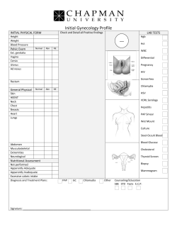


© Copyright 2026