
Primary Cryptococcal Prostatitis- Rare Occurrence Case Report Vinaya B Shah
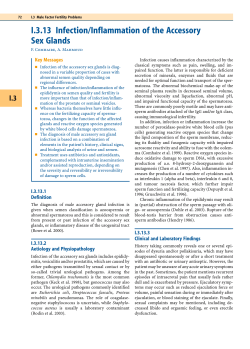
© JAPI • may 2012 • VOL. 60 57
Case Report
Primary Cryptococcal Prostatitis- Rare Occurrence
Vinaya B Shah*, Pallavi A Patil**, Vipul Agrawal***, Harish K Kaswan***
Abstract
Cryptococcosis is a well recognized infection in immunocompromised patients. Cryptococcal infection primarily
involves the lung and is hematogeneously spread to other organs. Sometimes it might affect the genitourinary
tract. The prostate gland is a rare site of primary infection due to cryptococcus neoformans. We report a case of
granulomatous inflammation in the prostate as a result of crypyococcus neoformans infection in a 70 year old
immunocompetent patient, a non diabetic, which was diagnosed by transrectal ultrasound guided biopsy.
C
Introduction
ryptococcosis is a well recognized infection in
immunocompromised patients. Cryptococcal infection
primarily involves the lung and is hematogeneously spread
to other organs. Sometimes it might affect the genitourinary
tract and rare cases have been reported involving the prostate
without systemic infection and still rarer occurrence in an
immunocompetent patient.
Case Report
A 70 year old non diabetic, non hypertensive was admitted
with complaints of frequency, dysuria and fever since 1 week.
His general condition was satisfactory. His temperature was
39.5º C. The rest of the physical examination was unremarkable.
His laboratory results showed a Hb of 14.2 gm%, total white cell
count of 6x 10⁹ /dl, platelet count of 185x 109/dl and ESR of 20mm/
hour. Urine microscopy showed 10-12 white blood cells per
high power field, but urinary culture was negative. Ultrasound
of prostate showed symmetrical prostatomegaly weighing 120
gms. Prostatic specific antigen (PSA) levels were high i.e 45ng/ml.
Transrectal biopsy of prostate was done from both the
lobes and sent for the histopathological examination, which
showed total 4 linear bits which showed fibrous stroma with
multiple foci of granulomatous inflammation comprising of
lymphocytes, macrophages, epithelioid cells, histiocytes and
multinucleate giant cells (Figure 1). High power revealed
histiocytes and multinucleate giant cells with engulfed grey
yeast forms surrounded by a clearing of surrounding tissue, a
halo representing the capsular material (Figures 2a, 2b).
prostatitis was made. He was treated with high dose of
fluconozole 400mg/day and venous amphotericin at doses 0.4
mg/kg/day for 2-3 weeks.
Discussion
Cryptococcus infection is acquired through the respiratory
route. In most hosts who encountered Cryptococcus neoformans
the infection is contained or eliminated. Immunocompromised
patients are at risk of systemic disease and dissemination.
The clinical presentation of cryptococcosis varies depending
on the site and host, but the dominant involved organ are the
lungs and CNS. Early reports dated back to the 60`sand 70`s.1
Subsequently prostatic involvement were reported in patients
immunocompromised by steroids.2 organ transplantation,3
human immunodeficiency syndrome (HIV) infection and
Hodgkins lymphoma. 4 It is also described in apparently
immunocompetent host.5
The prostate gland has also emerged as a potential site of
relapse of cryptococcosis after apparently successful therapy of
cryptococcal meningitis.6 Systemic spread from primary focus
of cryptococcal infection commonly involves CNS, manifested
as meningitis.
Untreated meningitis are invariably fatal. Our patient was
treated with high doses of fluconazole and recovered. We
Gomori methenamine silver (GMS) very efficient to visualize
the fungi and also advantageous since it stains old and nonviable
fungal elements more efficiently as seen in (Figure 2c). Special
stains like mucicarmine helped in delineating the capsule
(Figure 2d).
Diagnosis of granulomatous cryptococcal prostatitis was
given. Subsequently the patient was investigated for any
immunocompromised status. Patient was seronegative for HIV-I,
HIV-II, HbsAg, VDRL. Culture from blood and cerebro spinal
fluid(CSF) was negative for cryptococcus. X ray chest showed
clear lung fields. Thus the diagnosis of primary cryptococcal
Associate Professor, Pathology Department, **Resident Pathology,
Pathology Department, ***Senior Resident, Urology Department,
TNMedical College & BYLNair Hospital, Mumbai- 400034
Received: 18.06.2010; Revised: 29.03.2011; Accepted: 02.06.2011
*
Fig. 1 : Prostatic tissue with fibrous stroma and multiple foci of
granulomas. [Scanner view of Hematoxylin & Eosin(H & E) stain x
40]
58
© JAPI • may 2012 • VOL. 60
by histopathology).10 The cryptococcal CSF antigen had the
best overall sensitivity (94.1%) followed by the serum antigen
(93.6%).10 However, some differences were observed in the
different categories of hosts, with lower sensitivity in AIDS and
immunocompetent patients (92%) and higher sensitivity among
the other immunocompromised hosts without HIV infection.10
Though our case had negative CSF culture, histopathological
demonstration of the organism was done in the prostatic tissue
which had multiple foci of granulomatous inflammation with
the aid of special stains.
In the needle biopsies of the prostate, if granulomas are
observed on histology, then the usual common differential
diagnosis include tuberculous, BCG induced idiopathic non
specific granulomatous, malackoplakia, xanthogranulomatous,
iatrogenic postsurgical induced and fungal etiology.
Fig. 2 : (a) H & E stain x100 shows a granuloma comprising of
lymphocytes, macrophages, multinucleate giant cells and (b) High
power: H & E stain x400 shows grey yeast bodies of cryptococcus
engulfed by the giant cell (arrow). Gomori methenamine silver
(GMS) stained the fungus black and numerous such cryptococci
were seen in (c) and Mucin stain: mucicarmine delineated the
capsule by staining magenta pink (arrow) (d).
feel that a persistent prostatic focus of infection needs to be
scrutinized vigilantly.
The rising incidence of cryptococcosis in India is posing a
serious threat. Due to lack of sensitive methods for diagnosis,
high morbidity and mortality are associated with the disease.
Early diagnosis and institution of specific antifungal therapy are
imperative to minimize the severity of infection. The laboratory
diagnosis of cryptococcosis is based on direct demonstration,
culture, and antigen detection by latex agglutination test (LAT).
Histopathological examination of organisms on tissue
biopsy is also a effective means of diagnosing cryptococcosis.
Special stains include gomori`s silver and calcofluor white
stain are sensitive means of identifying this organism in tissue.
The mucicarmine stain is particularly helpful in that it stains
the capsule of this organism, allowing the observer to make a
presumptive diagnosis of cryptococcossis.
Cryptococcossis is easily cultured on standard fungal media
that do not contain cycloheximide. Large, cream to yellow
colored colonies appear in 3-5 days when yeast is cultured on
Saborauds dextrose agar. Encapsulated colonies are mucoid.
Cryptococcus neoformans can be distinguished from most other
species of cryptococcossis by its ability to produce melanin
through phenol oxidase. Cultures may require several weeks
for a positive result and these are not helpful in the immediate
management of clinical patients.
Microscopic methods and culture, though specific, show a
sensitivity of 50–80 %.7 Also, culture takes time and requires
more labour and large volumes of samples. Latex agglutination
test (LAT) is more sensitive but suffers from the limitation of
false positivity.8,9
The latex- Cryptococcus antigen test is simple, sensitive test
capable of detecting C. neoformans polysaccharide antigens in
serum and CSF and is superior to india- ink mount. Clinical
studies established the prognostic value of aid in establishing a
diagnosis when culture was negative.10,11
In one study, despite negative CSF culture, there was
disseminated cryptococcal disease at autopsy (demonstrated
To summarize, thorough histopathological examination
for classic histologic clues and clinical history will aid in
diagnosing most of the lesions considered in the differential
diagnosis of granulomatous prostatitis. But in fungal prostatitis,
histopathology is one of the major tools of diagnosis. The major
advantages of histopathology are speed, low-cost, and the
ability to provide a presumptive identification of the infecting
fungus, as well as demonstrating the tissue reaction. A number
of histologic stains are available that are routinely used to
visualize fungi in tissue sections Gomori methenamine silver
(GMS), Gridley’s fungus (GF), and periodic acid-Schiff (PAS)
are special for and very efficient to visualize the fungi. Mucin
stains, like Mayer’s mucicarmine, stain the mucopolysaccharide
capsule of Cryptococcus neoformans which was done in our case.
Conclusion
Cryptococcosis can be easily misdiagnosed in uncompromised
host both clinically and pathologically because of misconception
that the disease affects only immunocompromised individuals.
Hence, awareness and thorough histopathological examination
of granulomatous lesion in a prostate would avoid misdiagnosis.
References
1.
Dreyfuss ML, Sommer RI. Granulomatous prostatitis due to
Cryptococcus neoformans (Torula) with disseminated cryptococcus
and meningitis. NY State J Med 1961;61:158-92.
2.
Braman RT. Cryptococcosis (torulosis) of prostate. Urology
1981;17:284-5.
3. Plunkett JM, Turner BI, Tallent MB, Johnson HK. Cryptococcal
septicaemia associated with urologic instrumentation in a renal
allograft recipient. J Urol 1981;125:241-2.
4. Lief M, Sartarazi F. Prostatic cryptococcosis in acquired immune
deficiency syndrome. Urology 1986;28:318-9.
5. King C, Finley R, Chapman SW. Prostatic cryptococcal infection.
Ann Intern Med 1990;113:720.
6. Hinchey WW, Someren A. Cryptococcal prostatitis. Am J Clin Pathol
1981;75:257-60.
7. Snow RM and Dismukes WE. Cryptococcal meningitis: diagnostic
value of cryptococcal antigen in cerebrospinal fluid. Arch Intern Med
1975;135:1155–7.
8. Stoeckli TC and Burman W J. Inactivated pronase as the cause of
false-positive results of serum cryptococcal antigen tests. Clin Infect
Dis 2001;32:836–7.
9. Whittier S, Hopfer R L and Gilligan P. Elimination of false-positive
serum reactivity in latex agglutination test for cryptococcal antigen
in human immunodeficiency virus-infected population. J Clin
Microbiol 1994;32:2158–61.
10. Antinori S, Radice A, Galimberti L, Magni C, Fasan M, Parrvicini
© JAPI • may 2012 • VOL. 60 C. The Role of Cryptococcal Antigen Assay in Diagnosis
and Monitoring of Cryptococcal Meningitis. J Clin Microbiol
2005;43:5828-9.
59
11. Cunha BA. Central nervous system infections in the compromised
host: a diagnostic approach. Infect. Dis Clin N Am 2001;15:567-590.


© Copyright 2026