
Dear Author, Journal of Nuclear Medicine Technology
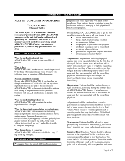
Journal of Nuclear Medicine Technology
Dear Author,
This letter describes how to proof your manuscript slated for an upcoming issue of the Journal of
Nuclear Medicine Technology. This PDF is the only set of proofs you will receive for your article.
This file contains:
Important notice to authors
Reprint order form
Page proofs and queries for your article
After printing the PDF file, please read the page proofs carefully and:
1) indicate changes or corrections in the margin of the page proofs;
2) answer all queries (AQ1, AQ2, AQ3, etc.) on the last page;
3) proofread any tables and equations carefully;
4) check that any Greek, especially µ (mu), has translated correctly.
Within 48 hours, please return the original PDF set of page proofs to the address given below.
If there are color images in your article, you will be receiving a hard-copy color proof before
publication to review color reproduction and orientation.
If you have any problems or questions, please contact me. PLEASE ALWAYS INCLUDE
YOUR ARTICLE NUMBER WITH ALL CORRESPONDENCE.
Sincerely,
Susan Alexander
Associate Director of Publications
Society of Nuclear Medicine
1850 Samuel Morse Drive
Reston, VA 20190-5316
Tel: (703) 708-9000
Fax: (703) 708-9018
Journal of Nuclear Medicine Technology
IMPORTANT NOTICE TO AUTHORS
You must proofread and return these page proofs to the
Society of Nuclear Medicine within 48 hours of receipt.
Failure to do so may result in your article being published
without your corrections.
Your cooperation in adhering to our tight production deadline is
greatly appreciated.
Please return these page proofs marked with your corrections to:
Susan Alexander
Associate Director of Publications
Society of Nuclear Medicine
1850 Samuel Morse Drive
Reston, VA 20190-5316
USA
Corrections may also be faxed to (703) 708-9018 or e-mailed
to [email protected].
If you have any questions, please call Susan Alexander at (703) 708-9000.
JNMT Reprint Order Form
cust. #3006
Please place your order at the time you review your page proofs. Reprints ordered later are priced much higher if the article contains color
images. Your order will be processed and shipped within 10 business days after the article has been published and payment has been
received.
Prices*
Length of article
U.S. customers
1–4 pages
5–8 pages
9–12 pages
13–16 pages
17–20 pages
Cover page**
Non-U.S. customers
1–4 pages
5–8 pages
9–12 pages
13–16 pages
17–20 pages
Cover page**
100 reprints
Quantity ordered
200 reprints 300 reprints
$381
$602
$828
$1049
$1268
$211
$407
$647
$878
$1090
$1332
$303
$472
$730
$986
$1248
$1500
$393
$483
$758
$1050
$1328
$1611
$283
$528
$846
$1156
$1444
$1782
$414
$627
$908
$1341
$1702
$2058
$548
*Prices are valid through the December 2006 issue.
**Cover page includes journal title, article title, and authors’ names.
The minimum order is 100 copies. For orders over 300 copies, call for additional information and pricing: Steve Klein, Production
Manager, Society of Nuclear Medicine, 703-708-9000, ext. 1213.
Reprint Information
Title of article: ___________________________________________________________________________________________
Length of article: ______ printed pages
Publication date of article: ___________________
Cost of reprints:
$__________
Cost of cover page (if ordered):
$__________
Total cost:
$__________
Customer Information
Name: ____________________________________________________________________
Shipping address: ____________________________________________________________
City: ______________________________________________________________________
State or country: ________________________________________ Postal code: __________
Phone number: _________________________
Fax number: ___________________________
E-mail address: _________________________
Payment Information
Orders must be paid for in advance (purchase orders are not accepted). VISA and MasterCard are the preferred methods of payment.
Type of credit card:
□ VISA
□ MasterCard
Card number: ______________________________________________________________
Expiration date: ____________________________________________________________
Signature: _________________________________________________________________
Name as it appears on the card (please print): ______________________________________
Do you require a receipt? □ Yes (include e-mail address or fax number above)
□ No
You may also pay by sending a check or money order (made payable to Sheridan Reprints) to the following address:
Sheridan Reprints
Attn: Tamara Groft
450 Fame Avenue
Hanover, PA 17331
Regardless of payment method, please fax this Reprint Order Form to Sheridan Reprints at 717-633-8929.
The 717-633-8929 number is for reprint orders only. Do not fax your article proofs to this number.
Essential Role of Nuclear Medicine Technology in
Tositumomab and 131I-Tositumomab Therapeutic
½AQ1 Regimen for Non-Hodgkin’s Lymphoma*
William C. Cole1, Jennifer Barrickman2, and Glen Bloodworth, CNMT2
1University of Colorado Health Sciences Center, Denver, Colorado; and 2GlaxoSmithKline Oncology, Research Triangle Park,
North Carolina
Nuclear medicine technology traditionally has focused on
diagnostic imaging, with therapeutics left mostly to other medical
disciplines. However, after many years in development, radioimmunotherapy (RIT) finally has become a clinical reality in many
nuclear medicine departments. The nuclear medicine technologist is a key player in the successful implementation of RIT.
Delivery of a therapeutic regimen of tositumomab and 131Itositumomab provides a model for the technologist’s roles and
responsibilities in the developing field of RIT. This article examines the clinical rationale, logistic requirements, and imaging
and dosimetry procedures required by this treatment regimen.
Upon completion of this article, the reader should be able to
describe the target patient population and identify the roles
and responsibilities of various members of the treatment team.
The reader also will gain an understanding of the treatment process, including drug administration, imaging, and therapeutic
dose calculations.
V1
Key Words: non-Hodgkin’s lymphoma; radioimmunotherapy
J Nucl Med Technol 2006; 34:1–7
tic effect, the radionuclide selected is generally a b-emitter,
such as 131I or 90Y, on the basis of the higher absorbed
fraction of energy deposited in tissue by b-emitters than by
pure g-emitters. The nuclear medicine technologist is a key ½AQ2
player in the successful implementation of RIT. The delivery of tositumomab and 131I-tositumomab (the BEXXAR
therapeutic regimen; GlaxoSmithKline; approved by the
U.S. Food and Drug Administration in June 2003) provides
a model for the technologist’s roles and responsibilities in
the developing field of RIT.
BEXXAR is an RIT regimen containing an unlabeled
anti-CD20 IgG2a murine monoclonal antibody, tositumomab, and a radiolabeled antibody, 131I-tositumomab. This
therapeutic regimen is indicated for the treatment of patients with CD20 antigen–expressing relapsed or refractory,
low-grade, follicular or transformed non-Hodgkin’s lymphoma (NHL), including patients with rituximab-refractory
NHL.
That being said, why is a new treatment for NHL
needed? NHL is the fifth leading and second-fastest-growing
cause of cancer-related deaths in the United States. The
American Cancer Society reported that in 2005, the total
number of patients with newly diagnosed NHL was expected
to reach 56,390, and 19,200 patients would be expected to
succumb to this disease (1). Patients with low-grade NHL
often have multiple relapses and eventually become refractory to therapy. With each subsequent treatment, patients
are less likely to respond, and the duration of the response is
shortened (Fig. 1) (2). As in many other cancers, radiation ½Fig: 1
and chemotherapy are mainstays of the initial treatment for
NHL. RIT is part of a relatively new category of therapy
called biologic therapy and has yielded some impressive
clinical results that were summarized in a recent review (3).
Kaminski et al. (4) recently reported a complete response
for 75% of 76 patients who received the tositumomab and
131I-tositumomab therapeutic regimen as the initial treatment for follicular lymphoma. Forty (70%) of these patients
remained in remission for 4.3–7.7 y.
Here we describe the logistics of the tositumomab and
131I-tositumomab therapeutic regimen and the vital role that
nuclear medicine can play in its effective implementation.
N
uclear medicine technology has long been focused on
diagnostic imaging. With few exceptions (e.g., thyroid
ablation with 131I-NaI), therapeutics have been left to other
medical disciplines. A new era for nuclear medicine is at
hand: radioimmunotherapy (RIT). In RIT, the anticancer
properties of specific immunoglobulins, commonly referred
to as antibodies, are combined with the cell-killing potential of radionuclides. Antibodies are believed to act against
cancer cells through the processes of antibody-dependent
cellular cytotoxicity, complement-mediated cytotoxicity, and
apoptosis induction. With the addition of a radionuclide to
the antibody, there is an additional cell-killing effect resulting from ionizing radiation. To maximize the therapeu-
Received Sep. 16, 2005; revision accepted Mar. 14, 2006.
For correspondence or reprints contact: Glen Bloodworth, CNMT,
GlaxoSmithKline Oncology, 229 White Oak Dr., Wilmington, NC 28409.
E-mail: [email protected]
*NOTE: FOR CE CREDIT, YOU CAN ACCESS THIS ACTIVITY THROUGH
THE SNM WEB SITE (http://www.snm.org/ce_online) THROUGH JUNE 2007.
ROLE
OF
NUCLEAR MEDICINE
jnmt24380-pe n 4/22/06
IN
RIT • Cole et al.
1
process, each treatment site must demonstrate competency
in patient dose administration and calculations. Competency
is documented by the completion of the imaging and dose
determination steps after 3 successful attempts in 6 possible
attempts. Information is collected on manufacturer-provided
dosimetry worksheets, which are faxed to the Bexxar Service Center (BSC), a single point of contact for all aspects
of the tositumomab and 131I-tositumomab therapeutic regimen. A template (Excel; Microsoft Corp.) has been developed to aid in performing the dosimetry calculations.
Once a site has successfully completed this certification
process, it is no longer necessary to submit the dosimetry
worksheets to the BSC. However, the BSC is always willing to provide assistance even after the certification process
is complete.
Premedications
FIGURE 1.
of (2).)
Duration of response. (Reprinted with permission
METHODOLOGIES
Medical Professionals
A number of medical professionals may become involved with the tositumomab and 131I-tositumomab therapeutic regimen, and each is important for successful
treatment. Among these individuals may be a hematologist
or oncologist, oncology nurse, nuclear medicine physician,
radiation oncologist, nuclear medicine technologist, nuclear
pharmacist, hospital pharmacist, radiation safety officer,
and medical physicist. The U.S. Food and Drug Adminis½AQ3 tration mandates that these medical professionals receive
½Table 1 training by GlaxoSmithKline on-site trainers. Table 1 lists
some of the main functions required in the successful
implementation of the tositumomab and 131I-tositumomab
therapeutic regimen.
Throughout the process, a site coordinator is responsible
for communication among all team members in an effort to
provide a cohesive treatment regimen for the patient.
Specifically, the site coordinator plans the treatment sched½AQ4 ule, submits order documents to the nuclear pharmacist,
and coordinates patient scheduling with all participating
team members. The site coordinator may be any one of the
health professionals listed above or another staff member
capable of effectively managing simultaneous tasks. Given
the nature of the tositumomab and 131I-tositumomab therapeutic regimen, the nuclear medicine technologist is often
½Fig: 2 the logical choice for site coordinator. Figure 2 summarizes
the main steps in the tositumomab and 131I-tositumomab
regimen.
Thyroid-blocking agents are commonly used in nuclear
medicine. For the tositumomab and 131I-tositumomab therapeutic regimen, one of the following regimens must be
started at least 24 h before the dosimetric dose and continued for 2 wk after the therapeutic dose: a saturated
solution of potassium iodide (4 drops orally 3 times per day),
Lugol’s solution (20 drops orally 3 times per day), and
potassium iodide tablets (130 mg orally once per day).
In addition to thyroid protection, patients should receive
premedication with 650 mg of acetaminophen and 50 mg
of diphenhydramine 30–60 min before the administration
of each of the unlabeled (‘‘cold’’) tositumomab doses to
reduce the incidence of possible infusion-related events
(e.g., fever, rigor or chills, sweating, hypotension, dyspnea,
bronchospasm, and nausea). If any of these events do occur,
reduction of the infusion to 50% the initial rate (450 mg of
unlabeled tositumomab over 60 min) is recommended. It
may be necessary to interrupt the infusion if the symptoms
are severe. Many times, the infusion of unlabeled tositumomab is conducted in the oncology infusion area so that a
nuclear medicine technologist may actually administer
only 131I-tositumomab—a brief 20-min infusion followed
by a flush.
V1
Training
Training of site personnel is a critical component of
successful implementation of the tositumomab and 131Itositumomab therapeutic regimen. As part of the training
2
JOURNAL
OF
Dosage and Dose Administration
The tositumomab and 131I-tositumomab therapeutic regimen is administered intravenously in 2 steps, the dosimetric step and the therapeutic step. Each step consists of 2
separate components: the unlabeled, or cold, infusion and
the radiolabeled, or hot, infusion. The prescribed dosage for
each step is shown in Table 2. The therapeutic dose of ½Table 2
131I-tositumomab is given in centigrays, the unit of absorbed
dose. Dosimetry, the process of relating the prescribed
absorbed dose to a specific megabecquerel value, is de- ½AQ5
scribed in detail in the next section.
Both the dosimetric and the therapeutic infusions involve
2 nearly identical components: cold-antibody (tositumomab)
infusion and hot-antibody (131I-tositumomab) infusion. The
only difference between the 2 is the activity of the hot antibody infused (diagnostic versus therapeutic activity levels).
NUCLEAR MEDICINE TECHNOLOGY • Vol. 34 • No. 2 • June 2006
jnmt24380-pe n 4/22/06
TABLE 1
Roles of Health Care Team Members During Administration of Tositumomab and
131I-Tositumomab
Therapeutic Regimen
Responsible Party
Role
Review radioactive material
license
Identify and refer patients
Prescribe therapy
Educate patients
Place order with BSC
Report platelet counts for
dose calculation
Prescribe thyroid block
Prescribe premedications
Receive packaging and
prepare doses
Administer cold infusion
Monitor patient
Administer hot infusion
Report adverse events
Perform quality control
procedures and acquire
images
Calculate estimated dose
(after second scan)
Calculate therapeutic dose
Send completed dosimetry
worksheets to nuclear
pharmacy
Report final dose calculation
to BSC
Determine when patient can
be released
Instruct patient regarding
radiation safety precautions
Perform patient follow-up
Nuclear
Nuclear Radiation
Hematologist Radiation medicine medicine
safety Oncology Nuclear
Hospital Medical
or oncologist oncologist physician technologist officer
nurse pharmacist pharmacist physicist
3
3
3
3
3
3
3
3
3
3
3
3
3
3
3
3
3
3
3
3
3
3
3
3
3
3
3
3
3
3
3
3
3
3
3
3
3
3
3
3
3
3
3
3
3
3
3
3
3
3
V1
3
3
3
3
3
Dose shielding, prevention of spills and contamination, and
minimization of exposure to personnel are important considerations for each step.
After the administration of premedications, 450 mg of
unlabeled tositumomab antibody are first delivered to the
patient over 1 h. The antibody is prepared in a volume of
3
3
3
3
3
3
3
3
3
3
50 mL and is delivered to the patient via an infusion pump. ½AQ6
This step often takes place in the oncology infusion area.
Immediately after the cold infusion, the patient is administered the radiolabeled, or hot, 131I-tositumomab antibody. This step often is performed in the nuclear medicine
department. The hot antibody is prepared and delivered to
FIGURE 2. Steps in tositumomab and
131I-tositumomab therapeutic regimen.
ROLE
OF
NUCLEAR MEDICINE
jnmt24380-pe n 4/22/06
IN
RIT • Cole et al.
3
TABLE 2
Prescribed Dosage for Each Step of Tositumomab and
131I-Tositumomab Therapeutic Regimen
Dose
Tositumomab
Dosimetric
Therapeutic
450 mg
450 mg
131I
Tositumomab
185 MBq (5 mCi)
75 cGy (platelet count of
.150,000/mm3)
65 cGy (platelet count
of between .100,000/mm3
and ,150,000/mm3)
Regimen is not indicated for
patients with platelet
count of ,100,000/mm3
the site of the infusion in a 30-mL total volume in a 60-mL
syringe . The syringe is loaded into a syringe pump, and the
total volume is delivered over 20 min; this step is followed
by a thorough flush of the tubing with 0.9% saline. It is
strongly recommended that personnel responsible for setting up and administering the hot infusion thoroughly
familiarize themselves with the procedure by making several practice runs with nonradioactive syringes, tubing,
syringe pump, and shielding before patient arrival to minimize unanticipated problems that may arise. To minimize
the risk of contamination, exposed work surfaces, such as
countertops, infusion chairs, and floors, should be draped
with absorbent pads. Throughout the infusion process, every
effort should be made to adhere to the concept of ‘‘as low as
½AQ7 reasonably achievable’’ by minimizing the amount of time
spent near the source, maximizing the distance from the
source, and using shielding whenever possible to reduce the
dose to infusion personnel. Finally, after the infusion is complete, the area should be surveyed for contamination with a
survey meter.
imen, such as extent of disease, bone marrow involvement,
spleen size, and renal function (5). In addition to enabling
an estimation of TBRT, imaging also provides an opportunity for the nuclear medicine physician to evaluate biodistribution with the same isotope, unlike other regimens
that use a g-emitting surrogate to predict biodistribution.
However, unlike most nuclear medicine imaging procedures, this process is intended to evaluate gross biodistribution only, not provide detailed diagnostic information.
The expected biodistribution after a dosimetric dose of 131Itositumomab is illustrated by whole-body scans of a patient
with the typical pattern for NHL (Fig. 3), whereas biodis- ½Fig: 3
tribution may be different in patients with solid tumors or
other cancer types, such as a retroperitoneal tumor with
thyroid uptake (Fig. 4) or cutaneous lymphoma (Fig. 5), ½Fig: 4
respectively. In addition to visual inspection of the images, ½Fig: 5
biodistribution also may be assessed by evaluating the TBRT
(day 6 or 7). For the tositumomab and 131I-tositumomab
therapeutic regimen, the expected biodistribution is defined
as a TBRT of between 50 and 150 h.
As imaging takes place at 3 different times (day 0 within
1 h of dosing; day 2, 3, or 4; and day 6 or 7) after the
dosimetric administration of 131I-tositumomab, it is important to maintain constant imaging parameters. For example,
it is important that images are acquired by use of the same
camera with the same head-to-table distance, scan length
(in cm), scan speed, collimator, field of view, region of
interest, and energy window. Simply put, the imaging parameters must be exactly the same for each imaging session.
This requirement also applies to obtaining background and
standard counts.
V1
Imaging and Dosimetry
Nuclear medicine technologists are focused primarily on
patient imaging. However, what role does imaging have in
therapy? The ultimate goal in drug therapy is to provide
the most effective dose to optimize benefit and minimize
toxicity. The tositumomab and 131I-tositumomab therapeutic regimen seeks to approach this goal by evaluating individual patient drug distribution and elimination over time.
These aspects of drug behavior are termed pharmacokinetics. With conventional pharmaceutical agents, the best
that clinicians can do in this regard is to measure the time
course for the drug (or metabolites) in the blood. With
radiolabeled agents, however, a much more accurate estimate of patient-specific pharmacokinetics is possible. Because 131I is both a g-emitter and a b-emitter, it is possible
to obtain whole-body g-counts from imaging of the patient
after a relatively small (dosimetric) dose (184 MBq [5 mCi])
of 131I-tositumomab to estimate the total body residence
time (TBRT). This determination allows consideration of
the factors shown to be influential in the pharmacokinetics
of the tositumomab and 131I-tositumomab therapeutic reg-
4
JOURNAL
OF
FIGURE 3. Expected biodistribution showing blood-pool activity in heart and major vessels on first scan (A) and decreasing
in intensity on second scan (B) and third scan (C) for patient
with NHL. Moderate uptake in liver and spleen on first scan ½AQ16
progressively diminishes in intensity on second and third scans.
Anterior views.
NUCLEAR MEDICINE TECHNOLOGY • Vol. 34 • No. 2 • June 2006
jnmt24380-pe n 4/22/06
attenuated to 65 cGy. The tositumomab and 131I-tositumomab
therapeutic regimen is not indicated for patients with a
platelet count of less than 100,000/mm3. Although it is
important to understand the basic concepts for calculating
the patient-specific dose, the template mentioned previously
will automatically perform all calculations and curve fitting.
Residual Activity
FIGURE 4. Expected biodistribution showing blood-pool activity in heart and major vessels on first scan (A) and decreasing
½AQ17 in intensity on second scan (B) and third scan (C) for patient
with retroperitoneal adenopathy. Moderate uptake in liver and
spleen on first scan progressively diminishes in intensity on
second and third scans. Tumor uptake is seen as 2 focal areas
of moderate uptake in upper to middle abdomen, best seen on
second scan, corresponding to retroperitoneal adenopathy
seen on CT scan. ‘‘Butterfly’’-shaped uptake seen in neck on
third scan represents thyroid uptake. Anterior views.
The determination of residual activity is another critical
element required to obtain an accurate measure of the
injected activity, thereby ensuring that the patient received
the full prescribed dose of 131I-tositumomab. The process is
simply to gather all infusion-related equipment, including
the syringe used for the radioactive dose, and analyze the
equipment with a dose calibrator as soon as practicable after
the infusion is completed. The residual activity is subtracted
from the measured activity in the syringe before administration and results in the net administered activity. Measuring and
recording residual activity is required in both the dosimetric
and the therapeutic steps and is critically important.
Patient Release Methodology
Although it will be the responsibility of the radiation
safety officer to determine when a patient can be released
after a therapeutic dose of tositumomab and 131I-tositumomab,
the nuclear medicine technologist may be called upon to
assist, and it is useful to review the principles governing
that determination. Patient release is not dependent on the
V1
Determining the TBRT is done by simply drawing a
‘‘best-fit’’ straight line between the whole-body counts obtained on day 0 and those obtained on subsequent days on a
½AQ8 semilogarithmic graph of percentage injected activity on
the y-axis and time from the dosimetric dose on the x-axis.
In the first estimate of TBRT after the second imaging time,
one simply connects the dots. This first estimation is done
only to determine whether the radiopharmacy may require
2 vials of 131I-tositumomab instead of 1 to prepare the
patient-specific dose. The need for 2 vials becomes important if the estimated therapeutic dose to the patient may
exceed 3,700 MBq (100 mCi). The final estimation of
TBRT at 3 imaging times requires the best-fit straight
½Fig: 6 line between the second and third time points (Fig. 6).
The TBRT can be used in the calculation of the required
megabecquerel amount of 131I-tositumomab: 131I activity
½AQ9 (MBq) 5 [activity hours (MBq/h)/residence time (h)] ·
[desired total body dose/75 cGy].
½AQ10
The value for activity hours (describing the amount of
activity in the body and the time that it is there, on the basis
of patient sex and effective mass or weight) is obtained
simply from a table provided in the training. The last element of the equation describes the fact that the recommended total body dose (cGy) for the tositumomab and
131I-tositumomab therapeutic regimen is 75 cGy for patients with a platelet count of more than 150,000/mm3. For
cases of mild thrombocytopenia (platelet count of 100,000/
mm3–150,000/mm3), the recommended total body dose is
FIGURE 5. Expected biodistribution for patient with lymphomatous nodules. Multiple areas of various sizes, configuration,
and intensity are seen in right upper chest wall laterally,
epigastric region just to right of midline, lower right abdomen
laterally, bilateral inguinal regions, and proximal right thigh on
second scan (B). Some of these areas persist on third scan (C).
These areas represent cutaneous lymphomatous nodules.
Focal intense uptake is first seen in left upper abdomen on
third scan, most likely representing activity in fundus of
stomach. Anterior views.
ROLE
OF
NUCLEAR MEDICINE
jnmt24380-pe n 4/22/06
IN
RIT • Cole et al.
5
FIGURE 6. Example of TBRT after dosimetric administration of tositumomab
and 131I-tositumomab. Best-fit line for 2
assessment time points and 100% initial
time point are shown.
½AQ11
dosimetric dose of 131I-tositumomab, as the 185-MBq (5mCi) dosimetric dose is below the 1,221-MBq (33-mCi) limit
specified by 10CFR35.75 for immediate outpatient release.
The therapeutic dose of 131I-tositumomab, however, usually
will exceed the 1,221-MBq (33-mCi) limit. Nuclear Regulatory Commission agreements in most states do allow the release of patients receiving this dose according to 10CFR35.75,
which notes that a licensee may release a patient administered
greater than 1,221 MBq (33 mCi) of a radiopharmaceutical or
a permanent radioactive implant, provided that the total
effective dose equivalent (TEDE) to any other individual
from exposure to the released patient is not likely to exceed 5
mSv (500 mrem). (It should be noted that some states have
specific requirements for compliance, while still allowing
tositumomab and 131I-tositumomab therapy to be performed
in an outpatient setting. The BSC can assist with statespecific information.) In order to comply with this ‘‘5-mSv
rule,’’ 2 parameters are used: the TBRT and the dose rate at
1 m from the patient immediately after the therapeutic dose
(6,7). The TBRT is determined in the dosimetric step. Therefore, the next step in determining when a patient can be
released is measuring the dose rate at 1 m with a calibrated
ionization chamber. An ion chamber is necessary because
these instruments are energy independent and yield accurate
dose rates regardless of the radionuclide being measured. The
official equation used to determine the TEDE (mSv) to any
exposed individual is 0.25 · dose rate · (8.95 1 0.99TBRT).
The value 0.25 refers to the occupancy factor, which is
an estimate of the percentage of time that a patient will
spend at a distance of 1 m from another individual. Other
values, for example, 0.5 or 0.125, are used if it is assumed
that a greater or a lesser fraction of the day will be spent
within this proximity to others. The dose rate is measured
at 1 m with the calibrated ionization chamber. Training
provided for the tositumomab and 131I-tositumomab therapeutic regimen includes a table to assist in this calculation.
In addition to determining the TEDE, an appropriate patient
release procedure requires the following additional steps.
An evaluation of the patient’s living and working conditions is needed to limit radiation exposure to others. Factors
that should be considered include the patient’s willingness and
ability to follow patient safety guidelines, to perform self-care,
and to delay returning to work, living arrangements, potential
for exposure to others during the trip home after treatment,
procedures for notification of health care workers in the event
that the patient is hospitalized or in need of medical care, and
the presence of urinary incontinence. A record of the interview used to make these evaluations must be kept for 3 y.
Instructions need to be provided to the patient ready for
release. Some of the instructions, which should be adhered
to on a temporary basis, typically 1–2 wk, include sleeping
in a separate bed (at least 1.8 m [6 ft] from anyone else),
avoiding long trips (4 h or more) sitting near others, staying
at least 1.8 m (6 ft) from children and pregnant women,
minimizing the time spent in close contact with others, and
delaying returning to work. The length of time needed to
comply with any of the instructions is dependent on the
TEDE as determined above. A table is provided to simplify
this determination.
The template designed for determining the TBRT and the
therapeutic dose calculation also contains spreadsheets to
assist in determining when a patient can be released and
providing instructions to the patient ready for release.
V1
½AQ12
½AQ13
½AQ14
6
JOURNAL
OF
CONCLUSION
Nuclear medicine plays a major role in the successful
implementation of RIT, as exemplified by the tositumomab
and 131I-tositumomab therapeutic regimen. This factor signifies a new era of involvement for nuclear medicine in
patient care with respect to cancer treatment and completes
the circle for nuclear medicine as a resource for diagnostic
NUCLEAR MEDICINE TECHNOLOGY • Vol. 34 • No. 2 • June 2006
jnmt24380-pe n 4/22/06
and therapeutic interventions. Nuclear medicine professionals now are afforded the opportunity to expand the impact of our direct patient contact to benefit patients at 2 very
important stages of their interaction in the larger disease
management environment. Nuclear medicine professionals
should welcome these new challenges and responsibilities as
further demonstration of their professionalism and commitment to the best patient care possible.
REFERENCES
½AQ15
1. American Cancer Society. Cancer Facts and Figures, 2005. Atlanta, GA:
American Cancer Society; 2006.
2. Johnson PW, Rohatiner AZ, Whelan JS, et al. Patterns of survival in patients with
recurrent follicular lymphoma: a 20-year study from a single center. J Clin Oncol.
1995;13:140–147.
3. Wahl RL. Tositumomab and 131I therapy in non-Hodgkin’s lymphoma. J Nucl
Med. 2005;46(suppl 1):128S–140S.
4. Kaminski MS, Tuck M, Estes J, et al. 131I-Tositumomab therapy as initial
treatment for follicular lymphoma. N Engl J Med. 2005;352:441–449.
5. Kostakoglu L, Leonard JP, Coleman M, et al. Patient-specific characteristics correlate
with the effective half-life of 131I-tositumomab in non-Hodgkin’s lymphoma
[abstract]. J Nucl Med. 2000;41(suppl):79P.
6. Siegel JA, Kroll S, Regan D, Kaminski MS, Wahl RL. A practical methodology
for patient release after tositumomab and 131I-tositumomab therapy. J Nucl Med.
2002;43:354–363.
7. GlaxoSmithKline. BEXXAR Site Training Manual. Research Triangle Park, NC:
GlaxoSmithKline; 2005.
V1
ROLE
OF
NUCLEAR MEDICINE
jnmt24380-pe n 4/22/06
IN
RIT • Cole et al.
7
QUERIES - jnmt24380q
[AQ1]
Brand name was deleted from title and from Abstract per journal style (generic name was used instead). Brand
name is allowed at first use in text.
[AQ2]
Sense of sentence beginning ‘‘To maximize’’ OK as edited?
[AQ3]
‘‘on-site’’ correct for ‘‘site’’?
[AQ4]
OK to change ‘‘submits order documents with’’ to ‘‘submits order documents to’’?
[AQ5]
Per journal style, the SI unit ‘‘becquerel’’ is used in place of ‘‘millicurie.’’ Check changes throughout.
[AQ6]
‘‘mL’’ (the preferred SI unit) OK for ‘‘cc’’? Please check changes throughout. Also, do you need to say what the
antibody is mixed with to create the 50-mL solution? e.g., saline? Please check similar uses throughout.
[AQ7]
Is ‘‘ALARA’’ spelled out correctly?
[AQ8]
‘‘semilogarithmic’’ correct for ‘‘semilog’’?
[AQ9]
‘‘MBq/h’’ correct here for ‘‘mCi hr’’? Also, please check sense of equation as edited.
[AQ10] OK to change ‘‘term’’ to ‘‘value for’’?
[AQ11] What is ‘‘10CRF35.75’’?
[AQ12] Which instruments are ‘‘these instruments’’? Also, why is yielding accurate dose rates a reason for an ion
chamber being necessary?
[AQ13] Equation OK as edited? In particular, is ‘‘DR’’ spelled out correctly?
[AQ14] In what unit is this value of 0.25 given? hours?
[AQ15] Is 2006 the correct publication date for reference 1?
[AQ16] OK to add ‘‘for patient with NHL’’ to distinguish first statement in Fig. 3 legend from those in Fig. 4 and Fig. 5
legends? See similar changes in the latter two legends.
[AQ17] Correct to change ‘‘second’’ to ‘‘third’’ in Fig. 4C legend? See also Fig. 5C legend.


© Copyright 2026