
Prostatic disorders in the dog S.D. Johnston , K. Kamolpatana , M.V. Root-Kustritz
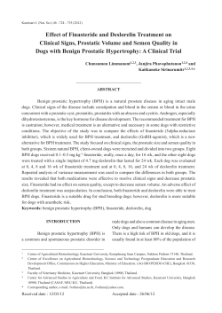
Animal Reproduction Science 60–61 Ž2000. 405–415
www.elsevier.comrlocateranireprosci
Prostatic disorders in the dog
S.D. Johnston a,) , K. Kamolpatana b, M.V. Root-Kustritz c ,
G.R. Johnston a
a
College of Veterinary Medicine, Western UniÕersity of Health Sciences,
309 East Second Streetr College Plaza, Pomona, CA 91766-1854 USA
b
Faculty of Veterinary Medicine, Kasetsart UniÕersity, Kamphaengsaen, Nakhon-Pathom, 73140 Thailand
c
College of Veterinary Medicine, UniÕersity of Minnesota, St. Paul MN 55108 USA
Abstract
Common canine prostatic disorders include benign prostatic hypertrophy ŽBPH., prostatitis,
prostatic cysts and prostatic adenocarcinoma. BPH is a spontaneous and age-related disorder of
intact male dogs, which occurs in more than 80% male dogs over 5 years of age, and which is
associated with clinical signs of sanguinous prostatic fluid, constipation and dysuria. BPH signs
respond to castration or to finasteride treatment Ž0.1–0.5 mgrkg per os once daily., as finasteride
inhibits conversion of testosterone to dihydrotestosterone, causing prostatic involution via apoptosis. BPH often occurs concurrently with prostatic infection, abscessation, cysts and neoplasia in
the intact dog, and finasteride-induced prostatic involution may be beneficial in treatment of all of
these conditions except neoplasia. Two studies suggest that risk of prostatic adenocarcinoma is
increased in neutered, compared to intact male dogs. Although canine prostatic neoplasia, unlike
human prostatic neoplasia, usually does not respond to androgen deprivation, recent reports of
prostatic intraepithelial neoplasia ŽPIN. in a high percentage of older male dogs, with and without
prostatic adenocarcinoma, suggests that PIN may be a precursor to adenocarcinoma in the dog as
it is believed to be in man. q 2000 Elsevier Science B.V. All rights reserved.
Keywords: Canine prostatic hypertrophy; Canine prostatitis; Canine prostatic neoplasia; Finasteride
1. Introduction
The canine prostate is an androgen-dependent, ovoid-shaped, bilobed gland composed
of glandular and stromal elements, which encircles the urethra of the male dog caudal to
)
Corresponding author. Tel.: q1-909-469-5628; fax: 909-469-5635.
E-mail address: [email protected] ŽS.D. Johnston..
0378-4320r00r$ - see front matter q 2000 Elsevier Science B.V. All rights reserved.
PII: S 0 3 7 8 - 4 3 2 0 Ž 0 0 . 0 0 1 0 1 - 9
406
S.D. Johnston et al.r Animal Reproduction Science 60–61 (2000) 405–415
the neck of the urinary bladder. The prostate contributes fluid to the first and third
fractions of the ejaculate ŽEngland et al., 1990.. Prostatic fluid is secreted continuously
in intact male dogs, and flows retrograde into the urinary bladder or antegrade out the
external urethral orifice in volumes ranging from a few drops to several milliliters,
dependent on prostate size.
Prostatic growth and secretion are mediated by dihydrotestosterone ŽDHT., a metabolite of testosterone ŽT. formed in the presence of the enzyme, 5a-reductase. Canine
prostatic size decreases by 50% within 3 weeks of castration, and by 70% within 9
weeks of castration ŽBarsanti and Finco, 1995..
The caudalrdorsal aspect of the prostate in most dogs can be palpated per rectum.
The normal gland is not easily seen in survey radiographs, but can be identified using
retrograde contrast urethrocystography with bladder distension or by ultrasonography
ŽJohnston et al., 1991.. Hyperplastic prostates usually are visible radiographically, and
Fig. 1. Radiographic image of the prostate, lateral projection. The black line represents the distance between
the sacral promontory and the pubis ŽDSPP.. The greatest cranio–caudal distance of the prostate is measured
parallel to this line. Reprinted with permission from Kamolpatana Ž1998..
S.D. Johnston et al.r Animal Reproduction Science 60–61 (2000) 405–415
407
the prostate is considered enlarged when the prostatic diameter, as visualized on the
lateral radiographic view, is greater than 70% of the pubic–sacral promontory distance
ŽDSPP. ŽFig. 1. ŽFeeney et al., 1987..
Prostatic volume in cubic centimeters in the dog can be estimated from greatest
craniocaudal Ž L., transverse ŽW . and dorsoventral Ž D . diameters detected using ultrasonographic examination, and applied to the formula: volume in cubic centimeterss Žw L
= W = D xr2.6. q 1.8 ŽKamolpatana et al., 1999..
2. Benign prostatic hypertrophy in the dog
Benign prostatic hypertrophy ŽBPH. is a spontaneous and age-related condition in
humans and intact male dogs. The pathogenesis of BPH is not completely understood;
however, DHT is accepted as a key hormone in stimulating enlargement of the canine
prostate by enhancing growth in both stromal and glandular components. A major
protein in canine seminal plasma, canine secretory prostatic esterase ŽCPSE., is produced by prostatic epithelial cells under the influence of T; serum CPSE is elevated in
dogs with BPH Žmean concentration of 189.7 ngrml, n s 25. compared to normal intact
dogs Žmean concentration of 41.8 ngrml, n s 20. ŽBell et al., 1995..
More than 80% of intact male dogs over 5 years of age exhibit BPH, and prostatic
volume in affected dogs is 2 to 6.5 times greater than that of normal dogs of similar
weight ŽLaroque et al., 1994, 1995; Hornbuckle et al., 1978; Kamolpatana, 1998..
Prostatic volume in beagles ŽBW s 7–13 kg. with BPH has been reported to range from
20 to 31 cm3 ŽWheaton et al., 1979., 19 to 21 cm3 Žmeasured by MRI. ŽLaroque et al.,
1995; Cohen et al., 1995. and 5 to 24 cm3 Žmeasured by ultrasonography. ŽIguer-Ouada
and Verstegen, 1997..
Clinical signs in affected dogs include sanguinous prostatic fluid dripping from the
tip of the penis, blood in the urine or semen, constipation, and difficult urination
ŽKrawiec and Heflin, 1992.. Diagnosis of BPH is based on detection of blood in
prostatic fluid of the ejaculate or in prostatic fluid emitted from the end of the penis, and
on detecting uniform prostatic enlargement by palpation, radiography andror ultrasonography.
The treatment objective in dogs with BPH is to decrease prostatic size, which
alleviates signs related to BPH; castration is the recommended treatment in most dogs
ŽBasinger, 1987.. Diethystilbestrol at the dose of 0.2–1.0 mg, PO, every 2–3 days, for
3–4 weeks was reported, historically, as an effective medical treatment of BPH in dogs,
but potential adverse side effects of diethystilbestrol in dogs include bone marrow
suppression and pancytopenia, and squamous metaplasia of the prostate, with ductal
obstruction and cyst formation. Medroxyprogesterone acetate Ž3 mgrkg, SQ, given
twice at 4-week intervals, and megestrol acetate, 0.55 mgrkg, per os, for 4 weeks. have
been used to treat BPH in dogs, and are reported not to adversely affect semen quality or
libido ŽBamberg-Thalen and Linde-Forsberg, 1993.. However, only 53% of BPH dogs
treated with medroxyprogesterone acetate showed decreased prostatic size by 6 weeks of
treatment, and the potential adverse sequelae of progesterone administration in dogs,
408
S.D. Johnston et al.r Animal Reproduction Science 60–61 (2000) 405–415
development of diabetes mellitus and mammary nodules, may argue against use of this
regime.
In all mammals, T is metabolized to DHT under the influence of the inhibitable
enzyme, 5a-reductase ŽRussell and Wilson, 1994.. Two isoenzymes of 5a-reductase,
type I and type II, translated from genes on different chromosomes, have been identified
in tissues of humans, nonhuman primates, dogs, rats and mice ŽLiang et al., 1985.. In
humans, the prostate, epididymis, seminal vesicles and genital skin have type II
5a-reductase, and nongenital skin has type I. The canine prostate is reported to contain
both type I and type II 5a-reductase, although there are no data regarding localization or
amounts of each isoenzyme in specific lobes of the prostate of this species ŽKamolpatana, 1998..
Finasteride ŽProscar TM Merck, West Point, PA 19486, USA. is a commercially
available synthetic steroid Žazasteroid. which inhibits type II 5a-reductase and which,
therefore, blocks conversion of T to DHT ŽCohen et al., 1995; Span et al., 1998.. It is an
effective treatment for BPH in humans when given at a dose of 5 mg, PO, every 24 h for
12–24 months. Finasteride decreases serum concentrations of DHT, without affecting
serum concentrations of T, cortisol, estradiol, luteinizing hormone, prolactin, or
thyroid-stimulating hormone in men with BPH. Experimental beagles over 5 years of
age with BPH that were treated with finasteride Ž1 mgrkg BW, PO, every 24 h, for
16–21 weeks. showed prostatic atrophy, and decrease in prostatic volume by 50–70%
Ždetermined by MRI. as compared to placebo-treated controls ŽCohen et al., 1995,
Laroque et al., 1995; Iguer-Ouada and Verstegen, 1997.. This dose of finasteride also
decreased serum concentrations of DHT in dogs, with no adverse effect on testis
histology or semen quality.
A dose–response study of finasteride at 0.10–0.50 mgrkg, PO, every 24 h for 7 days
Ž5 mg per dog in dogs weighing 10–50 kg., in normal intact male dogs caused
significant decrease in serum concentrations of DHT without changing serum concentrations of T, suggesting that the 5 mg per dog dose for dogs ranging from 10 to 50 kg may
be as effective as the 1 mgrkg dose reported earlier in research dogs ŽKamolpatana et
al., 1998.. Finasteride at all doses was associated with a mean decrease in concentration
of DHT of 55% Ždecrease from mean of 155 " 32.3 pgrml to 70 " 15 pgrml., and had
no effect on serum T concentrations Žpretreatment 2.6 " 0.38 ngrml; posttreatment
2.2 " 00.63 ngrml..
In a double blind, placebo-controlled clinical study to determine the effect of
finasteride at 0.10–0.50 mgrkg of body weight, PO, every 24 h, on prostatic diameter
and volume in dogs with spontaneous BPH, finasteride treatment decreased prostatic
diameter by an average of 20%, prostatic volume by an average of 43%, and serum
concentrations of DHT by an average of 58% after 16 weeks of treatment ŽKamolpatana,
1998.. The nine experimental dogs were client-owned animals with spontaneous BPH;
they ranged in age from 2.7 to 11 years, in weight from 10.3 to 49 kg, and represented
seven breeds. Pretreatment prostatic diameters ranged from 52.8 to 109 Žmean " SEMs
70.6 " 6.3.% DSPP, and prostatic volume ranged from 10.4 to 82 Žmean " SEM s 35.5
" 9.1. cm3. Posttreatment, the average prostatic diameter in treatment dogs ranged from
46.6 to 61 Žmean " SEM s 53.8 " 2.5.% DSPP, and mean prostatic volume in treatment
dogs ranged from 7.72 to 36.8 Žmean " SEM s 30 " 7.9. cm3. Prostatic volume
S.D. Johnston et al.r Animal Reproduction Science 60–61 (2000) 405–415
409
decreased by 43%, suggesting that ultrasonography is comparable to radiography
Žprostatic diameter decreased by 20%. in evaluating change in prostatic size during
medical treatment in BPH dogs, and may suggest uniform distribution of type II 5 a
reductase in the dog prostate. Constipation and presence of blood in the seminal fluid
decreased continuously after 1 week of treatment. By 4 weeks of treatment there was no
blood in seminal fluid of the treatment dogs, except one; in that dog, blood persisted
during the 16 weeks of treatment, even though his prostatic volume decreased by 53%.
These results are similar to those in a study in which finasteride administration Ž1
mgrkg, PO, every 24 h, for 16 weeks, in BPH dogs. resulted in decreased prostatic
volume Žby 50–52%. measured by MRI ŽCohen et al., 1995; Laroque et al., 1995..
Finasteride treatment in dogs causes decrease in semen volume, but has no adverse
effect on semen quality and no effect on serum concentration of T. The average seminal
volume of nine BPH dogs before treatment was 17 " 11 Žrange s 1–70, n s 6., and
seminal volume declined to 14 " 12 Ž n s 5., 9 " 6 Ž n s 7., 7.9 " 7 Ž n s 5., 7.6 " 4.8
Ž n s 6., 5.9 " 3 Ž n s 7., and 7.8 " 4 ml Žn s 6. at 1, 2, 3, 4, 8, and 16 weeks of
finasteride treatment, respectively ŽKamolpatana, 1998.. There was not a significant
change in seminal volume in control dogs over 16 weeks of treatment. There was no
significant difference in total number of sperm per ejaculation or in the spermiogram in
treatment and control dogs before or during 16 weeks of treatment Ž P ) 0.1. ŽKamolpatana, 1998.. Five treatment dogs that, during and after finasteride treatment, had
normal libido during copulation, were successfully bred to bitches, and the bitches
became pregnant with normal pregnancy, gestation length and litter size.
One study has reported that, after cessation of finasteride treatment, prostatic volume
in research beagles with induced BPH slowly increased, and, by 12 weeks following
cessation of treatment, prostatic volume was similar to pretreatment volume ŽIguer-Ouada
and Verstegen, 1997.. In that study, however, pretreatment prostatic volume was as low
as 5 cm3 Žmeasured by ultrasonography., and clinical signs related to BPH were not
described, so whether this time of BPH recurrence is the same in dogs with spontaneous
BPH is unknown.
To determine whether finasteride-induced prostatic involution in dogs with spontaneous BPH occurs by programmed cell death Žapoptosis. of prostatic cells, nine BPH
dogs were randomly assigned to treatment or control groups and treated with finasteride
Ž n s 5. at the dose of 0.10–0.50 mgrkg BW, PO, every 24 h, or placebo Ž n s 4. for 16
weeks. Prostatic cells from the third fraction of the ejaculate were concentrated by
cytospin centrifugation and examined for apoptosis using the terminal deoxyribonucleotidyl transferase ŽTdT.-mediated dUTP nick end labeling ŽTUNEL. method for
detecting apoptotic cells ŽOncor Apop Tag Plus In Situ Apoptosis Detection Kit, Oncor,
Gaithersburg, MD 20877, USA. ŽMesner and Kaufmann., 1997.. When the four control
BPH dogs finished the 16 weeks of placebo, they were then treated with finasteride
Ž0.10–0.50 mgrkg BW, PO, every 24 h, for 16 weeks., and followed for another 16
weeks. These results were not significantly different than those from the first 5 treatment
dogs, so treatment dogs results were pooled Ž n s 9.. Percentage apoptotic prostatic cells
in the prostatic fluid of treatment dogs increased significantly, from 9% before treatment
to 33%, 31%, 26%, 24%, 27%, and 18% after 1, 2, 3, 4, 8 and 16 weeks treatment,
respectively ŽKamolpatana, 1998.. There was no significant change in percentage of
410
S.D. Johnston et al.r Animal Reproduction Science 60–61 (2000) 405–415
apoptotic prostatic cells in the prostatic fluid ejaculate of control BPH dogs.
Finasteride-induced prostatic involution, therefore, appears to occur via apoptosis, not
necrosis, in BPH dogs.
3. Infectionr
r inflammation of the canine prostate gland
Both acute and chronic infections occur in the canine prostate gland, usually as a
result of ascent of normal aerobic urethral bacteria Žincluding mycoplasma. into a gland
with benign prostatic hypertrophy. Escherichia coli is the most common bacterial
organism identified in dogs with bacterial prostatitis, followed by Staphylococcus
aureus, Klebsiella spp., Proteus mirabilis, Mycoplasma canis, Pseudomonas aeruginosa, Enterobacter spp., Streptococcus spp., Pasteurella spp., and Haemophilus spp.
Brucella canis may infect the canine prostate, but is more commonly associated with
epididymal and testicular infection and clinical signs referable to those organs. Infections with anaerobic bacteria also have been observed, as have infections with fungal
agents Ž Blastomyces dermatitidis, Cryptococcus neoformans, or Coccidioides immitis .
via hematogenous spread, urethral ascent, or penetration through the scrotum with
descending prostate infection from a testicular source ŽKlausner et al., 1995..
Clinical signs in dogs with acute prostatitis include depression, pain on rectal
palpation of the prostate, fever, straining to urinate or defecate, a ‘‘stiff-legged’’ gait,
hematuria, edema of the scrotum, prepuce or hindlimb, and pollakiuria. One, some or all
of these signs may be present. In addition, dogs with prostatic abscessation, peritonitis or
septicemia may show signs of septic shock. Laboratory findings in most affected dogs
include regenerative leukocytosis, although occasional animals are leukopenic. Urine
collected by cystocentesis may contain blood, bacteria, and leukocytes, because prostatic
fluid constantly drips retrograde from the prostatic urethra into the urinary bladder in the
intact male dog.
Clinical signs in dogs with chronic prostatitis may be absent, or may consist of poor
semen quality Ždecreased percentage of motile or morphologically normal sperm in the
ejaculate. with infertility or sometimes decreased libido if prostatic contraction is
painful. Urine may contain blood, bacteria a leukocytes as with acute prostatitis, and
dogs with chronic prostatitis may be admitted to a veterinarian for suspected lower
urinary tract disease.
Presumptive diagnosis of acute prostatitis is based on presence of clinical signs in an
intact or recently castrated male dog. Presumptive diagnosis of chronic prostatitis is
based on signs of infertility or decreased libido. Definitive diagnosis is made by
detecting inflammatory exudate in prostatic fluid collected by ejaculation or prostatic
massage. In general, needle aspiration of an infected prostate or prostatic abscess should
be avoided when possible, so as to prevent seeding of the needle track with bacteria. The
exudate should be characterized cytologically and microbiologically, which permits
diagnosis of bacterial and fungal disease as well as determination of bacterial sensitivity
to antibiotics. Normal canine semen andror prostatic fluid should contain less than
10,000 bacteria per milliliter, and sediment should not contain significant numbers of
inflammatory cells. Quantitative culture of prostatic fluid collected by ejaculate or
S.D. Johnston et al.r Animal Reproduction Science 60–61 (2000) 405–415
411
prostatic massage has been demonstrated to have an 80% to 100% correlation with
culture of prostatic tissue ŽBarsanti et al., 1983; Ling et al., 1983, 1990; Cowan et al.,
1991.. Prostatic ultrasonography is recommended for all dogs with prostatic disease
because, diagnostically, this procedure can detect more than one type of prostatic disease
in a single prostate, and because, therapeutically, presence of cysts or abscesses may
indicate need for surgical drainage or medical therapy for BPH to shrink the prostate. B.
canis serology is indicated in male dogs with prostatitis in order to rule out canine
brucellosis.
Treatment strategies for dogs with prostatitis include specific antimicrobial therapy,
and consideration of castration or antiandrogen therapy to cause decrease in prostatic
size. Antibiotic therapy should be selected based on sensitivity of bacteria cultured from
inflammatory exudates and on the ability of the antibiotic to diffuse into prostatic fluid
in therapeutic concentrations. The blood–prostate barrier in the normal dog prevents
diffusion of drugs with low lipid solubility or those that are highly protein bound in
plasma from entering the prostatic fluid in therapeutic concentrations. In addition, pH
gradients between blood and prostatic fluid may influence ionized drug trapping in
prostatic fluid. In general, antibiotics known to diffuse into the prostatic fluid of the
normal dog in therapeutic concentrations include trimethoprim-sulfa, chloramphenicol,
and enrofloxacin, all of which are effective in treating most aerobic bacterial infections
of the canine prostate ŽDorfman et al., 1995.. Enrofloxacin also is effective against
mycoplasma infections. Trimethoprim-sulfa and enrofloxacin are not effective against
anaerobic infections; chloramphenicol is indicated in these cases. Inflammatory compromise of the blood–prostate barrier in acute and chronic prostatitis, which might permit
therapeutic concentrations of other antibiotics to reach the site of infection, has not been
well studied. Fungal prostatitis usually is a part of systemic fungal infection in the dog,
which should be treated with systemic antifungal regimens.
Surgical drainage has been recommended for prostatic abscessation, as antibiotic
therapy alone will not result in cure ŽMullen et al., 1990; Glennon and Flanders, 1993..
Drainage is described using needle aspiration or surgical placement of multiple Penrose
drains drawn through and fixed to the ventral lateral abdominal wall. Partial prostatectomy and marsupialization also are described for prostatic abscess drainage. Complications may occur with all methods. In one study of 92 dogs treated with multiple Penrose
drain applications, three dogs died during surgery, and 19 died or were euthanized in the
immediate postoperative period because of sepsis, shock and peritonitis ŽMullen et al.,
1990.. Long-term follow-up of 57 dogs was associated with good to excellent results in
33, fair results in 14, and poor results in 9. Postoperative complications included painful
abdomen, scrotalrpreputialrhindlimb edema, hypoproteinemia, hypoglycemia, anemia,
sepsisrshock, hypokalemia, and urine leakage from Penrose drains. Poor results in
surgical drainage of abscessed prostates in the dog have prompted some clinicians to
substitute aggressive antibiotic and antiandrogen Žfinasteride. therapy for this procedure.
Castration should be considered in dogs with prostatitis if the disorder is a recurrent
one, if infection is associated with a hyperplastic or abscessed gland, or if reproductive
potential is unimportant to the client. Castration results in prostatic atrophy, and has
been shown to reduce duration of chronic bacterial prostatitis and number of bacterial
colony-forming units per milliliter of urine in experimentally infected dogs ŽCowan et
412
S.D. Johnston et al.r Animal Reproduction Science 60–61 (2000) 405–415
al., 1991. Castration is not recommended in the presence of acute infection, as such
surgery may result in presence of scirrhous spermatic cords. Therapy with megestrol
acetate Ž0.11 mgrkg per os once daily. or finasteride Ž5 mg per os once daily. may be
used to accomplish decrease in prostate size temporarily, until infection is controlled
with antibiotics and castration, or until breeding, if desired, can be accomplished.
4. Prostatic cysts
Prostatic cysts in the dog include diffuse cystic change associated with androgen-dependent BPH, as well as retention or paraprostatic cysts, which are cavitating lesions
with a distinct wall, containing clear to turbid fluid, either within Žretention. or outside
Žparaprostatic. the prostatic parenchyma ŽWhite et al., 1987.. Pathogenesis is unknown,
but the observation of retention cysts occurring concurrently with estrogen-secreting
Sertoli cell tumors has prompted speculation that they may occur as dilation of prostatic
acini secondary to estrogen-induced squamous metaplasia. Paraprostatic cysts usually
arise craniolateral to the prostate, displacing the bladder cranially and ventrally, or
caudal to the gland within the pelvis, as possible dilated embryonal remnants of
Wolffian ducts. Mean age at time of diagnosis is reported as 8.0 years ŽStowater and
Lamb, 1989.. Affected dogs may be asymptomatic, or signs referable to concurrent BPH
or to physical displacement of abdominal viscera may be present. Diagnosis is confirmed by radiography and ultrasonography, and surgical resection, with or without
concurrent castration, is recommended treatment.
5. Prostatic neoplasia in dogs
Prostatic adenocarcinoma is an uncommon, highly invasive malignant tumor of intact
and castrated male dogs. Prevalence from necropsy studies is reported at 0.2–0.6% ŽBell
et al., 1991., and age at diagnosis has been reported to range from 5 to 17 Žmedian 10.
years ŽCornell et al., 1997.. Two studies suggest increased risk of prostatic adenocarcinoma in castrated male dogs compared to intact male dogs ŽObradovich et al.,
1987; Bell et al., 1991.; in one of these, the risk of a castrated dog developing the
disease was 2.38 times greater than that of an intact dog ŽBell et al., 1991.. Unlike
prostatic adenocarcinoma in humans, this tumor in dogs does not appear to respond to
androgen deprivation Žantiandrogens or castration..
Environmental chemicals that have hormonal activity Ženvironmental estrogens. or
act as endocrine disruptors are theorized to cause preneoplastic or overly neoplastic
changes in many tissues, including reproductive tissues. Significance of these chemicals
in the pathogenesis of spontaneous prostatic adenocarcinoma in the dog is unknown.
In humans, prostatic intraepithelial neoplasia ŽPIN. is considered a precursor of
prostate cancer ŽWaters, 1999; Waters and Bostwick, 1997; Waters et al., 1997b.. PIN
includes cytologic features of cell crowding, loss of polarity, and nuclear and nucleolar
enlargement. PIN has been identified in 6 of 11 Ž55%. of sexually intact dogs 7–17
years old with no other evidence of prostatic disease, and in 19 of 29 Ž66%. of dogs with
prostatic adenocarcinoma ŽWaters and Bostwick, 1997., suggesting that PIN may be a
S.D. Johnston et al.r Animal Reproduction Science 60–61 (2000) 405–415
413
precursor to adenocarcinoma in the dog as well. Foci of high-grade PIN were detected in
only 1 of 11 Ž9%. elderly castrated dogs from 7 to 17 years of age. Canine and human
PIN are similar in basal cell disruption, proliferative index, and microvessel density,
suggesting that the canine prostate may be a useful model for studying carcinogenesis
and prostate cancer progression in humans ŽWaters et al., 1997b..
Clinicopathologic findings reported in 168 dogs with prostatic adenocarcinoma
include stranguria Ž45%., tenesmus Ž44%., hematuria Ž29%. anorexia Ž23%. and weight
loss Ž15%. ŽCornell et al., 1997.. Radiographic findings from 185 affected dogs
included: prostatic enlargement Ž82%., prostatic mineralization Ž32%., sublumbar lymphadenopathy Ž24%., axial skeletal metastasis Ž16%., lung metastasis Ž15%. and appendicular skeletal metastasis Ž8%.. Ultrasonographic appearance of the neoplastic canine
prostate includes prostatomegally, mineralization of the parenchyma, presence of focal
to diffuse hyperechoic areas, and irregularrdiscontinuous prostatic contour ŽBell et al.,
1991..
Prevalence of metastasis of canine prostatic adenocarcinoma, which may be as high
as 80%, was reported as higher in neutered affected dogs in one study Ž n s 31; Bell et
al., 1991. and as higher in intact affected dogs in another Ž n s 168; Cornell et al., 1997..
Bell et al. Ž1991. reported that pulmonary metastases were significantly more common
in neutered than intact dogs with prostatic adenocarcinoma, while Cornell et al. Ž1997.
reported that intact dogs were 9.5 times more likely than neutered dogs to have
metastatic disease to other organs. Reported sites of metastasis, from most to least
common, are lungs, regional lymph nodes, liver, urethra, spleen, colon and rectum,
urinary bladder, bone, heart, kidney, distant lymph nodes, and adrenal glands. Skeletal
metastasis reported in 29 of 127 Ž23%. dogs with spontaneous prostate cancer included
59 lesions; the lumbar vertebrae and pelvis were most frequently affected, and only 7%
of the lesions occurred distal to the elbow or stifle. Bone metastasis leading to
myelopathy or lameness was the initial clinical manifestation of malignancy. Dogs with
skeletal metastases were younger at prostate cancer diagnosis than dogs without skeletal
metastases, and skeletal metastases were high-grade carcinomas ŽWaters et al., 1997a..
Collection of prostatic cells by transrectal or transabdominal fine needle aspiration,
with or without ultrasound guided placement, is reported to diagnose prostatic carcinoma
correctly in about 80% of affected dogs.
Treatment of canine prostate cancer generally is unrewarding. Castration results in
involution of the non-neoplastic portion of the prostate, but does not affect progression
of the neoplastic disease. Surgical resection usually is not recommended, because the
disease is not usually diagnosed at an early stage, and prostatic surgery often results in
urinary incontinence. External beam radiation therapy is reported to shrink some canine
prostatic tumors with relief of urinary outflow obstruction and obstipation ŽKlausner et
al., 1995.. Maximum survival following radiation therapy of affected dogs at the
University of Minnesota College of Veterinary Medicine has been 5 months.
Acknowledgements
We thank the Orthopedic Foundation for Animals, the German Shepherd Dog Club of
America, and the Shetland Sheepdog Club of America for support of this work.
414
S.D. Johnston et al.r Animal Reproduction Science 60–61 (2000) 405–415
References
Bamberg-Thalen, B., Linde-Forsberg, C., 1993. Treatment of canine benign prostatic hyperplasia with
medroxyprogesterone acetate. J. Am. Anim. Hosp. Assoc. 29, 221–226.
Barsanti, J.A., Finco, D.R., 1995. Medical management of canine prostatic hyperplasia. In: Kirk, R.W. ŽEd..,
Current Veterinary Therapy XII. W.B. Saunders, Philadelphia, PA, pp. 1033–1034.
Barsanti, J.A., Prasse, K.W., Crowell, W.A., Shotts, E.B., Finco, D.R., 1983. Evaluation of various techniques
for diagnosis of chronic bacterial prostatitis in the dog. J. Am. Vet. Med. Assoc. 183, 219–224.
Basinger, R.R., 1987. Surgical management of prostatic diseases. Compendium Small Animal: Continuing
Education Article a 2 9, 993–999.
Bell, F.W., Klausner, J.S., Hayden, D.W., Feeney, D.A., Johnston, S.D., 1991. Clinical and pathologic features
of prostatic adenocarcinoma in sexually intact and castrated dogs: 31 cases Ž1970–1987.. J. Am. Vet. Med.
Assoc. 199, 1623–1630.
Bell, F.W., Klausner, J.S., Hayden, D.W., Lund, F.M., Liebenstein, B.L., Feeney, D.A., Johnston, S.D.,
Shivers, J., Isaacs, W.B., 1995. Evaluation of serum and seminal plasma markers in the diagnosis of canine
prostatic disorders. J. Vet. Intern. Med. 9, 149–153.
Cohen, S.M., Werrmann, J.G., Rasmusson, G.H., Tanaka, W.K., Malatesta, P., Prahalada, S., Jacobs, J.G.,
Harris, G., Nett, T.M., 1995. Comparison of the effects of new specific azasteroid inhibitors of steroid 5a
reductase on canine hyperplastic prostate: suppression of prostatic DHT correlated with prostate regression.
The Prostate 26, 55–71.
Cornell, K.K., Waters, D.J., Cooley, D.M., Pauli, B., Harvey, H.J., Hall, G., Render, J., Hendrick, M., Sweet,
D., Stoica, G., 1997. Canine prostate carcinoma; clinicopathologic findings in 168 cases. In: Proceedings,
Ann. Meeting, Am. Coll. Vet. Radiol. p. 86.
Cowan, L.A., Barsanti, J.A., Crowell, W.A., Brown, J., 1991. Effects of castration on chronic bacterial
prostatitis in dogs. J. Am. Vet. Med. Assoc. 199, 346–350.
Dorfman, M., Barsanti, J., Budsberg, S.C., 1995. Enrofloxacin concentrations in dogs with normal prostate and
dogs with chronic bacterial prostatitis. Am. J. Vet. Res. 56, 386–390.
England, G.C., Allen, W.E., Middleton, D.J., 1990. An investigation into the origin of the first fraction of the
canine ejaculate. Res. Vet. Sci. 49, 66–70.
Feeney, D.A., Johnston, G., Klausner, R., Perman, J.S., Leininger, V., Tomlinson, J.R., 1987. Reports of
reproductive studies: canine prostatic disease — comparison of radiographic appearance with morphologic
and microbiologic findings: 30 cases Ž1981–1985.. J. Am. Vet. Med. Assoc. 190, 1018–1026.
Glennon, J.C., Flanders, J.A., 1993. Decreased incidence of postoperative urinary incontinence with a
modified Penrose drain technique for treatment of prostatic abscesses in dogs. Cornell Vet. 83:, 189–198.
Hornbuckle, W.E., MacCoy, D.M., Allan, G.A., Gunther, R., 1978. Prostatic disease in the dog. Cornell Vet.
68 ŽSuppl. 7., 284–305.
Iguer-Ouada, M., Verstegen, J.P., 1997. Effect of finasteride ŽProscar MSD. on seminal composition, prostate
function and fertility in male dogs. J. Reprod. Fertil., Suppl. 51, 139–149.
Johnston, G.R., Feeney, D.A., Rivers, W., Walter, P.A., 1991. Diagnostic imaging of the male canine
reproductive organs. Methods and limitations. Vet. Clin. North Am.: Small Anim. Pract. 21, 553–589.
Kamolpatana, K., 1998. Effect of finasteride on benign prostatic hypertrophy in dogs. PhD thesis, Graduate
School, Washington State University, Pullman, WA 99164, p. 151.
Kamolpatana, K., Johnston, S.D., Hardy, S.K., Castner, S., 1998. Effect of finasteride on serum dihydrotestosterone and testosterone concentrations in healthy intact adult male dogs. Am. J. Vet. Res. 59, 762–764.
Kamolpatana, K., Johnston, G.R., Johnston, S.D., 1999. Determination of canine prostatic volume using
transabdominal ultrasonography. Vet. Radiol. Ultrasound, in press.
Klausner, J.S., Johnston, S.D., Bell, F.W., 1995. Canine prostatic diseases. In: Kirk, R.W. ŽEd.., Current
Veterinary Therapy XII. W.B. Saunders, Philadelphia, PA, pp. 1103–1108.
Krawiec, D.R., Heflin, D., 1992. Reports of retrospective studies: Study of prostatic disease in dogs: 177 cases
Ž1981–1986.. J. Am. Vet. Med. Assoc. 200, 1119–1122.
Laroque, P.A., Prahalada, S., Gordon, L.R., Noblot, S.M., Bagdon, W.J., Duprat, P., Peter, C.P., Van Zwieten,
M.J., 1994. Effects of chronic oral administration of a selective 5a-reductase inhibitor, finasteride, on the
dog prostate. The Prostate 24, 93–100.
S.D. Johnston et al.r Animal Reproduction Science 60–61 (2000) 405–415
415
Laroque, P.A., Prahalada, S., Molon-Noblot, S., Cohen, S.M., Soper, K., Duprat, P., Peter, C.P., van Zwieten,
M.J., 1995. Quantitative evaluation of glandular and stromal compartments in hyperplastic dog prostates:
effect of 5-alpha reductase inhibitors. The Prostate 27, 121–128.
Liang, T., Cascieri, M.A., Cheung, A.H., Reynolds, G.F., Rasmusson, G.H., 1985. Species difference in
prostatic steroid 5a-reductases of rat, dog, and human. Endocrinology 117, 571–579.
Ling, G.V., Branam, J.E., Ruby, A.L., Johnson, D.L., 1983. Canine prostatic fluid: techniques of collection,
quantitative bacterial culture, and interpretation of results. J. Am. Vet. Med. Assoc. 183, 201–206.
Ling, G.V., Nyland, T.G., Kennedy, P.C., 1990. Comparison of two sample methods for quantitative
bacteriologic culture of canine prostatic fluid. J. Am. Vet. Med. Assoc. 196, 1479–1482.
Mesner, P.W., Kaufmann, S.H., 1997. Methods utilized in the study of apoptosis. In: Kaufmann, P.W. ŽEd..,
Advances in Pharmacology. Academic Press, San Diego.
Mullen, H.S., Matthiesen, D.T., Scavelli, T.D., 1990. Results of surgery and postoperative complications in 92
dogs treated for prostatic abscessation by a multiple Penrose drain technique. J. Am. Anim. Hosp. Assoc.
26, 369–379.
Obradovich, J., Walshaw, R., Goullaud, E., 1987. The influence of castration on the development of prostatic
carcinoma in the dog: 43 cases Ž1978–1985.. J. Vet. Intern. Med. 1, 183–187.
Russell, D.W., Wilson, J.D., 1994. Steroid 5a-reductase: two genesrtwo enzymes. Annu. Rev. Biochem. 63,
25–61.
Span, P.N, Schalken, J.A., Sweep, F.G., Smals, A.G., 1998. Identification and partial characterization of two
steroid 5a-reductase isozymes in the canine prostate. The Prostate 34, 222–230.
Stowater, J.L., Lamb, C.R., 1989. Ultrasonographic features of paraprostatic cysts in nine dogs. Vet. Rad. 30,
232–239.
Waters, D.J., 1999. High-grade prostatic intraepithelial neoplasia in dogs. Eur. Urol. 35, 456–458.
Waters, D.J., Bostwick, D.G., 1997. Prostatic intraepithelial neoplasia occurs spontaneously in the canine
prostate. J. Urol. 157, 713–716.
Waters, D.J., Hayden, D.W., Bell, F.W., Klausner, J.S., Render, J., Sweet, D.W., Hendrick, M.J., Pauli, B.,
Cornell, K.K., Cooley, D.M., Xian, J., Bostwick, D.G., 1997a. Skeletal metastases in dogs with prostate
carcinoma. In: Proceedings, Ann. Meeting, Am. Coll. Vet. Radiol. p. 5.
Waters, D.J., Hayden, D.W., Bell, F.W., Klausner, J.S., Qian, J., Bostwick, D.G., 1997b. Prostatic intraepithelial neoplasia in dogs with spontaneous prostate cancer. Prostate 30, 92–97.
Wheaton, L.G., Klerk, D.P., Standberg, J.D., Coffey, D.S., 1979. Relationship of seminal volume to size and
disease of the prostate in the beagle. Am. J. Vet. Res. 40, 1325–1328.
White, R.A.S., Herrtage, M.E., Dennis, R., 1987. The diagnosis and management of paraprostatic and prostatic
retention cysts in the dog. J. Small Anim. Pract. 28, 551–574.


© Copyright 2026