
β Inflammation of islets and apoptosis And how to protect the
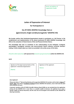
Inflammation of islets and apoptosis
And how to protect the β-cell from Death
Kathrin Maedler, Larry L. Hillblom Islet Research Center, UCLA
Apoptosis in T1DM
Insulin Caspase 3
T1DM
beta cell area
(% total pancreatic area)
1.4
*
1.2
1.0
0.8
0.6
0.4
0.2
0.0
control
% beta cells
positive for cleaved Caspase 3
T1DM
control
8.0
7.0
6.0
5.0
4.0
*
3.0
2.0
1.0
0.0
T1DM
control
Meier et al, Diabetologia 2005
Apoptosis in T2DM
control
T2DM
Butler et al, Diabetes 2003
Apoptosis in transplanted islets
Extrinsic Signals:
Fas
Cytokines
GLP-1
?
A
B
J.Leuc.Biol 77, May 2005
human islet
intraportal porcine islet
Diabetes 48, Oct. 1999
allograft
Environment in the liver
Hypoxia
c-FLIP
Intrinsic Signals:
Hypoxia
Reactive oxygen
Species
Nutrient Withdrawal
DNA damage
XIAP
Clin.Exp.Imm144, Feb. 2006
Activation of intrahepatic endothelial and Kupffer cells,
resulting in inflammation and thrombosis.
TLR-4 up-regulation
TLR-4
Diabetes 55, July 2006
insulin
FASEB J. 21, May 2007
Apoptosis in mouse islet transplanted, inhibited by
anticoagulant APC, Diabetes 53, Nov.2004
The Mechanisms of β-cell apoptosis
in T1DM
Islet antigen
Mathis et al, Nature 2002
The Mechanisms of β-cell apoptosis
in T2DM
β-cells
The Mechanisms of β-cell apoptosis
in T2DM
β-cells
The Mechanisms of β-cell apoptosis
in T2DM
β-cell mass
β-cell dysfunction
β-cell apoptosis
β-cells
insulin resistance
GLUCOSE
Free fatty acids
free radicals/
oxidative stress
IAPP
ER stress
iNOS
pro-inflammatory
Cytokine
anti-inflammatory
cytokines
Adipokines
diabetic genes:
PPARγ, Fas/FasL
Calpain 10, HNF4
TCF7L2, Znt-8,
CDKAL-1
Glucose is an important fuel for the β-cell
β-Cells
Glucose
insulin
secretion
Proliferation
Glucose Regulation
Glucose-induced β-cell turnover
Glucose
β-Cells
Apoptosis
Impaired
function
Diabetes
Glucose-induced β-cell turnover
ZB4
Glucose
FasL
Fas
Caspase
activation
β-Cells
Apoptosis
Diabetes
il p
Impaired
+F
function
Proliferation
Are there other regulator of the Fas pathway
in the β-cell?
The effect of IL-1β on the β-cell
IL-1β
IL-1R1
IL-1AcP
Fas, Caspases,
JNK/p38/ERK
iNOS, SOCS-3,
IκB
PKCδ
bcl-2...
NFκB
PDX-1, Isl-1,
GLUT2...
β-cell
Apoptosis
impaired function
Regulation of IL-1β & IL-1Ra in the β-cell in vivo
IL-1β
Insulin
Merge
IL-1Ra
Insulin
Merge
nondiabetic
control
patient
with T2DM
nondiabetic
control
patient
with T2DM
Maedler et al,
PNAS 2004
Glucose-induced β-cell turnover
ZB4
Glucose
IL-1β
IL-1Ra
FasL
Fas
Caspase
activation
β-Cells
Apoptosis
Diabetes
Impaired
function
Inhibit β-cell apoptosis by blocking IL-1β
signaling?
The effect of IL-1β/ IL-1Ra on the β-cell
IL-1β
IL-1Ra
IL-1R1
IL-1AcP
Fas, Caspases,
JNK/p38/ERK
iNOS, SOCS-3,
IκB
PKCδ
bcl-2...
NFκB
PDX-1, Isl-1,
GLUT2...
β-cell
Apoptosis
impaired function
IL-1Ra is protective against β-cell apoptosis
and improves β-cell function
glucose 5.5 mM
33.3 mM
33.3 mM+IL-1Ra
TUNEL Insulin
insulin secretion
(pmol/islet)
2.5
basal
stimulated
2.0
1.5
**
1.0
*
0.5
0.0
IL-1Ra (ng/ml) 0
500
*
0.5
β-cell proliferation
after 4 days of culture
0.0
glucose (mM) 5.5
IL1-Ra
-
% Ki67pos β -cells
1.0
5.5
+
33.3
-
33.3
+
* p<0.05 to 5.5 mM
**p<0.05 to 33.3 mM
Is IL-1Ra protective against diabetes
progression in vivo?
Apoptosis
Glucose
IL-1Ra
Does IL-1Ra influence
β-cell function?
β-cell survival?
β-cell gene transcription?
Impaired
function
Is IL-1Ra protective against diabetes
progression in vivo?
•
Start with five weeks old male C57BL/6
•
Daily injections of IL-1Ra or solvent for 12 weeks
so
t
n
e
lv
control diet
I
HFD: high fat/
high sucrose diet
a
R
1
L-
control diet
HFD: high fat/
high sucrose diet
IL-1Ra improves glucose tolerance
Intraperitoneal glucose tolerance test after 12 weeks
of diet and treatment
35
control
control + IL-1Ra
30
Blood glucose (mM)
25
*
20
15
10
5
0
-20
0
20
40
60
80
time after injection (min)
100
120
*p<0.05 to untreated same diet
+p<0.05 to control diet
IL-1Ra improves glucose tolerance
Intraperitoneal glucose tolerance test after 12 weeks
of diet and treatment
35
control
control + IL-1Ra
High fat diet
30
Blood glucose (mM)
25
*
20
+
15
10
5
0
-20
0
20
40
60
80
time after injection (min)
100
120
*p<0.05 to untreated same diet
+p<0.05 to control diet
IL-1Ra improves glucose tolerance
Intraperitoneal glucose tolerance test after 12 weeks
of diet and treatment
35
control
control + IL-1Ra
High fat diet
30
High fat diet + IL-1Ra
Blood glucose (mM)
25
*
20
+
15
*
10
5
0
-20
0
20
40
60
80
time after injection (min)
100
120
*p<0.05 to untreated same diet
+p<0.05 to control diet
IL-1Ra improves insulin secretion
Intraperitoneal glucose tolerance test
after 12 weeks of treatment
2.0
*
0 min
30 min
Insulin secretion
µg/L
1.5
1.0
*
0.5
0.0
IL-1Ra
-
+
control
* p<0.05 to basal same treatment
IL-1Ra improves insulin secretion
Intraperitoneal glucose tolerance test
after 12 weeks of treatment
2.0
*
0 min
30 min
Insulin secretion
µg/L
1.5
1.0
*
0.5
0.0
IL-1Ra
-
+
control
High fat diet
* p<0.05 to basal same treatment
IL-1Ra improves insulin secretion
Intraperitoneal glucose tolerance test
after 12 weeks of treatment
2.0
*
0 min
30 min
*
Insulin secretion
µg/L
1.5
1.0
*
0.5
0.0
IL-1Ra
-
+
control
-
+
High fat diet
* p<0.05 to basal same treatment
IL-1Ra improves β-cell turnover
Ki67-positive β-cells
% of control
control
250
High fat diet
*
untreated
200
*
150
Ki67
Insulin
DAPI
100
50
0
IL-1Ra
-
+
control diet
-
+
IL-1Ra
High fat diet
* p<0.05 to untreated same diet
β-cell apoptosis
• no increase in β-cell apoptosis in HFD-mice in pancreas sections
• BUT: higher sensitivity in isolated islets
TUNEL-positive β-cells
% of control
control
500
High fat diet
*
400
untreated
300
200
100
0
IL-1Ra
-
+
control diet
-
+
IL-1Ra
High fat diet
*p<0.05 to control same treatment
+p<0.05 to control HFD
HFD reduces insulin mRNA which is
restored by IL-1Ra treatment…
Quantitative RT-PCR analysis
Insulin
Insulin/GAPDH
% control
300
200
100
*
0
IL-1Ra
-
+
control
-
+
High fat diet
*p<0.05 HFD to control same treatment
Summary & Conclusion
• IL-1Ra protects from diet-induced diabetes.
• IL-1Ra improves β-cell function.
•IL-1Ra improves β-cell survival.
IL-1Ra overexpression increases beta cell replication
and mass in transplanted islets, and improves metabolic
outcome. (Diabetologia. 2007 Mar;50(3):602-11).
Results from a recent clinical study in patients with
T2DM showed that IL-1Ra improved glycaemic control
and β-cell function (NEnglJMed 356:15 2007).
The fact that IL-1Ra improves β-cell function is in favor
for the critical role of IL-1β signaling in the β-cell in type
1, type 2 diabetes and in islet transplantation. Our data
implements IL-1Ra for a potential therapy of diabetes.
Acknowledgements
Andres Quiros
Valerie Zamudio
Ruth Schruefer
Fabienne Schulthess
Nadine Sauter
Heather Gerber
Lendy Lee
Michelle Peng
Luan Shu
Michael Muehle
Leena Haataja, Anil Bhushan & Peter Butler, LHIRC, UCLA
Jake Lusis, Dept. of Genetics, UCLA
Jose Oberholzer, University of Illinois at Chicago
Marc Donath, USZ Zurich
American Diabetes Association
Larry L. Hillblom Research Foundation
NIH
Swiss National Foundation
German Research Foundation
ICR for human islets


© Copyright 2026