
Document 192394
Copyright ERS Journals Ltd 1997
European Respiratory Journal
ISSN 0903 - 1936
Eur Respir J 1997; 10: 522–529
DOI: 10.1183/09031936.97.10030522
Printed in UK - all rights reserved
Accurate measurement of intrinsic positive endexpiratory pressure: how to detect and correct for
expiratory muscle activity
S.G. Zakynthinos, T. Vassilakopoulos, E. Zakynthinos, C. Roussos
Accurate measurement of intrinsic positive end-expiratory pressure: how to detect and
correct for expiratory muscle activity. S.G. Zakynthinos, T. Vassilakopoulos, E. Zakynthinos,
C. Roussos. ©ERS Journals Ltd 1997.
ABSTRACT: It has been shown that expiratory muscle contraction leads to an
overestimation of intrinsic positive end-expiratory pressure (PEEPi). To quantify
this overestimation, we compared PEEPi, measured during spontaneous breathing
(SB) by the end-expiratory airway occlusion technique (PEEPi,occl) with static
PEEPi (PEEPi,st). PEEPi,st was measured using end-expiratory airway occlusion
during simulation of SB by the ventilator with the patient relaxed, and was considered to represent the "gold standard" for PEEPi,occl.
Twelve ventilator-dependent patients were studied during SB (pressure support
5–7 cmH2O). Full mechanical ventilation was resumed when they were unable to
sustain SB. Subsequently, by manipulating the variables of the ventilator, we simulated the pattern of SB and measured PEEPi,st, corresponding to PEEPi,occl. On
the basis of the presence or absence of expiratory rise in gastric pressure (Pga)
(rapid drop of end-expiratory Pga at the beginning of inspiration, Pga,exp,rise), and
abdominal muscle electromyographic (EMG) activity, patients were subdivided
into those either actively (Group 1) or passively expiring (Group 2).
In Group 1 (8 patients), PEEPi,occl was higher than PEEPi,st (13.3±2.0 vs 6.8±1.1
cmH2O; p<0.01). PEEPi,occl - Pga,exp,rise (6.9±1.1 cmH2O) was quite similar to PEEPi,st;
their mean difference was 0.03 cmH2O with limits of agreement -0.48 to +0.53
cmH2O. In Group 2, PEEPi,occl was similar to PEEPi,st.
We conclude that, in actively expiring patients, an accurate estimation of the
actual PEEPi,st can be obtained by subtracting Pga,exp,rise from PEEPi,occl.
Eur Respir J 1997; 10: 522–529.
In actively breathing patients, either during spontaneous or assisted ventilation, intrinsic positive end-expiratory pressure (PEEPi) can be measured by two methods.
In the first method, the airway occlusion method, PEEPi
is measured as the plateau in airway pressure between
occluded inspiratory efforts and expresses the elastic recoil pressure of the total respiratory system (PEEPi,occl)
[1]. In the second method, requiring the placement of an
oesophageal balloon, the decrease in oesophageal pressure needed to abruptly bring expiratory flow to the
point of zero during unoccluded breathing represents the
dynamic PEEPi (PEEPi,dyn) [1]. PEEPi measurements
obtained by these methods have been considered to indicate the presence of dynamic hyperinflation and have
been used, especially the second method, for its indiscriminate quantification. Recently, however, it has been
shown that expiratory muscle activity during expiration
increases the end-expiratory alveolar pressure independently of dynamic hyperinflation, leading to an overestimation of PEEPi. Indeed, during airway occlusion,
spontaneously breathing patients, using their expiratory
muscles, react by increasing the end-expiratory airway
pressure between inspiratory efforts, thus preventing the
appearance of a plateau to measure PEEPi [2, 3]. In this
instance, even if an apparent plateau in airway pressure
is present, PEEPi is overestimated [4]. During unoc-
Dept of Critical Care and Pulmonary Services, Athens University Medical School,
Evangelismos Hospital, Athens, Greece.
Correspondence: S. Zakynthinos
Evangelismos Hospital
Critical Care Department
45–47 Ipsilandou St
GR-106 75 Athens
Greece
Keywords: Abdominal muscles
expiratory muscles
intrinsic positive end-expiratory pressure
Received: May 8 1996
Accepted after revision November 25 1996
Supported by a NATO Scientific Grant
and The Thorax Foundation.
Presented in part at the Annual International
Conference of the American Thoracic Society in Seattle, WA, May 21–24, 1995.
cluded breathing, in turn, either in patients with stable
chronic obstructive pulmonary disease (COPD) [2] or
in mechanically-ventilated patients [3, 4], expiratory muscles can contract during expiration so that part of the
decrease in oesophageal pressure preceding inspiration
measured as PEEPi,dyn is due to relaxation of the expiratory muscles rather than to contraction of the inspiratory muscles to counterbalance PEEPi.
The amount of PEEPi overestimation due to the use
of expiratory muscles that should be subtracted from
measured PEEPi to calculate the actual ("true") PEEPi
produced by dynamic hyperinflation, remains unclear.
Thus, the purpose of this study was to quantify the influence of expiratory muscle activity on PEEPi measurement in spontaneously breathing patients. Towards this
goal, we measured the static PEEPi (PEEPi,st) caused by
dynamic hyperinflation and compared this value with
PEEPi,occl measured in actively expiring patients. We
used a method described recently [5] to measure PEEPi
during spontaneous breathing under real static conditions, i.e. by simulating the exact pattern of spontaneous
breathing with the ventilator during controlled mechanical ventilation and occluding the airway at the end of
a tidal expiration [6]. PEEPi,st values obtained in this
way were considered to represent the "gold standard"
for PEEPi,occl. The study was performed on patients with
523
I N T R I N S I C P E E P A N D E X P I R ATO RY M U S C L E A C T I V I T Y
acute respiratory failure, spontaneously breathing after
discontinuation from mechanical ventilation and, subsequently, needing resumption of mechanical assistance. Since this condition may be considered as a good
model of acute respiratory failure with the patients being in severe respiratory distress, we expected that at least
some of them would actively expire during the spontaneous breathing trial. In addition to the actively expiring patients, the passively expiring patients were also
studied to compare the results and confirm the accuracy of PEEPi,occl measurement in this latter condition.
Methods
Patients
Twelve patients (7 males, and 5 females) admitted to
the intensive care unit for management of acute respiratory failure of different aetiologies (table 1), participated in the study. Diagnosis in four patients with COPD
was based upon previous clinical history and routine
lung function tests. Three patients with adult respiratory
distress syndrome (ARDS) met the conventional criteria for the diagnosis of the syndrome; their lung injury
scores on the day of the study were 1.8, 2.0 and 2.1.
Four patients had severe sepsis according to the currently
accepted criteria for the diagnosis of septic syndrome;
three of them had positive blood cultures associated with a
localized site of infection. The investigative protocol was
approved by the institutional Ethics Committee and in-
formed consent was obtained from the family of each
patient.
All patients were intubated with a Portex cuffed endotracheal tube (Portex Ltd, Kent, UK), 8–9 mm inner diameter, cut to a length of 25 cm. The patients had been
mechanically-ventilated for a period of 4–10 days prior
to the present investigation and were clinically stable
(systolic blood pressure >100 mmHg; heart rate <120
beats·min-1; and with no significant fluctuations in blood
pressure, heart rate, arterial blood gas values, urine output, or mental status) for the preceding 12 h. All sedative and paralysing medication was discontinued at least
12 h prior to the study. Other drug and fluid therapy
continued throughout the study. All patients were studied early in the course of acute respiratory failure, at a
time when discontinuation from full ventilatory assistance was felt to be impossible. Mechanical ventilation
was delivered by a Siemens 300 servo ventilator (Siemens-Elema, Solna, Sweden) in the assist-control (A/C)
mode, with the ventilator settings prescribed by the primary physicians. Tidal volume (VT) was set on the ventilator at 0.47–0.78 L and respiratory frequency (f R)
ranged 16–28 breaths·min-1. The time of mechanical
inflation (t I) ranged 0.49–0.95 s, whereas the duration
of expiration (t E) ranged 1.30–2.35 s. Mean inspiratory
flow (VT/t I) ranged 0.69–1.20 L·s-1, and PEEP was 0–6
cmH2O. Thirty minutes before the beginning of the study,
PEEP was removed and fraction of inspired oxygen
(FI,O2) increased to 100% in all patients. Baseline blood
gas values were obtained (ABL 300) 25 min later. Arterial
oxygen tension (Pa,O2) (mean±SD) was 20.1±6.4 kPa
Table 1. – Patient clinical characteristics, pertinent respiratory mechanics, and behaviour during spontaneous breathing after discontinuation from mechanical ventilation
Pt
No.
Sex
Age
yrs
F
F
M
M
M
54
60
56
64
59
F
M
M
42
70
70
Group 2
9
10
F
F
76
75
11
12
M
M
50
48
Group 1
1
2
3
4
5
6
7
8
Diagnosis
Rrs,max*
Cst,rs*
L·cmH2O-1 cmH2O·L-1·s-1
Duration of
Expiratory
discontinPa,CO2
Rrs,min*
uation
increase+
muscle
cmH2O·L-1·s-1
min
mmHg
activity‡
COPD
COPD
COPD
COPD
ARDS, flail
chest
ARDS
ARDS
Coronary
bypass, CPE
0.068
0.066
0.071
0.060
0.036
29.5
31.1
17.5
15.3
13.5
21.7
22.4
11.2
10.8
8.1
30
35
50
42
15
37
30
28
20
15
+
+
+
+
+
0.030
0.026
0.048
12.1
13.0
14.7
7.8
6.5
10.6
20
26
29
16
14
41
+
+
+
Sepsis
Atelectasis,
sepsis
Sepsis
Pneumonia,
sepsis
0.070
0.059
12.0
13.1
9.1
8.4
60
23
29
30
-
0.065
0.049
12.4
14.5
9.2
8.7
30
34
32
18
-
Pt: patient; M: male; F: male; COPD: exacerbated chronic obstructive pulmonary disease; ARDS: adult respiratory distress syndrome; CPE: cardiogenic pulmonary oedema; Cst,rs: static compliance of the total respiratory system; Rrs,max and Rrs,min: total
and minimal (airway) respiratory system resistance (after subtraction of the endotracheal tube resistance [7]). Pa,CO2: arterial carbon dioxide tension (1 mmHg=0.133 kPa). *: assessed during control mechanical ventilation with the patient sedated and/or paralysed, using the constant flow end-inspiratory occlusion method [7]; +: increase in Pa,CO2 at the end of spontaneous breathing trial,
compared to its value at the beginning of this trial; ‡: Based on the presence or absence of expiratory muscle activity during
expiration, patients were divided into two groups. +: denotes the presence of expiratory muscle activity at expiration during spontaneous breathing; -: denotes the absence of expiratory muscle activity.
524
S . G . ZAKYNTHINOS ET AL .
(151±48 mmHg), and arterial carbon dioxide tension
(Pa,CO2) was 6.3±1.2 kPa (47±9 mmHg). During the
study, a physician not involved in the procedure was
always present to provide for patient care as needed.
The electrocardiogram, the heart rate, the systemic arterial blood pressure and the arterial oxygen saturation
(Sa,O2) (Nellcor, CA, USA) were monitored continuously.
Experimental set-up
All measurements were made with the patients in a
semirecumbent position. Airflow (V') was measured with
a heated pneumotachograph (Fleisch No. 2; Lausanne,
Switzerland) inserted between the endotracheal tube
and the Y-piece of the ventilator, and a differential pressure transducer (Validyne MP-45, ±2 cmH2O; Northridge,
CA, USA). VT was obtained by integrating the flow signal. Oesophageal (Poes) and gastric (Pga) pressures were
measured with conventional balloon-catheter systems
placed in the midoesophagus and the stomach, respectively. The oesophageal balloon was filled with 0.5 mL
of air, and the gastric balloon contained 1.0 mL of air.
Both balloons were connected to separate differential
pressure transducers (Validyne MP-45, ±100 cmH2O).
Appropriate placement of the oesophageal balloon was
verified by an occlusion test [8]. Airway pressure (Paw)
was recorded at the distal end of the endotracheal tube
with a differential pressure transducer (Validyne MP45, ±100 cmH2O). Rib cage (RC) and abdominal (AB)
displacements were measured with a respiratory inductive plethysmograph (Respitrace Ambulatory Monitoring, Ardsley, NY, USA). The bands were placed circumferentially around the RC and AB, in such a way
that they were at the level of the nipples and umbilicus, respectively. Care was taken to avoid overlap of
the AB band with the lower RC. The electrical activity of the abdominal muscles (EMGab) was recorded
with surface electrodes placed in the right anterior axillary line, midway between the costal margin and the iliac
crest, and conditioned with a Nihon-Kohden electromyograph amplifier (band-pass between 20 Hz and 1
kHz). All signals, except EMGab, were continuously recorded on an eight-channel electrostatic recorder (Gould
ES 1000; Gould Instruments, Cleveland, OH, USA) at
a paper speed of 10 or 25 mm·s-1, and taped on a videorecorder (including EMGab) via an analogue-to-digital
converter. The data were played back to a personal computer (Wyse 486) by the same analogue-to-digital converter, at a sampling rate of 1,500 Hz (EMGab) or 200
Hz (other variables), for subsequent data analysis.
Protocol
Patients were initially allowed to breathe spontaneously through the ventilator with a small inspiratory assistance (pressure support 5–7 cmH2O) to compensate
for the additional work due to the endotracheal tube and
inspiratory circuit [9]. Fifteen to sixty minutes (33±13
min) after the beginning of spontaneous breathing, Pa,CO2
increased ≥1.9 kPa (14 mmHg) in all patients (3.5±1.2
kPa) (26±9 mmHg)) (table 1), and reinstitution of mechanical ventilation on A/C was required. In every patient,
Sa,O2 remained higher than 90% throughout the spontaneous breathing trial. Pa,O2 was 17.2±7.3 kPa (129±55
mmHg) at the end of this trial. During spontaneous
breathing, the airway was occluded with the end-expiratory hold knob of the ventilator. Each occlusion lasted 8–12 s, and was repeated 2–3 times in every patient.
As soon as mechanical ventilation on the A/C mode
had been reinstituted, the patients were sedated (midazolam) and some were also paralysed (pancuronium
bromide). After an initial bolus dose of these drugs (15
mg midazolam, 0.06 mg·kg-1 pancuronium bromide)
had been given, the patients continued to receive the
same sedative and paralysing medication in order to
maintain undetectable respiratory muscle activity (judged by the lack of inspiratory swings of Poes, airway
pressure wave contour representative of passive inflation and clinical assessment).
Recordings of V' and volume during spontaneous breathing were analysed in terms of VT, f R and duty cycle
(t I/t tot) to obtain the breathing pattern. The period of
time selected for analysis in every patient was just before the initiation of an airway occlusion (randomly
selected among the 2–3 performed in each patient). The
value obtained was the mean of three consecutive breaths. With the patient ventilated with control mechanical
ventilation and constant inspiratory flow, we subsequently tried to simulate this pattern of spontaneous
breathing in each patient by regulating the appropriate
buttons of the ventilator [5]. Inclusion criteria for accepting simulated breaths as representative of the breathing pattern during spontaneous ventilation were: 1)
VT ±0.02 L; 2) f R ±0.1 breaths; and 3) t I/t tot ±0.02. For
the patients as a group, the breathing pattern before the
initiation of the airway occlusion was (mean±SD): VT
0.42±0.06 L; f R 28±6 breaths·min-1; and t I/t tot 0.33±
0.03. Breaths simulated by the ventilator were almost
identical, i.e: VT 0.42±0.07 L; f R 28±6 breaths·min-1;
and t I/t tot 0.33± 0.02.
Measurements and data analysis
During spontaneous breathing, expiratory muscle activity was assessed as follows. Firstly, the rise of Pga
was measured during the expiration from its end-inspiratory level to the maximum at end-expiration, and its
subsequent abrupt decrease at the beginning of next
inspiration (Pga,exp,rise) (fig. 1). This pattern, associated with a decrease in abdominal cross-sectional area
during the expiration and a subsequent increase during
the inspiration, gives characteristic Pga-AB displacement
loops [2, 3] and clearly indicates expiratory contraction of the abdominal muscles. The increase in Pga occurring during the expiratory phase of the breathing cycle
can be considered as a reflection of the direct mechanical effect of this contraction [2]. Secondly, the increase
in end-expiratory Poes (∆Poes) was measured, relative to
its level at the onset of spontaneous breathing trial,
when expiratory muscle activity was nil (fig. 1). This
increase in end-expiratory oesophageal pressure indicates recruitment of the expiratory muscles during expiration which, at least in normal subjects, leads to an
end-expiratory lung volume below the subject's normal
functional residual capacity (FRC) [10]. Thirdly, the
EMG activity of the abdominal muscle (EMGab) was
measured. Fourthly, palpation of the abdominal wall was
performed.
525
I N T R I N S I C P E E P A N D E X P I R ATO RY M U S C L E A C T I V I T Y
b)
∆Poes
a)
Poes
cmH2O
10
0
Pga
cmH2O
10
0
Pdi
cmH2O
10
0
V'
L·s-1
1
0
Paw
cmH2O
5
0
Poes
cmH2O
Pga,exp,rise
AB
10
0
Pga
10
cmH2O 0
Pdi
cmH2O
10
0
V'
L·s-1
1
0
Paw
cmH2O
10
0
PEEPi,occl
1s
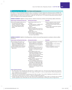
Fig. 1. – Tracings of oesophageal pressure (Poes), gastric pressure
(Pga), transdiaphragmatic pressure (Pdi), flow (V '), airway pressure
(Paw), and cross-sectional area of the abdomen (AB) in a representative patient (patient No. 1) actively expiring during spontaneous
breathing through the ventilator (pressure support 6 cmH2O). After
discontinuation from full mechanical assistance, the patient was
unable to sustain spontaneous breathing and resumption of mechanical ventilation was needed. a) corresponds to the beginning of spontaneous breathing (SB). b) corresponds to the end of spontaneous
breathing, 1 min before the reinstitution of control mechanical ventilation. The three vertical lines are passed through the onset of inspiratory muscle activity (i.e. beginning of Poes decay) and the beginning
and the end of inspiratory flow, respectively. In contrast to the beginning of spontaneous breathing (a), note the large increase of Pga and
Poes during expiration due to expiratory muscle recruitment at the
end of spontaneous breathing trial (b). For further explanation, see
text.
PEEPi during spontaneous breathing was measured by
two methods described previously [1, 5]. In the first method, the airway was occluded at end-expiration using
the end-expiratory hold button of the ventilator, and the
occlusion was maintained for several consecutive inspiratory efforts. The value of end-expiratory plateau in
Paw between occluded inspiratory efforts represents the
elastic recoil pressure of the total respiratory system [1],
and is called "PEEPi,occl" in the present study. An expiratory plateau in Paw postocclusion was apparent in
all patients who did not use their expiratory muscles
during spontaneous breathing (fig. 2). In contrast, this
was not the case in the actively expiring patients (fig.
3). For the sake of consistency, in these patients the
value of Paw at the end of expiration of the first postocclusion inspiratory effort was also referred to here as
"PEEPi,occl". However, it is important to emphasize that,
in this circumstance, the term PEEPi,occl was only used
to indicate the presence of a positive alveolar pressure
at end-expiration produced through whatever mechanism. This pressure does not reflect the elastic recoil
of the total respiratory system, since expiratory muscle
contraction obviously contributes to its value. The second method was performed under static conditions [5].
With the patient sedated and sometimes paralysed during the simulation of spontaneous breathing by the ventilator, the airway was occluded at the end of a tidal
expiration using the end-expiratory hold button of the
ventilator (fig. 3). The end-expiratory plateau of Paw
1s
Occlusion
Fig. 2. – Recordings of oesophageal pressure (Poes), gastric pressure (Pga), transdiaphragmatic pressure (Pdi), flow (V ') and airway
pressure (Paw) during end-expiratory airway occlusion in a representative patient (patient No. 9) passively expiring during spontaneous breathing through the ventilator (pressure support 5 cmH2O).
Airway occlusion was performed with the expiratory hold knob of
the Siemens 300 ventilator. An apparent end-expiratory plateau in
Paw is present between occluded inspiratory efforts representing the
elastic recoil of the total respiratory system. Note that expiration remains passive in all consecutive inspiratory efforts post-occlusion.
directly reflects intrinsic PEEP (PEEPi,st) [6]. With the
ventilator, we simulated one pattern of spontaneous breathing in each patient, i.e. that corresponding to the period just prior to an airway occlusion.
When the patient was ventilated with controlled mechanical ventilation during the simulation of spontaneous
breathing, respiratory mechanics (table 1) were assessed
by the constant flow end-inspiratory occlusion method
described in detail previously [7].
Results are expressed as mean±SE, unless otherwise
specified. Statistical analysis was performed using Student's paired t-test and linear regression analysis. A pvalue of 0.05 was considered significant. The agreement
between PEEPi,occl (corrected for expiratory muscle activity) and PEEPi,st which measure the same physiological variable, i.e. the static PEEPi, was evaluated by the
method of BLAND and ALTMAN [11]. The degree of agreement was summarized by calculating the mean difference and the standard deviation of the differences (SD).
If differences within the limits of agreement, i.e. mean
difference ±2SD, are not clinically important, the two
methods can be used interchangeably.
Results
Patients were divided in two groups on the basis of
expiratory muscle activity during the spontaneous breathing trial (table 1). Group 1 consisted of patients who
were using their expiratory muscles at expiration (mostly COPD and ARDS). In every Group 1 patient, active
expiration was detected by the coexistence of a typical
S . G . ZAKYNTHINOS ET AL .
526
b)
a)
Poes
cmH2O
∆Poes
c)
10
0
10
Pga
cmH2O 0
▲
Pga,exp,rise
▲
Pdi 10
cmH2O
0
V'
L·s-1
1
0
PEEPi,occl
PEEPi,st
Paw 10
cmH2O 0
1s
Occlusion
Fig. 3. – Representative recordings of oesophageal pressure (Poes), gastric pressure (Pga), transdiaphragmatic pressure (Pdi), flow (V ') and airway pressure (Paw) during airway occlusion in a patient (patient No. 3) actively expiring during spontaneous breathing (SB) through the ventilator (pressure support 5 cmH2O). a) corresponds to the beginning of spontaneous breathing, after discontinuation from full mechanical ventilation.
b) Thirty minutes later the airway was occluded with the end-expiratory hold knob of the ventilator. Twenty minutes after airway occlusion (50
minutes from the beginning of spontaneous breathing), the patient was unable to sustain spontaneous breathing and reinstitution of control mechanical ventilation was needed. c) With the patient relaxed, the pattern of breathing just prior to the airway occlusion was subsequently simulated
by the ventilator. PEEPi,st was measured during the simulation under static conditions by the end-expiratory occlusion technique [6]. For further
explanation, see text. ∆Poes: increase in end-expiratory Poes relative to its level at the onset of the spontaneous breathing trial (a); Pga,exp,rise: gastric pressure decay from its maximal value to its minimal value at the beginning of the next inspiration; PEEPi,occl: end-expiratory value of airway pressure of the first inspiratory effort after airway occlusion at the end of expiration during spontaneous breathing, using the end-expiratory
hold button of the ventilator; PEEPi,st: PEEPi measured as the value of end-expiratory plateau of airway pressure during simulation of the pattern of spontaneous breathing with the ventilator, occluding the airway by the end-expiratory hold button.
Table 2. – Respiratory mechanics at the airway occlusion during spontaneous breathing in actively (Group 1)
and passively expiring patients (Group 2)
PEEPi,occlPEEPi,occlPEEPi,occl
Pga,exp,rise
∆Poes
PEEPi,st
Pga,exp,rise
Pt.
PEEPi,st
No.
cmH2O
cmH2O
cmH2O
cmH2O
cmH2O
cmH2O
Group 1
1
6.6
18.7
12.0
17.6
12.1
6.7
2
7.2
18.6
11.0
8.3
11.4
7.6
3
12.3
19.5
7.5
5.4
7.2
12.0
4
10.1
15.8
6.0
5.9
5.7
9.8
5
2.5
5.5
3.1
3.4
3.0
2.4
6
4.1
7.2
3.0
2.8
3.1
4.2
7
4.8
7.5
2.7
2.0
2.7
4.8
8
7.1
13.4
6.0
7.4
6.3
7.4
6.8±1.1**
13.3±2.0
6.4±1.3
6.6±1.8
6.4±1.3
6.9±1.1
Mean±SE
Group 2
9
6.5
6.6
0
0
0.1
6.6
10
2.3
2.3
0
0
0
2.3
11
5.0
5.2
0
0
0.2
5.2
12
4.0
4.1
0
0
0.1
4.1
4.5±0.9
4.6±0.9
0
0
0.1±0.0
Mean±SE
Pt: patient. **: p<0.01, significantly different from PEEPi,occl. For further definitions see legend to figure 3.
Pga - AB displacement loop produced by the increasingly positive Pga at the same time that abdominal crosssectional area decreased during expiration, and a positive
∆Poes and EMGab. It is notable that palpation of the
abdominal wall also revealed the abdominal muscle
contraction during expiration in all Group 1 patients. In
Group 2, the patients did not use their expiratory muscles at expiration; all were septic. In the Pga - AB dis-
4.6±0.9
placement loops, Pga increased from the beginning to the
end of inspiration and decreased during expiration, indicating passive expiration. Moreover, ∆Poes and EMGab
were zero. Palpation of the abdominal wall did not detect abdominal muscle contraction in any patient of this
group.
The results of measurements performed during airway occlusion are summarized in table 2. During the
I N T R I N S I C P E E P A N D E X P I R ATO RY M U S C L E A C T I V I T Y
a)
PEEPi cmH2O
**
Pga,exp,rise
5
b)
PEEPi,occl
PEEPi,st
20
15
PEEPi cmH2O
10
5
0
10
0
15
0
20
15
20
PEEPi,st cmH2O
airway occlusion (fig. 3) in Group 1 patients, measurements showed that PEEPi,occl was significantly higher
than PEEPi,st (13.3±2.0 vs 6.8±1.1 cmH2O; p<0.01; n=8).
The difference, PEEPi,occl - Pga,exp,rise (6.9±1.1 cmH2O),
was very close to PEEPi,st (6.8±1.1 cmH2O) (fig. 4a).
Accordingly, the difference, PEEPi,occl - PEEPi,st, was
quite similar to Pga,exp,rise (and ∆Poes) (table 2). Good
correlations were detected between PEEPi,occl - PEEPi,st
and ∆Poes (r=0.869; p<0.01), and PEEPi,occl - Pga,exp,rise
and PEEPi,st (r=0.997; p<0.001) (fig. 5). The values of
both the above regressions were very close to the line
of identity. The mean difference between PEEPi,occl Pga,exp,rise and PEEPi,st was 0.03 cmH2O. The limits of
agreement (i.e. mean difference -2SD to mean difference
527
10
5
10
15
PEEPi-occl-Pga,exp,rise cmH2O
20
Fig. 5. – Identity plot for values of the difference PEEPi,occl - Pga,exp,rise
and PEEPi,st in actively expiring patients (n=8). Regression equation
(dotted line) is shown (p<0.001); all values are very close to the line
of identity (solid line). This finding indicates that PEEPi,occl overestimates the actual static PEEPi (represented by PEEPi,st) by an amount
equal to Pga,exp,rise. For definitions see legend to figure 3.
±2SD) were -0.48 to +0.53 cmH2O. These data convincingly indicate that, in spontaneously breathing and
actively expiring patients, the so-called PEEPi,occl, overestimates the "true" static PEEPi by an amount equal to
Pga,exp,rise (or ∆Poes).
In contrast to Group 1, in Group 2 during the airway
occlusion (fig. 2), PEEPi,occl, and PEEPi,st had quite similar mean values (4.6±0.9 and 4.5±0.9 cmH2O, respectively; n=4), (table 2, and fig. 4b). Individual values of
PEEPi,occl and PEEPi,st were well-correlated (r=0.999;
p<0.001) and were positioned close to the identity line.
Their mean difference was 0.1 cmH2O, and the limits of
agreement were -0.06 to +0.26 cmH2O. Therefore, in
those patients who did not use their expiratory muscles at
expiration, PEEPi,occl accurately represented the PEEPi,st.
Discussion
5
0
PEEPi,occl
PEEPi,st
Fig. 4. – Values of PEEPi,occl and PEEPi,st in: a) actively expiring
patients (Group 1) (n=8) and b) passively expiring patients (Group
2) (n=4). PEEPi,occl was measured from the end-expiratory plateau in
airway pressure between occluded inspiratory efforts. Although an
apparent plateau was not present in actively expiring patients, for the
sake of consistency, in these patients the value of airway pressure at
the end-expiration of the first postocclusion inspiratory effort was also
referred to here as PEEPi,occl. PEEPi,st was considered to represent
the actual static PEEPi and was measured from end-expiratory plateau
of airway pressure during simulation by the ventilator of the pattern
of spontaneous breathing just before airway occlusion. PEEPi,occl was
higher than PEEPi,st in actively expiring patients (a). Subtracting the
mean value of Pga,exp,rise from that of PEEPi,occl, the difference
obtained was almost equal to PEEPi,st. In contrast to actively expiring
patients, in passively expiring patients PEEPi,occl was similar to PEEPi,st
(b), indicating that PEEPi,occl reliably measures the actual static
PEEPi. **: p<0.01, compared to PEEPi,st. For definitions see legend
to figure 3.
This study yielded two principal findings. Firstly, in
spontaneously breathing and actively expiring patients,
PEEPi measured with the airway occlusion (PEEPi,occl)
technique overestimates the actual PEEPi. Pga,exp,rise
can accurately express the amount of this overestimation.
Thus, by subtracting Pga,exp,rise from PEEPi,occl, an accurate estimation of the actual PEEPi,st can be made.
Secondly, in passively expiring patients, the airway occlusion technique measures the actual PEEP i,st accurately.
PEEPi,occl measurement in the presence of expiratory
effort
The measurement of PEEPi during spontaneous breathing by the end-expiratory airway occlusion technique (PEEPi,occl) is of particular interest for two reasons.
Firstly, the value of PEEPi is obtained under static conditions, and is considered to represent an average level
528
S . G . ZAKYNTHINOS ET AL .
of regional PEEPi reflecting the alveolar pressure after
readjustment of dynamic regional volume and pressure
differences [1]. In contrast, the value of PEEPi,dyn indicates the pressure required to initiate inspiratory flow
into those lung units with the lowest levels of end-expiratory alveolar pressure [1], thus underestimating the
PEEPi in the presence of regional time constant inequalities, as in patients with COPD. Secondly, and probably more important in clinical practice, PEEPi,occl is
measured by monitoring the Paw or simply by observing the airway pressure manometer of the ventilator
noninvasively, that is, without the need to position an
oesophageal balloon. In the presence of expiratory muscle activity during expiration, however, an accurate estimation of alveolar pressure reflecting the elastic recoil
of the total respiratory system by the measurement of
PEEPi,occl, cannot be obtained. In this instance, what is
measured as PEEPi,occl simply indicates the presence of
positive alveolar pressure at the end of expiration postocclusion; besides the elastic recoil of the respiratory
system, expiratory muscle contraction apparently contributes to its amount.
The results of this study clearly indicate that, by subtracting Pga,exp,rise from PEEPi,occl measured at the
end-expiration of the first inspiratory effort after the airway occlusion, an accurate value of the actual PEEPi,st
can be obtained. The values of Pga,exp,rise used were the
average of three consecutive breaths prior to the airway
occlusion. These values were almost the same as the
Pga,exp,rise of the first inspiratory effort against the occluded airway (6.4±1.3 vs 6.5±1.3 cmH2O), indicating
that expiratory muscle activity during expiration of the
first inspiratory effort postocclusion was nearly the
same as that during unoccluded spontaneous breathing
just prior to the airway occlusion. It stands to reason,
therefore, that the positive alveolar pressure measured
as PEEPi,occl at the end of expiration of the first inspiratory effort after the airway occlusion is the sum of the
actual PEEPi,st and Pga,exp,rise.
Clinical detection of expiratory muscle contraction
To interpret the rise in Pga as being a consequence of
expiratory contraction of the abdominal muscles, it is
necessary to check that it occurs together with a decrease
in abdominal dimensions [2, 4]. In every patient included in the present study, as in the four cases of LESSARD
et al. [4], the rise in Pga during active expiration occurred simultaneously with a decrease in abdominal dimensions. This finding suggests that Pga recording may
be adequate to detect abdominal muscle contraction during expiration, without necessitating measurement of
the AB displacement.
In the present study, palpation of the abdominal wall
had 100% accuracy in revealing abdominal muscle activity during expiration. Therefore, when palpation of
the abdominal wall can not demonstrate any abdominal muscle activity during expiration, the PEEPi,occl
measurements obtained may be considered as accurate,
without the need for Pga recording. On the contrary, when
abdominal muscle contraction exists, the positioning of
a gastric balloon is necessary to correct PEEPi,occl for
the contribution of expiratory muscle activity. It must
be stressed, however, that the patients in the present
study belonged to a special category; they had acute respiratory failure and were allowed to breathe spontaneously, being in severe respiratory distress when the
palpation of the abdominal wall was performed. Hence,
in these actively expiring patients, the powerful abdominal muscle contraction producing a Pga,exp,rise of 6.4±
1.3 cmH2O (range 2.7–12 cmH2O) (table 2) was easy to
detect clinically; the absence of such an abdominal muscle contraction was indicative of passive expiration. This
may not be the case when active expiration is associated with a weak contraction of the abdominal muscles.
For example, in stable COPD [2] or during exacerbations of COPD not needing intubation [3], Pga,exp,rise
may be only a few cmH2O. In this instance, palpation of
the abdominal wall may not be accurate in detecting abdominal muscle contraction at expiration.
Nevertheless, we have observed in this study that actively expiring patients after airway occlusion react by
further increasing their expiratory muscle activity in an
unpredictable manner. In contrast, passively expiring
patients after airway occlusion do not react by recruiting their expiratory muscles and expiration remains passive throughout the airway occlusion manoeuvre (fig.
2). Therefore, an airway occlusion manoeuvre may increase the ability of palpation of the abdominal wall to
detect the presence or absence of expiratory muscle contraction at expiration during unoccluded spontaneous
breathing. Abdominal muscle activity during expiration
that is small and undetectable by palpation during unoccluded breathing, may become apparent during airway
occlusion. On the contrary, absence of abdominal muscle contraction by palpation of the abdominal wall during the airway occlusion indicates that expiration was
passive during the preceding unoccluded breathing. In
this instance it is not necessary to position a gastric balloon to detect expiratory muscle contraction.
Clinical implication
The possible clinical implication of these findings is
related to the accurate and easy measurement of PEEPi,st
during spontaneous breathing in order to apply the appropriate level of external continuous positive airway pressure (CPAP) or positive end-expiratory pressure (PEEP)
to decrease inspiratory muscle effort due to PEEPi. Airway occlusion appears to be the method of choice for
measuring PEEPi in this instance, since the Paw recording that is needed to obtain PEEPi,occl may be routinely performed in almost any clinical setting. When Paw
tracing and/or palpation of the abdominal wall during
airway occlusion indicates expiratory muscle contraction during expiration, the placement of a gastric balloon will provide Pga fluctuations required to correct
Paw tracing for the contribution of expiratory muscle
activity and obtain the actual static PEEPi. Assessment
of PEEPi,dyn carries the disadvantage of necessitating
an additional invasive measurement, that is, Poes. In addition, the clinical implications of PEEPi,dyn have not
yet been fully defined [12].
In conclusion, the present study demonstrates that in
spontaneously breathing and passively expiring patients the airway occlusion technique (PEEPi,occl) accurately measures PEEPi,st. On the contrary, in actively
I N T R I N S I C P E E P A N D E X P I R ATO RY M U S C L E A C T I V I T Y
expiring patients, this technique overestimates the actual PEEPi,st by an amount equal to the mechanical effect
produced by expiratory muscle contraction at end-expiration. This effect is accurately expressed by Pga,exp,rise
during PEEPi,occl measurement. Thus, to correct PEEPi,occl
for the contribution of expiratory muscle activity, we
should subtract Pga,exp,rise measured either exactly before
the airway occlusion or at the end-expiration of the first
inspiratory effort postocclusion. Palpation of the abdominal wall may help in the detection of abdominal muscle activity. When this activity is absent by palpation
even during the airway occlusion manoeuvre, it is not
necessary to position a gastric balloon to detect expiratory muscle contraction, and PEEPi,occl measurements
accurately reflect the actual PEEPi,st. When it is present,
Pga recording is needed to correct PEEPi measurements
for the contribution of expiratory muscle contraction.
Acknowledgements: The authors are grateful to G.
Tzelepis for his helpful suggestions and criticisms, and
to M. Titcomb for her secretarial assistance.
References
1.
2.
3.
Petrof BJ, Legare M, Goldberg M, Milic-Emili J, Gottfried
SB. Continuous positive airway pressure reduces work
of breathing and dyspnea during weaning from mechanical ventilation in severe chronic obstructive pulmonary
disease. Am Rev Respir Dis 1990; 141: 281–289.
Ninane V, Yernault, JC, De Troyer A. Intrinsic PEEP
in patients with chronic obstructive pulmonary disease:
role of expiratory muscles. Am Rev Respir Dis 1993;
148: 1037–1042.
Appendini L, Patessio A, Zanoboni S, et al. Physiologic
effects of positive end-expiratory pressure and mask
4.
5.
6.
7.
8.
9.
10.
11.
12.
529
pressure support during exacerbations of chronic obstructive pulmonary disease. Am J Respir Crit Care Med
1994; 149: 1069–1076.
Lessard MR, Lofaso F, Brochard L. Expiratory muscle
activity increases intrinsic positive end-expiratory pressure independently of dynamic hyperinflation in mechanically-ventilated patients. Am J Respir Crit Care Med
1995; 151: 562–569.
Zakynthinos S, Vassilakopoulos T, Roussos Ch. The
load of inspiratory muscles in patients needing mechanical ventilation. Am J Respir Crit Care Med 1995; 152:
1248–1255.
Rossi A, Gottfried SB, Zocchi L, et al. Measurement of
static compliance of total respiratory system in patients
with acute respiratory failure during mechanical ventilation. Am Rev Respir Dis 1985; 131: 672–678.
Rossi A, Gottfried SB, Higgs BD, Zocchi L, Grassino A,
Milic-Emili J. Respiratory mechanics in mechanicallyventilated patients with respiratory failure. J Appl Physiol 1985; 58: 1849–1858.
Baydur A, Behrakis PK, Zin WA, Jaeger M, Milic-Emili
J. A simple method for assessing the validity of the
esophageal balloon technique. Am Rev Respir Dis 1982;
126: 788–791.
Brochard L, Rua F, Lorino H, Lemaire F, Harf A. Inspiratory pressure support compensates for the additional work of breathing caused by the endotracheal
tube. Anesthesiology 1991; 75: 739–745.
Abbrecht PH, Rajagopal KR, Kyle RR. Expiratory muscle recruitment during inspiratory flow-resistive loading
and exercise. Am Rev Respir Dis 1991; 144: 113–120.
Bland JM, Altman DG. Statistical methods for assessing agreement between two methods of clinical measurement. Lancet 1986; 1: 307–310.
Maltais F, Reissmann H, Navalesi P, et al. Comparison
of static and dynamic measurements of intrinsic PEEP
in mechanically-ventilated patients. Am J Respir Crit
Care Med 1995; 150: 1318–1324.


© Copyright 2026