
Positron-Emission Computed Tomography in Cyst Infection Diagnosis in Patients with Autosomal
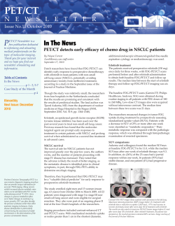
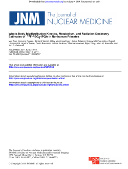
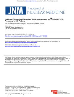
CJASN ePress. Published on June 23, 2011 as doi: 10.2215/CJN.06900810
Article
Positron-Emission Computed Tomography in Cyst
Infection Diagnosis in Patients with Autosomal
Dominant Polycystic Kidney Disease
François Jouret,* Renaud Lhommel,† Claire Beguin,‡ Olivier Devuyst,* Yves Pirson,* Ziad Hassoun,§
and Nada Kanaan*
Summary
Background Cyst infection remains a challenging issue in patients with autosomal dominant polycystic kidney disease (ADPKD). In most patients, conventional imaging techniques are inconclusive. Isolated observations suggest that 18fluorodeoxyglucose (18FDG) positron-emission computed tomography (PET/CT) might
help detect cyst infection in ADPKD patients.
Design, setting, participants, & measurements Comparative assessment of administrative databases from January 2005 to December 2009 identified 27 PET/CT scans performed in 24 ADPKD patients for suspicion of
abdominal infection. Cyst infection was definite if confirmed by cyst fluid analysis. Cyst infection was
probable if all four of the following criteria were met: temperature of ⬎38°C for ⬎3 days, loin or liver tenderness, C-reactive protein plasma level of ⬎5 mg/dl, and no CT evidence for intracystic bleeding. Episodes with only two or three criteria were grouped as “fever of unknown origin”.
Results Thirteen infectious events in 11 patients met all criteria for kidney (n ⫽ 3) or liver (n ⫽ 10) cyst infection. CT was contributive in only one patient, whereas PET/CT proved cyst infection in 11 patients
(84.6%). In addition, 14 episodes of “fever of unknown origin” in 13 patients were recorded. PET/CT identified the source of infection in nine patients (64.3%), including 2 renal cyst infections. Conversely, PET/CT
showed no abnormal 18FDG uptake in 5 patients, including 2 intracystic bleeding. The median delay between the onset of symptoms and PET/CT procedure was 9 days.
Divisions of
*Nephrology, †Nuclear
Medicine, ‡Medical
Information and
Statistics, and
§
Gastroenterology,
Cliniques Universitaires
Saint-Luc, Université
catholique de Louvain,
Brussels, Belgium
Correspondence: Dr.
François Jouret,
Division of Nephrology,
Cliniques Universitaires
Saint-Luc, Université
catholique de Louvain,
Avenue Hippocrate, 10,
B-1200 Brussels,
Belgium. Phone: 32-27641855; Fax: 32-27642836; E-mail:
francois.jouret@
uclouvain.be
Conclusions This retrospective series underscores the usefulness of PET/CT to confirm and locate cyst infection and identify alternative sources of abdominal infection in ADPKD patients.
Clin J Am Soc Nephrol 6: 1644 –1650, 2011. doi: 10.2215/CJN.06900810
Introduction
Autosomal dominant polycystic kidney disease
(ADPKD, Mendelian Inheritance in Man #173900)
represents the most common inherited kidney disease
and is characterized by the development of numerous
renal and hepatic cysts from various renal tubular
segments and biliary ducts, respectively (1,2). Cyst
growth causes organ enlargement leading to abdominal and/or loin discomfort. Cysts are also associated
with acute complications, such as bleeding and infection. Cyst infection (CI) represents the most challenging diagnostic issue in patients with ADPKD, with a
substantial risk for abscess formation and lifethreatening sepsis (3). The diagnosis of CI remains
uneasy because of the lack of specific symptoms and
signs and the limitations of conventional imaging
procedures (4,5). The elevation of serum carbohydrate
antigen 19.9 (CA19.9) has been recently proposed as a
diagnostic marker for hepatic CI (6). In addition, isolated reports have suggested that positron-emission
1644
Copyright © 2011 by the American Society of Nephrology
tomography (PET) after intravenous injection of
18
fluorodeoxyglucose (18FDG) might help identify tissue infection in ADPKD patients (7–11). Inflammatory cells are indeed characterized by a high metabolic activity and increased uptake of glucose analog,
18
FDG (9). Moreover, the modern combination of PET
with computed tomography (CT) may improve the
localization of pyocysts by integrating metabolic and
anatomical data (12). This study retrospectively investigates the accuracy of PET/CT imaging in diagnosing CI in ADPKD patients.
Study Population and Methods
The assessment of the computerized billing database of the Université catholique de Louvain Academic Saint-Luc Hospital (Brussels, Belgium) listed
all ADPKD patients admitted for suspicion of abdominal infection from January 2005 to December 2009.
Among these patients, those who underwent a
PET/CT were further identified using the administrawww.cjasn.org Vol 6 July, 2011
Clin J Am Soc Nephrol 6: 1644 –1650, July, 2011
tive records of the Division of Nuclear Medicine. All of the
medical files were reviewed.
Diagnostic criteria for ADPKD were the presence of
polycystic kidneys in at-risk patients or, in the absence of
family history, the presence of more than three renal cysts
for individuals aged between 15 and 39 years, more than
two cysts in each kidney for individuals aged 40 to 59
years, and more than four cysts in each kidney for individuals aged ⬎60 years (13). The diagnosis of renal and
liver CI was on the basis of the following criteria recently
proposed by Sallée et al. (4): (1) CI was definite when
confirmed by cyst aspiration showing neutrophils and bacteria; (2) CI was probable in the concurrent manifestation
of four conditions—fever (temperature of ⬎38°C for ⬎3
days), abdominal tenderness in kidney or liver area, increased C-reactive protein (CRP, ⬎5 mg/dl), and the absence of CT arguments for recent intracystic bleeding, i.e.
spontaneous intracystic content above 50 Hounsfield units
in the original reads. Infectious episodes which met 2 or 3
of these criteria were categorized as “fever of unknown
origin” (FUO).
The PET/CT procedure was performed using the
PET/CT Philips GEMINI TF (Philips Medical Systems,
Brussels, Belgium) after intravenous injection of a mean
dose of 300 ⫾ 40.7 MBq (8.1 ⫾ 1.1 mCi) of 18FDG. PET
imaging was acquired in list mode and rebuilt in 4-mm
transaxial slices with isometric voxels using time-of-flight
reconstruction. Two transverse series of 2- and 5-mm slices
were concurrently generated from low dose helical CT. No
intravenous or oral radiologic contrast agent was administered at the time of PET/CT imaging. All of the PET/CT
were initially performed for suspicion of abdominal infection at the physicians’ discretion and read by 2 nuclear
medicine physicians with at least 10 years of PET experience. PET findings were further confronted to CT results
and discussed with 2 radiologists with at least 15 years of
experience. For the purposes of this study, all 27 of the
PET/CT images were methodically reviewed by one nuclear medicine physician with 10 years of experience unaware of any clinical, biologic, and radiologic data.
PET/CT was considered as positive for CI when 18FDG
uptake was focally increased lining at least one cyst, in
strong contrast with the physiologic accumulation in parenchyma and distant from the pelvicalyceal physiologic
excretion (14). The maximal standardized uptake value
(SUVmax) was calculated for each patient using the following formula: [Pixel value (Bq/ml) ⫻ patient weight
(Kg)]/[injected dose (Bq) ⫻ 1000 (g/kg)].
In addition, radiologic investigations performed independently from PET/CT imaging, i.e. CT and magnetic
resonance (MR), were reviewed. The slice thickness of
CT images was 3 mm. Intravenous contrast agents were
systematically administered, except in patients with elevated serum creatinine level and/or declared allergy to
iodide. CT and MR imaging (MRI) were considered as
positive when enhanced wall thickening and focal inflammatory infiltrate were detected in at least one cyst.
This study was approved by the Commission of Biomedical Ethics of the Université catholique de Louvain
(Brussels, Belgium).
PET/CT in Cyst Infection Diagnosis, Jouret et al.
1645
Results
From January 2005 to December 2009, 268 ADPKD patients were admitted to the Université catholique de Louvain Academic Saint-Luc Hospital (Brussels, Belgium). Abdominal infection was suspected in 46 patients. Among
these, 27 PET/CT were performed in 24 patients. Thirteen
infectious events in 11 patients met all of the criteria for
kidney (n ⫽ 3) or liver (n ⫽ 10) CI, whereas 14 episodes in
13 patients only encountered 2 or 3 of them. The cohort
included 2 patients under chronic hemodialysis and 14
renal transplant recipients (RTR).
Liver Cyst Infections
Three liver CI in 3 patients were definite, as confirmed
by cyst fluid analysis. Clinical and biologic data are summarized in Table 1. Blood cultures grew Gram-negative
bacteria in all cases (Table 2). CT imaging without contrast
agent was performed in all cases but was contributive in
only one. Conversely, PET/CT demonstrated a pathologic
increase of 18FDG uptake lining liver pyocyst (Figure 1) in
all cases, which guided cyst drainage.
In addition, seven liver CI were probable in five patients
(Table 1). Blood cultures grew Escherichia coli in two patients (Table 2). CT imaging with (n ⫽ 2) or without (n ⫽
3) contrast agent was performed in four patients but did
not meet diagnostic criteria for CI in any of them. One
patient had two separate CT studies. PET/CT demonstrated CI in six patients (85.7%) (Table 3). PET/CT was
negative in a 74-year-old woman admitted for fever, chills,
nausea, and pain in the right upper abdominal quadrant,
in whom kidney transplantation from a deceased donor
had been performed 19 days earlier. Clinical examination
confirmed liver tenderness. Plasma CRP level was 23.9
mg/dl (normal value [NV], ⬍1 mg/dl); neutrophil white
blood cells (nWBC) count was 16.4 ⫻ 103/mm3 (NV, ⬍7 ⫻
103/mm3); serum CA19.9 level was 80.1 units/ml (NV,
⬍35 units/ml). Blood culture grew E. coli. CT with contrast
medium identified no infectious site and ruled out intracystic bleeding. PET/CT performed 3 days after admission
revealed abnormal 18FDG uptake only in a thyroid nodule.
Empirical antibiotherapy was initiated with ceftazidim for
suspicion of liver CI and continued orally for 3 weeks with
amoxycillin/clavulanic acid. Symptoms and signs rapidly
resolved. The patient was discharged on day 10. Twelve
days after discharge, the patient presented with similar
symptoms. Antibiotics had been stopped 2 days earlier.
Blood analyses showed: CRP, 16.4 mg/dl; nWBC, 13.5 ⫻
103/mm3; CA19.9, 89.0 units/ml. Urine and blood cultures remained germ-free. CT with contrast medium
showed no evidence of cyst infection or bleeding. Conversely, PET/CT performed on day 8 after admission identified a pathologic 18FDG accumulation lining cystic structures in the left part of the liver, in association with
segmental 18FDG uptake in the right colon. Ciprofloxacine
and metronidazole were started and planned for 6 weeks.
The patient was discharged on day 13. One week later, she
complained of two-sided calcaneal tendinitis. Ciprofloxacine and metronidazole were interrupted, and amoxycillin/clavulanic acid was resumed for 3 weeks. Ten days
later, she complained of epigastric pain. Blood analyses
showed increased CRP (15.7 mg/dl) and CA 19.9 (100.7
The data are presented as the means ⫾ SD. nWBC, neutrophils white blood cells; CRP, C-reactive protein; PET, positron emission tomography; CT, computed tomography.
a
Upper normal ranges: neutrophils counting, ⬍7 ⫻ 103/mm3; CRP, ⬍1 mg/dl.
3
2
5
0
0
4
43.3 ⫾ 11.5
49 ⫾ 17
56 ⫾ 17
67 ⫾ 3
61 ⫾ 13
60 ⫾ 13
1/2
2/3
4/9
3/3
5/7
13/14
0
0
2
12.6 ⫾ 7.8
8.6 ⫾ 4.6
8.2 ⫾ 3.2
29 ⫾ 9
23 ⫾ 8
16 ⫾ 10
1
2
关9, 10, 65兴
56 ⫾ 5
1/2
Renal cyst infection (probable)
Liver cyst infection
definite
probable
Fever of unknown origin
3/3
0
13.2 ⫾ 5.8
26 ⫾ 14
Positive Blood
Culture (n)
GFR
(ml/min per
1.73 m2)
Age
(years)
Patients/PET/CT
(n/n)
Gender
(M/F)
ESRD
Patients (n)
nWBC Counting
(103/mm3)a
CRP
(mg/dl)a
Positive Urine
Culture (n)
Clinical Journal of the American Society of Nephrology
Table 1. Characteristics of 24 autosomal dominant polycystic kidney disease (ADPKD) patients who underwent 27 positron-emission computed tomography (PET/CT) imaging procedures
1646
units/ml) levels. Blood and urine cultures were sterile.
PET/CT imaging evidenced an increased 18FDG uptake in
distinct small cysts located in the right part of liver parenchyma. Because of the number, size, and location of infected cysts, aspiration was not feasible. Intravenous metronidazole and ceftriaxone were administered for 3 weeks,
with clinical and biologic improvement. Eighteen months
later, the patient remained free of liver CI, with baseline
CA19.9 at 47.6 units/ml.
Kidney Cyst Infections
Three kidney CIs were probable in 3 ADPKD patients
(Table 1). Urine cultures disclosed E. coli infection in two
patients, with one case of bacteriemia (Table 2). CT was
performed for all patients, with injection of contrast agent
in only one. No cyst infection or bleeding was found in any
case. MRI was performed in one of these patients and
identified a thick-walled, heterogeneous cyst spontaneously contrasting on T1-weighted images with parietal
enhancement after gadolinium injection (8). PET/CT confirmed CI in two patients by demonstrating a focal increase
of 18FDG uptake lining pyocysts (Figure 2) but failed to
evidence CI in a 62-year-old woman with stage IV chronic
kidney disease (CKD). She was admitted for fever, chills,
nausea, and right loin pain of 12 days of duration. Clinical
examination revealed a positive right lumbar percussion.
CRP was 10.0 mg/dl, and nWBC count was 6.5 ⫻ 103/
mm3. Urinalyses disclosed leucocyturia, but urine and
blood cultures remained negative. CT without contrast
agent did not evidence cyst infection or bleeding, and
PET/CT performed 8 days after admission showed a
pathologic 18FDG accumulation in the gastric wall secondary to active and diffuse Helicobacter pylori infection. Classical H. pylori treatment was administered for 7 days, and
antibiotherapy was continued for 21 days using ciprofloxacine for suspicion of CI. The symptoms and signs progressively regressed. The 1-year follow-up showed no recurrence of CI.
Fever of Unknown Origin in Patients with ADPKD
Fourteen PET/CT were performed in 13 ADPKD patients with suspicion of abdominal infection, which met
only 2 or 3 of the above-mentioned criteria for CI. Mean
nWBC count and CRP level were 8.2 ⫾ 3.2 ⫻ 103/mm3 and
16 ⫾ 10 mg/dl, respectively (Table 1). Twelve CT scans
were performed in 11 patients, with no contrast agent
injection in eight patients because of CKD. CT identified
renal intracystic bleeding in two patients, renal CI in one,
and diverticulitis of the small intestine in one, whereas no
infectious site could be identified in the remaining eight
episodes (66.7%). PET/CT showed no pathologic 18FDG
uptake in 5 patients (35.7%) including both cases of intracystic bleeding, but identified distinct infectious diseases,
such as gastritis (n ⫽ 1), angiocholitis (n ⫽ 1), small intestine diverticulitis associated with psoas abscess (n ⫽ 1),
right colon diverticulitis (n ⫽ 1), prostatitis (n ⫽ 1), kidney
graft pyelonephritis (n ⫽ 1), and infection of abdominal
aorta aneurysm (n ⫽ 1). Typical 18FDG uptake lining renal
cysts was found in 2 patients (14.3%). The first patient was
an 81-year-old woman under hemodialysis for 6 years,
who had previously suffered from kidney CI. She com-
Clin J Am Soc Nephrol 6: 1644 –1650, July, 2011
PET/CT in Cyst Infection Diagnosis, Jouret et al.
1647
Table 2. Bacterial strains retrieved in 27 infectious episodes in 24 autosomal dominant polycystic kidney disease (ADPKD) patients
Urine
Renal cyst infections (n)
Liver cyst infections (n)
E. coli (2)
Fever of unknown origin (n)
E. coli (3)
K. pneumoniae (1)
Blood
E. coli (1)
E. coli (4)
K. pneumoniae (1)
E. coli (2)
K. oxytoca (2)
E. cloacae (1)
Cyst Fluid
E. coli (2)
K. pneumoniae (1)
One patient may present with positive cultures of urine and/or blood and/or cyst fluid samples. No causative bacterium was
found in 1 renal cyst infection (33%), 5 liver cyst infections (50%), and 6 episodes of fever of unknown origin (42.8%).
E. cloacae, Enterobacter cloacae; K. pneumoniae, Klebsiella pneumoniae; E. coli, Escherichia coli; K. oxytoca, Klebsiella oxytoca.
Figure 1. | Representative imaging of liver cyst infection diagnosis (arrow) using positron emission tomography (PET) after 18fluorodeoxyglucose injection, coupled with conventional computed tomography (CT). The merge between CT (A) and PET (C) imaging corresponds to
the central pictures (B). Coronal and transverse planes are shown in the upper and lower panels, respectively.
plained of abnormal fatigue and macroscopic hematuria in
the absence of fever or pain. CRP was 24.2 mg/dl, and
nWBC count was 7.5 ⫻ 103/mm3. Urine sediment showed
both red and white blood cells, but the cultures remained
sterile. Blood cultures grew Klebsiella oxytoca. CT without
contrast agent because of iodide allergy showed no evidence of cyst infection or bleeding. Conversely, PET/CT
identified a significant accumulation of 18FDG lining cystic
structures in the lower part of the left kidney. She was
treated with ciprofloxacine for 4 weeks with rapid improvement. The 2-year follow-up showed no recurrence of
CI.
The second patient was a 58-year-old RTR, with a past
history of 3 CI and right nephrectomy. At the outpatient
1648
Clinical Journal of the American Society of Nephrology
Table 3. Positron-emission computed tomography (PET/CT) imaging results (n ⴝ 27) in 24 autosomal dominant polycystic kidney
disease (ADPKD) patients
Cyst Infection
Fever of Unknown Origin
Liver
PET/CT (⫹)
PET/CT (⫺)
n
Kidney
Definite
Probable
Probable
Cyst
Infection
No Cyst
Infection
3
0
3
6
1
7
2
1
3
2
0
2
0
12
12
Cyst infection is considered as definite when cyst aspiration shows neutrophil debris and bacteria. Cyst infection is probable in
the presence of all of the following symptoms and signs: fever (temperature above 38°C for more than 3 days), abdominal
tenderness located in the renal or liver area, increased C-reactive protein (⬎5 mg/dl), and the absence of CT arguments for recent
intracystic bleeding. Infectious episodes that do not meet all of these criteria are referred to as “fever of unknown origin.”
Figure 2. | Representative imaging of kidney cyst infection diagnosis (arrow) using position emission tomography (PET) after 18fluorodeoxyglucose injection, coupled with conventional computed tomography (CT). The merge between CT (A) and PET (C) imaging corresponds
to the central pictures (B). Coronal and transverse planes are shown in the upper and lower panels, respectively.
transplant clinic, she reported a 6-day fluctuating lower
abdominal pain and chills, without urinary or digestive
symptoms. Clinical examination confirmed hypogastrium
tenderness. Urine culture was contaminated by Gram-neg-
ative bacteria. CRP was 8.1 mg/dl, and nWBC count was
8.9 ⫻ 103/mm3. CT without contrast medium showed one
large pyocyst in the left kidney with thickened wall and
distended renal fascia. PET/CT on day 9 after the onset of
Clin J Am Soc Nephrol 6: 1644 –1650, July, 2011
abdominal symptoms confirmed the hypermetabolic status
of the infected cyst. Ciprofloxacine was administered for 4
weeks, with favorable evolution.
Features of PET/CT Imaging in Patients with ADPKD
The median delay between the onset of symptoms and
PET/CT imaging was 9 days. Seven patients (29.2%) were
diabetic, with a need for insulin in 3 of them. The mean
glycaemia at the time of 18FDG injection was 117.9 ⫾ 38.3
mg/dl. PET/CT imaging was usually acquired 95.3 ⫾ 31.1
minutes after 18FDG injection and yielded positive results
in 86.7% of CI cases (Table 3). Mean SUVmax measured in
the most metabolic pyocyst reached 5.1 ⫾ 1.7 g/ml.
PET/CT results significantly changed the management of 7
ADPKD patients presenting with suspicion of abdominal
infection (25.9%). By contrast, conventional CT failed to
detect the infected cyst in 84.6% of patients and yielded
negative results in 2 patients with definite liver CI (66.7%).
Discussion
The main diagnostic objectives in ADPKD patients presenting with a suspicion of CI are (1) the exclusion of
noncystic infections; (2) the location and extension of infected cysts; and (3) the identification of concomitant conditions, like urinary tract obstruction (3). The lack of specific signs frequently retards the diagnosis and subsequent
treatment. The definite diagnosis of CI requires cyst fluid
analysis showing causative bacteria and neutrophils. However, this is not always possible or indicated, so that common criteria for CI rely on a constellation of clinical and
biologic parameters (4).
Conventional imaging procedures, like ultrasound, CT,
and MRI, frequently fail to confidently identify CI (3,4).
Radiologic features of pyocysts are unspecific and may be
secondary to previous infections and hemorrhages, as well
as chronic parenchyma injury. Cyst content of pus or blood
may be indistinguishable (3), and contrast enhancement
lining cyst walls can be caused by residual functional
parenchyma (15,16). Furthermore, CT is most frequently
performed in ADPKD patients without intravenous contrast medium because of CKD. Here, injection of contrast
medium was performed in 30.4% of patients, and CT imaging yielded contributive results in only five patients
(21.7%), including one with liver CI, one kidney with CI,
one with diverticulitis, and two with intracystic bleeding.
The accuracy of MRI in CI diagnosis, with and without
gadolinium injection, remains unknown.
To complement conventional radiologic procedures and
improve the accuracy of CI diagnosis, techniques using
radiolabeled tracers, like 111indium-labeled leukocytes and
18
FDG, have been developed. The 111In-leukocyte scanning
allowed the identification of renal CI in a few ADPKD
patients in whom other noninvasive imaging procedures
had failed to locate the infection (17,18). However, this
technique requires the handling of blood derivatives and
the ex temporane in vitro labeling process, as well a 48-hour
delay before imaging. 111In-leukocyte scintigraphy is characterized by low image resolution, uneasy coregistration,
and high interobserver variability (19). Moreover, the use
of 111In-leukocyte scanning in febrile RTR is inadequate
because of unspecific accumulation of leukocytes in renal
PET/CT in Cyst Infection Diagnosis, Jouret et al.
1649
and pulmonary parenchymae (20). Recently, PET/CT using the glucose analog, 18FDG, has been proposed in the
detection and localization of tissue infection. Inflammatory
cells are indeed characterized by a high metabolic status
and increased uptake of 18FDG (9). PET/CT helped diagnose renal and hepatic CI in ADPKD patients with renal
function ranging from mildly reduced GFR to ESRD (7–11).
The advantages of 18FDG PET/CT are rapid imaging, high
target-to-background ratio, and a direct coregistration with
low-dose CT without radiologic contrast medium administration (21). Limitations include its relatively high cost
and restricted availability. In addition, 18FDG uptake is not
specific for infection and may be increased in other conditions, like tumors. The actual risk of malignancy in ADPKD patients does not seem to be increased (22). Liver
cystadenocarcinomas are uncommon, and most kidney tumors show low-grade malignancy leading to low 18FDG
uptake (14). The role of alternative tracers, like 18F-FLT, is
currently being addressed (23). Similarly, the differentiation of 18FDG accumulation in residual parenchyma versus
that in inflammatory cells lining pyocysts remains debated
(24,25). Here, SUVmax measured in renal and hepatic pyocysts significantly differed from baseline 18FDG uptake
(24). Moreover, 18FDG uptake in pyocyst wall was clearly
distinguishable using CT data coregistration (Figures 1 and 2).
The retrospective design of this monocentric study
based on administrative databases represents the main
limitation. This trial includes a small number of patients,
with only five renal CI, and relies upon past clinical reports. All of the PET/CT images were blindly reviewed,
but the lack of accurate medical literature about PET/CT in
CI hampered the use of objective criteria, like SUVmax.
PET/CT imaging was considered as positive for CI when
18
FDG uptake was focally increased lining at least one cyst
in strong contrast with the surrounding tissue. The mean
SUVmax reached 5.1 ⫾ 1.7 g/ml. This value needs to be
further assessed in prospective trials. PET/CT imaging
was contributive in 13 of 15 renal or hepatic CI, whereas
conventional CT identified only 2 CI. Moreover, PET/CT
evidenced 2 kidney CI, although both patients did not
meet all of the conventional criteria, and identified alternative sources of abdominal infection in 7 patients.
PET/CT yielded 2 false-negative results in one diabetic
RTR during the immediate post-transplantation period
and in a 62-year-old nondiabetic woman with stage IV
CKD. Technical conditions might partly explain these
false-negative results. In the first patient, the delay between the onset of symptoms and PET/CT was short (5
days), and the patient’s glycaemia at the time of 18FDG
injection (155 mg/dl) was significantly higher than the
mean of all procedures. In the other patient, PET/CT was
performed 20 days after the onset of fever, with an extended imaging delay (191 minutes). Current European
guidelines for PET/CT recommend (1) the intravenous
injection of 2.5 MBq/kg of body weight 18FDG activity
(⫾10%), and (2) an interval between 18FDG administration
and the acquisition of 60 minutes (26). The efficiency of
PET/CT in immunocompromised patients remains poorly
established. PET/CT has been shown to distinguish AIDSrelated opportunistic infections from malignancies (27). In
1650
Clinical Journal of the American Society of Nephrology
RTR and CKD patients, prospective trials investigating
PET/CT efficiency in identifying infectious sites are lacking.
The main differential diagnosis of CI in ADPKD patients
is intracystic bleeding. Significant cyst hemorrhage is usually detected by conventional CT, and the ability of
PET/CT to distinguish cyst infection versus bleeding remains unknown. Increased 18FDG accumulation has been
reported in both acute and resolving hematoma in extrarenal sites (28,29). Here, 2 kidney cyst hemorrhages were
identified by CT, with no pathologic 18FDG accumulation.
Further prospective evaluations are required to establish
PET/CT accuracy in excluding intracystic bleeding and to
define the optimal delay between the onset of symptoms
and PET/CT imaging.
In conclusion, this retrospective series underscores the
usefulness of PET/CT to confirm and locate CI, as well as
to identify alternative sources of abdominal infection, in
ADPKD patients.
Acknowledgments
The authors thank Max Lonneux and Francois-Xavier Hanin for
the original readings of PET/CT and Laurence Annet and Etienne
Danse for the original readings of CT images.
Disclosures
None.
References
1. Pirson Y, Chauveau D, Devuyst O: Autosomal dominant
polycystic kidney disease. In: Oxford Textbook of Clinical
Nephrology, 3rd Ed., edited by Davison AM, Oxford, Oxford
University Press, 2005, pp 2304 –2324
2. Torres VE, Harris PC, Pirson Y: Autosomal dominant polycystic kidney disease. Lancet 369: 1287–1301, 2007
3. Gibson P, Watson ML: Cyst infection in polycystic kidney
disease: A clinical challenge. Nephrol Dial Transplant 13:
2455–2457, 1998
4. Sallée M, Rafat C, Zahar JR, Paulmier B, Grünfeld JP, Knebelmann B, Fakhouri F: Cyst infections in patients with autosomal dominant polycystic kidney disease. Clin J Am Soc
Nephrol 4: 1183–1189, 2009
5. Pirson Y: Extrarenal manifestations of autosomal dominant
polycystic kidney disease. Adv Chronic Kidney Dis 17: 173–
180, 2010
6. Kanaan N, Goffin E, Pirson Y, Devuyst O, Hassoun Z: Carbohydrate antigen 19-9 as a diagnostic marker for hepatic cyst
infection in autosomal dominant polycystic kidney disease.
Am J Kidney 55: 916 –922, 2010
7. Bleeker-Rovers CP, de Sevaux RG, van Hamersvelt HW, Corstens FH, Oyen WJ: Diagnosis of renal and hepatic cyst infections by 18-F-fluorodeoxyglucose positron emission tomography in autosomal dominant polycystic kidney disease.
Am J Kidney Dis 41: E18 –E21, 2003
8. Migali G, Annet L, Lonneux M, Devuyst O: Renal cyst infection in autosomal dominant polycystic kidney disease. Nephrol Dial Transplant 23: 404 – 405, 2008
9. Keidar Z, Gurman-Balbir A, Gaitini D, Israel O: Fever of unknown origin: The role of 18F-FDG PET/CT. J Nucl Med 49:
1980 –1985, 2008
10. Soussan M, Sberro R, Wartski M, Fakhouri F, Pecking AP,
Alberini JL: Diagnosis and localization of renal cyst infection
by 18F-fluorodeoxyglucose PET/CT in polycystic kidney disease. Ann Nucl Med 22: 529 –531, 2008
11. Desouza RM, Prachalias A, Srinivasan P, O’Doherty M, Olsburgh J: Differentiation between infection in kidney and liver
cysts in autosomal dominant polycystic kidney disease: Use
of PET/CT in diagnosis and to guide management. Transplant
Proc 41: 1942–1945, 2009
12. Kaim AH, Burger C, Ganter CC, Goerres GW, Kamel E,
Weishaupt D, Dizendorf E, Schaffner A, von Schulthess GK:
PET/CT-guided percutaneous puncture of an infected cyst in
autosomal dominant polycystic kidney disease: Case report.
Radiology 221: 818 – 821, 2001
13. Pei Y, Obaji J, Dupuis A, Paterson AD, Magistroni R, Dicks E,
Parfrey P, Cramer B, Coto E, Torra R, San Millan JL, Gibson
R, Breuning M, Peters D, Ravine D: Unified criteria for ultrasonographic diagnosis of ADPKD. J Am Soc Nephrol 20:
205–212, 2009
14. Aide N, Cappele O, Bottet P, Bensadoun H, Regeasse A, Comoz F, Sobrio F, Bouvard G, Agostini D: Efficiency of (18)FFDG PET in characterising renal cancer and detecting distant
metastases: A comparison with CT. Eur J Nucl Med Mol Imaging 30: 1236 –1245, 2003
15. Levine E, Hartman DS, Meilstrup JW, Van Slyke MA, Edgar KA,
Barth JC: Current concepts and controversies in imaging of renal
cyst diseases. Urol Clin North Am 24: 523–543, 1997
16. Gupta S, Seith A, Sud K, Gupta S, Seith A, Sud K, Kohli HS,
Singh SK, Sakhuja V, Suri S: CT in the evaluation of complicated autosomal dominant polycystic kidney disease. Acta
Radiol 41: 280 –284, 2000
17. Gilbert BR, Cerqueira MD, Eary JF, Simmons MC, Nabi HA,
Nelp WB: Indium-111 white blood cell scan for infectious
complications of polycystic renal disease. J Nucl Med 26:
1283–1286, 1985
18. Bretan PN Jr., Price DC, McClure RD: Localization of abscess
in adult polycystic kidney by indium-111 leukocyte scan.
Urology 32: 169 –171, 1988
19. Rennen HJ, Boerman OC, Oyen WJ, Corstens FH: Imaging
infection/inflammation in the new millennium. Eur J Nucl
Med 28: 241–252, 2001
20. Sebrechts C, Biberstein M, Klein JL, Witztum KF: Limitations
of indium-111 leukocyte scanning in febrile renal transplant
patients. AJR 146: 823– 829, 1986
21. Meller J, Sahlmann CO, Scheel AK: 18F-FDG PET and
PET/CT in fever of unknown origin. J Nucl Med 48: 35– 45,
2007
22. Bonsib SM: Renal cystic diseases and renal neoplasms: A
mini-review. Clin J Am Soc Nephrol 4: 1998 –2007, 2009
23. Lawrentschuk N, Davis ID, Bolton DM, Scott AM: Functional imaging of renal cell carcinoma. Nat Rev Urol 7: 258–266, 2010
24. Engel H, Steinert H, Buck A, Berthold T, Huch Böni RA, von
Schulthess GK: Whole-body PET: Physiological and artifactual fluorodeoxyglucose accumulations. J Nucl Med 37: 441–
446, 1996
25. Raddatz D, Ramadori G: Carbohydrate metabolism and the
liver: Actual aspects from physiology and disease. Z Gastroenterol 45: 51– 62, 2007
26. Boellaard R, O’Doherty MJ, Weber WA, Mottaghy FM, Lonsdale MN, Stroobants SG, Oyen WJ, Kotzerke J, Hoekstra OS,
Pruim J, Marsden PK, Tatsch K, Hoekstra CJ, Visser EP, Arends B, Verzijlbergen FJ, Zijlstra JM, Comans EF, Lammertsma AA, Paans AM, Willemsen AT, Beyer T, Bockisch A,
Schaefer-Prokop C, Delbeke D, Baum RP, Chiti A, Krause BJ:
FDG PET and PET/CT: EANM procedure guidelines for tumour PET imaging. Eur J Nucl Med Mol Imaging 37: 181–
200, 2010
27. Sathekge M, Goethals I, Maes A, van de Wiele C: Positron
emission tomography in patients suffering from HIV-1 infection. Eur J Nucl Med Mol Imaging 36: 1176 –1184, 2009
28. Ryan A, McCook B, Sholosh B, Pryma DA, Jablonowski E,
Fuhrman C, Blodgett TM: Acute intramural hematoma of the
aorta as a cause of positive FDG PET/CT. Clin Nucl Med 32:
729 –731, 2007
29. Repko BM, Tulchinsky M: Increased F-18 FDG uptake in resolving atraumatic bilateral adrenal hemorrhage (hematoma)
on PET/CT. Clin Nucl Med 33: 651– 653, 2008
Received: August 10, 2010 Accepted: March 22, 2011
Published online ahead of print. Publication date available at
www.cjasn.org.


© Copyright 2026