
Cytologic Features of Prostatic Adenocarcinoma in Urine: Comparison with Urothelial Carcinoma
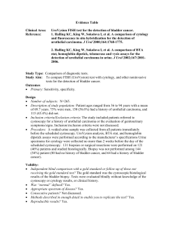
The Korean Journal of Pathology 2011; 45: 79-86 DOI: 10.4132/KoreanJPathol.2011.45.1.79 Cytologic Features of Prostatic Adenocarcinoma in Urine: Comparison with Urothelial Carcinoma Lucia Kim ∙ Joo Young Song Suk Jin Choi ∙ In Suh Park Jee Young Han ∙ Joon Mee Kim Young Chae Chu Department of Pathology, Inha University School of Medicine, Incheon, Korea Received: March 9, 2010 Accepted: November 19, 2010 Corresponding Author Young Chae Chu, M.D. Department of Pathology, Inha University Hospital, 7-206 Sinheung-dong 3-ga, Jung-gu, Incheon 400-711, Korea Tel: +82-32-890-3984 Fax: +82-32-890-3464 E-mail: [email protected] *This study was supported by Inha University Research Grants. Background: Prostate adenocarcinoma (PACa) cells are rarely identified in urine cytology specimens and might be easily overlooked or misdiagnosed as urothelial neoplasm when clinically unsuspected. Methods: We reviewed 19 urine cytology specimens obtained from 13 patients with PACa and evaluated the characteristic features discriminating PACa from urothelial carcinoma (UCa). For comparison, 27 cases of high-grade UCa (HGUCa) and 10 cases of urothelial carcinoma in situ (UCis) were also evaluated. Results: The urine cytologic evaluation of PACa revealed clustered cells forming 3-dimensional syncytial fragments with occasional microacinar grouping in a clean background. Most tumor cells were small and uniform with a high nuclear-to-cytoplasmic ratio and indistinct cell borders. The nuclei were round-to-oval and the cytoplasm was scanty and thin. One or more centrally-located prominent nucleoli were characteristically noted in one half of the cases. The nucleoli had a well-defined, large, round and eosinophilic appearance. In four high-grade cases, large tumor cells were encountered and had relatively monotonous cells with smooth-outlined cell clusters, well-defined and thin cytoplasm, and round nuclei with characteristic prominent nucleoli. Conclusions: Combining the information of prostate cancer and the recognition of cytomorphologic features of PACa will help differentiate PACa from HGUCa and UCis. Key Words: Prostatic neoplasms; Urine; Cytology; Carcinoma, transitional cell MATERIALS AND METHODS Carcinoma cells of a prostate origin are rarely detected in urine cytology specimens.1,2 Although urine cytology is routinely performed in patients with urologic complaints, the clinical value is limited for making the diagnosis of prostate adenocarcinoma (PACa), and pathologists may not be familiar with the cytomorphology of these cells. However, it is important that cancer cells are recognized to be of a prostate origin and they should be distinguished from urothelial neoplasm because of the therapeutic implications. PACa cells have been frequently overlooked or misdiagnosed as urothelial malignancies in cases that have a lack of clinical information. Even though PACa may be clinically suspected, the cancer cells in urine specimens may reflect a synchronous carcinoma of the bladder.3 The cytologic features of PACa cells on urine cytology have been described;1-7 however, the clinical application of these cytologic features is challenging. The purpose of this study was to evaluate the cytologic findings of PACa in urine specimens, to determine the characteristic features favoring a prostate origin of cancer cells and to discriminate PACa from high-grade urothelial carcinoma (HGUCa) and urothelial carcinoma in situ (UCis). We searched the medical records of Inha University Hospital and we identified 359 patients who had histologically-confirmed acinar-type PACa at our hostpital between 2000 and December 2008; we reviewed these patients’ urine cytology from the pa thologic archives. Synchronous or metachronous urothelial neoplasm in the urinary tract was excluded. Nineteen urine specimens obtained from 13 patients with PACa were diagnosed as atypia in 2 cases and suspicious or positive for malignancy in 17 cases. The specimens consisted of voided urine in 13 cases, bladder washing in 4 cases and catheterized urine in 2 cases. Cytohistologic correlations were done for all the cases and the urine cytology was reviewed by two pathologists (L. Kim and YC Chu). We reviewed the medical records to ascertain the clinical findings, including age, the chief complaints, the serum prostate specific antigen (PSA) level, the cystoscopic findings, the initial cytologic diagnosis and the clinical diagnosis at the time of posi tive urine cytology. The urine cytology of 27 cases of histologically-proven HGUCa and 10 cases of UCis were evaluated for comparison with the 79 80 Lucia Kim·Joo Young Song·Suk Jin Choi, et al. PACa cases. The cytologic specimens of HGUCa consisted of voided urine in 18 cases, catheterized urine in 3 cases and bladder washing in 6 cases; the cytologic specimens of UCis were voided urine in 8 cases and bladder washing in 2 cases. To determine the differential diagnostic features of these neoplasms, the following cytologic features were reviewed with respect to cellularity, the background, the shape of cell fragments, the presence of microacinar grouping and papillary clusters, the size and shape of individual cells, the cytoplasmic features, the cell borders, the nuclear shape and the nuclear border, the chromatin pattern, the features of the nucleoli, the nuclear-to-cytoplasmic ratio (N/C ratio) and the presence of pleomorphism. RESULTS Clinical features of the prostate adenocarcinoma The clinical findings of the 13 cases of PACa are summarized in Table 1. The mean age of the patients was 66 years (range, 44 to 89 years). The chief complaints were hematuria (n=6), voiding difficulty (n=5), urgency and frequency (n=1), and weight loss and lymph node enlargement (n=1). The initial clinical impressions were benign prostatic hyperplasia (n=5), bladder tumor (n=3), urethral polyp (n=1), chronic prostatitis (n=2), prostatic cancer (n=1) and malignant lymphoma (n=1). Cystoscopic examination was performed in seven patients. Protruding masses or polyps in the prostatic urethra were noted in four patients, essentially normal findings were noted in two patients, and severe trabeculations and edema of the bladder mucosa were noted in one patient. In all the patients, the serum PSA level was elevated (range, 7.05 to 404 ng/mL). In 11 cases, urine cytology was performed before the diagnosis of prostate cancer was confirmed. In these cases, the cytologic diagnosis was suspicious or positive for UCa (6 cases), small cell carcinoma (2 cases), atypia (2 cases), and combined HGUCa and PACa (1 case). In 8 patients with a known history of prostate cancer, the cytologic diagnosis was PACa (7 cases) or suspicious for car- Table 1. Clinical observations for the 19 urine cytology specimens of the 13 prostatic adenocarcinoma patients Case Age No. (yr) Initial serum Gleason PSA score (ng/mL) Initial cystoscopic findings Initial clinical Dx 62 Gross hematuria Polyp in prostatic urethra Urethral polyp 12.11 4+5 76 Gross hematuria ND Bladder ca 32.1 4 + 5 Voided urine 3A 3B 64 Hematuria ND BPH 4 71 Voiding difficulty ND Prostatic ca 404 5A 5B 56 Weight loss, LN enlargement ND Malignant lymphoma 226 6 44 Voiding difficulty Normal finding Chronic prostatitis 7 66 Gross hematuria ND 8A 8B 72 Urgency, frequency 9 65 Hematuria 1A Chief complaint No Bladder washing No Bladder invasion Yes No No Suspicious for urothelial ca Urothelial ca No Yes Urothelial ca Adenoca No 5 + 4 Voided urine No Atypia Yes 5 + 4 Voided urine Voided urine Yes Yes Prostatic adenoca Prostatic adenoca No 31.4 5 + 5 Bladder washing Yes Prostatic adenoca Yes Chronic prostatitis 35.2 5 + 4 Voided urine No Suspicious for urothelial ca No Protruding mass in prostatic urethra Bladder cancer 13.8 5 + 4 Bladder washing Bladder washing No Yes Urothelial ca Prostatic adenoca Yes Normal finding BPH 3 + 3 Voided urine Yes Suspicious for ca No 3 + 5 Voided urine Yes Prostatic adenoca No 5 + 5 Voided urine Voided urine No Yes Urothelial ca Adenoca Yes 2B Voided urine Cytologic diagnosis Suspicious for small cell ca Small cell ca 1B 2A Cytology specimens Presence of clinical information at the time of cytologic Dx Catheterized urine 8.06 7.05 316 5 + 4 Voided urine Voided urine No 10 89 Voiding difficulty ND BPH 11A 11B 88 Voiding difficulty Protruding mass in prostatic urethra BPH 12 85 Voiding difficulty BPH 117 5 + 5 Voided urine No Atypia No 13 65 Hematuria Severe trabeculation and edema of bladder mucosa Protruding mass in bladder neck Bladder ca 350 5 + 5 Voided urine No Urothelial ca & prostatic adenoca Yes 8.07 Dx, diagnosis; PSA, prostate specific antigen; Ca, carcinoma; ND, not done; BPH, benign prostatic hyperplasia; Adenoca, adenocarcinoma; LN, lymph node. 81 Prostatic Adenocarcinoma in Urine Cytology cinoma (1 case). The Gleason score of the tumors ranged from 6 to 10, and most of tumors except one were high-grade (Gleason score >7). In 6 patients, bladder wall invasion by the tumor was identified on the cystoscopic and radiologic studies. Cytologic features of the prostate adenocarcinoma The urine specimens exhibited variable cellularity depending on the collection method, but the cellularity was usually low. The smear background was clean (10 cases) or inflamed (4 cases). Two cases exhibited tumor diathesis and three cases were bloody. The cancer cells were commonly seen as aggregates along with singly scattered cells. At low magnification, the cancer cells formed small, crowded, three-dimensional syncytial clusters with overlapping nuclei (Fig. 1A). Several small, loosely cohesive clusters were also scattered. Approximately one half of the cases showed microacinar grouping in cellular aggregates (Fig. 1B), but any papillary clusters were not noted. Two cytologic patterns were identified. The predominant pattern encompassed 15 cases and the tumor cells were generally small, uniform and hyperchromatic with a high N/C ratio. The cytoplasm was scanty, thin, or vacuolated. The cytoplasmic border was indistinct and naked nuclei were occasionally seen (Fig. 1C). The nucleus was centrally-located, small and round-to-oval. The nuclei had coarsely granular or vesicular chromatin, and a smooth and thin nuclear border. One or more large, centrally located, well defined, eosinophilic and round nucleoli were occasionally seen in 8 cases (Fig. 1D). The remaining four cases had different cytologic features from the aforementioned cases. Three of the four cases displayed large cells with abundant cytoplasm, yet the pleomorphism was not marked. Some tumor cells formed three-dimensional clusters with a smooth outer contour (Fig. 1E). The tumor cells were round or irregular in shape and they showed abundant, thin and vacuolated cytoplasm with well-defined borders, and round-to-oval nuclei with vesicular chromatin. One or two large, centrally-located, prominent nucleoli were characteristically present; the nucleoli were well-defined, round and eosinophilic like those noted in the former pattern (Fig. 1F). Tumor giant cells were occasionally present, but any squamoid feature was not detected. The other case showed singly scattered monotonous tumor cells which were medium in size and round-to-oval in shape. The cytoplasm was well-defined and granular, thin or vacuolated. The nuclei had coarsely granular chromatin, a smooth nuclear border and a centrally-located prominent nucleolus (Fig. 1G). Cytologic features of the high grade urothelial carcinoma The cytologic features of the 27 cases of histologically-proven HGUCas were reviewed. The smears showed variable cellularity, but generally high cellularity. In most cases, a predominant pattern was present with individually-scattered tumor cells along with loosely cohesive or large, three-dimensional clusters (Fig. 2A). The tumor cells were less cohesive than that of the PACa cells. Papillary clusters were occasionally present in 14 cases, but microacinar grouping was not present in any of the cases. Tumor diatheses were present in two-thirds of the cases and only two cases showed a clean background. The tumor cells were medium-to-large in size, pleomorphic and partly squamoid (Fig. 2B). The tumor cells had more cytoplasm, resulting in a lower N/C ratio than that of the PACa cells. The cell shape was oval, pyramidal or irregular, the cell border was fairly well-defined and the cytoplasm was dense or vacuolated. The nuclei were usually oval or irregular in shape and eccentric in location (Fig. 2C). The nu clear chromatin was coarsely granular with a clumping and clearing pattern. One or more large nucleoli were occasionally present in eight cases. In two cases, very large nucleoli were seen, which were similar to that of the PACa cells. However, the nucleoli were irregular in shape and location, usually not well-defined and less prominent (Fig. 2D). Cellular pleomorphism and hyperchromasia were marked with occasional tumor giant cells in most cases. Squamoid features were occasionally seen in six cases, but glandular differentiation was not identified. Cytologic features of the urothelial carcinoma in situ The smears were usually hypocellular and most of the cancer cells were singly scattered and rarely formed small clusters (Fig. 3A). The background was clean or inflamed and tumor diathesis was not seen. The cancer cells were medium-to-large in size and showed a similar cytomorphology to that of HGUCa cells. The cancer cells were oval, pyramidal or irregular in shape, with variable amounts of cytoplasm. The cell border was well-defined and irregular, and the cytoplasm was dense or vacuolated. The nuclei were eccentrically-located and oval or irregular in shape, with coarsely granular chromatin and distinct nuclear margins (Fig. 3B). The nucleoli were indistinct or distinct and irregularly-located (Fig. 3C). In three cases, prominent nucleoli were occasionally present, but irregular in shape and location, not welldefined and smaller than PACa cells (Fig. 3D). The UCis cells were usually monotonous and smaller with a higher N/C ratio and had less prominent nucleoli than that of the HGUCa cells. 82 Lucia Kim·Joo Young Song·Suk Jin Choi, et al. A B C D E F Fig. 1. Cytologic features of prostate adenocarcinoma. (A) At low magnification, the cells form tight, three-dimensional syncytial clusters with overlapping nuclei and singly-scattered cells in the clean background. (B) Microacinar grouping is noted in some cellular aggregates and the tumor cells are small, uniform and hyperchromatic. (C) The individually-scattered cells show an indistinct cytoplasmic border and naked nuclei. (D) The cytoplasm is scanty, thin or vacuolated and the nucleus is small and round with coarsely granular chromatin. One or more large, centrally-located, well-defined, eosinophilic and round nucleoli are characteristic. (E) In the poorly differentiated cases, three-dimensional clusters with smooth external border are seen. (F) The tumor cells are large in size, and they show well-defined, abundant and vacuolated cytoplasm, and oval nuclei with vesicular chromatin and one or two large, prominent nucleoli. The pleomorphism is not distinct. (continued to the next page) 83 Prostatic Adenocarcinoma in Urine Cytology DISCUSSION G Fig. 1. (continued from the previous page) (G) Other poorly differentiated cases show singly-scattered, monotonous, round-to-oval cells with well-defined cell borders, round nuclei, clumped chromatin, and a prominent nucleolus. PACa cells are very rarely encountered in urine specimens and pathologists may not be familiar with the cytomorphology of these cells. When atypical cells are encountered in urine specimens, cytologic examination alone might not be enough to reach the diagnosis of PACa in most cases. However, when clinical information of prostate cancer is provided, the PACa can usually be diagnosed without difficulty. Our cases showed that when the clinical data about prostate cancer was provided, the cytologic diagnosis was easily made, but without the clinical information the diagnostic accuracy was markedly decreased. Therefore, the clinical information is important and awareness of the cytomorphology of PACa cells will help achieve an accurate diagnosis. We summarized the characteristic cytologic features of PACa cells that are helpful for differentiation them from HGUCa and A B C D Fig. 2. Cytologic features of high-grade urothelial carcinoma. (A) The smear shows high cellularity and a necrotic background with individually-scattered or clustered tumor cells. (B) The tumor cells are medium-to-large in size, pleomorphic and occasionally squamoid. (C) The cell shape is oval or pyramidal, the cell border is well-defined, the cytoplasm is dense or vacuolated, and the nucleus is eccentric in location. (D) Very large nucleoli are occasionally seen, but they are irregular in shape and location. 84 Lucia Kim·Joo Young Song·Suk Jin Choi, et al. A B C D Fig. 3. Cytologic features of urothelial carcinoma in situ. (A) The cancer cells are singly-scattered and they occasionally form small clusters. (B) They are smaller and less pleomorphic than the high-grade urothelial carcinoma cells. (C) The tumor cells are oval or pyramidal in shape with a moderate amount of dense or vacuolated cytoplasm and well-defined cell borders. The nucleus is eccentrically located with coarsely granular chromatin and irregularly located nucleoli. (D) Prominent nucleoli are occasionally present and they are irregular in shape and location and not well-defined. UCis cells in urine specimens (Table 2). The tumor cells of PACa are characteristically uniform and small with a high N/C ratio. The cytoplasm is scanty in amount and thin with indistinct cytoplasmic borders. The nuclei are round-to-oval with prominent nucleoli. These features are similar to those described in previous studies.1-5,7 Koss3 reported that PACa cells were usually small and round in shape with scanty cytoplasm and spherical nuclei. In addition, the presence of enlarged nucleoli was one of the most valuable features of prostate carcinoma. Bardales et al.5,7 commented that higher cell cohesiveness, vesicular chromatin and readily visible nucleoli were more evident in prostatic carcinoma. Varma et al.4 emphasized the presence of small tumor cells showing faintly-stained, finely granular or vacuolated cytoplasm, round-to-oval nuclei with finely granular chromatin and a prominent central nucleolus in PACa cells. Krishnan and Truong1 suggested that the presence of an oval or round nucleus with a well-defined border, fine chromatin and a lack of significant pleomorphism might be more reliable for suspecting PACa. In the literature, the presence of prominent nucleoli is described as the most helpful differential diagnostic feature.2-5,7 However, the rare occurrence or absence of nucleoli in PACa cases has been reported.1,8,9 Poor fixation and degeneration can make the nuclear and nucleolar features obscure, even in the same cases. HGUCa cells also showed prominent nucleoli, the same as seen in PACa.3,7 In this study, prominent nucleoli were present in 12 of the 19 PACa specimens (63.2%), 8 of the 27 HGUCa specimens (29.6%) and 3 of the 10 UCis specimens (30%). However, the nucleolar features were slightly different between these tumors. The nucleoli of the PACa cells were large, well-defined, round, eosinophilic and centrally-located, whereas the nucleoli of the UCa cells were smaller, irregular in shape and 85 Prostatic Adenocarcinoma in Urine Cytology Table 2. Comparison of the cytologic features of prostatic adenocarcinoma, urothelial carcinoma in situ and high grade urothelial carcinoma in the urine cytology Cytologic features Smear patterns Cellularity Shape of cell fragments Microacinar grouping Papillary clusters Cellular features Cell size Cell shape Cytoplasmic features Cell borders Nuclear shape Nuclear border Nucleoli N/C ratio Pleomorphism Background Prostatic adenocarcinoma Urothelial carcinoma in situ High grade urothelial carcinoma Usually low 3-dimensional syncytial fragments and single cells, clusters with smooth external border Occasionally present Very rare Usually low Single cell predominant, rarely small clusters Absent Very rare Variable, usually high Variable, single cells, loose clusters and 3-dimensional syncytial fragments Absent Present Small, uniform, rarely large Round to oval Scanty, thin Indistinct with naked nuclei or well defined Round to oval Smooth and thin Usually prominent, centrally located, large, well defined, round, eosinophilic Very high Monotonous Medium to large, relatively uniform Oval, pyramidal, irregular Variable amount, dense or vacuolated Well defined, irregular borders Oval or irregular, eccentrically located Irregular and prominent Indistinct or distinct, irregular shape and location, not well defined, smaller size High Less pleomorphic Medium to large, variable Oval, pyramidal, irregular Variable amount, dense or vacuolated Well defined, irregular borders Oval or irregular, eccentrically located Irregular and prominent Variable, irregular shape and location, not well defined, smaller size High Pleomorphic Clean or inflammatory Clean or inflammatory Tumor diathesis or bloody N/C ratio, nuclear to cytoplasmic ratio. location, and not well-defined. Therefore, when present, the nu cleolar findings must be the most useful diagnostic feature for PACa cells based on cytologic evaluation. However, this should be supported by other cytologic features before a definite diagnosis is made. The cells of PACa tend to be round, small and monotonous, whereas UCa cells are larger and more pleomorphic. The cytoplasm of the PACa cells is scanty, pale and not readily identified and the N/C ratio is very high, whereas the UCa cells have well-defined, more abundant, dense or squamoid cytoplasm and eccentrically-located nuclei. Although UCis cells are less pleomorphic and they are smaller cells than the HGUCa cells, the distinct cytomorphology of urothelial cells tends to be preserved. Therefore, recognition of the aforementioned cytologic features of PACa cells will help make the diagnosis of PACa. Four difficult cases of PACa with large tumor cells that were hard to discriminate from the UCa cells were included in this study. Ancillary tests such as immunocytochemical staining for PSA were required. If a biopsy is performed, then the cyto-histologic correlation should be reviewed. However, other cytologic features favoring a prostatic origin might be preserved. Several studies have commented on the patterns or the background of smears, as well as the cellular features of tumor cells, as diagnostic features of PACa.1,3,6,7 Higher cell cohesiveness is more evident in prostate carcinoma when discriminating these cancer cells from urothelial carcinoma,7 and a prostate origin is suggested when multiple small clusters of monotonous cancer cells are observed.3 Within the aggregates, the microacinar gro uping alone is a sufficient finding for diagnosis of PACa.3,6 In our cases, the PACa cells had a tendency to form small, threedimensional tight clusters. Microacinar grouping was present in one-half of the cases, but papillary clusters were not identified. In cases of UCis, isolated tumor cells were the dominant cytologic features. In HGUCa, the tumor cells are individually scattered or they form loose clusters or large papillary clusters without microacinar grouping. Our cases also showed compatible findings for the PACa; the background was clean or inflamed and tumor diathesis was rarely identified. In cases of UCis, the smear was as clean as that of PACa. HGUCa is hypercellular, with a necrotic or bloody background. The tumor cells of UCis are usually less pleomorphic, smaller in size and have a higher N/C ratio than HGUCa cells. Therefore, when the monotonous atypical cells are present in the clean background, the possibility of PACa as well as UCis should be considered, and histologic confirmation is required. When poorly differentiated cytomorphology such as a high N/C ratio, hyperchromasia, small cells and not readily identifiable cytoplasm is present, the PACa cells might be confused with small cell neuroendocrine carcinoma.9 This distinction is clinically important because the latter is unresponsive to hormonal treatment.10 In this situation, the helpful differential cytologic features for PACa would be a clean background without cellular necrosis, round or oval nuclei without nuclear molding, distinct-to-prominent nucleoli and scanty, thin or granular cy- 86 Lucia Kim·Joo Young Song·Suk Jin Choi, et al. toplasm. But the differential diagnosis may occasionally require the use of immunocytochemical markers.6 We should keep in mind that the cases of PACa with neuroendocrine differentiation are rarely encountered.9 In conclusion, the characteristic cytologic features of PACa in urinary cytology are as follows: 1) one or more central prominent nucleoli, 2) small tight clusters with microacinar grouping in a clean background, 3) uniform and small cells with a very high N/C ratio, and 4) round-to-oval nuclei with scanty and thin cytoplasm. These features would lead to suspecting the possibility of PACa, even in the absence of the clinical information at the time of cytologic examination, and help to differentiate the specimen from HGUCa and UCis. 163-94. 4.Varma VA, Fekete PS, Franks MJ, Walther MM. Cytologic features of prostatic adenocarcinoma in urine: a clinicopathologic and immunocytochemical study. Diagn Cytopathol 1988; 4: 300-5. 5.Bardales RH, Pitman MB, Stanley MW, Korourian S, Suhrland MJ. Urine cytology of primary and secondary urinary bladder adenocarcinoma. Cancer 1998; 84: 335-43. 6.Orell SR. Prostate: benign and malignant. In: Gray W, Mckee GT, eds. Diagnostic cytopathology. Philadelphia: Churchill Livingstone, 2003; 617-28. 7.Bardales RS. Secondary neoplasms of the urinary tract. In: Bardales RH, ed. Practical urologic cytopathology. New York: Oxford University Press, 2002; 203-33. 8.Rosa M, Chopra HK, Sahoo S. Fine needle aspiration biopsy diagnosis of metastatic prostate carcinoma to inguinal lymph node. Di- REFERENCES agn Cytopathol 2007; 35: 565-7. 9.Parwani AV, Ali SZ. Prostatic adenocarcinoma metastases mimick- 1.Krishnan B, Truong LD. Prostatic adenocarcinoma diagnosed by urinary cytology. Am J Clin Pathol 2000; 113: 29-34. ing small cell carcinoma on fine-needle aspiration. Diagn Cytopathol 2002; 27: 75-9. 2.Rupp M, O’Hara B, McCullough L, Saxena S, Olchiewski J. Prostat- 10.Zhou M, Magi-Galluzzi C, Epstein JI. Neoplasms of the prostate ic carcinoma cells in urine specimens. Cytopathology 1994; 5: 164- and seminal vesicles. In: Zhou M, Magi-Galluzzi C, eds. Genitouri- 70. nary pathology. A volume in the series: foundations in diagnostic 3.Koss LG. Diagnostic cytology of the urinary tract with histopathologic and clinical correlation. Philadelphia: Lippincott-Raven, 1996; pathology. Philadelphia: Churchill Livingstone, 2007; 56-108.


© Copyright 2026