
Kelly P. O’Keefe, M.D., F.A.C.E.P. Tracy G. Sanson, M.D., F.A.C.E.P.
Chapter 1: Signs, Symptoms, and Presentations
Kelly P. O’Keefe, M.D., F.A.C.E.P.
Tracy G. Sanson, M.D., F.A.C.E.P.
Signs and symptoms do not exist as an island by themselves, but must be looked at in the
greater context of the entire clinical picture. All the patient’s information such as age,
past medical history, prior surgeries, behavioral risk factors, and other data help us to
intelligently complete the diagnostic puzzle. Classic presentations taken directly from
authoritative textbooks often predominate on board exams. In real life, patients
frequently skip the book and present with their own collection of complaints and
findings, often differing from the classic by varying degrees. This list of signs and
symptoms discussed is taken directly from the Model for the Clinical Practice of
Emergency Medicine.
GENERAL PRESENTATIONS
Altered mental status
Altered mental status (AMS) is a relative term, and includes many distinctly different
clinical states such as delirium, dementia, coma, and psychiatric conditions. Delirium is
abrupt in onset, and characterized by a fluctuating course of confusion and disordered
attention. It may be caused by infection, dysfunction of a variety of organ systems, an
acute neurologic event, hypoxia, hypoglycemia, and a variety of drugs and medications.
Table 1-1 lists the classic diagnosis to consider when evaluating altered mental status in
conjunction with certain other complaints or findings.
TABLE 1-1
Classic Diagnosis with Altered Mental Status
Clinical Presentation of AMS and …
Visual or auditory hallucinations
Auditory hallucinations
Insulin or oral hypoglycemics
Fruity smell on breath
Alcohol smell on breath
Consider…
Delirium
Psychiatric causes
Hypoglycemia
Ketosis / Hyperglycemia
Alcohol intoxication
Hypoglycemia
Head trauma
Confabulation
Thiamine deficiency
Headache
Acute CNS event or infection
Carbon Monoxide
Pinpoint pupils
Narcotic use
Pontine bleed
Infants/ Children
Accidental ingestion
Hypoglycemia
Intussusception
Young adults
Substance abuse
Elderly / demented patients
Urinary tract infection
Polypharmacy
Depression
Unequal pupils
Head trauma / herniation
Brain aneurysm
Focal neurologic findings
Acute CNS event, abscess
Enlarged thyroid
Myxedema coma
Fever
Meningitis, encephalitis
Brain abscess (HIV?)
Sepsis
Seizure
Heat stroke
Cocaine intoxication
Very high fever, add …
History of seizures
Supratherapeutic drug levels
Post-ictal state
Head trauma
Asterixis, liver disease
Hepatic encephalopathy
Chronic Renal Failure
Acid Base disorder
Electrolyte disturbance
History of COPD / CHF / MI
Hypoxia
History of HIV / AIDS
Brain abscess
Toxoplasmosis
Cryptococcus
Hypotension
Acute cardiac event
Hypoxia
Sepsis
Trauma
Drug ingestion
Syncope
Acute neurologic event
Pulmonary embolism
Dysrhythmia
Severe hypertension
… With papilledema
Acute CNS event
…Hypertensive encephalopathy
The mnemonic “AEIOU TIPS” is helpful to recall the various causes of altered mental
status quickly.
TABLE 1-2: AEIOU TIPS for Altered Mental Status
A
E
I
O
U
T
I
P
S
Alcohol, acidosis, Addison’s
Encephalopathy
Infection (meningitis), ingestion, iron
Opiates, oxygen (hypoxia)*
Uremia
Trauma, thyroid,
Inflammatory (vasculitis), Intussusception
Psychiatric
Salicylates
*When considering narcotics as an etiology for AMS, several narcotics, such as
meperidol and propoxyphene, in overdose do not cause small pupils. Propoxyphene may
require much larger doses of naloxone to reverse its effects.
Emergent measures in the evaluation of the patient with altered mental statues include an
assessment of bedside glucose level, oxygen saturation, and the patient’s ability to protect
the airway.
Anxiety
Anxiety is commonly associated with lower acuity states of psychiatric disorders, but
such statements as the sensation of an “impending sense of doom” have been associated
with significant medical issues such as pulmonary embolism or ventricular fibrillation.
Always consider psychiatric disorders after a thorough medical evaluation. Other
medical causes of anxiety include hypoxia from any etiology, hyperthyroidism or thyroid
storm, and withdrawal syndromes. Autonomic signs such as palpitations, chest tightness,
sweats, and tremulousness commonly accompany anxiety states, making it more difficult
to differentiate from certain medical etiologies. The alteration in mental status associated
with hypoglycemia can mimic anxiety. Several over the counter drugs, prescription
medications, and drugs of abuse can produce symptoms of anxiety, to include niacin,
ginseng, caffeine, laxatives, thyroid medications, stimulants, beta agonists, theophylline,
antidepressants, benzodiazepines, ketamine, ecstasy, cocaine, LSD, and PCP.
Apnea
Apnea, the cessation of breathing for longer than 10-20 seconds, should always be
considered abnormal, and in general signifies a significant disorder. Neonates should be
evaluated for sepsis, and admitted for monitoring. Apnea may be the first sign of RSV
infection and bronchiolitis. In adults, consider respiratory failure, sepsis, high spinal cord
injury or elevated intracranial pressure as a cause. Other neurologic or neuromuscular
diseases, and metabolic alkalosis can cause apnea. Obesity is commonly associated with
sleep apnea in adults.
Ataxia
Ataxia is commonly attributed to either a sensory problem (severe peripheral
neuropathies) or motor issues, such as acute cerebellar vascular events (look for headache
or other focal neurologic findings). Acute cerebellar hemorrhage presents with ataxia,
nausea, vomiting, and severe headache, and is a neurosurgical emergency. Ataxia is also
a common symptom of anti-convulsant toxicity, or metabolic deficiencies associated with
alcoholism. Ataxia, altered mental status, and ophthalmoplegia suggests the Wernicke
syndrome, and should be treated with thiamine. Confabulation is another classic
component of the Wernicke-Korsakoff syndrome, but is not universally present, and
clears fairly rapidly with treatment .
Back Pain
Back pain is most commonly associated with lower acuity diagnosis, but can imply an
emergency situation. A list of critical and emergent situations associated with back pain
is given in Table 1-3. Always consider an abdominal or genitourinary source for the back
pain as well (peptic ulcer, pancreatitis, stone, pyelonephritis).
Table 1-3
Classic Diagnosis Associated with Back Pain
Clinical Presentation of Back Pain and ….
Risk factors of coronary artery disease,
Family history of vascular disease
Consider…
Abdominal aortic aneurysm
Fever and low back pain
Epidural abscess
UTI, prostatitis
History of cancer
Age greater than 50
…With neurologic deficit
Spinal column metastatic lesion
Trauma
Vertebral body fracture or
compression
Urinary or bowel incontinence,
Decreased rectal tone,
Perianal numbness
Cauda equina syndrome
Radicular syndromes
Herniated disc
…cord compression
Pain with walking, pain in bilateral legs
Spinal stenosis
Peripheral vascular disease
Bleeding
Patients with significant, recurrent abnormal bleeding should be evaluated for disease
processes affecting the clotting system and platelets. Patients with Hemophilia can
present with a normal PT, PTT, and bleeding time. Patients with Von Willebrand disease
will have normal platelet counts, increased bleeding times (which are typically not
measured in the ED), and low von Willebrand Factor levels. Aspirin, warfarin, or
heparin injestion should always be considered as a potential contributor to bleeding of
any source.
Crying and/or Fussiness
Excessive crying is most commonly due to intestinal colic, with an incidence of 10-15%
of all neonates. Table 4 lists other potentially related conditions. All require physician
diligence to uncover the etiology. It is always helpful to gather a thorough history from
the parents including whether or not this is a first child. After serious pathology has been
excluded, some first time parents simply need reassuring to help them cope with a crying
child.
Table 4
Conditions associated with Excessive Crying / Fussiness in Infants
Occult infection
Inborn error of metabolism
Congenital heart disease
Dehydration
Herpes encephalitis
Corneal abrasion
Hair Tourniquet (toe, penis)
Stomatitis
Trauma: subdural hematoma, fractures (Accidental vs. Non accidental injuries)
Inadequate feeding (especially in the breast fed child)
Cyanosis
Although it is not unusual to see cyanosis in the first few minutes after birth, central
cyanosis in infants generally requires admission and thorough evaluation. Unlabored
tachypnea and cyanosis imply cyanotic heart disease and right-to-left shunting. Labored
breathing with grunting and retractions suggests a pulmonary issue such as pneumonia.
Irregular, shallow breathing and cyanosis is associated with sepsis, meningitis, or
elevated intracranial pressure, due to cerebral edema or intracranial hemorrhage.
Cyanosis is also associated with dyshemoglobinemias, such as methemoglobinemia
(chocolate brown blood) and carboxyhemoglobinemia (cherry red cyanosis), which may
present with a normal PaO2. Peripheral cyanosis can be due to reduced cardiac output,
cold exposure, or arterial or venous obstruction to blood flow.
Dehydration
Signs of dehydration include changes in mental status, sunken eyes, absent tears, dry
mucous membranes, deceased urine output, and delayed capillary refill. The most
common cause of dehydration in children in the United States is viral gastroenteritis.
Dehydration in adults and children can also be a result of environmental conditions and
an inability to care for self.
Dizziness
Dizziness is a layperson’s term that can signify weakness, lightheadedness or feeling of
presyncope, balance problems, or vertigo. Further questioning by the healthcare provider
is required to elucidate the meaning and true symptoms. One must consider anemia,
dysrhythmias, myocardial infarction, hypovolemia, vasovagal event, infection, or
psychiatric problems such as anxiety disorder with hyerventiliation, and depression when
patient’s present with this vague complaint. Vertigo, commonly referred to as dizziness,
is detailed later in this chapter.
Edema
Edema, the collection of fluid in spaces where it would not normally occur, can be due to
a variety of reasons. Peripheral edema, ranging from trace to 4+ and pitting, may be due
to sodium overload, renal disease, hepatic disease, or cardiac disease. Other causes
include vascular insufficiency, discontinuation of diuretics, and heat edema, a mild, selflimited swelling of the dependent extremities upon new exposure to a hot environment.
Edema may also occur in other areas, such as the abdomen (ascites), the lungs (cardiac or
non-cardiac pulmonary edema), the scrotum and genitalia, or the brain (high altitude,
malignancy, infection, diffuse injury, pediatric diabetic ketoacidosis). Edema of the
upper extremities and face is seen with the Superior Vena Cava Syndrome, most
commonly associated with a malignancy, and caused by compression, infiltration, or
thrombosis. Similar processes involving the inferior vena cava result in pelvic
congestion and lower extremity edema. Deep venous thrombosis of an extremity results
in edema of the affected limb. Peripheral edema is commonly associated with certain
medications, such as nifedipine. Peripheral edema in pregnancy can be normal; beware
of generalized edema, hypertension, and proteinuria in later pregnancy as they indicate
the presence of preeclampsia.
Failure to thrive
Failure to thrive (FTT) is a general term applied most commonly to the pediatric
population, signifying the failure to meet normal weight, size and other developmental
milestones. FTT may be a sign of underlying illness, but also raises the possibility of
child neglect or abuse. Signs include lack of subcutaneous tissue, protruding ribs, or loose
folds of skin over the buttocks. Malnutrition, dehydration, electrolyte abnormalities, and
behavioral disturbances can be present. Adults, especially the elderly, can also present
with FTT, which may likewise be due to neglect or abuse, medical or psychiatric illness,
a decline in mental status such as dementia, and the general inability to care for self.
Fatigue
Fatigue, a general sense of becoming tired with minimal or no exertion, can be a
symptom of a wide range of medical or psychiatric illnesses. Look for corresponding
signs of an infectious disease, anemia, cardiac disease, hypoxia, inflammatory condition
or autoimmune process, metabolic abnormality, endocrine disorder, environmental
changes, pregnancy, or depression. Medications may contribute to or cause fatigue as
well.
Feeding Problems
Feeding problems in infants can be multifaceted, including caregiver inexperience. Poor
feeding is also recognized as a nonspecific sign of neonatal illness, and should be the
clinician’s initial pursuit.
Fever
Fever, an abnormally elevated body temperature (generally greater than 100.5° F core
temperature or 99.5° F oral temperature), may accompany a wide variety of conditions,
both normal and pathologic. Infectious disorders are the most common cause of fever,
but a variety of non-infectious conditions may cause an elevated temperature as well.
(See table 1-5). Fever generally leads to an alteration of other vital signs, including
tachycardia and tachypnea as the body attempts to cool itself. Medications or drugs,
typhoid fever, brucellosis, leptospirosis, viral myocarditis, endocarditis, Lyme disease,
and Rheumatic fever may cause bradycardia and fever. Life threatening causes of fever
include sepsis (look for hypotension), meningitis (stiff neck, headache, altered mental
status, meningococcal petechial rash), brain abscess (focal neurologic deficit), epiglottitis
(airway obstruction), pneumonia (respiratory failure), and peritonitis (abdominal pain).
Fever in an immunocompromised patient (chemotherapy, neutropenia, splenectomy
patient, transplant recipient, newborn) must be considered an emergency no matter how
good the patient looks. Deterioration can be rapid and fatal. When in doubt as to the
etiology of the fever, if the patient appears ill, collect blood cultures and administer
broad spectrum antibiotics. It is also important to remember that some cancers can
present with fever.
Table 1-5
Emergent Non-infectious causes of Fever
Clinical Presentation of Fever and …
Consider…
Chest pain, shortness of breath
Acute MI
Pulmonary embolism
Pulmonary infarction
Pulmonary edema / CHF
Recent neuroleptic use
Neuroleptic Malignant Syndrome
Altered mental status
Heat Stroke
Cocaine use
Thyroid storm
Cerebrovascular accident
Intracranial hemorrhage
Acute adrenal insufficiency
Seizure
Blood transfusion
Transfusion reaction
Transplant patient
Transplant rejection
Hypotension
Hypotension, generally accepted as an adult systolic blood pressure less than 90 mm Hg.,
should be viewed as a sign of significant disease. Some patients may have a natural
blood pressure in the range of 80 –90 mmHg, so comparison to previously documented
vital signs is recommended.
Table 1-6 Causes of Hypotension
Table1- 6
Causes of Hypotension
Volume depletion
Dehydration
Blood loss
Cardiogenic
Acute MI
Cardiac failure
Massive PE
Cardiac depressants (drugs, poisonings)
Valve failure
Loss of peripheral vascular tone
Cervical spine injury
Sepsis
Anaphylaxis
Poisoning
Medications
Hypotension accompanied by altered mental status, nausea and vomiting, and
hyperpigmentation of the mucosa or skin suggests Addison’s disease (adrenal
insufficiency). Hypotension accompanied by evidence of decreased organ perfusion and
function is known as shock, although early shock states may exhibit normal blood
pressures. Hypotension rarely exists with severe head injury, except as a terminal event,
and therefore other causes of inadequate blood pressure should be searched for in the
traumatized patient.
Jaundice
Jaundice is a yellowish discoloration of the skin, sclera, or mucous membranes, and
results from elevations of the bilirubin level. Unconjugated bilirubin elevations occur
from increased bilirubin production, or a problem in the liver affecting the uptake and
conjugation of bilirubin. Elevation of conjugated bilirubin occurs with intrahepatic or
extrahepatic cholestatsis and decreased excretion of conjugated bilirubin.
An indirect fraction of bilirubin greater than 85% suggests an unconjugated bilirubin
elevation, while a direct fraction of 30% or greater suggests a conjugated bilirubin
problem. Jaundice is first demonstrated in the sclera at total bilirubin levels greater than
2 mg/dl. Table 1-7 reviews some causes of jaundice. Kernicterus is due to toxic levels of
bilirubin in the neonatal brain, and is characterized by lethargy and poor feeding, and
may progress to muscular rigidity, opisthotonos, seizures, and death.
Table 1-7
Causes of Jaundice
Other presenting signs / factors
Consider…
Newborn:
Physiologic jaundice (most common)
Breast milk jaundice (2nd most common)
ABO incompatibility / hemolysis
Sepsis / TORCH infection
Intrahepatic or extrahepatic structural disease
Hypothyroidism
Congenital metabolic / genetic disorders
Sudden onset, fever, tender liver
Hepatitis
Heavy ethanol use
Alcoholic hepatitis
Cirrhosis
Family history, asymptomatic
Gilbert syndrome
Older patient, painless
Malignancy (pancreatic or hepatobilliary)
Known prior malignancy,
Hard nodular liver
Hepatic metastases
Prior biliary tract disease
Inflammatory bowel disease
Biliary tract scarring or stricture
Cholecystitis
Common bile duct gallstone
Hepatomegaly, edema, JVD
Chronic heart failure
Anemia
Hemolysis
Pregnancy
Fatty liver of pregnancy
Cholestasis of pregnancy
Jaundice in the setting of pelvic inflammatory disease and right upper quadrant pain
suggests perihepatitis or the Fitz-Hugh-Curtis syndrome.
Joint Pain and/or Swelling
The number of joints involved classifies joint pain. A monoarthritis involves one joint,
an oligoarthritis involves 2-3 joints, and a polyarthritis involves more than three. Septic
arthritis is the most worrisome condition, characterized by a red, hot, swollen and painful
joint. It may be associated with systemic signs of illness such as fever, chills, and
malaise. Staphylococcus and gram negatives are the most common causative organisms.
Patients with sickle cell disease are prone to infection with Salmonella. A young adult
with pustular skin lesions, a migratory arthritis or tenosynovitis, and systemic symptoms
preceding a monoarthritis or oligoarthritis suggests gonococcal arthritis. The classis triad
of urethritis, conjunctivitis, and arthritis supports the diagnosis of Reiter’s Syndrome.
Crystalline joint disease (gout, pseudogout) is brought on by minor trauma, surgery, or
dietary indiscretions, and most commonly affects the first MTP joint, the ankle, or the
knee. Fluid from the inflamed joint reveals the typical crystals and an inflammatory
response. Anklylosing spondylitis is associated with the radiograph findings of bamboo
spine, with sacroilitis and squaring of the vertebral bodies. The disease is associated with
the HLA-B27 antigen. Rheumatoid arthritis may be associated with a variety of
inflammatory conditions, such as pericarditis, myocarditis, pneumonitis, pleural
effusions, and mononeuritis multiplex. The disease is chronic, systemic, polyarticular,
and associated with morning stiffness, fatigue, myalgias, and depression. The distal
interphalangeal joints are generally spared. Osteoarthritis typically involves the DIP
joints, and has a lack of constitutional symptoms. Lyme arthritis classically follows the
primary symptoms of Lyme disease by variable amounts of time (weeks to years), is a
monoarticular or symmetric oligoarthritis primarily of the large joints and requires
antibiotic therapy to eradicate the organism.
Limp
Limp may occur for a variety of reasons, including several serious disease processes. A
child with a limp requires due diligence in excluding serious etiologies. Table 1-8
examines causes of limp by age and etiology. Exclude serious causes first. The child
will often refuse to bear weight and assume the frog-leg position (hip flexed, abducted,
and externally rotated) when the hip is involved and the joint capsule swollen. Injury and
arthritis are the most common etiologies in the adult population.
Table 1-8
Classic Etiologies of Limp in the Child
Other presenting signs / factors
Boys, age 3-10
Inflammatory process involving hip or knee
Little or no systemic symptoms
Consider…
Toxic synovitis
Fever, malaise, decreased feeding
Septic joint
Boys, age 11-13, peak up to age 17
May be bilateral
Insidious process
No systemic symptoms
Slipped Capital Femoral Epiphysis
Boys, age 2-10
15% bilateral
Perthes Disease (avascular
necrosis of femoral head)
No systemic symptoms
Lymphadenopathy
Lymphadenopathy is a marker of our immune response to a wide variety of infectious
organisms, and may be widespread or focal. Persistent, generalized lymphadenopathy
requires further evaluation. Lymph nodes generally remain small, but can become quite
large (tennis ball size) and suppurative in certain disease processes, such as Cat Scratch
Fever. Prominent, firm, persistent nodes suggest metastatic malignancy. An abnormal
chest radiograph in children and young adults with cervical lymphadenopathy is strongly
associated with malignant neoplasm, commonly lymphoma. Other disease processes,
such as granulomatis disease and autoimmune disorders present with persistent
lymphadenopathy. Fever of varying degrees is a common presenting finding for many of
these disease processes.
Malaise
Malaise is defined as a vague feeling of debility or lack of health, often indicative of or
accompanying the onset of an illness. Malaise is associated with infectious diseases,
environmental conditions such as heat illness, and other processes such as menstruation.
As a non-specific complaint, it may result from a variety of medical and psychiatric
conditions, to include electrolyte abnormalities, hematologic and oncologic disease,
connective tissue disorders, metabolic irregularities, chronic pain syndromes, and
depression.
Paralysis
Paralysis is the loss of strength or impairment of motor function due to a lesion of the
neural or muscular mechanism. Paralysis may be focal, such as the isolated cranial nerve
seven weakness of Bell’s Palsy, or more widespread, such as paralysis following a stroke,
or spinal cord injury. The most common cause of a bilateral Bell’s Palsy is Lyme disease.
The saliva of certain ticks may induce a general paralysis, known as Tick Paralysis,
which is readily reversible upon removal of the offending tick. The Guillian-Barre
syndrome often presents as an ascending paralysis with loss of deep tendon reflexes. The
Eaton Lambert Syndrome is characterized by muscular weakness that improves with
repetitive muscle use, in contrast to the weakness of Myasthenia Gravis. Myasthenia
Gravis primarily affects ocular or bulbar muscles; weakness is exacerbated by repetitive
muscle use. Familial periodic paralysis (FPP) is hereditary, affects primarily Asian
males, and may be associated with hyperkalemia, hypokalemia, or normal potassium
levels. Attacks generally follow high carbohydrate intake. Thyrotoxic periodic paralysis
is similar to FPP, but is associated with hyperthyroidism. Botulism is a toxin mediated
illness presenting as a descending, symmetric paralysis and can lead to respiratory failure.
Infantile botulism is commonly associated with the ingestion of honey in children less
than one year of age. Paralytic shellfish poisoning results from the ingestion of shellfish
exposed to toxins produced from dinoflagellates and other marine microbiologic
lifeforms. Blooms of these organisms are commonly associated with “Red Tides” in our
oceans. Polymyositis and Dermatomyositis are the most common inflammatory muscular
conditions, and present primarily with proximal muscle weakness. Paralysis of an
affected limb may occur with acute arterial occlusion, vascular injury, or acute
compartment syndrome. A scuba diving mishap can cause spinal decompression
syndrome, with distal weakness progressing proximally, and arterial gas embolism.
Todd’s paralysis is the reversible, focal paralysis that occurs in some post seizure
patients. Complicated migraine cephalgia may include reversible focal weakness. These
patients may be at increased risk for stroke later in life.
Paresthesis and Dysesthesia
Paresthesias are abnormal sensations, such as prickling, burning, numbness, tingling, and
hyperesthesia. Dysesthesia implies that the abnormal sensation is unpleasant. A variety
of conditions affecting nerve transmission cause these sensations. Pure sensory strokes
can lead to numbness. Any nerve lesion, whether vascular, demyelinating, or
compressive may cause these symptoms. Other causes include vascular insufficiency to a
limb, decompression illness, frostbite, and a variety of electrolyte abnormalities.
Ciguatera toxin, from the ingestion of affected large fish, can cause perioral dysesthesia
lasting up to a year, and is associated with a hot-cold reversal phenomenon. Cold stimuli
are perceived as hot, and vice versa. Alcohol may cause symptoms to reoccur. In
addition chronic burning feet syndrome, similar to an alcoholic or diabetic peripheral
neuropathy, may result. The combination of paresthesias and wrist drop implicate lead
poisoning. Perioral paresthesias occur with hyperventilation and subsequent acute acidbase and electrolyte changes. Hypocalcemia is also associated with perioral and
peripheral paresthesias.
Poisoning
A specific agent may be identified for a variety of Toxidromes. These are listed in
Table 1-9
Table 1-9
Agents used in Poisonings and Specific Symptoms of Toxidromes
Acetaminophen
Hepatic injury
Amanita mushrooms
Narcotics
Depression of CNS, respirations,
Miosis
Sympathomimetics
(cocaine, amphetamines)
Agitation, mydriasis, tachycardia,
Hyperthermia, diaphoresis, hypertension
Cholinergics
(organophosphates, carbamates)
SLUDGE:
Salivation, lacrimation, urination,
defecation, gastric emptying
fasciculations
Anticholinergics
(atropine, scopolamine)
Altered mental status, dry mm. urinary
retention, hyperthermia, mydriasis
(Mad as a hatter, hot as a hare,
red as a beet, dry as a bone)
Salicylate toxicity
(aspirin, oil of wintergreen)
,
Altered mental status, respiratory alkalosis
Metabolic acidosis, tinnitus, hyperpnea,
Tachycardia, GI symptoms
Insulin
Oral hypoglycemics
Altered mental status, hypoglycemia
Hypertension, tachycardia, diaphoresis
Serotonin syndrome
Altered mental status, “wet dog shakes”,
Increased muscle tone, hyperreflexia,
Hyperthermia
Beta-blockers
Calcium channel blocker
Clonidine
Bradycardia, hypotension
Digoxin
High grade AV block
Hyperkalemia
INH
Seizures unresponsive to usual treatment,
history of tuberculosis
Tricyclic antidepressants
Tachycardia, hypotension, widened QRS
Ventricular dysrhythmias, seizures
Pruritus
Pruritus, an itching sensation, occurs from a variety of reasons. Pruritus in an allergic
reaction may be the first sign of anaphylaxis. Itching occurs with significant liver or
renal disease, as an occult manifestation of malignancy, or from parasitic infections.
Other causes include aging, dry skin, contact dermatitis, heat rash, medication side
effects, and unknown reasons. HIV disease is associated with chronic rash and pruritus.
Treatment is symptomatic with antihistamines and occasionally corticosteroids either
topically or systemically and then directed at the underlying etiology if one can be
identified.
Rash
Rashes are skin eruptions, with a variety of appearances, and arise from a multitude of
causes. Rashes may be a manifestation of a local irritation, malignancies, infectious
disease, endocrine disorders, autoimmune processes, nutritional disorders, or a systemic
reaction to allergens/medications. Rashes can be asymptomatic, or life threatening.
Table 1-10 contains a list of important rashes.
Table 1-10
Important Rashes for the EP
Disease
Impetigo
Description, associated factors
Bullae, crusting, Staphylococcus,
Streptococcus
Erysipelas
Red plaque, sharply demarcated border
Fever, systemic symptoms
Scarlet fever
Exudative pharyngitis
Red rash, punctate, blanches, rough,
sandpaper feel
Accentuated at flexural creases (Pastia’s lines)
Strawberry red tongue
Rocky Mountain Spotted Fever
Fever, headache, myalgias, systemic illness
Rash appears day 3
Red macules progressing to maculopapular and
petechial
Ankles and wrists first, central spread
Tick bite, Rickettsia rickettsii
Hand, foot and mouth (and buttock) Fever, anorexia, sore mouth
disease
Rash day 2-3, mouth first
Painful, ulcerating oral lesions
Palms, soles, buttocks
Enterovirus
Erythema infectiosum
appearance
(Fifth disease)
Abrupt, bright red, slapped cheek
Circumoral pallor
Fever, systemic symptoms
Measles
URI prodrome, fever, Coryza, conjunctivitis
Tiny white spots on buccal mucosa first
(Koplik’s spots); Red, blanching maculopapular
rash
Head to feet spread
Infectious mononucleosis
Worst exudative pharyngitis you’ve ever seen
Splenomegaly, lymphadenopathy
Generalized maculopapular rash, soft palate
petechia
Ampicillin or amoxicillin cause rash
Chickenpox (Varicella)
Diffuse dewdrop on rose petal rash
(Clear vesicles on a red base)
Roseola infantum
Abrupt, high fever
Maculopapular rash on neck, trunk, and buttocks
Develops as fever resolves
Erythema nodosum
Tender, discrete nodules on shins, extensor
prominences, up to 5 cm; sarcoid, other diseases
Kawasaki disease
Mucocutaneous lymph node syndrome
Conjunctivitis, rash, lymphadenopathy
Oropharyngeal mucous membranes involvement
Pityriasis rosea
Initial herald patch
Pink, maculopapular patches over ribs
(Christmas tree distribution)
Erythema multiforme
Stevens-Johnson syndrome
Malaise, myalgias, fever, diffuse pruritus
Erythematous papules develop later
Infection, medications
Erythema chronicum migrans
(Lyme disease: Borrelia
burgdorferi)
Systemic symptoms, Target lesion
Expanding rash with red, nonscaling border
Geographic distribution of illness
tick bite (often missed)
Toxic Epidermal necrolysis
(TEN)
Generalized warm, tender erythema to skin
Skin shears with lateral pressure (Nikolsky sign)
Systemic illness, toxic appearing
Toxic shock syndrome (TSS)
Streptococcal TSS
Fever, diffuse erythema
Subsequent desquamation
Mucous membrane involvement
Multi system manifestations
Tampon use, wound packing
Meningococcemia
Headache, fever, stiff neck
Petechia, hemorrhagic vesicles
Sudden Infant Death Syndrome (SIDS)
Consider potential child abuse with SIDS, especially with a similar history in a sibling.
Infants with sleep apnea are at increased risk for SIDS. Accidental asphyxiation and
hyperthermia play a part in some SIDS deaths. Approximately 90% of all SIDS
deaths occur during the first 6 months of life. In those rare circumstances
where the event is witnessed, it is noted that the baby suddenly becomes
cyanotic, apneic, and limp without emitting a cry or struggling. There is a
high frequency of upper respiratory infections preceding the fatal event.
The term apparent life-threatening event (ALTE) is used when intervention
or resuscitation are effective after such an episode. Infants with an ALTE
are often siblings of SIDS victims, and have frequent or prolonged apnea.
Physiologic abnormalities in these babies include diminished
chemoreceptor sensitivity to hypercarbia and hypoxia, problems with
control of heart and respiratory rate, and impaired vagal tone.
Sleeping Problems
Sleep disturbances are a common symptom of psychiatric disorders, including
depression, mania, and anxiety. Careful questioning may indicate a problem with
substance abuse. Look carefully for findings suggesting cardiac or pulmonary disease.
The typical Pickwickian body habitus, or spousal complaints of excessive snoring should
suggest sleep apnea. These patients will typically be fatigued and prone to falling asleep
during normal waking hours.
Syncope
Syncope, a transient loss of consciousness, is generally a benign event, but can portend a
life threatening illness, particularly in the elderly. A vasovagal episode is usually benign.
A patient typically has warning symptoms such as lightheadedness, nausea, or
diaphoresis, and an appropriate stimulus, such as blood drawing, or fear. Certain
situations may predispose to benign syncope, such as urination, defecation, or fits of
coughing. Orthostatic syncope may be due to volume depletion, or simple postural
changes, autonomic dysfunction, or medications. Cardiac syncope may be due to
tachydysrhythmias such as ventricular tachycardia, bradydysrhythmias such as third
degree heart block, or structural abnormalities, such as aortic stenosis in the elderly, or
hypertrophic cardiomyopathy in younger patients. Pulmonary embolism can cause
significant cardiac outflow problems, and lead to syncope. Less common as an etiology
is cerebrovascular disease, usually associated with focal neurologic deficits or
symptoms. Drop attacks, although not truly associated with a loss of consciousness, are
sudden falls due to a brief loss of muscle tone, and are seen with vertebrobasilar
ischemia, excessive movement of the odontoid with compression of the brain stem in a
patient with an unstable C1-C2 vertebral body articulation, the chronic tonsilar herniation
of a Chiari malformation, or severe, congenital cervical spinal stenosis . Syncope in a
patient with a sudden, severe headache should suggest a subarachnoid hemorrhage.
Tremor
Tremor is seen in a variety of acute and chronic conditions in the ED. Tremor is usually
seen in the extremities, but may be present in the head and neck as well. Perioral tremor
(the rabbit syndrome) is seen with acute extrapyramidal syndromes. Tremor is seen with
multiple withdrawal syndromes, and chronic alcohol use as well. Tremor in a neonate is
associated with neonatal abstinence syndromes, particularly with amphetamine-exposed
babies. Tremor is seen in a variety of neurologic conditions, and may be classified as
being present at rest, with action (postural), or with intention (kinetic tremor). The
symptoms of Parkinson’s disease include the classic pill-rolling tremor. Medications and
elements causing tremor include mercury, copper, (Wilson’s disease) lead, arsenic,
amiodarone, tricyclic antidepressants, beta agonists, dopamine agonists, neuroleptics,
lithium, amphetamines, theophylline, caffeine, and valproic acid. Table 1-11 identifies
additional causes of tremor.
Table 1-11
Causes of Tremor
Clinical Presentation includes Tremor and ….
Consider…
Tachycardia, hypertension
Alcohol withdrawal
Nausea, anorexia, anxiety
Abstinence of 6-24 hours with prior heavy use
Nervousness, tachycardia, sweating
Altered mental status
Hypoglycemia
Muscle weakness, hyperreflexia, tetany
Positive Chvostek’s or Trousseau’s sign
Dysrhythmia
Hypomagnesaemia
Cerebellar findings, “hung up” reflexes
Generalized non pitting edema
Bradycardia, altered mental status
Thick tongue, hyponatremia, hypothermia
Hypothyroidism
Bipolar disorder, lethargy
Dehydration, change in medication
Lithium toxicity
Altered mental status, chronic lung disease
Headache, asterixis, blurred vision
CO2 narcosis
Altered mental status, fever, agitation
Myoclonus, ataxia, diaphoreses
Hyperreflexia, shivering, diarrhea
(Wet dog shakes)
Serotonin syndrome
Weakness
Weakness is a general term often used to signify anything from malaise to myalgias. It
commonly accompanies the complaint of dizziness, hence the classic “weak and dizzy”.
The clinician should evaluate the patient for anemia, electrolyte disturbances,
dehydration, occult infection (especially UTI’s or prostatitis), hepatic or renal
dysfunction, hyperglycemia, or hypothyroidism. Weakness with anorexia, nausea and
vomiting, hypotension, and changes in mucosal or cutaneous pigmentation suggests
Addison’s disease. Polypharmacy or medication side effects should always be
considered in the elderly patient. In the patient with a cardiac history, consider the
possibility of silent ischemia or dysrhythmia. If the patient has a pacemaker, consider
the possibility of malfunction. Please see the discussion under “paralysis” for other
possible etiologies.
Weight loss
While it might be hard to believe in America today, weight loss can be unintentional and
a symptom of significant illness. Virtually any chronic, debilitating disease, to include
chronic infectious disease, malignancy, heart disease, pulmonary disease, autoimmune
illness and a variety of other processes can be linked with weight loss. Use the clues in
Table 1-12 to evaluate the etiology of weight loss.
Table 1-12 Causes of Unintentional Weight Loss
Clinical Presentation includes Weight Loss and…
Consider…
HIV disease
Chronic diarrhea
Weakness
HIV wasting syndrome
Hyperpigmentation, hypotension
Altered mental status
Nausea and vomiting
Addison’s Disease
Dysphagia, chest pain,
Regurgitation, coughing
Achalasia
Smoking history
Family history of malignancy
Change in bowel habits / caliber of stool
Painless jaundice
Chronic cough, fatigue
Malignancy
Fever, abdominal pain
Bloody diarrhea
Infectious diarrhea
Crohn’s disease
Colitis
Polydipsia, polyuria, polyphagia
New onset diabetes mellitus
Dental erosions, electrolyte disturbances
Dysrhythmias, depression, female
Eating disorder
Itchy rash of foot (or other entry point),
Diarrhea, anemia
Hookworm infestation
Palpitations, nervousness
Heat intolerance, tachycardia
Exophthalmos, goiter
Hyperthyroidism
Painless Lymphadenopathy
Lymphoma
ABDOMINAL PRESENTATIONS
Abnormal vaginal bleeding
Abnormal vaginal bleeding is best classified as related to pregnancy, unrelated to
pregnancy, pre-monarchal, and post-menopausal. In pre-pubertal girls, vaginitis is the
most common cause of pelvic pain and vaginal bleeding. Intermittent bleeding and foul
discharge should suggest a vaginal foreign body, and bleeding with trauma to the genital
area should alert the physician to the possibility of sexual abuse. In post-menopausal
women, malignancy accounts for 40% of bleeding, while other causes include the use of
exogenous estrogens and atrophic vaginitis.
Vaginal bleeding in pregnancy is best addressed by the relationship to the last menstrual
period. The classic triad of a missed period, abdominal pain, and vaginal bleeding
suggest an ectopic pregnancy. Vaginal bleeding in the first 20 weeks of pregnancy with a
closed os is termed a threatened abortion, becomes an inevitable abortion when the
cervix dilates, and a complete abortion with passage of all fetal tissue. Incomplete
abortion occurs with the partial passage of fetal tissue, and is most common between 6
and 14 weeks gestational age. A missed abortion occurs with fetal death and failure to
pass tissue. A septic abortion occurs with evidence of infection during any part of a
miscarriage, presenting with pelvic pain, fever, cervical motion or uterine pain, and
purulent discharge.
Placenta previa is the implantation of the placenta over the cervical os, and is generally a
cause of vaginal bleeding in the second half of pregnancy. The patient presents with
painless, bright red vaginal bleeding. Pelvic examination should be deferred and the
diagnosis made by ultrasound when the diagnosis is suspected. Abruptio placentae
involves the early separation of the placenta from the uterine wall, and presents during
the second half of pregnancy as painful vaginal bleeding, abdominal and uterine pain,
increased uterine tone, and fetal distress. Bleeding may be contained within the uterine
cavity, masking the severity of the process.
Dysfunctional uterine bleeding is the general term used for non-pregnancy related vaginal
bleeding in woman of childbearing age and a normal pelvic examination. Anovulatory
cycles lead to irregular cycles, prolonged bleeding, and bleeding between periods. Other
causes include uterine fibroids, polyps, cervicitis, malignancy, trauma, or foreign body.
Anuria
The lack of any urine output at all is known as anuria. It occurs in chronic renal failure,
although some patients with CRF produce some quantity of urine. Acute renal failure for
the most part leads to oliguria rather than anuria in the short-term, although urine output
may remain above oliguric levels (400 ml/day). Post renal azotemia leading to anuria
occurs in less than 5% of patients with acute renal failure. Pre renal causes of anuria
would include severe dehydration or blood loss. Complete occlusion of blood flow to the
kidneys (or kidney) such as renal artery thrombosis or aortic dissection would cause
anuria, but are unusual. Obstruction of the urine outflow may be caused by benign
prostatic hypertrophy, alone or in conjunction with acute inflammation (prostatitis) or
various medications (narcotics, anticholinergics, antihistamines). An unusual cause of
complete urine outflow is bilateral obstructing renal calculi (rare). Anuria has occurred
when fungal bladder infections form fungus balls large enough to occlude the urethra.
Whenever a urinary catheter fails to produce urine output, remember to irrigate the
catheter, ensuring patency and correct placement.
Ascites
Ascites is often a result of hepatic failure and portal hypertension. Ascites occurs with a
variety of processes, which hinder forward flow, such as constrictive pericarditis or
tricuspid regurgitation. Other signs of liver disease /cirrhosis include spider angioma,
testicular atrophy, gynecomastia, muscle wasting, and superficial bruising. Inflammatory
conditions of the abdomen such as pancreatitis can be associated with ascites.
Malignancy of the abdomen or pelvis may cause ascites due to metastasis to the liver and
subsequent liver disease, or direct extension into the abdominal cavity. Patients on
peritoneal dialysis suffer from iatrogenic ascites. These patients can subtly manifest
spontaneous bacterial peritonitis. Patients with ascites should have a paracentesis to
identify the exact etiology of the fluid. At times a paracentesis is needed for therapeutic
purposes( rather than diagnostic) to relieve the pressure of the protuberant abdomen and
allow the patient to breath more easily.
Colic
Intestinal Colic is thought to be a common cause of excessive crying in the newborn.
The cause is unclear, and may be related to diet or other factors. Examination and lab
findings are unremarkable. Colic is not generally an ED diagnosis.
Constipation
Constipation, the presence of difficult to pass, hard stools, is a common gastrointestinal
complaint. Acute constipation necessitates an evaluation for bowel obstruction,
suggested by vomiting and obstipation (the inability to pass rectal gas). Physical
examination should focus on detecting abdominal masses, hernias, and hematochezia
(consider inflammatory disease or diverticulitis). Chronic constipation is associated
with a variety of disease processes, as listed in Table 1-13.
Table 1-13
Chronic Constipation
Cinical Presentation of Constipation and ,,,
Consider…
Cold intolerance
Hypothyroidism
Chronic pain
Narcotic use
Diverticulitis
Inflammatory stricture
Nephrolithiasis
Hyperparathyroidism
Cramps
Abdominal cramps are a non-specific marker of gastrointestinal distress and are generally
of a non-emergent nature. Cramps may accompany constipation, as the intestines
contract to move hard stools forward, or diarrhea and vomiting, as peristalsis occurs in a
hyperactive fashion. A variety of infectious disorders, inflammatory bowel conditions,
and irritable bowel syndrome will present with significant abdominal cramping.
Muscle cramps can be associated with electrolyte disturbances (especially hyperkalemia
and hypocalcemia), dehydration, heat illness, tetanus, end-stage renal disease,
respiratory alkalosis, and a variety of medications with cholinergic effects. They can
occur post dialysis in chronic renal failure, if too much fluid is removed.
Mestrual cramps are a common cause of abdominal-pelvic pain, and can be severe.
Dysmenorhea presents with painful cramping of the lower abdomen and may be
accompanied by sweating, tachycardia, headaches, nausea, vomiting, diarrhea, and
tremulousness. Endometriosis, an aberrant location of glands and stroma normally
found in the uterus, can present with significant, cyclical cramping pain of the abdomen
and pelvis, infertility, bowel obstruction, hematuria, gastrointestinal bleeding, and other
symptoms.
Diarrhea
Not to be confused with an occasional loose stool, diarrhea implies the frequent and
massive discharge of intestinal contents through the anus. Causes of bloody diarrhea are
discussed under hematochezia. Causes of diarrhea are legion, and include infectious
agents, inflammatory processes, food allergies, misuse of laxatives, and a variety of
medications and toxins. Traveler’s diarrhea is by far the most common travel related
illness due to contamination of water or food, and changes in the bowel flora with E. coli.
In addition to bacteria, viruses (rotavirus, Norwalk agent) and parasites are also common
culprits. The most common parasite to cause diarrhea worldwide is Giardia.
Immunocompromised patients and especially AIDS patients are prone to significant
diarrhea from a variety of agents such as Cytomegalovirus, Cryptosporidia, Isospora
belli, Cyclospora, MAC, and others. In addition, a majority of the agents used to combat
the progression of HIV cause diarrhea as a side effect.
Dysmenorrhea
Painful menstruation, which may be accompanied by sweating, nausea and vomiting,
diarrhea, headaches, and tremulousness, is classified as primary (not associated with
pelvic pathology), and secondary. Primary dysmenorrhea occurs in young woman, with
an estimated prevalence of 75%. Causes of secondary dysmenorrhea include pelvic
congestion, cervical stenosis, endometriosis and adenomyosis, pelvic infection,
adhesions, and stress. Endometriosis is generally associated with infertility and chronic
pelvic pain, although the range of symptoms is great.
Dysuria
The most common cause of dysuria is infection of the urinary tract. Table 1-14
discusses other causes of this common complaint.
Table 1-14 Causes of Dysuria
Clinical Presentation of Dysuria and …
Consider…
Elderly males
Prostatitis
BPH
Postmenopausal females
Atrophy and dryness
Females
Trauma of intercourse
Sensitivity to scented items
Unprotected intercourse
Penile discharge
Penile lesions
Sexually transmitted disease
Vaginal discharge
Vaginitis (yeast, Trichomoniasis)
Foreign body
Back pain, hematuria
Calculi
Neoplasm
Associated spondyloarthropathy
Reiter’s syndrome
Behcet’s syndrome
Lupus
Biking, horseback riding, running
Dysuria related to strenuous physical
activity
Pyuria with negative urine culture
Tuberculosis
Chlamydia
Children
Associated UTI’s
Congenital abnormality of GU tract
Hematemesis
Vomiting of blood is associated with upper GI bleeding (proximal to the ligament of
Treitz) from a variety of causes, including peptic ulcer disease, gastritis, esophagitis, and
duodenitis.
Esophageal varices are often the culprit in the alcoholic patient, or the
patient with chronic liver disease and portal hypertension. Repetitive non-bloody
vomiting may be followed by hematemesis as a Mallory-Weiss tear of the esophagus
occurs. Hematemesis in the neonate occurs with necrotizing enterocolitis. Penetrating
neck trauma and hematemesis should lead to the investigation of an esophageal injury.
Gastritis progressing to hematemesis in the elderly is often caused by chronic NSAID
use. Acute iron ingestion causes local toxicity and upper GI bleeding, in association with
altered mental status, an anion gap acidosis, and shock. Melena, the passage of black
stools, is also associated with upper GI bleeding.
Hematochezia
Hematochezia refers to the passage of bright red or dark red / maroon stools, and is a sign
of lower GI bleeding. It may occur with upper GI bleeding with rapid passage of the
blood through the GI tract. Please see the section on rectal bleeding as well. The
Hemolytic Uremic syndrome (E. coli 0157:H7) is generally preceded by bloody diarrhea
1-2 weeks before the onset. A variety of other enteric pathogens cause an invasive
enteritis and bloody diarrhea. Campylobacter is associated with wilderness waters,
Salmonella is linked to poultry and pet turtles, and Vibrio parahaemolyticus with raw
seafood. Vibrio vulnificans is associated with seawater exposure, liver disease, and
invasive bullous ulcers of the extremities. Shigella in addition to bloody diarrhea
commonly causes high fevers and seizures. Prior antibiotic use is associated with the
development of Clostridia difficile overgrowth. Consider Cytomegalovirus in the HIV
patient. Noninfectious causes of lower GI bleeding include diverticulosis (the most
common cause of massive lower GI bleeding), angiodysplasia, cancer or polyps, and
inflammatory bowel disease. In the child, intussusception is classically associated with
currant jelly stool, intermittent abdominal pain, and altered mental status. A Meckel’s
diverticulum may cause pain similar to appendicitis, or can cause massive, painless lower
GI bleeding. Food dyes and milk allergy should be considered when other causes have
been excluded. Henoch Schonlein purpura is a systemic vasculitis causing abdominal
pain and lower GI bleeding and a typical, purpuric rash of the lower extremities and
buttocks.
Hematuria
Hematuria can be very frightening to the patient, as a little blood goes a long way.
Hematuria may be grossly visible to the eye, or microscopic. The most common
etiologies are infection, generally associated with burning, frequency, or voiding small
amounts. Systemic symptoms may also be prominent, especially fever, back pain, and
vomiting. Malignancies of the kidney or bladder may present with hematuria, and
require timely evaluation. Sudden onset of severe back, flank or abdominal pain with
hematuria suggests renal or ureteral calculus, although abdominal aortic aneurysm should
never be overlooked with these symptoms in the patient with risk factors for the disease.
Trauma may lead to hematuria form a variety of sources, to include renal contusion,
hematoma, or laceration, or bladder injury. Blood at the urinary meatus and a high-riding
prostate post blunt trauma suggests urethral injury. Simple or complex cysts of
polycystic kidney disease are associated with flank pain and hematuria. Additional
etiologies of hematuria include glomerulonephritis from a variety of causes, radiation
treatment, papillary necrosis, renal arteriovenous fistula, bladder neck varicosities,
interstitial cystitis, and urethral prolapse. Hematuria and hemoptysis suggests
Goodpasture’s syndrome. Consider foreign bodies of the GU tract in children or adults at
risk for such behavior. Hematuria following pharyngitis suggests a post-streptococcal
glomerulonephritis. Other systemic diseases associated with hematuria include lupus,
sickle cell anemia, infectious mono, Henoch-Schonlein purpura, and endocarditis. Cyclic
hematuria consider endometriosis affecting the bladder.
Nausea and Vomiting
These symptoms may be direct related to a gastrointestinal disease, or to a variety of
other processes, both benign and serious. Vomiting in a woman of childbearing age
should always prompt a pregnancy test. Excessive vomiting in the first trimester occurs
with hyperemesis gravidarum. Vomiting in the third trimester with hypertension is
associated with preeclampsia. Emesis following head trauma, or associated with severe
headache suggests elevated intracranial pressure. Vomiting with a red, painful eye
should focus the clinician on a diagnosis of glaucoma. In the patient with cardiac risk
factors, nausea and vomiting may be an associated symptom with chest pain of cardiac
origin, or may be the sole manifestation of an inferior wall myocardial infarction. Emesis
in a patient with vascular disease suggests intestinal ischemia. Vomiting in a diabetic
occurs with diabetic ketoacidosis, or with a history of abdominal surgery consider
intestinal obstruction. Projectile vomiting in an infant suggests pyloric stenosis, or may
be a sign of volvulus, intestinal atresia, or malrotation of the gut. Bilious vomiting
speaks against gastric outlet obstruction. Vomiting in the patient on chronic medications
(digoxin, lithium) suggests drug toxicity. One of the most common causes of vomiting is
a viral gastroenteritis, which may present with or without diarrhea, and commonly will
produce evidence of an ileus on abdominal radiographs. A history of prior abdominal
surgery should always prompt consideration of adhesions and subsequent bowel
obstruction, which may be complete or partial.
Abdominal Pain
Abdominal pain can be a marker of significant disease, or may be present in a variety of
more benign conditions. Important distinguishers with abdominal pain include type of
pain (sharp, crampy), timing (constant, intermittent), relation to food or bowel movement,
associated symptoms (vomiting, fever), and radiation (to the back, testicles, shoulder).
Symptoms of referred pain suggest specific diagnosis as well. Table 1-15 identifies some
of the more common causes of abdominal pain and their associated risk factors and
findings.
Table 1-15 Etiologies of Abdominal Pain
Clinical Presentation of Abdominal Pain and…
Consider…
Cardiac disease, vascular disease
Age greater than 50, radiation to back,
Butt, hip, testicles
Hypotension
Abdominal aortic aneurysm
Periumbilical pain, migrating to RLQ
Appendicitis
Meckel’s Diverticulum
RUQ pain, Murphy’s sign
Female, fertile, overweight, age 40
Cholecystitis
General abdominal pain
Pancreatitis
Heavy alcohol use, history of gallstones
Radiates to the back
Sudden onset, severe flank pain
Radiates to genitalia
Hematuria
Renal, ureteral calculus
Right upper quadrant pain
Pelvic pain, STD
Fitz-Hugh-Curtis syndrome
(perihepatitis)
Epigastric pain
Radiates to back
Melena
NSAID use, alcohol use
Peptic ulcer, duodenal ulcer
Gastritis
LLQ pain, constipation
Blood in stool
Diverticulitis
Vomiting feculent material
Constipation, obstipation
Bowel obstruction
Intussusception
Volvulus
Projectile vomiting
Newborn, male
Palpable cherry pit in epigastric area
Visible peristalsis
Pyloric stenosis
Age 3 mo. to 6 yrs
Intermittent symptoms
Currant jelly stool, sausage shaped mass
Intussusception
Bilious vomiting in neonate
Malrotation of the gut
Volvulus
Focal pain and swelling
Hernia
Incarcerated hernia
Pelvic pain
Pelvic pain in the non-pregnant female has a variety of causes, and is outlined in Table 116.
Table 1-16 Etiologies of Pelvic Pain
Clinical Presentation of Pelvic Pain and …
Consider…
Cervical motion tenderness
Fever
Pelvic inflammatory Disease
Tubo-ovarian abscess
Risk factor for sexually transmitted diseases
Vaginal discharge
Vulvar erythema / irritation
Vulvovaginitis
Adnexal pain
Normal menstrual cycles
Ovarian cysts
Hypotension
Ruptured hemorrhagic corpus luteum
Sudden onset, severe pain
Unilateral pain and mass
Ovarian torsion
Onset with menses
Dyspareunia
Dysmenorrhea
Endometriosis
Enlarged uterus or palpable uterine mass
Leiomyomas
Weight gain, increased thirst
In vitro fertilization in process
Severe form with pericardial effusion,
Hepatorenal failure, ascites, thromboembolism
Ovarian hyperstimulation syndrome
Peritonitis
The classic signs of peritonitis (inflammation of the peritoneum, associated with exudates
and pus) include abdominal pain to palpation, rebound tenderness (pain worse when
releasing focal palpation), and guarding. Fever, anorexia, nausea, vomiting, loose stools
or constipation may also be present. Specific peritoneal signs include the ileopsoas sign
(pain with passively extending the hip or actively flexing the hip against resistance), the
obturator sign (internal or external rotation of the flexed hip causes pain), the heel tap
sign (painful pushing on the heel of the patient causing abdominal jiggling), and
percussion tenderness (pain to gentle percussion). Peritonitis is the most common
complication of peritoneal dialysis. Table 1-15 (under abdominal pain) lists the more
common causes of peritonitis by location of abdominal pain.
Rectal pain and Rectal bleeding
Hemorrhoids are the most common cause of painless rectal bleeding, usually noted upon
wiping. Hemorrhoids may be internal or external. They may be complicated by pain,
prolapse, or thrombosis. A thrombosed hemorrhoid is evident by a deep purplish
discoloration and a palpable clot. Anal fissures, superficial linear tears of the anal canal,
lead to painful (sharp, ripping pain with bowel movements) rectal bleeding. These are
generally midline, and associated with hard stools. The examiner may see a sentinel pile.
Fissures not in the midline should raise the suspicion of more worrisome diagnosis, such
as Crohn’s disease, ulcerative colitis, carcinoma, or sexually transmitted diseases.
Cryptitis occurs with anal spasm and trauma from the hard bowel movements, leading to
inflammation of the anal glands. Anorectal abscesses cause deep, throbbing pain, and
may invade deep spaces. Drainage and bleeding may occur spontaneously. Swelling and
discoloration will be visible, and fever may be present. These, as well as other
inflammatory bowel conditions, may lead to the development of fistula in ano, with a
persistent bloody, foul smelling discharge present. Consider a carcinoma of the rectum
or sigmoid colon in all patients over age 40 with pain, bleeding, or a change in stool size.
Rectal prolapse presents with an obvious protruding mass, bleeding, and pain. A history
of foreign bodies in the rectum is often not readily elicited, but should be considered.
Bloody diarrhea with fever and abdominal cramping suggest inflammatory bowel
disease, or infection with an invasive organism.
Urinary incontinence
Among the most common causes of urinary incontinence is simple stress incontinence,
often occurring in older woman with a history of multiparity. Incontinence may be
caused by straining or coughing. Urinary retention can present as overflow incontinence
due to any of the reasons listed below. Serious problems leading to urinary incontinence
include any of the spinal cord syndromes (anterior, central, Brown – Sequard, conus
medularis), and the cauda equina syndrome. Incontinence is also associated with acute
transverse myelitis, multiple sclerosis, and organophosphate poisoning.
Urinary Retention
The patient most likely to present with acute urinary retention is an elderly male with
benign prostatic hypertrophy. Urinary retention in these patients may present as overflow
incontinence, confounding the history. Prostate cancer, severe prostatitis, and bladder
neck contracture are other causes. In females, the most common cause of urinary
retention is an atonic bladder, resulting from years of infrequent voiding. In younger
patients, consider multiple sclerosis, tabes dorsalis, diabetes mellitus, and syringomyelia.
Other, less frequent causes include phimosis, paraphimosis, and urethral stenosis.
Urethral foreign bodies, to include calculi, may also contribute. Medications can cause
urinary retention acutely, and include agents with antihistamine or anticholinergic effects
or stimulants (ephedrine, amphetamines), which increase the tone of the bladder neck.
Other neurologic causes include spinal shock, and the spinal cord syndromes, including
the cauda equina syndrome (pain radiating into one or both legs, numbness in the
perineum, and trouble starting or stopping urination or defecation). After ruling out other
etiologies, consider psychogenic urinary retention.
CHEST
Chest Pain
Causes of chest pain are legion, and it is imperative that the EP addresses all potentially
lethal etiologies in the evaluation process. Chest pain is generally judged by the
company it keeps, but diseases such as acute cardiac syndrome and pulmonary embolism
frequently present with “atypical” symptoms and types of pain. Table 1-17 addresses the
classic presentations of the most concerning o common diagnosis.
Table 1-17 Classic Diagnoses Associated with Chest Pain
Clinical Presentation of Chest Pain and…
Radiation to L. shoulder, neck, jaw
Associated nausea, shortness of breath, sweating
Induced by activity, alleviated by rest
Occurs in early awakening period
Chest “pressure”
Risk Factors for CAD
Consider…
Acute MI
Acute Coronary Syndrome
Rapid onset, severe pain
Migrates distally
Tearing sensation
Vascular Disease Risk Factors
Associated with pregnancy
Associated neurologic deficit
Discrepancy in peripheral pulses
New pericardial rub or valve failure
Aortic Dissection
Pleuritic pain, sudden onset
Pain may be recurrent
Dyspnea, relative hypoxemia
Syncope
Risk factors, associated with pregnancy
Anxiety
Pulmonary embolism
Pleuritic pain, sudden onset
Dyspnea
Trauma (but also spontaneous)
… With hypotension and altered mental status
Pneumothorax
Pain preceded by vomiting
Located along the esophagus
Persistent and unrelenting
Increased by swallowing and flexion of the neck
Esophageal rupture
Dull, aching, or pleuritic
May be positional: increased supine
Radiation to trapezial ridge
Recent viral illness
Uremia, SLE, cancer
Dyspnea, fever
Rub?
… If hypotensive, narrow pulse pressure
Pericarditis
Associated myocarditis
Cough
Tension pneumothorax
Cardiac tamponade
Cough is the rapid expulsion of air from the airways to clear mucous, liquid, or foreign
material. A cough reflex is initiated in response to any source of irritation of the
tracheobronchial tree. Any irritative process such as inflammation or infection of the
upper or lower respiratory system may lead to cough. Certain medications, such as ACE
inhibitors, cause cough as a side effect. Cough, rather than wheezing may be the
presenting sign of reactive airway disease. Cough is a significant pathway for the spread
of infectious disease.
Hemoptysis
Hemoptysis, the expectoration of blood from the bronchopulmonary system, is generally
classified as minor or major based on the amount of blood involved. Major hemoptysis is
generally due to advanced pulmonary malignancy (erosion into blood vessels), trauma
(pulmonary contusion, tracheobronchial disruption), or vasculitides (Goodpasture’s
syndrome, Wegner’s granulomatosis). Minor hemoptysis is generally caused by
repetitive coughing, irritation of the airways, or pulmonary infection. Hemoptysis with
chest pain should prompt consideration of pulmonary embolism. Hemoptysis with
dyspnea on exertion, orthopnea, and a heart murmur suggests mitral valve stenosis.
Pulmonary tuberculosis should be considered until proven otherwise for all infectious
etiologies. Superinfection with Aspergillosis in the patient with tuberculosis may lead to
the formation of large, invasive fungus balls and fatal, massive hemoptysis.
Hiccup
Also known by the Latin term “singultus”, have been associated throughout the medical
literature with a variety of conditions, including ants in the external auditory canal,
sarcoidosis, multiple sclerosis, and subphrenic abscess. In practice, many cases of
hiccups remain of idiopathic origin.
Palpitations
Palpitations, the sensation of irregular and/ or strong beating of the heart, may
accompany a variety of dysrhythmias, or may have no cardiac etiology at all.
Remarkably, some patients with significant cardiac dysrhythmias or other problems may
have no sense of palpitations at all. Evaluation should be directed towards cardiac issues,
electrolyte abnormalities, and the use of stimulants. Frequently, a specific cause is
elusive, and the patient remains otherwise asymptomatic.
Shortness of Breath or Dyspnea
Dyspnea is the subjective sensation of difficult, labored, or uncomfortable breathing. A
patient may complain of dyspnea, and lack objective findings. The majority of causes of
dyspnea are cardiac or pulmonary (two thirds). Dyspnea commonly accompanies chest
pain with coronary artery disease, or it may be the sole presentation of an acute coronary
syndrome as an “anginal equivalent.” Likewise, dyspnea may accompany many other
cardiac disease states, such as pericarditis or pericardial effusion, the cardiomyopathies,
and left sided congestive heart failure. Dyspnea may be the sole presentation of a
pulmonary embolus. Other pulmonary causes include a variety of chronic lung
conditions such as asthma, emphysema, cystic fibrosis, or pulmonary hypertension.
Acute pulmonary causes include pneumothorax, airway foreign body, allergic reactions,
and respiratory infections. Other non-cardiopulmonary causes include acid-base
disorders, medications, anemia, infection, toxins, high altitude, poor conditioning, and
others. Symptoms of altered mental status, hypotension, or respiratory failure require
immediate intervention by the clinician, while in other circumstances the search for the
etiology may proceed at a more relaxed pace.
Tachycardia
Tachycardia is defined by age, with a heart rate of 100 or greater used in adults.
Tachycardia accompanies a host of diseases (of the body or the mind) and symptoms, and
like other cardiac symptoms, should be judged by the company it keeps. Determination of
the origin of the fast heart requires a good history, physical examination, and
electrocardiogram. Please examine Table 1-18 for more on some of the causes of
tachycardia.
Table 1-18 Classic Diagnoses Associated with Tachycardia
Clinical presentation of tachycardia and… Consider…
Outdoor exposure
Hypothermia (typically replaced by
bradycardia)
Altered mental status
Hypoglycemia
Hypoxia
Illicit drugs
Fever and altered mental status
Hyperthermia/ heat stroke
Thyroid storm
Sepsis
Cocaine
Neuroleptic Malignant Syndrome
Delirium tremens
Seretonin syndrome
Episodic palpitations, diaphoresis,
headache
Pheochromocytoma
Chest pain
Acute MI
Pericarditis
Pulmonary embolism
Pneumothorax
Dyspnea
Pulmonary edema
Allergic reacion
Pulmonary embolism
Fever
Infection
Cocaine
Dehydration
Trauma, or blood loss
Anemia
Pain
Overdose/ suicide attempt
Stimulants
Cyclic antidepressants
Anticholinergics
Antihistamines
Calcium channel antagonists,
ethanol, iron, nitrites, arsenic,
salicylates
Many others…
Alcohol or substance abuse
Withdrawal syndromes
History of hypertension
Beta-blocker withdrawal
Wheezing
“All that wheezes is not asthma” is the mantra, additionally; the worst asthma (with little
airflow) may have no wheezing at all! Wheezing describes the musical, high-pitched
sounds produced by the flow of air through obstructed central and lower airways. Of
note inspiratory stridor may be confused with wheezing. (Table 1-24) Causes of airway
obstruction, and therefore wheezing, include asthma (increased secretions, smooth
muscle constriction, muscle hypertrophy, peribronchial inflammation), bronchiolitis,
COPD, transient hyper reactivity of the airway, and foreign body. Cardiovascular causes
of wheezing include congestive heart failure (cardiogenic pulmonary edema), ARDS
(noncardiogenic pulmonary edema), and pulmonary embolism Gastroesophageal refux
can induce wheezing via aspiration of gastric contents, or by mediation of a vagal reflex
arc. Like stridor, wheezing can also be psychogenic and created by the patient.
Wheezing is generally accompanied by dyspnea.
HEAD AND NECK
Diplopia
Binocular Diplopia commonly occurs with disorders of the extraocular muscles, or of the
cranial nerves supplying them III,IV,and VI). Diplopia, ptosis, and a CN III palsy with
pupillary sparing suggests a diabetic cranial mononeuropathy as the cause.
Please see Table 1-19 for other causes of diplopia.
Table 1-19 Diplopia
Clinical Presentation of Diploplia and…____
Consider…
Monocular
Lens dislocation
Lens opacities (cataracts)
Binocular
Vertigo, vomiting, ataxia, tinnitus
Hemiparesis
Unilateral facial weakness
Vertebral artery dissection
Binocular, with Bulbar symptoms
Botulism
Myasthenia Gravis
Trauma
Medial or inferior orbit injuries
Nerve / muscle entrapment
Binocular
Intranuclear ophthalmoplegia
Multiple sclerosis
Dysphagia
Dysphagia, or difficulty in swallowing, should be differentiated from odynaphagia, or
pain on swallowing. Clarify if the trouble occurs with swallowing liquids or solids.
An inability to swallow liquids or saliva indicates an obstruction, usually due to a food
bolus and/or an underlying stricture of the esophagus. A variety of neurologic and
neuromuscular disorders may lead to dysphagia, including stroke, amyotrophic lateral
sclerosis, and myasthenia gravis. Other obstructive etiologies include Superior vena cava
syndrome, thyroid enlargement, neck masses, and local abscesses. Dysphagia in a child
can be associated with the ingestion of a foreign body.
Eye pain
Eye pain is generally due to trauma, infection, or inflammation. Table 1-20 uses other
findings to differentiate among the causes.
Table 1-20 Eye Pain
Clinical Presentation of Eye Pain and…
Consider…
Vesicular rash, involving tip of nose
Herpes Keratitis
Headache
Dendritic lesion by fluorescein staining of cornea
Red eye
Periorbital swelling and erythema
Orbital cellulitis
Proptosis
Fever
Pain on eye movement/ restriction of movement
Red eye, may be focal
Contact lens wear
Fluorescein stain defect with infiltrate,
shaggy borders
Corneal ulcer
Hypopyon
“Something in my eye”
Red eye, may be focal
Pain relief with topical anesthetic
Fluorescein uptake
Corneal abrasion
Foreign body
Metal rust ring
Linear abrasion noted to conjunctiva
Conjunctival abrasion
Foreign body
Blunt ocular trauma
Hyphema
Decreased visual acuity
…With proptosis
Ruptured globe
Chemical exposure
Acid or alkali burn
(Irrigate!!!!)
Blunt ocular trauma, 1-2 days ago
Red eye (ciliary flush)
Photophobia
Mildly decreased visual acuity
Anterior chamber cells / flare
Pain not relieved with topical anesthetics
Traumatic iritis
Red eye (ciliary flush)
Photophobia
Mildly decreased visual acuity
History of autoimmune disease
Pain not relieved by topical anesthetics
Iritis / uveitis
Focal injection below bulbar conjunctiva
Normal visual acuity
Dull pain
Episcleritis
Red eye (ciliary flush)
Midposition pupil
Headache
GI symptoms
Elevated intraocular pressure
Acute glaucoma
Red eye, history of corneal transplant
photophobia
Transplant rejection
…Retrobulbar hematoma
Herpetic keratitis requires immediate involvement of the ophthalmologist. Always
consider the presence of an ocular foreign body with conjunctiva or corneal abrasions. If
a ruptured globe is suspected, cover the eye and consult an ophthalmologist.
Headache
Headache is a non-specific finding in a variety of disease processes or in normal states.
Specific headache patterns and their associated symptoms must be recognized and acted
upon rapidly by the EP. See Table 1-21 for more details. Nausea and vomiting are nonspecific symptoms associated with a number of headache syndromes, including trauma,
glaucoma, tumor, and migraine cephalgia.
Table 1-21 Classic Diagnoses associated with Headache
Clinical Presentation of Headache and…
Fever, stiff neck
Consider…
Meningitis
“Worst headache of my life”
Subarachnoid hemorrhage
Worse in morning upon waking
History of cancer
Brain tumor
Immunocompromised state
HIV disease / AIDS
Fever variable
Brain abscess,
Intracranial infection
Trauma, loss of consciousness
…Lucid period and then deterioration
Intracranial bleed
… Epidural bleed
Female, obese
Visual complaints
Pseudotumor cerebri
(Idiopathic intracranial
hypertension)
Transient scotomata
Subsequent headache
GI symptoms
… With transient focal neurologic deficit
Classic migraines
Eye pain, red eye, mid-position pupil
Abdominal pain, vomiting
Acute glaucoma
Hemicrania, rhinorrhea, congestion
Partial Horner’s syndrome (transient)
Male
Increased at night, or cold exposure
Associated with polymyalgia rheumatica
Scalp tenderness
Tender, inflamed temporal artery
Cluster headache
… Complicated migraine
Temporal arteritis (Giant Cell)
Severe, unilateral posterior headache
Facial pain
Neurologic deficit
Vertebral dissection
Unilateral headache
Ipsilateral partial Horner’s syndrome
Contralateral hemispheric findings
Carotid dissection
Post dural puncture
Relieved when supine
Neck stiff, backache
Facial pain
Exacerbated by chewing, shaving, smoking
Excruciating, lightning pains
Distribution of branches of CN V
Post dural puncture headache
Trigeminal neuralgia
(tic douloureux)
Loss of Hearing
Acute hearing loss is most commonly idiopathic, but may be related to viral illness,
vascular disease, hematologic disease (leukemia, sickle cell disease), or metabolic
abnormalities. Unilateral hearing loss with tinnitus should prompt an evaluation for
acoustic neuroma. Benign and reversible, cerumen impaction is easily diagnosed and
remedied.
Loss of vision
In the absence of trauma acute loss of vision or reduction in visual acuity requires
immediate evaluation for potentially reversible causes, such as acute glaucoma and
central retinal artery occlusion. Symptoms and the diagnosis they suggest are listed in
Table 1-22.
Table 1-22 Loss of Vision
Clinical Presentation of Vision Loss and…
Painful, red eye
Midposition pupil
Headache
GI symptoms – pain,N/V
Consider…
Acute glaucoma
Red desaturation (decreased color vision)
Afferent pupillary defect
May have pain with eye movement
Optic neuritis
(anterior or retrobulbar)
Sudden, painless loss
History of amaurosis fugax
Partial field cut or complete
Whitening of the retina
Cherry red spot at macula
Central retinal artery occlusion
Acute, painless
“Blood and Thunder” fundus
(Edema, cotton wool spots, hemorrhage)
Central retinal vein occlusion
Headache, jaw pain
History of Polymyalgia rheumatica
Scalp or temporal artery tenderness
Fever, fatigue
Elevated ESR and CRP
Temporal arteritis (Giant cell)
Visual loss, full or partial
Preceded by visual “floaters”
or flashes of light
Retinal detachment
Vitreal hemorrhage
Rhinorrhea
Rhinorrhea is most commonly associated with a viral URI or seasonal allergies. Purulent
rhinorrhea suggests a bacterial process or sinusitis. The presence of a discharge from any
orifice should always prompt a search for a foreign body, especially in a child, and the
nose is no exception. Clear rhinorrhea, dripping out the nose or down the throat
following head trauma suggests a basilar skull fracture and dural leak. Similar symptoms
following certain ENT procedures or neurosurgical procedures should also raise the
suspicion of a post-operative leak. Suspect cerebrospinal fluid leak when a drop of the
discharge collected on a piece of filter paper produces a rapidly advancing ring, or halo.
Sore throat
Sore throat (pharyngitis) is most commonly caused by viral illness. This may be difficult
to differentiate clinically from a bacterial or other infectious process. Table 1-23
differentiates causes of a sore throat. Etiologies include overuse (yelling at a rock
concert), chemicals (aspiration of gasoline) or foreign bodies (swallowed chicken bones).
Table 1-23 Sore Throat
Clinical Presentation of Sore Throat and…
URI symptoms
Exanthem
Mild erythema and edema of pharynx
Consider…
Common viruses
HIV disease / AIDS
Other immunocompromised state
Thrush
Odynophagia
Candida esophagitis
CMV
Fever, significant sore throat
Thick, white exudates
Infectious Mononucleosis*
Splenomegaly
Generalized lymphadenopathy
Vesicles on an erythematous base
Painful oral ulcers
Herpes infection
Fever
Tonsillar exudates
Erythema of pharynx
Cervical adenopathy
Scarlet fever rash
Group A beta hemolytic strep
Fever
Diphtheria
Gray-green pseudomembrane
Hoarseness
Tender, diffuse cervical adenopathy (“Bull neck”)
Mild erythema & symptoms
Concomitant GU symptoms
History of oro-genital sex
Gonococcus
Chlamydia trachomatis
Chronic tonsillitis
Multiple trials of antibiotics
“hot-potato” voice
trismus, drooling
inferior, medial displacement of the tonsil
contralateral deflection of the uvula
Peritonsillar abscess
Dysphagia, intense neck pain, limitation of
cervical motion, fever
Cervical lymphadenopathy
Muffled voice
Respiratory distress
Stridor and neck edema in children
Inflammatory torticollis
Retropharyngeal abscess
*A significant portion (90%) of patients with infectious mononucleosis will develop a
diffuse macular rash from the interaction of the virus and the use of amoxicillin or
Ampicillin. These patients are then often mislabeled as penicillin allergic.
Stridor
Stridor is an audible noise caused by an obstruction of airflow at the trachea or above.
Table 1-24 identifies some of the common causes and clues to their diagnosis.
Table 1-24 Stridor
Clinical Presentation of Stridor and …
Consider…
Expiratory stridor
High fever
Drooling
Epiglottitis
Pharyngeal abscess
Expiratory stridor without fever
Supraglottic foreign body
Congenital defect
Hypertrophied tonsils
Biphasic stridor
Vocal cord paralysis
Foreign body at the vocal cords
Laryngomalacia
Inspiratory stridor
High-pitched stridor
Fever
Croup
Bacterial tracheitis
Inspiratory stridor without fever
Congenital
Foreign body
Acquired subglottic stenosis
Stridor is much more likely to be found in a child with an infectious etiology than in an
adult due to the relative size of the airways. Stridor is also an easily produced
psychosomatic symptom.
Tinnitus
Tinnitus, a ringing or buzzing sensation in the ear, is most commonly associated with
otologic disease, including hearing loss and acoustic neuroma. Intermittent bouts of
tinnitus, hearing loss, and vertigo define Meniere’s disease. Tinnitus can be objective,
and heard by the examiner when applying a stethoscope to head and neck structures near
the ear. Common causes include vascular tumors, A-V malformations, and arterial bruits.
Aspirin, loop diuretics, and Aminoglycoside can cause tinnitus.
Vertigo
Vertigo is defined as a sense of rotation and disequilibrium, and is generally
accompanied by nausea and vomiting. There are a multitude of causes, some of which
are important to expediently address. The clinician should be able to determine if there is
a peripheral or central (more worrisome) cause. Clues to discriminate central vs.
peripheral are provided in Table 1-25.
Table 1-25 Peripheral versus Central Vertigo
Clinical Presentation of Vertigo and …
Consider…
Worsened with movement
Sudden onset, severe symptoms
Hearing normal
Nystagmus present, but extinguishes
Normal neurologic examination
Peripheral etiology
Present when lying still
Central etiology
Headache
Nystagmus, other symptoms present at all times
Tinnitus, or hearing problem present
Focal neurologic abnormalities
Vertigo following scuba diving should suggest the presence of a perilymphatic fistula,
requiring surgical repair. Central positional vertigo also exists, and is suggested by
positional vertigo, no latency of nystagmus / vertigo, prolonged duration (over 20
seconds) of nystagmus/vertigo, and non-fatiguing of nystagmus. Many commonly
prescribed medications, including anticonvulsants and diuretics, can cause vertigo.
OTHER SPECIFIC SIGNS, SYMPTOMS, AND PRESENTATIONS
Blue dot sign
This is the appearance of the cyanotic, torsed appendix testis, a mullerian duct remnant.
The “blue dot” can be visualized through the scrotal skin on the affected side, and occurs
in about 20% of affected cases.
Chvostek’s sign and Trousseau’s sign
Tapping the muscles of the face leading to spasm is a positive Chvostek’s sign. This is
primarily clinical evidence of severe hypocalcemia. Trousseau’s sign refers to carpopedal
spasm and paresthesias when the upper arm is compressed by a tourniquet or blood
pressure cuff and also occurs with hypocalcemia. Both findings also occur with
Hypomagnesemia.
Hamman’s crunch
Mediastinal emphysema causes a crunching noise as the heart beats.
Homan’s sign
This refers to pain in the calf upon passive plantar flexion of the foot and stretching of the
gastrocnemius. It is discussed as a potential sign of deep venous thrombosis.
Unfortunately, the finding is unreliable.
Hutchinson’s sign
This sign describes a herpetic rash involving the tip of the nose. This site indicates the
likely involvement (76% chance) of the cornea due to the shared innervation of the two
areas by the nasociliary nerve.
Ice Rink sign
Fluorescein staining of the cornea reveals multiple vertically oriented linear corneal
abrasions under cobalt blue lighting, indicative of the presence of a foreign body under
the upper eyelid. Each time the patient blinks or moves the eye, another mark is made.
Murphy sign
Palpation in the right subcostal area during deep inspiration produces pain. Described as
a positive Murphy sign, it is indicative of acute cholecystitis. The sign may be elicited by
the hand of the examiner, or by the ultrasound probe during examination of the right
upper quadrant.
Nikolsky sign
Minimal lateral skin pressure results skin sloughing. This sign is seen in patients with in
Toxic Epidermal Necrolysis and Staphylococcal Scalded Skin Syndrome,.
Phalen’s sign
The patient is asked to fully flex the wrist for 60 seconds while the forearm is held
vertically. Numbness or paresthesias is the distribution of the median nerve suggests a
carpal tunnel syndrome.
Prehn’s sign
This refers to the relief of pain upon elevation of the scrotum in cases of epididymitis.
Unfortunately, it is unreliable for the differentiation of causes of testicular pain.
Seidel’s Test
This test indicates a perforation of the globe. It is termed positive when fluorescein stain
is placed on the surface of the cornea and streaming of the aqueous humor is noted under
cobalt blue light.
Snuffbox tenderness
The abductor pollicis longus, extensor pollicis brevis, and the extensor pollicis longus
tendons border the anatomical snuffbox. It overlies the scaphoid carpal bone.
Tenderness upon palpation is a clinical sign of an oft-occult scaphoid (or navicular)
fracture.
Seatbelt sign
This refers to a pattern of bruising on the lower abdomen from the seatbelt of a restrained
motor vehicle collision victim. Its presence should raise the suspicion for an enteric or
mesenteric injury.
Tinels’s sign
This test is positive for median nerve compression at the wrist when light tapping over
the nerve produces pain or paresthesias in the distribution of the nerve.
Finkelstein’s test
In de Quervain’s tendonitis the tendons of the anatomical snuffbox are inflamed.
Finkelstein’s test is the relatively specific for this condition. The thumb is held in the
palm by the fingers and the wrist is deviated in the ulnar direction, stretching the affected
tendons, and resulting in pain near the radial styloid.
Sister Mary Joseph nodule
This subcutaneous periumbilical nodule represents the metastasis of a gastric carcinoma,
and is named after the nun who first recognized its occurrence.
Virchow node
For obscure reasons, the first sign of an occult gastric neoplasm is often the metastasis of
the disease to the supraclavicular lymph nodes, known as a Virchow node.
Vin Rose urine
This refers to the red wine color of urine post iron poisoning deferoxamine therapy. This
color results from the iron chelation and elimination in the urine.


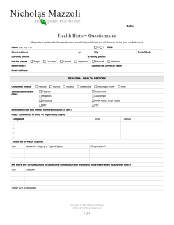


© Copyright 2026