
Inositol activity in oligoasthenoteratospermia – An study M. COLONE
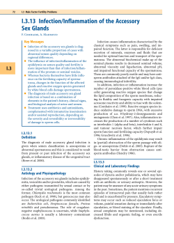
European Review for Medical and Pharmacological Sciences
2010; 14: 891-896
Inositol activity in oligoasthenoteratospermia –
An in vitro study
M. COLONE1, G. MARELLI*, V. UNFER**, G. BOZZUTO1,
A. MOLINARI1, A. STRINGARO1
1
Department of Technologies and Health, National Institute of Health, Rome (Italy)
*Obstetric-Gynaecologic Clinic, Scientific Institute for Admission and Treatment, Saint Raphael
Hospital, Milan (Italy)
**Obstetric-Gynaecologic Clinic, University of Perugia, Perugia (Italy)
Abstract. – Background: Oligoasthenoteratospermia, a reduction in motilty and number of
spermatozoa and a change in their morphology,
is one of the most relevant causes of infertility in
men. One of the factors, which may influence
male infertility is linked to the production of reactive oxygen species (ROS) by morphologically altered spermatozoa. Spermatozoa are more susceptible than other cell species to the detrimental
activity of these chemical compounds. In particular ROS can affect motility, morphology and DNA
stability of spermatozoa.
Aim: In the present in vitro study the role of a
natural substance, inositol, has been investigated as a possible antioxidant agent both for the
systemic treatment of male infertility and for the
improvement in the in vitro quality of the sperm
used for the fertilization applied to medically assisted reproductive procedures.
Materials and Methods: The collected samples, belonging to subjects suffering from
oligoasthenoteratospermia and of healthy subjects were submitted to phase constrast microscopy in order to evaluate spermatozoa
motility, treated with inositol 2 mg/ml and then
submitted to scansion electron microscopy
(SEM) and to transmission electron microscopy
(TEM). SEM allowed to study both the surface
morphology of the biological samples and the
changes induced on them by the treatment with
inositol. TEM allowed to study ultrastructural details of the biological samples.
Results: In the samples of subjects suffering
from oligoasthenoteratospermia the spermatozoa appear entirely covered with an amorphous
fibrous material, that gives an excessive viscosity to the seminal fluid, and reduces or avoids
cell mobility. The micrographs of these samples
show that the mitochondria, in their intermediate
tract, appear to be altered with markedly damaged cristae. After treatment with inositol the
pathologic samples clearly shows the absence
of the amorphous material, perhaps due to a
variation in seminal fluid pH. Furthermore, they
show the presence of mitochondria morphologi-
cally more similar to control specimen mitochondria, with less damage involving mitochondrial cristae.
Conclusions: These preliminary data appear
to suggest that inositol, on account of its antioxidant activity, could preferentially aim at the mitochondrium. Further studies are requested to
the purpose of better defining the combination
between ROS values of the samples, inositol in
vitro treatment and oligoasthenoteratospermia.
Key Words:
Oligoasthenoteratospermia, Reactive oxygen
species, Scansion electron microscopy, Transmission
electron microscopy.
Introduction
A reduction in number and motility of spermatozoa together with a change in their morphology
is called “oligoasthenoteratospermia”. In 30-35%
of the infertile couples presenting with it the etiology is unknown. Several are the therapies suggested for counteracting such a disease. However, they are often ineffective. In the past few
years the scientific community has addressed the
research towards the study of the oxidative stress,
since it had been observed that one of the factors,
which may influence male infertility, is linked to
the production of reactive oxygen species (ROS)
generated by morphologically altered spermatozoa1-8. In fact, if from a point of view a certain
amount of spermatozoa-produced free radicals
appears to be important both for the process of
their maturation and for their reproductive function9, from another point it should be taken into
consideration the fact that spermatozoa are cells
devoid of those defensive mechanisms character-
Corresponding Author: Marisa Colone, MD; e-mail: [email protected]
891
M. Colone , G. Marelli, V. Unfer, G. Bozzuto, A. Molinari, A. Stringaro
izing other cell species4,10. Consequently, spermatozoa are much more susceptible to the negative
effects produced by the oxidative reactions.
Many of the exceeding radicals are produced by
leukocytes of seminal plasma or by immature
spermatozoa, prematurely released from the seminiferous tubules. Such radicals generate some
stress in male gametes. In certain cases not only
the morphological features of such gametes (i.e.,
head and tail structures) result altered, but, above
all, they influence in a negative fashion also the
expression and the stability of the spermatozoal
core, namely the genetic code11.
Therefore, the oxidative stress affecting male
gametes is a condition capable of modifying not
only cell motility and morphology but also DNA
stability and normal mitochondrial structure. Mitochondria are the organelles producing the energy (ATP) required by spermatozoa for the performance of their function. ROS, in fact, may induce changes in mitochondrial membranes and
may thus compromise spermatozoal function12.
This leads to a vicious circle since the damaged
membranes, in turn, induce the production of additional ROS13 with the following end results: reduced fertilizing capacity of man, early miscarriages and, in certain instances, genetic changes
involving the embryo.
Several are the situations capable of inducing
an oxidative stress in seminal fluid: morbid conditions involving the reproductive apparatus
(varicocele, prostatitis)14-16, particular life styles
(smoking, alcohol abuse, drug addiction)17-20, environmental pollution (radiation, smog, industrial
gases)21-23 and nutritional errors (unbalanced hyperlipidic diet)24.
The majority of body cells is supplied with endogenous antioxidant systems; some of them are
of enzymatic type, such as superoxide desmutase,
catalase and glutathione/peroxidase-glutathione/
reductase, whereas others are not enzyms type,
such as vitamin C, vitamin E, coenzyme Q10, etc.
However, when such defensive systems are inadequate, ROS are produced in excess and may lead
to pathological damages by oxidizing cell membrane lipids and proteins as well as DNA structure, thus altering cell function.
In the present in vitro study the role of a natural substance, inositol, has been investigated as a
possible antioxidant agent both for the systemic
treatment of male infertility and for the improvement in the in vitro quality of the sperm used for
the fertilization applied to medically assisted reproductive procedures25,26.
892
Inositol is a cyclic sugar and a vitaminic factor
belonging to the vitamin B2 complex; it is involved in calcium-dependent intracellular signalling pathways and in cellular signal transduction through the plasmatic membrane27. Inositol
regulates several cellular processes, including cell
differentiation and proliferation through calcium
release28. It can induce stimulant effects both at a
motor level and at a central nerve system (CNS)
level. It is an organic compound participating in
the composition of membrane phospholipids. Inositol is used in the treatment of muscular dystrophy29 and of liver diseases30. In addition, it controls the secretion of certain endocrine glands, including gonads31. Its natural sources are untreated
whole-meal cereals, citrus fruits, brewer’s yeast.
Recent studies32 have suggested inositol involvement both in maturation of spermatozoa and in
their migration from the epididymis.
Materials and Methods
After subscribing an informed consent, samples of seminal fluid were obtained from healthy
subjects and from subjects with oligoasthenoteratospermia. The collected samples were submitted to phase constrast microscopy in order to
evaluate spermatozoa motility. The specimens
were stained with tripan blue for assessing their
vitality and then were treated with inositol 2
mg/ml either for 30 minutes, 1 hour or 2 hours at
37°C under a controlled CO2 atmosphere. Following the above mentioned procedures, the
samples were submitted to scansion electron microscopy (SEM) and to transmission electron microscopy (TEM).
The study was approved by the local Ethical
Committee before starting the samples collection.
Scansion Electron Microscopy
SEM allows to study both the surface morphology of properly prepared biological samples
and the prospective changes induced on them by
the treatment with several types of agents, the development of any pathological situations, and the
changes in environmental conditions. The samples were prepared according to the following
procedure: the spermatozoa were stuck on glassslides, 13-mm in diameter, then covered with
polylysine and fixed by a 2.5% glutaraldheyde
solution in 0.2 M cacodylate buffer (at pH 7.3)
Inositol activity in oligoasthenoteratospermia – An in vitro study
and kept for 60 minutes at room temperature.
Following a few washings in the same buffer solution, a post-fixation was carried out by using
1% osmium tetroxide (OsO4) in 0.2 M cacodylate buffer at pH 7.2 for 60 minutes at room temperature. The samples were then dehydrated by
means of increasing scalar solutions of ethanol,
exsiccated in the presence of CPD and covered
with a 30-nm layer of gold by means of a sputtering technique. Finally the samples were examined through a scansion electron microscope
Cambridge Stereoscan 360.
(Epon 812, Agar Scientific Ltd, Stansted, U.K.).
Ultrathin specimen sections were obtained by an
ultramicrotome Nova (LKB, Bromma, Sweden).
Prior to performing the examinations, the sections were contrasted through incubation in
uranyl acetate and lead citrate. The specimens
were finally checked with a transmission electron
microscope Philips 208 (FEI Company, Eindhowen, The Netherland).
Transmission Electron Microscopy
TEM allows to study ultrastructural details of
biological samples. The preparation of samples
for TEM examination must aim at achieving an
extremely thin specimen, which should be intersected by the electrons originated by the source.
The following are the main phases of the procedure: fixation and post-fixation, dehydration, infiltration and polymerization of the resin, specimen sectioning and contrasting. In short, the
samples were fixed with 2.5% (v/v) glutaraldheyde solution in 0.1 M (pH 7.4) cacodylate
buffer at 4°C temperature overnight. Then the
samples were embedded into 2% Agar Noble in
water, dehydrated by increasing scalar solutions
of ethanol and embedded into epoxidic resin
A number of observations by SEM were performed on samples of seminal fluid belonging to
normal sperm donors and to donors suffering
from oligoasthenoteratospermia (Figure 1). The
qualitative analysis of spermatozoal cell surface
shows the presence, in the control subjects, of
spermatozoa with normal morphology (Figure
1a), whereas in the samples of subjects suffering
from oligoasthenoteratospermia the spermatozoa
appear entirely covered with an amorphous fibrous material. Such a covering layer could be
responsible for the excessive viscosity of seminal
fluid and consequently for the reduced or absent
cell mobility observed at the phase contrast microscope. Figures 1c and 1d show the micrographies pertinent to the same specimens following
Results
Figure 1. Scansion electron microscopy. A, Control specimen. B, Oligoasthenoteratospermia specimen covered with amorphous material. C, Control spermatic cell following 2-hour treatment with inositol. D, Pathological spermatic cell following
inositol treatment. Arrow heads point out intermediate tract thickening.
893
M. Colone , G. Marelli, V. Unfer, G. Bozzuto, A. Molinari, A. Stringaro
a 2-hour treatment with inositol (2 mg/ml) at
37°C. The spermatic cell of the control (Figure
1c) shows a normal, unaltered surface morphology associated with a slight thickening of the intermediate tract (arrow head) which, as it is well
known, contains the mitocondrial sheath. The
pathological specimen treated with inositol (Figure 1d) clearly shows the absence of the amorphous material present in the untreated specimen
(Figure 1b). In addition, a marked increase in the
thickness of spermatozoal intermediate tract is
visible even in such a specimen (arrow head).
The data found would apparently suggest that inositol treatment might be able to dissolve the initially observed amorphous fibrous material by inducing, perhaps, a variation in seminal fluid pH.
Such a hypothesis has been substantiated by the
findings obtained on the same specimens through
TEM. The observations made on ultrathin sections of the control specimen show a spermatozoon with many intact mitochondria surrounding
the axoneme of the spermatic flagellum (Figure
2a). On the contrary, micrographs of seminal fluid specimen belonging to the subject suffering
from oligoasthenoteratospermia (Figure 2b)
show an impaired spermatozoon with an altered
acrosome (Aa) associated with the presence of
marginalized chromatin (Cm). In addition, mitochondria, in their intermediate tract, appear to be
altered with markedly damaged cristae (Figure
2b, arrow heads). Following a 2-hour inositol
treatment (2 mg/ml) at 37°C, the pathological
specimen shows the presence of mitochondria
morphologically more similar to control specimen mitochondria (Figures 2c and 2d), with less
damage involving mitochondrial cristae.
Discussion
An increase in the number of infertile couples
has been observed in the past few decades, with
particular reference to an increment of cases ascribable to male factors. Recent data indicate that
oxidative stress plays a fundamental role in infertility development33-35. Spermatozoa are especially sensitive to the action of radicals both because
of the composition of their plasmatic membrane,
which is rich in polyunsaturated fatty acids, and
because of the loss of antioxidant enzymes when
they lose the cytoplasm during their maturation36.
Figure 2. Transmission electron microscopy. A-B, Ultrathin section of a control spermatozoon (A) and a pathologic spermatozoon (B). Marginalized chromatin (Cm) and markedly altered acrosome (Aa) are visible in (B). Arrow heads point out altered mitochondrial cristae. C-D, Ultrathin sections of pathological spermatozoa treated with inositol for a 2-hour period. D,
Magnification of the intermediate tract: some mitochondria are visible with partially restored original morphology.
894
Inositol activity in oligoasthenoteratospermia – An in vitro study
This is one of the reasons why the medical and
scientific interest has been prompted to search
for new substances able to improve the expectations in the field of male infertility.
High concentrations of ROS in the reproductive
apparatus and in the seminal fluid are associated
with important dysfunctions and cellular damages
involving spermatozoa9,37. Recent scientific evidence24,38-40 indicates that the use of antioxidants,
both through the systemic route and as a supplement employed during in vitro techniques for the
preparation of seminal fluid, appears to be effective
in order to induce a significant reduction in excessively high levels of ROS. This represents a promising therapeutic strategy against male infertility.
The present ultrastructural morphologic study has
evidenced that inositol, with its antioxidant activity,
may be preferentially aiming at the mitochondrium.
Both the altered intramitochondrial metabolism with
elevated free iron concentrations and the defective
mitochondrial respiratory chain, associated with an
increased production of free radicals and a subsequent oxidative damage, may very likely constitute a
mechanism capable of compromising the vitality of
cells, including spermatozoa. Especially the latter
cells, which are devoid of cytoplasm, suffer more
heavily the damages due to the high concentrations
of ROS produced within themselves.
These preliminary data appear to suggest that
inositol, on account of its antioxidant activity,
could preferentially aim at the mitochondrium.
Several studies41-43 have carried out in man and
they have shown a correlation between an altered
mitochondrial function (oxidative damage) and a
reduction in motility and in fertilizing ability.
Our future research will aim at clarify the inositol’s mechanism of action, by correlating the
data resulting from the structural analysis (qualitative findings) with the quantitative sensibility
tests (flow cytometry), for the purpose of better
defining the combination between ROS values of
the samples, inositol in vitro treatment and
oligoasthenoteratospermia.
References
1) MAKKER K, AGARWAL A, SHARMA R. Oxidative stress &
male infertility. Indian J Med Res 2009; 129: 357-367.
2) CHABORY E, DAMON C, LENOIR A, HENRY-BERGER J,
VERNET P, CADET R, SAEZ F, DREVET JR. Mammalian
glutathione peroxidases control acquisition and
maintenance of spermatozoa integrity. J Anim Sci
2010; 88: 1321-1323.
3) TUNC O, TREMELLEN K. Oxidative DNA damage impairs global sperm DNA methylation in infertile
men. J Assist Reprod Genet 2009; 26: 537-544.
4) JONES R, MANN T, SHERINS R. Peroxidative breakdown of phospholipids in human spermatozoa,
spermicidal properties of fatty acid peroxides, and
protective action of seminal plasma. Fertil Steril
1979; 31: 531-537.
5) AITKEN RJ, CLARKSON JS. Cellular basis of defective
sperm function and its association with the genesis of reactive oxygen species by human spermatozoa. J Reprod Fertil 1987; 81: 459-469.
6) AITKEN RJ, BUCKINGHAM D, WEST K, WU FC, ZIKOPOULOS K, RICHARDSON DW. Differential contribution of
leucocytes and spermatozoa to the generation of
reactive oxygen species in the ejaculates of oligozoospermic patients and fertile donors. J Reprod
Fertil 1992; 94: 451-462.
7) AITKEN RJ, BAKER MA. Oxidative stress and male reproductive biology. Reprod Fertil Dev 2004; 16:
581-588.
8) DE IULIIS GN, WINGATE JK, KOPPERS AJ, MCLAUGHLIN
EA, AITKEN RJ. Definitive evidence for the nonmitochondrial production of superoxide anion by human spermatozoa. J Clin Endocrinol Metab 2006;
91: 1968-1975.
9)
DE LAMIRANDE E, GAGNON C. Impact of reactive oxygen species on spermatozoa: a balancing act between beneficial and detrimental effects. Hum Reprod 1995; 10(Suppl 1): 15-21.
10) AITKEN RJ. Founders’ Lecture. Human spermatozoa: fruits of creation, seeds of doubt. Reprod
Fertil Dev 2004; 16: 655-664.
11) COCUZZA M, SIKKA SC, ATHAYDE KS, AGARWAL A. Clinical relevance of oxidative stress and sperm chromatin damage in male infertility: an evidence
based analysis. International Braz J Urol 2007;
33: 603-621.
12) ARMSTRONG JS, RAJASEKARAN M, CHAMULITRAT W, GATTI
P, HELLSTROM WJ, SIKKA SC. Characterization of reactive oxygen species induced effects on human
spermatozoa movement and energy metabolism.
Free Radic Biol Med 1999; 26: 869-880.
13) KOPPERS AJ, DE IULIIS GN, FINNIE JM, MCLAUGHLIN EA,
AITKEN RJ. Significance of mitochondrial reactive
oxygen species in the generation of oxidative
stress in spermatozoa. J Clin Endocrinol Metab
2008; 93: 3199-3207.
14) PASQUALOTTO FF, SUNDARAM A, SHARMA RK, BORGES E Jr,
PASQUALOTTO EB, AGARWAL A. Semen quality and oxidative stress scores in fertile and infertile patients
with varicocele. Fertil Steril. 2008; 89: 602-607.
15) HENDIN BN, KOLETTIS PN, SHARMA RK, THOMAS AJ Jr,
AGARWAL A. Varicocele is associated with elevated
spermatozoal reactive oxygen species production
and diminished seminal plasma antioxidant capacity. J Urol 1999; 161: 1831-1834.
16) POTTS JM, PASQUALOTTO FF. Seminal oxidative stress
in patients with chronic prostatitis. Andrologia
2003; 35: 304-308.
895
M. Colone , G. Marelli, V. Unfer, G. Bozzuto, A. Molinari, A. Stringaro
17) KIZILER AR, AYDEMIR B, ONARAN I, ALICI B, OZKARA H,
GULYASAR T, AKYOLCU MC. High levels of cadmium
and lead in seminal fluid and blood of smoking
men are associated with high oxidative stress and
damage in infertile subjects. Biol Trace Elem Res
2007; 120: 82-91.
18) S ALEH RA, A GARWAL A, S HARMA RK, N ELSON DR,
THOMAS AJ Jr. Effect of cigarette smoking on levels
of seminal oxidative stress in infertile men: a
prospective study. Fertil Steril 2002; 78: 491-499.
19) KOCH OR, PANI G, BORRELLO S, COLAVITTI R, CRAVERO
A, FARRE S, GALEOTTI T. Oxidative stress and antioxidant defenses in ethanol-induced cell injury. Mol
Aspects Med 2004; 25: 191-198.
20) WU D, CEDERBAUM AI. Alcohol, oxidative stress,
and free radical damage. Alcohol Res Health
2003; 27: 277-284.
21) CHITRA KC, SUJATHA R, LATCHOUMYCANDANE C, MATHUR
PP. Effect of lindane on antioxidant enzymes in
epididymis and epididymal sperm of adult rats.
Asian J Androl 2001; 3: 205-208.
22) LATCHOUMYCANDANE C, MATHUR PP. Induction of oxidative stress in the rat testis after short-term exposure to the organochlorine pesticide methoxychlor. Arch Toxicol 2002; 76: 692-698.
23) L ATCHOUMYCANDANE C, C HITRA KC, M ATHUR PP.
2,3,7,8-tetrachlorodibenzo-p-dioxin (TCDD) induces oxidative stress in the epididymis and epididymal sperm of adult rats. Arch Toxicol 2003;
77: 280-284.
24) ESKENAZI B, KIDD SA, MARKS AR, SLOTER E, BLOCK G,
WYROBEK AJ. Antioxidant intake is associated with
semen quality in healthy men. Hum Reprod 2005;
20: 1006-1012.
25) PAPALEO E, UNFER V, BAILLARGEON JP, DE SANTIS L, FUSI
F, BRIGANTE C, MARELLI G, CINO I, REDAELLI A, FERRARI
A. Myo-inositol in patients with polycystic ovary
syndrome: a novel method for ovulation induction.
Gynecol Endocrinol 2007; 23: 700-703.
26) PAPALEO E, UNFER V, BAILLARGEON JP, FUSI F, OCCHI F,
D E S ANTIS L. Myo-inositol may improve oocyte
quality in intracytoplasmic sperm injection cycles.
A prospective, controlled, randomized trial. Fertil
Steril 2009; 91: 1750-1754.
27) ONNEBO SM, SAIARDI A. Inositol pyrophosphates
modulate hydrogen peroxide signalling. Biochem
J 2009; 423: 109-118.
31) HINTON BT, WHITE RW, SETCHELL BP. Concentrations
of myo-inositol in the luminal fluid of the mammalian testis and epididymis. J Reprod Fertil
1980; 58: 395-399.
32) BONI R, GUALTIERI R, TALEVI R, TOSTI E. Calcium and
other ion dynamics during gamete maturation and
fertilization. Theriogenology 2007; 68(Suppl 1):
S156-S164.
33) SALEH RA, AGARWAL A, NADA EA, EL-TONSY MH, SHARMA RK, MEYER A, NELSON DR, THOMAS AJ. Negative
effects of increased sperm DNA damage in relation to seminal oxidative stress in men with idiopathic and male factor infertility. Fertil Steril 2003;
79(Suppl 3): 1597-1605.
34) WANG X, SHARMA RK, SIKKA SC, THOMAS AJ JR, FALCONE T, AGARWAL A. Oxidative stress is associated
with increased apoptosis leading to spermatozoa
DNA damage in patients with male factor infertility. Fertil Steril 2003; 80: 531-535.
35) WANG X, SHARMA RK, GUPTA A, GEORGE V, THOMAS
AJ, FALCONE T, AGARWAL A. Alterations in mitochondria membrane potential and oxidative stress in
infertile men: a prospective observational study.
Fertil Steril 2003; 80(Suppl 2): 844-850.
36) MAKKER K, AGARWAL A, SHARMA R. Oxidative stress &
male infertility. Indian J Med Res. 2009; 129: 357367.
37) AITKEN J, FISHER H. Reactive oxygen species generation and human spermatozoa: the balance of
benefit and risk. Bioessays 1994; 16: 259-267.
38) ROSS C, MORRISS A, KHAIRY M, KHALAF Y, BRAUDE P,
COOMARASAMY A, EL-TOUKHY T. A systematic review
of the effect of oral antioxidants on male infertility.
Reprod Biomed Online 2010 Mar 10. [Epub
ahead of print].
39) CHI HJ, KIM JH, RYU CS, LEE JY, PARK JS, CHUNG DY,
CHOI SY, KIM MH, CHUN EK, ROH SI. Protective effect of antioxidant supplementation in spermpreparation medium against oxidative stress in
human spermatozoa.Hum Reprod 2008; 23:
1023-1028.
40) TUNC O, THOMPSON J, TREMELLEN K. Improvement in
sperm DNA quality using an oral antioxidant therapy. Reprod Biomed Online 2009; 18: 761-768.
28) BERRIDGE MJ, BROWN KD, IRVINE RF, HESLOP JP. Phosphoinositides and cell proliferation. J Cell Sci
Suppl 1985; 3: 187-198.
41) ZORN B, VIDMAR G, MEDEN-VRTOVEC H. Seminal reactive oxygen species as predictors of fertilization, embryo quality and pregnancy rates after
conventional in vitro fertilization and intracytoplasmic sperm injection. Int J Androl 2003; 26:
279-285.
29) BASSET O, BOITTIN FX, DORCHIES OM, CHATTON JY, VAN
B REEMEN C, R UEGG UT. Involvement of inositol
1,4,5-trisphosphate in nicotinic calcium responses in dystrophic myotubes assessed by nearplasma membrane calcium measurement. J Biol
Chem 2004; 279: 47092-47100.
42) HAMMADEH ME, RADWAN M, AL-HASANI S, MICU R,
ROSENBAUM P, LORENZ M, SCHMIDT W. Comparison of
reactive oxygen species concentration in seminal
plasma and semen parameters in partners of pregnant and non-pregnant patients after IVF/ICSI. Reprod Biomed Online 2006; 13: 696-706.
30) LEE HJ, LEE SA, CHOI H. Dietary administration of
inositol and/or inositol-6-phosphate prevents
chemically-induced rat hepatocarcinogenesis.
Asian Pac J Cancer Prev 2005; 6: 41-47.
43) AMARAL S, OLIVEIRA PJ, RAMALHO-SANTOS J. Diabetes
and the impairment of reproductive function: possible role of mitochondria and reactive oxygen
species. Curr Diabetes Rev 2008; 4: 46-54.
896


© Copyright 2026