
Committed to the advancement of Clinical & Industrial Disinfection &... Editorial
Committed to the advancement of Clinical & Industrial Disinfection & Microbiology
VOLUME - IV
ISSUE - VI
NOV-DEC 2011
Editorial
Contents
Editorial
n
1
Mini review
n
2
Current Trends
n
9
In Profile
n
Relax Mood
n
Bug of the
n
6
10
Month 11
Did you Know
n
13
Best Practices
n
14
In Focus
n
16
It is important to note that in the mid-1800s, studies by Ignaz Semmelweis in Vienna,
Austria, and Oliver Wendell Holmes in Boston, USA, established that hospital-acquired
diseases were transmitted via the hands of Healthcare workers. As a consequence,
Semmelweis recommended that hands be scrubbed in a chlorinated lime solution before
every patient contact and particularly after leaving the autopsy room. Unfortunately, both
Holmes and Semmelweis failed to observe a sustained change in their colleagues’
behaviour. In particular, Semmelweis experienced great difficulties in convincing his
colleagues and administrators of the benefits of this procedure. In the light of the principles
of social marketing today, his major error was that he imposed a system change (the use of
the chlorinated lime solution) without consulting the opinion of his collaborators. Also
people are more resistant to change in the system. But following the implementation of this
measure, the mortality rate fell dramatically to 3% in the clinic most affected and remained
low thereafter. This caused a major paradigm shift in the field of hand hygiene. And today it
is one of the major concerns to eliminate Nosocomial Infection.
In the section mini review, WHO Guidelines on Hand Hygiene in Healthcare is discussed.
Health care-associated infection (HCAI) is a major problem for patient safety and its
surveillance and prevention must be a first priority for settings and institutions committed
to making health care safer.
In the section current trends we are going to explore various aspects of Environmental
Monitoring in Pharmaceutical industry. It is necessary to follow these guidelines in the
areas like clean-rooms for drug fill/finish, formulation tank rooms, laminar flow hoods,
biological safety hoods, isolators, glove boxes, molding machines, kit assembly lines,
Intravenous compounding areas and sterile packaging.
In profile segment covers biography of Subhash Mukhopadhyay (physician). His feat was
recognized posthumously and was given belated recognition as the Indian physician who
in 1986 was "officially" regarded as being the first doctor to perform in-vitro fertilization in
India.
Chlamydia trachomatis will be discussed in the Bug of the month segment. It is an obligate
intracellular human pathogen and Gram-negative. It can cause numerous disease states in
both men and women. Both sexes can display urethritis, proctitis (rectal disease and
bleeding), trachoma, and infertility. The bacterium can cause prostatitis and epididymitis
in men.
Did You Know emphasizes on Proteus syndrome, which is a complex disorder consisting
variably of disproportionate, asymmetric overgrowth of body parts; cerebriform
connective tissue nevi; epidermal nevi; vascular malformations of the capillary, venous,
and lymphatic types; and dysregulated adipose tissue. Serious complications may ensue,
such as pulmonary embolism, cystic lung disease, and various neoplasms.
Best Practices segment will encompass aseptic media-fill testing, which is used to quantify
the aseptic technique of compounding personnel or processes and to ensure that the
processes used are able to produce a sterile product without microbiological
contamination.
JHS team thanks all our readers for their support and contribution. Feedback and
suggestions are always invited.
Microxpress
Quick Reliable Microbiology
Group
www.tulipgroup.com
1
Mini Review
NOV-DEC 2011
WHO Guidelines on Hand
Hygience in Health Care
Definition of terms
Antiseptic handwashing. Washing hands with soap and water, or
other detergents containing an antiseptic agent.
Hospital in Europe Link for Infection Control through
Surveillance (HELICS) (http://helics.univ-lyon1.fr/ helicshome.
htm), approximately 5 million HCAIs are estimated to occur in
acute care hospitals in Europe annually, representing around
million extra days of hospital stay and a corresponding economic
burden of €13–24 billion. In general, attributable mortality due to
HCAI in Europe is estimated to be 1% (50 000 deaths per year),
but HCAI contributes to death in at least 2.7% of cases (135 000
deaths per year). The estimated HCAI incidence rate in the USA
was 4.5% in 2002, corresponding to 9.3 infections per 1000
patient-days and 1.7 million affected patients; approximately 99
000 deaths were attributed to HCAI. The annual economic impact
of HCAI in the USA was approximately US $6.5 billion in 2004.
Antiseptic handrubbing (or handrubbing). Applying an antiseptic
handrub to reduce or inhibit the growth of microorganisms
without the need for an exogenous source of water and requiring
no rinsing or drying with towels or other devices.
Hand antisepsis/decontamination/degerming. Reducing or
inhibiting the growth of microorganisms by the application of an
antiseptic handrub or by performing an antiseptic handwash.
Hand care. Actions to reduce the risk of skin damage or irritation.
Handwashing. Washing hands with plain or antimicrobial soap
and water.
Burden of health care-associated infection in developing
countries
The magnitude of the problem is particularly relevant in settings
where basic infection control measures are virtually nonexistent.
This is the result of the combination of numerous unfavorable
factors such as understaffing, poor hygiene and sanitation, lack or
shortage of basic equipment, and inadequate structures and
overcrowding, almost all of which can be attributed to limited
financial resources. In addition to these specific factors, an
unfavorable social background and a population largely affected
by malnutrition and other types of infection and/or diseases
contribute to increase the risk of HCAI in developing countries.
Under these conditions, thousands of infections – in particular
due to hepatitis B and C viruses and human immunodeficiency
virus (HIV) transmission – are still acquired from patients, but
also from HCWs through unsafe use of injections, medical
devices and blood products, inadequate surgical procedures, and
deficiencies in biomedical waste management. In one-day
prevalence surveys recently carried out in single hospitals in
Albania,Morocco,Tunisia,and the United Republic of
Tanzania,HCAI prevalence rates were 19.1%, 17.8%, 17.9%, and
14.8%, respectively. Given the difficulties to comply with the
USA Centers for Disease Control and Prevention (CDC)
definitions of nosocomial infection,the most frequently surveyed
type of infection is SSI, which is the easiest to define according to
clinical criteria. The risk for patients to develop SSI in developing
countries is significantly higher than in developed countries (e.g.
30.9% in a paediatric hospital in Nigeria,23% in general surgery
in a hospital in the United Republic of Tanzania,and 19% in a
maternity unit in Kenya).
Hand cleansing. Action of performing hand hygiene for the
purpose of physically or mechanically removing dirt, organic
material, and/or microorganisms.
Hygienic hand antisepsis. Treatment of hands with either an
antiseptic handrub or antiseptic handwash to reduce the transient
microbial flora without necessarily affecting the resident skin
flora.
Hygienic handrub. Treatment of hands with an antiseptic handrub
to reduce the transient flora without necessarily affecting the
resident skin flora. These preparations are broad spectrum and
fast-acting, and persistent activity is not necessary.
Hygienic handwash. Treatment of hands with an antiseptic
handwash and water to reduce the transient flora without
necessarily affecting the resident skin flora. It is broad spectrum,
but is usually less efficacious and acts more slowly than the
hygienic handrub.
Surgical hand antisepsis/surgical hand preparation. presurgical
hand preparation. Antiseptic handwash or antiseptic handrub
performed preoperatively by the surgical team to eliminate
transient flora and reduce resident skin flora. Such antiseptics
often have persistent antimicrobial activity. Surgical handscrub
(bing)/ presurgical scrub refer to surgical hand preparation with
antimicrobial soap and water. Surgical handrub (bing) refers to
surgical hand preparation with a waterless, alcohol-based
handrub.
The burden of health care-associated infection
Health care-associated infection (HCAI) is a major problem for
patient safety and its surveillance and prevention must be a first
priority for settings and institutions committed to making health
care safer.
Historical perspective
In the mid-1800s, studies by Ignaz Semmelweis in Vienna,
Austria, and Oliver Wendell Holmes in Boston, USA, established
that hospital-acquired diseases were transmitted via the hands of
HCWs. In 1847, Semmelweis was appointed as a house officer in
one of the two obstetric clinics at the University of Vienna
Allgemeine Krankenhaus (General Hospital). He observed that
maternal mortality rates, mostly attributable to puerperal fever,
were substantially higher in one clinic compared with the other
(16% versus 7%).He also noted that doctors and medical students
often went directly to the delivery suite after performing
Health care-associated infection in developed countries
In developed countries, HCAI concerns 5–15% of hospitalized
patients and can affect 9–37% of those admitted to intensive care
units (ICUs). Recent studies conducted in Europe reported
hospital-wide prevalence rates of patients affected by HCAI
ranging from 4.6% to 9.3%. According to data provided by the
Microxpress
2
Group
www.tulipgroup.com
NOV-DEC 2011
Mini Review
autopsies and had a disagreeable odour on their hands despite
handwashing with soap and water before entering the clinic. He
hypothesized therefore that “cadaverous particles” were
transmitted via the hands of doctors and students from the
autopsy room to the delivery theatre and caused the puerperal
fever. As a consequence, Semmelweis recommended that hands
be scrubbed in a chlorinated lime solution before every patient
contact and particularly after leaving the autopsy room.
Following the implementation of this measure, the mortality rate
fell dramatically to 3% in the clinic most affected and remained
low thereafter.
CFU/cm2 on the forearm. Total bacterial counts on the hands of
HCWs have ranged from 3.9 x 10⁴ to 4.6 x 10⁶ CFU/cm2. Fingertip
contamination ranged from 0 to 300 CFU when sampled by agar
contact methods. Price and subsequent investigators documented
that although the count of transient and resident flora varies
considerably among individuals, it is often relatively constant for
any given individual.
Unfortunately, both Holmes and Semmelweis failed to observe a
sustained change in their colleagues’ behaviour. In particular,
Semmelweis experienced great difficulties in convincing his
colleagues and administrators of the benefits of this procedure. In
the light of the principles of social marketing today, his major
error was that he imposed a system change (the use of the
chlorinated lime solution) without consulting the opinion of his
collaborators. Despite these drawbacks, many lessons have been
learnt from the Semmelweis intervention; the “recognizeexplain-act” approach has driven many investigators and
practitioners since then and has also been replicated in different
fields and settings. Semmelweis is considered not only the father
of hand hygiene, but his intervention is also a model of
epidemiologically driven strategies to prevent infection.
1) Organisms are present on the patient’s skin, or have been shed
onto inanimate objects immediately surrounding the patient.
The number of organisms such as S. aureus, Proteus mirabilis,
Klebsiella spp. and Acinetobacter spp. present on intact areas of
the skin of some patients can vary from 100 to 106 CFU/cm2
gowns, bed linen, bedside furniture and other objects in the
immediate environment of the patient become contaminated with
patient flora. Contamination of the inanimate environment has
also been detected on ward handwash station surfaces and many
of the organisms isolated were staphylococci Tap/faucet handles
were more likely to be contaminated and to be in excess of
benchmark values than other parts of the station.
Transmission of pathogens by hands
Transmission of health care-associated pathogens from one
patient to another via HCWs’ hands requires five sequential steps:
2) Organisms must be transferred to the hands of HCWs.
Nurses could contaminate their hands with 100–1000 CFU of
Klebsiella spp. during “clean” activities such as lifting patients;
taking the patient’s pulse, blood pressure or oral temperature; or
touching the patient’s hand, shoulder or groin. cultured the hands
of nurses who touched the groin of patients heavily colonized
with P. mirabilis and found 10–600 CFU/ml in glove juice
samples. Estimating the frequency of HCWs’ glove
contamination with methicillin resistant S. aureus (MRSA) after
contact with a colonized patient, HCWs were intercepted after a
patient-care episode and cultures were taken from their gloved
hands before handwashing had occurred; 17% (confidence
interval (CI) 95%) of contacts with patients, a patient’s clothing
or a patient’s bed resulted in transmission of MRSA from a patient
to the HCWs’ gloves. In another study involving HCWs caring
for patients with vancomycin-resistant enterococci (VRE), 70%
of HCWs contaminated their hands or gloves by touching the
patient and the patient’s environment.
Normal bacterial flora on hands
In 1938, Price established that bacteria recovered from the hands
could be divided into two categories, namely resident or transient.
The resident flora (resident microbiota) consists of
microorganisms residing under the superficial cells of the stratum
corneum and can also be found on the surface of the skin.
Staphylococcus epidermidis is the dominant species, and
oxacillin resistance is extraordinarily high, particularly among
HCWs. Other resident bacteria include S. hominis and other
coagulase-negative staphylococci, followed by coryneform
bacteria (propionibacteria, corynebacteria, dermobacteria, and
micrococci). Among fungi, the most common genus of the
resident skin flora, when present, is Pityrosporum (Malassezia)
spp. Resident flora has two main protective functions: microbial
antagonism and the competition for nutrients in the ecosystem. In
general, resident flora is less likely to be associated with
infections, but may cause infections in sterile body cavities, the
eyes, or on non-intact skin.
3) Organisms must be capable of surviving for at least several
minutes on HCWs’ hands.
Enterococcus faecalis and E. faecium survive for at least 60
minutes on gloved and ungloved fingertips. Pseudomonas
aeruginosa and Burkholderia cepacia are transmissible by
handshaking for up to 30 minutes when the organisms are
suspended in saline, and up to 180 minutes when they are
suspended in sputum. Shigella dysenteriae type 1 showed its
capacity to survive on hands for up to 1 hour. Survival
percentages for rotavirus at 20 minutes and 60 minutes after
inoculation were 16.1% and 1.8%, respectively. Bacterial
contamination increases linearly over time. In the absence of
hand hygiene action, the longer the duration of care, the higher
the degree of hand contamination.
Transient flora (transient microbiota), which colonizes the
superficial layers of the skin, is more amenable to removal by
routine hand hygiene. Transient microorganisms do not usually
multiply on the skin, but they survive and sporadically multiply
on skin surface. They are often acquired by HCWs during direct
contact with patients or contaminated environmental surfaces
adjacent to the patient and are the organisms most frequently
associated with HCAIs. Some types of contact during routine
neonatal care are more frequently associated with higher levels of
bacterial contamination of HCWs’ hands: respiratory secretions,
nappy/diaper change, and direct skin contact. The
transmissibility of transient flora depends on the species present,
the number of microorganisms on the surface, and the skin
moisture. The hands of some HCWs may become persistently
colonized by pathogenic flora such as S. aureus, Gram-negative
bacilli, or yeast. Normal human skin is colonized by bacteria,
with total aerobic bacterial counts ranging from more than 1 x 10⁶
colony forming units (CFU)/cm2 on the scalp, 5 x 10⁵ CFUs/cm2
in the axilla, and 4 x 10⁴ CFU/cm2 on the abdomen to 1 x 10⁴
Microxpress
4) Handwashing or hand antisepsis by the HCW must be
inadequate or entirely omitted, or the agent used for hand hygiene
inappropriate.
In a laboratory-based study, it was found that using only 1 ml of
liquid soap or alcohol-based handrub yielded lower log
3
Group
www.tulipgroup.com
Mini Review
reductions (greater number of bacteria remaining on hands) than
using 3 ml of product to clean hands. The findings have clinical
relevance since some HCWs use as little as 0.4 ml of soap to clean
their hands. A comparative, crossover study of microbiological
efficacy of handrubbing with an alcohol-based solution and
handwashing with an unmedicated soap was conducted. The
study results were: 15% of HCWs’ hands were contaminated with
transient pathogens before hand hygiene; no transient pathogens
were recovered after handrubbing, while two cases were found
after handwashing. Another comparative study of three hand
hygiene agents (62% ethyl alcohol handrub, medicated
handwipe, and handwashing with plain soap and water) in a
group of surgical ICUs was conducted. The impact of ring
wearing on hand contamination was also studied. The results
showed that hand contamination with transient organisms was
significantly less likely after the use of an alcohol-based handrub
compared with the medicated wipe or soap and water. Ring
wearing increased the frequency of hand contamination with
potential health care-associated pathogens.
denature proteins. solutions containing 60–80% alcohol are most
effective, with higher concentrations being less potent. paradox
results from the fact that proteins are not denatured easily in the
absence of water.
Alcohols have excellent in vitro germicidal activity against
Gram-positive and Gram-negative vegetative bacteria (including
multidrug-resistant pathogens such as MRSA and VRE), M.
tuberculosis, and a variety of fungi.
However, they have virtually no activity against bacterial spores
or protozoan oocysts, and very poor activity against some nonenveloped (non-lipophilic) viruses.
Chlorhexidine
CHG, a cationic bisbiguanide, was developed in the United
Kingdom in the early 1950s and introduced into the USA in the
1970s. base is barely soluble in water, but the digluconate form is
water-soluble. The antimicrobial activity of chlorhexidine
appears to be attributable to the attachment to, and subsequent
disruption of cytoplasmic membranes, resulting in precipitation
of cellular contents. The antimicrobial activity of Chlorhexidine
is not seriously affected by the presence of organic material,
including blood. A scrub agent based on CHG (4%) was shown to
be significantly more effective to reduce bacterial count than
povidone iodine (7.5%) scrub agent. Chlorhexidine has
significant residual activity. Addition of low concentrations
(0.5–1%) of chlorhexidine to alcohol-based preparations results
in significantly greater residual activity than alcohol alone.
5) The contaminated hand or hands of the caregiver must come
into direct contact with another patient or with an inanimate
object that will come into direct contact with the patient.
Cross-transmission of organisms occurs through contaminated
hands. Factors that influence the transfer of microorganisms from
surface to surface and affect cross-contamination rates are type of
organism, source and destination surfaces, moisture level, and
size of inoculum. Contaminated hands could contaminate a clean
paper towel dispenser and vice versa. The transfer rates ranged
from 0.01% to 0.64% and 12.4% to 13.1%, respectively. Fingers
contaminated with norovirus could sequentially transfer virus to
up to seven clean surfaces, and from contaminated cleaning
cloths to clean hands and surfaces. Contaminated HCWs’ hands
have been associated with endemic HCAIs. Endemic S.
marcescens was transmitted from contaminated soap to patients
via the hands of HCWs. During an outbreak investigation of S.
liquefaciens, BSI, and pyrogenic reactions in a haemodialysis
centre, pathogens were isolated from extrinsically contaminated
vials of medication resulting from multiple dose usage,
antibacterial soap, and hand lotion. VRE could be transferred
from a contaminated environment or patients’ intact skin to clean
sites via the hands of HCWs in 10.6% of contacts. Several HCAI
outbreaks have been associated with contaminated HCWs’
hands. An outbreak of multidrug-resistant A. baumannii
documented identical strains from patients, hands of staff, and the
environment. The outbreak was terminated when remedial
measures were taken. Contaminated HCWs’ hands were clearly
related to outbreaks among surgical and neonatal patients.
Triclosan
Triclosan (chemical name 2,4,4’–trichloro-2’-hydroxydiphenyl
ether) is known commercially as Irgasan DP-300. It is a nonionic,
colourless substance developed in the 1960s; it is poorly soluble
in water, but dissolves well in alcohols. Concentrations ranging
from 0.2% to 2% have antimicrobial activity. Triclosan has been
incorporated in detergents (0.4% to 1%) and in alcohols (0.2% to
0.5%) used for hygienic and surgical hand antisepsis or
preoperative skin disinfection; it is also used for antiseptic body
baths to control MRSA. This agent is incorporated into some
soaps (at a 1% w/v concentration) and a variety of other consumer
products (deodorants, shampoos, lotions, etc.), as well as being
integrated also into various dressings and bandages for release
over time onto the skin.
Triclosan enters bacterial cells and affects the cytoplasmic
membrane and synthesis of RNA, fatty acids, and proteins.
Recent studies suggest that this agent’s antibacterial activity is
attributable in large part to binding to the active site of enoylacyl
carrier protein reductase.
Preparations used for hand hygiene
Alcohols
Most alcohol-based hand antiseptics contain either ethanol,
isopropanol or n-propanol, or a combination of two of these
products. Concentrations are given as either percentage of
volume (= ml/100 ml, abbreviated % v/v), percentage of weight
(= g/100 g, abbreviated % m/m), or percentage of weight/volume
(= g/100 ml, abbreviated % m/v). Studies of alcohols have
evaluated either individual alcohols in varying concentrations
(most studies), combinations of two alcohols, or alcohol
solutions containing small amounts of hexachlorophene,
quaternary ammonium compounds (QAC), povidone-iodine,
triclosan or CHG.
Triclosan has a fairly broad range of antimicrobial activity.
inhibitory concentrations (MICs) range from 0.1 to 10 µg/ml,
while minimum bactericidal concentrations are 25–500 µg/ml.
Triclosan’s activity against Gram-positive organisms (including
MRSA) is greater than against Gram-negative bacilli,
particularly P. aeruginosa. agent possesses reasonable activity
against mycobacteria and Candida spp., but has little activity
against filamentous fungi and most viruses of nosocomial
significance. Triclosan (0.1%) reduces bacterial counts on hands
by 2.8 log10 after a 1-minute hygienic handwash.
Iodine and iodophors
Iodine has been recognized as an effective antiseptic since the
1800s, though iodophors have largely replaced iodine as the
The antimicrobial activity of alcohols results from their ability to
Microxpress
NOV-DEC 2011
4
Group
www.tulipgroup.com
NOV-DEC 2011
Mini Review
active ingredient in antiseptics because iodine often causes
irritation and discolouring of skin.
Required time for the procedure
Scientists compared hand bacterial counts after 3-minute and 5minute scrubs with seven different formulations. showed that the
3-minute scrub could be as effective as the 5-minute scrub,
depending on the formula of the scrub agent. Scrubbing for longer
than 2 minutes did not confer any advantage. This study
recommended a 4-minute scrub for the surgical team’s first
procedure and a 2-minute scrub for subsequent procedures.
Iodine molecules rapidly penetrate the cell wall of
microorganisms and inactivate cells by forming complexes with
amino acids and unsaturated fatty acids, resulting in impaired
protein synthesis and alteration of cell membranes. are composed
of elemental iodine, iodide or triiodide, and a polymer carrier
(complexing agent) of high molecular weight.
The amount of molecular iodine present (so-called “free” iodine)
determines the level of antimicrobial activity of iodophors.
“Available” iodine refers to the total amount of iodine that can be
titrated with sodium thiosulfate. 10% povidoneiodine
formulations contain 1% available iodine and yield free iodine
concentrations of 1 ppm.
Surgical hand preparation with alcohol-based handrubs
Several alcohol-based handrubs have been licensed for the
commercial market frequently with additional, long acting
compounds (e.g. chlorhexidine gluconate or quaternary
ammonium compounds) limiting regrowth of bacteria on the
gloved hand. antimicrobial efficacy of alcohol based
formulations is superior to that of all other currently available
methods of preoperative surgical hand preparation. Numerous
studies have demonstrated that formulations containing 60–95%
alcohol alone, or 50–95% when combined with small amounts of
a QAC, hexachlorophene or chlorhexidine gluconate, reduce
bacterial counts on the skin immediately post-scrub more
effectively than do other agents.
Combining iodine with various polymers increases the solubility
of iodine, promotes sustained-release of iodine, and reduces skin
irritation. The most common polymers incorporated into
iodophors are polyvinyl pyrrolidone (povidone) and ethoxylated
nonionic detergents (poloxamers). antimicrobial activity of
iodophors can also be affected by pH, temperature, exposure
time, concentration of total available iodine, and the amount and
type of organic and inorganic compounds present (e.g. alcohols
and detergents). The concentrations used in antiseptics,
iodophors are not usually sporicidal. Iodophors demonstrated
poor persistent activity. The in vivo antimicrobial activity of
iodophors is significantly reduced in the presence of organic
substances such as blood or sputum. iodine has been found to be
less effective than alcohol 60% (v/v) and hydrogen peroxide 3%
and 5% on S. epidermidis. Iodophor antiseptics have become
contaminated with Gram-negative bacilli as a result of poor
manufacturing processes and have caused outbreaks or pseudooutbreaks of infection. outbreak of P. cepacia involving 52
patients in four hospitals in New York over six months was
attributed to the contamination of a 10% povidone iodine solution
used as an antiseptic and disinfectant solution.
Technique for the application of surgical hand preparation using
alcohol-based handrub
The application technique has not been standardized throughout
the world. The WHO approach for surgical hand preparation
requires the six basic steps for the hands as for hygienic hand
antisepsis, but requires additional steps for rubbing the forearms.
The hands should be wet from the alcohol-based rub during the
whole procedure, which requires approximately 15 ml depending
on the size of the hands. One study demonstrated that keeping the
hands wet with the rub is more important than the volume used.
Once the forearms and hands have been treated with an emphasis
on the forearms – usually for approximately 1 minute – the
second part of the surgical handrub should focus on the hands,
following the identical technique as outlined for the hygienic
handrub. The hands should be kept above the elbows during this
step.
Surgical hand preparation: state-of-the-art
Objective of surgical hand preparation
Surgical hand preparation should reduce the release of skin
bacteria from the hands of the surgical team for the duration of the
procedure in case of an unnoticed puncture of the surgical glove
releasing bacteria to the open wound. contrast to the hygienic
handwash or handrub, surgical hand preparation must eliminate
the transient and reduce the resident flora. It should also inhibit
growth of bacteria under the gloved hand. Rapid multiplication of
skin bacteria occurs under surgical gloves if hands are washed
with a non-antimicrobial soap, whereas it occurs more slowly
following preoperative scrubbing with a medicated soap.
Required time for the procedure
For many years, surgical staff frequently scrubbed their hands for
10 minutes preoperatively, which frequently led to skin damage.
Surgical hand antisepsis using an alcohol-based handrub required
3 minutes.
Surgical handscrub with medicated soap or surgical hand
preparation with alcohol-based formulations, both methods are
suitable for the prevention of SSIs. However, although medicated
soaps have been and are still used by many surgical teams
worldwide for presurgical hand preparation, it is important to
note that the antibacterial efficacy of products containing high
concentrations of alcohol are far more superior than that of any
medicated soap presently available.
The skin flora, mainly coagulase-negative staphylococci,
Propionibacterium spp., and Corynebacteria spp., are rarely
responsible for SSI, but in the presence of a foreign body or
necrotic tissue even inocula as low as 100 CFU can trigger such
infection. The virulence of the microorganisms, extent of
microbial exposure, and host defence mechanisms are key factors
in the pathogenesis of postoperative infection, risk factors that are
largely beyond the influence of the surgical team. Therefore,
products for surgical hand preparation must eliminate the
transient and significantly reduce the resident flora at the
beginning of an operation and maintain the microbial release
from the hands below baseline until the end of the procedure.
Microxpress
5
Group
www.tulipgroup.com
Current Trends
NOV-DEC 2011
Environmental Monitoring in
Pharmaceutical Industry
Environmental Monitoring (E/M) is a program designed to
demonstrate the control of viable (living microorganisms) and nonviable particles in critical areas. These areas include clean-rooms
for drug fill/finish, formulation tank rooms, laminar flow hoods,
biological safety hoods, isolators, glove boxes, molding machines,
kit assembly lines, Intravenous (IV) compounding areas and sterile
packaging.
Viable monitoring refers to testing for the detection and
enumeration of bacteria, yeast and mold. It includes the monitoring
of personnel, air and area surfaces for microbial contamination.
And differs from non-viable environmental monitoring which is a
reference for particle counts measured by a laser counter. Viable
counts provide metrics on the potential for contamination of a
company’s products as well as demonstrating the veracity that a
clean room is functioning as designed and being properly
maintained. Surface and air monitoring exhibit the asepsis of the
product manufacturing operation.
In aseptic processing, one of the most important laboratory controls
is the environmental monitoring program. This program provides
meaningful information on the quality of the aseptic processing
environment (e.g., when a given batch is being manufactured) as
well as environmental trends of ancillary clean areas.
Environmental monitoring should promptly identify potential
routes of contamination, allowing for implementation of corrections
before product contamination occurs.
Evaluating the quality of air and surfaces in the cleanroom
environment should start with a well-defined written program and
scientifically sound methods. The monitoring program should cover
all production shifts and include air, floors, walls, and equipment
surfaces, including the critical surfaces that come in contact with the
product, container, and closures. Written procedures should include
a list of locations to be sampled. Sample timing, frequency, and
location should be carefully selected based upon their relationship to
the operation performed. Samples should be taken throughout the
classified areas of the aseptic processing facility (e.g., aseptic
corridors, gowning rooms) using scientifically sound sampling
procedures. Sample sizes should be sufficient to optimize detection
of environmental contaminants at levels that might be expected in a
given clean area.
It is important that locations posing the most microbiological risk to
the product be a key part of the program. It is especially important to
monitor the microbiological quality of the critical area to determine
whether or not aseptic conditions are maintained during filling and
closing activities. Air and surface samples should be taken at the
locations where significant activity or product exposure occurs
during production. Critical surfaces that come in contact with the
sterile product should remain sterile throughout an operation. When
identifying critical sites to be sampled, consideration should be
given to the points of contamination risk in a process, including
factors such as difficulty of setup, length of processing time, and
impact of interventions. Critical surface sampling should be
performed at the conclusion of the aseptic processing operation to
avoid direct contact with sterile surfaces during processing.
Detection of microbial contamination on a critical site would not
necessarily result in batch rejection. The contaminated critical site
sample should prompt an investigation of operational information
and data that includes an awareness of the potential for a low
incidence of false positives.
Microxpress
Environmental monitoring methods do not always recover
microorganisms present in the sampled area. In particular, low-level
contamination can be particularly difficult to detect. Because false
negatives can occur, consecutive growth results are only one type of
adverse trend. Increased incidence of contamination over a given
period is an equal or more significant trend to be tracked. In the
absence of any adverse trend, a single result above an action level
should trigger an evaluation and a determination about whether
remedial measures may be appropriate. In all room classes, remedial
measures should be taken in response to unfavorable trends.
All environmental monitoring locations should be described in
SOPs with sufficient detail to allow for reproducible sampling of a
given location surveyed. Written SOPs should also address elements
such as (1) frequency of sampling, (2) when the samples are taken
(i.e., during or at the conclusion of operations), (3) duration of
sampling, (4) sample size (e.g., surface area, air volume), (5)
specific sampling equipment and techniques, (6) alert and action
levels, and (7) appropriate response to deviations from alert or
action levels.
Establishing Levels and a Trending Program
Microbiological monitoring levels should be established based on
the relationship of the sampled location to the operation. The levels
should be based on the need to maintain adequate microbiological
control throughout the entire sterile manufacturing facility. One
should also consider environmental monitoring data from historical
databases, media fills, cleanroom qualification, and sanitization
studies, in developing monitoring levels. Data from similar
operations can also be helpful in setting action and alert levels,
especially for a new operation.
Environmental monitoring data will provide information on the
quality of the manufacturing environment. Each individual sample
result should be evaluated for its significance by comparison to the
alert or action levels. Averaging of results can mask unacceptable
localized conditions. A result at the alert level urges attention to the
approaching action conditions. A result at the action level should
prompt a more thorough investigation. Written procedures should
be established, detailing data review frequency and actions to be
taken. The quality control unit should provide routine oversight of
near-term (e.g., daily, weekly, monthly, quarterly) and long-term
trends in environmental and personnel monitoring data.
Trend reports should include data generated by location, shift, room,
operator, or other parameters. The quality control unit should be
responsible for producing specialized data reports (e.g., a search on
a particular isolate over a year period) with the goal of investigating
results beyond established levels and identifying any appropriate
follow-up actions. Significant changes in microbial flora should be
considered in the review of the ongoing environmental monitoring
data.
Written procedures should define the system whereby the most
responsible managers are regularly informed and updated on trends
and investigations.
Disinfection Efficacy
The suitability, efficacy, and limitations of disinfecting agents and
procedures should be assessed. The effectiveness of these
disinfectants and procedures should be measured by their ability to
6
Group
www.tulipgroup.com
NOV-DEC 2011
Current Trends
ensure that potential contaminants are adequately removed from
surfaces.
To prevent introduction of contamination, disinfectants should be
sterile, appropriately handled in suitable containers and used for no
longer than the predefined period specified by written procedures.
Routinely used disinfectants should be effective against the normal
microbial vegetative flora recovered from the facility. Many
common disinfectants are ineffective against spores. For example,
70 percent isopropyl alcohol is ineffective against Bacillus spp.
spores. Therefore, a sound disinfectant program also includes a
sporicidal agent, used according to a written schedule and when
environmental data suggest the presence of sporeforming
organisms.
Disinfection procedures should be described in sufficient detail
(e.g., preparation, work sequence, contact time) to enable
reproducibility. Once the procedures are established, their adequacy
should be evaluated using a routine environmental monitoring
program. If indicated, microorganisms associated with adverse
trends can be investigated as to their sensitivity to the disinfectants
employed in the cleanroom in which the organisms were isolated.
Assessing microbial quality of air should involve the use of active
devices including but not limited to impaction, centrifugal, and
membrane (or gelatin) samplers. Each device has certain advantages
and disadvantages, although all allow testing of the number of
organisms per volume of air sampled. We recommend that such
devices be used during each production shift to evaluate aseptic
processing areas at carefully chosen locations. Manufacturers
should be aware of a device's air monitoring capabilities, and the air
sampler should be evaluated for its suitability for use in an aseptic
environment based on collection efficiency, cleanability, ability to
be sterilized, and disruption of unidirectional airflow.20 Because
devices vary, the user should assess the overall suitability of a
monitoring device before it is placed into service. Manufacturers
should ensure that such devices are calibrated and used according to
appropriate procedures.
Air Sampler
Environmental Monitoring is a must in the pharmaceutical industry.
The FDA guidelines for Aseptic Processing from September 2004
suggests that the air sampler should be evaluated based on collection
efficiency, cleaning ability, ability to be sterilized and disruption of
unidirectional air flow. Further devices have to be calibrated and
used according to appropriate procedures.
A specific Standard for the purpose of Environmental Monitoring is
ISO 14698 Biocontamination control. It is a valid worldwide
published standard, and is mandatory in Europe as there is a
reference in the EU GMP Guide.
Monitoring Methods
Industries that have their clean room facilities monitored do so to
ensure their desired/required quality standards are met. The areas
that are sampled in a manufacturer’s clean room include:
Acceptable methods for monitoring the microbiological quality of
the environment include:
Types of Air Sampler
Methods of Air sampling in a Clean Room
Air - the air in a clean room is controlled and Monitored on a regular
basis (e.g., daily, weekly, quarterly) for particle counts, viable
counts, temperature and humidity. HEPA filters are used to control
the viable and non-viable particulate counts within the air. HEPA
filters have the capability to filter out particulates down to 0.2 µm in
size. These filters usually run continuously at a calibrated flow rate
in order to maintain the required air quality within the room.
Humidity is usually kept at a low level in order to help prevent the
proliferation of microbes within the room such as bacteria and mold,
which tend to prefer damp conditions in order to replicate.
Sieve Impactor: This acts by aspiration of air through the sieves at
the top of the sampler. The apparatus consists of a container
designed to accommodate a petri dish containing growth medium.
The cover of the unit is perforated and a vacuum pump draws a
volume of air through the cover. The particles in the air containing
microorganisms impact on the agar medium. It works on wellknown Andersen’s sieve impaction principle.
Centrifugal Sampler: Centrifugal samplers create a centrifugal
force, which causes particles and microorganisms to impact the
medium at a rate dependent on the size of the particle.
Slit-to-Agar Air Sampler: This sampler draws a known volume of
air by vacuum through a slit opening and then accelerated and
directed toward the surface of a petri dish containing growth media.
What is Air Sampling?
Ø
Air Sampling is used to quantify specific chemical, biological
and physical agents that may present in occupational
environment.
Ø
Biological Air Sampling is routinely used to monitor the
populations of air borne particles of the surrounding area.
Ø
In the context of microbiological assessment air sampling is the
collection of air borne microbial contaminants that may impact
on product spoilage, product safety and human health.
Gelatin Filter Sampler: In this air sampler air is sampled at a
programmable flow rate and passes through a gelatin membrane
filter, which captures the microbes.
2. Passive Air Monitoring (Settling Plates)
In Pharmaceutical industry all manufacturing activities are carried
out in clean areas as recommended by FDA guidelines. Different
standards of clean room are used.
Eg- Class 100, Class 10,000, Class 1,00,000
Significance Of Air Sampling
Ø
Measures the number of viable air borne particles (i.e. the
concentration of microorganisms in the air).
Ø
Evaluation of the effectiveness of control methods.
Ø
Compliance status with respect to various occupational health
standards.
Ø
Routine surveillance.
There are two principle means of monitoring the microbiological
population of the air, active air monitoring and passive air
monitoring. Both have a part to play, but active sampling methods
have become an essential environmental monitoring tool, especially
in the pharmaceutical and medical device sectors.
1. Active Air Monitoring
Microxpress
According to USP chapter<1116> microbial limits for sterile
products are as follows:
Class
100
10,000
100,000
3
CFU/m Air
<3
<20
<100
Class 100- all sterile fillings (Injectables/eye drops) are carried out.
Class 10,000- Manufacturing activities like blending mixing,
7
Group
www.tulipgroup.com
Current Trends
NOV-DEC 2011
Ø
Incoculate one SCDA Plate with 24 hours old culture of S.aureus
0
incubate at 37+2 C for 5 days. Inoculate another SCDA Plate
with 24 hours old culture C.albicans incubate at 25-30 0C for 5
days. (Positive Control).
filtration, stirring and punching of tablets.
Class 1,00,000- Air locks, Packing area, manufacturing of noninjectable product.
Environmental monitoring by settling plate
Interpretation
Passive monitoring is usually done using ‘settle plates’ – standard
Petri dishes containing appropriate (usually non-selective) culture
media that are opened and exposed for a given time and then
incubated to allow visible colonies to develop and be counted. Settle
plates are very limited in their application since they are only really
capable of monitoring viable biological particles that sediment out
of the air and settle onto a surface over the time of exposure. They
will not detect smaller particles or droplets suspended in the air and
they cannot sample specific volumes of air, so the results are not
quantitative. They are also vulnerable to interference and
contamination from non-airborne sources and the agar growth
medium in the plates may deteriorate if they are exposed for too
long. Settle plates may easily become overgrown in heavily
contaminated conditions and interpretation of the data they produce
can be difficult.
Ø
Negative Control Plates should remain sterile till end of the
incubation period.
Ø
Positive Control Plates should give growth of standard colonies.
Methods for surface monitoring in a Clean Room
Surfaces (including floors, walls, equipment, etc.) are cleaned and
monitored on a regular basis for viable counts by using specially
designed contact plates that contain a growth medium called
Trypticase Soy Agar (TSA) and Sabouraud Dextros Agar (SDA).
The TSA is a growth medium designed for bacteria and the SDA and
a growth medium designed for mold and yeast. TSA and SDA are
typically incubated at different temperatures, TSA at 30-350C which
is mainly the optimal growing temperature for most environmental
0
bacteria, and 20-25 C which is the optimal growing temperature for
most mold and yeast species.
Their value in critical areas will be enhanced by ensuring that plates
are positioned in locations posing the greatest risk of product
contamination. As part of methods validation, the quality control
laboratory should evaluate what media exposure conditions
optimize recovery of low levels of environmental isolates. Exposure
conditions should preclude desiccation (e.g., caused by lengthy
sampling periods and/or high airflows), which inhibits recovery of
microorganisms. The data generated by passive air sampling can be
useful when considered in combination with results from other types
of air samples.
1. Contact Plates- as mentioned above are special Petri dishes which
contain sterile growth medium prepared in a manner so the surface
of the media protrudes above the rim of the plate. The contact plate is
pressed against any flat surface the needs to be sampled. Any viable
microorganisms on the surface will stick to the agar surface and will
grow upon proper incubation. This technique reveals the number of
viable microorganisms on a surface.
2. Swabs- are sterile and stored in a suitable sterile liquid. The swabs
are rubbed over the test surface. The microbiologist can determine
the type of microorganisms on the swab by subculturing it to media.
Swabs are used for surfaces that are not flat, and can be used to
sample hard to reach areas of machinery that could not be sampled
with a contact plate. Swabbing is more qualitative than quantitative.
On the other hand, settle plates are inexpensive and easy use,
requiring no special equipment. They are useful for qualitative
analysis of airborne microorganisms and the data they produce may
detect underlying trends in airborne contamination and provide
early warning of problems. They are also useful for directly
monitoring airborne contamination of specific surfaces. In an
environment such as a low risk food factory, settle plates may
provide an adequate means of monitoring biological air quality.
How Personnel are monitored in a Clean Room?...
Personnel - Personnel are the biggest source of contamination in
clean areas. Personnel harbor millions of bacteria, carrying them
with them everywhere they go. Gowning is the most effective way to
protect the cleanroom environment from ourselves. To asses the
effectiveness of the gowning program personnel may be monitored
on a regular basis for viable counts. Personnel monitoring employs
contact plates to assess microbial contamination of clean room
personnel.
Requirements
Dehaydrated Culture Media- Soyabean Casein Digest Agar
Microorganisms (ATCC Culture)- S.aureus 25923 and C.albicans
10231
Equipments
Ø
Autoclave
Ø
Incubator
Contact Plates - Personnel in critical areas may be monitored for
microbial contamination utilizing contact plates. The contact plates
monitor areas of the body that may interact with the sterile field or
product exposure areas. These may include gloved hands, forearms,
or other areas. Personnel monitoring is a good indication of how
well personnel are gowning when they enter the clean room. Many
companies utilize this testing for proficiency based training
programs for clean room personnel.
Procedure
Ø
Prepare sterile plates of Soyabean Casein Digest Agar.
Ø
Expose the plates in specified area as per respective protocol.
0
Ø
After exposure as per specified time incubate one set at 37+2 C
0
and set another set at 25 -30 C for 5 days.
Ø
Count the number of colonies from both sets.
Reference
United state Pharmacopoeia, 2008. Pharmaceutical CompoundingSterile Preparations, <797>. Pg: 323-328.
st
1 set will give bacterial colonies.
nd
2 set will give Yeast/Mould colonies.
Ø
Count the colonies from both sets and calculate final CFU value.
0
Ø
Incubate one sterile plate of SCDA at 37+2 C and another at 25 30 0C for 5 days. (Negative control).
Microxpress
8
Group
www.tulipgroup.com
In Profile
NOV-DEC 2011
involved in initiating life outside the body.
Harsha's birth opened up the much-sought opportunity for
treating couples incapable of natural reproduction. The ICMR
estimates at least 10 per cent of couples in India face infertility.
Apart from factors like low sperm count, infections and erectile
dysfunction in males, damaged fallopian tubes, low egg
production and fibroids in females, lifestyle changes have also
adversely affected fertility. In Vitro Fertilisation (IVF) is a
complex process in which the ovum is fertilised outside the body
and the fertilised egg is then implanted to the uterus.
Subhash Mukhopadhyay
(Physician)
Early life
He was born on January 16, 1931 in Hazaribag, Bihar (now in
Jharkhand), India. He studied and graduated (in 1955) with an
honours degree in physiology from the Calcutta National
Medical College. He later earned a doctorate from the University
of Calcutta in 1958 reproductive physiology under the
stewardship of Prof. Sachchidananda Banerjee, followed by a
second doctorate from the University of Edinburgh in 1967 in
reproductive endocrinology.
Presently, there are more than 400 IVF clinics in India that even
treat those coming from abroad due to the comparatively lower
costs and, in some cases, for Indian donor eggs.
Mukhopadhyaya's life and death has been the subject of countless
newspaper reviews and inspired the Hindi movie 'Ek Doctor Ki
Maut' (Death of a doctor), directed by Tapan Sinha.
Career
The country's first successful in-vitro fertilization to produce
"Durga" alias Kanupriya Agarwal--second test tube baby in the
world--was performed by Dr Subhash Mukhopadhyay on
October 3, 1978. The Calcutta-based doctor got belated
recognition eight years later but it was tragically late.
Kumar is currently active in setting up a research institute in
reproductive biology in memory of Mukhopadhyay.
Late recognition
According to Scientific records, “Harsha” (born 16 August 1986)
become the first human test tube baby of India. The credit for this
achievement went to T.C Anand Kumar, Director of IRR (ICMR).
In 1997, he went to Kolkata to participate in a Science Congress.
It was there that all the research documents of Mukhopadhyay
were handed over to him. After meticulously scrutinising and
having discussions with Durga’s parents, he became certain that
Mukhopadhyay was the architect of first human test tube baby in
India. This eminent scientist once mentioned in a journal on ‘A
critique of Mukherjee ’s technique’: “The brief description given
by Mukherjee in his letter dated 19 October 1978 to the Director
of Health Services, Government of West Bengal, the reports he
gave over the television interviews and reported in the lay press
describe how Mukherjee carried out the procedure of in vitro
fertilization.
The world's first test tube baby Louise Brown was born just three
months earlier on July 25, 1978 in the UK when Edwards' efforts
were crowned by success.
Some of his remarkable achievements are- 1) Used HMG for
ovarian stimulation for IVF, 2) First to use transvaginal approach
for ovum pick up, 3) First to cyropreserve human embryos, 4)
First to transfer embryo in a subsequent untreated cycle.
Durga's birth was caught in ethical and moral controversies with
the West Bengal government even denouncing Mukhopadhyay's
claim that he had created history in India. The physician's
achievements were not recognised at first.
Facing social ostracization, bureaucratic negligence, reprimand
and insult from the government and refusal to allow him to attend
international conferences, the physician committed suicide in his
Calcutta residence on June 19, 1981.
In the ‘Dictionary of Medical Biography,’ published by World
Foundation, enlists names of 1100 Medical Scientists from 100
countries around the world for their path breaking contributions
to the medical science. Only three names found their place in that
dictionary from the city of Kolkata. The names are: Sir Ronald
Ross, U.N. Bramhachari and Dr. Mukhopadhyay. What is more
ridiculous is that after his death, in 1983, one by one three
scientists Howard Jones, Gleicher and Trounson (Australia) in
three separate research claimed the invention of Human test tube
baby. Dr. Subhas Mukherjee is still respected and remembered as
someone who invented the most efficient process for the birth of
test tube babies. In Sao Paulo, during the eve of 30 years
completion of IVF, Brazilian Medical Society recognized and
honored him for his incredible achievements.
The physician was subjected to repeated questioning by a
committee formed by the West Bengal government. The
committee said the doctor's work was bogus.
His feat has since been posthumously recognised. He has been
given belated recognition as the Indian physician who in 1986
was "officially" regarded as being the first doctor to perform invitro fertilization in India.
Eight years after the birth of 'Durga', India's second Test-tube
baby Harsha was born. Indira Hinduja was the gynaecologist
responsible for the birth of Harsha Chawda at the state-run
hospital K.E.M. Hospital in Mumbai on August 16, 1986. Some
records say that Harsha is the 'first' Test-tube baby because of the
controversy involving Mukhopadhayay.
Dr. Subhas Mukherjee's life and work was published in the
"Dictionary of Medical Biography" by Wellcome Trust Centre
for the History of Medicine at UCL, London England. Edited by
W. F. Bynum and Helen Bynum and published Greenwood Press,
Westport, CT 06881. ISBN 0-313-32881-1.
Like Brown's birth, Durga's birth had caused public debate,
criticism and even social professional ostracism of those
Microxpress
9
Group
www.tulipgroup.com
Relax Mood
r
u
o
m
u
h
e
Enjoy th
NOV-DEC 2011
Touch-screen monitors
A distraught patient phoned her doctor's office. Was it true, the woman
wanted to know, that the medication the doctor had prescribed was for
the rest of her life? She was told that it was. There was a moment of
silence before the woman continued, “I'm wondering, then, just how
serious my condition is, because this prescription is marked NO
REFILLS.”
Evolution of medicine
I have a headache ...
2000 BC - Eat this root.
1000 AD - That root is infected. say this prayer.
1850 AD - That prayer is superstition. drink this potion.
1940 AD - That potion is snake oil. swallow this pill.
1985 AD - That pill is ineffective. take this antibiotic.
2000 AD - That antibiotic is artificial. eat this root.
Customer: “How much is that banana for?”
Shopkeeper: “10 rupees?
Customer: “Can you sell it to me for 6 rupees?”
Shopkeeper: “At that rate, you’ll only get the banana peel!”
Customer: “Okay… I’ll buy the banana for 4 rupees, but you can keep the
peel!”
The doctor took his patient into the room and said,
“I have some good news and some bad news.”
The patient said, "Give me the good news.”
"They're going to name a disease after you."
70 Year Old Tom went to the Doctor for a Check up.
Days later the Doctor sees Tom with a Gorgeous Young Woman and says,
Wow, Aren't you really Doing Great.?!
Tom : I'm just doing what You Said, Doc. You said “Get a Hot Mamma
and Be Cheerful”.
Doc.: God. I Only Said, “you've Got a Heart Murmur: Be Careful”.
A salesman was demonstrating unbreakable combs in a department store.
He was impressing the people who stopped by to look by putting the
comb through all sorts of torture and stress.
Finally to impress even the skeptics in the crowd, he bent the comb
completely in half, and it snapped with a loud crack.
Without missing a beat, he bravely held up both halves of the
'unbreakable' comb for everyone to see and said, "And this, ladies and
gentlemen, is what an unbreakable comb looks like on the inside."
A software manager, a hardware manager, and a marketing manager are
driving to a meeting when a tire blows. They get out of the car and look at
the problem.
The software manager says, "I can't do anything about this - it's a
hardware problem.”
The hardware manager says, "Maybe if we turned the car off and on
again, it would fix itself.”
The marketing manager says, "Hey, 75% of it is working - let's ship it!"
Touch-screen monitors have become more and more
commonplace as their price has steadily dropped over the past
decade. There are three basic systems that are used to
recognize a person's touch:
l
Resistive
l
Capacitive
l
Surface acoustic wave
The resistive system consists of a normal glass panel that is
covered with a conductive and a resistive metallic layer. These
two layers are held apart by spacers, and a scratch-resistant
layer is placed on top of the whole setup. An electrical current
runs through the two layers while the monitor is operational.
When a user touches the screen, the two layers make contact in
that exact spot. The change in the electrical field is noted and
the coordinates of the point of contact are calculated by the
computer. Once the coordinates are known, a special driver
translates the touch into something that the operating system
can understand.
In the capacitive system, a layer that stores electrical charge is
placed on the glass panel of the monitor. When a user touches
the monitor with his or her finger, some of the charge is
transferred to the user, so the charge on the capacitive layer
decreases. This decrease is measured in circuits located at each
corner of the monitor. The computer calculates, from the
relative differences in charge at each corner, exactly where the
touch event took place and then relays that information to the
touch-screen driver software. One advantage that the
capacitive system has over the resistive system is that it
transmits almost 90 percent of the light from the monitor,
whereas the resistive system only transmits about 75 percent.
This gives the capacitive system a much clearer picture than
the resistive system.
On the monitor of a surface acoustic wave system, two
transducers (one receiving and one sending) are placed along
the x and y axes of the monitor's glass plate. Also placed on the
glass are reflectors - they reflect an electrical signal sent from
one transducer to the other. The receiving transducer is able to
tell if the wave has been disturbed by a touch event at any
instant, and can locate it accordingly. The wave setup has no
metallic layers on the screen, allowing for 100-percent light
throughput and perfect image clarity. This makes the surface
acoustic wave system best for displaying detailed graphics
(both other systems have significant degradation in clarity).
Another area in which the systems differ is in which stimuli
will register as a touch event. A resistive system registers a
touch as long as the two layers make contact, which means that
it doesn't matter if you touch it with your finger or a rubber ball.
A capacitive system, on the other hand, must have a conductive
input, usually your finger, in order to register a touch. The
surface acoustic wave system works much like the resistive
system, allowing a touch with almost any object -- except hard
and small objects like a pen tip.
As far as price, the resistive system is the cheapest; its clarity is
the lowest of the three, and its layers can be damaged by sharp
objects. The surface acoustic wave setup is usually the most
expensive. Most of the Instruments in biological segments
with touch-screen interface are generally the resistive type.
Microxpress
10
Group
www.tulipgroup.com
NOV-DEC 2011
Bug of the Month
Chlamydia Trachomatis
Chlamydia trachomatis, an obligate intracellular human pathogen, is
one of three bacterial species in the genus Chlamydia. C. trachomatis
is a Gram-negative bacteria, therefore its cell wall components retain
the counter-stain safranin and appear pink under a light microscope.
The inclusion bodies of Chlamydia trachomatis were first described
in 1907, the Chlamydia trachomatis agent was first cultured in the
yolk sacs of eggs by Feifan Tang et al in 1957. Also, there are
diagnostic isolation of Chlamydia, including TRIC agent, from the
eye, genital tract, and rectum. C. trachomatis includes three human
biovars: trachoma (serovars A, B, Ba or C), urethritis (serovars DK), and lymphogranuloma venereum (LGV, serovars L1, 2 and 3).
Many, but not all, C. trachomatis strains have an extra-chromosomal
plasmid.
blindness worldwide. The World Health Organization (WHO)
estimates that it accounted for 15% of blindness cases in 1995, but
only 3.6% in 2002.
Signs and symptoms Genital disease
Chlamydial cervicitis in a female patient is characterized by
mucopurulent cervical discharge, erythema, and inflammation.
Male patients may develop a white, cloudy or watery discharge from
the tip of the penis. Women Chlamydial infection of the neck of the
womb (cervicitis) is a sexually transmitted infection which is
asymptomatic for about 50-70% of women infected with the disease.
Of those who have an asymptomatic infection that is not detected by
their doctor, approximately half will develop pelvic inflammatory
disease (PID), a generic term for infection of the uterus, fallopian
tubes, and/or ovaries. PID can cause scarring inside the reproductive
organs, which can later cause serious complications, including
chronic pelvic pain, difficulty becoming pregnant, ectopic (tubal)
pregnancy, and other dangerous complications of pregnancy.
Chlamydia is known as the "Silent Epidemic" because in women, it
may not cause any symptoms in 75% of cases, and can linger for
months or years before being discovered. Symptoms that may occur
include unusual vaginal bleeding or discharge, pain in the abdomen,
painful sexual intercourse (dyspareunia), fever, painful urination or
the urge to urinate more frequently than usual (urinary urgency).
In men, chlamydia shows symptoms of infectious urethritis
(inflammation of the urethra) in about 50% of cases. Symptoms that
may occur include: a painful or burning sensation when urinating, an
unusual discharge from the penis, swollen or tender testicles, or
fever. Discharge, or the purulent exudate, is generally less viscous
and lighter in color than for gonorrhea. If left untreated, it is possible
for chlamydia in men to spread to the testicles causing epididymitis,
which in rare cases can cause sterility if not treated within 6 to 8
weeks. Chlamydia is also a potential cause of prostatitis in men,
although the exact relevance in prostatitis is difficult to ascertain due
to possible contamination from urethritis.
Identification
Chlamydia species are readily identified and distinguished from
other chlamydial species using DNA-based tests. Most strains of C.
trachomatis are recognized by monoclonal antibodies (mAbs) to
epitopes in the VS4 region of MOMP. However, these mAbs may
also cross-react with two other Chlamydia species, C. suis and C.
muridarum.
C. trachomatis is an obligate intracellular pathogen and can cause
numerous disease states in both men and women. Both sexes can
display urethritis, proctitis (rectal disease and bleeding), trachoma,
and infertility. The bacterium can cause prostatitis and epididymitis
in men. In women, cervicitis, pelvic inflammatory disease (PID),
ectopic pregnancy, and acute or chronic pelvic pain are frequent
complications. C. trachomatis is also an important neonatal
pathogen, where it can lead to infections of the eye (trachoma) and
pulmonary complications. Chlamydia trachomatis is the single most
important infectious agent associated with blindness; approximately
600million worldwide suffer C. trachomatis eye infections and
20million are blinded as a result of the infection. C. trachomatis may
be treated with any of several antibiotics: azithromycin,
erythromycin or doxycycline/tetracycline.
Chlamydia infection
Chlamydia infection is a common sexually transmitted infection
(STI) in humans caused by the bacterium Chlamydia trachomatis.
The term Chlamydia infection can also refer to infection caused by
any species belonging to the bacterial family Chlamydiaceae. C.
trachomatis is found only in humans. Chlamydia is a major
infectious cause of human genital and eye disease. Chlamydia
infection is one of the most common sexually transmitted infections
worldwide; it is estimated that about 1 million individuals in the
United States are infected with chlamydia.
C. trachomatis is naturally found living only inside human cells.
Chlamydia can be transmitted during various means of sexual
intercourse, and can be passed from an infected mother to her baby
during vaginal childbirth. Between half and three-quarters of all
women who have a chlamydia infection of the neck of the womb
(cervicitis) have no symptoms and do not know that they are
infected. In men, infection of the urethra (urethritis) is usually
symptomatic, causing a white discharge from the penis with or
without pain on urinating (dysuria). Occasionally, the condition
spreads to the upper genital tract in women (causing pelvic
inflammatory disease) or to the epididymis in men (causing
epididymitis). If untreated, chlamydial infections can cause serious
reproductive and other health problems with both short-term and
long-term consequences.
Chlamydia conjunctivitis or trachoma is a common cause of
Microxpress
Conjunctivitis due to chlamydia.
Chlamydia conjunctivitis or trachoma was once the most important
cause of blindness worldwide, but its role diminished from 15% of
blindness cases by trachoma in 1995 to 3.6% in 2002. The infection
can be spread from eye to eye by fingers, shared towels or cloths,
coughing and sneezing and eye-seeking flies. Newborns can also
develop chlamydia eye infection through childbirth. Using the
SAFE strategy (acronym for surgery for in-growing or in-turned
lashes, antibiotics, facial cleanliness, and environmental
improvements), the World Health Organisation is aiming for the
global elimination of trachoma by 2020 (GET 2020 initiative).
Rheumatological conditions
Chlamydia may also cause reactive arthritis (reiter's syndrome) - the
triad of arthritis, conjunctivitis and urethritis (inflammation of the
urethra) - especially in young men. About 15,000 men develop
reactive arthritis due to chlamydia infection each year in the U.S.,
and about 5,000 are permanently affected by it. It can occur in both
sexes, though is more common in men.
Perinatal infections
As many as half of all infants born to mothers with chlamydia will be
born with the disease. Chlamydia can affect infants by causing
spontaneous abortion; premature birth; conjunctivitis, which may
lead to blindness; and pneumonia. Conjunctivitis due to chlamydia
11
Group
www.tulipgroup.com
Bug of the Month
voided urine. Urine and self-collected swab testing facilitates the
performance of screening tests in settings where genital examination
is impractical. At present, the NAATs have regulatory approval only
for testing urogenital specimens, although rapidly evolving research
indicates that they may give reliable results on rectal specimens.
Because of improved test accuracy, ease of specimen management,
convenience in specimen management, and ease of screening
sexually active men and women, the NAATs have largely replaced
culture, the historic gold standard for chlamydia diagnosis, and the
non-amplified probe tests. The latter test is relatively insensitive,
successfully detecting only 60-80% of infections in asymptomatic
women, and often giving falsely positive results. Culture remains
useful in selected circumstances and is currently the only assay
approved for testing non-genital specimens.
typically occurs one week after birth (compared with chemical
causes (within hours) or gonorrhea (2–5 days)).
Other conditions
Chlamydia trachomatis is also the cause of lymphogranuloma
venereum, an infection of the lymph nodes and lymphatics. It
usually presents with genital ulceration and swollen lymph nodes in
the groin, but it may also manifest as proctitis (inflammation of the
rectum), fever or swollen lymph nodes in other regions of the body.
Pathophysiology
Chlamydiae have the ability to establish long-term associations with
host cells. When an infected host cell is starved for various nutrients
such as amino acids (for example, tryptophan), iron, or vitamins, this
has a negative consequence for Chlamydiae since the organism is
dependent on the host cell for these nutrients. Long-term cohort
studies indicate that approximately 50% of those infected clear
within a year, 80% within two years, and 90% within three years.
The starved chlamydiae enter a persistent growth state wherein they
stop cell division and become morphologically aberrant by
increasing in size. Persistent organisms remain viable as they are
capable of returning to a normal growth state once conditions in the
host cell improve.
There is much debate as to whether persistence has in vivo
relevance. Many believe that persistent chlamydiae are the cause of
chronic chlamydial diseases. Some antibiotics such as β-lactams can
also induce a persistent-like growth state, which can contribute to the
chronicity of chlamydial diseases.
Treatment
C. trachomatis infection can be effectively cured with antibiotics
once it is detected. Current Centers for Disease Control guidelines
provide for the following treatments: Azithromycin 1 gram oral as a
single dose, or Doxycycline 100 milligrams twice daily for seven to
14 days. Tetracycline, Erythromycin, Ciprofloxacin 500 milligrams
twice daily for 3 days. Agents recommended for pregnant women
include erythromycin or amoxicillin. b
-lactams are not suitable
drugs for the treatment of chlamydia. While they have the ability to
halt growth of the organism (i.e. are microbistatic), these antibiotics
do not eliminate the bacteria. Once treatment is stopped, the bacteria
will begin to grow once more.
Research
Recent phylogenetic studies have revealed that Chlamydia likely
shares a common ancestor with cyanobacteria, the group containing
the endosymbiont ancestor to the chloroplasts of modern plants,
hence, Chlamydia retains unusual plant-like traits, both genetically
and physiologically. In particular, the enzyme L,L-diaminopimelate
aminotransferase, which is related to lysine production in plants, is
also linked with the construction of chlamydia's cell wall. The
genetic encoding for the enzymes is remarkably similar in plants,
cyanobacteria, and Chlamydia, demonstrating a close common
ancestry. This unexpected discovery may help scientists develop
new treatment avenues: if scientists could find a safe and effective
inhibitor of L,L-diaminopimelate aminotransferase, they might
have a highly effective and extremely specific new antibiotic against
chlamydia.
Screening for women
For sexually active women who are not pregnant, screening is
recommended in those under 25 and others at risk of infection. Risk
factors include a history of chlamydial or other sexually transmitted
infection, new or multiple sexual partners, and inconsistent condom
use. For pregnant women, guidelines vary: screening women with
age or other risk factors is recommended by the U.S. Preventive
Services Task Force (USPSTF) (which recommends screening
women under 25) and the American Academy of Family Physicians
(which recommends screening women aged 25 or younger). The
American College of Obstetricians and Gynecologists recommends
screening all at risk, while the Centers for Disease Control and
Prevention recommend universal screening of pregnant women. The
USPSTF acknowledges that in some communities there may be
other risk factors for infection, such as ethnicity. Evidence-based
recommendations for screening initiation, intervals and termination
are currently not possible. There is no universal agreement on
screening men for chlamydia.
In England and Wales the NHS National Chlamydia Screening
Programme (NCSP) aims to- Prevent and control chlamydia
infection through early detection and treatment of asymptomatic
infection; Reduce onward transmission to sexual partners; Prevent
the consequences of untreated infection; Test at least 25 percent of
the sexually active under 25 population annually.
Through the programme chlamydia testing is available for free to
men and women under 25 in a range of local venues including
Contraceptive and Sexual Health Clinics, Community Pharmacy,
General Practice and in some areas postal testing is available.
References
(1) U.S. Preventive Services Task Force. Screening for chlamydial
infection: U.S. Preventive Services Task Force recommendation
statement. Ann Intern Med. 2007;147:128-134. (2) Stamm WE,
Batteiger BE. Chlamydia trachomatis (trachoma, perinatal
infections, lymphogranuloma venereum, and other genital
infections). In: Mandell GL, Bennett JE, Dolin R, eds. Principles and
Practice of Infectious Diseases. 7th ed. Philadelphia, Pa: Elsevier
Churchill Livingstone; 2009:chap 180. (3) Budai I (March 2007).
"Chlamydia trachomatis: milestones in clinical and microbiological
diagnostics in the last hundred years: a review". Acta microbiologica
et immunologica Hungarica 54 (1): 5–22. doi:10.1556
/AMicr.54.2007.1.2. PMID 17523388. (4) Chlamydia fact sheet
from the Centers for Disease Control and Prevention. (5) Diagnosis
and Treatment of Chlamydia trachomatis Infection - April 15, 2006 American Family Physician Mpiga P, Ravaoarinoro M (2006). (6)
Chlamydia trachomatis persistence: an update. Microbiol. Res. 161
(1): 9–19. doi:10.1016/j.micres.2005.04.004. PMID 16338585
Diagnosis
The diagnosis of genital chlamydial infections evolved rapidly from
the 1990s through 2006. Nucleic acid amplification tests (NAAT),
such as polymerase chain reaction (PCR), transcription mediated
amplification (TMA), and the DNA strand displacement
amplification (SDA) now are the mainstays. NAAT for chlamydia
may be performed on swab specimens collected from the cervix
(women) or urethra (men), on self-collected vaginal swabs, or on
Microxpress
NOV-DEC 2011
12
Group
www.tulipgroup.com
NOV-DEC 2011
Did You Know
Proteus Syndrome
Proteus syndrome is a complex disorder consisting variably of
disproportionate, asymmetric overgrowth of body parts; cerebriform
connective tissue nevi; epidermal nevi; vascular malformations of
the capillary, venous, and lymphatic types; and dysregulated adipose
tissue. Serious complications may ensue, such as pulmonary
embolism, cystic lung disease, and various neoplasms. Somatic
mosaicism, lethal in the nonmosaic state, is the best working
hypothesis. Although Proteus syndrome data are consistent with this
hypothesis, it has not been proven. The etiology is unknown to date.
Diagnostic criteria are emphasized because misdiagnosis of Proteus
syndrome is common. Proteus syndrome, also known as
Wiedemann's syndrome (named after the German paediatrician
Hans-Rudolf Wiedemann). Proteus syndrome is highly variable, and
is named after the Greek sea-god Proteus, who could change his
shape. The condition appears to have been first described in the
American medical literature by Drs. Samia Temtamy and John
Rogers in 1976. Dr. Michael Cohen described it in 1979, only a few
more than 200 cases have been confirmed worldwide, with estimates
that about 120 people are currently alive with the condition. As
attenuated forms of the disease may exist, there could be many
people with Proteus syndrome who remain undiagnosed. Those most
readily diagnosed are also the most severely disfigured.
Proteus syndrome is a progressive condition wherein children are
usually born without any obvious deformities. Tumors and skin and
bone growths appear as they age. The severity and locations of these
various asymmetrical growths vary greatly but typically the skull,
one or more limbs, and soles of the feet will be affected. There is a
risk of premature death in affected individuals due to deep vein
thrombosis and pulmonary embolism caused by the vessel
malformations that are associated with this disorder. Because of
carrying excess weight and enlarged limbs, arthritis and muscle pain
may also be symptoms — as is the case for Mandy Sellars, a woman
living with a form of Proteus syndrome. Further risks may occur due
to the mass of extra tissue — Joseph Merrick, the most famous
sufferer of Proteus syndrome, himself died when the weight of his
head dislocated his neck while asleep.
The disorder itself does not uniformly cause learning impairments:
the distribution of intelligence deficits among sufferers of Proteus
syndrome appears higher than that of the general population,
although this is difficult to determine with statistical significance. In
addition, the presence of visible deformity may have a negative
effect on the social experiences of the sufferer, causing cognitive and
social deficits.
mosaic state gene. This mutation was not present in more than 1,000
persons who were unaffected by this disorder. Previous research had
suggested the condition linked to PTEN on chromosome 10, while
other research pointed to chromosome 16. Prior to the determination
of the cause of the disease in 2011, other researchers expressed doubt
regarding the involvement of PTEN or GPC3.
Diagnosis
The following are the 3 general criteria necessary for clinical
diagnosis without regard to specific clinical features: Lesions follow
a mosaic distribution or pattern. Problems follow a progressive
course. The disorder appears to be sporadic (ie, not inherited).
Diagnostic confirmation also requires the presence of manifestations
listed under the following categories: Category A (1 required) Connective tissue nevus, Category B (2 required), Epidermal nevus.
Disproportionate overgrowth of one or more of the following: limbs,
digits, cranium, vertebrae, external auditory meatus, spleen, or
thymus. Bilateral ovarian cystadenomas or a parotid monomorphic
adenoma in a patient younger than 20 years. Category C (all 3
required): Lipomas or focal atrophy of adipose tissue; Capillary,
venous, or lymphatic malformation; Facial features including
dolichocephaly, a long face, down-slanting palpebrae, ptosis,
depressed nasal bridge, anteverted nares, and open mouth position
while at rest.
Treatment
A team of doctors in Australia have trial tested the drug Rapamycin in
the treatment of a patient said to have Proteus syndrome and have
found it to be an effective remedy. However, the diagnosis of Proteus
syndrome in this patient has been questioned by others.
References
(1) Cohen MM Jr. Proteus syndrome: an update. Am J Med Genet C
Semin Med Genet. Aug 15 2005;137C(1):38-52. [Medline]. (2)
Turner JT, Cohen MM Jr, Biesecker LG. Reassessment of the Proteus
syndrome literature: application of diagnostic criteria to published
cases. Am J Med Genet A. Oct 1 2004;130A(2):111-22. [Medline].
(3) Pazzaglia UE, Beluffi G, Bonaspetti G, et al. Bone malformations
in Proteus syndrome: an analysis of bone structural changes and their
evolution during growth. Pediatr Radiol. Aug 2007;37(8):829-35.
[Medline]. (3) Biesecker L. The challenges of Proteus syndrome:
diagnosis and management. Eur J Hum Genet. Nov
2006;14(11):1151-7. [Medline]. (4) Hoey SE, Eastwood D, Monsell
F, Kangesu L, Harper JI, Sebire NJ. Histopathological features of
Proteus syndrome. Clin Exp Dermatol. May 2008;33(3):234-8.
[Medline]. (5) Buis J, Enjolras O, Soupre V, Roman S, Vazquez MP,
Picard A. 980-nm laser diode and treatment of subcutaneous mass in
Proteus-like syndrome. J Eur Acad Dermatol Venereol. Jan
2010;24(1):109-11. [Medline]. (6) Turner J, Biesecker B, Leib J, et
al. Parenting children with Proteus syndrome: experiences with, and
adaptation to, courtesy stigma. Am J Med Genet A. Sep 15
2007;143A(18):2089-97. [Medline]. (7) Becktor KB, Becktor JP,
Karnes PS, Keller EE. Craniofacial and dental manifestations of
Proteus syndrome: a case report. Cleft Palate Craniofac J. Mar
2002;39(2):233-45. [Medline]. (8) Cohen MM Jr. Further diagnostic
thoughts about the Elephant Man. Am J Med Genet. Apr
1988;29(4):777-82. [Medline]. (9) Hamm H. Cutaneous mosaicism
of lethal mutations. Am J Med Genet. Aug 6 1999;85(4):342-5.
[Medline]. (10) Hoeger PH, Martinez A, Maerker J, et al. Vascular
anomalies in Proteus syndrome. Clin Exp Dermatol. May
2004;29(3):222-30. [Medline]. (11) Nguyen D, Turner JT, Olsen C,
et al. Cutaneous manifestations of proteus syndrome: correlations
with general clinical severity. Arch Dermatol. Aug 2004;140(8):94753. [Medline].
Frequency
Proteus syndrome is believed to be exceedingly rare, with less than
100 confirmed affected individuals reported worldwide. This
suggests that prevalence is less than 1 case per 1,000,000 live births.
Prevalence based on Sex
Males are almost twice as likely to be affected as females and also
appear to be at greater risk for thrombosis as females.
Genetics
Manifestations probably result from somatic mosaicism for a
dominant lethal gene, but the gene locus has yet to be identified.
Because hyperplasia and hypoplasia often occur together, another
hypothesis suggests that the postzygotic event that results in these
clinical manifestations is embryonic somatic recombination leading
to at least 3 subsets of cells. These subsets include normal,
overgrowth (pleioproteus), and atrophy (elattoproteus) cells.
Reports of discordance for Proteus syndrome in monozygotic twins
supports the theory that the condition arises postzygotically.
In 2011 researchers determined the cause of Proteus syndrome. In 26
of 29 patients who met strict clinical criteria for the disorder
Lindhurst et al. identified an activating mutation in AKT1 kinase in a
Microxpress
13
Group
www.tulipgroup.com
Best Practices
NOV-DEC 2011
Media-Fill Test
‘Sterile’ is a powerful word, with harsh legal implications
surrounding non-compliance. Global regulatory authorities would
define sterile as ‘free of viable organisms’, and sterility assurance
has become one of the most scrutinised areas of pharmaceutical and
medical device manufacture. The favoured method of production
of sterile pharmaceutical products includes a terminal sterilisation
process, such as autoclaving or irradiation. Since it is not practical
to examine every unit for confirmation of sterility, terminal
sterilisation processes use biological indicators (BIs) to provide
levels of sterility assurance. BIs are substrates carrying high loads
of resistant micro-organisms, at levels far greater than the
bioburden of the load being sterilised. If everything on the BI is
killed, it is reasonable to assume that the load is also free of viable
organisms and can be deemed sterile. However, many therapeutic
agents would not withstand terminal sterilisation, so aseptic
manufacture and aseptic filling processes are required.
Aseptic processing used to produce sterile parenteral drug products
and Active Pharmaceutical Ingredients (APIs) involves the
handling of pre-sterilised products in a highly controlled
environment. Using the BI correlation approach is not applicable
here, as aseptic processing involves ensuring a great deal of process
control, with sensitive handling of products until they are sealed
within their final containers.
All efforts are made to minimise the risk of contamination:
Ø
Filling and support areas are engineered to minimise
contamination
Ø
Air in critical areas is supplied at point-of-use as highefficiency particulate air (HEPA) filtered, laminar flow air at a
velocity sufficient to sweep particles away from the filling and
closing areas
Ø
Positive air pressure is used to prevent ingress of airborne
contamination: anything that can be sterilised must be rendered
sterile before it can be taken into the clean area where the
process is performed
Ø
Human intervention is kept to a minimum
Ø
Cleaning is thorough and validated
Ø
Disinfection practices are tight and validated
Ø
Monitoring is done to prove the process and environment are
under control
Despite such measures, contamination is an ever-present threat,
since there will always be a risk that materials and surfaces may
carry organisms, and inefficiencies in air filtration may pose a risk.
The largest source of potentially viable contamination comes from
people – the operators running the filling process. Aseptic
processing is a process being operated in a controlled – but not
sterile – environment; the probability of non-sterility cannot be
calculated. The industry works to recognised, accepted
contamination levels, so the probability of viable contamination is
recognised and calculated. Routine sampling for sterility testing is
not sensitive enough to detect such low level contamination.
Sample numbers are too small, and only gross contamination is
likely to be detected. Pharmaceutical manufacturers, therefore,
need other means of guaranteeing the quality of their product. This
is why process simulations (media fills) – supported by
environmental monitoring and other related processes – are
required. These are used to demonstrate control of the process to the
industry standard for allowable contamination levels.
manipulation skills. These guidelines apply to all organizations that
prepare compounded sterile preparations (CSPs), and are
enforceable by the FDA, individual state boards of pharmacy, and
accreditation organizations. The aim of USP Chapter <797> is to set
consistent compounding standards and increase patient safety.
What is a media-fill test?
Aseptic media-fill testing is used to quantify the aseptic technique
of compounding personnel or processes and to ensure that the
processes used are able to produce a sterile product without
microbiological contamination. During this test, a microbiological
growth medium, such as soybean-casein digest medium (SCDM),
also known as trypticase soy broth (TSB), is substituted for the
actual drug product to simulate admixture compounding. This
process simulation, normally includes exposing the
microbiological growth medium to product contact surfaces of
equipment, container closure systems, critical environments, and
process manipulations to closely simulate the same exposure that
the product itself will undergo. After using TSB instead of actual
drug product to prepare a simulated sterile preparations (CSP), the
final container is then incubated and checked for turbidity, which
indicates the presence of microbial contaminants. Results are then
interpreted to assess the potential for a unit of drug product to
become contaminated during actual operations (e.g., start-up,
sterile ingredient additions, aseptic connections, filling, closing).
Environmental monitoring data from the process simulation can
also provide useful information for the processing line evaluation.
Process simulation studies should be designed to emulate the
routine production process as closely as possible, including
formulation, filtration and filling stages. Processes will vary in
relation to the type of product to be filled, e.g. liquid or solid dosage
forms, and each process simulation is a unique event whereby
extrapolation of outcomes cannot be directly linked to actual
process contamination rates.
The study will be performed using microbiological growth media in
place of active pharmaceutical ingredients (API). Microbiological
growth medium, such as soybean casein digest medium, should be
used. Use of anaerobic growth media (e.g., fluid thioglycollate
medium) should be considered in special circumstances. The media
selected should be demonstrated to promote growth of grampositive and gram-negative bacteria, and yeast and mold (e.g., USP
indicator organisms). The QC laboratory should determine if USP
indicator organisms sufficiently represent production-related
isolates. Environmental monitoring and sterility test isolates can be
substituted (as appropriate) or added to the growth promotion
challenge. Growth promotion units should be inoculated with a
<100 CFU challenge. If the growth promotion testing fails, the
origin of any contamination found during the simulation should
nonetheless be investigated and the media fill promptly repeated
(The cause of the growth promotion failure should also be
investigated.) The production process should be accurately
simulated using media and conditions that optimize detection of
any microbiological contamination. Each unit should be filled with
an appropriate quantity and type of microbial growth medium to
contact the inner container closure surfaces (when the unit is
inverted or thoroughly swirled) and permit visual detection of
microbial growth. Some drug manufacturers have expressed
concern over the possible contamination of the facility and
equipment with nutrient media during media fill runs. However, if
the medium is handled properly and is promptly followed by the
cleaning, sanitizing, and, where necessary, sterilization of
UNITED STATES PHARMACOPEIA (USP) GENERAL
CHAPTER <797>: PHARMACEUTICAL
Compounding – Sterile Preparations recommends minimal
requirements for personnel training and evaluation in aseptic
Microxpress
14
Group
www.tulipgroup.com
Best Practices
NOV-DEC 2011
The USP Chapter <797> Proposed Revisions indicate that vials
should be incubated within a range of 20 to 35°C for 14 days.
“Positive” test, is indicated by visible turbidity in the medium on or
before 14 days. The American Society for Microbiology (ASM)
asserts that in their “experience and opinion a range of 32°C ± 2°C
covers a broader spectrum of potential contaminants and
pathogens”. Therefore, a temperature range of 30 to 35°C would
likely be acceptable to both the USP and ASM as a range of
incubator temperature. It is suggested that a “media-fill results log”
be kept, with results documented at days seven and 14 of the
incubation process.
equipment, subsequently processed products are not likely to be
compromised.
Modern culture media, designed for media fill trials, possess certain
attributes that facilitate process simulations; they will be irradiated
making them suitable for introduction into compounding areas, will
dissolve in cold water and have known filtration performance as
standard broth can be slow to filter or block the filter.
How is a media-fill test prepared?
USP Chapter <797> provides examples of media-fill test
procedures that are considered an adequate representation of each
of the three risk levels – low, medium, and high – assigned to CSPs.
Media is generally available in sterile vials and bags, and as sterile
and nonsterile powder. Each organization should review the types
of sterile compounding performed and mimic their own procedure
as closely as possible. For example, the USP low- and medium-risk
media-fill examples do not specifically mention the use of a sterile,
lyophilized powder for reconstitution as part of the process. If any
organization reconstitutes antibiotics from lyophilized powder in
vials, this should be included in their media-fill validation process.
The media-fill tests should mimic the most challenging or stressful
conditions that might be encountered during the preparation (and
sterilization, when applicable) of CSPs. Additionally, be sure to
clear the compounding area of any real patient compounding
records, labels, and drug vials, to assure that the TSB media will not
be dispensed in error to a patient. Table 1 for the suggested
examples listed in the current proposed revisions to USP Chapter
<797>.
CSP Risk Level
Example of Media-Fill Test Procedure
Low
Within an ISO Class 5 environment,
transfer, with same sterile 10-mL syringe
and needle or dispensing pin, three sets of
four 5-mL aliquots of sterile SCDM into
three separate 30-mL sterile vials.
Medium
Within an ISO Class 5 environment, transfer
six 100- mL aliquots of sterile SCDM by
gravity through separate tubing sets into
separate evacuated sterile containers.
Arrange the six containers as three pairs,
and use a sterile 10-mL syringe and 18gauge needle to exchange two 5-mL
aliquots of medium from one container to
the other in the pair. Then, inject a 5-mL
aliquot from each container into a sterile 10mL clear vial (three total), using a sterile 10mL syringe and vented needle or pin.
High
Dissolve 3 g of non-sterile commercially
available SCDM powder in 100 mL of nonbacteriostatic water to make a 3% nonsterile solution. Withdraw 25 mL of the
medium into each of three 30-mL syringes
and transfer 5 mL from each syringe into
separate sterile 10-mL vials. (These vials
are positive controls and will generate
exponential microbial growth, for
comparison). Next, in an ISO Class 5
environment, affix a 0.2-micron filter and
20-gauge needle to the previously prepared
syringes and inject 10 mL from each syringe
into three separate 10-mL sterile vials.
Repeat for three more vials, affix sterile
adhesive seal, and label.
What does a turbid or “positive”media test mean?
A positive test could indicate that the compounding employee
needs additional training and instruction regarding aseptic
technique. Often, simple touch contamination can be the culprit,
but a turbid test could also indicate that the controlled cleanroom
environment was negatively compromised, possibly due to a
malfunctioning blower/motor in a laminar air flow workstation
(LAFW) or biological safety cabinet (BSC) or a leak in a hood or
cleanroom HEPA-filter. In addition, for highrisk compounding, a
positive test could indicate that the integrity of the sterilizing 0.2micron filter was compromised.
What should be analyzed if the media-fill test is positive?
As part of the aseptic media-fill validation process, written policies
and procedures should describe how your organization will meet
the USP Chapter <797> requirements and provide employees with
a step-by-step process for the media-fill activity. It is suggested that
each employee performing media-fill activities have a “mentor” for
the activity – someone to verbalize the instructions, step-by-step
throughout the activity, and to provide additional observation of
aseptic technique. Additionally, the written policy should define the
steps to take if a media-fill is positive. Recommended steps include
re-training of aseptic technique (with a mentor) and a repeat mediafill test. If tests continue to indicate microbial contamination,
further testing of the hoods and cleanroom environment may be
necessary. If highrisk compounding is performed during the mediafill, filter integrity may need to be examined as a potential culprit. If
environmental air sampling is being performed on a weekly (highrisk) or monthly (low- or medium-risk) basis, review these results
to see if any compelling trends are noticed in relation to areas where
the positive media-fill activity was performed. All corrective
actions and re-testing should be documented as part of the overall
quality assurance process.
Along with other quality assurance measures, a robust media-fill
program is a necessary step to validate processes of organizations
that prepare CSPs. Media-fill testing is just one part of a necessary
overall quality assurance program.
Alone, it may not provide enough data to fully validate
compounding, but it is an important step in the overall pharmacy
quality assurance process.
References
1. The United States Pharmacopeia (USP) 2008. General Chapter
<797> Pharmaceutical Compounding – Sterile Preparations.
Pg: 319-336.
2. American Society for Microbiology. November 8, 2005 – ASM
Comments on U.S. Pharmacopeia (USP), Proposed Changes to
Chapter <797>.
How is the prepared final container of media incubated?
Microxpress
15
Group
www.tulipgroup.com
In Focus
Sterile Ready Prepared Media Plate…….
BioShields Presents Nusept
Composition - 1% w/v Poly (hexamethylene biguanide) hydrochloride,
Perfume, Fast green FCF as color.
Microxpress is proud and happy to present latest range of Sterile
Ready Prepared Media Plate in service of the customer.
Description: NUSEPTTM is a new generation, powerful, non stinging,
safe, highly effective and resistance-free microbicidal antiseptic
solution. NUSEPTTM is an ideal antiseptic for use in medical settings. The
main active ingredient of NUSEPTTM is poly (hexamethylenebiguanide)
hydrochloride (PHMB). PHMB is a polymeric biguanide. There is no
evidence that PHMB susceptibility is affected by the induction or hyper
expression of multi-drug efflux pumps, neither there have been any
reports of acquired resistance towards this agent.
Microxpress manufacturing setup for Sterile Ready Prepared
Plates has been designed as per current GMP (Good
Manufacturing Practice) guidelines. The modern set up
encompasses manufacturing rooms of class 10000, plate filling
carried out in class 100 with Laminar Air flow. Qualified
dedicated team of well trained personnel in aseptic and
microbiology techniques to handle this critical manufacturing
process. Well equipped state of the art automated machines for
media preparation and plate filling designed for speed with
accuracy.
ACTIVITY : Broad spectrum: Bactericidal, Fungicidal & Virucidal.
CONTACT TIME :1 min (undiluted & 10% v/v solution), 5 min (5%
v/v solution), 10 min (2.5% v/v solution).
APPLICATIONS :
Medical: In Hospitals, Nursing homes, Medical colleges, Pathological
laboratories for Inter-operative irrigation. Pre & post surgery skin and
mucous membrane disinfection. Post-operative dressings. Surgical &
non-surgical wound dressings. Surgical Bath/Sitz bath. Routine
antisepsis during minor incisions, catheterization, scopy etc. First aid.
Surface disinfection.
Industrial: In Pharmaceutical industry, Food & beverage industry,
Hotel industry etc. General surface disinfection. Eliminating biofilms.
Microxpress range of Sterile Ready Prepared Media Plate...
Cat no.
Product (Plate size : 90mm x 15mm)
SP000304
Chocolate Agar Plate
SP000303
Chromogenic UTI Agar Plate
SP000308
Dey Engley Neutralizing Agar Plate
SP000504
MacConkey Agar without Crystal Violet,
NaCl and with 0.5% Sodium Taurocholate
plate
SP000507
Mueller Hinton Agar Plate
SPG000507
Mueller Hinton Agar Plate (γ- irradiated)
SP000508
Mueller Hinton Agar with 5% Sheep Blood
Plate
SP000601
Nutrient Agar Plate
SP000101
5% Sheep Blood Agar Plate
SP000903
Sabouraud Dextrose Agar Plate
SPG000903
Sabouraud Dextrose Agar Plate
(γ- irradiated)
SP000908
NOV-DEC 2011
USAGE DIRECTIONS :
Surgical, postoperative,
non surgical dressings
Pre & post surgery, skin cleaning
& disinfection
Surgical/Sitz bath
– Use undiluted
– Use undiluted
– Add 50 ml of NUSEPTTM in 1L
of water & use
– Use undiluted scopy, first
aid, bites, cuts stings etc
– Use undiluted
– Add 100 ml of NUSEPTTM in
1L of water and gently mop the
floor or surfaces
Antisepsis during minor incisions,
catheterization,
Midwifery, nursery & sickroom
General surface disinfection
the
ht s o f
g
i
l
gh
Hi
coming issue
B 2012
JAN-FE
UE - I
Soyabean Casein Digest Agar Plate
ISS
ME - V
VOLU
SPG000908
Soyabean Casein Digest Agar Plate
(Triple Layer Pack, γ- irradiated)
SP000909
Soyabean Casein Digest Agar with β
Lactamase Plate
(Triple Layer Pack, γ- irradiated)
SP001001
Tryptone Soya Agar with Lecithin and
Polysorbate 80 Plate
SPG001001
Tryptone Soya Agar with Lecithin and
Polysorbate 80 Plate
(Triple Layer Pack, γ- irradiated)
eview
Mini R n Agar.
r Hinto
Muelle
nds
ery.
nt Tre
Curre for Safe Surg
s
e
n
Guideli
file
WHO
In Pro Bhatnagar.
aroop
anti Sw
h
S
r.
onth
D
the M
Bug of m difficile.
iu
Clostrid
w
u Kno
Did Yo fectant.
Disin
Sterile
s
ractice
Best P esting.
yT
Sterilit
NOTE: Also available all Harmonized Media Plate (Sterile
Ready Plate) as triple wrapped gamma Irradiated.
m
group.co
p
Grou
TM
TM
xpress
biology
Micro
ble Micro
Quick Relia
Printed and published by D.G. Tripathi, Edited by Suman Kr Mandal for and on behalf of Tulip
Diagnostics (P) Ltd., Tulip House, Dr. Antonio Do Rego Bagh, Alto Santacruz, Bambolim Complex,
Post Office Goa - 403 202, India. Fax: 0832 2458544, Website: www.tulipgroup.com.
16
Microxpress
Quick Reliable Microbiology
www.tulip

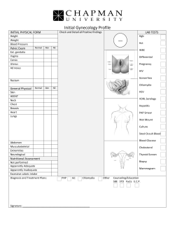


© Copyright 2026