
Introduction
Introduction 1| General introduction chapter 1 general introduction Outline introduction 1. General introduction 1 General introduction 1.1 Historical perspective of Chlamydia trachomatis 1.2 Taxonomy of Chlamydiales 1.3 Chlamydia trachomatis developmental cycle 1.4 Chlamydia trachomatis plasmid and genes 1.4.1 Endogenous plasmid 1.4.2 MOMP and OmpA gene 1.4.3 Polymorphic Membrane Protein and pmp genes 2Chlamydia trachomatis epidemiology, serovar distribution and clinical manifestation 2.1 Epidemiology of urogenital Ct infections 2.2 Distribution of Ct serovars 2.3 Clinical manifestation and complications 2.4 Anogential serovars & symptoms 2.5 Lymphogranuloma venereum 3 Chlamydia trachomatis diagnostic methods 4 Chlamydia trachomatis typing 4.1 Chlamydia serotyping 4.2 Chlamydia genotyping 5 Chlamydia trachomatis and the development of cervical cancer 5.1 Cervical cancer 5.2 High-risk HPV types 5.3 Chlamydia trachomatis and cervical cancer 6 Thesis Outline 1.1 Historical perspective of Chlamydia trachomatis Chlamydia trachomatis (Ct) comprises a collection of serological d istinguishable Gram-negative intracellular bacteria. Ct can colonize different types of tissue, like the conjunctiva, cervix, urethra, oropharynx, proctum and lymph nodes, and leads to a variety of human diseases depending on the anatomical site of infection (i.e., trachoma, lymphogranuloma venereum, pelvic inflammatory diseases, reactive arthritis). Using current highly sensitive methods, viable Ct is also detected in peripheral blood mononuclear cells in skin lesions and synovial fluid (14-15). Although trachoma and lymphogranuloma venereum have been known for centuries, the etiology remained mysterious (128). More insight in the disease trachoma was obtained in 1907 when Halberstädter and von Prowazek inoculated the eyes of Orang-utans with conjunctival swabs of trachoma patients (36) (figure 1). In Giemsa-stained epithelial cells from the Orang utan’s conjunctiva they discovered intracytoplasmic vacuoles containing numerous particles. Some small particles, now recognized as Ct elementary bodies, were also detected outside the epithelial cells. The intracytoplasmic vacuoles contained larger particles, known as reticulate bodies. Professor Tang isolated Ct in 1957 for the first time and initially recognized it as a viral infection (126). In the 1960s, the use of tissue culture techniques and electron microscopy revealed evidence for bacterial, ribosomes and cell wall structures, suggesting that Ct is a bacterium. During the sexual revolution in the late 1960s it became clear that Ct can be sexually transmitted (78) and in the late 1970s it was even recognized as the most prevalent sexually transmitted bacterial infection (100, 154). Currently, the total of infections is estimated over 92 million cases each year, which is supposed to be an underestimation (153). In 1963, Wang and Grayston described for the first time the existence of different Ct serotypes (146). They inoculated egg yolk sacs with Ct and subsequently injected the crude extracts into mice to trigger the mice’s immune response. In some of these mice, a protective mechanism against the Ct infection was observed, while other mice were only partly protected, apparently by cross-protection. This observation suggested the existence of different Ct serotypes. Because this method was time-consuming and complex, more easier tests were needed for larger scale epidemiological studies, leading to the development of polyclonal microimmunofluorescence tests (147) and subsequently to the more specific monoclonal microimmunofluorescence tests, targeting the major outer membrane protein (MOMP) (150). While m icroimmunofluorescence tests contributed to more knowledge on the etiology 12 13 1 chapter 1 general introduction of the different Ct serovars in the early days of Ct research, the technique was replaced at the end of previous century by more sensitive genotyping methods targeting the gene coding for the MOMP (OmpA gene). Currently, different types of genotyping methods are used in sexual network studies, phylogenetic studies and serovar distribution studies resulting in a better understanding of the epidemiology and etiology of the different Ct serovars. Figure 1 Halberstädter and von Prowazek inoculating the eyes of an Orang-utan (1906). the former family Chlamydiacea into two genera and add three new non- Chlamydiacea families. Thus, according the new taxonomy, the species Chlamydia trachomatis, together with the species Chlamydia suis and Chlamydia muridarum, belong to the genus Chlamydia, the family Chlamydiaceae and the order Chlamydiales while the species C.pneumoniae and C.psittaci belong to the new genus Chlamydophila (figure 2). The new taxonomy is better structured than the previous taxonomy, although not commonly accepted by all chlamydiologist (103). 1.3 Chlamydia trachomatis developmental cycle Ct has a biphasic developmental cycle characterized by alternating between two morphological forms, the electron dense round elementary body (EB) and the reticulate body (RB) (Table 1) (73). EBs are small (300nm), metabolically inactive, extracellular forms, that are responsible for dissemination of Ct and are able to attach to and invade susceptible cells. Table 1 Review of within normal limits cervical smears within five years Property Adapted from: Pospischil A. (2009). Drug. Today. 45(suppl. B): 141-146 1.2 Taxonomy of Chlamydiales All members of the order Chlamydiales are gram-negative, non-motile bacteria with a biphasic developmental cycle. At the end of the 20 th century, a proposal for revision of the order Chlamydiales was developed, due to the discovery of a number of Chlamydia-like organisms such as the Simkania strain and Parachlamydia acanthamoehae (26). The revision is based on 16S rRNA sequence similarity and analysis of full-length 16S and 23S rDNAs. Members of the order Chlamydiales share at least 80% sequence identity for the genes encoding the 16S rRNA and 23S rRNA. The new Chlamydiales taxonomy splits 14 before the diagnosis of cervical cancer. Size Rigid cell Wall Extracellular stability Infective Induces phagocytosis Inhibits phago-lysosomal fusion Metabolic active Replication Elementary body Reticulate body 0.2-0.4 µm Yes Yes Yes Yes Yes No No 0.6-1.0 µm No No No No No Yes Yes The EBs interact with the host cell membrane in a two-stage process. First, initial attachment occurs through reversible electrostatic interactions of the Ct MOMP with heparin sulfate containing glycosaminoglycans present on the surface of epithelial cells (118, 124-125, 127). The second, irreversible binding is possibly associated with protein disulfide isomerase, a component of the estrogen receptor complex (20). This irreversible binding step is immediately followed by a type three secretion system (TTSS) mediated injection of the translocated actin-recruiting phosphoprotein (TARP) into the host cells cytoplasma (figure 3). Inside the host cell, TARP is rapidly phosphorylated, 15 1 chapter 1 general introduction 16 Figure 3 T he three steps seems necessary for Elementary Body uptake Adapted from: Bush, R.M. and Everett, K.D.E. (2001). Int. J. Syst. Evol. Microbiol. 51: 203 - 220 Figure 2 Taxonomy diagram of the old and new Chlamydiales classification. triggering phagocytosis of the EB attached to the host membrane (19). Once entered into the cell by phagocytosis, the EB starts an immediate differentiation to the larger (1μm) metabolically active non infectious RB. The RB expresses directly early genes, associated with inclusion body modification (i.e., inc-like genes CT228, CT229 and EEA1-like gene CT147). The modification of the inclusion body prevents the inclusion body from entry into the endocytic pathway leading to lysosomal fusion. Six to 14 hours after phagocytosis, the RB starts replicating, resulting in extensive growth of the inclusion body, containing up to 10.000 Chlamydia particles. Approximately 30 hours after phagocytosis, the RBs differentiate back to EBs and are released by exocytosis or host cell lysis (Figure 4) (1). in the host cell. First, a reversible electrostatic binding between MOMP and heparin-sulfate occurs, followed by an irreversible binding involving protein disulfide isomerase. Finally, Tarp injection occurs causing endocytose. Adapted from: Y.M.AbdelRahman et al. FEMS Microbiology Reviews 2005 29;949-959 1.4 Chlamydia trachomatis endogenous plasmid and genomic genes The first complete Ct genome (serovar D) was sequenced during the late 1990 as part of the Chlamydia genome project (117). This provides valuable information about the genome organization and allowed further research in protein expression and gene regulation. The Ct genome comprises 1.042.519 basepairs with 894 open reading frames encoding for predicted proteins. The endogenous plasmid, consisting of 7.493 basepairs, was also sequenced. Subsequently, the endogenous plasmid, the chromosomal OmpA gene and the 17 1 chapter 1 Figure 4 Developmental cycle of Chlamydia trachomatis. Adapted from Gerald J Stine 1992, the biology of sexually transmitted diseases Wm. C. Brown, USA. chromosomal pmp genes will be discussed in detail, since those genes are the most frequent DNA targets for diagnostic Ct assays. 1.4.1 The endogenous plasmid Almost all Ct isolates carry a small plasmid which contains 8 open reading frames. Although the exact function of the endogenous plasmid and its proteins remain unclear, it is supposed that the plasmid genes plays an important role in transcriptional regulation of chromosomal genes and contributes to the virulence of the Ct strain (13). Naturally occurring plasmid-free strains of Ct have been reported, but are exceedingly rare and do not spread into the population, possibly due to low virulence (27, 123). Plasmid-free Ct isolates are also unable to accumulate glycogen in their inclusion body, which is a typical characteristic of Ct. For the viability of Ct, the plasmid seems important but not necessary (87). 18 general introduction Recently, a new variant of Ct has been discovered in Sweden (designated as the Swedish variant). This Swedish variant carries a plasmid showing a 377 basepairs deletion within coding sequence 1 (93). Although the Swedish Ct variant seems normally transmittable, the infection seems to remain more frequently asymptomatic than regular Ct strains (7). Importantly, most nucleic acid amplification Ct detection tests are targeting the endogenous plasmid, because the plasmid copy number is approximately 10 times higher than chromosomal Ct DNA and therefore permits more sensitive Ct detection. The 377bp deletion found in the Swedish strain overlaps the target region for 2 frequently used nucleic acid amplification tests ( Roche Diagnostics and Abbott Laboratories). This results in false-negative Ct tests results by these assays for the Swedish strain (61), causing a major underestimation of the Ct prevalence in Sweden (113). The underestimation has a further impact on the Swedish healthcare system, since infected individuals remain untreated, which leads to persistent Ct infections with possible severe late complications. Infected and untreated individuals have also more time to transmit the bacteria to other persons. Immediately after identification of the Swedish strain, Roche and Abbott developed next generation dual target assays, targeting the endogenous plasmid as well as chromosomal DNA (OmpA gene) (35). The proportion of the Swedish variant has declined significantly, since the introduction of the upgraded detection assays, possibly due to faster treatment (48). 1.4.2 MOMP and the OmpA gene The Major Outer Membrane Protein (MOMP) is the most studied Ct protein and is encoded by the chromosomal OmpA gene. The protein structure comprises 5 transmembrane constant domains and 4 variable domains outside the membrane (figure 5). The differences in MOMP variable epitopes permits classification of the Ct strains into 3 major serogroups and 19 different serovars, designated serogroup B (comprising serovars B, Ba, D, Da, E, L2 and L2a), serogroup C (comprising serovars A, C, H, I, Ia, J, K, L1 and L3) and the intermediate serogroup (comprising serovars F, G, Ga) (52, 79). Serovars A, B and C are mainly detected in conjunctival samples, while the serovars D-K are mostly found in the anogenital tract as common STD confined to the mucosal layer. In contrast, the serovars L1, L2 and L3 invade the submucosal layer and lymph nodes causing the disease lymphogranuloma venereum (106). It seems that the disease type is not determined by MOMP but other proteins, although it is possible to cluster the serovars into the 3 disease groups (biovars). 19 1 chapter 1 general introduction Figure 5 T he Major Outer Membrane Protein and the OmpA gene (V= variable, C=constant, D=domain, R=region, OM= outer membrane). complete Ct genome revealed a gene family, consisting of 9 genes that encode for autotransporter proteins (pmp genes), (117). While most pmp genes are highly conserved among the different Ct serovars, one pmp gene (pmpH) can be clustered into three clades that are compatible with the biovars (ie ocular trachoma clade, STD clade and LGV clade; Table 2) (122). Table 2 T he three biovars (pmpH clade) and the relation with serovars and disease type. Biovar (pmpH clade) Serovars (OmpA) Trachoma Clade A, B, Ba, C STD Clade Lymphogranuloma Venereum Clade Figure was kindly provided by SA Morré. Since MOMP is the most commonly available protein on the membrane of the elementary body, several vaccine developing studies are performed targeting MOMP. (56, 82). However, so far now no adequate Ct vaccine has been developed. The OmpA gene, encoding MOMP, comprises of 4 variable regions and 5 conserved regions, encoding the different protein domains. At present, DNA technology has replaced the conventional sero-determination and dozens of new genovariants have been identified, like genovariant Ja and L2b, due to an increased resolution (24, 114). Although OmpA genotyping has replaced MOMP serotyping, most literature still refers to serovars instead of genovariants, because it is a clear and stable nomenclature reflecting clinical relevance. 1.4.3 Polymorphic Membrane Protein and pmp genes Like MOMP, other surface-exposed proteins are considered important mediators in interaction between Ct and host cells. Sequence analysis of the 20 Major infection site Conjunctivae, urogenital tract (rare) D, Da, E, F, G, Ga, Anogenital tract H, I, Ia, J, K L1, L2, L2a, L3 Inguinal lymph nodes, proctum Sequelae Trachoma, blindness, reactive arthritis PID in women, ectopic pregnancy, infertility, reactive arthritis Genital Ulcer Disease, Anal fistulas, elephantiasis, frozen pelvic syndrome The compatibility of the 3 clades with disease suggests that this protein might play a role in pathogenesis, virulence an tropism. Nevertheless, there is still an incomplete understanding of the virulence and host immune response to these proteins, despite the potential importance of pmp in chlamydial biology. 2. Chlamydia trachomatis epidemiology, serovar distribution and clinical manifestation 2.1 Epidemiology of urogenital Ct infections Genital Ct infections are the most common bacterial sexually transmittable disease worldwide, with between the 90 and 92 million new infections each year(32). Over the past decade, the detection rate of Ct has increased, due to the application of more sensitive Ct detection assays and the rise of Ct screening programs. Infection may occur at any age, but peaks in the 16-25 year age groups, since these persons show high-risk behavior. Important risk groups are female sex workers, homosexual men, unmarried young adults and swingers (25, 141, 159). Risk factors for a Ct infection are high frequency of 21 1 chapter 1 partner change, and unprotected sexual intercourse (60). In the USA, more than a million cases of chlamydial infections were reported in 2007, while the estimated amount of infected individuals was around 3 million (16, 152). In the past decade, the incidence of Ct infections in Europe has also increased. In 2005, over 200.000 cases were reported in 17 European countries, which is certainly an underestimation, since in the Netherlands alone 60.000 Ct infection occur annually (94, 130). This underestimation might be due to the asymptomatic course in the majority of Ct infections and the lack of extensive diagnosis in other countries. 2.2 Distribution of Ct serovars Throughout the world many serovar-distribution studies are performed. Most distribution studies focus at Ct infections retrieved from the urogenital tract (serovars D-K). In general, similar serovar distributions are observed in different countries. Although some regional differences are observed, serovar E is the most prevalent serovar worldwide. The serovar E prevalence varies between the 30-60% of the Ct positive samples in the countries USA (Alabama), Australia, China, Costa Rica, France, Iceland, Iran, Korea, Russia, Sweden, Taiwan and the Netherlands (6, 28-29, 41, 44, 46, 53, 89, 95, 108, 116, 155). In contrast, serovar E was far less prevalent in Uganda, Thailand, India and Columbia (3, 67, 91, 107). This variation might be due to the different Ct genotyping methods (Thailand) or a small isolated study population (Uganda, India). Overall, serovars D, E and F contribute for over 70% of all typed urogenital serovars, making it difficult to study an association with symptoms (23, 71, 132). In specific subgroups, like men who have sex with men (MSM), a different serovar distribution is observed. The majority of anogenital Ct infections in MSM are caused by serovars G, D and J (4, 31, 49, 55, 120, 143). The difference in serovar distributions between MSM and the general population can be network related, like observed among heterosexual individuals living in an isolated town in Canada (11). Apart from network-associated factors like sexual prevalence, tissue tropism could explain the difference in serovar distribution. In a recent study, an association between rectal tropism and polymorphisms of open reading frames within genomic DNA of serovar G is observed (43). Genovariants from the serovars G, D and J might be more affected by the polymorphisms associated with rectal tropism than other serovars, although those polymorphisms are not located on the OmpA gene. So for a better understanding of tissue tropism we should focus on other Ct genes, independent of the OmpA gene. 22 general introduction 2.3 Clinical manifestation and complications An urogenital Ct infection can cause urethritis in men and vaginitis, cervicitis and urethritis in women. Since most infections are asymptomatic (women:70%, men: 50%), the great majority of infections remain unnoticed and thus untreated (131). Symptoms in men are dysuria and a clear urethral discharge, both occurring 7-21 days after primary infection. Complications in men can be epididymitis, prostatitis and reactive arthritis (85). At this moment, the role of Ct in the development of male infertility is not fully understood, since contradicting results are published (17, 21). It is suggested that the infertility is caused by strictures of the Vas deferens, due to previous inflammation. Reactive arthritis (previously called Reiter’s syndrome) can be triggered by a urogenital Ct infection and is more frequently observed in men (39). Besides the triad of urethritis, conjunctivitis and arthritis, a reactive arthritis can also give rise to mucocutaneaous lesions on the penis, mouth, hands and feet (90). The majority of women have no complaints, but symptoms can be a mucopurulent vaginal discharge or post-coital bleeding. A cervical Ct infection might induce an ascending infection causing endometritis, salpingitis, pelvic inflammatory disease (PID) and perihepatitis (Fitz-Hugh-Curtis syndrome). Although high percentages of progression have been given for these complications, it appears that most of the complications as described are the result of symptomatic Ct infections (72). In Europe, Ct infections are responsible for at least 60% of the PID cases (98). PID can lead to late severe reproductive complications later in life due to tubal adhesions. It is estimated that two-third of women with tubal factor infertility and one-third of the women with ectopic pregnancies have Ct antibodies detectable in the serum, indicating a Ct infection earlier in life (81, 85). The course and duration of an untreated, asymptomatic Ct infection is not totally clear. It would be relatively easy to investigate the course and duration of an untreated Ct infection, but ethical considerations, regarding the prominent health risk for untreated individuals, do not allow those prospective cohort studies and restrict the investigations to retrospective studies. However, the primary time point at which a person had been initially infected is hard to determine in retrospective studies. Also, discrimination between a recurrent or persistent Ct infection is not possible, although Ct genotyping might give an indication. Still, various studies have investigated the natural course of Ct infections in minimal prospective studies and larger retrospective studies (30, 45, 62, 68, 72, 84, 97, 105, 134, 140). In general, approximately 50% of the asymptomatic Ct infections were cleared spontaneously within the first year and no complications occurred. After 5 years, 95% of the women had resolved their Ct infection spontaneous without receiving antibiotic treatment. Those results indicate a currently overrated fear for complications due to a Ct infection. 23 1 chapter 1 general introduction 2.4 Anogential serovars & symptoms The serovars D-K are most prevalent in the anogenital tract, although other serovars are sporadically observed. A number of reports have been published regarding clinical manifestation and the D-K serotypes (Table 3) (2, 5, 23-24, 28, 71, 132, 139). In men, serovars F and G are associated with less severe symptoms compared to other serovars and the serovars H and J are associated with urethral discharge (132, 139). In women, the serovars F and G are associated with lower abdominal pain, while serovar K frequently causes vaginal discharge (71, 139). Also, the serogroup C serovars are associated with persistence of the Ct infection (24). Serovar E has an asymptomatic course of infection, that possibly contributes to a higher prevalence and transmission rate, since asymptomatic infections remain more frequently unnoticed and thus untreated (23). Table 3 T he main outcome of studies that observed a correlation between serovars and clinical manifestation. Investigators Sample Main outcome numbers Duynhoven et al. 310 Dean et al. 56 Van de Laar et al. 275 Batteiger et al. 124 Morre et al. 221 Gao et al. 240 Antilla et al. 530.000 (cohort) Dean et al. 24 552 Urethral discharge associated with serovars H and J in men. Lower abdominal pain was associated with serovars F and G in women. Serovar F associated with severe endometrial disease and serovar E associated with asymptomatic milder infections. Serovar F and G were associated with less severe symptoms in men, while in women no associations were observed. Serovars F en G were associated with more polymorpho nuclear leukocytes in smears from men. Overall no association with symptoms was observed Serovar G was associated with lower abdominal pain and serovar F with infection at a younger age. Serovar Ga was associated with symptoms in men, especially dysuria. Serovar K was associated with vaginal discharge. Serovar G, D and I antibodies were associated with cervical squamous cell carcinoma. Serovars belonging to serogroup C are associated with persistence. However, it remains important to realize that most of those associations are observed in single studies that cannot be reproduced. Several other studies do not confirm the associations between serovars and symptoms (57, 65, 76, 86, 121). So more studies will be required to confirm or reject the association between symptoms and serovars. 2.5 Lymphogranuloma venereum Lymphogranuloma venereum (LGV) is a sexually transmitted disease, caused by Ct serovars L1, L2, L2a or L3. Originally, LGV was known as an endemic disease in Africa, Southeast Asia, Central/South America and the Caribbean. Until 2003, LGV was only diagnosed sporadically in the Netherlands as a traveler’s disease imported from foreign countries (142). The disease has an aggressive nature with serious late sequelae. The disease LGV comprises 3 stages. In the first stage, a painless erosion/ulcer in the anogenital region appears 3-30 days after infection. This erosion frequently remains unnoticed and is observed in only 4% of the LGV patients (102). A visible erosion or ulcus is difficult to differentiate from a trauma or other ulcerative diseases, like primary Syphilis and Herpes simplex infections. The second disease stage occurs 1-4 weeks after infection and is characterized by lymphadenopathy in the so-called bubonic form of the inguinal lymph nodes in men or retro-peritoneal lymph nodes in women (Figure 6). However, other lymph nodes (e.g., femoral) in the lower abdomen can also be infected, depending on the infection site. Over time, the infected lymph nodes become abscesses and may rupture, leading to fistulae. If LGV infections remains untreated a tertiary (final) stage will develop, due to progressive spread and tissue destruction. At this stage, irreversible damage to the lymph nodes will lead to an impaired lymph drainage and possibly elephantiasis. Also signs of more cachexic illness can be observed (135). Since 2003, LGV proctitis (the anorectal variant) is observed in developed countries among men who have sex with men (MSM), caused by the genovariant L2b (37, 77, 114). The majority of the infected MSM (80%) are co-infected with blood borne diseases like HIV and hepatitis C (133). First it was thought that LGV proctitis in MSM was a new epidemic, but retrospective studies performed in San Francisco revealed that LGV was already present in rectal swabs from MSM in 1981, indicating an old unnoticed epidemic (115, 135). Differentiation between an LGV and non-LGV Ct infection is important, since an LGV infection need prolonged antibiotic treatment (22). Several in-house PCRs are available for differentiation between a LGV serovar and an anogenital (D-K) serovar (18, 37, 70). 25 1 chapter 1 Figure 6 E xample of a bubonic inguinal lymph node in a men. The picture was kindly provided by Henry de Vries. 3. Chlamydia trachomatis diagnostic methods In the 1970s Ct culture was the standard reference method for diagnosing Ct infections. However, the restrictive specimen collection, transport and storage conditions limited the availability of the test for clinical purposes. During the 1980s, two antigen detection methods were developed. The first assay was a direct immunofluorescence assay (DFA), utilizing a fluorescein-tagged monoclonal antibody to allow Ct elementary bodies to be microscopically visualized in cellular smears collected from the endocervix and conjunctiva (MicroTrak, Trinity Biotech, Ireland) (129). The second detection method was a solid phase enzyme-linked immunosorbent assay (EIA), using polyclonal anti-chlamydial antibodies to capture Ct antigen onto beads (Chlamydiazyme, Abbott Laboratories, USA) (88). Both assays generate a fare more rapid result (2-3 hours) as compared to Ct culture (3-7 days), but had a slightly lower 26 general introduction sensitivity. A limitation of DFA is the requirement of expert microscopists, making it laborious and thus less suitable for high-throughput studies compared to the EIA. Currently, other Ct detection methods are available like point of care (POC) rapid assays, DNA probe assays and highly sensitive nucleic acid amplification tests (NAATs). The three POC assays most commonly used are the Clearview (Unipath, UK), Biostar (Thermo Electron Corp, USA) and the QuickVue (Quidel, USA), all based on direct detection of Ct antigens(136). The POC assays give a result within 30-40 minutes ensuring that patients can immediately receive antibiotic treatment. Like HIV rapid tests, the assays have a very high specificity, but a low to moderate sensitivity indicating that a second more sensitive assay is needed to confirm the obtained test result. The main utility of those POC tests are in applications where return for treatment is unlikely or in population screening programs (59, 99). The two most commonly used DNA probe assays are the PACE2 (Gen-Probe Inc, USA) and the Hybrid Capture 2 (HC2) Ct assay (Qiagen, USA) (66, 137). The PACE2 system uses a labeled DNA probe that binds to Ct rRNA, generating a DNA/RNA hybrid. No amplification is required, due to the high copy number of Ct rRNA. Although this assay has a very good specificity, the test is being replaced by the cheaper and more sensitive HC2 assay. The HC2 targets both the genomic OmpA gene and the endogenous plasmid DNA by using a labeled RNA probe. The HC2 does not need DNA amplification, due to the use of signal amplification with multiple labeled antibodies specific for DNA/RNA hybrids. The use of signal amplification instead of DNA amplification prevents the HC2 assay against carry-over contamination. The HC2 system is a good alternative for NAATs, although the sensitivity of DNA probe assays is slightly lower (table 4) (8). Table 4 O verview of the mean costs, mean throughput and mean sensitivity of the 3 major assay types. Assay type Costs Throughput Sensitivity EIA DNA-Probe NAATs Low Moderate High Very good Very good Very good Moderate/Good Very good Excellent NAAT: Nucleic Acid Amplification Test EIA: Enzyme Immuno Assay 27 1 chapter 1 NAATs are in general the best diagnostic tools for Ct detection, although they are relatively expensive (Table 4) (8, 40). However, NAATs are nowadays the gold standard for Ct detection. Different types of commercially available NAATs like the rRNA based Aptima Combo 2 assay (Gen-probe), the COBAS Amplicor (Roche Diagnostics), the COBAS TaqMan (Roche Diagnostics) and the Ct-DT DEIA (Labo Biomedical Products BV) are used in routine diagnostics. Most assays have different targets (i.e., endogenous plasmid, chromosomal DNA or rRNA), but have more or less the same sensitivity. 4. Chlamydia trachomatis typing 4.1 Chlamydia serotyping During the sixties, the first experiments involving Ct serotyping were performed by Wang and Grayston, who developed the mouse toxicity prevention test (MTPT) (146). Briefly, they inoculated egg yolk sacs with “trachoma-inclusion conjunctivitis organisms”. Extracts were subsequently intravenously injected into mice that subsequently triggered an immune response. A protective mechanism in the mice was described when the mice were challenged for the second time with the same isolates of Chlamydia. When the mice were challenged with other isolates, the protective mechanism was less pronounced indicating cross-protection, but more important, the existence of different serovars. The MTPT method was very time-consuming, since it took 7 years to classify 80 Ct strains into 6 serotypes (150). Therefore, a more rapid microimmunofluorescence (MIF) test was developed which was based on culturing of elementary bodies of known serotypes in yolk sacs and the production of polyvalent antiserum in mice (147, 149). The combination of the MIF test and the ability of culture of cells with Ct inclusion bodies facilitated the discovery of 15 different Ct strains (A-L3) based on variability in the major outer membrane protein (MOMP) (51, 145, 148). Later, other subserovars like Ba, Da and Ga, were discovered. During the 1980s the relatively aspecific polyclonal antibodies were replaced by more specific monoclonal antibodies (119). The above-described principle was the basis for the development of several immunoassays that were used until the early 1990 when DNA sequencing of the OmpA gene became available (138). A disadvantage of immunotyping was the requirement of Ct culture which is time-consuming, expensive and requires expert staff. Also, the sensitivity of culture is low compared to DNA analysis. 28 general introduction 4.2 Chlamydia genotyping During the 1990s serotyping was replaced by molecular genotyping of the OmpA gene, encoding MOMP. The first genotyping method consists of a PCR followed by restriction fragment length polymorphism (RFLP) and pattern analyses on a polyacrylamide gel. With this method it was possible to differentiate between 13 genotypes (96). The patterns observed by RFLP were compatible with the results obtained by serotyping, so the name “serovar” could be maintained. The strength of the PCR-RFLP methods is the ability to detect double serovar infections (156). At this moment, RFLP is still performed to differentiate between an LGV and anogenital serovar, even with other alternatives available (38, 54). With the availability of fluorophore-marked nucleotides enabling DNA sequence analyses, OmpA sequencing became an alternative for RFLP. Sequencing has a higher resolution than RFLP and serotyping, leading to the discovery of new genovariants (69). A drawback with sequencing is the inability to detect double serovar infections, because a multiple serovar infection will lead to a mixed and therefore non-interpretable sequence result. Currently, different reverse line blot assays are available that can identify the different serovars (6, 155, 158). Those assays are sensitive simple technology reverse hybridization methods, targeting the variable regions in the OmpA gene. The method is highly suitable for epidemiological studies due to its ability to detect multiple serovar infections, the relatively fast results and the ability to perform the assay manually with the use of simple technology (ie., PCR). However a drawback of the reverse line blot is the lower discriminating power compared to OmpA sequencing. At this moment, new high throughput microsphere suspension assays are developed, based on reverse hybridization technology for the Luminex 100 platform, which can be used in large-scale serovar distribution studies (42). We can consider those microsphere suspension assays as second generation hybridization methods. At STD clinics, Real-Time PCRs are used for discrimination between a LGV infection and a non-LGV Ct infection (12, 33). The probe target for those Real Time PCRs is a L-specific gap in the polymorphic protein H (pmpH). Those PCRs are all in-house validated methods, since no commercial pmpH Real-Time PCRs are available. Although this method belongs to the Ct genotyping methods, it cannot identify the different serovars and is thus not used in epidiological studies. An upcoming genotyping method is the high-resolution typing of Ct strains by multi locus sequence typing (MLST) that uses several other genes as targets (47). This method has a higher discriminatory power then OmpA sequencing, 29 1 chapter 1 but uses another nomenclature. This makes it difficult to compare the results obtained with previous studies. However, a new nomenclature may lead to further understanding of the natural history of Ct and its relation to symptoms. In general, most Ct genotyping is now performed in research settings. The only clinical application for genotyping is the differentiation between LGV serovars and non-LGV serovars. New typing methods may lead to a better understanding of the natural history and might reveal more clinical relevance for Ct genotyping. Whether this goal will be reached with the highly established serovar nomenclature or a new nomenclature is unknown. 5. Chlamydia trachomatis and the development of cervical cancer 5.1 Cervical cancer Cervical cancer is the second most common cancer worldwide, with annually over 500.000 cases (145). The majority of cases (>80%) occur in developing countries, due to poor screening programs for precancerous lesions (104). Since the introduction of population based screening programs in developed countries, in which high grade precursor lesions of cervical cancer are diagnosed and treated, a reduction of approximately 75% of the cervical cancer cases in the past half century have been observed. It is commonly accepted that infections with specific types of Human Papillomavirus (HPV) are a necessary cause for the development of squamous cell- and adenocarcinoma of the cervix. During life, 80% of all sexually active women get infected with HPV, although the great majority of these infected women (90-95%) clear the HPV infection without any symptoms. The remaining 5-10% of the women with a persistent HPV infection are at a higher risk of developing (pre)cancerous lesions. The most common histological types of cervical cancers are squamous cell carcinoma (80%) and endocervical adenocarcinoma (20%) (10, 109, 151). Precancerous lesions can be categorized into the low-grade cervical intraepithelial neoplasia 1 (CIN1) and high-grade CIN2 and 3 lesions. Most of the CIN1 lesions will regress, making progression to cervical cancer a rare event(34, 80). Even the majority of high grade CIN lesions will spontaneously regress (63). Thus, only a small proportion (<1%) of all women infected with a high risk HPV infection will finally develop cancer. 5.2 High-risk HPV types As mentioned above, HPV has been recognized as a necessary cause for the development of cervical cancer. HPV types can be divided into low risk (lr) 30 general introduction HPV types, associated with genital warts and high risk (hr) HPV types, associated with cervical cancer. Besides cervical cancer, HPV is also associated with other carcinomas (e.g., anus, vulva/vagina, penis and oropharynx) (83). Worldwide, approximately 53% of the cervical cancers are caused by hr-HPV type 16 and 17.2% by hr-HPV type 18 (111). Taken together, 94.6% of the cervical cancers are caused by the hr-HPV types 16, 18, 45, 31, 33, 52, 58, 35, 59, 56, 51, 39, 68, 73 and 82 (74). The remaining 5.4% are caused by unknown HPV types. 5.3 Chlamydia trachomatis and cervical cancer Several co-factors, like smoking, oral anticonceptive usage, and other sexually transmitted infections (e.g., HIV infection, Herpes Simplex 2 infection, Ct infection) have been suggested to facilitate the persistence of an HPV infection or even the development of cancer(9). The first report of an association between Ct infections and cervical cancer was already published during the 1970s (101). Multiple, but not all, studies determined an association between Ct antibodies in the serum and squamous cell carcinoma of the Cervix (SCC) or persistence of carcinogenic HPV types (2, 50, 75, 110, 112). However, no association between Ct antibodies or Ct DNA and adenocarcinomas of the cervix were observed, while the columnar cells of the endocervix are the major Ct infection site (58, 92, 157). So based on the pathogenesis of Ct we would expect an association with adenocarcinomas instead of SCCs. Most studies observed only an association between Ct antibodies and SCC, but one study also observed Ct DNA in cervical tumors (144). Serovar G antibodies were even associated with a higher risk for SCC (2). Overall, these results suggest a role for Ct as a co-factor in cervical carcinogenesis for SCC either by facilitating a persistent HPV infection via interactions with the immune system or by facilitating HPV induced SCC itself due to the inflammatory status of (parts of ) the genital tract (64). While the association between Ct antibodies and SCC has been interpreted by some groups as a causal link, others think it might be caused by residual confounding of other factors that are related to an HPV-positive status. Ct and HPV are both common sexually transmitted infection that share the same risk factors, such as sex at young age or multiple sexual partners. This strong relationship between HPV and Ct can result in inadequate adjustment for HPV, especially when low sensitive HPV serology is used as a marker for an HPV infection. So further studies, restricted to HPV positive cases and controls only, should be performed to overcome the issue of residual confounding. 31 1 chapter 1 Thesis outline The current thesis comprises of three sections, In the first section (Chapter 2-5) comprises a technical part, in which several Ct detection and genotyping methods are evaluated. The second section (Chapter 6-9) comprises several studies regarding the Ct serovar distribution and the relation with clinical signs and symptoms. In the third part (Chapter 10, 11), the association between Ct and the development of cervical cancer is investigated. Briefly, the aim of the specific chapters are mentioned below. Section 1 Chapter 2 describes the evaluation of a sensitive Ct amplification, Ct detection and Ct genotyping method (Ct-DT assay) that allows detection and identification of the 14 major Ct serovars (A-L3). Also, the serovar distribution was determined in samples obtained from the Netherlands and Uganda. Chapter 3 evaluates the Ct amplification (PCR) and detection step (DEIA) in clinical samples from Costa Rican women by a comparison with Hybrid Capture 2 Ct detection assay results. Also the Ct serovar distribution among Costa Rican women was determined with the Ct genotyping step. In Chapter 4 a comparison between the Ct-DT RHA (genotyping) and 2 other Ct genotyping methods (ie., OmpA sequencing and LGV/non-LGV differentiation by a pmpH real time PCR) was made. Chapter 5 describes the evaluation of a new high-throughput microsphere suspension (MS) assay for simultaneously detection and identification of the 14 serovars on the Luminex platform. general introduction infections were determined in LGV positive MSM. Besides the serovar distribution, this study serves as an evaluation of the LGV/non-LGV differentiation test used at the STI-outpatient clinic in Amsterdam. Chapter 9 investigates the correlation between the different serogroups and the level of Ct IgG antibodies in the serum. Section 3 In Chapter 10 the Ct co-factor role in the development of CIN2+ lesions among Costa Rican women was investigated on Ct antibody level and Ct DNA level. Chapter 11 investigates whether Ct DNA was detected in cervical adenocarcinomas. Also, HPV genotyping was performed for all adenocarcinomas. Chapter 12 consists of a general discussion and conclusion. Section 2 Chapter 6 contains a description of the first Russian serovar distribution study (St Petersburg). The obtained Russian distribution results were compared with previous performed distribution studies in the Netherlands. Chapter 7 describes the incidence of concurrent serovar infections at Ct genotyping level. Also, the incidence of multiple anatomical sites (i.e., vagina/ cervix, urethra, rectum and pharynx) on detection and genotyping level were investigated. In Chapter 8 the D-K serovar distribution was determined among MSM infected with non-LGV serovars. Also the percentage of mixed LGV/non-LGV 32 33 1 chapter 1 general introduction References: 1. 2. 3. 4. 5. 6. 7. 8. 9. 10. 11. 12. 13. 14. 15. 16. 34 Abdelrahman, Y. M., and R. J. Belland. 2005. The chlamydial developmental cycle. FEMS Microbiol Rev 29:949-959. Anttila, T., P. Saikku, P. Koskela, A. Bloigu, J. Dillner, I. Ikaheimo, E. Jellum, M. Lehtinen, P. Lenner, T. Hakulinen, A. Narvanen, E. Pukkala, S. Thoresen, L. Youngman, and J. Paavonen. 2001. Serotypes of Chlamydia trachomatis and risk for development of cervical squamous cell carcinoma. JAMA 285:47-51. Bandea, C. I., K. Kubota, T. M. Brown, P. H. Kilmarx, V. Bhullar, S. Yanpaisarn, P. Chaisilwattana, W. Siriwasin, and C. M. Black. 2001. Typing of Chlamydia trachomatis strains from urine samples by amplification and sequencing the major outer membrane protein gene (omp1). Sex Transm Infect 77:419-422. Barnes, R. C., A. M. Rompalo, and W. E. Stamm. 1987. Comparison of Chlamydia trachomatis serovars causing rectal and cervical infections. J Infect Dis 156:953-958. Batteiger, B. E., W. Lennington, W. J. Newhall, B. P. Katz, H. T. Morrison, and R. B. Jones. 1989. Correlation of infecting serovar and local inf lammation in genital chlamydial infections. J Infect Dis 160:332-336. Beni, B. T., H. Motamedi, and M. R. Ardakani. 2010. Genotyping of the prevalent Chlamydia trachomatis strains involved in cervical infections in women in Ahvaz, Iran. J Med Microbiol 59:1023-1028. Bjartling, C., S. Osser, A. Johnsson, and K. Persson. 2009. Clinical manifestations and epidemiology of the new genetic variant of Chlamydia trachomatis. Sex Transm Dis 36:529-535. Black, C. M., J. Marrazzo, R. E. Johnson, E. W. Hook, 3rd, R. B. Jones, T. A. Green, J. Schachter, W. E. Stamm, G. Bolan, M. E. St Louis, and D. H. Martin. 2002. Head-to-head multicenter comparison of DNA probe and nucleic acid amplification tests for Chlamydia trachomatis infection in women performed with an improved reference standard. J Clin Microbiol 40:3757-3763. Bosch, F. X., and S. de Sanjose. 2007. The epidemiology of human papillomavirus infection and cervical cancer. Dis Markers 23:213-227. Bray, F., B. Carstensen, H. Moller, M. Zappa, M. P. Zakelj, G. Lawrence, M. Hakama, and E. Weiderpass. 2005. Incidence trends of adenocarcinoma of the cervix in 13 European countries. Cancer Epidemiol Biomarkers Prev 14:2191-2199. Cabral, T., A. M. Jolly, and J. L. Wylie. 2003. Chlamydia trachomatis omp1 genotypic diversity and concordance with sexual network data. J Infect Dis 187:279-286. Cai, L., F. Kong, C. Toi, S. van Hal, and G. L. Gilbert. 2010. Differentiation of Chlamydia trachomatis lymphogranuloma venereum-related serovars from other serovars using multiplex allele-specific polymerase chain reaction and high-resolution melting analysis. Int J STD AIDS 21:101-104. Carlson, J. H., W. M. Whitmire, D. D. Crane, L. Wicke, K. Virtaneva, D. E. Sturdevant, J. J. Kupko, 3rd, S. F. Porcella, N. Martinez-Orengo, R. A. Heinzen, L. Kari, and H. D. Caldwell. 2008. The Chlamydia trachomatis plasmid is a transcriptional regulator of chromosomal genes and a virulence factor. Infect Immun 76:2273-2283. Carter, J. D., H. C. Gerard, and A. P. Hudson. 2008. Psoriasiform lesions induced by tumour necrosis factor antagonists: a skin-deep medical conundrum. Ann Rheum Dis 67:1181-1183. Carter, J. D., and A. P. Hudson. 2009. Reactive arthritis: clinical aspects and medical management. Rheum Dis Clin North Am 35:21-44. CDC 2009, posting date. Chlamydia prevalence monitoring project annual report 2007: sexually transmitted disease surveillance 2007 supplement. Centers for disease control and prevention. [Online.] 17. Cengiz, T., L. Aydoganli, M. Baykam, N. A. Mungan, E. Tuncbilek, M. Dincer, K. Yakupoglu, and Z. Akalin. 1997. Chlamydial infections and male infertility. Int Urol Nephrol 29:687-693. 18. Chen, C. Y., K. H. Chi, S. Alexander, C. A. Ison, and R. C. Ballard. 2008. A real-time quadriplex PCR assay for the diagnosis of rectal lymphogranuloma venereum and non-lymphogranuloma venereum Chlamydia trachomatis infections. Sex Transm Infect 84:273-276. 19. Clifton, D. R., K. A. Fields, S. S. Grieshaber, C. A. Dooley, E. R. Fischer, D. J. Mead, R. A. Carabeo, and T. Hackstadt. 2004. A chlamydial type III translocated protein is tyrosinephosphorylated at the site of entry and associated with recruitment of actin. Proc Natl Acad Sci U S A 101:10166-10171. 20. Davis, C. H., J. E. Raulston, and P. B. Wyrick. 2002. Protein disulfide isomerase, a component of the estrogen receptor complex, is associated with Chlamydia trachomatis serovar E attached to human endometrial epithelial cells. Infect Immun 70:3413-3418. 21. de Barbeyrac, B., A. Papaxanthos-Roche, C. Mathieu, C. Germain, J. L. Brun, M. Gachet, G. Mayer, C. Bebear, G. Chene, and C. Hocke. 2006. Chlamydia trachomatis in subfertile couples undergoing an in vitro fertilization program: a prospective study. Eur J Obstet Gynecol Reprod Biol 129:46-53. 22. de Vries, H. J., V. Smelov, J. G. Middelburg, J. Pleijster, A. G. Speksnijder, and S. A. Morre. 2009. Delayed microbial cure of lymphogranuloma venereum proctitis with doxycycline treatment. Clin Infect Dis 48:e53-56. 23. Dean, D., E. Oudens, G. Bolan, N. Padian, and J. Schachter. 1995. Major outer membrane protein variants of Chlamydia trachomatis are associated with severe upper genital tract infections and histopathology in San Francisco. J Infect Dis 172:1013-1022. 24. Dean, D., R. J. Suchland, and W. E. Stamm. 2000. Evidence for long-term cervical persistence of Chlamydia trachomatis by omp1 genotyping. J Infect Dis 182:909-916. 25. Dukers-Muijrers, N. H., A. M. Niekamp, E. E. Brouwers, and C. J. Hoebe. 2010. Older and swinging; need to identify hidden and emerging risk groups at STI clinics. Sex Transm Infect 86:315-317. 26. Everett, K. D., R. M. Bush, and A. A. Andersen. 1999. Emended description of the order Chlamydiales, proposal of Parachlamydiaceae fam. nov. and Simkaniaceae fam. nov., each containing one monotypic genus, revised taxonomy of the family Chlamydiaceae, including a new genus and five new species, and standards for the identification of organisms. Int J Syst Bacteriol 49 Pt 2:415-440. 27. Farencena, A., M. Comanducci, M. Donati, G. Ratti, and R. Cevenini. 1997. Characterization of a new isolate of Chlamydia trachomatis which lacks the common plasmid and has properties of biovar trachoma. Infect Immun 65:2965-2969. 28. Gao, X., X. S. Chen, Y. P. Yin, M. Y. Zhong, M. Q. Shi, W. H. Wei, Q. Chen, R. W. Peeling, and D. Mabey. 2007. Distribution study of Chlamydia trachomatis serovars among high-risk women in China performed using PCR-restriction fragment length polymorphism genotyping. J Clin Microbiol 45:1185-1189. 29. Geisler, W. M., R. J. Suchland, and W. E. Stamm. 2006. Association of Chlamydia trachomatis Serovar Ia infection with black race in a sexually transmitted diseases clinic patient population in Birmingham, Alabama. Sex Transm Dis 33:621-624. 30. Geisler, W. M., C. Wang, S. G. Morrison, C. M. Black, C. I. Bandea, and E. W. Hook, 3rd. 2008. The natural history of untreated Chlamydia trachomatis infection in the interval between screening and returning for treatment. Sex Transm Dis 35:119-123. 31. Geisler, W. M., W. L. Whittington, R. J. Suchland, and W. E. Stamm. 2002. Epidemiology of anorectal chlamydial and gonococcal infections among men having sex with men in Seattle: utilizing serovar and auxotype strain typing. Sex Transm Dis 29:189-195. 35 1 chapter 1 32. Gerbase, A. C., J. T. Rowley, D. H. Heymann, S. F. Berkley, and P. Piot. 1998. Global prevalence and incidence estimates of selected curable STDs. Sex Transm Infect 74 Suppl 1:S12-16. 33. Goldenberger, D., F. Dutly, and M. Gebhardt. 2006. Analysis of 721 Chlamydia trachomatispositive urogenital specimens from men and women using lymphogranuloma venereum L2-specific real-time PCR assay. Euro Surveill 11:E061018 061014. 34. Gray, L. A., and W. M. Christopherson. 1975. The treatment of cervical dysplasias. Gynecol Oncol 3:149-153. 35. Hadad, R., H. Fredlund, and M. Unemo. 2009. Evaluation of the new COBAS TaqMan CT test v2.0 and impact on the proportion of new variant Chlamydia trachomatis by the introduction of diagnostics detecting new variant C trachomatis in Orebro county, Sweden. Sex Transm Infect 85:190-193. 36. Halberstadter L, P. S. 1907. Uber Zelleinschlusse parasitarer Natur beim trachom. . Arbeiten aus dem Kaiserlichen Gesundheitsamte 26:44-47. 37. Halse, T. A., K. A. Musser, and R. J. Limberger. 2006. A multiplexed real-time PCR assay for rapid detection of Chlamydia trachomatis and identification of serovar L-2, the major cause of Lymphogranuloma venereum in New York. Mol Cell Probes 20:290-297. 38. Herida, M., B. de Barbeyrac, P. Sednaoui, C. Scieux, N. Lemarchand, G. Kreplak, M. Clerc, J. Timsit, V. Goulet, J. C. Desenclos, and C. Semaille. 2006. Rectal lymphogranuloma venereum surveillance in France 2004-2005. Euro Surveill 11:155-156. 39. Hicks, D. 2008. Complications of Chlamydia trachomatis infection in men., p. 99-109. In T. R. Moss (ed.), International handbook of Chlamydia 3rd edn. Alden Press, Halsemere, UK. 40. Horner, P., S. Skidmore, A. Herring, J. Sell, I. Paul, O. Caul, M. Egger, A. McCarthy, E. Sanford, C. Salisbury, J. Macleod, J. Sterne, and N. Low. 2005. Enhanced enzyme immunoassay with negative-gray-zone testing compared to a single nucleic Acid amplification technique for community-based chlamydial screening of men. J Clin Microbiol 43:2065-2069. 41. Hsu, M. C., P. Y. Tsai, K. T. Chen, L. H. Li, C. C. Chiang, J. J. Tsai, L. Y. Ke, H. Y. Chen, and S. Y. Li. 2006. Genotyping of Chlamydia trachomatis from clinical specimens in Taiwan. J Med Microbiol 55:301-308. 42. Huang, C. T., W. W. Wong, L. H. Li, C. C. Chiang, B. D. Chen, and S. Y. Li. 2008. Genotyping of Chlamydia trachomatis by microsphere suspension array. J Clin Microbiol 46:1126-1128. 43. Jeffrey, B. M., R. J. Suchland, K. L. Quinn, J. R. Davidson, W. E. Stamm, and D. D. Rockey. 2010. Genome sequencing of recent clinical Chlamydia trachomatis strains identifies loci associated with tissue tropism and regions of apparent recombination. Infect Immun 78:2544-2553. 44. Jonsdottir, K., M. Kristjansson, J. Hjaltalin Olafsson, and O. Steingrimsson. 2003. The molecular epidemiology of genital Chlamydia trachomatis in the greater Reykjavik area, Iceland. Sex Transm Dis 30:249-256. 45. Joyner, J. L., J. M. Douglas, Jr., M. Foster, and F. N. Judson. 2002. Persistence of Chlamydia trachomatis infection detected by polymerase chain reaction in untreated patients. Sex Transm Dis 29:196-200. 46. Jurstrand, M., L. Falk, H. Fredlund, M. Lindberg, P. Olcen, S. Andersson, K. Persson, J. Albert, and A. Backman. 2001. Characterization of Chlamydia trachomatis omp1 genotypes among sexually transmitted disease patients in Sweden. J Clin Microbiol 39:3915-3919. 47. Klint, M., H. H. Fuxelius, R. R. Goldkuhl, H. Skarin, C. Rutemark, S. G. Andersson, K. Persson, and B. Herrmann. 2007. High-resolution genotyping of Chlamydia trachomatis strains by multilocus sequence analysis. J Clin Microbiol 45:1410-1414. 48. Klint, M., R. Hadad, L. Christerson, B. Lore, C. Anagrius, A. Osterlund, I. Larsson, S. Sylvan, H. Fredlund, M. Unemo, and B. Herrmann. 2010. Prevalence trends in Sweden for the new variant of Chlamydia trachomatis. Clin Microbiol Infect. 49. Klint, M., M. Lofdahl, C. Ek, A. Airell, T. Berglund, and B. Herrmann. 2006. Lymphogranuloma venereum prevalence in Sweden among men who have sex with men and characterization of Chlamydia trachomatis ompA genotypes. J Clin Microbiol 44:4066-4071. 36 general introduction 50. Koskela, P., T. Anttila, T. Bjorge, A. Brunsvig, J. Dillner, M. Hakama, T. Hakulinen, E. Jellum, M. Lehtinen, P. Lenner, T. Luostarinen, E. Pukkala, P. Saikku, S. Thoresen, L. Youngman, and J. Paavonen. 2000. Chlamydia trachomatis infection as a risk factor for invasive cervical cancer. Int J Cancer 85:35-39. 51. Kuo, C. C., S. P. Wang, J. T. Grayston, and E. R. Alexander. 1974. TRIC type K, a new immunologic type of Chlamydia trachomatis. J Immunol 113:591-596. 52. Kuo, C. C., S. P. Wang, K. K. Holmes, and J. T. Grayston. 1983. Immunotypes of Chlamydia trachomatis isolates in Seattle, Washington. Infect Immun 41:865-868. 53. Lee, G., J. Park, B. Kim, S. A. Kim, C. K. Yoo, and W. K. Seong. 2006. OmpA genotyping of Chlamydia trachomatis from Korean female sex workers. J Infect 52:451-454. 54. Lister, N. A., A. Smith, T. Read, and C. K. Fairley. 2004. Testing men who have sex with men for Neisseria gonorrhoeae and Chlamydia trachomatis prior to the introduction of guidelines at an STD clinic in Melbourne. Sex Health 1:47-50. 55. Lister, N. A., S. N. Tabrizi, C. K. Fairley, A. Smith, P. H. Janssen, and S. Garland. 2004. Variability of the Chlamydia trachomatis omp1 gene detected in samples from men tested in male-only saunas in Melbourne, Australia. J Clin Microbiol 42:2596-2601. 56. Lu, H., H. Wang, H. M. Zhao, L. Zhao, Q. Chen, M. Qi, J. Liu, H. Yu, X. P. Yu, X. Yang, and W. M. Zhao. 2010. Dendritic cells (DCs) transfected with a recombinant adenovirus carrying chlamydial major outer membrane protein antigen elicit protective immune responses against genital tract challenge infection. Biochem Cell Biol 88:757-765. 57. Lysen, M., A. Osterlund, C. J. Rubin, T. Persson, I. Persson, and B. Herrmann. 2004. Characterization of ompA genotypes by sequence analysis of DNA from all detected cases of Chlamydia trachomatis infections during 1 year of contact tracing in a Swedish County. J Clin Microbiol 42:1641-1647. 58. Madeleine, M. M., T. Anttila, S. M. Schwartz, P. Saikku, M. Leinonen, J. J. Carter, M. Wurscher, L. G. Johnson, D. A. Galloway, and J. R. Daling. 2007. Risk of cervical cancer associated with Chlamydia trachomatis antibodies by histology, HPV type and HPV cofactors. Int J Cancer 120:650-655. 59. Mahilum-Tapay, L., V. Laitila, J. J. Wawrzyniak, H. H. Lee, S. Alexander, C. Ison, A. Swain, P. Barber, I. Ushiro-Lumb, and B. T. Goh. 2007. New point of care Chlamydia Rapid Test--bridging the gap between diagnosis and treatment: performance evaluation study. BMJ 335:1190-1194. 60. Manavi, K. 2006. A review on infection with Chlamydia trachomatis. Best Pract Res Clin Obstet Gynaecol 20:941-951. 61. Marions, L., M. Rotzen-Ostlund, L. Grillner, K. Edgardh, A. Tiveljung-Lindell, A. Wikstrom, and P. Lidbrink. 2008. High occurrence of a new variant of Chlamydia trachomatis escaping diagnostic tests among STI clinic patients in Stockholm, Sweden. Sex Transm Dis 35:61-64. 62. McCormack, W. M., S. Alpert, D. E. McComb, R. L. Nichols, D. Z. Semine, and S. H. Zinner. 1979. Fifteen-month follow-up study of women infected with Chlamydia trachomatis. N Engl J Med 300:123-125. 63. McCredie, M. R., K. J. Sharples, C. Paul, J. Baranyai, G. Medley, R. W. Jones, and D. C. Skegg. 2008. Natural history of cervical neoplasia and risk of invasive cancer in women with cervical intraepithelial neoplasia 3: a retrospective cohort study. Lancet Oncol 9:425-434. 64. Meijer, C. J., J. J. Calame, E. J. de Windt, E. K. Risse, O. P. Bleker, P. Kenemans, W. G. Quint, and M. J. Meddens. 1989. Prevalence of Chlamydia trachomatis infection in a population of asymptomatic women in a screening program for cervical cancer. Eur J Clin Microbiol Infect Dis 8:127-130. 65. Millman, K., C. M. Black, W. E. Stamm, R. B. Jones, E. W. Hook, 3rd, D. H. Martin, G. Bolan, S. Tavare, and D. Dean. 2006. Population-based genetic epidemiologic analysis of Chlamydia trachomatis serotypes and lack of association between ompA polymorphisms and clinical phenotypes. Microbes Infect 8:604-611. 37 1 chapter 1 66. Modarress, K. J., A. P. Cullen, W. J. Jaffurs, Sr., G. L. Troutman, N. Mousavi, R. A. Hubbard, S. Henderson, and A. T. Lorincz. 1999. Detection of Chlamydia trachomatis and Neisseria gonorrhoeae in swab specimens by the Hybrid Capture II and PACE 2 nucleic acid probe tests. Sex Transm Dis 26:303-308. 67. Molano, M., C. J. Meijer, S. A. Morre, R. Pol, and A. J. van den Brule. 2004. Combination of PCR targeting the VD2 of omp1 and reverse line blot analysis for typing of urogenital Chlamydia trachomatis serovars in cervical scrape specimens. J Clin Microbiol 42:2935-2939. 68. Molano, M., C. J. Meijer, E. Weiderpass, A. Arslan, H. Posso, S. Franceschi, M. Ronderos, N. Munoz, and A. J. van den Brule. 2005. The natural course of Chlamydia trachomatis infection in asymptomatic Colombian women: a 5-year follow-up study. J Infect Dis 191:907-916. 69. Morre, S. A., J. M. Ossewaarde, J. Lan, G. J. van Doornum, J. M. Walboomers, D. M. MacLaren, C. J. Meijer, and A. J. van den Brule. 1998. Serotyping and genotyping of genital Chlamydia trachomatis isolates reveal variants of serovars Ba, G, and J as confirmed by omp1 nucleotide sequence analysis. J Clin Microbiol 36:345-351. 70. Morre, S. A., S. Ouburg, M. A. van Agtmael, and H. J. de Vries. 2008. Lymphogranuloma venereum diagnostics: from culture to real-time quadriplex polymerase chain reaction. Sex Transm Infect 84:252-253. 71. Morre, S. A., L. Rozendaal, I. G. van Valkengoed, A. J. Boeke, P. C. van Voorst Vader, J. Schirm, S. de Blok, J. A. van Den Hoek, G. J. van Doornum, C. J. Meijer, and A. J. van Den Brule. 2000. Urogenital Chlamydia trachomatis serovars in men and women with a symptomatic or asymptomatic infection: an association with clinical manifestations? J Clin Microbiol 38:2292-2296. 72. Morre, S. A., A. J. van den Brule, L. Rozendaal, A. J. Boeke, F. J. Voorhorst, S. de Blok, and C. J. Meijer. 2002. The natural course of asymptomatic Chlamydia trachomatis infections: 45% clearance and no development of clinical PID after one-year follow-up. Int J STD AIDS 13 Suppl 2:12-18. 73. Moulder, J. W. 1991. Interaction of chlamydiae and host cells in vitro. Microbiol Rev 55:143-190. 74. Munoz, N., F. X. Bosch, X. Castellsague, M. Diaz, S. de Sanjose, D. Hammouda, K. V. Shah, and C. J. Meijer. 2004. Against which human papillomavirus types shall we vaccinate and screen? The international perspective. Int J Cancer 111:278-285. 75. Naucler, P., H. C. Chen, K. Persson, S. L. You, C. Y. Hsieh, C. A. Sun, J. Dillner, and C. J. Chen. 2007. Seroprevalence of human papillomaviruses and Chlamydia trachomatis and cervical cancer risk: nested case-control study. J Gen Virol 88:814-822. 76. Ngandjio, A., M. Clerc, M. C. Fonkoua, J. Thonnon, F. Njock, R. Pouillot, F. Lunel, C. Bebear, B. De Barbeyrac, and A. Bianchi. 2003. Screening of volunteer students in Yaounde (Cameroon, Central Africa) for Chlamydia trachomatis infection and genotyping of isolated C. trachomatis strains. J Clin Microbiol 41:4404-4407. 77. Nieuwenhuis, R. F., J. M. Ossewaarde, H. M. Gotz, J. Dees, H. B. Thio, M. G. Thomeer, J. C. den Hollander, M. H. Neumann, and W. I. van der Meijden. 2004. Resurgence of lymphogranuloma venereum in Western Europe: an outbreak of Chlamydia trachomatis serovar l2 proctitis in The Netherlands among men who have sex with men. Clin Infect Dis 39:996-1003. 78. Oriel, J. D., P. Reeve, P. Powis, A. Miller, and C. S. Nicol. 1972. Chlamydial infection. Isolation of Chlamydia from patients with non-specific genital infection. Br J Vener Dis 48:429-436. 79. Ossewaarde, J. M., M. Rieffe, A. de Vries, R. P. Derksen-Nawrocki, H. J. Hooft, G. J. van Doornum, and A. M. van Loon. 1994. Comparison of two panels of monoclonal antibodies for determination of Chlamydia trachomatis serovars. J Clin Microbiol 32:2968-2974. 80. Ostor, A. G. 1993. Natural history of cervical intraepithelial neoplasia: a critical review. Int J Gynecol Pathol 12:186-192. 38 general introduction 81. Paavonen, J., and W. Eggert-Kruse. 1999. Chlamydia trachomatis: impact on human reproduction. Hum Reprod Update 5:433-447. 82. Pal, S., E. M. Peterson, R. Rappuoli, G. Ratti, and L. M. de la Maza. 2006. Immunization with the Chlamydia trachomatis major outer membrane protein, using adjuvants developed for human vaccines, can induce partial protection in a mouse model against a genital challenge. Vaccine 24:766-775. 83. Parkin, D. M., and F. Bray. 2006. Chapter 2: The burden of HPV-related cancers. Vaccine 24 Suppl 3:S3/11-25. 84. Parks, K. S., P. B. Dixon, C. M. Richey, and E. W. Hook, 3rd. 1997. Spontaneous clearance of Chlamydia trachomatis infection in untreated patients. Sex Transm Dis 24:229-235. 85. Peipert, J. F. 2003. Clinical practice. Genital chlamydial infections. N Engl J Med 349:2424-2430. 86. Persson, K., and S. Osser. 1993. Lack of evidence of a relationship between genital symptoms, cervicitis and salpingitis and different serovars of Chlamydia trachomatis. Eur J Clin Microbiol Infect Dis 12:195-199. 87. Peterson, E. M., B. A. Markoff, J. Schachter, and L. M. de la Maza. 1990. The 7.5-kb plasmid present in Chlamydia trachomatis is not essential for the growth of this microorganism. Plasmid 23:144-148. 88. Pothier, P., and A. Kazmierczak. 1986. Comparison of cell culture with two direct Chlamydia tests using immunof luorescence or enzyme-linked immunosorbent assay. Eur J Clin Microbiol 5:569-572. 89. Quint, K., C. Porras, M. Safaeian, P. Gonzalez, A. Hildesheim, W. Quint, L. J. van Doorn, S. Silva, W. Melchers, M. Schiffman, A. C. Rodriguez, S. Wacholder, E. Freer, B. Cortes, and R. Herrero. 2007. Evaluation of a novel PCR-based assay for detection and identification of Chlamydia trachomatis serovars in cervical specimens. J Clin Microbiol 45:3986-3991. 90. Quint, K. D., A. H. van der Helm-van Mil, W. Bergman, and A. P. Lavrijsen. 2010. [Mucocutaneous abnormalities in Chlamydia trachomatis-induced reactive arthritis]. Ned Tijdschr Geneeskd 154:A1614. 91. Quint, K. D., L. J. van Doorn, B. Kleter, M. N. de Koning, H. A. van den Munckhof, S. A. Morre, B. ter Harmsel, E. Weiderpass, G. Harbers, W. J. Melchers, and W. G. Quint. 2007. A highly sensitive, multiplex broad-spectrum PCR-DNA-enzyme immunoassay and reverse hybridization assay for rapid detection and identification of Chlamydia trachomatis serovars. J Mol Diagn 9:631-638. 92. Rahm, V. A., V. Odlind, and H. Gnarpe. 1990. Chlamydia trachomatis among sexually active teenage girls: inf luence of sampling location and clinical signs on the detection rate. Genitourin Med 66:66-69. 93. Ripa, T., and P. A. Nilsson. 2007. A Chlamydia trachomatis strain with a 377-bp deletion in the cryptic plasmid causing false-negative nucleic acid amplification tests. Sex Transm Dis 34:255-256. 94. RIVM 2008, posting date. Urogenitale Chlamydia trachomatis en lymfogranuloma venereum. RIVM. [Online.] 95. Rodriguez, P., B. de Barbeyrac, K. Persson, B. Dutilh, and C. Bebear. 1993. Evaluation of molecular typing for epidemiological study of Chlamydia trachomatis genital infections. J Clin Microbiol 31:2238-2240. 96. Rodriguez, P., A. Vekris, B. de Barbeyrac, B. Dutilh, J. Bonnet, and C. Bebear. 1991. Typing of Chlamydia trachomatis by restriction endonuclease analysis of the amplified major outer membrane protein gene. J Clin Microbiol 29:1132-1136. 97. Rogers, S. M., W. C. Miller, C. F. Turner, J. Ellen, J. Zenilman, R. Rothman, M. A. Villarroel, A. Al-Tayyib, P. Leone, C. Gaydos, L. Ganapathi, M. Hobbs, and D. Kanouse. 2008. Concordance of chlamydia trachomatis infections within sexual partnerships. Sex Transm Infect 84:23-28. 39 1 chapter 1 98. Rogstad, K. 2008. complications in the female and their management, p. 111-121. In T. Moss (ed.), International handbook of Chlamydia 3rd edn. Alden Press, Halsemere, UK. 99. Sabido, M., G. Hernandez, V. Gonzalez, X. Valles, A. Montoliu, J. Figuerola, V. Isern, B. Vinado, L. Figueroa, and J. Casabona. 2009. Clinic-based evaluation of a rapid point-of-care test for detection of Chlamydia trachomatis in specimens from sex workers in Escuintla, Guatemala. J Clin Microbiol 47:475-476. 100.Schachter, J., L. Hanna, E. C. Hill, S. Massad, C. W. Sheppard, J. E. Conte, Jr., S. N. Cohen, and K. F. Meyer. 1975. Are chlamydial infections the most prevalent venereal disease? JAMA 231:1252-1255. 101.Schachter, J., E. C. Hill, E. B. King, V. R. Coleman, P. Jones, and K. F. Meyer. 1975. Chlamydial infection in women with cervical dysplasia. Am J Obstet Gynecol 123:753-757. 102.Schachter, J., and A. O. Osoba. 1983. Lymphogranuloma venereum. Br Med Bull 39:151-154. 103.Schachter, J., R. S. Stephens, P. Timms, C. Kuo, P. M. Bavoil, S. Birkelund, J. Boman, H. Caldwell, L. A. Campbell, M. Chernesky, G. Christiansen, I. N. Clarke, C. Gaydos, J. T. Grayston, T. Hackstadt, R. Hsia, B. Kaltenboeck, M. Leinonnen, D. Ojcius, G. McClarty, J. Orfila, R. Peeling, M. Puolakkainen, T. C. Quinn, R. G. Rank, J. Raulston, G. L. Ridgeway, P. Saikku, W. E. Stamm, D. T. Taylor-Robinson, S. P. Wang, and P. B. Wyrick. 2001. Radical changes to chlamydial taxonomy are not necessary just yet. Int J Syst Evol Microbiol 51:249; author reply 251-243. 104.Schiffman, M., P. E. Castle, J. Jeronimo, A. C. Rodriguez, and S. Wacholder. 2007. Human papillomavirus and cervical cancer. Lancet 370:890-907. 105.Sheffield, J. S., W. W. Andrews, M. A. Klebanoff, C. Macpherson, J. C. Carey, J. M. Ernest, R. J. Wapner, W. Trout, A. Moawad, M. Miodovnik, B. Sibai, M. W. Varner, S. N. Caritis, M. Dombrowski, O. Langer, and M. J. O’Sullivan. 2005. Spontaneous resolution of asymptomatic Chlamydia trachomatis in pregnancy. Obstet Gynecol 105:557-562. 106.Simms, I. 2008. epidemiology of Chlamydia trachomatis, p. 1-11. In T. Moss, R (ed.), International handbook of chlamydia, Haslemere, UK. 107. Singh, V., S. Salhan, B. C. Das, and A. Mittal. 2003. Predominance of Chlamydia trachomatis serovars associated with urogenital infections in females in New Delhi, India. J Clin Microbiol 41:2700-2702. 108.Smelov, V., K. D. Quint, J. Pleijster, P. H. Savelkoul, K. Shalepo, E. Shipitsyna, M. Domeika, A. Gorelov, A. Savicheva, W. G. Quint, H. J. de Vries, S. Ouburg, and S. A. Morre. 2009. Chlamydia trachomatis serovar distributions in Russian men and women: a comparison with Dutch serovar distributions. Drugs Today (Barc) 45 Suppl B:33-38. 109.Smith, H. O., M. F. Tiffany, C. R. Qualls, and C. R. Key. 2000. The rising incidence of adenocarcinoma relative to squamous cell carcinoma of the uterine cervix in the United States--a 24-year population-based study. Gynecol Oncol 78:97-105. 110.Smith, J. S., C. Bosetti, N. Munoz, R. Herrero, F. X. Bosch, J. Eluf-Neto, C. J. Meijer, A. J. Van Den Brule, S. Franceschi, and R. W. Peeling. 2004. Chlamydia trachomatis and invasive cervical cancer: a pooled analysis of the IARC multicentric case-control study. Int J Cancer 111:431-439. 111.Smith, J. S., L. Lindsay, B. Hoots, J. Keys, S. Franceschi, R. Winer, and G. M. Clifford. 2007. Human papillomavirus type distribution in invasive cervical cancer and high-grade cervical lesions: a meta-analysis update. Int J Cancer 121:621-632. 112.Smith, J. S., N. Munoz, R. Herrero, J. Eluf-Neto, C. Ngelangel, S. Franceschi, F. X. Bosch, J. M. Walboomers, and R. W. Peeling. 2002. Evidence for Chlamydia trachomatis as a human papillomavirus cofactor in the etiology of invasive cervical cancer in Brazil and the Philippines. J Infect Dis 185:324-331. 113.Soderblom, T., A. Blaxhult, H. Fredlund, and B. Herrmann. 2006. Impact of a genetic variant of Chlamydia trachomatis on national detection rates in Sweden. Euro Surveill 11:E061207 061201. 40 general introduction 114.Spaargaren, J., H. S. Fennema, S. A. Morre, H. J. de Vries, and R. A. Coutinho. 2005. New lymphogranuloma venereum Chlamydia trachomatis variant, Amsterdam. Emerg Infect Dis 11:1090-1092. 115.Spaargaren, J., J. Schachter, J. Moncada, H. J. de Vries, H. S. Fennema, A. S. Pena, R. A. Coutinho, and S. A. Morre. 2005. Slow epidemic of lymphogranuloma venereum L2b strain. Emerg Infect Dis 11:1787-1788. 116.Spaargaren, J., I. Verhaest, S. Mooij, C. Smit, H. S. Fennema, R. A. Coutinho, A. Salvador Pena, and S. A. Morre. 2004. Analysis of Chlamydia trachomatis serovar distribution changes in the Netherlands (1986-2002). Sex Transm Infect 80:151-152. 117. Stephens, R. S., S. Kalman, C. Lammel, J. Fan, R. Marathe, L. Aravind, W. Mitchell, L. Olinger, R. L. Tatusov, Q. Zhao, E. V. Koonin, and R. W. Davis. 1998. Genome sequence of an obligate intracellular pathogen of humans: Chlamydia trachomatis. Science 282:754-759. 118.Stephens, R. S., K. Koshiyama, E. Lewis, and A. Kubo. 2001. Heparin-binding outer membrane protein of chlamydiae. Mol Microbiol 40:691-699. 119.Stephens, R. S., M. R. Tam, C. C. Kuo, and R. C. Nowinski. 1982. Monoclonal antibodies to Chlamydia trachomatis: antibody specificities and antigen characterization. J Immunol 128:1083-1089. 120.Stevens, M. P., J. Twin, C. K. Fairley, B. Donovan, S. E. Tan, J. Yu, S. M. Garland, and S. N. Tabrizi. 2010. Development and evaluation of an ompA quantitative real-time PCR assay for Chlamydia trachomatis serovar determination. J Clin Microbiol 48:2060-2065. 121.Stothard, D. R., G. Boguslawski, and R. B. Jones. 1998. Phylogenetic analysis of the Chlamydia trachomatis major outer membrane protein and examination of potential pathogenic determinants. Infect Immun 66:3618-3625. 122.Stothard, D. R., G. A. Toth, and B. E. Batteiger. 2003. Polymorphic membrane protein H has evolved in parallel with the three disease-causing groups of Chlamydia trachomatis. Infect Immun 71:1200-1208. 123.Stothard, D. R., J. A. Williams, B. Van Der Pol, and R. B. Jones. 1998. Identification of a Chlamydia trachomatis serovar E urogenital isolate which lacks the cryptic plasmid. Infect Immun 66:6010-6013. 124.Su, H., and H. D. Caldwell. 1998. Sulfated polysaccharides and a synthetic sulfated polymer are potent inhibitors of Chlamydia trachomatis infectivity in vitro but lack protective efficacy in an in vivo murine model of chlamydial genital tract infection. Infect Immun 66:1258-1260. 125.Su, H., L. Raymond, D. D. Rockey, E. Fischer, T. Hackstadt, and H. D. Caldwell. 1996. A recombinant Chlamydia trachomatis major outer membrane protein binds to heparan sulfate receptors on epithelial cells. Proc Natl Acad Sci U S A 93:11143-11148. 126.Tang, F. F., H. L. Chang, Y. T. Huang, and K. C. Wang. 1957. Studies on the etiology of trachoma with special reference to isolation of the virus in chick embryo. Chin Med J 75:429-447. 127.Taraktchoglou, M., A. A. Pacey, J. E. Turnbull, and A. Eley. 2001. Infectivity of Chlamydia trachomatis serovar LGV but not E is dependent on host cell heparan sulfate. Infect Immun 69:968-976. 128.Taylor, H. 2008. Trachoma: A Blinding Scourge from the Bronze Age to the twenty-first century. Centre for Eye Research Australia, East Melbourne. 129.Thomas, B. J., R. T. Evans, D. A. Hawkins, and D. Taylor-Robinson. 1984. Sensitivity of detecting Chlamydia trachomatis elementary bodies in smears by use of a f luorescein labelled monoclonal antibody: comparison with conventional chlamydial isolation. J Clin Pathol 37:812-816. 130.van Bergen, J., H. M. Gotz, J. H. Richardus, C. J. Hoebe, J. Broer, and A. J. Coenen. 2005. Prevalence of urogenital Chlamydia trachomatis increases significantly with level of urbanisation and suggests targeted screening approaches: results from the first national population based study in the Netherlands. Sex Transm Infect 81:17-23. 131.van de Laar, M. J., and S. A. Morre. 2007. Chlamydia: a major challenge for public health. Euro Surveill 12:E1-2. 41 1 chapter 1 132.van de Laar, M. J., Y. T. van Duynhoven, J. S. Fennema, J. M. Ossewaarde, A. J. van den Brule, G. J. van Doornum, R. A. Coutinho, and J. A. van den Hoek. 1996. Differences in clinical manifestations of genital chlamydial infections related to serovars. Genitourin Med 72:261-265. 133.van de Laar, T. J., A. K. van der Bij, M. Prins, S. M. Bruisten, K. Brinkman, T. A. Ruys, J. T. van der Meer, H. J. de Vries, J. W. Mulder, M. van Agtmael, S. Jurriaans, K. C. Wolthers, and R. A. Coutinho. 2007. Increase in HCV incidence among men who have sex with men in Amsterdam most likely caused by sexual transmission. J Infect Dis 196:230-238. 134.van den Brule, A. J., C. Munk, J. F. Winther, S. K. Kjaer, H. O. Jorgensen, C. J. Meijer, and S. A. Morre. 2002. Prevalence and persistence of asymptomatic Chlamydia trachomatis infections in urine specimens from Danish male military recruits. Int J STD AIDS 13 Suppl 2:19-22. 135.van der Ham, R., and H. J. de Vries. 2009. Lymphogranuloma venereum, where do we stand?: clinical recommendations. Drugs Today (Barc) 45 Suppl B:39-43. 136.van Der Pol, B. 2008. Chlamydia diagnostic methods, p. 39-54. In T. R. Moss (ed.), International handbook of Chlamydia, Haslemere, UK. 137. Van Der Pol, B., J. A. Williams, N. J. Smith, B. E. Batteiger, A. P. Cullen, H. Erdman, T. Edens, K. Davis, H. Salim-Hammad, V. W. Chou, L. Scearce, J. Blutman, and W. J. Payne. 2002. Evaluation of the Digene Hybrid Capture II Assay with the Rapid Capture System for detection of Chlamydia trachomatis and Neisseria gonorrhoeae. J Clin Microbiol 40:3558-3564. 138.van der Pol, B. J., and R. B. Jones. 1992. Comparison of immunotyping of Chlamydia trachomatis by indirect f luorescent-antibody staining and radioimmunoassay. J Clin Microbiol 30:1014-1015. 139.van Duynhoven, Y. T., J. M. Ossewaarde, R. P. Derksen-Nawrocki, W. I. van der Meijden, and M. J. van de Laar. 1998. Chlamydia trachomatis genotypes: correlation with clinical manifestations of infection and patients’ characteristics. Clin Infect Dis 26:314-322. 140.van Valkengoed, I. G., S. A. Morre, A. J. van den Brule, C. J. Meijer, L. M. Bouter, J. T. van Eijk, and A. J. Boeke. 2002. Follow-up, treatment, and reinfection rates among asymptomatic chlamydia trachomatis cases in general practice. Br J Gen Pract 52:623-627. 141.van Veen, M. G., F. D. Koedijk, and M. A. van der Sande. 2010. STD coinfections in The Netherlands: Specific sexual networks at highest risk. Sex Transm Dis 37:416-422. 142.van Weel, J. 2004. Rare sexually transmitted disease hits Europe. Lancet Infect Dis 4:720. 143.Waalboer, R., E. M. van der Snoek, W. I. van der Meijden, P. G. Mulder, and J. M. Ossewaarde. 2006. Analysis of rectal Chlamydia trachomatis serovar distribution including L2 (lymphogranuloma venereum) at the Erasmus MC STI clinic, Rotterdam. Sex Transm Infect 82:207-211. 144.Wallin, K. L., F. Wiklund, T. Luostarinen, T. Angstrom, T. Anttila, F. Bergman, G. Hallmans, I. Ikaheimo, P. Koskela, M. Lehtinen, U. Stendahl, J. Paavonen, and J. Dillner. 2002. A population-based prospective study of Chlamydia trachomatis infection and cervical carcinoma. Int J Cancer 101:371-374. 145.Wang, S., and J. T. Grayston. 1975. Chlamydia trachomatis immunotype J. J Immunol 115:1711-1716. 146.Wang, S. P., and J. T. Grayston. 1963. Classification of Trachoma Virus Strains by Protection of Mice from Toxic Death. J Immunol 90:849-856. 147. Wang, S. P., and J. T. Grayston. 1970. Immunologic relationship between genital TRIC, lymphogranuloma venereum, and related organisms in a new microtiter indirect immunof luorescence test. Am J Ophthalmol 70:367-374. 148.Wang, S. P., J. T. Grayston, E. R. Alexander, and K. K. Holmes. 1975. Simplified microimmunof luorescence test with trachoma-lymphogranuloma venereum (Chlamydia trachomatis) antigens for use as a screening test for antibody. J Clin Microbiol 1:250-255. 149.Wang, S. P., J. T. Grayston, and J. L. Gale. 1973. Three new immunologic types of trachomainclusion conjunctivitis organisms. J Immunol 110:873-879. 42 general introduction 150.Wang, S. P., C. C. Kuo, R. C. Barnes, R. S. Stephens, and J. T. Grayston. 1985. Immunotyping of Chlamydia trachomatis with monoclonal antibodies. J Infect Dis 152:791-800. 151.Wang, S. S., M. E. Sherman, A. Hildesheim, J. V. Lacey, Jr., and S. Devesa. 2004. Cervical adenocarcinoma and squamous cell carcinoma incidence trends among white women and black women in the United States for 1976-2000. Cancer 100:1035-1044. 152.Weinstock, H., S. Berman, and W. Cates, Jr. 2004. Sexually transmitted diseases among American youth: incidence and prevalence estimates, 2000. Perspect Sex Reprod Health 36:6-10. 153.WHO. 2001. Global prevalence and incidence of selected curable sexually transmitted infections: Overview and estimates. 154.Woolfitt, J. M., and L. Watt. 1977. Chlamydial infection of the urogenital tract in promiscuous and non-promiscuous women. Br J Vener Dis 53:93-95. 155.Xiong, L., F. Kong, H. Zhou, and G. L. Gilbert. 2006. Use of PCR and reverse line blot hybridization assay for rapid simultaneous detection and serovar identification of Chlamydia trachomatis. J Clin Microbiol 44:1413-1418. 156.Yang, C. L., I. Maclean, and R. C. Brunham. 1993. DNA sequence polymorphism of the Chlamydia trachomatis omp1 gene. J Infect Dis 168:1225-1230. 157. Zereu, M., C. G. Zettler, E. Cambruzzi, and A. Zelmanowicz. 2007. Herpes simplex virus type 2 and Chlamydia trachomatis in adenocarcinoma of the uterine cervix. Gynecol Oncol 105:172-175. 158.Zheng, H. P., L. F. Jiang, D. Y. Fang, Y. H. Xue, Y. A. Wu, J. M. Huang, and Z. Y. Ou. 2007. Application of an oligonucleotide array assay for rapid detecting and genotyping of Chlamydia trachomatis from urogenital specimens. Diagn Microbiol Infect Dis 57:1-6. 159.Znazen, A., O. Frikha-Gargouri, L. Berrajah, S. Bellalouna, H. Hakim, N. Gueddana, and A. Hammami. 2010. Sexually transmitted infections among female sex workers in Tunisia: high prevalence of Chlamydia trachomatis. Sex Transm Infect. 43 1


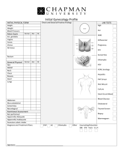


© Copyright 2026