
Veterinary Pathology Online
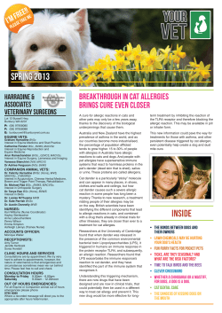
Veterinary Pathology Online http://vet.sagepub.com/ Genital Lesions Associated with Visceral Leishmaniasis and Shedding of Leishmania sp. in the Semen of Naturally Infected Dogs S. A. Diniz, M. S. Melo, A. M. Borges, R. Bueno, B. P. Reis, W. L. Tafuri, E. F. Nascimento and R. L. Santos Vet Pathol 2005 42: 650 DOI: 10.1354/vp.42-5-650 The online version of this article can be found at: http://vet.sagepub.com/content/42/5/650 Published by: http://www.sagepublications.com On behalf of: American College of Veterinary Pathologists, European College of Veterinary Pathologists, & the Japanese College of Veterinary Pathologists. Additional services and information for Veterinary Pathology Online can be found at: Email Alerts: http://vet.sagepub.com/cgi/alerts Subscriptions: http://vet.sagepub.com/subscriptions Reprints: http://www.sagepub.com/journalsReprints.nav Permissions: http://www.sagepub.com/journalsPermissions.nav >> Version of Record - Sep 1, 2005 What is This? Downloaded from vet.sagepub.com by guest on June 15, 2014 Vet Pathol 42:650–658 (2005) Genital Lesions Associated with Visceral Leishmaniasis and Shedding of Leishmania sp. in the Semen of Naturally Infected Dogs S. A. DINIZ, M. S. MELO, A. M. BORGES, R. BUENO, B. P. REIS, W. L. TAFURI, E. F. NASCIMENTO, AND R. L. SANTOS Departamento de Clı́nica e Cirurgia Veterinárias, Escola de Veterinária da Universidade Federal de Minas Gerais, Belo Horizonte, MG, Brazil (SAD, MSM, AMB, RB, BPR, EFN, RLS); and Departamento de Patologia Geral, Instituto de Ciências Biológicas da Universidade Federal de Minas Gerais, Belo Horizonte, MG, Brazil (WLT) Abstract. Although visceral leishmaniasis is primarily transmitted by a biological invertebrate vector, transmission in the absence of the vector has been reported, including venereal transmission in humans. Considering the possibility of venereal transmission, we studied genital lesions in dogs naturally infected with visceral leishmaniasis and shedding of Leishmania sp. in the semen. Approximately 200 dogs were serologically tested for anti-Leishmania antibodies and divided into three groups: 1) serologically negative dogs (n ⫽ 20), 2) asymptomatic serologically positive dogs (n ⫽ 20), and 3) symptomatic serologically positive dogs (n ⫽ 20). Samples from both testes, all segments of both epididymes, prostate gland, glans penis, and prepuce were histologically evaluated and processed for immunodetection of Leishmania sp. Semen samples were obtained from 22 symptomatic serologically positive dogs and processed for detecting Leishmania DNA by polymerase chain reaction. A significantly higher frequency of inflammation was observed in the epididymes, glans penis, and prepuce of dogs with visceral leishmaniasis, which was associated with a high frequency of immunohistochemically positive tissues (up to 95% of tissues from symptomatic dogs were positive by immunohistochemistry). Leishmania DNA was detected in eight of 22 semen samples from symptomatic dogs. Together these findings indicate that genital lesions and shedding of Leishmania sp. (donovani complex) in the semen are associated with visceral leishmaniasis. Additional studies should address the possibility of venereal transmission of the disease in the dog. Key words: Genital system; Leishmania sp.; semen; visceral leishmaniasis. Leishmaniasis is a zoonotic disease caused by intracellular protozoa belonging to the genus Leishmania. According to the World Health Organization, the disease is present in 88 countries, mostly in tropical areas and in the Mediterranean basin. Depending on which Leishmania species is involved in the infection and the immunocompetence of the human host, the infection can result in visceral, cutaneous, or mucocutaneous leishmaniasis. Visceral leishmaniasis in dogs is associated with variable clinical manifestations ranging from unapparent subclinical infections to a systemic disease characterized by progressive weight loss, hepatomegaly, splenomegaly, and lymphadenopathy, which is frequently associated with dermatological signs.14,21 The amastigote form of the parasite occurs intracellularly in the vertebrate host, whereas the promastigote and paramastigote forms are found within the digestive tract of the invertebrate host.14,24 Several vertebrate species can be infected with Leishmania sp., but the dog is recognized as the most important reservoir for human visceral leishmaniasis caused by Leishmania chagasi and Leishmania infantum.15 Indeed, the spatial distribution of serologically positive dogs and human infections is spatially correlated.18 Transmission of the disease usually occurs between the invertebrate host, which in the New World is the sand fly Lutzomia longipalpis, and the vertebrate host.14,24 However, transmission of visceral leishmaniasis in the absence of the invertebrate vector has been reported. Recently, a case of transmission through blood transfusion was documented in a dog.19 Similarly, transmission between drug users through contaminated needles has been reported.2 Furthermore, autochthonous cases of visceral leishmaniasis have been reported in the USA7 and in the UK,9 where there is no suitable biological vector. In these cases, exposure to an alternative insect vector, direct transmission, or vertical transmission were considered possible routes of infection, although the actual mechanism for transmission was not identified.7 In spite of the occurrence of vertical transmission in humans,12 a recent study indicates that this route of trans- 650 Downloaded from vet.sagepub.com by guest on June 15, 2014 Genital Leishmaniasis Vet Pathol 42:5, 2005 mission is not likely to occur in dogs.1 A case of venereal transmission between a man and a woman in an area free of the disease and in the absence of the vector has been well documented.22 Venereal transmission has not been reported in dogs, although it is thought to be likely to occur in this species5 because genital lesions associated with visceral leishmaniasis have been described both in the dog6 and man.3,4,10,20 There is also a report of canine transmissible venereal tumor in which intralesional amastigotes of Leishmania sp. were observed.13 Considering the potential for venereal transmission of visceral leishmaniasis and the lack of information about genital lesions associated with the disease in dogs, the goal of this study was to identify lesions in the genital system and to detect amastigote forms of Leishmania sp. in genital organs and in the semen of dogs with visceral leishmaniasis. Materials and Methods Animals and sampling The dogs from this study came from the Center for Zoonosis Control (CZC) and consisted of sexually mature male dogs of varied ages and varied breeds. Approximately 200 dogs were submitted to serological tests, including indirect fluorescence, complement fixation, and enzyme-linked immunosorbent assay for detection of anti-Leishmania antibodies. Sixty dogs were then selected. Serologically positive dogs were assigned to groups on the basis of clinical symptoms and visceral lesions observed at necropsy. A control group was derived from serologically negative animals at the CZC that were in good health clinically and devoid of visceral lesions. Therefore the 60 dogs were divided into one of the following three groups: 1) control group composed of healthy dogs serologically negative for leishmaniasis, 2) asymptomatic serologically positive dogs (with neither clinical signs nor lesions suggestive of visceral leishmaniasis), and 3) symptomatic serologically positive dogs (with clinical signs and lesions suggestive of visceral leishmaniasis). At necropsy, samples from both testes, all segments of both epididymides, and the prostate gland were collected. Another group of 60 dogs were selected and grouped on the same criteria described above and used for the study of the external genitalia; samples of the glans penis and prepuce were then collected. Senile and prepubertal dogs were excluded from the study. A third group of 22 serologically positive sexually mature male dogs with clinical signs of variable intensity were subjected to semen sampling for PCR detection of Leishmania DNA as described below. All tissue samples were fixed in 10% neutral buffered formalin for 24 and 48 hours and embedded in paraffin. Sections (5 m) were mounted onto silane-coated slides and stained with hematoxylin and eosin (HE) or further processed for immunohistochemistry (IHC). The frequency of inflammation was estimated from the number of dogs with any inflammatory change over the total number of dogs in each group. Histological sections were scored from 0 (no inflammation) to 3 (severe inflammation). The score of each 651 slide was established by a single pathologist without knowledge of group assignment (control, asymptomatic, or symptomatic). Selected sections of the testes were stained with Congo Red for detection of amyloid. Immunohistochemistry The immunohistochemical protocol employed here has been recently described.23 Monoclonal and polyclonal antibodies and a canine hyperimune serum were employed as primary antibodies on serial sections. A commercially available anti-Leishmania lipophosphoglycan monoclonal antibody (Cedarlane Laboratories, Hornby, Canada) was used as a primary antibody and diluted 1 : 100 (0.01 M phosphatebuffered saline [PBS]). For production of the polyclonal antibody, promastigotes of a canine isolate of L. chagasi were grown to the stationary phase in alfa-MEM medium at 25⬚C, supplemented with 10% heat-inactivated fetal calf serum, and injected intramuscularly into a rabbit. The immunization protocol included three injections with 3-week intervals. For the first injection, 107 inactivated promastigotes were suspended in 0.5 ml of complete Freud’s adjuvant. For the following two injections, the same number of inactivated promastigotes were suspended in 0.5 ml of incomplete Freud’s adjuvant. Three weeks after the third injection, serum was collected, stored at ⫺20⬚C, and used as primary antibody at 1 : 100 dilution. A heterologous hyperimmune serum of one dog naturally infected with L. chagasi (immunofluorescent assay title ⱖ 1 : 40), diluted 1 : 100 (0.01 M PBS), was also used as primary antibody. Sections (5 m) were hydrated and incubated in 4% hydrogen peroxide in PBS solution (0.01 M, pH 7.2), incubated with normal goat serum (1 : 100 dilution) as blocking antibody, and then incubated with one of the primary antibodies described above 18–22 hours at 4⬚C in a humid chamber. After washing in PBS, the slides were incubated with biotinylated secondary antibody for 20 minutes at room temperature, washed in PBS again, and then incubated with streptavidin–peroxidase complex (LSAB⫹ Kit, DAKO Corporation, Carpinteria, CA) for 20 minutes at room temperature. The reaction was developed with a 0.024% diaminobenzidine (Sigma, St. Louis, MO) solution and 0.16% hydrogen peroxide. The slides were counterstained with Harris’ hematoxylin. Negative controls comprised tissues known to be free of amastigotes and by switching the primary antibody by PBS. Samples of liver and spleen from serologically and parasitologically positive dogs were used as positive controls. The number of immunolabeled amastigotes was scored from 0 to 3 (0 ⫽ no amastigotes detected, 1 ⫽ small number of amastigotes [1–3 amastigotes or macrophage-containing amastigotes per higher magnification microscopic field], 2 ⫽ moderate number of amastigotes [4–10 amastigotes or macrophage-containing amastigotes per higher magnification microscopic field], and 3 ⫽ large number of amastigotes [⬎10 amastigotes or macrophage-containing amastigotes per higher magnification microscopic field]). Polymerase chain reaction Samples of semen were collected by digital manipulation from 22 dogs seropositive for Leishmania sp. None of these dogs were part of the groups described above that were used Downloaded from vet.sagepub.com by guest on June 15, 2014 652 Diniz, Melo, Borges, Bueno, Reis, Tafuri, Nascimento, and Santos Table 1. Frequency of inflammation in the internal genitalia of symptomatic, asymptomatic, and control dogs (serologically negative for leishmaniasis). Table 2. Frequency of inflammation in the external genitalia of symptomatic, asymptomatic, and control dogs (serologically negative for leishmaniasis). Frequency of Inflammatory change (%)* Group Control (n ⫽ 20)† Asymptomatic (n ⫽ 20) Symptomatic (n ⫽ 20) Vet Pathol 42:5, 2005 Frequency of Inflammatory change (%)* Testis Epididymis Prostate Group Glans penis Prepuce 15.8a 20.0a 45.0a 36.8a 70.0ab‡ 85.0b§ 65.0a 68.4a 40.0a Control (n ⫽ 20) Asymptomatic (n ⫽ 20) Symptomatic (n ⫽ 20) 10.0a 55.0b† 90.0c§ 20.0a 80.0b‡ 95.0b㛳 * Different letters in the same column indicate a statistically significant difference (P ⬍ 0.05). † The samples from the testes and epididymes from one control dog were lost during processing. ‡ Control versus asymptomatic (P ⫽ 0.0562). § Control versus symptomatic (P ⫽ 0.0031). for the morphological and immunohistochemical studies. All 22 dogs had clinical signs of variable intensity compatible with visceral leishmaniasis, but none of the dogs had an advanced clinical disease with either cachexia or chronic renal failure. These samples were processed for DNA extraction by digesting the semen in FE buffer (100 mM Tris-HCl, 100 mM NaCl, 1 mM EDTA pH 8.0, Sarcosyl 20%, proteinase K 20 mg/ml, dithiothreitol 154 mg/ml), followed by phenol-chloroform extraction. DNA was precipitated in isopropanol at ⫺20⬚C, washed in 70% ethanol, and resuspended in nuclease-free water. PCR was performed according to a previously described method.11 DNA (1 g) was added to 15.6 l of a solution containing 5.1 l of 10⫻ PCR buffer, 5.1 l of a 200 M dNTP solution, 0.2 M primer A (5⬘CTTTTCTGGTCCCGCGGGTAGG-3⬘), 0.2 M primer B (5⬘-CCACCTGGCCTATTTTACACCA-3⬘), 3 mM MgCl2, and 1.5 U of Taq polymerase. Cycling parameters were denaturation at 94⬚C for 4 minutes; 49 cycles of denaturation (94⬚C for 30 seconds), annealing (59⬚C for 30 seconds), and extension (72⬚C for 30 seconds); and a final extension at 72⬚C for 10 minutes. The PCR products (145 bp) were resolved by agarose gel electrophoresis. To test the sensitivity of this PCR protocol for semen samples, promastigotes of L. chagasi were cultured as described and serially diluted in semen samples from a serologically negative dog. Concentrations ranged from 1 to 10,000 organisms/ml of semen, with an aliquot of noninoculated semen used as negative control. Statistical analysis The frequency of lesions and IHC positivity were compared among groups by Fisher’s exact test with Graphpad Instat software (version 3.05, Graphpad Software, Inc., San Diego, CA). The inflammation score and the score for the number of immunolabeled amastigotes were compared by the Kuskal–Wallis test, and a correlation analysis between these scores was performed with Spearman’s nonparametric correlation test. The Graphpad Instat software was used for these nonparametric analyses. Results No gross lesions were detected in either the internal or external genitalia. The frequencies of inflammatory * Different letters in the same column indicate a statistically significant difference (P ⬍ 0.05). † Control vs. asymptomatic (P ⫽ 0.0057). ‡ Control vs. asymptomatic (P ⬍ 0.0004). § Control vs. symptomatic (P ⬍ 0.0001). 㛳 Control vs. symptomatic (P ⬍ 0.001). changes in the testis, epididymis, and prostate are summarized in Table 1. The frequency of inflammation in the glans penis and prepuce are summarized in Table 2. Although the frequency of testicular inflammation was not significantly different among the groups, seropositive dogs, particularly symptomatic dogs, tended to have a higher frequency of inflammation in the testis (Table 1). Indeed, the mean score of inflammation was higher in testes of symptomatic dogs, indicating a stronger testicular inflammatory reaction associated with clinical visceral leishmaniasis (Table 3). These dogs had multifocal to diffuse nonsegmental lymphoplasmacytic interstitial orchitis associated with secondary mild to severe testicular degeneration. The degenerative process was more intense in the seminiferous tubules of symptomatic dogs (Fig. 1). Either under regular light microscopy or polarized light, no amyloid was observed in Congo Red–stained sections of the testes from seropositive dogs with testicular histopathological changes (data not shown). A predominantly histiolymphocytic epididymitis (Fig. 2) was observed in all groups, and the frequency Table 3. Score* of inflammation in the internal genitalia of symptomatic, asymptomatic, and control dogs (serologically negative for leishmaniasis). Score of Inflammatory Changes (n)† Group Control‡ Asymptomatic Symptomatic Testis 0.26 (19) 0.20 (20)aA 0.75 (20)bA abA Epididymis Prostate 0.37 (19) 1.20 (20)bBC 1.58 (20)bB 1.05 (20)aB 0.95 (20)aB 0.85 (19)aA aA * 0 ⫽ no inflammation, 1 ⫽ mild, 2 ⫽ moderate, and 3 ⫽ severe inflammation. † n ⫽ total number of dogs in a given group. Different lowercase letters in the same column indicate a statistically significant difference (P ⬍ 0.05). Different uppercase letters in the same row indicate a statistically significant difference (P ⬍ 0.05). ‡ The samples from the testes and epididymes from one control dog were lost during processing. Downloaded from vet.sagepub.com by guest on June 15, 2014 Vet Pathol 42:5, 2005 Genital Leishmaniasis 653 Fig. 1. Testis; dog. Diffuse histiocytic orchitis associated with severe degeneration of the seminiferous epithelium in a symptomatic serologically positive dog. HE. Bar ⫽ 100 m. Fig. 2. Epididymis; dog. Focal epididymitis characterized by a lymphohistiocytic inflammatory infiltrate in a symptomatic serologically positive dog. HE. Bar ⫽ 100 m. Fig. 3. Prepuce; dog. Histiocytic balanoposthitis with intracellular amastigotes in macrophages (inset) in a symptomatic serologically positive dog. HE. Bar ⫽ 50 m. Fig. 4. Glans penis; dog. Urethral mucosa with intraepithelial macrophage containing amastigotes (arrow) in a symptomatic serologically positive dog. HE. Bar ⫽ 20 m. of epididymitis was significantly higher (P ⫽ 0.0031) in the symptomatic group compared with the seronegative controls (Table 1). The difference between frequencies of epididymitis between asymptomatic dogs and seronegative controls was just marginally significant (P ⫽ 0.0562). Both asymptomatic and symptomatic seropositive dogs had a significantly higher score of inflammation compared with the controls (Table 3). The frequency of inflammatory infiltrate and the score of inflammation in the prostate gland was not significantly different among the groups (Tables 1, 3), and in the sections with inflammation, a lymphoplasmacytic infiltrate predominated. Interestingly, in spite of these results, the highest score of inflammation in the control group was observed in the prostate gland compared with other organs of the genital system (Table 3). Taken together, these results indicate that there is no association between visceral leishamaniasis and prostatitis. The inflammatory reaction in the glans penis was predominantly histiocytic with morphological features of a granulomatous reaction. The frequency and the score of inflammation in the glans penis were significantly higher in seropositive dogs compared with controls, with symptomatic dogs developing a higher frequency and score of inflammation (Tables 2, 4). Several seropositive dogs (10 symptomatic and five asymptomatic) had a large number of macrophages containing intracellular amastigotes with virtual absence of other inflammatory cells (Fig. 3). In one of the symptomatic dogs, macrophages containing intracellular amastigotes were observed within the epithelial layer of the urethra, indicating transepithelial migration of Leishmania-containing phagocytes (Fig. 4). Downloaded from vet.sagepub.com by guest on June 15, 2014 654 Diniz, Melo, Borges, Bueno, Reis, Tafuri, Nascimento, and Santos Table 4. Score* of inflammation in the external genitalia of symptomatic, asymptomatic, and control dogs (serologically negative for leishmaniasis). Score of Inflammatory Change (n)† Group Glans Penis Prepuce Control Asymptomatic Symptomatic 0.20 (20)aA 1.05 (20)bA 1.65 (20)cA 0.20 (20)aA 1.55 (20)bB 1.70 (20)bA * 0 ⫽ no inflammation, 1 ⫽ mild, 2 ⫽ moderate, and 3 ⫽ severe inflammation. † n ⫽ total number of dogs in a given group. Different lowercase letters in the same column indicate a statistically significant difference (P ⬍ 0.05). Different uppercase letters in the same row indicate a statistically significant difference (P ⬍ 0.05). In the prepuce, the frequency and score of inflammation between seronegative controls and seropositive dogs was significantly different, but no difference was detected between symptomatic and asymptomatic dogs (Tables 2, 4). A hystioplasmacytic balanoposthitis was observed in all groups, with variable frequencies. In addition some seropositive dogs either symptomatic or asymptomatic had a granulomatous dermatitis characterized by a large number of histiocytes diffusely distributed throughout the mucosa associated with multifocal areas of erosions and ulcerations with intense infiltration of neutrophils. Large numbers of amastigotes were observed intracellularly in macrophages. Amastigotes were observed in HE-stained sections of the epididymis from eight symptomatic and one asymptomatic dog, in the testes of three symptomatic dogs, in the glans penis of seven symptomatic and one asymptomatic dog, and in the prepuce of eleven symptomatic and three asymptomatic dogs. To improve the sensitivity of detection of amastigotes, a immunohistochemical approach was employed. Canine hyperimmune serum was superior to monoclonal and polyclonal antibodies; thus, sections in which the canine hyperimmune serum was employed as primary antibody were examined in detail to generate the data presented here. The frequency of amastigotes detected by IHC is summarized in Tables 5 and 6. As expected, a much higher sensitivity for detection of amastigotes was obtained with IHC compared with HE-stained sections. This notion is supported by the absence of amastigotes in HE-stained sections of the prostate gland, which is in sharp contrast to the IHC results, which demonstrated amastigotes in more than a half of the sections from both symptomatic and asymptomatic dogs. Amastigotes were specifically stained, with the vast majority located intracellularly in macrophages (Figs. 5–8). The distribution of immunostaining was variable and correlated with the inflammatory changes, with the excep- Vet Pathol 42:5, 2005 Table 5. Frequency of immunohistochemical (IHC) detection of Leishmania sp. amastigotes in the internal genitalia of symptomatic, asymptomatic, and control dogs (serologically negative for leishmaniasis). Frequency of IHC-Positive Organs [% (n)]* Group Testis Control Asymptomatic Symptomatic 0.0 (18) 35.0 (20)b 75.0 (20)c a Epididymis Prostate 0.0 (18) 60.0 (20)b 95.0 (20)c 0.0 (18)a 57.9 (19)b 52.6 (19)b a * n ⫽ total number of dogs in a given group that was evaluated by IHC. Different letters in the same column indicate a statistically significant difference (P ⬍ 0.05). tion of the prostate, in which a small number of macrophages containing amastigotes were observed scattered throughout the interstitium. Amastigotes were not detected by IHC in any of the genital organs from seronegative dogs. Higher numbers of positive sections of the testis and epididymis were observed in symptomatic compared with asymptomatic dogs. No immunolabeled amastigotes were observed either in the lumen of the seminiferous tubules or within the epididymal duct. In contrast, no significant differences in the number of IHC-positive sections of the prostate gland, glans penis, and prepuce were observed between these groups (Tables 5, 6). The number of amastigotes was higher in the epididymis and prepuce of symptomatic dogs (Tables 7, 8), which is in good agreement with the frequency and intensity (score) of inflammation. In fact, a highly significant positive correlation (P ⬍ 0.0001) was observed between the score of inflammation and the number of amastigotes in the epididymis (r ⫽ 0.6730), prepuce (r ⫽ 0.6911), and glans penis (r ⫽ 0.6178), whereas no significant correlation was observed in the testes and prostate gland (r ⫽ 0.2479 and 0.0261, respectively). PCR analysis of semen samples spiked with promastigotes of L. chagasi resulted in a level of detection of at least 1 organism/ml of semen. All dilutions from 10,000 to 1 organism/ml were PCR positive, whereas Table 6. Frequency of immunohistochemical (IHC) detection of Leishmania sp. amastigotes in the external genitalia of symptomatic, asymptomatic, and control dogs (serologically negative for leishmaniasis). Frequency of IHC-Positive Organs [% (n)]* Group Glans Penis Prepuce Control Asymptomatic Symptomatic 0.0 (20) 75.0 (20)b 75.0 (20)b 0.0 (20)a 80.0 (20)b 95.0 (20)b a * n ⫽ total number of dogs in a given group that was evaluated by IHC. Different letters in the same column indicate a statistically significant difference (P ⬍ 0.05). Downloaded from vet.sagepub.com by guest on June 15, 2014 Genital Leishmaniasis Vet Pathol 42:5, 2005 655 Fig. 5. Testis; dog. Immunolabeling of amastigotes of Leishmania sp. inside macrophages (inset) associated with a lymphohistiocytic inflammatory infiltrate in a symptomatic serologically positive dog. Streptavidin–peroxidase complex. Bar ⫽ 50 m. Fig. 6. Epididymis; dog. Immunolabeling of amastigotes of Leishmania sp. inside macrophages (inset) associated with a lymphoplasmacytic infiltrate in a symptomatic serologically positive dog. Streptavidin–peroxidase complex. Bar ⫽ 40 m. Fig. 7. Prepuce; dog. Immunolabeling of a large number of Leishmania-containing macrophages diffusely distributed throughout the mucosa in a symptomatic serologically positive dog. Streptavidin–peroxidase complex. Bar ⫽ 80 m. Fig. 8. Prepuce; dog. Immunolabeling of a large number of amastigotes of Leishmania sp. inside macrophages in a symptomatic serologically positive dog. Streptavidin–peroxidase complex. Bar ⫽ 20 m. the noninoculated control was negative (data not shown). PCR analysis of semen samples from 22 serologically positive dogs resulted in 36.36% (8/22) positivity (Fig. 9). Discussion We demonstrated that visceral leishmaniasis is associated with a high frequency of inflammatory lesions in most of the male genital organs in dogs. Our results clearly indicate that inflammation of the epididymis, glans penis, and prepuce is more frequent in dogs serologically positive for visceral leishmaniasis, and among those, symptomatic dogs develop lesions more often than asymptomatic dogs. A similar trend was observed for the occurrence of orchitis, but not for prostatitis. The frequency and intensity of inflammatory lesions in the testis correlated with clinical manifestation of the disease as well as the parasite load in the testis. The number of amastigotes in the testis apparently acts as a causing factor triggering the inflammatory response, whereas testicular degeneration is probably a consequence of inflammation rather than any direct parasitic effect on the seminiferous epithelium. This notion is supported by the fact that testicular degeneration is often secondary to testicular inflammation.16 It is noteworthy that no amastigotes were detected within the seminiferous tubules, and although the seminiferous tubules often developed degenerative changes, they remained morphologically intact in all seropositive dogs. Experimental infection of hamsters with Leishmania donovani results in testicular amyloidosis, Downloaded from vet.sagepub.com by guest on June 15, 2014 656 Diniz, Melo, Borges, Bueno, Reis, Tafuri, Nascimento, and Santos Table 7. Score* of the number of immunolabeled amastigotes in the internal genitalia of symptomatic, asymptomatic, and control dogs (serologically negative for leishmaniasis). Table 8. Score* of the number of immunolabeled amastigotes in the external genitalia of symptomatic, asymptomatic, and control dogs (serologically negative for leishmaniasis). Score of the Number of Immunolabeled Amastigotes (n)† Group Testis Control Asymptomatic Symptomatic 0.0 (18) 0.50 (20)bA 1.15 (20)cA aA Score of the Number of Immunolabeled Amastigotes (n)† Epididymis Prostate 0.0 (18) 0.80 (20)bA 1.85 (20)c,B 0.0 (18) 0.68 (19)bA 0.78 (19)bA aA Vet Pathol 42:5, 2005 aA * 0 ⫽ no amastigotes detected, 1 ⫽ small number of amastigotes, 2 ⫽ moderate number of amastigotes, and 3 ⫽ large number of amastigotes. † n ⫽ total number of dogs in a given group that was evaluated by IHC. Different lowercase letters in the same column indicate a statistically significant difference (P ⬍ 0.05). Different uppercase letters in the same row indicate a statistically significant difference (P ⬍ 0.05). degeneration, progressive atrophy, and azospermia. In this experimental model, the degenerative changes are also associated with infiltration of lymphocytes and macrophages containing amastigotes in the testes.8 Testicular amyloidosis was not observed in this study, which contrasts with the previously described lesions in hamsters.8 In human visceral leishmaniasis, testicular involvement is not well studied, but there one study reported amastigotes in macrophages obtained by fine needle aspiration of the testes of a boy with a severe lymphoblastic leukemia.10 An explanation for the finding of amastigotes in cases of epididymitis could be the presence of a pre-existing inflammatory process, which would recruit macrophages carrying Leishmania. In this case, the presence of amastigotes associated with epididymitis would just be an incidental finding. However, our data indicate that through an unknown mechanism, Leishmania- or macrophage-containing amastigotes are attracted to the epididymis because the frequency of epididymitis is significantly increased in dogs with vis- Group Glans Penis Prepuce Control Asymptomatic Symptomatic 0.0 (20) 0.60 (20)b 0.75 (20)bB 0.0 (20)a 1.15 (20)b 1.85 (20)c a * 0 ⫽ no amastigotes detected, 1 ⫽ small number of amastigotes, 2 ⫽ moderate number of amastigotes, and 3 ⫽ large number of amastigotes. † n ⫽ total number of dogs in a given group that was evaluated by IHC. Different lowercase letters in the same column indicate a statistically significant difference (P ⬍ 0.05). ceral leishmaniasis and the intensity of inflammation correlates with the number of amastigotes. In sharp contrast, although the occurrence of prostatitis is relatively high in healthy seronegative dogs, no amastigotes were detected in HE-stained slides, and just a very low parasitic load was detected by IHC in the prostate gland of symptomatic or asymptomatic seropositive dogs. In addition, the frequency of inflammation in the prostate gland did not differ among the groups. Considering that epididymitis is often associated with the presence of inflammatory cells in canine semen,17 epididymitis associated with visceral leishmaniasis could result in shedding of Leishmania in the semen, favoring venereal transmission of the disease, which has been reported in humans.22 To further characterize this process, we employed PCR for detection of Leishmania in the semen, resulting in a relatively high percentage of seropositive dogs shedding the organism in the semen. Importantly, this frequency could be underestimated because we sampled dogs that were not conditioned to digital sampling, which resulted in some samples composed mostly of the prostatic frac- Fig. 9. Representative PCR reaction for detection of Leishmania DNA in semen samples from dogs serologically positive for leishmaniasis. M ⫽ molecular mass marker (the size of the marker is indicated on the left, where an arrow indicates the position for the predicted 145-bp PCR product); C⫺ ⫽ negative control; C⫹ ⫽ positive control (Leishmaniafree semen sample spiked with cultured promastigotes of Leishmania chagasi); 19, 8, and 26 ⫽ negative semen samples; 15, 23, and 7 ⫽ positive semen samples. Downloaded from vet.sagepub.com by guest on June 15, 2014 Vet Pathol 42:5, 2005 Genital Leishmaniasis tion with a very low cell content. In addition, serial sampling was not doable because the dogs were euthanatized soon after sampling. Considering the method of digital manipulation employed for semen sampling, the Leishmania DNA detected in the semen most likely originated from the internal genitalia because contamination with Leishmania from the external genitalia would not be expected under these circumstances. However, natural breeding in dogs often results in trauma to both the male and female, increasing the possibility of transference of amastigotes from the external genitalia of the dog in addition to the organisms secreted from the internal genital organs. These aspects could increase the possibility of venereal transmission. The increased frequency of inflammation in the epididymis, glans penis, and prepuce was directly proportional to the severity of clinical signs and to the number of amastigotes in the testis as evidenced by IHC. However, this pattern was not observed in the prostate gland, in which the frequency of inflammation was similar between seropositive and control dogs, although a few amastigotes were detected in most of the prostate sections by IHC. These findings are in good agreement with previous report6 that described an increase in the inflammatory infiltrate associated with more evident clinical signs, although these authors studied a limited number of dogs by transmission electron microscopy. Our results demonstrate that sensitivity for identification of amastigotes in HE-stained sections is extremely low compared with IHC. In addition, the use of canine hyperimune serum as primary antibody yielded results comparable or even superior to either the monoclonal or the polyclonal antibodies used in this study. These results are in good agreement with the previous description of this immunohistochemical method.23 The high frequency of inflammation associated with erosions and/or ulcerations and amastigotes in the glans penis and prepuce, as well as the finding of macrophages containing amastigotes migrating through the urethral epithelium, might also contribute to shedding of Leishmania in the semen. Although the biological vector is the most important route of transmission, the possibility of venereal transmission could prove to have epidemiological significance, mostly in relation to the implementation of an eradication program. According to early reports,5 venereal transmission of visceral leishmaniasis is likely to occur in dogs because it was observed that, in an area with low vector population and low number of human cases, a high number of infected dogs was observed, which is not compatible with exclusively vector-mediated transmission. Lesions in the glans penis and prepuce have been re- 657 ported in cases of human visceral leishmaniasis4,20 and human cutaneous leishmaniasis.3 Furthermore, a case of venereal transmission of human visceral leishmaniasis has been well documented.22 Taken together, our findings indicate that genital lesions and shedding of Leishmania sp. (donovani complex) in the semen are frequently associated with visceral leishmaniasis. Additional studies should address the possibility of venereal transmission of the disease in the dog. Acknowledgements This work was supported by a grant from the Pró-Reitoria de Pesquisa da UFMG. We are grateful to Dr. M. S. Michalik for performing the serological tests. SAD has a scholarship from CAPES, Brası́lia, Brazil. RB is supported by the Pontifı́cia Universidade Católica de Minas Gerais, Brazil. We are thankful to the Centro de Controle de Zoonoses da Prefeitura Municipal de Belo Horizonte for providing the samples for this study. References 1 Andrade HM, Toledo VPCP, Marques MJ, Silva JCF, Tafuri WL, Mayrink W, Genaro O: Leishmania (Leishmania) chagasi is not vertically transmitted in dogs. Vet Parasitol 103:71–81, 2002 2 Bosch RJ, Rodrigo AB, Sánchez P, Gálvez MV, Herrera E: Presence of Leishmania organisms in specific and non-specific skin lesions in HIV-infected individuals with visceral leishmaniasis. Int J Dermatol 41:670–675, 2002 3 Cabello I, Caraballo A, Millán Y: Leishmaniasis in the genital area. Rev Inst Med Trop Sao Paulo 44:105–107, 2002 4 Cain C, Stone MS, Thieberg M, Wilson ME: Nonhealing genital ulcers. Arch Dermatol 130:1311–1316, 1994 5 Deane LM: Epidemiologia e profilaxia do Calazar Americano. Rev Bras Malariol Doenc Trop 10:431–449, 1958 6 Diaz MP, Arribas JLG, Garcia EG, Rosado MC: Lesiones testiculares y epididimarias en la leishmaniasis visceral canina, de presentacion natural. Hygia Pecoris 4:5–25, 1982 7 Gaskin AA, Schantz P, Jackson J, Birkenheuer A, Tomlinson L, Gramiccia M, Levy M, Steurer F, Kollmar E, Hegarty BC, Ahn A, Breitschwerdt EB: Visceral leishmaniasis in a New York foxhound kennel. J Vet Intern Med 16:34–44, 2002 8 Gonzalez JL, Gallego E, Castaño M, Rueda A: Testicular amyloidosis in hamsters experimentally infected with Leishmania donovani. Br J Exp Pathol 64:518–523, 1983 9 Harris MP: Suspected transmission of leishmaniasis. Vet Rec 135:339, 1994 10 Kapila K, Prakash MB, Mehrota R, Vermar K: Testicular leishmaniasis in a boy with acute lymphoblastic leukemia. Acta Cytol 38:878–879, 1994 11 Lachaud L, Marchergui-Hammami S, Chabbert E, Dereure J, Dedet JP, Bastien P: Comparision of six PCR methods using peripheral blood for detection of canine Downloaded from vet.sagepub.com by guest on June 15, 2014 658 12 13 14 15 16 17 18 Diniz, Melo, Borges, Bueno, Reis, Tafuri, Nascimento, and Santos visceral leishmaniasis. J Clin Microbiol 40:210–215, 2002 Low GC, Edin MD, Lond FRCP, Cooke WE, Irel FRCS: A congenital case of Kala-azar. Lancet 11:1209–1211, 1926 Macri B, Galofaro V, Braca G: Leishmaniosi e neoplasmi nota II: correlazioni ed interferenze tra leishmaniosi e tumore venereo transmissibile (Sarcoma di Sticker) nel cane. Atti Soc Ital Sci Vet 36:510–512, 1982 Marsella R, Gopegui RR: Leishmaniasis: a re-emerging zoonoses. Int J Dermatol 37:801–814, 1998 Moreno J, Alvar J: Canine leishmaniasis: epidemiological risk and the experimental model. Trends Parasitol 18: 399–405, 2002 Nascimento EF, Santos RL: Patologia da Reprodução dos Animais Domésticos, 2nd ed., pp. 96–101. Guanabara Koogan, Rio de Janeiro, Brazil, 2003 Nelson RW, Couto CG: Fundamentos de Medicina Interna de Pequenos Animais, pp. 513–517. Guanabara Koogan, Rio de Janeiro, Brazil, 1994 Oliveira CL, Assunção RM, Reis IA, Proietti FA: Spatial distribution of human and canine visceral leishmaniasis 19 20 21 22 23 24 Vet Pathol 42:5, 2005 in Belo Horizonte, Minas Gerais State, Brasil, 1994– 1997. Cad Saude Publica 17:1231–1237, 2001 Owens SD, Oakley DA, Marryott K, Hatchett W, Walton R, Nolan TJ, Newton A, Steurer F, Schantz P, Giger U: Transmission of visceral leishmaniasis through blood transfusions from infected English foxhounds to anemic dogs. J Am Vet Med Assoc 219:1076–1083, 2001 Schubach A, Cuzzi-Maya T, Gonçalves-Costa SC, Pirmez C, Oliveira-Neto MP: Leishmaniasis of glans penis. J Eur Acad Dermatol Venereol 10:226–228, 1998 Slappendel RJ, Greene CE: Leishmaniasis. In: Infectious Disease of the Dog and Cat, ed. Greene CE, pp. 769– 778. Saunders, Philadelphia, PA, 1990 Symmers WSC: Leishmaniasis acquired by contagion: a case of marital infection in Britain. Lancet 16:127–132, 1960 Tafuri WL, Santos RL, Arantes RME, Gonçalves R, Melo MN, Michalick MSM, Tafuri WL: An alternative immunohistochemical method for detecting Leishmania amastigotes in paraffin-embedded canine tissues. J Immunol Methods 292:17–23, 2004 Vassı́lios JK: Old World canine leishmaniasis. Comp Contin Educ Vet Pract 15:941–949, 1993 Request reprints from Dr. Renato Lima Santos, Escola de Veterinária da UFMG, Depto. Clı́nica e Cirurgia Veterinárias, Av. Antônio Carlos, 6627, 31270-901 Belo Horizonte, MG (Brazil). E-mail: [email protected]. Downloaded from vet.sagepub.com by guest on June 15, 2014


© Copyright 2026