
Why are hatching and emergence success low? Mercury and selenium
Marine Pollution Bulletin 62 (2011) 1671–1682
Contents lists available at ScienceDirect
Marine Pollution Bulletin
journal homepage: www.elsevier.com/locate/marpolbul
Why are hatching and emergence success low? Mercury and selenium
concentrations in nesting leatherback sea turtles (Dermochelys coriacea)
and their young in Florida
Justin Perrault a,⇑, Jeanette Wyneken a, Larry J. Thompson b, Chris Johnson c, Debra L. Miller d,1
a
Department of Biological Sciences, Florida Atlantic University, Building 01, Sanson Science, 777 Glades Road, Boca Raton, FL 33431, United States
Nestlé Purina PetCare, 801 Chouteau Ave., St. Louis, MO 63102, United States
Loggerhead Marinelife Center of Juno Beach, 14200 US Highway One, Juno Beach, FL 33408, United States
d
The University of Georgia, College of Veterinary Medicine, Veterinary Diagnostic and Investigational Laboratory, 43 Brighton Road, Tifton, GA 31793, United States
b
c
a r t i c l e
Keywords:
Dermochelys coriacea
Hatching success
Emergence success
Hatchling
Mercury (Hg)
Selenium (Se)
Marine turtles
i n f o
a b s t r a c t
Leatherback sea turtles (Dermochelys coriacea) have low hatching and emergence success compared to
other sea turtle species. Postmortem examinations of hatchlings showed degeneration of heart and skeletal muscle that was similar to that found in other neonates with selenium deficient mothers. Selenium
deficiency can result from elevated concentrations of bodily mercury. Ingested mercury is detoxified by
the liver through mercury–selenium compound formation. In animals persistently exposed to mercury,
the liver’s ability to detoxify this element may decrease, especially if dietary selenium is insufficient.
We measured mercury and selenium concentrations in nesting female leatherbacks and their hatchlings
from Florida and compared the levels to hatching and emergence success. Both liver selenium and the
liver selenium-to-mercury ratio positively correlated with leatherback hatching and emergence success.
This study provides the first evidence for the roles of mercury and selenium in explaining low reproductive success in a globally imperiled species, the leatherback sea turtle.
Ó 2011 Elsevier Ltd. All rights reserved.
1. Introduction
Mercury (hereafter termed Hg) is found in marine organisms
ranging from primary producers to top carnivores and it accumulates in the body from both water and food sources (Caurant
et al., 1999; Guirlet et al., 2008). While Hg is present naturally in
the environment (Campbell et al., 2005), the largest source
(75%) of this element is traced to emissions from anthropogenic
sources, mainly from the burning of fossil fuels (Pacyna and Pacyna, 2002). Mercury and Hg compounds (i.e., methylmercury,
dimethylmercury, etc.) affect various functional processes including reproduction, growth, development, vision, and hearing. Mercury has no known normal physiological role in the body (EPA,
1985). Miller et al. (2009) conducted postmortem examinations
of dead-in-nest leatherback sea turtle (Dermochelys coriacea)
hatchlings from Juno Beach, Florida, as well as hatchlings that died
⇑ Corresponding author. Tel.: +1 561 297 0146; fax: +1 561 297 2749.
E-mail addresses: [email protected] (J. Perrault), [email protected] (J. Wyneken),
[email protected] (L.J. Thompson), [email protected] (C. Johnson),
[email protected] (D.L. Miller).
1
Present address: Center for Wildlife Health, Department of Forestry, Wildlife and
Fisheries University of Tennessee, Knoxville, TN 37996, United States.
0025-326X/$ - see front matter Ó 2011 Elsevier Ltd. All rights reserved.
doi:10.1016/j.marpolbul.2011.06.009
in the laboratory shortly after nest emergence and found heart and
skeletal muscle pathologies that were similar to those seen in bovid neonates whose mothers were selenium (Se) deficient (Orr and
Blakely, 1997). They suggested that these changes might reflect Hg
toxicity or Se deficiency because, in many animals, ingested Hg is
detoxified in the liver (where both Hg and Se are stored) through
formation of Hg–Se compounds. Thus, persistently elevated Hg
may eventually deplete Se stores (Se deficiency) if dietary levels
are not compensatory (Cardellicchio et al., 2002). Selenium deficiency may lead to Hg toxicity. Selenium is a trace element and
is found naturally in marine food sources that are high in protein
(Se is largely associated with proteins), but it has low bioavailability (Caurant et al., 1996; DRI, 2000). Selenium has known enzymatic, antioxidant, thyroidal, and immune functions (Rayman,
2000); however, at higher concentrations, Se can produce adverse
effects including reduced reproductive fitness, tissue lesions, physiological anomalies, and can lead to death (Ohlendorf et al., 1988;
Hoffman, 2002).
Worldwide, leatherback sea turtles have experienced a substantial population decrease (67%) resulting from habitat degradation, poaching, and fisheries bycatch (Pritchard, 1982; Spotila
et al., 1996; Sarti-Martinez, 2000). It is difficult for these organisms
to rebound from a population decline, as they have delayed
1672
J. Perrault et al. / Marine Pollution Bulletin 62 (2011) 1671–1682
maturity (Avens et al., 2009) and exhibit low hatching success
(25.8–56.0%; Whitmore and Dutton, 1985; Leslie et al., 1996; Bell
et al., 2003; Hilterman and Goverse, 2003; Hernández et al.,
2007); however, average hatching success at some rookeries is as
high as 67–73.3% in leatherbacks (Livingstone, 2006; Stewart and
Johnson, 2006), which suggests variability within the species
across populations. Despite this higher hatching success at some
rookeries, the high mortality observed during early life history
stages (Zug and Parham, 1996; Davenport, 1997; Bell et al., 2003)
limits the potential recovery of this imperiled species.
Although low hatching and emergence success in the leatherback turtle has been explored from several nest-based perspectives, the causes remain unidentified. Redfearn (2000) found that
leatherback nest depth did not contribute to mortality in Florida.
Bell et al. (2003) documented high early embryonic mortality in
an eastern Pacific population and hypothesized that maternal
reproductive health, chemical contaminants, or bacterial infection
may be the cause. Wallace et al. (2004) determined that neither
hypoxia nor high nest temperatures (within the thermal tolerance
range) were correlated with low hatching success at that same
rookery. Surprisingly, no hypotheses regarding the roles of toxicants (e.g., Hg or persistent organic pollutants [POPs]) on hatching
and emergence success have been tested, despite known and potential negative impacts on health and reproductive success of
other vertebrates (loons: Burgess and Meyer, 2008; panthers:
USEPA, 2000; marine mammals: Beland et al., 1993).
Leatherback sea turtles make long distance migrations from foraging grounds to nesting beaches, and then subsequently return to
foraging grounds (Eckert et al., 2006; Hays et al., 2006). At those
foraging grounds, non-essential (e.g., Hg) and essential elements
(e.g., Se) can accumulate in the body through food (e.g., pelagic
medusa; Bjorndal, 1997) and water intake (Caurant et al., 1999;
Guirlet et al., 2008). Females returning to nesting beaches to lay eggs
can transfer these elements to their offspring via egg components
(i.e., albumen, yolk), as has previously been shown in leatherbacks
(Guirlet et al., 2008) and other reptiles (Roe et al., 2004; Rainwater
et al., 2005; Unrine et al., 2006). Elevated concentrations of these
elements could impact leatherback turtle hatching and emergence
success. Leatherbacks lay 6–11 nests per season depositing an
average of 70–80 eggs with a mean interclutch interval of 9–
10 days (Miller, 1997; Bell et al., 2003; Stewart and Johnson,
2006). In this study, our objectives were to (i) identify or exclude
Se deficiency as a factor affecting leatherback turtle nest success,
(ii) determine how Hg and Se in hatchling blood, liver, and yolk
sac are correlated with each other and with maternal blood Hg
and Se concentrations, and (iii) characterize Hg and Se concentrations in nesting leatherbacks (blood), hatchlings (blood from live
turtles and liver and yolk sac from dead turtles), and egg shells
and shelled albumen globs (SAGs).
2. Materials and methods
2.1. Study period and site description
Western Atlantic female leatherbacks and their young were
sampled on the nesting beach during the 2007 and 2008 nesting
seasons. Samples were collected along Florida’s east coast (Juno
Beach and Jupiter Beach, FL), primarily in the 20 km area of peak
nesting between Jupiter Inlet (26°560 3600 N, 80°040 1500 W) and the
Lake Worth Inlet (26°460 2400 N, 80°010 5300 W, Fig. 1). This rookery
has been monitored nightly for leatherback nesting activity since
2001 (Stewart, 2007). Nesting season runs from March through
early July in southeastern Florida. Over 400 individual nesting
leatherbacks have been identified in Florida since tagging programs began (K. Stewart, personal communication).
2.2. Sample collection
2.2.1. Nesting females
Sampling in Florida was conducted in conjunction with the Loggerhead Marinelife Center’s (LMC) leatherback tagging project. The
beach was patrolled on foot or with all-terrain vehicles each night
to encounter nesting leatherbacks during the nesting season. From
10:30 p.m. to 5:30 a.m., night-vision scopes were used to identify
turtle crawls and the stage of nesting before approaching a nesting
turtle to minimize the chances of disturbance. Nesting females
were approached once they entered a physiological ‘‘trance’’,
which begins after oviposition commences (Dutton and Dutton,
1994).
Individuals were identified based on their flipper tags and/or
internal PIT (passive integrated transponder) tags. However, if neither of these tags were present, PIT and/or flipper tags were applied by the LMC staff. Blood was collected into 7 mL BD K3EDTA
VacutainerÒ tubes (Becton–Dickinson and Co. Franklin Lakes, NJ,
USA) using an 18 gauge venous collection needle fitted in a VacutainerÒ tube holder. Before insertion of the needle, the entire area
was swabbed with a sterile 70% isopropyl alcohol swab. Approximately 5 mL of blood were taken from the femoral rete system
(Dutton, 1996). The location of this vascular network was identified by palpation of the area approximately 10 cm posterior to
the knee. Blood collection ceased when a complete sample was obtained or egg-laying terminated (after approximately 7–11 min).
The venipuncture site was then disinfected with a new alcohol
swab and pressure was applied until the blood site clotted. The
samples were chilled immediately. After blood collection, the turtle’s minimum curved carapace length was recorded (from nuchal
notch, to posterior tip of the caudal peduncle, CCLmin, after
Wyneken, 2001). Blood was frozen at 20 °C, and later shipped
overnight, on ice, to the Veterinary Diagnostic and Investigational
Laboratory at the University of Georgia (UGA-VDIL, P.O.
Box 1389, 43 Brighton Road, Tifton, GA 31793, USA) for analysis.
2.2.2. Hatchlings
We monitored the progress of all nests and recorded any incidents of predation, sea water inundation, or wash outs due to
storm. On nights when hatchlings were expected to emerge (55–
60 days after egg deposition), a 61 cm 61 cm 10 cm cage was
placed on top of the chamber in order to retain the hatchlings so
that blood could be collected. The cages were covered with mesh
so that predators were excluded if emergence occurred in the absence of the beach patrollers. The cages were monitored every
30–60 min.
Upon emergence, up to 10 normal hatchlings were selected
and weighed to the nearest 0.5 g with a PESOLAÒ 100 g scale.
Their straight carapace lengths (SCL), straight carapace widths
(SCW), and body depths (BD) were also recorded to the nearest
0.05 mm using Vernier calipers. Body condition (mass:SCL ratio)
was also calculated. The dorsal and lateral neck of each hatchling
was swabbed with a sterile 70% isopropyl alcohol pad and blood
was collected from the external jugular vein (dorsal cervical sinus;
Owens and Ruiz, 1980) using a 1 mL BD SafetyGlide™ allergy syringe. Approximately 0.1–0.2 mL of blood was collected from each
of the hatchlings and was pooled by clutch into a K3EDTA tube for
Se (and Hg if the quantity was sufficient) analysis. This amount
(0.1–0.2 mL) is less than 5% blood volume for turtles weighing
greater than 40 g and is considered safe for collection (Jacobson,
2007; Strik et al., 2007). Blood was chilled on ice immediately
after collection. All samples were frozen at 20 °C until they were
later shipped overnight, on ice, to the UGA-VDIL for analysis.
Hatchlings were observed for 1 h and released after blood
collection.
J. Perrault et al. / Marine Pollution Bulletin 62 (2011) 1671–1682
1673
Fig. 1. Juno Beach/Jupiter Beach, FL, nesting beach. This area is located in northern Palm Beach County, and lies on the east coast of Florida in the western Atlantic Ocean.
2.2.3. Nest inventory
The nests from the study females were excavated and the contents inventoried 3 days following the major emergence date to
determine hatching and emergence success, which were calculated
after Miller (1999):
Hatching success:
# hatched eggs
:
# hatched eggs þ # unhatched eggs þ # pipped live þ # pipped dead
Emergence success:
# hatched eggs # ðlive hatchlings in nest þ # dead hatchlingsÞ
:
total eggsðhatched eggs þ unhatched eggs þ dead pipped þ live pippedÞ
A pipped egg is one in which the turtle cuts through the egg
shell, but does not exit the egg. Inventory data include the number of SAGs (Bell et al., 2003), but they are not counted as part
of hatching success as they never contain yolks or embryos. Up
to five dead-in-nest turtles/nest that were in good condition
were collected, frozen, and sent to UGA-VDIL for later sampling
of the liver and yolk sac. Because the sacrifice of endangered
species was prohibited and deemed unethical, we have to assume that all individuals in a clutch (dead-in-nest and those that
emerged) have similar Hg and Se concentrations. Previous studies support this assumption (Heinz et al., 1987; Sakai et al.,
1995; Bryan et al., 2003). Additionally, three hatched eggshells
and three SAGs were collected for Hg analyses (and Se analyses
when available).
1674
J. Perrault et al. / Marine Pollution Bulletin 62 (2011) 1671–1682
2.3. Sample analyses
All sample analyses were standardized by using the same laboratory equipment and procedures. Mercury concentrations were
determined using a flow injection analysis system (FIAS 400, Perkin-Elmer, Norwalk, CT) by atomic absorption spectrophotometry
(AAS, AAnalyst 100, Perkin-Elmer). To quantify total Hg, approximately 1 mL of blood (or 1 g of liver tissue, yolk, albumen, or shell)
from each animal was transferred to a TeflonÒ microwave vessel
and mixed with 5 mL of 65% nitric acid (HNO3, Fisher Scientific)
and 2 mL of 30% hydrogen peroxide (H2O2, Fisher Scientific). The
samples then were digested using a laboratory microwave oven
(MARS5, CEM Corporation, Matthews, SC, USA) and heated to
200 °C under high pressure over 10 min.
The samples were cooled to room temperature (1 h). Approximately 15–20 mL of distilled water was added to the flask so that
the contents could be easily mixed. Potassium permanganate
(KMnO4, 1%) was added until the purple color from the KMnO4 persisted for 2–3 s. The sample mixture was diluted to volume with
water (25 mL). The solutions were capped, and inverted 10 times
to mix. The use of HNO3 in AAS can result in artificially high Hg
concentrations; however, the back titration of KMnO4 corrects this
error. The average of triplicate analyses is reported. Appropriate
blanks (deionized water), standards (1, 4, and 5 ppb solutions
made from Fisher Scientific’s Mercury Reference Standard Solution,
1000 ppm ± 1%, Certified), and controls (UTAK Laboratories, Inc.
Metals Level 1 Whole Blood Toxicology Control for blood; NIST Bovine Liver Standard Reference MaterialÒ 1577c for liver, yolk sac,
and albumen) were used for each run.
Blood Se was quantified using graphite furnace AAS; 250 lL of
blood was mixed with a prepared diluent (0.2% nitric acid [Fisher
Scientific] and 0.1% Triton X-100 [Acros Organics]). Liver tissue,
yolk, and albumen were digested using same procedures as for
Hg analyses. Appropriate blanks (nitric acid/Triton X-100 diluent),
standards (50 and 100 ppb solutions made from Fisher Scientific’s
Selenium Reference Standard Solution, 1000 ppm ± 1%, Certified)
and controls (UTAK Laboratories, Inc. Metals Level 1 Whole Blood
Toxicology Control for blood; NIST Bovine Liver Standard Reference
MaterialÒ 1577c for liver, yolk sac, albumen, and shell) were employed for each analytical run (Table 1). The average of triplicate
analyses is reported.
2.4. Statistical analyses
Data were tested for normality using the Shapiro–Wilk statistic.
Least-squares linear regressions were run with hatching and emergence success as the dependent variable and nesting female Hg or
Se as the independent variables. A multiple regression analysis was
carried out to determine if Hg and Se were significantly related to
hatching and emergence success. Simple regression analysis was
carried out in order to determine if nesting female blood Hg and
Se concentrations correlated with body size (CCLmin), clutch size,
or hatchling blood, liver, and/or yolk sac Hg and Se concentrations.
In order to determine if Hg or Se concentrations were correlated
with hatchling mass, SCL, SCW, BD, or mass:SCL ratio, simple
regressions were performed. Where data were not normally distributed, rank regressions were performed. Nesting female blood
Hg and Se were compared to hatchling blood and liver Hg and Se
concentrations using a Mann–Whitney U test. Hatchling blood Hg
and liver Hg were compared using a Mann–Whitney U test, in addition to hatchling blood Se and liver Se. Simple linear regressions
were used to determine if a relationship existed between hatchling
blood, liver, and/or yolk sac Hg and Se concentrations and hatching
and/or emergence success. Paired t-tests were carried out to determine if blood Hg or Se concentrations differed significantly in nesting females from the first nesting encounter to the second nesting
encounter. Similarly, paired t-tests were carried out to compare
hatchling blood and liver Hg or Se from subsequent nests from
the same nesting female. Lastly, SAG albumen Hg concentrations
were compared to yolk sac Hg concentrations using a Mann–Whitney U test (Sokal and Rohlf, 1995). Data were analyzed using Systat
12 Software (Systat, Inc., Evanston, IL, USA).
3. Results
3.1. Nesting female Hg and Se concentrations and hatching/emergence
success
Blood from 60 nesting females was sampled during two nesting
seasons, with 39 in 2007, and 21 in 2008, yielding a total of 52 Hg
samples and 71 Se samples. Fewer Hg tests (than Se) were run because an insufficient volume of blood was collected from some
individuals. Eleven females were sampled during more than one
nesting event during the nesting season: one turtle was sampled
on three occasions for Se, 11 turtles twice for Se, and eight turtles
were sampled twice for Hg. These subsequent nesting events were
not necessarily sequential.
Size, mean CCLmin of the turtles (mean ± SD = 152 ± 8 cm,
range = 125–174 cm) did not significantly correlate with either
blood Hg or Se concentration (p > 0.05). Additionally, the clutch
size of the nest (mean ± SD = 72 ± 14, range = 34–102 eggs, n = 60
clutches) did not significantly correlate with either blood Hg or
Se concentration (p > 0.05). During excavations in 2007 and 2008,
10 nests could not be located (due to washout or inundation)
and two were predated. Neither hatching nor emergence success
met the assumptions of normality; therefore the median and range
are reported (hatching success: median = 59.7%, range 0–93.4%;
emergence success: median = 50.8%, range = 0–93.4%).
Maternal blood Hg and Se (Table 2) were not significantly correlated (p > 0.05). Neither maternal blood Hg nor Se significantly
correlated with hatching or emergence success (p > 0.05). When
Hg and Se were combined into a multiple regression, no significant
correlation was observed (p > 0.05).
Maternal blood Hg concentrations were negatively correlated
(by rank regression) with days into the nesting season (r2 = 0.10,
p = 0.02, Fig. 2). Day 1 of the nesting season was set as 1 March.
No significant trends in Se concentrations were observed. Blood
Hg concentrations in each of the eight females sampled more than
Table 1
Quality control results (ppm dry weight) attained with reference materials.
a
b
Reference material
Element
Reference value
Attained value
n
UTAK Metals Level 1 Control
Hg
Se
0.0006a
0.123b
0.0004 ± 0.0002
0.101 ± 0.020
8
4
NIST Bovine Liver SRMÒ 1577c
Hg
Se
0.00536 ± 0.00017
2.031 ± 0.045
0.00488 ± 0.00474
2.865 ± 0.153
9
7
Expected range: 0.00051–0.00069.
Expected range: 0.105–0.141.
1675
J. Perrault et al. / Marine Pollution Bulletin 62 (2011) 1671–1682
Table 2
Synopsis of Hg and Se concentrations (mean ± SD or range) in leatherback turtle tissues and eggs from the literature and this study. Values are in ppm (wet weight) and were
converted from dry weight if indicated.
a
b
c
d
e
f
g
Location
Year
Tissue
Stage/sex/
maturity
CCL range (cm)
n
[Hg]
[Se]
Reference
Wales, UK
Wales, UK
Wales, UK
Wales, UK
Wales, UK
Scotland, UK
Scotland, UK
Scotland, UK
Scotland, UK
Gabon, Africa
French Guiana
French Guiana
Georgia/Massachusetts, USA
California, USA
California, USA
St. Croix, USVI
Juno Beach/Jupiter Beach, FL
1988
Liver
Muscle
Blubber
Liver
Muscle
Liver
Muscle
Liver
Muscle
Blood
Blood
Egg
Blood
Blood
Blood
Blood
Blood
Mature male, D/Sa
159
Mature male, E/Da
170
Mature male, E/Da
151
a
141
1
1
1
1
1
1
1
1
1
6
78
76
16
3
9
11
52 (Hg), 71 (Se)
0.12 ± 0.01b
0.04 ± 0.01b
0.06 ± 0.01b,c
0.37
0.013
0.26d
0.1d
0.09d
0.04d
0.200 ± 0.200
0.011 ± 0.003
0.012 ± 0.003
0.01 ± 0.00
0.014 ± 0.035
0.007–0.048
BDLe-0.013
0.003–0.088
0.45 ± 0.01b
1.20 ± 0.16b
<0.03b,c
6.5
4.3
N/A
N/A
N/A
N/A
N/A
9.98 ± 0.05
1.44 ± 0.38
7.55 ± 2.83
N/A
N/A
N/A
0.39–21.27
Davenport et al., 1990
Davenport et al., 1990
Davenport et al., 1990
Godley et al., 1998
Godley et al., 1998
Godley et al., 1998
Godley et al., 1998
Godley et al., 1998
Godley et al., 1998
Deem et al., 2006
Guirlet et al., 2008
Guirlet et al., 2008
Innis et al., 2010
Harris et al., 2011
Harris et al., 2011
Harris et al., 2011
This studyf,g
1996
1993
1995
2001–2002
2006
2006
2007–2008
2005–2007
2005–2007
2007–2008
Mature male, E/D
Nesting female
Nesting female
146–158
143–170
Males/females, Ea
Foraging males
Foraging females
Nesting females
Nesting females
136–161.5
144–160
143–170
N/A
125–174
D, drowned; S, stranded; E, entangled in fishing gear.
Reported as mg metal/kg dry weight; converted to ppm wet weight using dry tissue percentage given in Godley et al. (1998); SD given as four replicates were performed.
Assume 45% moisture as given in Godley et al. (1998).
Converted to ppm wet weight using dry tissue percentage given in Godley et al. (1998).
BDL, below detection limit.
Hg detection limit for this study: 0.000009 ppm.
Se detection limit for this study: 0.00005 ppm.
Fig. 2. Trends in nesting female blood Hg (ppm wet weight) across the nesting
season. The regression weakly shows that Hg concentrations (ppm wet weight)
significantly decreased as the nesting season progressed. Nesting season begins
March 1; our first sample was collected on April 22.
once tended to decrease, in subsequent samples, although not significantly so (mean decrease = 0.016 ppm, paired t-test: t = 2.025,
df = 7, p = 0.08, Fig. 3a). Blood Se concentrations of nesting females
showed no pattern of increase or decrease (paired t-test:
t = 0.914, df = 10, p = 0.38, Fig. 3b).
for normally distributed data; median and range for non-normal
data). We found no significant correlations between clutch size,
hatchling mass, SCL, SCW, BD, or mass:SCL ratio and blood, liver
or yolk sac Hg or Se concentrations (p > 0.05).
Nesting female blood Hg was significantly higher than hatchling
blood Hg (U0.05 (2), 52, 15 = 753.5, p < 0.001) and hatchling liver Hg
(U0.05 (2), 52, 22 = 347, p = 0.008). Nesting female blood Se was significantly higher than hatchling blood Se (U0.05 (2), 71, 44 = 473,
p < 0.001) and hatchling liver Se (U0.05 (2), 71, 18 = 98, p < 0.001). Female blood Hg did not significantly differ from hatchling yolk sac
Hg concentrations (U0.05 (2), 52, 5 = 196, p = 0.06).
Using simple regressions, neither nesting female blood Hg nor
blood Se concentrations significantly correlated with that of hatchling blood, liver, or yolk sac Hg or Se concentrations (p > 0.05).
Additionally, neither hatchling blood Hg and Se concentrations
nor hatchling liver Hg and Se concentrations were significantly correlated (p > 0.05). Using paired t-tests, we found that neither
hatchling Hg nor Se (blood or liver) decreased in subsequent nests
(p > 0.05) from the same nesting female.
Within hatchling samples, liver Hg was significantly higher (24fold) than blood Hg (U0.05 (2), 15, 22 = 330, p < 0.001). The reverse
relationship was observed with Se; hatchling blood Se was 1.5-fold
higher than liver Se (U0.05 (2), 44, 18 = 583, p = 0.0039). Hatchling liver Se positively correlated with hatching success (r2 = 0.35,
p = 0.02, Fig. 4a) and emergence success (r2 = 0.46, p = 0.004,
Fig. 4b). When the ratio of hatchling liver Se to liver Hg was compared to hatching success, a stronger, significant positive correlation was found (r2 = 0.55, p = 0.002, Fig. 5a) than for liver Se
concentrations alone. A similar trend in emergence success was
also found (r2 = 0.72, p < 0.001, Fig. 5b).
3.2. Hatchlings
3.3. Eggshells and SAGs
A total of 301 live hatchlings were sampled from 53 nests. Fifteen pooled blood samples were of sufficient volume for Hg analyses and 43 for Se analyses. Twenty-two dead-in-nest hatchlings
were sampled for Hg concentrations in the liver, while 19 were
sampled for liver Se. Seven hatchlings were sampled for Hg concentrations in the yolk sac. Table 3 summarizes morphometrics
and Hg and Se concentrations in hatchlings (mean ± SD and range
Mercury and Se concentrations in eggshells and SAGs (Table 4)
were not correlated with Hg or Se concentrations in maternal
blood or in dead hatchlings (p > 0.05) SAG albumen Hg concentrations were significantly lower than Hg concentrations in the yolk
sacs of the hatchlings by two orders of magnitude (U0.05 (2), 7, 14 =
98, p = 0.0003).
1676
J. Perrault et al. / Marine Pollution Bulletin 62 (2011) 1671–1682
4.1. Baseline Hg and Se concentrations
Fig. 3. Changes in individual nesting female blood Hg ((A) ppm wet weight,
mean ± SE = vertical bar) and Se ((B) ppm wet weight, mean ± SE = vertical bar)
concentrations for subsequent samples. Turtles (A–H) correspond to the same
individuals in (A and B). The numbers in parentheses represent the intervals (in
days) between sampling. Individual (A) was sampled a third time for Se, 20 days
after the second sample was collected. The third sample was 11.48 ppm lower than
the second sample and is not shown in the figure.
4. Discussion
This study is the first study to document Hg and Se concentrations in nesting female leatherback sea turtles from the continental
U.S. (Florida). Our results provide baseline concentrations of these
elements in nesting female leatherbacks, in addition to hatchling
leatherback turtles. This is the first study to (i) document Hg and
Se levels in hatchling sea turtles (for blood, liver, and yolk sac),
(ii) provide evidence for the transfer of these elements from
mother to offspring in this region, (iii) document Hg and Se concentrations in SAGs of nesting leatherbacks, and (iv) identify factors that may contribute to the low hatching and emergence
success, a feature common in this species (Whitmore and Dutton,
1985; Leslie et al., 1996; Bell et al., 2003; Hilterman and Goverse,
2003; Hernández et al., 2007).
Mercury concentrations of leatherbacks from Florida ranged
from 0.003 to 0.088 ppm (median = 0.026 ppm). These levels were
similar in magnitude to those reported in western Atlantic loggerheads (Caretta caretta, Day et al., 2005) and Kemp’s ridleys (Lepidochelys kempii) from the Gulf of Mexico (Kenyon et al., 2001). This
similarity was unexpected since loggerheads and Kemp’s ridleys
feed at higher trophic levels (e.g., crustaceans, horseshoe crabs,
mollusks, Bjorndal, 1997) than leatherbacks and therefore should
accumulate higher Hg concentrations. A possible explanation for
these findings might be environmental because satellite tagged
leatherbacks have been observed feeding in waters also used by
loggerheads and Kemp’s ridleys (Eckert et al., 2006; Evans et al.,
2008). Also of interest, nesting female leatherback blood Hg samples from Gabon, Africa (Deem et al., 2006) were an order of magnitude higher than blood samples collected from nesting
leatherback turtles in Florida (this study), French Guiana (Guirlet
et al., 2008), St. Croix (Harris et al., 2011), and from leatherbacks
in waters off Georgia and Massachusetts (Innis et al., 2010), and
coastal California (Harris et al., 2011; Table 2). Together, these results suggest that leatherbacks from separate populations that may
forage in diverse areas (TEWG, 2007) and, depending on location,
accumulate Hg at higher or lower concentrations from their food
and water. This trend was found in diamondback terrapins (Malaclemys terrapin) from South Carolina and Georgia, where one site
(located near a site’s chlor-alkali plant Hg discharge) showed blood
Hg concentrations that were an order of magnitude higher than for
the other sites (Blanvillain et al., 2007). Also, there were population
level differences in heavy metal levels (Hg, cadmium, lead) in loggerhead sea turtle eggs from Florida, Georgia and North Carolina,
suggesting that distinct, non-interbreeding groups (demes; Mayr,
1977) are present and that heavy metal contamination of these
organisms can occur at different concentrations associated with
different foraging grounds and populations (Stoneburger et al.,
1980).
Selenium concentrations in Florida’s nesting population ranged
from 0.39 to 21.27 ppm (median = 7.64 ppm). These values were
similar to those seen in nesting leatherbacks from French Guiana
(Guirlet et al., 2008) and to leatherbacks entangled in fishing gear
(Georgia and Massachusetts, USA; Innis et al., 2010; Table 2), signifying that organisms in these areas/populations could be foraging
in similar areas. Leatherback turtle Se concentrations are high
compared to other reptiles for reasons that remain unknown (Innis
et al., 2010). Monitoring of toxicants and other elements in the
food chain of leatherbacks has not been conducted. The Se concentration of one moon jelly (Aurelia aurita) caught off the coast of
Florida was 10.07 ppm (Perrault, unpublished data), which is similar to blood and liver concentrations of the leatherbacks in this
study. The high concentrations of Se in leatherbacks are most likely
due to diet. Further comparative studies of prey items and nesting
and at-sea caught individuals are necessary to determine if populations differ in Se levels and if levels vary among and between
nesting season(s).
Table 3
Body mass, morphometrics, and Hg and Se concentrations (blood, liver, and yolk sac) of hatchling leatherback sea turtles. Mercury and Se concentrations given in ppm wet
weight.
Range
Mean ± SD or Median
n
a
b
c
Mass (g)
SCLa (mm)
SCWa (mm)
BDa (mm)
Mass:SCL
Blood [Hg]
Blood [Se]
Liver [Hg]
Liver [Se]
Yolk sac [Hg]
31.0–53.5
43.7 ± 3.9
301
49.0–65.6
59.5
301
26.9–44.3
39.8
301
21.6–29.5
25.6 ± 1.3
301
0.61–0.91
0.73
301
0.0004–0.001
0.0007 ± 0.0003
16
1.21–6.97
3.17 ± 1.36
45
0.009–0.031
0.017 ± 0.007
22
0.97–8.29b
1.91c
19
0.004–0.083
0.048 ± 0.029
7
SCL, straight carapace length; SCW, straight carapace width; BD, body depth.
8.29 is an outlier (more than 3 SDs away from the mean) and was removed for statistical analyses; range without this data point = 0.97–4.54.
Median without outlier is 1.78.
1677
J. Perrault et al. / Marine Pollution Bulletin 62 (2011) 1671–1682
Fig. 4. Linear regression between hatchling liver Se (log transformed, ppm wet
weight) and (A) hatching success and (B) emergence success (black triangles).
Hatching and emergence success increased with increasing Se concentrations.
Fig. 5. Linear regression between the ratio of hatchling liver Se:Hg (log transformed, ppm wet weight) and (A) hatching success and (B) emergence success.
Hatching and emergence success increased with increasing Se:Hg ratios.
For reptile hatchlings, Hg concentrations are reported for only
one species (Burger, 1992) and Se concentrations for only two species (Burger, 1992; Nagle et al., 2001). Burger (1992) reported skin
and whole body Hg and Se levels (ppm dry weight) in pine snake
hatchlings (Pituophis melanoleucus). Assuming 75% moisture content, the Hg values converted to wet weight (for comparison) were
body: 0.033 ± 0.007 ppm; skin: 0.070 ± 0.012 ppm. Selenium
concentrations were 0.686 ± 0.110 ppm and 0.487 ± 0.055 ppm
for body and skin, respectively (Burger, 1992). The Hg concentrations in the Burger (1992) pine snake study were higher and the
Se concentrations were lower when compared to values from marine turtles. Nagle et al. (2001) reported whole body Se concentrations (ppm dry weight) in hatchling red-eared sliders (Trachemys
scripta) from a coal ash basin (ASH, polluted) and a reference site
(REF). Assuming 75% moisture content (for comparative purposes),
the converted values to wet weight ranged on average from 0.41
(REF) to 1.84 ppm (ASH). These values overlapped with the concentrations found in our leatherback hatchlings (blood and liver).
Additionally, Nagle et al. (2001) allowed their hatchlings to live
for 9 days post-hatching, which would allow the turtles to absorb
most of their yolk sac, changing the bodily concentration of the element from when it first hatched, as Se is present in the yolk. The
variations observed between the sliders/snakes and leatherbacks
are most likely due to multiple differences including tissues sampled (blood, liver, and yolk v. skin and whole body), habitat differences, and prey preference/availability. Red-eared slider adults are
omnivorous and opportunistic, feeding on potentially any prey
item ranging from fallen leaves to amphibian tadpoles (Cagle,
1950). Snakes and leatherbacks both feed at or near the top of their
respective food chains (Pitman and Dutton, 2004); however, the
leatherback food chain is much shorter than that of snakes. Fewer
levels of the food chain would account for lower biomagnification
of Hg. It can be assumed that Hg levels in eggs reflect Hg levels in
nesting reptiles (from their diets prior to nesting). Therefore, the
snakes could be expected to pass on greater Hg loads to their offspring, which may explain the lower concentrations of Hg that
was seen in leatherbacks.
Selenium is found naturally in the marine environment, often in
high concentrations. Gelatinous zooplankton, particularly jellyfish,
form the primary diet of leatherbacks. Jellyfish can exhibit high
Table 4
Mercury and Se concentrations (ppm wet weight) of eggshells, SAG shells, and SAG albumen from nesting leatherback sea turtles.
Median
Range
n
a
Eggshell [Hg]
SAG shell [Hg]
SAG albumen [Hg]
SAG albumen [Se]
0.002
0.0009–0.016
15
0.001
0.0004–0.002
13
0.0003
BDLa–0.002
15
0.02
BDL–0.09
15
BDL, below detection limit.
1678
J. Perrault et al. / Marine Pollution Bulletin 62 (2011) 1671–1682
levels of Se (Perrault, unpublished data). Cnidaria is the only phylum that characteristically contains a particular species of Se (SelJ
selenoprotein homologs) in their tissues (Stillwell and Berry,
2005). This finding, along with the large volume of food that
leatherbacks are hypothesized to consume daily (Lutcavage and
Lutz, 1986; Wallace et al., 2006a), could account for the higher concentration of Se in leatherback hatchlings than pine snake hatchlings (through maternal transfer). The reason for high Se
concentrations is unclear; however, it may be that high Se concentrations in leatherbacks are physiologically necessary to offset the
negative consequences associated with high Hg consumption due
to high prey volume intake. Overall, studies on trace element concentrations in hatchling sea turtles are lacking and greater attention is needed in this area in order to define baseline
concentrations of these elements.
4.2. Maternal transfer and bodily elimination
Up to 10 months prior to oviposition and when conditions are
favorable, follicular growth occurs in the ovary of sea turtles (Wibbels et al., 1990; Rostal et al., 1997). In Kemp’s ridleys, vitellogenesis occurs approximately 4–6 months before the mating period
(Rostal et al., 1998); however, in leatherbacks, it is unknown exactly how far in advance this occurs (Rostal et al., 1996, 2001).
Vitellogenin (VTG) is produced in the liver and is transported to
the oocytes via the plasma (Heck et al., 1997). In the body, Hg
and Se are stored in the liver and these elements (along with others
including copper, iron, manganese, and zinc) are likely transported
by vitellogenin or other egg proteins (e.g., lipovitellin; Unrine et al.,
2006) from maternal liver to the egg (Richards, 1997; Nagle et al.,
2001; Roe et al., 2004; Hopkins, 2006). Based on this evidence, if
vitellogenesis occurs while the females are foraging, higher concentrations of contaminants and/or necessary nutrients may be
sequestered in the follicles if mothers offload Hg and Se that bioaccumulate during routine food and water intake (Caurant et al.,
1999; Guirlet et al., 2008; Innis et al., 2010). These compounds
would then be passed onto the developing embryos at higher concentrations than if the females were fasting.
We found that Hg burdens in nesting females tended to progressively decrease with each subsequent nest sampled throughout the season (Figs. 2 and 3a). In contrast, no noticeable trend
was reported for the nesting turtles in the French Guiana population, even for females laying over six clutches (Guirlet et al.,
2008). However, in the same study, they observed a decrease in
blood Hg concentrations between the first and the second clutches,
which is consistent with our findings. Because the Florida turtle
nest over a wide range of available beach sites, just two of our
sequential samples represented a single clutch interval. Blood Se
levels showed no significant seasonal trends in either Florida or
French Guiana. Blood Se concentrations, reported here, increased
in 7 of 11 of the nesting turtles sampled more than once, and the
average showed an increasing trend, although not significantly so
(Fig. 3b). Selenium can be stored in blubber of marine organisms
(ringed seals, Phoca hispida: Kari and Kauranen, 1978; short-finned
pilot whales, Globicephala macrorhynchus: Stoneburger, 1978;
leatherback sea turtles: Davenport et al., 1990), although usually
in lesser concentrations than those of other tissues. Feeding, drinking (Casey et al., 2010), and fat metabolism during the nesting season could mobilize Se, causing a small increase in the
concentrations in the blood (Day et al., 2005).
Deem et al. (2009) observed that blood Hg concentrations in
nesting loggerheads were greater than foraging loggerheads, indicating that nesting females mobilize Hg during the nesting season
and probably deposit it into the albumen of their eggs. Therefore,
hatchlings acquire Hg from both yolk absorption and ingestion of
albumen. It is likely that, as embryos develop in ovo, Hg and Se
concentrations may change as a result of water and gas exchange
between the egg and the nest environment (Roe et al., 2004;
Guirlet et al., 2008). Therefore, the environment, as well as maternal
input, may influence Hg and Se loads in developing sea turtles.
We did not find significant correlations between maternal blood
Hg and Se concentrations and hatchling Hg and Se concentrations.
This was unexpected for Se, as Guirlet et al. (2008) found a significant correlation between maternal Se and Se concentration of the
egg contents. Theoretically, this lack of correlation may reflect
dilution due to uptake of water vapor (and possibly liquid water)
from the environment during development (Ackerman et al.,
1985; Kam and Ackerman, 1990; Wallace et al., 2006b). However,
Nagle et al. (2001) found that there was no difference between
bodily burdens of trace elements in red-eared slider (Trachemys
scripta) hatchlings reared in substrate with elevated concentrations
of trace elements and hatchlings reared in substrate from a reference site. This indicates that parchment-shelled turtle eggs (common to both T. scripta and D. coriacea) and egg membranes may
act as a protective barrier to certain trace elements. If so, maternal
input is the most important source of essential and non-essential
elements to developing turtle embryos. This was verified by Nagle
et al. (2001), when they found that hatchlings from mothers that
resided in Se-laden waters had Se concentrations over four times
that of those with mothers from the reference site.
The lack of correlation between nesting females and hatchlings
differs from trends found for other contaminants. For example,
persistent organic pollutants (POPs including RDDTs, pp0 -DDE
and PCB 153 + 105, PCB 180, PCB 138) in blood and eggs were positively correlated nesting leatherbacks from French Guiana (Guirlet
et al., 2010). Blood sampled from nesting green turtles from Terengganu, Malaysia showed concentrations of POPs (RPCBs,
RPBDEs, cHCH, and trans-chlordane, mirex) that significantly correlated with egg contents and blood of hatchling turtles (van de
Merwe et al., 2010). The lack of correlation observed in our study
may be explained by the toxicokinetics of Hg and Se in tissues
when compared to POPs. POP concentrations in the blood at the
time of nesting are thought to represent the contamination accrued at foraging grounds prior to nesting (4–10 months prior, Rostal et al., 1997; Guirlet et al., 2010), whereas Hg and Se in the blood
may indicate more recent uptake (days–weeks prior). The lack of
correlation between nesting females and hatchlings is consistent
with this hypothesis as the majority of Hg and Se found in hatchlings comes from the yolk.
Mercury concentrations in the yolk sacs of the hatchlings did
not significantly differ from blood Hg concentrations in the nesting
females, a finding comparable to that in Guirlet et al. (2008).
Maternal blood Hg concentrations were extremely similar
(0.001 ppm difference in means) to the concentrations of egg contents (yolk and albumen). Because all yolked follicles are formed
before turtles arrive at the nesting beach (Rostal et al., 1996),
leatherbacks may pass on similar concentrations of trace elements
to their offspring in each subsequent clutch (we found no significant differences in Hg and Se concentrations in hatchlings from
subsequent clutches). This would ensure that all hatchlings were
equally provisioned with the necessary nutrients in their yolk sacs,
and that no single clutch is burdened with a high contaminant load
(Sakai et al., 1995), unless high concentrations of these compounds
are deposited into the albumen during the nesting season, which is
unlikely. We found significantly lower concentrations (two orders
of magnitude) of Hg in the albumen of the SAGs (we did not test for
Se in yolk sac or albumen) when compared to the yolk sacs of the
hatchlings, indicating the yolk is the main source of this toxicant
for the hatchlings. Sakai et al. (1995) found little intraclutch and
interclutch variation in Hg concentrations of yolks of loggerhead
sea turtle and green sea turtle eggs, respectively. Interclutch Se
concentrations were shown to vary minimally in the eggs of
J. Perrault et al. / Marine Pollution Bulletin 62 (2011) 1671–1682
captive bred mallards (Anas platyrhynchos; Heinz et al., 1987) and
in eggs from a reference population of common grackles (Quiscalus
quiscala; Bryan et al., 2003).
We found significantly higher concentrations of Hg in the livers
of hatchlings than in the blood. In other vertebrates, Hg is stored in
the liver where it can be or has been detoxified; assuming hatchling leatherbacks process Hg similarly, this may account for the
lower concentration in the blood. Interestingly, we found significantly higher concentrations of Se in the blood than the liver.
Magat and Sell (1979) observed that Se binds to the yolk in hens, but,
in mallards fed Se as selenomethionine (an amino acid containing
Se), a greater proportion of this dietary Se was transferred to the
eggs and was bound at greater levels in the albumen. Selenomethionine is a natural form of Se most often encountered by animals
(Spallholz and Hoffman, 2002). It is not regulated homeostatically.
Since embryos of oviparous species consume albumen during
development (Romanoff, 1967; Palmer and Guillette, 1991), this
could contribute to their total Hg and Se concentrations. In hatchlings, absorption of the yolk and ingestion of egg albumen/amniotic fluid may cause blood Se levels to be elevated over those in
the liver when this form is present due to the lack of bodily regulation (Burk and Levander, 1999). Overall, blood Se concentrations
in hatchlings were 4500 times higher than blood Hg concentrations; liver Se concentrations were 130 times higher than liver
Hg concentrations (nesting leatherback blood Se:Hg: 907:1, Guirlet
et al., 2008; green turtle liver Se:Hg: 12:1, Anan et al., 2001;
hawksbill turtle liver Se:Hg: 56:1; diamondback terrapin liver
Se:Hg: 1.4:1, Burger, 2002; Black-footed albatross liver Se:Hg:
0.78:1, Ikemoto et al., 2004a; common eider ducks liver Se:Hg:
7.9:1, Wayland et al., 2000; common eider ducks blood
Se:Hg: 17.8:1, Wayland et al., 2000; ringed and bearded seal liver
Se:Hg, <1:1, Smith and Armstrong, 1978, for review; Northern fur
seal liver Se:Hg, 1:1, Ikemoto et al., 2004a; Dall’s porpose liver
Se:Hg: 2.5:1, Ikemoto et al., 2004a; pilot whale blood Se:Hg:
3.9:1, Nielsen et al., 2000; sperm whale blood Se:Hg: 0.6:1, Nielsen
et al., 2000). Sea turtles have higher Se:Hg ratios than other
brackish and marine organisms. Therefore, nesting females pass
on essential elements to their young at greater concentrations than
non-essential and potentially harmful elements.
We found measurable concentrations of Hg in eggshells and Hg
and Se in the SAGs of leatherback turtles. Lam et al. (2006) and Burger (1994) reported Hg and Se in green turtle (Chelonia mydas) eggshells from Hong Kong and in avian eggshells from Long Island,
New York, respectively. Egg-laying may provide a means of toxicant elimination in reptiles (Burger, 1994), although the amount
excreted may be insignificant (Sakai et al., 1995; Guirlet et al.,
2008). SAGs may also reduce toxicant loads slightly in nesting female leatherbacks without causing harm to the offspring. The albumen and eggshells are produced progressively during the nesting
season. Together, these findings provide a plausible explanation
for the decrease in Hg burden as the nesting season progressed.
4.3. Hatching and emergence success
We found that the concentration of Se (a necessary nutrient) in
the liver of hatchlings as well as the higher ratio of hatchling Se to
Hg were both positively correlated with leatherback turtle hatching and emergence success at the Florida rookery. While we did
not find a significant correlation between hatching and emergence
success and maternal blood Hg and/or Se concentrations, this may
not be surprising. Blood concentrations are more indicative of the
current status in the body and of recent dietary intake. Evidence for
feeding by gravid leatherbacks (Casey et al., 2010) suggests prey
and seawater intake is low during the nesting season. Mercury
concentrations are almost always higher in liver than blood, but
this depends on the age, diet, and species being tested (Puls,
1679
1994; Blanvillain et al., 2007). In general, concentrations of these
elements in the liver are better indicators of long term status in
the body, but this sampling technique is not practical for live animals, particularly free-ranging wildlife. Day et al. (2005) found that
Hg concentrations in the scutes of loggerheads were accurate predictors of the Hg concentrations in the liver based on stranded
individuals. They also reported that blood and scute concentrations
were highly correlated in loggerheads from the eastern Atlantic;
these results provide guidance for sampling other cheloniids, but
not leatherbacks. Because leatherbacks lack thick keratinous scutes
(Deraniyagala, 1939), such a comparison cannot be made in
leatherbacks.
Miller et al. (2009) documented cardiac and skeletal muscle
anomalies in dead-in-nest and captive reared leatherback hatchlings and post-hatchlings. They noted that the cardiac changes
were similar to those seen in bovine neonates that were deficient
in Se (a condition that can result from elevated Hg concentrations;
Enjalbert et al., 1999). They hypothesized that maternal or hatchling Hg burden/Se status may have contributed to the muscular
anomalies and to low hatching and emergence success of the nests.
Our findings support the Miller et al. (2009) hypothesis. Both Se
status and Hg load in leatherback sea turtle hatchlings correlated
with hatching and emergence success (Figs. 4 and 5). Selenium
deficiency is linked to muscular degeneration in a variety of animals (Dierenfeld, 1989; Enjalbert et al., 1999; Miller et al., 2009),
which may explain decreased hatching/emergence success, as
hatchlings must rely on adequate skeletal muscle performance to
escape from eggs and the nest.
Selenium is important in detoxifying Hg and other toxicants
including cadmium and lead in the body (Naganuma et al., 1983;
Sasakura and Suzuki, 1998; Ikemoto et al., 2004a). Mercury concentrations have been documented in a number of fish, birds, and
mammals and its toxic effects include impairment of the nervous
system, immune system, growth, and development (Zelikoff et al.,
1994; Wiener et al., 2003; Day et al., 2007). In marine mammals
and seabirds, methylmercury, the most toxic form, is converted to
inorganic Hg (e.g., mercuric chloride, mercuric acetate, and mercuric sulfide) by Se (Iwata et al., 1982) and to a lesser extent, other
mechanisms (reactive oxygen species: Yasutake and Hirayama,
2001; gut bacteria: Rowland, 1988). The inorganic forms most
likely bind to metallothioneins and subsequently to proteins (high
molecular weight substances, HMWS) in the liver (Ikemoto et al.,
2004b). Decomposition of these Hg–Se bound HMWS produces an
insoluble form (Ikemoto et al., 2004a) that can be excreted (Ralston
and Raymond, 2010). While leatherback hatchlings have Hg and Se
concentrations that suggest detoxification is likely, embryos and
hatchlings may not be fully protected. Embryonic leatherbacks
may be more sensitive to heavy metal toxicity, as Se may fail to protect the developing embryo during early developmental stages (e.g.,
before functional liver formation) as occurs in developing carp
(Cyprinus carpio) and chicks (Huckabee and Griffith, 1974; Birge
et al., 1976; Cuvin-Aralar and Furness, 1991). If true in leatherback
hatchlings from Florida, such developmental limitations may explain the decrease in hatching and emergence success when Se
was present in lower ratios when compared to Hg.
In seabirds, methylmercury is the dominant form of Hg in the
blood, whereas in the liver and kidney, it is generally present as
the less toxic, inorganic form, suggesting that it has been detoxified
by Se or metallothioneins (Clarkson, 1994; Thompson et al., 1990).
Selenium, which is found at high concentrations in leatherbacks,
could act as a protective mechanism against Hg in this species.
Additionally, Se concentrations in hatchlings may be at the ideal
range for the leatherbacks, allowing for an optimal standard metabolic rate for growth and development (Mitchelmore et al., 2006).
Selenium in elevated concentrations can cause toxicity; however, few
data exist on Se and Hg toxicity in reptiles. The Se concentrations
1680
J. Perrault et al. / Marine Pollution Bulletin 62 (2011) 1671–1682
observed in this study (for both liver and blood; adults and
hatchlings) fell below or within the range of toxicity thresholds
for Se predicted for other egg-laying vertebrates (3–16 ppm, fish:
Skorupa, 1998; birds: Skorupa and Ohlendorf, 1991; Heinz, 1996;
Lemly, 1996; Fairbrother et al., 1999) and were much lower than
Se concentrations that caused anatomical and pathological anomalies in aquatic birds (eggs: 2.2–110 ppm; livers: 19–130 ppm;
Ohlendorf et al., 1986). Mercury concentrations of the hatchlings
(blood, liver, and yolk sac) were much lower than those predicted
to harm waterbirds (5 ppm in liver, Zillioux et al., 1993), wild
common terns (Sterna hirundo, 0.5–1.5 ppm; Fimreite, 1974),
and developing captive bred mallard embryos (1 ppm; Heinz
and Hoffman, 2003). Overall, Se’s protective effect against Hg and
other toxicants and its importance to growth and development offers no surprise that it correlates with the reproductive success of
marine organisms, including the leatherback sea turtle.
5. Conclusions
This study is the first to correlate both maternal and hatchling
contaminant loads with reproductive success in sea turtles and is
also the first to document Hg and Se concentrations (blood, liver,
and yolk sac) in any hatchling sea turtle species. It is also the first
to provide evidence that SAGs of leatherbacks may help decrease
the bodily burden of toxicants through elimination. Most importantly, we found that Se and the ratio of Se to Hg positively correlated with leatherback sea turtle hatching and emergence success.
Therefore, the physiological protection of Se against Hg may allow
for more live turtles to hatch and subsequently emerge from the
nests. This increase in hatchling production could lead to an increase in the leatherback sea turtle population, which is extremely
important for this globally imperiled species. Previous studies of
toxicant levels in sea turtles have documented the effects of contaminants on clinical health parameters in subadult and adult
sea turtles (POPs: Keller et al., 2004; Hg: Day et al., 2007; Hg and
Se: Innis et al., 2008) and their effects on hatchling body condition
(POPs: van de Merwe et al., 2010), but none have focused on nesting females and their consequences to reproduction. Further investigations of trace element levels and their toxicokinetics are
needed for hatchling sea turtles to establish baselines, mechanisms
of detoxification, and causal relationships.
Acknowledgements
The authors thank the Loggerhead Marinelife Center staff and
volunteers, including S. Bergeron, S. Fournies, K. Garrido, and M.
Merrill. K. Stewart provided valuable discussions regarding this
project. J.E. Knowles provided assistance with maps. The authors
also thank C.E. Proffitt for statistical advice and D. Scheurle for
use of equipment. Lastly, the authors thank L. Bryan, T. Cook, E.
Courtney, E. Dougherty, E. Eads, J. Lasala, M. Martin, A. Merrill, C.
Ross, R. Timmons, and J. Yost for sample analyses and field assistance. The authors thank the reviewer for his or her thorough review and thoughtful comments on this manuscript. This study
was supported, in part, by a Florida Sea Turtle License Plate Grant
to D.L.M., The University of Georgia Veterinary Diagnostic and
Investigational Laboratory, the FAU Nelligan Fund, and personal
funds. All procedures were in adherence to Florida Fish and Wildlife Conservation Commission Marine Turtle Permit #073 conditions and FAU IACUC approval A07-03.
References
Ackerman, R.A., Seagrave, R.C., Dmi’el, R., Ar, A., 1985. Water and heat exchange
between parchment-shelled reptile eggs and their surroundings. Copeia 1985,
703–711.
Anan, Y., Kunjito, T., Watanabe, I., Sakai, H., Tanabe, S., 2001. Trace element
accumulation in hawksbill turtles (Eretmochelys imbricata) and green turtles
(Chelonia mydas) from Yaeyama Islands. Jpn. Environ. Toxicol. Chem. 20, 2802–
2814.
Avens, L., Taylor, J.C., Goshe, L.R., Jones, T.T., Hastings, M., 2009. Use of
skeletochronological analysis to estimate age of leatherback sea turtles
Dermochelys coriacea in the western North Atlantic. Endangered Species Res.
8, 165–177.
Beland, P., De Guise, S., Girard, C., Lagace, A., Martineau, D., Michaud, R., Muir, D.C.G.,
Norstrom, R.J., Pelletier, E., Ray, S., Shugart, L.R., 1993. Toxic compounds and
health and reproductive effects in St. Lawrence beluga whales. J. Great Lakes
Res. 19, 766–775.
Bell, B.A., Spotila, J.R., Paladino, F.V., Reina, R.D., 2003. Low reproductive success of
leatherback turtles, Dermochelys coriacea, is due to high embryonic mortality.
Biol. Conserv. 115, 131–138.
Birge, W.J., Roberts, O.W., Black, J.A., 1976. Toxicity of metal mixtures to chick
embryos. Bull. Environ. Contam. Toxicol. 6, 314–318.
Bjorndal, K., 1997. Foraging ecology and nutrition of sea turtles. In: Lutz, P.L.,
Musick, J.A. (Eds.), The Biology of Sea Turtles. CRC Press, Boca Raton, pp. 199–
231.
Blanvillain, G., Schwenter, J.A., Day, R.D., Point, D., Christopher, S.J., Roumillat, W.A.,
Owens, D.W., 2007. Diamondback terrapins, Malaclemys terrapin, as a sentinel
species for monitoring mercury pollution of estuarine systems in South Carolina
and Georgia, USA. Environ. Toxicol. Chem. 26, 1441–1450.
Bryan Jr., A.L., Hopkins, W.A., Baionno, J.A., Jackson, B.P., 2003. Maternal transfer of
contaminants to eggs in common grackles (Quiscalus quiscala) nesting on coal
fly ash basins. Arch. Environ. Contam. Toxicol. 45, 273–277.
Burger, J., 1992. Trace element levels in pine snake hatchlings: tissue and temporal
differences. Arch. Environ. Contam. Toxicol. 22, 209–213.
Burger, J., 1994. Heavy metals in avian eggshells: another excretion method. J.
Toxicol. Environ. Health 41, 207–220.
Burger, J., 2002. Metals in tissues of diamondback terrapins from New Jersey.
Environ. Monit. Assess. 77, 255–263.
Burgess, N.M., Meyer, M.W., 2008. Methylmercury exposure associated with
reduced productivity in common loons. Ecotoxicology 17, 83–91.
Burk, R.F., Levander, O.A., 1999. Selenium. In: Shills, M.E., Olson, J.A., Shike, M., Ross,
A.C. (Eds.), Modern Nutrition in Health and Disease, ninth ed. Williams &
Wilkins, Baltimore, pp. 561–569.
Cagle, F.R., 1950. Life history of the slider turtle, Pseudemys scripta troostii
(Holbrook). Ecol. Monogr. 20, 31–54.
Campbell, L.M., Norstrom, R.J., Hobson, K.A., Muir, D.C., Backus, S., Fisk, A.T., 2005.
Mercury and other trace elements in a pelagic Arctic marine food web
(Northwater Polynyna, Baffin Bay). Sci. Total Environ. 351–352, 247–263.
Cardellicchio, N., Decataldo, A., Di Leo, A., Misino, A., 2002. Accumulation and tissue
distribution of mercury and selenium in striped dolphins (Stenella coeruleoalba)
from the Mediterranean Sea (southern Italy). Environ. Pollut. 116, 265–271.
Casey, J., Garner, J., Garner, S., Williard, A.S., 2010. Diel foraging behavior of gravid
leatherback sea turtles in deep waters of the Caribbean Sea. J. Exp. Biol. 213,
3961–3971.
Caurant, F., Bustamante, P., Bordes, M., Miramand, P., 1999. Bioaccumulation of
cadmium, copper and zinc in some tissues of three species of marine turtles
stranded along the French Atlantic coasts. Mar. Pollut. Bull. 38, 1085–1091.
Caurant, F., Navarro, M., Amiard, J.-C., 1996. Mercury in pilot whales: possible limits
to the detoxification process. Sci. Total Environ. 186, 95–104.
Clarkson, T.W., 1994. The toxicology of mercury and its compounds. In: Watras, C.J.,
Huckabee, J.W. (Eds.), Mercury Pollution – Integration and Synthesis. CRC Press,
Boca Raton, pp. 631–641.
Cuvin-Aralar, M.L., Furness, R.W., 1991. Mercury and selenium interaction: a
review. Ecotoxicol. Environ. Saf. 21, 348–364.
Davenport, J., 1997. Temperature and the life-history strategies of sea turtles. J.
Therm. Biol. 22, 479–488.
Davenport, J., Wrench, J., McEvoy, J., Camacho-Ibar, V., 1990. Metal and PCB
concentrations in the ‘‘Harlech’’ leatherback. Mar. Turtle News 48, 1–6.
Day, R.D., Christopher, S.J., Becker, P.R., Whitaker, D.W., 2005. Monitoring mercury
in the loggerhead sea turtle, Caretta caretta. Environ. Sci. Technol. 39, 437–446.
Day, R.D., Segars, A.L., Arendt, M.D., Lee, A.M., Peden-Adams, M.M., 2007.
Relationship of blood mercury levels to health parameters in the loggerhead
sea turtle (Caretta caretta). Environ. Health Perspect. 115, 1421–1428.
Deem, S.L., Dierenfeld, E.S., Sounguet, G.P., Alleman, A.R., Cray, C., Poppenga, R.H.,
Norton, T.M., Karesh, W.B., 2006. Blood values in free-ranging nesting
leatherback sea turtles (Dermochelys coriacea) on the coast of the Republic of
Gabon. J. Zoo. Wildl. Med. 37, 464–471.
Deem, S.L., Norton, T.M., Mitchell, M., Segars, A., Alleman, A.R., Cray, C., Poppenga,
R.H., Dodd, M., Karesh, W.B., 2009. Comparison of blood values in foraging,
nesting and stranded loggerhead turtles (Caretta caretta) along the coast of
Georgia, USA. J. Wildl. Dis. 45, 41–56.
Deraniyagala, P.E.P., 1939. The Tetrapod Reptiles of Ceylon, vol. I: Testudinates and
Crocodilians. Ceylon Journal of Science. Sunil Printers, New Delhi, pp. 1–242.
Dierenfeld, E.S., 1989. Vitamin E deficiency in zoo reptiles, birds, and ungulates. J.
Zoo. Wildl. Med. 20, 3–11.
Dietary Reference Intakes (DRI), 2000. National Research Council. National
Academy Press, Washington, pp. 284–319.
Dutton, P., 1996. Methods for collection and preservation of samples for sea turtle
genetic studies. In: Bowen, B.W., Witzell, W.N. (Eds.), Proceedings of the
International Symposium on Sea Turtle Conservation Genetics. NOAA Technical
Memorandum NMFS-SEFSC-396, 173 pp.
J. Perrault et al. / Marine Pollution Bulletin 62 (2011) 1671–1682
Dutton, P., Dutton, D., 1994. Use of PIT tags to identify adult leatherbacks. Mar.
Turtle News 67, 13–14.
Eckert, S.A., Bagley, D., Kubis, S., Ehrhart, L., Johnson, C., Stewart, K., DeFreese, D.,
2006. Internesting and postnesting movements and foraging habitats of
leatherback sea turtles (Dermochelys coriacea) nesting in Florida. Chel.
Conserv. Biol. 5, 239–248.
Enjalbert, F.P., Lebreton, P., Salat, O., Schelcher, F., 1999. Effects of pre- or
postpartum selenium supplementation on selenium status in beef cows and
their calves. J. Anim. Sci. 77, 223–229.
EPA, 1985. Ambient water quality criteria for mercury – 1984. U.S. Environmental
Protection Agency Report 440/5-84-026, 136 pp. Available from Natl. Tech.
Infor. Serv., 5285 Port Royal Road, Springfield, Virginia 22161.
Evans, D., Ordoñez, C., Troëng, S., Drews, C., 2008. Satellite tracking of leatherback
turtles from Caribbean Central America reveals unexpected foraging grounds.
In: Frick, M., Panagopoulou, A., Rees, A.F., Williams, K. (Eds.), Proceedings of the
Twenty-Seventh Annual Symposium on Sea Turtle Biology and Conservation.
NOAA Technical Memorandum NMFS-SEFSC-569, 262 p.
Fairbrother, A., Brix, K.V., Toll, J.E., McKay, S., Adams, W.J., 1999. Egg selenium
concentrations as predictors of avian toxicity. Hum. Ecol. Risk Assess. 5, 1229–
1253.
Fimreite, N., 1974. Mercury contamination of aquatic birds in northwestern Ontario.
J. Wildl. Manage. 38, 120–131.
Godley, B.J., Gaywood, M.J., Law, R.J., McCarthy, C.J., McKenzie, C., Patterson, I.A.P.,
Penrose, R.S., Reid, R.J., Ross, H.M., 1998. Patterns of marine turtle mortality in
British waters (1992–1996) with reference to tissue contaminant levels. J. Mar.
Biol. Assoc. UK 78, 973–984.
Guirlet, E., Das, K., Girondot, M., 2008. Maternal transfer of trace elements of
leatherback turtles of French Guiana. Aquat. Toxicol. 88, 267–276.
Guirlet, E., Das, K., Thomé, J.P., Girondot, M., 2010. Maternal transfer of chlorinated
contaminants in the leatherback turtles, Dermochelys coriacea, nesting in French
Guiana. Chemosphere 79, 720–726.
Harris, H.S., Benson, S.R., Gilardi, K.V., Poppenga, R.H., Dutton, P.H., Work, T.M.,
Mazet, J.A.K., 2011. Comparative health assessment of western Pacific
leatherback turtles (Dermochelys coriacea) foraging off the coast of California:
2005–2007. J. Wildl. Dis. 47, 321–337.
Hays, G.C., Hobson, V.J., Metcalfe, J.D., Righton, D., Sims, D.W., 2006. Flexible
foraging movements of leatherback turtles across the north Atlantic Ocean.
Ecology 87, 2647–2656.
Heck, J., MacKenzie, D.S., Rostal, D., Medler, K., Owens, D., 1997. Estrogen induction
of plasma vitellogenin in the Kemp’s ridley sea turtle (Lepidochelys kempii). Gen.
Comp. Endocrinol. 107, 280–288.
Heinz, G.H., 1996. Selenium in birds. In: Beyer, W.N., Heinz, G.H., Redmon-Norwood,
A.W. (Eds.), Environmental Contaminants in Wildlife: Interpreting Tissue
Concentrations. CRC Press, Boca Raton, pp. 447–458.
Heinz, G.H., Hoffman, D.J., 2003. Embryotoxic thresholds of mercury:
estimates from individual mallard eggs. Arch. Environ. Contam. Toxicol. 44,
257–264.
Heinz, G.H., Hoffman, D.J., Krynitsky, A.J., Weller, D.M., 1987. Reproduction in
mallards fed selenium. Environ. Toxicol. Chem. 6, 423–433.
Hernández, R., Buitrago, J., Guada, H., Hernández-Hamón, H., Llano, M., 2007.
Nesting distribution and hatching success of the leatherback, Dermochelys
coriacea, in relation to human pressures at Playa Parguito, Margarita Island,
Venezuela. Chel. Conserv. Biol. 6, 79–86.
Hilterman, M.L., Goverse, E., 2003. Aspects of nesting and nest success of the
leatherback turtle (Dermochelys coriacea) in Suriname, 2002. Guianas
Forests and Environmental Conservation Project (GFECP). Technical Report,
World Wildlife Fund Guianas/Biotopic Foundation, Amsterdam, the
Netherlands, 31 p.
Hoffman, D.J., 2002. Role of selenium toxicity and oxidative stress in aquatic birds.
Aquat. Toxicol. 57, 11–26.
Hopkins, W.A., 2006. Use of tissue residues in reptile ecotoxicology: a call for
integration and experimentalism. In: Gardner, S.C., Oberdörster, E. (Eds.),
Toxicology of Reptiles. CRC Press, Boca Raton, pp. 35–62.
Huckabee, J.W., Griffith, N.A., 1974. Toxicity of mercury and selenium to the eggs of
carp (Cyprinus carpio). Trans. Am. Fish. Soc. 103, 822–825.
Ikemoto, T., Kunito, T., Tanaka, H., Baba, N., Miyazaki, N., Tanabe, S., 2004a.
Detoxification mechanisms of heavy metals in marine mammals and seabirds:
interaction of selenium with mercury, silver, copper, zinc, and cadmium in liver.
Arch. Environ. Contam. Toxicol. 47, 402–413.
Ikemoto, T., Kunito, T., Anan, Y., Tanaka, H., Baba, N., Miyazaki, N., Tanabe, S., 2004b.
Association of heavy metals with metallothionein and other proteins in hepatic
cytosol of marine mammals and seabirds. Environ. Toxicol. Chem. 23, 2008–
2016.
Innis, C., Merigo, C., Dodge, K., Tlusty, M., Dodge, M., Sharp, B., Myers, A., McIntosh,
A., Wunn, D., Perkins, C., Herdt, T.H., Norton, T., Lutcavage, M., 2010. Health
evaluation of leatherback turtles (Dermochelys coriacea) in the Northwestern
Atlantic during direct capture and fisheries gear disentanglement. Chel.
Conserv. Biol. 9, 205–222.
Innis, C., Tlusty, M., Perkins, C., Holladay, S., Merigo, C., Weber III, E.S., 2008. Trace
metal and organochlorine pesticide concentrations in cold-stunned juvenile
Kemp’s ridley turtles (Lepidochelys kempii) from Cape Cod, Massachusetts. Chel.
Conserv. Biol. 7, 230–239.
Iwata, H., Masukawa, T., Kito, H., Hayashi, M., 1982. Degradation of methylmercury
by selenium. Life Sci. 31, 859–866.
Jacobson, E.R., 2007. Collection biological samples for clinical evaluation. Available
from: <http://www.iacuc.ufl.edu/AnimalUseGuides/BiolSamColl.doc>.
1681
Kam, Y.C., Ackerman, R.A., 1990. The effect of incubation media on the water
exchange of snapping turtle (Chelydra serpentine) eggs and hatchlings. J. Comp.
Physiol. B 160, 317–324.
Kari, T., Kauranen, P., 1978. Mercury and selenium contents of seals from fresh and
brackish water in Finland. Bull. Environ. Contam. Toxicol. 19, 273–280.
Keller, J.M., Kucklick, J.R., Stamper, M.A., Harms, C.A., McClellan-Green, P.D., 2004.
Associations between organochlorine contaminant concentrations and clinical
health parameters in loggerhead sea turtles from North Carolina, USA. Environ.
Health Perspect. 112, 1074–1079.
Kenyon, L.O., Landry Jr., A.M., Gill, G.A., 2001. Trace metal concentrations in blood of
the Kemp’s ridley sea turtle (Lepidochelys kempii). Chel. Conserv. Biol. 4, 128–
135.
Lam, J.C.W., Tanabe, S., Chan, S.K.F., Lam, M.H.W., Martin, M., Lam, P.K.S., 2006.
Levels of trace elements in green turtle eggs collected from Hong Kong:
evidence of risks due to selenium and nickel. Environ. Pollut. 144, 790–801.
Lemly, A.D., 1996. Selenium in aquatic organisms. In: Beyer, W.N., Heinz, G.H.,
Redmon-Norwood, A.W. (Eds.), Environmental Contaminants in Wildlife:
Interpreting Tissue Concentrations. CRC Press, Boca Raton, pp. 427–445.
Leslie, A.J., Penick, D.N., Spotila, J.R., Paladino, F.V., 1996. Leatherback turtle,
Dermochelys coriacea, nesting and nest success at Tortuguero, Costa Rica, in
1990–1991. Chel. Conserv. Biol. 2, 159–168.
Livingstone, S.R., 2006. Sea turtle ecology and conservation on the north coast of
Trinidad. Ph.D. Dissertation. University of Glasgow, Glasgow, Lanarkshire,
Scotland, UK.
Lutcavage, M., Lutz, P.L., 1986. Metabolic rate and food energy requirements of the
leatherback sea turtle, Dermochelys coriacea. Copeia 1986, 796–798.
Magat, W., Sell, J.L., 1979. Distribution of mercury and selenium in egg components
and egg-white proteins. Proc. Soc. Exp. Biol. Med. 161, 458–463.
Mayr, R., 1977. Populations, Species, and Evolution, sixth ed. Harvard University
Press, Cambridge.
Miller, J.D., 1997. Reproduction in sea turtles. In: Lutz, P.L., Musick, J.A. (Eds.), The
Biology of Sea Turtles. CRC Press, Boca Raton, pp. 51–82.
Miller, J.D., 1999. Determining clutch size and hatching success. In: Eckert, K.L.,
Bjorndal, K.A., Abreu-Grobois, F.A., Donnelly, M. (Eds.), Research and
Management Techniques for the Conservation of Sea Turtles. IUCN/SSC
Marine Turtle Specialist Group Publication No. 4.
Miller, D.L., Wyneken, J., Rajeev, S., Perrault, J., Mader, D.R., Weege, J., Baldwin, C.A.,
2009. Pathological findings in hatchling and post-hatchling leatherback sea
turtles (Dermochelys coriacea) from Florida. J. Wildl. Dis. 45, 962–971.
Mitchelmore, C.L., Rowe, C.L., Place, A.R., 2006. Tools for assessing contaminant
exposure and effects in reptiles. In: Gardner, S.C., Oberdörster, E. (Eds.),
Toxicology of Reptiles. CRC Press, Boca Raton, pp. 63–122.
Naganuma, A., Tanaka, T., Maeda, K., Matsuda, R., Tabata-Hanyu, J., Imura, N., 1983.
The interaction of selenium with various metals in vitro and in vivo. Toxicology
29, 77–86.
Nagle, R.D., Rowe, C.L., Congdon, J.D., 2001. Accumulation and selective maternal
transfer of contaminants in the turtle Trachemys scripta associated with coal ash
deposition. Arch. Environ. Contam. Toxicol. 40, 531–536.
Nielsen, J.B., Nielsen, F., Jørgensen, P.-J., Grandjean, P., 2000. Toxic metals and
selenium in blood from pilot whales (Globicephala melas) and sperm whales
(Physeter catodon). Mar. Pollut. Bull. 40, 348–351.
Ohlendorf, H.M., Hoffman, D.J., Saiki, M.K., Aldrich, T.W., 1986. Embryonic mortality
and abnormalities of aquatic birds: apparent impacts of selenium from
irrigation drainwater. Sci. Total Environ. 52, 49–63.
Ohlendorf, H.M., Kilness, A.W., Simmons, J.L., Stroud, R.K., Hoffman, D.J., Moore, J.F.,
1988. Selenium toxicosis in wild aquatic birds. J. Toxicol. Environ. Health 24,
67–92.
Orr, J.P., Blakely, B.R., 1997. Investigation of the selenium status of aborted
calves with cardiac failure and myocardial necrosis. J. Vet. Diagn. Invest. 9, 172–
179.
Owens, D.W., Ruiz, G.J., 1980. New methods of obtaining blood from cerebrospinal
fluid from marine turtles. Herpetologica 36, 17–20.
Pacyna, E.G., Pacyna, J.M., 2002. Global emission of mercury from anthropogenic
sources in 1995. Water Air Soil Pollut. 137, 149–165.
Palmer, B.D., Guillette, L.J., 1991. Oviductal proteins and their influence on
embryonic development in birds and reptiles. In: Deeming, D.C., Ferguson,
M.W.J. (Eds.), Egg Incubation: Its Effects on Embryonic Development in Birds
and Reptiles. Cambridge University Press, Cambridge, pp. 29–46.
Pitman, R.L., Dutton, P.H., 2004. Killer whale predation on a leatherback turtle in the
northeast Pacific. Pac. Sci. 58, 497–498.
Pritchard, P.C.H., 1982. Nesting of the leatherback turtle, Dermochelys coriacea, in
Pacific Mexico, with a new estimate of the world population status. Copeia
1982, 741–747.
Puls, R., 1994. Mineral Levels in Animal Health: Diagnostic Data, second ed. Sherpa
International, Clearbrook, British Columbia.
Rainwater, T.R., Reynolds, K.D., Cañas, J.E., Cobb, G.P., Anderson, T.A., McMurry, S.T.,
Smith, P.N., 2005. Organochlorine pesticides and mercury in cottonmouths
(Agkistrodon piscivorus) from northeastern Texas, USA. Environ. Toxicol. Chem.
24, 665–673.
Ralston, N.V.C., Raymond, L.J., 2010. Dietary selenium’s protective effects against
methylmercury toxicity. Toxicology 278, 112–123.
Rayman, M.P., 2000. The importance of selenium to human health. Lancet 356, 233–
241.
Redfearn, E.C., 2000. A comparative approach to understanding sea turtle hatchling
metabolism during emergence. Master’s Thesis. Florida Atlantic University,
Boca Raton, FL, USA.
1682
J. Perrault et al. / Marine Pollution Bulletin 62 (2011) 1671–1682
Richards, M.P., 1997. Trace mineral metabolism in the avian embryo. Poult. Sci. 76,
152–164.
Roe, J.H., Hopkins, W.A., Baionno, J.A., Staub, B.P., Rowe, C.L., Jackson, B.P., 2004.
Maternal transfer of selenium in Alligator mississippiensis nesting downstream
from a coal-burning power plant. Environ. Toxicol. Chem. 23, 1969–1972.
Romanoff, A.L., 1967. Biochemistry of the Avian Embryo. John Wiley & Sons Inc.,
New York.
Rostal, D.C., Grumbles, J.S., Byles, R.A., Márquez, M.R., Owens, D.W., 1997. Nesting
physiology of Kemp’s ridley sea turtles, Lepidochelys kempi, at Rancho Nuevo,
Tamaulipas, Mexico, with observations on population estimates. Chel. Conserv.
Biol. 2, 538–547.
Rostal, D.C., Grumbles, J.S., Palmer, K.S., Lance, V.A., Spotila, J.R., Paladino, F.V., 2001.
Changes in gonadal and adrenal steroid levels in the leatherback sea turtle
(Dermochelys coriacea) during the nesting cycle. Gen. Comp. Endocrinol. 122,
139–147.
Rostal, D.C., Owens, D.W., Grumbles, J.S., MacKenzie, D.S., Amoss Jr., M.S., 1998.
Seasonal reproductive cycle of the Kemp’s ridley sea turtle (Lepidochelys
kempii). Gen. Comp. Endocrinol. 109, 232–243.
Rostal, D.C., Paladino, F.V., Patterson, R.M., Spotila, J.R., 1996. Reproductive
physiology of nesting leatherback turtles (Dermochelys coriacea) at Las Baulas
National Park Costa Rica. Chel. Conserv. Biol. 2, 230–236.
Rowland, I.R., 1988. Interactions of the gut microflora and the host in toxicology.
Toxicol. Pathol. 16, 147–153.
Sakai, S., Ichihashi, H., Suganuma, H., Tatsukawa, R., 1995. Heavy metal monitoring
in sea turtles using eggs. Mar. Pollut. Bull. 30, 347–353.
Sarti-Martinez, A.L., 2000. Dermochelys coriacea. In: IUCN 2011. IUCN Red List of
Threatened Species. Version 2011.1. Available from: <www.iucnredlist.org>.
Sasakura, C., Suzuki, K.T., 1998. Biological interaction between transition metals
(Ag, Cd and mercury), selenide/sulfide and selenoprotein P. J. Inorg. Biochem.
71, 159–162.
Skorupa, J.P., 1998. Selenium poisoning of fish and wildlife in nature: lessons
from twelve real-world examples. In: Frankenberger, W.T., Engberg, R.A.
(Eds.), Environmental Toxicology, vol. 2. Elsevier Science, New York, pp. 59–
116.
Skorupa, J.P., Ohlendorf, H.M., 1991. Contaminants in drainage water and avian risk
thresholds. In: Dinar, A., Zilberman, D. (Eds.), The Economics and Management
of Water and Drainage in Agriculture. Kluwer Academic, Dordrecht, The
Netherlands, pp. 345–368.
Smith, T.G., Armstrong, F.A.J., 1978. Mercury and selenium in ringed and bearded
seal tissues from Arctic Canada. Arctic 31, 75–84.
Sokal, R.R., Rohlf, F.J., 1995. Biometry: The Principles and Practice of Statistics in
Biological Research, third ed. W.H. Freeman and Company, New York.
Spallholz, J.E., Hoffman, D.J., 2002. Selenium toxicity: cause and effect in aquatic
birds. Aquat. Toxicol. 57, 27–37.
Spotila, J.R., Dunham, A.E., Leslie, A.J., Steyermark, A.C., Plotkin, P.T., Paladino, F.V.,
1996. Worldwide population decline of Dermochelys coriacea: are leatherback
turtles going extinct? Chel. Conserv. Biol. 2, 209–222.
Stewart, K., 2007. Establishment and growth of a sea turtle rookery: the population
biology of the leatherback in Florida. Ph.D. Dissertation. Duke University,
Durham, NC, USA.
Stewart, K., Johnson, C., 2006. Dermochelys coriacea – leatherback sea turtle. In:
Meylan, P.A. (Ed.), Biology and Conservation of Florida Turtles. Chelonian
Research Monographs, vol. 3, pp. 144–157.
Stillwell, R.J., Berry, M.J., 2005. Expanding the repertoire of the eukaryotic
selenoproteome. Proc. Natl. Acad. Sci. USA 102, 16123–16124.
Stoneburger, D.L., 1978. Heavy metals in tissues of stranded short-finned pilot
whales. Sci. Total Environ. 9, 293–297.
Stoneburger, D.L., Nicora, M.N., Blood, E.R., 1980. Heavy metals in loggerhead sea
turtle eggs (Caretta caretta): evidence to support the hypothesis that demes
exist in the western Atlantic population. J. Herpetol. 14, 171–175.
Strik, N.I., Alleman, A.R., Harr, K.E., 2007. Circulating inflammatory cells. In:
Jacobson, E.R. (Ed.), Infectious Disease and Pathology of Reptiles. CRC Press,
Boca Raton, pp. 167–218.
Thompson, D.R., Stewart, F.M., Furness, R.W., 1990. Using seabirds to monitor
mercury in marine environments. Mar. Pollut. Bull. 21, 339–342.
Turtle Expert Working Group (TEWG), 2007. An assessment of the leatherback
turtle population in the Atlantic Ocean. NOAA Technical Memorandum NMFSSEFSC-555, 116 pp.
United States Environmental Protection Agency (USEPA), 2000. Mercury transport
and fate in watersheds. Star Report 10, National Center for Environmental
Research, vol. 4. USEPA, Washington, DC, pp. 1–8.
Unrine, J.M., Jackson, B.P., Hopkins, W.A., Romanek, C., 2006. Isolation and partial
characterization of proteins involved in maternal transfer of selenium in the
western fence lizard (Sceloporus occidentalis). Environ. Toxicol. Chem. 25, 1864–
1867.
van de Merwe, J.P., Hodge, M., Whittier, J.M., Ibrahim, K., Lee, S.Y., 2010. Persistent
organic pollutants in the green sea turtle, Chelonia mydas: nesting population
variation, maternal transfer, and effects on development. Mar. Ecol. Prog. Ser.
403, 269–278.
Wallace, B.P., Kilham, S.S., Paladino, F.V., Spotila, J.R., 2006a. Energy budget
calculations indicate resource limitation in Eastern Pacific leatherback turtles.
Mar. Ecol. Prog. Ser. 318, 263–270.
Wallace, B.P., Sotherland, P.R., Spotila, J.R., Reinal, R.D., Franks, B.F., Paladino, F.V.,
2004. Biotic and abiotic factors affect the nest environment of
embryonicleatherback turtles, Dermochelys coriacea. Physiol. Biochem. Zool.
77, 423–432.
Wallace, B.P., Sotherland, P.R., Tomillo, P.S., Bouchard, S.S., Reina, R.D., Spotila, J.R.,
Paladino, F.V., 2006b. Egg components, egg size, and hatchling size in
leatherback turtles. Comp. Biochem. Physiol. A 145, 524–532.
Wayland, M., Garcia-Fernandez, A.J., Neugebauer, E., Gilchrist, H.G., 2000.
Concentrations of cadmium, mercury and selenium in blood, liver and kidney
of common eider ducks from the Canadia Arctic. Environ. Monit. Assess. 71,
255–267.
Whitmore, C.P., Dutton, P.H., 1985. Infertility, embryonic mortality and nest-site
selection in leatherback and green sea turtles in Suriname. Biol. Conserv. 34,
251–272.
Wibbels, T., Owens, D.Wm., Limpus, C.J., Reed, P.C., Amoss Jr., M.S., 1990. Seasonal
changes in serum gonadal steroids associated with migration, mating, and
nesting in the loggerhead sea turtle (Caretta caretta). Gen. Comp. Endocrinol. 79,
154–164.
Wiener, J., Krabbenhoft, D., Heinz, G., Scheuhammer, A., 2003. Ecotoxicology of
mercury. In: Hoffman, D., Rattner, B.A., Burton, G.A., Jr., Cairns, J., Jr. (Eds.),
Handbook of Ecotoxicology. CRC Press, Boca Raton, pp. 409–463.
Wyneken, J., 2001. The Anatomy of Sea Turtles. U.S. Department of Commerce
NOAA Technical Memorandum NMFS-SEFSC-470, pp. 1–172.
Yasutake, A., Hirayama, K., 2001. Evaluation of methylmercury biotransformation
using rat liver slices. Arch. Toxicol. 75, 400–406.
Zelikoff, J.T., Smialowicz, R., Bigazzi, P.E., Goyer, R.A., Lawrence, D.A., Maibach, H.I.,
Gardner, D., 1994. Immunomodulation by metals. Fundam. Appl. Toxicol. 22, 1–
7.
Zillioux, E.J., Porcella, D.B., Benoit, J.M., 1993. Mercury cycling and effects in
freshwater wetland ecosystems. Environ. Toxicol. Chem. 12, 2245–2264.
Zug, G.R., Parham, J.F., 1996. Age and growth in leatherback turtles, Dermochelys
coriacea: a skeletochronolgical analysis. Chel. Conserv. Biol. 2, 244–249.


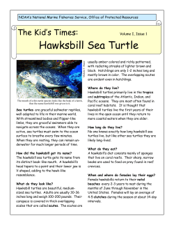


© Copyright 2026