
Why Relapse Occurs in PB Leprosy Patients After MDT Despite Mitsuda Reactivity
iNTERNATIONAL JOURNAL OF LEPROSY
^
Volume 66. Number 2
Printed in the U.S^.
(ISSN 0148-916X)
Why Relapse Occurs in PB Leprosy Patients After
Adequate MDT Despite They are Mitsuda Reactive:
Lessons from Convit's Experiment on Bacteria-Clearing
Capacity of Lepromin-Induced Granuloma'
S. Chaudhuri, S. K. Hajra, A. Mukherjee, B. Saha, B. Mazumder,
D. Chattapadhya, and Kunal Saha?
It is generally agreed that at the lepromatous pole of the leprosy spectrum there exists anergy toward Mycobacterium leprae
and an absence of protective immunity
against the parasite, resulting in unrestricted
growth of acid-fast bacilli (AFB) within
Virchowian macrophages and the development of extensive disease. On the other
hand, at the tuberculoid pole there is strong
Mitsuda positivity but incomplete protective immunity against M. leprae, permitting
its restricted multiplication and development of limited disease. Although tuberculoid (TT) and borderline tuberculoid (BT)
leprosy patients harbor few AFB, one million or less, and can mount a significant
granulomatous delayed-type hypersensitivity (DTH) reaction against the leprosy
bacilli, the tuberculoid granuloma especially within the nerve does not resolve in
some patients even after the recommended
short-term multidrug therapy (MDT) (s).
Often they stand a chance of relapsing after
discontinuation of MDT, perhaps due to
' Received for publication on 2 July 1997.
Accepted for publication in revised form on 22 April
1998.
S. Chaudhuri, M.B.B.S., D.C.P., Ph.D., Professor
of Leprology (deceased); S. K. Hajra, M.B.B.S.,
D.C.P., D.T.M.&11., Ph.D., Reader of Leprology (retired); A. Mukherjee, M.D., Ph.D., D.C.P., Professor of
Pathology and Director (retired); 13. Saha, M.D.,
D.T.M.&11., Assistant Professor; B. Mazumder,
M.B.B.S., D.D.V., Demonstrator, Department of Leprosy, School of Tropical Medicine, Calcutta, India. D.
Chattapadhya, M.D., Joint Director, National Institute
of Communicable Diseases, Delhi, India. K. Saha,
M.Sc., M.13.13.S., Ph.D. (U.S.A.), Professor of Immunology (retired), Delhi University, Delhi, India.
Reprint requests to Dr. K. Saha, 45A Soya 13zizar
Street, Calcutta 700 005, India.
182
persisting slow-growing M. Ieprae. Also
they develop nerve damage induced either
by the remnant M. leprae antigens, which
the host fails to eliminate ("), or by the
host's own antigenic determinants similar
to those of M. leprae ( 17 ).
Indeterminate leprosy is immunologically unstable. Some patients remain unchanged for years, even without treatment,
or undergo spontaneous regression, while a
small fraction, despite being Mitsuda-reactive, change their polarity mostly toward
the tuberculoid pole. Dharmendra and
Chatterjee (`') in a retrospective study have
shown that out of 524 lepromin-positive
healthy contacts, 17 (3.2%) developed tuberculoid leprosy. This indicated that their
cell-mediated immunity (CMI) was not
competent enough to eliminate all leprosy
bacilli, although they were able to form a
DTH granuloma at the lepromin injection
sites (`'). Kar and associates (") reported that
3 (6%) out of 50 indeterminate leprosy patients remained clinically active even at the
end of 1 year's treatment with MDT. Kaur
and her coworkers (I") reported inadequate
clinical response in 4.3% and 25% of her
tuberculoid and indeterminate leprosy patients, respectively, following MDT.
Why some of these patients fail to eliminate all leprosy bacilli although they are
Mitsuda positive and possess adequate CMI
against M. Ieprae is still not clearly understood. It appears that protective immunity is
the most important and under-investigated
aspect of the disease.
Keeping this in mind, in this study we attempted to investigate the nature of protective immunity in leprosy. We induced DTH
granulomas in Mitsuda-positive tuberculoid
66, 2^Chaudhuri, et al.: Differences in Iv! itsuda-Positive Individuals^183
and indeterminate leprosy patients as well
as in Mitsuda-positive healthy contacts by
intradermal injection of 6.4 x 10⁷ heatkilled M. leprae prepared from human leproma (') and studied any functional defect
of the lymphocytes and macrophages that
had infiltrated into the granulomas from the
vascular bed.
MATERIALS AND METHODS
Human materials
The study included three groups of human subjects. Group I included 52 histologically proved, tuberculoid leprosy patients.
They were all bacteriologically negative by
slit-skin-smear tests, and all were Mitsuda
positive. The lepromin test was performed
by intradermal injection of 0.1 ml standard
lepromin containing 1.6 x 10 heat-killed
M. leprae (human) per ml; 3 weeks thereafter the Mitsuda reaction was recorded (7)•
Short-term MDT was given to all patients.
Group II included 50 Mitsuda-positive,
histologically proved, indeterminate leprosy patients.
Group III consisted of 25 Mitsuda-positive healthy contacts (5 to 40 years old).
They were selected from families having
lepromatous (LL) and borderline lepromatous (BL) patients.
Laboratory tests
Three types of laboratory tests were performed.
Histological study of lepromin (megadose)-induced dermal granuloma. Briefly,
6.4 x 10⁷ heat-killed M. leprae (human)
(four times dose of a standard lepromin
test) were injected intradermally into each
subject. Six weeks thereafter biopsies were
taken from the inoculation sites, and histological sections were made and stained with
hematoxylin and eosin (H&E) to study the
histomorphology of the granuloma formed
therein (').
Capacity of clearing bacteria (CCB)
test. The CCB test was done by studying
the histological sections of the lepromin
(megadose)-induced granuloma (described
above) after Fite-Faraco staining for AFB.
The results were recorded according to the
method described by Convit and associates
('). Clearance of bacteria indicated a positive test.
Leukocyte migration inhibition (LMI)
test. Leukocyte-rich plasma samples were
obtained from each subject; cell suspensions containing 4 x 10' cells per ml of
minimal essential medium (MEM) were
used to fill the capillary tubes; these were
then incubated in migration chambers filled
with MEM and 10% fetal calf serum with
or without 1 x 10⁷ M. leprae sonicates. Areas of migration were measured and migration indices were calculated (3). A migration index below 80 indicates LMI positivity; above 80, LMI negativity.
The results of these three tests in the patients and contacts were compared.
RESULTS
The Table shows the results of the three
tests performed for the tuberculoid and indeterminate leprosy patients as well as for
the contacts. The histological characteristics
of a typical lepromin granuloma are: the
presence of Langhans' giant cells, epithelioid cells, extensive lymphoid-cell infiltrates, and circumscribed tuberculoid structure. An atypical granuloma is characterized
by the absence of macrophage differentiation, limited or no development of epithehold cells and scarce lymphoid infiltration
as well as the absence of giant cells (5)•
All 52 TT leprosy patients were Mitsuda
positive, but 6 patients had an atypical
granuloma and the remaining 46 patients
showed a typical granuloma. Fourteen IT
patients, including the above 6 patients
showing atypical granuloma and another 8
patients showing a typical granuloma,
failed to clear dead M. leprae (solid or fragmented) from the lepromin-induced granuloma (negative CCB test). Twelve patients
had a negative LMI test and the remaining
40 patients showed a positive LMI test.
Again, of the 40 LMI-positive TT leprosy
patients, 38 patients could show CCB positivity, indicating a close correlationship between the LMI and CCB tests. Curiously,
the remaining two Mitsuda-reactive TT patients were LMI positive but CCB negative.
Amazingly, of the 46 Mitsuda-positive
leprosy patients showing typical granuloma
at the lepromin injection sites, eight were
CCB negative with Lang,hans' giant cells
within granuloma showing intracellular
AFB (The Figure). Essentially all individuals were Mitsuda positive, while only a per-
184
International Journal of Leprosy 1998
Ti TAin.F.. Histological types of lepromin-induced granuloma, CCB and LM1 tests in
bacteriologically negative and Mitsuda-positive tuberculoid and indeterminate leprosy patients and in healthy contacts.
Immunological tests
Ilistological studies of
lepromin-induced granuloma.'
Leukocyte migration
inhibition (LNII) test
Capability of clearing
bacteria (CCI3) test
Positive^Negative
Typical
granuloma
with
giant cell''
Group
(no.)
I. Tuberculoid
leprosy
(52)
II. Indeterminate
(50)
III.^I lealthy
contacts
(25)
Atypical
granuloma
without
giant cell
No AFI3
within^AFI3 within
granuloma^,,ranuloma
and giant cell
Positive^Negative
(Nil <80)^(Nil >80)
No.
(4.
No.
46
88.4
6
11.6
38
73.1
14
26.9
40
77
12
23
49
98
I
2
28
56
11
44
38
76
12
24
25
100
0
0
23
92
8
23
92
No.
No.
No.
No.
8
Two types of granuloma ("typical" and "atypical') were observed at the sites of innoculation of 6.4 x 10'
heat-killed human M. lip o '.
'' A "typical granuloma had infiltrations of lymphocytes, epithehoid cells and Langhans' giant cells.
Seven "atypical'' granuloma had no Langhans' giant cell and showed AF13.
-
lion showed CCB and LMI reactivities.
Mitsuda reactivity could be a more sensitive detector of CMI but Mitsuda positivity
is not always associated with CCB and LMI
positivities.
The results of the three tests in the 50
Mitsuda-positive indeterminate leprosy patients were similar (The Table). Of these 50
patients, 49 (98%) showed a typical granuloma and only one showed atypical granuloma at the lepromin inoculation sites.
Forty-nine patients had typical and one had
atypical granulomas; 2$ (56c/0) patients
showed CCB positivity and 22 (44%) CCB
negativity (the latter showing mostly fragmented AFB even within Langhans' giant
cells).
An in vitro test was positive in 38 (76c/0)
out of the 50 indeterminate leprosy patients
but a CCB test was positive in only 28
(56%) cases which, again, indicated that all
LMI-positive patients were Mitsuda positive but not CCB positive. However, all
CCB-positive cases invariably showed Mitsuda and LMI positivities.
The results in the 25 Mitsuda-positive
healthy contacts were also similar (The
Table). All 25 Mitsuda-positive contacts
showed a typical lepromin granuloma but,
unlike the tuberculoid and indeterminate
patients, none showed an atypical granuloma. Nevertheless, two such typical giantcell granulomzis had AFB. Of these two
granulomas, one showed AFB within the
-
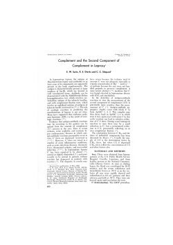
Ilu FRatkE. Langhans' giant cell with intracellular acid-fast bacilli within a "typical granuloma induced by intradermal injection of a large dose of lepromin in a "1 1' case (negative CCB test) (Fite-Faraco
x1000).
-
-
66, 2^Chaudhuri, et al.: Difierences in Mitsuda-Positive Individuals^185
Langhans' giant cells. Out of 25 contacts,
23 showed both CCB and LMI positivities,
again indicating a close correlation between
the results of these two tests. Importantly,
the bacteria-clearing competency of the lepromin-driven granuloma and lymphokine
release by specific T cells following challenge with M. leprae antigen in vitro were
impaired even in some Mitsuda-positive
healthy contacts. These data indicated that a
few Mitsuda-positive PB leprosy patients
and contacts showing typical giant-cell
granuloma induced by lepromin might not
clear a few, mostly fragmented. AFB from
the inoculation sites. Presumably they were
not resistant against M. /emit,.
.-
DISCUSSION
The study group composed of 127 subjects 152 tuberculoid (TT) patients, 50 indeterminate patients and 25 healthy contacts]
were all Mitsuda positive. They were subjected to histopathological examination of
lepromin (negative) injected for granuloma
function, and CCB and LMI tests.
Convit, et al. C) developed an in-vive
(CCB) test to find out the competency of
the macrophage to eliminate dead AFB
from the granulomas induced by intradermal injection of a megadose of lepromin in
patients suffering from various types of
leprosy. They found that Mitsuda-unresponsive LL patients formed an atypical and unprofessional granuloma, wherein macrophages not only failed to differentiate into
epithelioid and giant cells, but also showed
persistence of intracellular AFB. On the
other hand, a similar test performed in Mitsuda-positive tuberculoid leprosy patients
led to the formation of a typical and professional DTH granuloma, wherein most
macrophages underwent differentiation into
epithelioid cells and a few multinucleated
Langhans' giant cells, capable of eliminating intracellular AFB C"). It is well known
that the presence of Langhans' giant cells in
a lepromin granuloma is variable and does
not relate to the degree of CMI. Langhans'
giant cells are often absent in polar tuberculoid (TT) cases, conspicuous in subpolar
(TT, ) and small in size in borderline (BT)
leprosy cases (''). Curiously, we found both
typical and atypical granulomas at the lepromin injection sites of our Mitsuda-positive TT and indeterminate cases.
The results of the CCB tests in our 127
Mitsuda-positive patients and contacts
showed that 89 (707o) could clear AFB
from the lepromin-induced granuloma (The
Table) and obeyed Convit's rule, while the
rest did not (The Table). In their original paper, Convit, et al. (') considered the persistence of a few bacilli or bacillary debris in
some Mitsuda-positive patients as CCB
positive. However, in the present study to
avoid subjectivity persistence of fragmented acid-fast materials, although small
in number (The Figure), was taken as CCB
negative.
Also six (11%) TT patients of tile present
study, although Mitsuda-positive, formed
an atypical granuloma containing AFB
(CCB negative).
Furthermore, even giant cells within a
typical lepromin granuloma showed AFB
in 31 Mitsuda-reactive TT and indeterminate patients as well as contacts (The
Table). They were CCB negative, and of
them 26 cases showed negative LIVII tests
and the remaining five cases were LMI positive, indicating thereby that CCB and LMI
negativities may not give parallel results in
a few cases (The Table). M. leprae and their
debris persisting in these Mitsuda-positive,
CCB-negative cases even after completion
of short-term MDT might mount a cell-mediated immune response, eventually leading
to a type 1 reaction and nerve damage ('
These paradoxical findings, i.e., few Mitsuda-positive TT and indeterminate leprosy
patients forming typical and atypical granulomas at the lepromin injection sites and
showing CCB negativity and variable LMI
reactivities, are of interest. For the assessment of resistance of an individual against
M. leprae CCB positivity, an in of
protective immunity, outstrips the other two
parameters, i.e., Mitsuda reactivity, an indicator of granulomatous hypersensitivity, and
LMI positivity, a pointer of specific T-cell
responses against M. leprae in vitro (7).
It has now been established that intracellular killing of mycobacteria and their subsequent elimination involve a multistep
process; i.e., uptake and processing of AFB
by macrophages, presentation of the processed mycobacterial antigen onto the Th 1
subset of CD4 cells through MFIC molecules followed by the production of paracline gamma interferon and autocrine tu-
186^
International Journal of Leprosy^ 1998
mor necrosis factor-alpha. These stimulate
bacteria-laden macrophages to differentiate
into epithelioid cells and giant cells as well
as lymphocyte infiltration with eventful
killing and elimination of intracellular bacteria ( 2 . 1 "). The result of our LMI tests
showed that M. leprae-specific T cells in
101 (80%) of 127 of our Mitsuda-positive
patients and contacts were able to release
the migration inhibitory factor when they
were challenged with M. leprae sonicates in
vitro (LMI positivity) (The Table). However, 38 (37%) out of 101 LMI-positive
cases were CCB negative, suggesting impaired macrophage function despite having
an intact T-cell response. These findings indicate that either the LMI test is less sensitive than Mitsuda reactivity or that LMI
positivity is not an indicator of the resistance of macrophages against M. leprae ( 1 ).
However, both LMI and CCB positivities
are interdependent and need T cell help and
macrophage activation. Also, it was reported earlier that thymus-derived lymphocytes obtained from tuberculoid granulomas exhibited exaggerated activities as
evidenced by a high incorporation of
3 H-thymidine and "G-leucine ( 15 ), while the
bone-marrow-derived macrophages therein
were armed with HLA-DR (la) antigens after being activated by specific T cells (").
Thus, clearing of dead M. leprae from the
lepromin-induced granuloma demands the
T-cell-mediated activation of macrophages
and the subsequent differentiation into epithelioid and Langhans' giant cells. Atypical granulomas without any epithelioid or
giant cells were inert and could not eliminate AFB (CCB test negative), even Langhans' giant cells within typical granulomas
formed at the lepromin injection sites of 31
patients (The Table) contained AFB, showing thereby that Langhans' giant cells in
these patients have a slower digestive process than in others and perhaps were less
active.
We are tempted to postulate that differentiation of macrophages into multinucleated Langhans' giant cells and their ability
to digest and to eliminate intracellular
mycobacteria are two distinct events: the
first certainly needs T cell help, the second
needs intact macrophage function which is
perhaps genetically predetermined and associated with the Ir gene. The Ir gene is the
human equivalent to the bcg gene in mice
which is responsible for innate resistance or
susceptibility to mycobacterial infection ( 2 ).
The Ir gene controls the MHC class II antigen expression, affects respiratory burst,
and kills intracellular organisms ( 1)). An experimental proof is available. A strain of
mice susceptible to M. kin -aortal - him when
challenged with different doses of the
pathogen developed the disease at a certain
dose but showed strong skin DTH reaction
against the same pathogen ("). Thus, the
observed defective bacteria-clearing capacity of the macrophages within the tuberculoid granuloma of a patient is perhaps genetically predetermined and tends to persist
following adequate MDT. It is now known
that the bcg' allele confers resistance and is
dominant over bcg'. Also beg' macrophages
are superior to bcg' macrophages in the expression of surface markers (renamed as
natural resistance-associated macrophage
protein, NRAMP) and are associated with
the activation of toxic nitrogen and oxygen
radicals ( 2 . 21 ' 21 ). This inability of macrophages to differentiate into Langhans' giant
cells within the lepromin-driven granuloma
and their inability to clear intracellular AFB
in some of our Mitsuda-positive patients
and contacts is perhaps due to the fact that
gamma interferon released by Th 1 cells
fails to activate the human equivalent of the
kg' macrophages and to kill and release intracellular mycobacteria ( 2 'ft Is). Alternatively, defective autocrine release of tumor
necrosis factor-alpha by such macrophages
fails to cause them to mature into typical
Langhans' giant cells ( 2 ). Certainly this
needs experimental proof.
We would like to highlight that even today we do not understand the meaning of
the Mitsuda reaction, i.e., lepromin-induced
DTH granuloma. It is believed that naturally induced DTH reactivity against M.
leprae, which is life long, strongly correlates with protection ( 2 ). On the contrary, a
vaccine-induced Mitsuda positivity wanes
over the course of time ( 1 '). This difference
in the behavior of Mitsuda positivity, natural or induced, is perhaps of genetic origin
and needs to be addressed in a subsequent
study. However, the results of our present
study have shown that lepromin-induced
granuloma, typical or atypical, may or may
not eliminate AFB from the granuloma and,
66, 2^Chaudhuri, et al.: Differences in Mitsuda-Positive Individuals^187
thus, have put forward evidence that CCB
positivity is a better indicator of protective
immunity than Mitsuda positivity. These
two tests could be evaluated in PB patients
by doing a long-term follow up after therapy to determine if relapses were more frequent in the CCB-negative cases.
Very recently, Job and his coworkers (')
have reported persistence of AFB in the endoneurium of the tibial nerve of a BT leprosy patient even 21 years after adequate
antileprosy therapy. They have pointed out
that BT leprosy patients should be considered as a generalized disease and should be
given a longer duration of currently available antileprosy therapy. Such cases, if
CCB-negative, will not respond to shortterm WHO/MDT treatment and will not be
able to eliminate all AFB. We, therefore,
advocate that Mitsuda-positive but CCBnegative tuberculoid and indeterminate leprosy patients be administered low-dose
Convit vaccine plus MDT for quick clearance of bacteria, as done in Mitsuda-negafive multibacillary and paucibacillary leprosy patients (4.
SUMMARY
It is amazing how after years of scientific
research and therapeutic progress many
simple and basic questions about protective
immunity against Mrcobacterium !clime
remain unanswered. Although the World
Health Organization (WHO) has recommended short-term multidrug therapy
(WHO/MDT) for the treatment of paucibacillary (PB) leprosy patients, from time
to time several workers from different parts
of the globe have reported inadequate clinical responses in a few tuberculoid and indeterminate leprosy patients following adequate WHO/MDT despite the fact that they
are Mitsuda responsive. A few borderline
tuberculoid patients harbor acid-fast bacilli
(AFB) in their nerves for many years even
though they become clinically inactive following MDT, a fact which has been ignored
by many leprosy field workers. Keeping
these patients in mind, we have attempted
to investigate the cause of the persistence of
AFB in PB cases and have looked into the
question of why Mitsuda positivity in tuberculoid and indeterminate leprosy patients,
as well as in healthy contacts, is not invariably a guarantee for protectivity against the
leprosy bacilli. We have: a) analyzed the
histological features of lepromin-induced
granulomas, b) studied the bacteria-clearing
capacity of the macrophages within such
granulomas, and c) studied the in vitro
leukocyte migration inhibition factor released by the blood leukocytes of these subjects when M. leprae sonicates have been
used as an elicitor. The results of these three
tests in the three groups of subjects have
been compared and led us to conclude that
the bacteria-clearing capacity of the macrophages within lepromin-induced granuloma
(positive CCB test) may be taken as an indicator of the capability of elimination of
leprosy bacilli and protective immunity
against the disease. This important macrophage function is not invariably present in
all tuberculoid and indeterminate leprosy
patients or in all contacts even though they
are Mitsuda responsive and are able to
show a positive leukocyte migration inhibition (LMI) test. It is likely but not certain
that this deficit of the macrophage is genetically predetermined and persists after completion of short-term WHO/MDT. Thus,
after discontinuation of treatment slowgrowing, persisting M. leprae multiply
within macrophages leading to relapse.
RESUNIEN
Es sorprendente como despues de tantos altos de
investigacinn cientilica y de progreso teritp6utico todayia qUedell sin con testar mochas preguntas simples
y hiisicits sobre la imounidad protectora contra Ahcobucterium leprae. Aunque la OrganizaciOn Mundial
de la Sitlud (OMS) ha recomendado la poliquinnoterapia de corta doritchin apra el tratainiento de la lepra
paucihacilar (PB), todayia, de tiempo en liempo, algunos investigitdores de diclerentes panes del mond()
reportan respuestas clinicas inadectiadas en algunos
pacientes con lepra tuherculoide c indeterminada, a pesar de yeti han recibido el trationiento (PQT) apropiado y de so respuesta Mitsudit positiva. Algunos pacientes con lepra tuberculoid subpolar mantienen bacilos acido-resistentes (AFB) en sus neryios durante
muchos afios aunque Sc hayan tornado clinicamente
inactivos despues (le la PQT, on hecho que ha sido ignorado por muchos investigadores de la lepra. Manicniendo estos cases en lit mente, hemos intentado investigar la CaIlSa de la permanencia de AFI3 en los casos
PB y hemos enfocado la atenciOn a la pregunta de por
que la positividad a Mitstida en los pacientes tobercoloides, en los pacientes indeterminados y en los contactos sanos, no es invariablemente unit 1._!itrantia de
protecciOn contra el hacilo d e la leprit. Para csto hemos: a) analitado las caracteristicas histohigicits de los
I88^ International Journal of Leprosy^
granulomas inducidos con lepromina, h) estudiado la
capacidad de los macailagos de los granulomas para
depurar hacterias, y c) estudiado la liberaciiin del factor inhibidor de la migraciOn de leucocitos por los leucocitos circulantes de estos sujetos cuando se estimukin /if vitro con sonicados de M. /emit. Los resultados
comparitivos de estas 3 pruchas en los 3 grupos de individuos ban conducido a concluir que la capacidad
depuradora de hacterias de los macnifagos de los granulomas inducidos con lepromina puede tomarse comp
un indicador de la capacidad de eliminacifin del bacilo
de-la lepra y de la inmunidad protcctora contra la enferniedad. Esta importante funciOn macrofzigica no
esta invariablemente presente en todos los paciente tuherculoides c indeterminados, ni en todos los contactos
sanos aunque scan Mitsuda-positivos y capaces demostrar un prucha de inhihicion de leucocitos positiva.
Es probable que el deficit de los macrOfzigos este predetenninado geneticamente y que persista aun despues
de completarse el tratainiento (PQT) de corta duracifin
sugerido por la OMS. Asi, at descontinuar el tratamiento, los bacilos de la lepra persistentes pueden replicarse lentamente dcntro de los macnifzigos dando origen a las recaidas.
RESUME
II est surprenant de constater qu'apres tart d'an(lees de recherche scientifique et de progres thempcutiques, autant de questions simples ct de base demeurent sans reponse au sujet de l'iminunit6 protectrice
contre Mycobacterium /eprae. Ben clue l'Organisation
mondiale de la Sante (OMS) ail recommende une
polychimiotherapie (PCT/OMS) a court terme pour Ic
traitement des patients lepreux paucibacillaires (P13), it
n'en rests pas moins que plusieurs personnes a travers
Ic monde ont rapporte episodiquement des reponses
cliniques inadequates chez quelques patients tuberculotdes et indetermines, consectitivement a tine PCT
/0N1S adequate, malgre le fait que ces patients reagissent an test de N1itsuda. Quelques patients borderlines
tuberculoides presentent pendant des annees des
bacilles acido-alcolo-resistant (AAR), inimte s'ils deviennent cliniquement inactifs, tin fait qui a ete largeinent ignore par de nombreux leprologistcs de terrain.
Avcc l'histoire de ces patients en tete, nous avons tents
d'explorer la cause de persistancc de ces hacilles AAR
chez les patients PB et avons tente de savoir pourquoi
tine positivite au test de Mitsuda chez les patients tuberculoIdes ou indetermines, ainsi que les personnescontactes non alfectees, West pas necessairement tine
garantie de protection contre les bacilles de la lepre.
Nous avons:
a) analyse les caracteres histologiques des granulOmes
induits par l'injection de Lepromine.
1)) etudie la capacite d'elimination bacterienne des
macrophages provenant de tels granulômes.
c) etudie in vitro le factor ('inhibition de migration des
leucocytes secrete par les leucocytes sanguins de ces
sujets lorsque des sonicates de M. Leprae furent utilises comme stimulateurs.
1998
Les resultats de ces 3 tests chez les 3 groupcs de
sujets ont ete compares, cc qui nous a pennis de conclure que Ic test de czipacite a eliminer les bacteries des
macrophages provenant de grantilOines induits par Ic
lepromine petit etre considers comme un indicateur de
la capacite d'elimination du bacille de la lepre et de
l'immunite protectrice contre la maladie. Cctte importante function du macrophage nest pas invariablement
present chez tous les patients tuberculoIdes ou Indetermines ou chez toutes les personnes contactes meme
s'ils soot tolls positifs an test de Mitsuda et capables de
montrer un test ('inhibition de migration leucocytaire
(11‘11,) positif. II est probable, mais pas certain, que ce
deficit des macrophages est genetiquement predetermine et persiste apres l'arret de la PCT/OMS a court
tenne. Ainsi, apres l'interruption du traitement, des M.
leprue persistantes, a croissance lento, se multiplient
l'intericur des macrophages, aboutissant a des rechutes.
REFERENCES
I. BRINE, G.,
2.
3.
4.
5.
6.
7.
8.
9.
10.
11.
BARNETsoN, R. Sr. C., RIDLEY, J. S.
and KitoN \VALI., 0. Lymphocyte transformation
test in leprosy: correlation of response with inflammation of lesions. Chit. Exp. Immunol. 25
(1976) 80-94.
Cum 'Dunn, K. The immunology of leprosy: unveiling an enigma. (Editorial) Int. J. Lepr. 63
(1995) 430-447.
CliAtionmv, S., IIAJRA, S. K., SAIIA, B., MAsursfi)itt, 13., BiswAs, P. C., Cif,vrtApAnnvA, D. and
SAHA, K. An eight-year field trial on antileprosy
vaccines among high-risk household contacts in the
Calcutta metropolis. Int. J. Lepr. 62 (1994) 369-394.
CliAuniit:Ri, S..11mitA, S. K., SAHA, B. and SAILA,
K. Management of lepromin negative borderline
leprosy patients with low dose Convit vaccine as
an adjunct to multidrug therapy (a six-year follow
up study at Calcutta). Int. J. Lepr. 65 (1997) 56-62.
CoNvrr, J., AVII.A, J. L., Goifixims-YAfiR, M. and
PiNAkol, NI. E. A test for determination of competency in clearing bacilli in leprosy patients.
Bull. WHO 46 (1972) 821-826.
DIIARNIENDRA and Cii,vimtuu, K. R. Prognostic
value of the lepromin test in contacts of leprosy
cases. Lepr. India 27 (1955) 149-152.
HARBoi,, N1. The immunology of leprosy. In: Leprosy. I Listings, R. C., ed. Edinburgh: Churchill
Livingstone, 1985, pp. 53-87.
Jolt, C. K., BASKARAN, B., JAYAKUNIAR, J. and
Ascii} ion:, M. Pathologic changes in a tihial nerve
with surviving M. lepra' in a healed tuberculoid
leprosy patient. Int. J. Lepr. 65 (1997) 90-94.
KAR, P. K., JiiA, P. K. and SN11111, P. S. Indeterminate leprosy: a therapeutic evaluation. Indian J.
Lepr. 64 (1992) 163-167.
KAUR, S., SHARMA, V. K., BASAK, P. and KAUR, I.
Paucihacillary multi-druLt therapy in leprosy: 7'/ 2
years experience. Indian J. Lepr. 64 (1992) 153-161.
LoviK, M. and Gloss, 0. Repeated delayed-type
hypersensitivity reaction against Mycobacterium
66, 2^Chaudhuri, et^Diflia-ences in Milsuda-Positive Individuals^189
lepromurium antigens at the infection site do not
affect multiplication in CS!! mice. Infect. Immun.
36 (1982) 768-774.
12. M.v(1180iT, V., NIVKIIERR I., A., IlAtRA. S. K.,
S.miA, B. and SAILA, K.. Immunotherapy of far-advanced patients of lepromatous leprosy with low
dose Convit vaccine with multidrug therapy (a
Calcutta trial). Int. J. Lepr. 64 (1996) 26-36.
13. NAAFS, B. Features of relapse in paucibacillary
leprosy after multi-drug therapy. Indian J. Lepr. 67
(1995) 61-67.
14. NARAYANAN, R. B. Immunopathology ()I leprosy
granulomas: current status: a review. Leff. Rev. 59
(1988)75-82.
15. NARAYANAN, R. B. and GlimilAR, B. K. In-vitro
studies on dermal leprosy granulomas: assessment
of division and protein synthesis of cells. Acta
Leprol. 7 (1989) 13-17.
16. On EN1101E, Tom. II. M. Immunology of leprosy:
lesson front and for leprosy (Editorial). Int. J.
Lepr. 62 (1994) 108-12
17.
RANIIII:KKANA, A., Dvs, P. K., KkoR;, S., YoNG,
S., Si popl.f, I. C. and Bps, J. D. Nlycobacterial
65,000 MIA' heat-shock protein shares a clrboxyterminal epitope with human epidermal cyteberatin-1/2. Immunology 77 (1992) 267-276.
18. Riin.fv, D. S. Pathogenesis of Leprosy and Rehued Diseases. London: But
^Co. Ltd.,
1988, pp. 157-159.
19. RosEN, F. S. and GEIIN, R. S. Chronic granulomatoils disease. In: Case Studies in Immunology; A
Clinical ('omparison. London: Current Biology
Limited, 1996, pp. 67-72.
20. Sunrint, E., MAN), D., RADitocii, D., I'll ISCIINIAN.
F. MoRGAN, K., Guns, I'. and SKAMENE, K. Genetic control of innate resistance to mycobacterial
infections. Immunoparasitol. Today 12 (1991)
A42—A45.
21. Son:RR, E., MoRGAN, K., GRos, P. and SKANIENE,
F. Genetics of leprosy. Am. J. Trop. Med. Ilyg. 44
(199114-11.


© Copyright 2026