
ARTICLE COVER SHEET LWW—TECHNIQUES FLA, SF, LTE and Case Study & Review
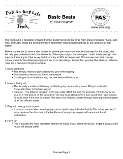
ARTICLE COVER SHEET
LWW—TECHNIQUES
FLA, SF, LTE and Case Study & Review
Article : BTK20114
Creator : CQ16
Date : 1/18/2008
Time : 15:26
Article Title :
Number of Pages (including this page) : 10
Template Version : 3.1
12/22/07
Scripts:
1. sc_Extract_Xml
2. sc_Multifig_Marker
3. Autopagination compliant
4. run_on
Copyright @ 2008 Lippincott Williams & Wilkins. Unauthorized reproduction of this article is prohibited.
Techniques in Knee Surgery 7(1):00–00, 2008
|
Ó 2008 Lippincott Williams & Wilkins, Philadelphia
T E C H N I Q U E
|
Deepening Trochleoplasty for Distal Femoral
Dysplasia in Patellar Instability
Simon T. Donell, FRCS(Orth), MD
Faculty of Health, University of East Anglia, and
Institute of Orthopaedics
Norfolk & Norwich University Hospital
Norwich, Norfolk, United Kingdom
| ABSTRACT
Awareness of the importance of the shape of the femoral
sulcus as a component of patellar instability has gained
acceptance over the last 15 years. As a result, operations
to correct the abnormality by deepening the sulcus have
been introduced. The main drive for this has been from
Europe, but is still only performed in a few specialist
centers. The indications, technique, rehabilitation, and
evaluation of results are therefore still being systematized and clarified. In this report, I describe the technique
of deepening by removing excess distal cancellous bone
and creating 2 triangular-shaped osteochondral flaps
which are then anchored with bioabsorbable screws
placed into the femoral condyles. The procedure requires
an appreciation of the abnormal geometry, but is not difficult to master, and the early results are encouraging.
Deepening of the femoral sulcus for those patients with
a significant dysplasia and symptomatic patellar instability is an operation that will become more common in the
future.
Keywords: trochleoplasty, dysplasia, patellar instability,
surgical technique
| HISTORICAL PERSPECTIVE
The importance of a shallow femoral sulcus as a component of patellar instability has been recognized since the
1890s. Elevation of the lateral trochlear facet was first described by Albee1 in 1915, but this fell out of favor because of subsequent patellofemoral osteoarthritis (OA).
In 1966, Masse2 described removing subchondral bone
and impacting the articular cartilage with a punch. This
technique was further developed by Henri Dejour in
Lyon, France, in the late 1980s.3,4 Dejour also classified
trochlear dysplasia on the lateral plain radiograph and developed the indications for his trochleoplasty.5
Address correspondence and reprint requests to Simon T. Donell,
FRCS(Orth), MD, Institute of Orthopaedics, Norfolk and Norwich
University Hospital, Colney Lane, Norwich, NR4 7UY, United
Kingdom. E-mail: [email protected].
The original Dejour technique uses a burr with a feeler
gauge attached. The gauge is placed on the chondral surface, and the burr removes subchondral bone at a set depth
giving a fixed-depth osteochondral flap. The new groove
is created by cutting the flap with an osteotome along the
desired line, impacting the 2 resulting flaps and anchoring
them with staples.3 He noted that the best results were in
those patients with instability and no pain where the trochleoplasty was part of the primary procedure.
| INDICATIONS/CONTRAINDICATIONS
The main indication for deepening trochleoplasty is
symptomatic patellar instability which has failed conservative therapy and which has a significant dysplasia with
a trochlear boss on lateral radiograph. It is not required
for the majority of patients with patellar instability. The
Dejour method for measuring the boss height is shown in
Figure 1 and requires a lateral radiograph with the posterior condyles strictly overlapping. A simpler method
uses a line drawn along the anterior cortex of the femur
and then measures a line dropped perpendicular to this
from the most anterior part of the femoral groove.
The history should establish significant functional
disability from patellar instability in a patient prepared
to undergo a major operation to control this. At least
one third of patients decline trochleoplasty during the
consent procedure. However, about half of these return
because of significant ongoing symptoms. The birth
and family history is important. Half the patients have
affected family members. Collagen disorders may be
present (eg, Marfan and Ehlers-Danlos syndromes), and
this may affect the anesthetic or operative procedure.
Physical examination should include an assessment
for hypermobility syndrome. This is present to some degree in more than half the patients and is associated with
a positive family history. The torsional alignment of the
lower limb is also important, noting hip version and tibial
torsion. From time to time, rotational osteotomies may
be needed to reorientate the extensor mechanism over
the distal femur. Trochleoplasty alone will not do this.
Volume 7, Issue 1
Copyright @ 2008 Lippincott Williams & Wilkins. Unauthorized reproduction of this article is prohibited.
1
F1
Donell
to the Dejour method (Fig. 3), with C and D being suitable for deepening trochleoplasty. The scans can also be
used to assess the patellar tilt angle and tibial tubercleYtrochlear groove distance. Correcting patellar maltracking is an important part of the procedure and may
require distal or proximal realignment operations according to the surgeon’s preference.
| SURGICAL TECHNIQUE
FIGURE 1. Lateral radiograph showing the Dejour method for calculating the boss height. Point ‘‘a’’ is the junction
between the posterior cortex and articular cartilage. Boss
height is measured between points ‘‘b’’ and ‘‘c.’’
Patellar tracking should be noted. With a dome-shaped
boss greater than 10 mm, the patella may show a ‘‘bayonet’’ tracking. When the knee is fully flexed, the boss can
be palpated directly through the quadriceps tendon.
Many patients in whom trochleoplasty is indicated
have had multiple previous procedures. The resulting
scars can be a problem if narrow skin bridges are created. Advice from a plastic surgeon may be needed.
Tightness of the quadriceps muscle may be present,
especially of there is a patella alta. Quadriceps dysplasia
with absence of the vastus medialis obliquus is almost
universal and can affect the postoperative course. Without decent quadriceps function, the symptoms of instability will persist.
The operation is performed under epidural analgesia,
which is also used for postoperative pain relief. A light
general anesthetic is usually added. Postoperative pain
control is critical otherwise early full motion cannot be
achieved and stiffness may result.
A single dose of a third-generation cephalosporin is
administered intravenously at induction, for example,
1.5 g cefuroxime. Chemical thromboprophylaxis is
not routinely given. A calf stimulator is applied to
the opposite leg during the operation. Cross-matching
blood for transfusion is not needed.
The patient is placed supine with the knee flexed at
90 degrees, held with foot bolster and thigh support. A
preliminary arthroscopy through anterolateral and superolateral portals is useful for assessing patellar tracking
and confirming that the tibiofemoral joint is normal.
Any loose bodies may be removed. An open arthrotomy
is performed through a midline longitudinal incision
commencing just medial to the patellar tendon at the
level of the tibiofemoral joint line progressing proximally and slightly laterally to the middle of the quadriceps
tendon. The proximal extent depends on the thickness
of the subcutaneous fat. A routine medial parapatellar
arthrotomy is performed. The incision is deepened
through the subcutaneous and prepatellar fascial layers
| PREOPERATIVE PLANNING
F2
Standard plain radiographs including a strict lateral and
skyline (tangential patella) are essential. The lateral
radiographs allow assessment of the degree of dysplasia.
The boss height should be measured (Fig. 2). Although a
height greater than 6 mm suggests suitability for trochleoplasty, consideration should be given to the size
of the patient. In a small female, this would represent a
greater dysplasia than a tall male. In the former, 4 mm
would be adequate to consider the procedure.
Further imaging with computed tomography (CT)
scan, or better, magnetic resonance imaging scan, helps
assess the shape of the groove in the first 20 degrees of
flexion. The CT scan shows the subchondral bone contour; the magnetic resonance imaging scan, the chondral
shape. The scan can be taken with the knee in extension
and the quadriceps relaxed. Coronal cuts through the distal femoral shaft down to the distal end of the tibial tubercle are ideal. The groove can be classified according
2
FIGURE 2. Lateral radiograph showing the author’s
method for calculating the boss height. The line ‘‘aYb’’ is
at right angles to the line of the anterior femoral cortex.
The boss height is the distance between ‘‘a’’ and ‘‘b.’’
Techniques in Knee Surgery
Copyright @ 2008 Lippincott Williams & Wilkins. Unauthorized reproduction of this article is prohibited.
F3
Deepening Trochleoplasty
FIGURE 3. Coronal section showing the Dejour classification of trochlear dysplasia on CT scans. A is a shallow groove, B
is flat, C is dome-shaped, and D shows a medial ‘‘cliff-face.’’
in the line of the incision and should pass onto the medial part of the patella. This will allow double-breasting
medial reefing at the end or a medial patellofemoral ligament reconstruction should it be needed. Skin flaps are
not developed. The approach continues through the
quadriceps tendon and medial retinaculum. Care should
be taken to avoid incising the articular cartilage. The patella is inverted and inspected. Any medial ossicle is removed, and any articular damage addressed. The patella
is then reverted and retracted into the lateral gutter.
The trochlea is then inspected, and the dysplasia
confirmed (Fig. 4). The synovium attached to the distal
FIGURE 4. The dysplastic trochlea of a left knee, seen
looking toward the hip.
FIGURE 5. The proximal femur has been exposed, seen
from lateral side.
Volume 7, Issue 1
Copyright @ 2008 Lippincott Williams & Wilkins. Unauthorized reproduction of this article is prohibited.
3
F4
Donell
femoral articular cartilage, along with the supracondylar
fat pad, is stripped off the bone using sharp dissection
and a small periosteal elevator. It is important to expose
the anterior cortex of the distal shaft of the femur (Fig.
5). This defines how much bone is to be removed. The
object is to create a groove at the level of the anterior
femoral cortex. The synovium is held retracted with
stay sutures.
Following this, the osteochondral flaps are marked
forming 2 triangles (Fig. 6). The objective is to obtain a
lateral buttress with a depressed medial flap. This is
achieved by marking the lateral flap with an acute line,
and the medial flap with a more obtuse one. The groove
line should pass more laterally than the original. This
helps patellar tracking and reduces the trochlear groove
part of the tibial tubercleYtrochlear groove distance. It is
important that the lateral flap is wide enough to take a
countersunk 3.5-mm screw. The apex of the flaps should
be close to the roof of the intercondylar notch. This allows
the whole length of the groove to be deepened.
The distal femoral cortex is then removed between
the marked flaps with an osteotome. This is shaped in
a triangular fashion with a more marked slope laterally
(Fig. 7). The horizontal component runs along the edge
of the chondral margin. The proximal cancellous bone
can be removed at the same time. Any bone removed
during the procedure should be saved, as it will be
used as graft at the end of the procedure to plug any
defects under the flaps.
A trench is then formed in the shape of a rhomboid,
which matches the marked flaps. This is most easily
achieved using a 4.5-mm drill passed in a fanlike manner
down to the apex. The index finger is placed at this
point, and the drill is passed until it is felt just under
the cartilage (Fig. 8). Broaching the articular cartilage
is not a significant problem and can be ignored. Any
bone between the passes of the 4.5-mm drill is then removed with rongeurs. A large rhomboid osteochondral
flap is now formed.
The new trochlear groove will be produced by
cutting along the marked groove line with an osteotome. If the flaps are now depressed, the cartilage will
crack open along the flap lines. The flaps are therefore
FIGURE 7. The distal femoral cortex is cut with an
osteotome in a V shape, seen from above.
FIGURE 8. A 4.5-mm drill is passed to the apex of the
marked flaps to produce a subchondral trench, seen from
lateral side.
FIGURE 6. The flaps marked out, seen from lateral side.
F5
F6
4
Techniques in Knee Surgery
Copyright @ 2008 Lippincott Williams & Wilkins. Unauthorized reproduction of this article is prohibited.
F7
F8
Deepening Trochleoplasty
F9
FIGURE 9. To allow the flap to be depressed, a gap is
fashioned with rongeurs along the flap edge until only the
distal flat part is left.
FIGURE 11. The flaps have been depressed and fixed
with absorbable screws, seen from above. Note that the
proximal groove widens.
undermined with a drill and/or burr. This can be visualized directly by using the arthroscope. The flap lines
are also transilluminated, confirming that adequate
bone has been removed. Slots are also made at the
proximal ends of each flap line with a rongeur. This
allows the flaps to fold down (Fig. 9).
To cut the groove, place an instrument such as the
rongeurs under the flap, and then cut with an osteotome.
The instrument will avoid inadvertent complete detach-
ment of a flap by its sudden collapse (Fig. 10). To obtain
the correct position with the 2 flaps making a smooth
groove, further targeted undermining of the subchondral
or cortical bone may be necessary, again aided by direct
visualization with the arthroscope. If a flap is unstable
(typically, the medial flap), it should be placed in the
correct position, and bone graft packed under it. The
flap should be checked to be stable, otherwise it will displace when using the tap for the absorbable anchoring
screw. Getting the tap through the subchondral bone
requires significant force, which easily displaces an unstable flap.
Once each flap position is satisfactory, each one
is secured with a countersunk 3.5 000D7 35-mm
FIGURE 10. The new groove is cut with an osteotome.
The rongeurs have been placed to protect the osteochondral flaps from inadvertent displacement. The view is from
the medial side.
FIGURE 12. Before and after the trochleoplasty, seen
looking toward the hip.
Volume 7, Issue 1
Copyright @ 2008 Lippincott Williams & Wilkins. Unauthorized reproduction of this article is prohibited.
5
F10
Donell
fascia is closed with continuous 2-0 Vicryl, and the
skin with subcuticular 3-0 Vicryl and SteriStrip. No
drain is used.
The wound is then covered with a clear waterproof
dressing, wool, and crepe bandage.
AQ1
| RESULTS
FIGURE 13. The synovium and periosteum are sutured
back to the articular edge, seen from above.
F11
F12
F13
SmartScrew (Linvatec, Finland). This screw is bioabsorbable and can be trimmed with a scalpel if an edge
is prominent. The screws need to be placed at the midpoint of the flap and directed into their respective femoral condyles (Fig. 11). If placed too distal, the proximal
end of the flap will not be secure. If placed too proximal,
the distal end of the flap may rotate, causing incongruency in the new groove. The new profile should
show a lateral flare (Fig. 12).
Any gaps under the flaps are packed with bone graft.
The proximal groove may also be packed but not above
the level of the subchondral bone plate. Any exposed
cancellous bone proximal to the articular surface is
sealed with bone wax. The synovium and supracondylar
fat pad are reattached to the articular edge using interrupted 2-0 Vicryl sutures (Fig. 13).
Patellar tracking should now be assessed. Any maltracking should be corrected using the surgeon’s preferred method. Unless the dysplasia is severe, a lateral
release is not typically needed, and closure with a double-breasted medial reefing is often sufficient. In severe
cases, a formal medial patellofemoral ligament reconstruction may be necessary.
Closure of the wound should include, if possible,
closure of the synovium with a continuous layer of 2-0
Vicryl. The extensor mechanism is repaired with interrupted 2-0 Vicryl along the medial border of the patella
and the distal quadriceps tendon. The whole of the medial retinaculum and quadriceps tendon is then closed with
a continuous 2-0 Vicryl. A double-breasting technique
may be used if appropriate.
If possible, the preretinacular fascia should be
closed with continuous 2-0 Vicryl. The subcutaneous
6
Trochleoplasty is in its infancy. My current series is
more than 55 patients over 13 years, with 18 performed
in 2006. The referral base is national. The early results of
this technique have been reported.6 In 15 patients with
17 knees treated and an average 3-year (range,
1Y9 years) follow-up, the boss height was reduced from
an average 7.5 mm (range, 6Y10 mm) to 0.7 mm (range,
j1 to 3 mm). Normal tracking was achieved in
11 knees, the rest having slight J-shaped tracking. Mild
residual apprehension was present in 7 knees. Seven
patients were very satisfied, including two who played
soccer and described their knees as normal. Six were satisfied, and 2 were disappointed. The average Kujala
score7 improved from 48 (range, 13Y75) to 75 (range,
51Y98). The disappointed patients had pain as a feature
both preoperatively and postoperatively, including one
who had presented late with a missed osteochondral fracture of the patella.
In Dejour’s series,3 32 patients underwent 40 trochleoplasties. Twenty-seven knees were subjectively
satisfied or very satisfied, with 36 achieving patellar stability postoperatively. Verdonk et al8 have also reported
on 7 patients with patellar instability out of 12 patients
(13 knees) using Dejour’s technique. The other patients
had retropatellar pain. Their results were poor, but this
was significantly influenced by the latter patients.
The results of the Bereiter trochleoplasty have also
been recently reported.9,10 This is also a deepening trochleoplasty, but the osteochondral flap is thin and flexible. It is suitable in patients in whom the articular surface
is normal, without chondral defects.
| COMPLICATIONS
The most important complication to avoid is stiffness.
Complete pain control and early movement help this.
After discharge, supervised sessions with a physiotherapist are important in patients who need encouragement.
Many patients have had multiple previous procedures
and incisions. Poor blood supply and early full movement can lead to wound breakdown. Advice from a plastic surgeon preoperatively can be helpful. Otherwise, if
there is concern for the wound, reducing knee flexion
may be needed. If this results in stiffness, arthroscopic
arthrolysis may be needed at 6 weeks.
Patellar pain and crepitus arises postoperatively if
the patellar articular surface is damaged. In fact, if the
Techniques in Knee Surgery
Copyright @ 2008 Lippincott Williams & Wilkins. Unauthorized reproduction of this article is prohibited.
AQ2
Deepening Trochleoplasty
patella is normal, the subjective and functional results
are very good. If not, the patient will notice crepitus
and may have mild anterior knee pain. However, patients feel that the knee is stronger and are pleased
with the result.
Some patients experience recurrent instability. A few
patients have patellar maltracking after the procedure.
Instability usually results from poor quadriceps control.
Often this is compounded by pre-existing quadriceps
dysplasia, with absence of the vastus medialis obliquus.
Overweight patients are particularly prone to poor quadriceps function. In the occasional patient who has maltracking, this has been because only a medial reefing
was performed to control patellar tracking in addition
to the trochleoplasty. In full extension, the patella falls
laterally and in flexion does not engage the new groove
properly. This is easily corrected with a medial patellofemoral ligament reconstruction using a free hamstring graft.
| POSTOPERATIVE MANAGEMENT
A continuous passive motion (CPM) machine is applied in the recovery room. The knee should be moved
as fully as possible, although it will initially be restricted by the bandages. Continuous passive motion is
continued until adequate pain control is achieved with
oral analgesics. Over the next 2 to 5 postoperative
days, the epidural dose is titrated against the oral analgesics. When the pain is controlled orally, the epidural
is discontinued.
The knee bandage is removed on the first postoperative day, and the knee redressed. Ice is applied intermittently to help reduce any swelling. The CPM need not be
on continuously. Once oral pain control is achieved, the
patient is mobilize full weight bearing, and full knee motion encouraged. Crutch support is required until quadriceps control is achieved.
By 6 weeks, most patients are walking free of support, and by 3 months, most have fully recovered.
| CONCERNS AND THE FUTURE
Both patients and clinicians have a number of concerns
and questions.
Chondrolysis
Chondrolysis has never occurred. Articular cartilage is
avascular, and keeping thick flaps preserves the subchondral bone and theoretically allows early blood supply through the cancellous bone.
Osteoarthritis
Subsequent OA is recognized as a late sequela of patellar stabilization surgery. In the early patients in this se-
ries, the osteochondral flaps were secured with metallic
screws. These were removed arthroscopically at 1 year.
The groove and drill holes had all filled in. At this
stage, it is often not possible to distinguish between
fibrocartilage and original hyaline cartilage macroscopically. No evidence of articular cartilage damage was
found. It is hoped that the rate of OA after patellar stabilization will decrease as a result of adding in a
trochleoplasty, because this reduces the joint reaction force.
Anterior Knee Pain and Trochlear Dysplasia
Trochleoplasty for anterior knee pain has been tried with
limited success. In established patellofemoral OA, it has
a place, being akin to an upper tibial osteotomy in tibiofemoral OA. Subsequent conversion to a patellofemoral
joint arthroplasty can then be performed as a fallback
procedure.
Return to Sports
About 20% of patients return to playing significant level
sports, including soccer. One patient, a sports teacher,
underwent autologous chondrocyte implantation (ACI)
on the lateral femoral condyle, but, on the tibial, not
the trochlear surface. This was as a result of a new injury
while playing basketball following trochleoplasty.
Operative Time
The operative time is between 90 and 120 minutes,
depending on whether a medial patellofemoral ligament
reconstruction is needed.
The technique and indications are still evolving. Ideally, the 3-dimensional shape of each groove should be
analyzed and then programmed through a computer to
define the new shape. An arthroscopic procedure using
navigation could then be used to produce the new groove
with a precise geometry. Development of this is unlikely,
as the commercial benefits would be outweighed by the
setup costs. However, the key area for improvement is
managing the damaged articular surface of the patella
when present. Current techniques of ACI would require
another major operation in a recovering knee. An offthe-shelf resurfacing matrix would change this. There is
no doubt that many patients labeled ‘‘chondromalacia patellae’’ in fact had an unrecognized trochlear dysplasia
with a traumatic injury to the cartilage. Furthermore,
some of the poor results from ACI on the patella may
be for the same reason. This area is ripe for research
and development.
| REFERENCES
1. Albee FH. The bone graft wedge in the treatment for
habitual dislocation of the patella. Med Rec. 1915;88:
257Y259.
Volume 7, Issue 1
Copyright @ 2008 Lippincott Williams & Wilkins. Unauthorized reproduction of this article is prohibited.
7
Donell
2. Masse Y. La trochleoplastie. Rev Chir Orthop. 1978;
64:3Y17.
7. Kujala UM, Jaakkola LH, Koskinen SK, et al. Scoring of
patellofemoral disorders. Arthroscopy. 1993;9:159Y163.
AQ3
3. Reynaud P. Les trochleoplastiesVcreusement. 8emes J
Lyon Chir Genou. 1995:176Y190.
AQ4
4. Dejour H, Neyret P, Walch G. Factors in patellar
instability. In: Aichroth PM, Dilworth Cannon W, eds.
Knee Surgery Current Practice. Martin Dunitz Ltd.
1992:403Y412.
8. Verdonk R, Jansegers E, Stuyts B. Trochleoplasty in
dysplastic knee trochlea. Knee Surg Sports Traumatol
Arthrosc. 2005;13:529Y533.
5. Dejour H, Walch G, Neyret Ph, et al. Dysplasia of the
femoral trochlea. Rev Chir Orthop. 1990;76:45Y54.
6. Donell ST, Joseph G, Hing C, et al. Trochleoplasty:
for dysplasia of the femoral sulcus. Knee. 2006;13:
266Y273.
8
9. Schottle PB, Fucentese SF, Pfirrmann C, et al. Trochleoplasty for patellar instability due to trochlear dysplasia: a
minimum 2-year clinical and radiological follow-up of 19
knees. Acta Orthop. 2005;76:693Y698.
10. von Knoch F, Bo¨hm T, Bu¨rgi ML, et al. Trochleoplasty for
recurrent patellar dislocation in association with trochlear
dysplasia: a 4- to 14-year follow-up study. J Bone Joint
Surg Br. 2006;88-B:1331Y1335.
Techniques in Knee Surgery
Copyright @ 2008 Lippincott Williams & Wilkins. Unauthorized reproduction of this article is prohibited.
AUTHOR QUERIES
AUTHOR PLEASE ANSWER ALL QUERIES
AQ1 0 Please provide the name and location of the manufaturer of SteriStrip.
AQ2 0 W6 to 1 mmW was changed to W6 to 10 mm.W Please check if correct.
AQ3 0 Please check journal title if correct (W8emes Journees Lyonnaises de Chirurgie
de GenouW).
AQ4 0 Please provide publisher location.
END OF AUTHOR QUERIES
Copyright @ 2008 Lippincott Williams & Wilkins. Unauthorized reproduction of this article is prohibited.
Author Reprints
For
Rapid Ordering
go to: www.lww.com/periodicals/author-reprints
Techniques in Knee Surgery
Order
Author(s) Name
Title of Article
*Article #
______
*Publication Mo/Yr
*Fields may be left blank if order is placed before article number and publication
month are assigned.
Quantity of Reprints ____ $
Reprint Pricing
Shipping
100 copies = $375.00
Covers (Optional)
$
200 copies = $441.00
$5.00 per 100 for
orders shipping
within the U.S.
Shipping Cost
$
Reprint Color Cost
$
Tax
$
Total
$
300 copies = $510.00
500 copies = $654.00
$20.00 per 100 for
orders shipping
outside the U.S.
Covers
Tax
400 copies = $585.00
$108.00 for first 100
copies
U.S. and Canadian
residents add the
appropriate tax or
submit a tax exempt
form.
$18.00 each add’l 100
copies
Reprint Color
($70.00/100 reprints)
You may have included color figures in your article. The costs to publish those will
be invoiced separately. If your article contains color figures, use Rapid Ordering
www.lww.com/periodicals/author-reprints.
Prices are subject to
change without
notice.
contact: Kimberly Chek,
VISA
Account #
Payment must be
received before
reprints can be
shipped. Payment is
accepted in the form
of a check or credit
card; purchase orders
are accepted for
orders billed to a
U.S. address.
For quantities over 500
Payment
MC
Use this form to
order reprints.
Publication fees,
including color
separation charges
and page charges will
be billed separately,
if applicable.
Discover
/
/
American
Express
Exp. Date
Name
ph. 410-528-4227 or
e-mail Kimberly.Chek@
wolterskluwer.com
Outside the U.S. call
4420.7981.0700
MAIL
Address
Dept/Rm
City
State
Zip
Country
Telephone
your order to:
Lippincott Williams &
Wilkins
Author Reprints Dept.
351 W. Camden St.
Baltimore, MD 21201
FAX:
Signature
410.528.4434
For questions
regarding reprints or
publication fees,
Ship to
E-MAIL:
reprints@wolters
kluwer.com
Name
Address
Dept/Rm
City
State
Zip
Country
Telephone
For
Rapid Ordering
go to: www.lww.com/periodicals/author-reprints
Copyright @ 2008 Lippincott Williams & Wilkins. Unauthorized reproduction of this article is prohibited.


© Copyright 2026