
New Techniques for Sample Preparation in Analytical Chemistry Zeki Altun
Faculty of Technology and Science
Chemistry
Zeki Altun
New Techniques for Sample
Preparation in Analytical
Chemistry
Microextraction in Packed Syringe (MEPS)
and Methacrylate Based Monolithic Pipette Tips
DISSERTATION
Karlstad University Studies
2008:4
Zeki Altun
New Techniques for Sample
Preparation in Analytical
Chemistry
Microextraction in Packed Syringe (MEPS)
and Methacrylate Based Monolithic Pipette Tips
Karlstad University Studies
2008:4
Zeki Altun. New Techniques for Sample Preparation in Analytical Chemistry - Microextraction in Packed Syringe (MEPS) and Methacrylate Based Monolithic Pipette Tips
DISSERTATION
Karlstad University Studies 2008:4
ISSN 1403-8099
ISBN 978-91-7063-161-0
© The author
Distribution:
Faculty of Technology and Science
Chemistry
SE-651 88 Karlstad
SWEDEN
+46 54-700 10 00
www.kau.se
Printed at: Universitetstryckeriet, Karlstad 2008
Abstract
Sample preparation is often a bottleneck in systems for chemical analysis. The
aim of this work was to investigate and develop new techniques to address
some of the shortcomings of current sample preparation methods. The goal has
been to provide full automation, on-line coupling to detection systems, short
sample preparation times and high-throughput.
In this work a new technique for sample preparation that can be connected online to liquid chromatography (LC) and gas chromatography (GC) has been
developed. Microextraction in packed syringe (MEPS) is a new solid-phase
extraction (SPE) technique that is miniaturized and can be fully automated. In
MEPS approximately 1 mg of sorbent material is inserted into a gas tight
syringe (100-250 µL) as a plug. Sample preparation takes place on the packed
bed. Evaluation of the technique was done by the determination of local
anaesthetics in human plasma samples using MEPS on-line with LC and
tandem mass spectrometry (MS-MS). MEPS connected to an autosampler was
fully automated and clean-up of the samples took about one minute. In
addition, in the case of plasma samples the same plug of sorbent could be used
for about 100 extractions before it was discarded.
A further aim of this work was to increase sample preparation throughput. To
do that disposable pipette tips were packed with a plug of porous polymer
monoliths as sample adsorbent and were then used in connection with 96-well
plates and LC-MS-MS. The evaluation of the methods was done by the analysis
of local anaesthetics lidocaine and ropivacaine, and anti-cancer drug roscovitine
in plasma samples. When roscovitine and lidocaine in human plasma and water
samples were used as model substances, a 96-plate was handled in about two
minutes. Further, disposable pipette tips may be produced at low cost and
because they are used only once, carry-over is eliminated.
3
To my Parents; “scholarship and books are
the building blocks of a better world”.
5
Abbreviations
γ-MAPS
ACN
AIBN
ATP
BMA
BP
BPO
C2
C8
C18
CA
Cdks
CEC
COC
DNA
EGDMA
EMI
ESI
GC
GMA
HPLC
I.D.
I.S.
k´
LC
LLE
LPME
MEPS
MeOH
MMA
MS
MS-MS
Mw
PC
PDMS
PMMA
PP
PPX
Q
RNA
RP-LC
SBSE
SEM
SME
SPDE
SPE
SPME
SRM
t0
tr
UV
6
tri((methoxysilyl)propyl)methacrylate
Acetonitrile
2,2’-Azobis(2-methylpropionitrile)
Adenosine Triphosphate
Butyl Methacrylate
Benzophenone
Benzoyl Peroxide
Ethyl Silica
Octyl Silica
Octadecyl Silica
Contact Angle
Cyclin Dependent Kinases
Capillary Electrochromatography
Cyclic Olefin Copolymer
Deoxyribonucleic Acid
Ethylene Glycol Dimethacrylate
Electron Membrane Isolation
Electrospray Ionization
Gas Chromatography
Glycidyl Methacrylate
High Performance Liquid Chromatography
Inner Diameter
Internal Standard
Retention Factor
Liquid Chromatography
Liquid-Liquid Extraction
Liquid Phase Microextraction
Microextraction in Packed Syringe
Methanol
Methyl Methacrylate
Mass Spectrometry
Tandem Mass Spectrometry
Molecular Weight
Polycarbonate
Polydimethylsiloxane
Poly(methyl methacrylate)
Polypropylene
Pipecoloxylidide
Quadrupole
Ribonucleic Acid
Reversed-Phase Liquid Chromatography
Stir Bar Sorbtive Extraction
Scanning Electron Microscopy
Supported Membrane Extraction
Solid-Phase Dynamic Extraction
Solid-Phase Extraction
Solid-Phase Microextraction
Selected Reaction Monitoring
Retention Time Unretained Analyte
Retention Time Retained Analyte
Ultra Violet
List of Papers Included in the Thesis
I
Microextraction in Packed Syringe (MEPS) for Liquid and Gas
Chromatography Applications. Part II - Determination of
Ropivacaine and its Metabolites in Human Plasma Samples Using
MEPS with Liquid Chromatography/Tandem Mass Spectrometry.
Mohamed Abdel-Rehim, Zeki Altun and Lars G. Blomberg, J. Mass
Spectrom. 39 (2004) 1488-1493.
II
New Trends in Sample Preparation: On-line Microextraction in
Packed Syringe (MEPS) for LC and GC Applications. Part III Determination and Validation of Local Anaesthetics in Human
Plasma Samples Using a Cation-exchange Sorbent, and MEPS-LCMS-MS.
Zeki Altun, Mohamed Abdel-Rehim and Lars G. Blomberg, J. Chromatogr.
A 813 (2004) 129-135.
III Increasing Sample Preparation Throughput Using Monolithic
Methacrylate Polymer as Packing Material for 96-Tips: 2 Minutes
per 96-Well Plate.
Zeki Altun, Lars G. Blomberg and Mohamed Abdel-Rehim, J. Liq.
Chromatogr. & Relat. Technol. 29 (2006) (10) 1477-1489.
IV
Surface Modified Polypropylene Pipette Tips Packed with a
Monolithic Plug of Adsorbent for High Throughput Sample
Preparation.
Zeki Altun, Anette Hjelmström, Mohamed Abdel-Rehim and Lars G.
Blomberg, J. Sep. Sci. 30 (2007) 1964-1972.
V
Some Factors Effecting the Performance of the Microextraction in
Packed Syringe (MEPS).
Zeki Altun, Lars I. Andersson, Lars G. Blomberg, Mohamed AbdelRehim, Anal. Chim. Acta (2008) Submitted.
Paper I-IV: The author was responsible for the experimental work, except
analysis and contact angle measurements, and for writing most of the paper.
Paper V: The author was responsible for the experimental work and for writing
the paper.
7
Papers/manuscripts not included in this thesis
I
Use of Carbon Dioxide and Ammonia as Nebulizer Gases in Mass
Spectrometry.
Zeki Altun, Mohamed Abdel-Rehim, Rapid Comm. Mass Spectrom. 16 (2002)
738-739.
II
Drug Screening Using Microextraction in a Packed Syringe (MEPS)
/ Mass Spectrometry Utilizing Monolithic-, Polymer-, and SilicaBased Sorbents.
Zeki Altun, Lars G. Blomberg, Eshwar Jagerdeo and Mohamed AbdelRehim, J. Liq. Chromatogr. & Relat. Technol. 29 (2006) (6) 829-839.
III Microextraction in Packed Syringe Online with Liquid
Chromatography-Tandem Mass Spectrometry: Molecularly
Imprinted Polymer as Packing Material for MEPS in Selective
Extraction of Ropivacaine from Plasma.
Mohamed Abdel-Rehim, Lars I. Andersson, Zeki Altun and Lars G.
Blomberg, J. Liq. Chromatogr. & Relat. Technol. 29 (2006) (12) 1725-1736.
IV
8
Evaluation of Monolithic Packed 96-Tips for Solid-Phase
Extraction of Local Anaesthetics from Human Plasma for
Quantitaion by Liquid Chromatography Tandem Mass
Spectrometry.
Zeki Altun, Anette Hjelmström, Lars G. Blomberg and Mohamed AbdelRehim, J. Liq. Chromatogr. & Relat. Technol. In press.
Table of Contents
List of Papers Included in the Thesis ................................................................. 7
Papers/manuscripts not included in this thesis............................................................... 8
1 Introduction...................................................................................................... 11
1.1 Sample Preparation...................................................................................................... 11
1.2 Commonly Used Sample Preparation Techniques ................................................. 11
1.2.1 Liquid-Liquid Extraction (LLE) ........................................................................... 11
1.2.2 Solid-Phase Extraction (SPE) ................................................................................. 12
1.2.2.1 Sample Clean-up Procedure in SPE.................................................................. 14
1.3 Trends in Sample Preparation.................................................................................... 15
1.4 Microextraction in Packed Syringe (MEPS) ............................................................ 17
1.5 Pipette Tips SPE.......................................................................................................... 18
1.6 Polymer Monoliths ...................................................................................................... 19
1.6.1 Methacrylate Based Porous Polymer Monoliths.......................................................... 21
1.7 Separation Techniques ................................................................................................ 23
1.7.1 Liquid Chromatography (LC) .................................................................................. 23
1.8 Mass Spectrometry (MS)............................................................................................. 24
1.9 Surface Modification of Polypropylene (PP) ........................................................... 25
1.9.1 Contact Angle (CA) Measurement ........................................................................... 27
1.9.2 Scanning Electron Microscopy (SEM) ...................................................................... 28
1.10 Analytes....................................................................................................................... 28
1.10.1 Local Anaesthetics.................................................................................................. 28
1.10.2 Roscovitine and Olomoucine .................................................................................... 29
2 The Aim of This Work ..................................................................................... 31
3 Results and Discussions ..................................................................................32
3.1 MEPS Considerations (Papers I, II and V).............................................................. 32
3.2 Method Development (Papers I, II and V) ............................................................. 33
3.2.1 The Sorbent.............................................................................................................. 33
3.2.2 The Washing Solvent................................................................................................ 36
3.2.3 The Elution Solvent.................................................................................................. 38
3.3 Carry-over ..................................................................................................................... 41
3.4 MEPS Reproducibility (Paper V) .............................................................................. 42
3.5 High Sample Throughput Using Monolithic Packed Tips (Paper III-IV) .......... 44
3.5.1 The Preparation of Porous Polymer Monoliths........................................................... 45
3.5.2 Surface Modification of Polypropylene (PP)................................................................ 47
3.5.2.1 Photoinduced Grafting....................................................................................... 47
3.5.2.2 Surface Modification by Grafting....................................................................... 50
3.5.3 Synthesis of Monoliths for Pipette Tips...................................................................... 51
3.5.4 Sampling .................................................................................................................. 53
3.5.5 Selectivity.................................................................................................................. 54
4 Conclusions and Future Aspects .....................................................................57
Acknowledgements.............................................................................................59
References...........................................................................................................60
9
1 Introduction
1.1 Sample Preparation
Analytes of environmental or biological origin usually occur in complex
matrices. Most of analytical instruments cannot handle such sample matrixes
directly. Cleaning up of such samples, isolation of analytes of interest and
enrichment of the analyte to a suitable concentration level may be required
before instrumental analysis. Sample preparation is the series of steps required
to prepare a sample in a suitable form for analysis. The faster these steps can be
done, the more quickly the analysis will be completed. The procedure must be
reproducible with high recovery of the analytes.
Historically, sample preparation has been considered not as a part of the
analytical process, rather the “procedure” that had do be done to develop and
perform analytical methods [1]. The technology has been crude and of low tech.
However, the significance of the sample preparation for the total analytical
performance is nowadays widely recognized. The final results of the
experiments depend on the starting conditions.
An ideal sample preparation method should involve a minimum number of
working steps, be easy to learn, be environmentally friendly and be economical
[2]. Further, as the number of samples grows high-throughput and fully
automated analytical techniques becomes required.
1.2 Commonly Used Sample Preparation Techniques
1.2.1 Liquid-Liquid Extraction (LLE)
Liquid-liquid extraction (LLE) is one of the oldest and most widely used sample
preparation techniques. In LLE separation of a sample is based on distribution
between two immiscible liquid phases. The classical form of LLE is using a
separatory funnel for separating liquid phases from each other. LLE involves
mixing an immiscible organic solvent with an aqueous solvent (e.g. plasma,
11
urine, serum) to extract the analyte into the organic phase. The organic phase
may be transferred, evaporated to dryness and reconstituted prior to analysis.
Compared to other sample preparation techniques LLE has advantages such as
large sample capacity and direct analysis after concentration of the clean organic
extract. Disadvantages of the classical LLE approach are that it is labor
intensive, difficult to automate and uses large volume of expensive/ and
environmentally harmful organic solvents [3]. To phase out these disadvantages,
some modern approaches to classical LLE have appeared during the past 10
years. Examples of modern approaches to LLE are single drop-liquid phase
microextrection (LPME) [4], liquid phase microextrection (LPME) [5-8] and
supported membrane extraction (SME) [9-13]. Single drop-LPME is based on a
drop of organic solvent hanging at the end of a syringe needle. As the organic
droplet is placed in the aqueous sample, based on passive diffusion, the analytes
are extracted into the droplet. After extraction, the droplet is withdrawn into
the syringe for further analysis. The group of Pedersen-Bjergaard and
Rasmussen developed an alternative concept based on the use of porous hollow
fibers made of polypropylene. In this, the extraction phase (acceptor phase) is
contained within the lumen of a porous hollow fiber. Further, the pores of the
hollow fiber are filled with an immobilized organic liquid. As the fiber assembly
is placed in a sample vial, the analytes are extracted through the organic liquid
immobilized within the pores of the hollow fiber before they are trapped in the
acceptor phase. To speed up the extraction time, the samples may be stirred or
vibrated. Although, this procedure is much more robust than single dropLPME, it suffers from disadvantages such as difficulty to automate and long
extraction times (up to 60 minutes per sample). However, by utilizing an
electrical potential difference across the membrane the researchers could speed
up analysis time considerably (5 minutes per sample). This procedure was
named electro membrane isolation (EMI) [14].
1.2.2 Solid-Phase Extraction (SPE)
Liquid-solid extraction or, as it is often called solid-phase extraction (SPE) is
the method used for concentration and isolation of target analytes using a solid
support. SPE is today the most commonly used sample preparation method in
many areas of chemistry including clinical, environmental and pharmaceutical
applications [15]. SPE was initially developed as a complement or replacement
for LLE [15-17].
12
The first analytical application of SPE started to the best of my knowledge in
the early 1950s [18]. Using an iron cylinder packed with 1200-1500 g of granular
activated carbon, Braus et al. isolated organic material samples from six water
plants on the Ohio River [19]. The study was performed for the determination
of causes to tastes and odours of these waters. Since then an increased
development of SPE has occurred with new formats and new phases with
different chemistries [16]. The cartridge formats are today the most popular
formats [15]. A typical construction of the cartridge device is shown in Figure 1.
Syringe Barrel
Filters or Frits
Solid Phase
Luer Tip
Figure 1. Schematic diagram showing a typical SPE cartridge.
Generally, the device consists of an open polypropylene or polyethylene syringe
barrel containing a sorbent packed between frits. SPE cartridges are available in
sizes containing from 10 mg to 10 g of sorbent with the 50 mg to 500 mg
sorbent cartridges being the most widely used. The most commonly used
packing materials are silica-based with chemically bonded functional groups and
highly cross-linked polymers such as styrene-divinylbenzene and
polymethacrylate. To further eliminate causes of carry-over and memory effects
the SPE cartridges are used only once, therefore the sorbent has to be cheap
and thus of relatively low quality.
13
1.2.2.1 Sample Clean-up Procedure in SPE
A typical solid-phase extraction involves four processing steps, Figure 2. In the
first step the sorbent is conditioned with 3-5 bed volumes of first an organic
solvent and then by water to remove impurities and ensure reproducible
retention of analytes. The second step is application of the sample solution
through the extraction device. This step is followed by rinsing and cleaning of
the sorbent from interferences without losing the analytes. Finally, the analytes
of interest are eluted from the sorbent using a strong solvent [15, 18, 20].
1. Conditioning
2. Sample
application
3. Washing
impurities
4. Elution of compounds
of interest
Figure 2. The four processing steps in the operation of SPE experiment.
The parameters describing the processing steps in SPE are according to the
theoretical principles of liquid chromatography (LC) [16, 18, 20].
In SPE, separation is based on the selective distribution of analytes between the
solid packing material and the liquid mobile phase. There are different SPE
selectivities available and the classification of these is based on the type of
distribution applied in the extraction [15]. The dominating selectivity is
reversed-phase SPE. Here, the stationary phase is usually silica spheres with
chemically bonded alkyl and/or aryl functional groups onto the surface. C18
silica dominates but C8 silica is also used extensively. The packing material in
LC (see section 1.5) and SPE are basically the same except that the spheres are
larger in SPE. Most organic solvents will flow through the sorbent by gravity,
but for aqueous and other viscous samples a slight vacuum is commonly
employed. The retention of analytes onto these sorbents is due primarily to
14
hydrophobic interactions. To elute analytes from these sorbents non-polar
solvents are used.
For analytes that are charged when in solution, ion exchange SPE can be used.
Ionic interaction occurs between an analyte carrying a positive or negative
charge and a sorbent carrying the opposite charge. The pH of the matrix must
be adjusted so that both the analyte and the sorbent are ionized for retention to
occur. For example, to retain weakly basic local anaesthetics with pKa of 8.5,
the pH has to be at least two pH units below the pKa of the analyte (pH<8.5)
and at least two pH units above the pKa of the sorbent. The elution of the
analyte is achieved by adjusting the pH to suppress the charge on either the
analyte or the sorbent. With strong ion-exchange sorbents, the charge on the
phase cannot easily be suppressed, so elution is achieved by suppressing the
charge of the analyte. In addition, elution can be performed with use of a
counter-ion at high ionic strength.
1.3 Trends in Sample Preparation
Recent trends in the sample preparation area focus on how to miniaturize the
process, increase the sample throughput, use selective sorbents and on-line
couple the sample preparation units to separation system or detection systems
[15, 17, 20-23].
The first attempts to miniaturize the process and provide high sample
throughput were done with the introduction of new formats such as SPE disks
[24], pipette tips [24-26], column switching systems [15, 26-29] and multi-well
plates [30]. Further, miniaturization resulted in development of new extraction
techniques. Some examples of emerging new techniques will be given here.
Solid-phase microextraction (SPME) is presently the most commonly used
microextraction technique [1, 31-33]. SPME is used routinely with GC but can
also be coupled to LC. In SPME a fused-silica fibre coated on the outside with
an appropriate stationary phase is used for sampling. When the stationary phase
is placed in contact with the sample matrix a partitioning of the analytes
between the two phases takes place. After that the fibre is inserted in the inlet
of a GC injector for direct desorption. The extraction efficiency of SPME
depends on a number of factors such as extraction time, agitation, sample pH,
salt concentration and temperature. SPME enables extraction and preconcentration of analytes from gaseous, liquid and solid samples.
15
Solid-phase dynamic extraction (SPDE) is a technique which utilizes wall
coated needles prepared from stainless steel capillary columns for sampling [3436]. The inside wall of the needle is coated with a 7 µm thick film of
polydimethylsiloxane (PDMS) and activated carbon as stationary phase. The
dynamic sampling is performed by pulling and pushing a fixed volume of the
sample through for an appropriate number of times. The adsorbed analytes are
then recovered using carrier gas or indoor air into a GC injector. The technique
can be used for vapour and liquid samples.
Another technique that is used to extract analytes from liquid samples is stir bar
sorptive extraction (SBSE) [37-38]. Stir bar sorptive extraction (SBSE) was
introduced in 1999 as a solventless sample preparation method for the
extraction of organic compounds from aqueous matrices. The method is based
on sorptive extraction, where solutes are extracted into a polymeric coating on a
magnetic stirring rod [39]. The basic principles of SBSE are thus identical to
SPME using polydimethylsiloxane-coated fibers, but the volume of extraction
phase is 50-250 times larger [39]. Stir bars, 1 or 2 cm long coated with 0.5 or 1
mm thick film are commercially available (TwisterTM, Gerstel GmbH, Mülheim
an der Ruhr, Germany). After the extraction, the stir bar is removed, dipped in
a clean paper tissue to remove droplets, and introduced into a thermal
desorption unit. Alternatively desorption can be made in an appropriate
solvent. Difficulties of automation and lack of appropriate selective phases are
some of the drawbacks related to SBSE.
The extraction principles of these techniques, SPME, SPDE and SBSE, are
identical and they utilize the same extraction medium, PDMS, but the amounts
are different.
In addition to these emerging new techniques, a large number of non-selective
and selective sorbents has been developed to compensate for some of the
drawbacks of silica based materials, e.g. some irreversible adsorption of basic
analytes [15].
16
1.4 Microextraction in Packed Syringe (MEPS)
Microextraction in packed syringe (MEPS), also called Microextraction by
Packed Sorbent, (Invented at AstraZeneca AB, Södertälje, Sweden, by Prof.
Mohamed Abdel-Rehim) is a new sample preparation technique that uses a gas
tight syringe as extraction device [40-57]. In MEPS the sorbent material, about
1 mg, is either inserted into the syringe barrel as a plug with polyethylene filters
on both sides, or between the syringe barrel and the needle, Figure 3.
Figure 3. Schematic picture of microextraction in packed syringe (MEPS).
The sample processing steps in MEPS are similar to those of SPE, Figure 4.
Basically, after conditioning of the sorbent with an appropriate solvent(s), the
sample solution is drawn through the needle into the syringe up and down once
or several times. This is followed by a washing step to remove interferences
and, finally the analytes of interest are extracted directly into the LC or GC
injector.
17
PHASE
1
PHASE
2
PHASE
3
PHASE
4
Elution
solvent
Injecting in
test equipment
Absorption
bed
Sampling
(n-1 times)
Washing
Figure 4. Schematic diagram of MEPS fully automated processing steps.
MEPS can be connected on-line to LC [41-47, 50-53, 56-57] or GC [40, 48, 49,
54] without any modification of the instrument, and connected to a robot the
method can be fully automated. In MEPS any sorbent material can be used
either as packing bed or as coating. Compared to above mentioned
microextraction techniques which use PDMS as sorbent in most of the cases,
MEPS has a big advantage, because it can be used with a wide range of
available sorbents.
1.5 Pipette Tips SPE
There is a trend in analytical chemistry towards miniaturization of analytical
systems. This trend has in the sample preparation area for instance prompted
the development of new formats such as micropipette tips. The first
commercially available micropipette tip was based on chromatographic media,
micro particulates C18, embedded in the scaffold of a polymer (ZipTip,
Millipore, Bedford, MA, USA). Since then, different types of pipette tips based
on micro particulates, polymers and monoliths, and with different interaction
18
modes such as hydrophobic, ion-exchange and affinity have been introduced
[24-26, 58-62]. In addition to advantages such as reduced sample- and solvent
consumption, the main advantages of pipette tips based sample preparation is
that it can be used with micropipettors and may be used easily with
commercially available liquid-handling systems for automated high-throughput
applications.
Based upon different interactions micropipette tips based SPE involves four
different processing steps (see section 1.2.2). After the conditioning step with
appropriate solvent(s), the sample of interest is applied through the extraction
phase. Following this, excess salts and other interferences are rinsed out in the
washing step. Finally, the analyte(s) of interest are eluted from the sorbent using
an appropriate elution solution. In addition, different aspiring and dispersing
cycles may be needed depending on sample and matrices.
1.6 Polymer Monoliths
The term monolith appeared in the chromatographic area for the first time to
describe a single piece of cellulose sponge [63]. This simple term was
considered handier than multi-word expressions such as continuous polymer beds or
continuous polymer rods used earlier. The word “monolithos” (from Greek: “mon”,
which means ´one` and ”lithos”, which means ´stone`) in chromatographic
terms means constituting or acting as a single, often rigid and uniform whole.
That is, the material fills the entire volume of the tube, and the mobile phases
must flow through the pores in the stationary phase.
The first continuous support column prepared with polymethacrylate-based
monomers was introduced by Kubin et al., in 1967 [64]. As an alternative to, at
that time, popular crosslinked polysaccharides, they synthesized a spongy elastic
gel like structure in a 22 mm inner diameter glass tube, which they used later for
the separation of water soluble polymers in size-exclusion chromatographic
mode. Unfortunately, the resulted flow rates were too low to make this material
useful as chromatographic medium at that time.
About two decades later, columns based on this type of material, however
designed according to a different principle was introduced by Svec et al. [65].
These, “continuous polymer rods” which the columns were called, were based on
poly(glycidyl methacrylate-co-ethylene dimethacrylate). After functionalization
19
with diethylamine in the second step of polymerization, the columns were used
for the separation of proteins in anion-exchange mode. Since then, materials
based on this principle have been used in many chromatographic modes such
as HPLC [65-70], capillary electrochromatography (CEC) [71-73], gas
chromatography (GC) [74] and solid-phase extraction SPE [75-78], but also as
carriers for catalysts and in enzyme reactors [79-81].
At about the same time Stellan Hjérten at Uppsala University and co-workers
prepared “continuous polymer beds” as chromatographic media [82]. Using N’Nmethylenebis(acrylamide), acrylic acid and phosphate buffer they prepared
hydrophilic polymer beds which they afterwards compressed to about 20% of
original size. Upon compression, the distance between the pores was decreased
which in chromatographic terms means less zone broadening. Using this
approach but without compressing Hjérten and coworkers later prepared
continuous polymer beds in fused silica capillaries [83] for different applications
such as reversed-phase [84], ion-exchange [85] and affinity chromatography [8687].
Monolithic stationary phases can be classified into two main categories silicabased monoliths and polymer-based monoliths [88]. Generally, monolithic silica
columns are prepared by sol-gel technology [89]. The sol-gel process usually
involves hydrolysis of sol-gel precursors, such as alkoxysilanes (tetraethoxy- or
tetramethoxysilane), and catalytic polycondensation of the hydrolyzed products
to form a macromolecular network structure of the material. Different
parameters have been investigated to optimize the final structure of the
network. The most important factor affecting the resulting network structure is
the reaction starting conditions. To obtain monolithic structure with desired
properties different ligands and catalysts such as acids, bases and ions have
been investigated [90-93].
The majority of current polymer-based monoliths are based upon styrenedivinylbenzene copolymers [94], but monoliths based upon other technologies
such as methacrylate quod vide, acrylate [66-67, 71-72], acrylamide [82-87] and
nonbornene [69-70, 93] have also been successfully synthesized. Because of the
large number of commercially available monomers providing different
functionalities, and ease of fabrication, the methacrylate-based polymer
monoliths have attracted most attention among organic monoliths.
20
Compared to conventional packing with silica particles, the technology for the
preparation of monolithic packing is simple, cheap and easier to transfer to
micro-fluidic devices. Due to the highly porous structure and flow-through
pores, the monoliths are ideal for high throughput applications, and more
packing material can be used to obtain efficient separations without excessive
flow backpressure. Further, because monolithic supports are synthesized in situ
and may be covalently attached to the walls of the chromatographic device,
there is no need for frits to keep them inside the device. Wide pH-tolerance
(pH 2-12) is another advantage of monolithic materials when compared to
conventional silica particles [94]. However, problems with inadequate
reproducibility in monolith synthesis are still a major obstacle for their
commercialization break through.
1.6.1 Methacrylate Based Porous Polymer Monoliths
As mentioned above, the preparation of methacrylate porous polymer matrixes
and their use in chromatographic separation was published for the first time by
Kubín et al [64]. Since then, such monolithic supports have been used in many
chromatographic areas including GC, LC, CEC, SPE and microfluidic devices.
The polymerization mixture of methacrylate based monoliths consists of the
monomer(s), a cross-linker and an initiator in the presence of a combination of
porogenic solvents. The preparation procedure is simple and straightforward.
Basically, after mixing, the polymerization mixture is degassed using nitrogen
gas in order to remove oxygen and poured into a surface modified
chromatographic device for polymerization in situ, thermally or under UV-light.
Surface modification may be necessary in order to covalently attach the
monolithic polymer to the tubing. This results in a mechanically stable
chromatographic device without voids forming between the monolith and the
tubing walls. Before use, the monolithic material is washed with an organic
solvent to remove possible unreacted compounds. Figures 5-6 show the
structure of monomers and initiators used in this work.
21
O
O
O
O
O
O
Ethylene glycol dimethacrylate (EDMA)
O
Butyl methacrylate (BMA)
O
O
O
O
Glycidyl methacrylate (GMA)
Methyl methacrylate (MMA)
Figure 5. Chemical structure of monomers and crosslinker used in this thesis.
CH3
H 3C
CH3
N N
CH3
O
CH3
CH3
2,2´-Azobis(2-methylpropionitril) (AIBN)
Benzophenone (BP)
O
O
O O
Benzoyl peroxide (BPO)
Figure 6. Chemical structure of initiators used in this thesis.
There are a number of factors affecting the porous properties of the monoliths.
The factors to be considered are the composition and amount of the porogenic
solvents, the cross-linker, the type and amount of initiator, and the
polymerization temperature or the intensity of the UV-light [95-98]. To obtain
monoliths with desired pores properties, most often, the amount and
composition of the porogenic solvents in the polymerization mixture are
optimized.
22
1.7 Separation Techniques
1.7.1 Liquid Chromatography (LC)
Chromatography was invented by Michail Semenovich Tswett (1872-1919) in
his investigation of plant extracts [99]. Tswett named the technique
chromatography which literally means “writing in colours”. This refers to the
coloured band separated such that they were visible to the eye. Liquid
chromatography (LC) is presently the dominating separation technique in
analytical chemistry [100]. The separation mechanisms and classification
principles in LC and SPE are essentially the same (section 1.2.2).
Modern high-performance liquid chromatography (HPLC) arose in the 1960s
[101-103]. HPLC is a suitable technique for the analysis of compounds having
different polarities, molecular weights, thermal instability or tendency to ionize
in solution. This flexibility has resulted in different separation modes such as
reversed-phase, normal-phase, ion-pairing, ion-exchange and size exclusion.
Nowadays the dominating LC mode is reversed-phase liquid chromatography
(RP-LC). In this technique the stationary phase is usually silica spheres with
hydrophobic surfaces, and the mobile phase is hydrophilic and is often
prepared as a buffer for stable pH. The commonly used silica spheres have
particle sizes of 3-10 µm, depending on the application. The most common
analytical columns are made of stainless steel tubing of 2-5 mm internal
diameter and 50-200 mm length. A basic LC apparatus is shown in Figure 7.
Figure 7. Schematic setup of an LC system. 1. Pump, 2. Mobile phase mixer, 3. Injector
Valve, 4. Column and 5. Detector.
The sample is introduced at the top of the column by the injector. The pump(s)
create a flow of mobile phase through the column. The analytes move with
23
different rates through the column, elute one after another from the column
and finally they are measured by the detector.
The average rate, at which the analytes migrate through the column, depends
on the differences of distribution of analytes between the stationary and mobile
phases. The separation is thus based on the affinity of analyte to the stationary
phase. Analytes with higher affinity to the stationary phase will have longer
retention time in the column than analytes with lower affinity. This principle is
common for all types of chromatography.
The degree of retention, expressed by the retention or capacity factor k´, can be
calculated from the following equation:
k´ =
t r − t0
t0
(1)
where t0 is the retention time for an unretained analyte and tr is the retention
time for a retained analyte from point of injection to the apex of its peak.
1.8 Mass Spectrometry (MS)
Mass spectrometry (MS) was discovered by the physicist J. J. Thomson (18561940) [104]. MS is today one of the most important analytical techniques for
molecular analysis. The basis in MS instruments is the production of ions and
separation or filtration according to their mass-to-charge (m/z) ratios under
vacuum [105]. Presently mass spectrometry has the best compound selectivity,
sensitivity and specificity among chromatographic detectors. From a mass
spectrum qualitative as well as quantitative information can be obtained.
Combining an LC instrument with MS was considered as an “unnatural
marriage” [106]. LC operates in condensed phase while MS operates under
vacuum. Because of this difference, care must be taken when integrating these
two techniques. In order to maintain this intactness, the solvent molecules must
be evaporated and targeted analytes must be transferred into the gas phase. The
ionization technique used in this study was electrospray ionization (ESI),
operated in positive ion mode. The solvent from the LC was passed along a
stainless steel capillary tube, to the end of which a positive electrical potential
24
(3-5 kV) was applied. The electrical field causes the solution to be vaporized
into a spray of fine droplets. To assist this, drying gas, nitrogen (N2), flows
along and post the capillary. Before entering the mass spectrometer probe these
droplets passes through a slightly heated vacuum tube where the solvent
evaporates and droplets become smaller. As the droplets become smaller the
electrical charge density increases until such that neutral molecules are released
from the surface. Finally, the remained sample ions enter the analyser where
their mass to charge ratios can be determined [106-107].
In this study a triple quadrupole mass spectrometer (MS-MS) was used to
obtain the desired information about targeted analytes. As the targeted analytes
from the chromatographic source entered the MS-MS, the first quadrupole
(Q1) was focused on parent ions of the selected target analytes, Figure 8. In the
second quadrupole (Q2), using argon as collision gas, these ions were
fragmented into lower (m/z) ratio product ions. These product ions were then
accelerated into the third quadrupole (Q3), where only one characteristic
product ion from each targeted analyte was monitored. This technique is called
selected reaction monitoring (SRM). Because each analyte has unique SRM
pattern, each analyte can in principle be quantitatively analysed without
chromatographically being separated from each other [107-108]. This technique
was necessary in quantitative analysis in this study, when no column was used
(Paper I-II).
Figure 8. Schematic presentation of how SRM ion experiments in MS-MS are carried out.
1.9 Surface Modification of Polypropylene (PP)
Most often chromatographic devices for microanalysis are fabricated from
inorganic materials such as glass, quartz and silicone. The well established
25
surface pretreatment methods using the silane 3tri((methoxysilyl)propyl)methacrylate (γ-MAPS) [109-112] may be one of the
primary reason for this. However, plastic materials such as polypropylene are
becoming increasingly popular due to their low cost of production in large
quantities. This facilitates their one time disposable use.
Polyolefins such as polypropylene (PP) are based on carbon and hydrogen
originating from monomers containing a double bond in the 1-position, Figure
9. The performance of polymeric material relies largely upon the properties of
their surfaces. Polyolefins have hydrophobic inert surface properties which
often limit their further applicability. To improve the wettability and adhesion
properties of these polymers, a surface modification may be necessary. Ideally,
the modified layer should be a surface layer such that the bulk properties
beneath the polymer surfaces are not affected. Plasma treatment using gases
such as oxygen, nitrogen, argon or carbon dioxide is commonly used as a
surface modification technique [113-114]. The change of surface wettability
after plasma treatment seems to depend largely on the gas that has been used.
This technique has been further explained [115-116]. However, such
modification techniques seem to be difficult to control, they often cause
problems with respect to reproducibility and are therefore not suitable for the
modification of micro-devices intended for chromatography.
CH3
CH3
HC
CH2
Propylene
CH CH2
n
Polypropylene
Figure 9. The structure and reaction of polypropylene.
The application of UV initiated grafting is known to be a simple method for the
modification of polymeric surfaces [117-118]. Here UV-light and initiators that
can abstract protons, thereby generating surface radicals, are being used. This
technique has been applied for the modification of various types of polymers
e.g. COC, PDMS, PC and PMMA [119-125].
A slightly modified sequential photoinduced living graft polymerization method
developed by Bowman and coworkers was used in this work for the
modification of polypropylene [126]. This process consists of two steps. In the
first step, a surface initiator is covalently attached to the surface of the PP
26
substrate under UV irradiation. The initiator abstracts hydrogen from the
substrate to generate surface initiator sites. In the second step, the monomer
solution is added to the modified substrate and attached initiators initiate the
graft polymerization under UV irradiation. The effects of wetting and adhesion
behaviour of modified PP were examined using contact angle measurements.
Further, the mechanical stability and attachment of the monoliths to the plastic
walls of PP tips were studied using scanning electron microscopy (SEM) and by
using the modified pipette tips for the sample preparation of drugs in plasma
samples.
1.9.1 Contact Angle (CA) Measurement
When a liquid droplet is placed on a solid surface, a three-phase equilibrium
exists between the liquid molecules, solid surface molecules and vapor phase.
The shape of the liquid droplet is determined by forces existing between liquid
molecules, and between liquid and solid surface molecules. The liquid droplet
spreads out if the attractive (adhesion) force between liquid and solid is
stronger than, in our case, hydrogen forces that exist between the aqueous
liquid molecules. If the hydrogen forces are stronger, the opposite happens and
the liquid droplet beads up. The angle formed between liquid-solid interface
and the tangent to the droplet profile at the liquid-solid-vapor contact point is
referred to as contact angle (θ), Figure 10. CA is usually measured to
qualitatively and quantitatively represent wettability or surface energy of a solid.
Wettability is the degree to which a solid may be wetted by a liquid. Usually, the
higher the contact angle, the lower is the surface energy, the lower the
wettability and the liquid droplet beads up [127-128].
LV
SV
θ
SL
Figure 10. Sideview of aqueous liquid droplet
on a solid surface. Where SV, LV and SL are
solid-vapor-, liquid-vapor and solid-liquid
interfaces.
The hydrophilic properties of polymer surfaces may be improved by surface
modification. Commonly used modification techniques are flame treatment and
plasma treatment such as corona discharge [127-128]. In such, the surface
27
wettability is changed by introduction of polar groups on the surface. A
common way to characterize the surface wettability is by the drop shape
method. Using a microsyringe, one liquid drop (~2 µL) is applied at the solid
surface. The adhesion character is then evaluated by following the shape of the
liquid drop using a high-speed camera. As the droplet is released from the
syringe, images are recorded until equilibrium is established. The contact angle
is then measured using goniometry.
1.9.2 Scanning Electron Microscopy (SEM)
Scanning electron microscopy (SEM) is one of the oldest and one of the most
widely used techniques for surface analysis [129-130]. Scanning electron
microscopy (SEM) creates three dimensional visual images by using electrons
instead of light. The SEM images are formed by scanning a focused electron
beam across the sample of interest. Electrons thus hit the conducting sample
and knock out secondary electrons from the sample surface. These secondary
electrons are counted by a detector and sent to an amplifier. Thus, the shape of
final image is dependent on the number of secondary electrons knocked out
from the sample surface. In this work before imaging, the nonconductive
polypropylene polymer samples were placed on conductive carbon cement and
sputtered under vacuum with a thin layer of gold coating in order to make them
conductive.
1.10 Analytes
1.10.1 Local Anaesthetics
All local anaesthetics have three characteristic components: an aromatic head,
an intermediate portion and an amino group tail. The intermediate portions of
the anaesthetics in this work are an amide (CONH-). Ropivacaine, lidocaine
and bupivacaine are basic, amide type local anaesthetic drugs, Figure 11.
Ropivacaine and bupivacaine are mainly used for surgery and for postoperative
pain relief [131]. Unlike bupivacaine, which is used as a racemic mixture,
ropivacaine is exclusively the S-(-)-enantiomer. Ropivacaine has a lower central
nervous and cardiotoxic potential than bupivacaine [132]. Lidocaine has
antiarrhythmic effects and is used in the treatment of cardiac disorders [131].
28
Ropivacaine is metabolized before being excreted, mainly in the liver [133]. The
metabolic pathways include aromatic hydroxylation and N-dealkylation [134].
The major metabolites of ropivacaine are PPX and 3-OH-ropivacaine, Figure
11.
H
CH3 O
N
H
H
N
Ropivacaine, Mw 274 g/mol
HO
N
N
H
CH3
N
C2H5
C2 H 5
Lidocaine, Mw 234 g/mol
N
C 3D7
CH3
2H -ropivacaine,
7
H
CH3 O
CH3 O
N
H
C3H7
3-OH-ropivacaine, Mw 290 g/mol
H
H
CH3
H
CH3
N
PPX, Mw 232 g/mol
CH3 O
N
H
N
H
C3 H 7
CH3
CH3 O
Mw 281 g/mol
CH3 O
N
H
CH3
N
C4H9
Bupivacaine, Mw 288 g/mol
Figure 11. Structures and molecular weights of local anaesthetics utilized in this work.
1.10.2 Roscovitine and Olomoucine
Roscovitine and olomoucine, Figure 12, are purine derivatives considered as
possible new anti-cancer drugs [135-136]. The drugs selectively inhibit cyclindependent kinases (Cdks), which are enzymes that play a crucial role in cell
cycle regulation and several vital cell processes. The cellular effects of these
29
drugs include inhibition of cell proliferation, induction of DNA fragmentation,
inhibition of RNA and DNA synthesis, cell cycle arrest in S-phase, induction of
apoptosis and as competitive inhibitor for ATP [136-141].
NH
N
N
H 3C
HN
NH
N
N
H 3C
OH
Roscovitine, Mw 354 g/mol
N
N
N
H
CH3
N
N
CH3
OH
Olomoucine (I.S.), Mw 298 g/mol
Figure 12. The structures and molecular weights of roscovitine and olomoucine (I.S.).
30
2 The Aim of This Work
Short total analysis time is desired in chemical analysis. Most often the total
analysis time is limited by the sample preparation step in the analytical
procedure. Thus, sample preparation is often considered as a bottleneck in a
system for chemical analysis. The general aim of this work has been to
investigate and develop new techniques to address this problem. The
importance of these new techniques has been to provide full automation,
miniaturization, on-line coupling to detection systems, short sample preparation
time and high-throughput.
More specific aims were to:
-
apply and evaluate microextraction in packed syringe (MEPS) as a new
sample preparation method.
- prepare and evaluate disposable plastic pipette tips packed with porous
monolithic polymers in connection with 96-well sample plates.
- evaluate the methods by the analysis of local anaesthetics in biological
matrices such as plasma.
31
3 Results and Discussions
3.1 MEPS Considerations (Papers I, II and V)
In Papers I, II and V, MEPS was performed using a 250 µL gas tight syringe.
The sorbent in the form of particles was weighted and inserted into the syringe
barrel as a plug and tightened with a polyethylene filter (20 µm pore sizes) from
both sides. Before using for the first time, the sorbent was manually
conditioned with 50-100 µL methanol followed by 50-100 µL of water. After
that, when automated (Papers I and II), the syringe was connected to an
autosampler and spiked plasma sample (25 µL) was drawn onto the syringe by
the autosampler. It is important that plasma samples are drawn slowly (20 µLs1) to obtain good percolation between sample and solid support. The flow rate
also can affect the retention of the analytes. The sorbent was then flushed with
washing solvent to remove interferences and the targeted analytes were after
that desorbed by an elution solvent directly into the analytical system liquid
chromatography-tandem mass spectrometry.
In Paper V, above mentioned processing steps were performed manually.
Before using for the first time, the sorbent was manually conditioned as
mentioned above. After that, manually spiked plasma sample (100 µL) was
drawn onto the syringe and then washed once with 100 µL of water (0.1%
formic acid) to remove interferences. Finally, the analytes were desorbed by 100
µL methanol/water 95:5 (v/v) (0.25% ammonium hydroxide) directly into
polypropylene vials for analysis with liquid scintillation counter.
In MEPS, many extractions were performed with the same plug of sorbent. To
be able to do that, the sorbent was flushed between every extraction, first with
elution solvent then by the washing solvent. This step decreased memory
effects, but also functioned as conditioning step before the next extraction. In
the case of plasma samples the same plug of sorbent was used for about 100
extractions. Then the extraction efficiency was reduced, and therefore the
sorbent was exchanged.
To measure analyte response when using MEPS the recovery was defined. The
recovery was measured as response of a processed spiked plasma sample as
percentage of pure standard solution.
32
3.2 Method Development (Papers I, II and V)
To optimize the recovery for the selected analytes, some parameters affecting
the recovery were determined. These were: type and amount of sorbent, the
composition and volume of the washing solution, composition and amount of
the elution solution, selectivity and carry-over. Further, good understanding of
the interactions between the analyte, the sample matrix and the sorbent was
necessary for optimization of the extraction process.
3.2.1 The Sorbent
In Papers I, II and V the studied substances are amide type local anaesthetics,
weakly basic, and with pKa-values between 7.0 and 8.1. When these drugs are in
plasma samples they may be protein-bounded which reduces the recovery. To
disrupt these bindings the pH of the samples was shifted (pH~3) using 0.1%
formic acid. At this pH the studied analytes are positively charged. Thus, when
the type of sorbent was selected both ion-exchange and hydrophobic
interactions had to be considered. Sorbents containing strong cation-exchange
and non-polar functional groups was chosen for the extraction of these
analytes, Papers I-II and V.
In Paper I different silica based sorbents (C2, C8 and C18) and a hydroxylated
polystyrene-divinylbenzene polymer (ENV+) were investigated, see Figure 13.
The silica sorbents had particles with an average size of 50 µm and polymer
particles were 90 µm [142]. In LC, it is well known that the hydrophobic
retention depends on the length of the carbon chain as well as the number of
carbon chains bonded at the surface of the silica spheres. An increase of both
these factors increases the hydrophobicity. A large number of carbon chains
bonded at the silica surface also increases the specific surface area of the
sorbent. In addition, there are residual silanols at the surface of the silicas used
in this work. Because these silanol groups were not totally shielded from the
analytes, in addition to hydrophobic interactions, ionic interactions could occur
when analytes were positively charged. As can be seen from Figure 13, different
silicas (C2, C8 and C18) provided higher sample recovery than the polymer
(ENV+). Further, the recovery increased with decreased length of the carbon
chain at the silica surface. The highest recovery was obtained when silica based
C2 was used as sorbent material. This, probably because the targeted analytes
33
were slightly polar and were made positively charged in plasma matrices and/or
because the C2 was less hydrophobic the analytes could more easily be
desorbed.
60
Recovery (%)
50
40
30
20
10
0
ENV+
Ethyl silica (C2) Octyl silica (C8) Octadecyl silica
(C18)
Sorbent
Figure 13. Effect of type of sorbents on the recovery (%) of ropivacaine, Paper I.
In Paper V when ENV+ and C18 were used as sorbent, ENV+ showed slightly
higher recovery than C18. This may be due to the fact that the sorbents in Paper
I and V were from different batches and may have had different average
particle sizes. However, it has been reported [142] by the manufacturer of the
sorbent that ENV+ has generally higher surface area and thereby higher sample
capacity than silica based sorbents. In addition, polymeric sorbents such as
ENV+ do not have problems associated with silanol residual (hydroxyl groups
attached to the silicas) interaction, and may be used under wider pH ranges.
However, a slight swelling of the polymer may lead to increased backpressure.
In Paper II, a silica based benzenesulphonic acid was utilized as sorbent
material. This sorbent is strongly acidic (pKa~1) and thereby charged over the
entire pH range. The primary retention on this sorbent is due to strong cation
exchange, but there are also other interactions such as non-polar interactions.
When the pH of the sample matrix was low, the targeted local anaesthetics were
positively charged and the sorbent material was negatively charged. In such case
the analytes were adsorbed to the sulphonic functional groups at the surface of
the sorbent material mainly because of ionic interactions. To break these
34
interactions the analytes were made neutral by using an elution solvent with
high pH-value (pH~11).
To achieve acceptable recovery and eliminate carry-over (section 3.3), the
amount of sorbent was optimized in relation to the nature and amount of
sample, the washing solvent and elution solvent. As can be seen from Figures
14 (Paper I) and 15 (Paper II), using an amount of 0.5 mg sorbent material,
recovery was lower than with 1 mg of sorbent. This was probably due to
insufficient adsorption capacity of the sorbent. Also when 2 mg of packing bed
was used the recovery decreased. The reason for this could be that a larger
volume of the elution solvent was needed for desorption of the analytes. The
smallest amount of sorbent which resulted in about 50 % recovery was 1 mg
when 25 µL plasma sample was extracted, Figures 14 and 15. This amount of
sorbent was suitable for a concentration range of 2-5000 nM of the test
analytes. For higher concentrations the amount of sorbent should be increased.
100
Ropivacaine
PPX
3-OH-ropivacaine
Internal Standard
Recovery (%)
80
60
40
20
0
0,5
1
2
Amount of sorbent (mg)
Figure 14. Effect of amount of sorbent, C2, on the recovery of ropivacaine and its
metabolites PPX and 3-OH-ropivacaine compared to direct injection of pure standard
solutions, Paper I.
35
100
Ropivacaine
PPX
3-OH-ropivacaine
Internal Standard
Recovery (%)
80
60
40
20
0
0,5
1
2
Amount of sorbent (mg)
Figure 15. Effect of amount of sorbent, benzenesulphonic acid, on the recovery of
ropivacaine and its metabolites PPX and 3-OH-ropivacaine compared to direct injection of
pure standard solutions, Paper II.
3.2.2 The Washing Solvent
The purpose of the washing solvent in the MEPS process is to selectively
remove unwanted compounds from the sorbent without losing the analytes. As
mentioned above the analyzed local anaesthetics are weakly basic and their
extraction from the plasma samples is based on ionic and non-polar
interactions. In such a case, both the pH and the concentration of organic
solvent will have effect on desired washing performance.
In Paper I, water containing different concentrations of organic solvents was
tested to optimize the washing solvent. The volume of the washing solvent was
50 µL. As can be seen from Figure 16, increasing concentration of organic
solvents in the washing solution decreased the analyte response. The reason for
this could be that the sorbent was silica based C2, where the interactions are
primarily hydrophobic. The use of 10% methanol decreased the recovery by
about 10% compared to water alone.
36
Response
1
0,9
0,8
M
eO
H
/A
CN
A
/W
at
er
CN
1:
1
:8
10
%
20
%
H
eO
M
eO
H
M
W
ate
r
10
%
0,7
Figure 16. Effect of the washing solution on the response of ropivacaine, Paper I.
In Paper II, when benzenesulphonic acid cation exchange sorbent was used the
isolation of the analytes from plasma matrices was primarily due to ionic
interactions. To prevent too high analyte losses, control of the pH, ionic
strength of the washing solution as well as the concentration of organic solvent
was necessary. Using a solution at low pH, the analytes remained positively
charged and their interaction with the sorbent material was not interrupted.
Different mixtures of water and methanol containing 0.1% formic acid were
tested as washing solution. The lowest amount leakage (<0.2), with no
detectable interferences and highest recovery was obtained when using 100 µL
water containing 0.1% formic acid as washing solution.
Volume washing also has significant effect on resulted extract. Although cleaner
extract may be obtained, higher washing volumes result in more leakage of
analyte, Figure 17.
37
1,2
1
Response
0,8
0,6
0,4
0,2
0
250
500
1000
Washing volume (µ L)
Figure 17. Effect of volume washing solvent, water (0.1% formic acid), on analyte leakage.
1 mg of ENV+ was used as sorbent and 1970 nM [3H]-bupivacaine was used as sample,
Paper V.
3.2.3 The Elution Solvent
The elution solvent should be one which is able to displace targeted analytes
from the sorbent in a minimum volume, a quantity that is directly injected into
the analysis instrument, LC or GC. If the retention is based on hydrophobic
interactions only, a non-polar solvent would be enough to disrupt the forces
that bind analytes to the sorbent. Further, in cases when there are ion exchange
interactions, the pH of the elution solvent should be 2 pH units above pKa
values of the targeted analytes for their elution. In addition, in Paper II the
targeted analytes can be eluted by neutralization of the sorbent material or using
a solution with high ionic strength. However, because ESI-MS-MS was used the
latter options were avoided because of the risk for source contamination and
interferences with targeted analytes when using high salt concentrations [143144].
In Paper I, when silica based C2 was used as sorbent material, besides the
hydrophobic forces between the targeted analytes and the packing bed, there
were also ionic interactions. Residual silanols were probably the reason for the
latter interactions. In Paper II, the primary retention mechanism was ionic
38
interactions and secondary interactions were hydrophobic. This means that the
retention mechanisms in both cases were quite similar. To elute the targeted
analytes a solvent capable of breaking both hydrophobic and ionic forces was
needed. As can be seen from Figure 18, to optimize the recovery different
mixtures of water and methanol containing 0.25% ammonium hydroxide were
tested as elution solvents. Keeping the concentration of ammonium hydroxide
constant the recovery of ropivacaine increased as the concentration of
methanol increased, Figure 18. The percentage recovery was about 50% when
methanol/water 95:5 (v/v) containing 0.25% ammonium hydroxide was used.
This elution solvent was used in both papers.
Recovery (%)
100
C8
C2
80
60
40
20
0
MeOH/Water 8:2
(v/v) (0.25%
NH4OH)
MeOH/Water 9:1
(v/v) (0.25%
NH4OH)
MeOH/Water/ACN
8:1:1(v/v) (0.25%
NH4OH)
MeOH/Water 95:5
(v/v) (0.25%
NH4OH)
Figure 18. Effect of different elution solvents on the recovery of ropivacaine using 1 mg of
C2 and C8 as sorbents.
In paper V, to further investigate the effect of elution solvent, different elution
volumes were tested for optimizing amount extracted. As can be seen in Figure
19 the analyte response increases as elution volume increases up to 75 µL. This
breakthrough volume is significant for this particular application.
39
1,2
1
Response
0,8
0,6
0,4
0,2
0
25
50
75
100
Elution volume (µ
µ L)
Figure 19. Effect of elution volume on analyte response 1970 nM [3H]-bupivacaine was
used as sample, Paper V.
As has been pointed out above, sample solution may be drawn through the
needle into the syringe up and down once or several times. Figure 20 shows the
effect of such procedure using 1 mg of ENV+ and C18 respectively as sorbent.
As can be seen from Figure 20, sample response increases as applied sample
volume increases up to examined 750 µL sample. This corresponds to three
times, 250 µL, up and down.
100
Recovery (%)
80
60
C18
ENV+
40
20
0
100
250
500
750
Sample volume (µ L)
3
Figure 20. Response of 1970 nM [ H]-bupivacaine as function of applied sample volume.
Elution volume and washing volume were 100 µL respectively. The same syringe and
sorbent was used for all runs. Between the extractions the sorbent was washed with 2*250
µL of elution- and washing solution respectively, Paper V.
40
3.3 Carry-over
One of the limitations of automated systems is analyte carry-over. This effect
depends on many factors including adsorption properties of the analytes,
apparatus being employed and sensitivity of the method. In a worse case, carryover can severely affect the precision and accuracy of the method. The smaller
the carry-over the better the performance of the method will be [145].
In Papers I and II carry-over was tested by injecting the elution solvent after
the highest standard concentration (2000 nM) had been run. To eliminate carryover the sorbent was washed between every extraction as described in section
3.1. Using this procedure less than 0.5% carry-over was observed and after an
additional blank no carry-over could be observed. According to Rossi et al.
carry-over ranging from 0.01 to 0.5% is typical for automated systems [145].
Performance of MEPS regarding carry-over for ENV+ and C18 was further
investigated in Paper V. According to this, ENV+ results in higher carry-over
than C18 when same conditions were applied. As can be seen from Figure 21,
the percentage carry-over increases with volume sample extracted up to applied
750 µL of sample. Percentage carry-over for ENV+ is, in general, higher than
for C18 sorbent, Figure 21. This may be due to the higher interactions that exist
for ENV+ polymer.
5,0
Carry-over (%)
4,0
3,0
C18
ENV+
2,0
1,0
0,0
100
250
500
750
Sample Volume (µ
µ L)
3
Figure 21. Carry-over of C18 and ENV+ sorbents using different sample volumes. [ H]bupivacaine with the concentration 1970 nM, Paper V. Before analysis of carry-over the
sorbent was cleaned first with 250 µL of elution solution and then 250 µL washing solution.
41
To reduce carry-over the sorbent may be cleaned up more intensively between
extractions. As can be seen from Figure 22 the response of carry-over was
decreased from about 0.7 to 0.1 when the cleaning up volumes were increased
from 250 µL to the tested 3x250 µL between extractions. That is, first with
3x250 µL elution solvent and then with 3x250 µL of washing solvent.
1,0
Response
0,8
0,6
0,4
0,2
0,0
1x250
2x250
3x250
Washing volume (µ
µ L)
Figure 22. Effect of cleaning up of sorbent between the sample extractions on response of
carry-over. 1970 nM [3H]-bupivacaine was used as test sample and 1 mg of ENV+ as
stationary phase. Carry-over was examined with 100 µL of elution solution after the
cleaning up procedure where the sorbent was first cleaned with elution- and then with the
washing solution, Paper V.
3.4 MEPS Reproducibility (Paper V)
For the reproducibility measurements in Paper V, three different syringes
packed with about 1 mg of C18 sorbent each were compared. Reproducibility
reflects the between-syringes precision of the method. To evaluate
reproducibility different plasma samples were spiked with [3H]-bupivacaine
(1970 nM and 10 nM) and analyzed under the same experimental conditions.
Results from such evaluation of recovery and relative standard deviation (RSD
%) are shown in Table I-II. As can be seen from Table I-II, an average recovery
of about 44% and 35% was obtained for the concentrations 1970 nM and 10
nM of [3H]-bupivacaine respectively. Further, most of the adsorbed analyte was
desorbed with the first elution step, elution 1. Considering washing and carryover, less than 3% analyte leakage and 0.3% carry-over were obtained.
42
MEPS is also intended to be used for automated multiple pushing and pulling
of sample through the sorbent. The results of applied sample 3x100 µL show
an average recovery of about 60% (Table I). This is an increase in recovery of
about 16% compared to applied sample volume 100 µL. A similar trend is seen
for [3H]-bupivacaine (10 nM) although with somewhat lower recovery (Table
II).
Table I. Sample recovery and RSD% of [3H]-bupivacaine (1970 nM) in plasma samples.
RSD was measured as percentage standard deviation divided by mean value. Applied
sample was washed with 100 µL water (0.1 formic acid) before desorbing with 100 µL
MeOH/H2O 95:5 (v/v) (0.25% ammonium hydroxide) directly into liquid scintillation bottles
for measurements, Paper V.
Sample
Syringe 1
Applied sample 100 µL
Wash
Elution 1
Elution 2
Elution 3
Carry-over
Applied sample 100 µL x 3
Wash
Elution 1
Elution 2
Elution 3
Carry-over
44,1
2,1
51,4
2,2
0,2
0,0
30,5
2,5
63,1
2,9
0,8
0,2
Recovery (%)
Syringe 2
Syringe 3
48,4
2,1
44,1
4,8
0,6
0,1
30,8
2,7
59,9
5,6
0,9
0,2
55,2
1,9
38,6
3,4
0,8
0,1
30,7
3,1
59,3
5,9
0,9
0,2
Average (%)
RSD%
49,2
2,0
44,7
3,5
0,5
0,1
30,7
2,8
60,8
4,8
0,9
0,2
12,0
4,9
13,8
36,7
53,2
49,9
5,0
5,1
8,6
30,4
0,7
20,1
3
Table II. Sample recovery of [ H]-bupivacaine (10 nM) in plasma samples, Paper V. Other
experimental conditions see Table I.
Sample
Syringe 1
Applied sample 100 µL
Wash
Elution 1
Elution 2
Elution 3
Carry-over
Applied sample 100 µL x 3
Wash
Elution 1
Elution 2
Elution 3
Carry-over
44,8
2,3
28,1
10,8
7,5
6,7
29,2
2,3
39,8
7,4
18,0
3,2
Recovery (%)
Syringe 2
Syringe 3
34,8
1,7
36,2
14,0
8,0
5,4
30,6
3,5
44,5
10,2
5,9
5,3
33,4
2,2
41,2
10,6
6,6
6,1
31,1
3,6
46,0
8,3
5,8
5,3
Average (%)
RSD%
37,6
2,1
35,2
11,8
7,3
6,1
30,3
3,2
43,5
8,6
9,9
4,6
9,1
24,3
34,9
19,2
10,4
17,6
9,4
13,3
5,9
8,6
84,1
16,8
43
3.5 High Sample Throughput Using Monolithic Packed Tips (Paper IIIIV)
Pipette tips solid-phase extraction is getting increased attention for analytical
applications. As highly efficient, inexpensive and disposable, pipette tips are
commonly used in analysis for desalting, concentrating samples or removing
interferences. In Paper III and IV disposable pipette tips (550 µL) packed with
methacrylate based monolithic sorbent in connection with 96-well plates were
used for sampling. The 96-well plates were placed underneath the tips and the
samples were eluted into them for further analysis by LC-MS-MS. In this way a
96-plate could be handled in about two minutes.
Most often the sorbent material used for sample cleaning up in pipette tips is
silica spheres with chemically bonded functional groups, or a combination of
silica spheres and a polymer. For instance, the first commercially available
pipette tips ZipTip from Millipore (Bedford, MA, USA) contained silica spheres
embedded in a polymeric scaffold.
Monoliths as sorbent materials for high-throughput and on-line SPE have been
utilized by Xie et al. [146]. Further, several groups have recently used different
approaches for the fabrication of monolithic packed pipette tips. For example,
Hsu et al. [59] used photografting for the fabrication of disposable plastic
pipette tips which they called EasyTip. The bed contained silica spheres (C18)
blended with an acrylate polymer mixture. In order to physically stabilize the
adsorbent plug they inserted a 1 mm thick ring obtained from the sharp end of
a pipette tip into another pipette tip in which the monolith was prepared. In
this case, the polymer was not chemically bonded to the tip wall. Stachowiak et
al. [122] used photografting to fabricate monoliths covalently attached to the
walls of micropipette tips. The approach was made in two steps, where the first
step was to modify the surface of the tip.
Also monolithic silica based pipette tips have been developed [60]. Monolithic
silica modified with C18 or coated with a titania based phase was e.g. used for
analysis of proteins. The packing was fixed into 200 µL pipette tips by
supersonic adhesion.
In this work (Papers III and IV) we developed a simple synthesis strategy for
the fabrication of photopolymerized methacrylate based monolithic polymers
attached to the walls of polypropylene pipette tips. The pipette tips were then
44
used for the sample preparation of some local anaesthetics and roscovitine in
human plasma samples.
3.5.1 The Preparation of Porous Polymer Monoliths
Polypropylene based pipette tips with total volume of 550 µL, suitable for our
liquid handling system from Apricot Designs, Inc (Monrovia, CA, USA) were
used for sampling. The monolithic mixture was prepared using a modified
method originally suggested by Svec and Fréchet [65]. As can be seen from
Table III the relation monomers to porogenic solvent in the polymerization
mixture is 40:60 in both our papers. This is according to the optimized ratio
presented by Svec and coworkers. Before polymerization, the prepared mixture
was vortexed for 10 min to dissolve the initiator and purged with nitrogen for
10 min in order to remove oxygen.
Table III. Composition of the polymerization mixture used for the preparation of monolithic
plug in Paper III and IV respectively.
Chemical
BMA
EGDMA
GMA
1-dodecanol
Cyclohexanol
1-propanol
1,4-butanediol
Water
AIBN
BPO
Paper III
wt%
3,5%
15,5%
20,0%
30,0%
30,0%
Paper IV
wt%
30,0%
70,0%
65,0%
25,0%
10,0%
1,0%
2,0%
The final properties of the pores of a monolith depend on the composition of
the polymerization mixture as well as polymerization temperature or radiation
power used for the initiation. Of these factors, the type and composition of the
porogenic mixture seem to be the key factors for fine tuning of the final
properties of the polymer monoliths. A dissertation dealing with e.g.
optimization of the ternary porogenic mixture 1,4-butanediol/1propanol/water and its effect on the pore sizes distribution and surface area of
the final monolith was published by Eeltink [147]. According to this, median
45
pore sizes of the monolith increases as percentage 1,4-butanediol in the
polymerization mixture increases up to 25%. The pore sizes seem to decrease as
the percentage 1,4-butanediol in the polymerization mixture is above 25 %. In
addition, as was pointed out by Eeltink, small changes in the content of the
polymerization mixture have large effect on the pores properties of the final
monolith. On the basis of this, a polymerization mixture containing about 25%
1,4-butanediol was used for preparation of monolithic plug in pipette tips,
Table III (Paper IV). This resulted in a monolith with large pores properties
and thereby low backpressure necessary for sample cleanup by the robot.
Another commonly used porogenic mixture for the preparation of methacrylate
based monoliths is cyclohexanol and 1-dodecanol [148]. In such a porogenic
mixture the pore sizes seem to increase as the percentage dodecanol in the
polymeric mixture increases. For the preparation of the monolithic plug in
Paper III the porogenic solvent contained 30% 1-dodecanol and 30%
cyclohexanol. This resulted in monoliths with pores properties useful for low
backpressure sample cleanup by the robot, Figure 23.
600
Backpressure [kPa]
500
400
300
200
100
0
0
10
20
30
40
50
60
1-dodecanol (w t%)
Figure 23. Influence of the wt% 1-dodecanol in the polymerization mixture on the
backpressure. Test solvent was methanol at the flow rate 100 µL/min.
46
3.5.2 Surface Modification of Polypropylene (PP)
In Paper IV, the plastic walls of the pipette tip were modified prior to in situ
preparation of the monolith. The aim of the surface modification was to create
reactive groups and improve the wetting ability of the PP substrate that may
help to covalently attach a plug of monolithic polymer. To demonstrate surface
modification of pipette tips, contact angle measurements of easily handled
rectangular PP sheets, about 4 cm x 1 cm x 1 mm, were utilized as models. A
flat surface is required for the correct measurements of contact angles. Two
different surface modification approaches were tested. The first modification
approach was developed by Bowman et al. [126] and is referred to as
photoinduced grafting. The second modification approach was developed by
Svec and coworkers [122] and is referred to as surface modification by grafting.
3.5.2.1 Photoinduced Grafting
A hydrophilic character of the PP is important for the anchoring of a
monolithic plug to the PP polymer. To compare the hydrophilicity of the
original PP surface and the modified PP surface, contact angle measurements
of water were performed. A comparison was made between modified and nonmodified PP sheets after different irradiation times. As shown in Table IV the
original surface of PP is very hydrophobic with a contact angle of about 98°
(n=10), the manufacturer reports 92° to 100° [149]. This contact angle value is
in line with values between 84.6° and 103° which have been reported by other
research groups [126, 150]. Contact angle measurements are very sensitive to
impurities and irregularities at the surface. These in combination with slight
differences in manufacturing procedures and temperature may result in
somewhat different contact angle values. This may be the reason for the
relatively wide differences in results in contact angle values. The precision of
our measurements for non-modified PP, inter-day and intra-day values were
between 2% and 4%. Impurities at the PP surface, temperature changes or
surface unevenness may be the cause of this. In addition, small changes in
camera angle may have an impact on the result.
47
Table IV. Contact angles average and precision, intra- and inter-day assays for the
modification of polypropylene sheets with 5% benzophenone in methanol, Paper IV.
Polymerization Time
(s)
Contact Angle
(θ)
Average (n=10)
0
270
500
1500
2700
98.0
84.8
82.3
82.4
81.2
Precision (RSD)
(%)
(%)
Intra-day (n=10)
Inter-day (n=2)
2.8
10.2
3.8
6.6
7.2
3.2
10.2
3.4
5.9
6.3
n= number of measurements.
A significant decrease in contact angle values was observed with increasing
irradiation time, Figure 24A. Compared to unmodified PP the contact angle
values of water decreased from 97° to about 85° after only 270 seconds of
irradiation. Appearance of more hydrophilic groups at the surface of the PPsheets or grafting of initiators at the PP surfaces may result in the decrease of
contact angle values. Further increases in irradiation time to 2700 seconds
resulted in a decrease of the contact angle of water, to about 82°. This indicates
that most of the modification of the PP surface and thereby changes of the
contact angle values occur during the first 500 seconds of the irradiation. After
500 seconds the value remained almost constant. These results were in line with
results presented by Lee and coworkers [151]. The intra- and inter-day
variations were between 2.8% and 10.2%, and 3.2% and 10.2% respectively,
Table IV.
48
110
A
Contact Angle ( θ )
100
270s
500s
1500s
90
2700s
0s
80
70
0.00
5.00
10.00
15.00
20.00
25.00
Time (s)
110
B
Contact Angle ( θ )
100
0s
270s
90
500s
1500s
2700s
80
70
0.00
5.00
10.00
15.00
20.00
25.00
Time (s)
Figure 24. Contact angle (θ) values of water on polypropylene (PP) sheets after different
irradiation times. Modification mixture composed of (A) 5% BP in methanol and (B)
methanol/1,4-butanediol 1:1, Paper IV.
Different solvents were tested to optimize the surface modification method,
Figure 24B. Methanol may evaporate quickly and it is difficult to apply as an
even layer on the hydrophobic PP surface. To investigate the effect of the
solvent, a more viscous mixture with higher boiling point composed of
methanol/1,4-butanediol 50:50 (wt%/wt%) with 5% BP as initiator was tested.
Compared to the use of methanol as solvent this more viscous solution led to
slightly higher contact angle values at the PP surface. This result may be due to
the confining effect of solvent molecules, the cage effect, and it may lead to
secondary reactions including recombination of the radicals to regenerate the
49
initiator. This effect is assumed to increase as the viscosity increases [152]. The
energy gained by radicals from BP is partly lost in their collision with the
surrounding large liquid molecules of 1,4-butanediol and the radicals are
therefore in part stopped before reaching the PP surface.
To further evaluate the surface modification procedure, SEM images of nonmodified pipette tips and tips modified with 5% BP in methanol were
compared. The surface of a non-modified tip looks smooth without any
observable irregularities, Figure 25A. However, in Figure 25B showing a surface
modified tip, some irregularities can be seen. The cause of this may be the
traces of initiator remaining after the modification and the subsequent washing
procedure. Domains having different degrees of irregularity are visible and such
irregularities may be the cause of the lower precision of the contact angle
measurements as mentioned above.
A
B
Figure 25. Scan electron microscopy images (SEM) of (A) non-modified pipette tip and (B)
pipette tip modified with 5% benzophenone in methanol, Paper IV.
3.5.2.2 Surface Modification by Grafting
Figure 26 shows SEM images of surface grafted PP tips using BP and AIBN as
initiators respectively. To optimize the modification approach, amount of
initiator as well as initiation time were optimized so that polymerization resulted
in a thin layer on the walls of the pipette tips. In cases where high amounts of
initiator and long irradiation times were applied the polymer gel formed inside
the tips could not be forced out. In contrast, when low amounts of initiator and
short initiation times were applied, the degree of modification was not enough.
Sufficient degree of modification usually resulted in that the almost transparent
pipette tips became grayish and cloudy. When BP was used as initiator, an
50
initiation time of 10 minutes with 4% initiator was chosen as optimal. In cases,
when AIBN was used as initiator even longer irradiation time did not result in
changes that could be visually observed. However, as can be seen from Figure
26A these changes were visible when investigated with SEM.
A
B
Figure 26. SEM images of (A) pipette tip modified with MMA/EGDMA 1:1 with AIBN (wt
5%) (B) pipette tip modified with MMA/EGDMA 1:1 with BP (wt 5%). UV-light initiated
modification at 254 nm and a distance of 15 cm from the UV-lamps, Paper IV.
3.5.3 Synthesis of Monoliths for Pipette Tips
The porous monolith was synthesized directly inside disposable pipette tips by
in situ UV-light initiated polymerization at 254 nm. The pipette tips were placed
vertically inside the polymerization apparatus and polymerization was allowed
to proceed for 60-80 min with the sharp end of the tip down and at a distance
to the UV lamp of 15 cm, and then for 25-50 min with the sharp end up and at
a distance of 5 cm to the UV lamp.
After completion of polymerization the tips were removed, inspected under
microscope for bubbles, and washed with methanol to remove the porogenic
solvents and other compounds remaining in the monolith. Before use, the tips
were washed with elution solvent.
Figure 27A-B displays images of the monolithic structure inside a pipette tip
indicating complete filling of the polymer across the tube. No significant
differences and no voids between the polymer matrix and the tip can be seen.
51
A
B
C
D
Figure 27. (A) Photographic images of UV-polymerized monolith inside a disposable micro
pipette tip, Paper III. (B) SEM image of poly(ethylene glycol dimethacrylate-butyl
methacrylate) monolith inside polypropylene based pipette tip, Paper IV. The tip was
modified with 5 wt% BP in MeOH. No voids between plastic walls of the tip and monolith
are visible. Porous monolith inside pipette tip modified with (C) MMA/EGDMA 1:1
(wt%/wt%) with BP (5 wt%) and (D) 5 wt% BP in methanol, Paper IV. The selected area
indicates the gap between the plastic walls of the tip and the monolith.
In Figure 27C, the SEM image of a monolith inside a pipette tip modified with
MMA/EGDMA 1:1 with 5% BP shows binding between the monolith and the
pipette wall. The monolith inside the tip surface modified with 5%BP in MeOH
shows binding between monolith and wall in the upper part of the image,
Figure 27D. The lower part of the image shows a gap between the monolith
and the pipette wall. The surface of the pipette wall in this part appears to be
much smoother compared to the surface in the upper part of the image. Hence,
the surface modified part seems to have obtained a more adhesive surface.
However, because of the experimental simplicity and less sensitizing chemicals
needed this modification approach was used for further studies of packed tips
intended for sample preparation in Paper IV.
52
Another property important for pipette tips useful for sample preparation is
good mechanical stability to withstand a complete sample preparation run cycle
(conditioning, sample application, washing of the interferences and elution of
analyte). Further, monoliths should have good hydrodynamic properties for
easy and uniform pumping of solvent through the bed. In spite of the relatively
high backpressure that results when working with plasma samples and a large
number of aspirating/dispensing cycles, the monolith did not collapse or slip
out of the tips. In fact, even under the application of pressure up to 10 bars
(tested from both sides) the monolith did not slip out of the tip. This indicates
a sufficient mechanical stability as well as suitable hydrodynamic properties of
the tips. The same tip could be used for more than five complete run cycles as
described above without any visually observable damages on the monolith.
3.5.4 Sampling
To evaluate the performance of the monolithic tips, roscovitine and lidocaine in
human plasma were used as model substances in Paper III. In Paper IV
ropivacaine in human plasma was used as model substance.
Before use, the tips were thoroughly washed with methanol and then
conditioned with the wetting solution, water, to ensure optimum adsorption of
the analytes. The manually prepared 96 pipette tips were handled by a robot, a
Personal Pipettor (PP-550N-MS) obtained from Apricot Designs, Inc.
(Monrovia, CA, USA), Figure 28.
The spiked plasma samples were aspired and expired by the robot from the 96well plate. When the samples passed through the monolithic bed the analytes
were adsorbed to it. The monolithic bed was then washed once by water (100
µL) to remove hydrophilic interferences such as proteins and salts. To remove
traces of washing solution left, the sorbent was dried with a stream of air.
Finally, the analytes were eluted by methanol (100 µL) or the LC mobile phase
directly into the 96-well plate for further analysis by LC-MS-MS.
Using this procedure, the cleaning of the samples from possible interferences in
a 96-well plate was performed in about two minutes; the speed of this part of
the analytical procedure was thus greatly increased.
53
Figure 28. Packed 96-tips with monolithic methacrylate polymer.
3.5.5 Selectivity
The method selectivity is defined as non-interference with the endogenous
substances in the regions of interest. Figures 29A and 29B show representative
mass chromatograms of blank human plasma and human plasma spiked with
roscovitine and lidocaine, Paper III. According to these chromatograms, in the
LC-MS-MS analysis of roscovitine in human plasma using monolithic pipette
tips, no interfering compounds were detected at the same retention time as the
studied compounds. This indicates a good selectivity for the application of
monolith containing tips as sample preparation method in the analysis of
roscovitine and lidocaine.
54
A
041005048
MRM of 2 Channels ES+
235 > 86
1.92e4
100
Lidocaine
B
%
Blank plasma sample
0
1.00
2.00
3.00
4.00
5.00
6.00
7.00
Time
8.00
Figure 29. Mass chromatograms of (A) blank plasma and plasma spiked with 125 nM
roscovitine and (B) blank plasma and plasma spiked with lidocaine (LLOQ), Paper III.
Chromatographic conditions: column Zorbax 50 x 2.1 mm I.D., SB-C8, 3,5 µm connected
to a guard column, an Optiguard 10 x 1 mm I.D., C8; mobile phase (A) 0.1 % formic acid in
ACN-Water (10:90, v/v), (B) 0.1% formic acid in ACN-Water (80-20, v/v); linear gradient
0% B for 1 min, 0-80% B in 4 min, 80% B in 1 min; flow rate 150 µL/min.
Figure 30 shows the extraction performance of pipette tips using ropivacaine in
plasma samples at three different concentration levels, Paper IV. The results
indicate the good binding capacity for ropivacaine under current conditions. By
comparing the response factor (substance area/area internal standard) the
extraction performance of different tips was compared. The relative standard
deviation of quantitative determination using tips was 9%, 6% and 10% for
concentration levels 30 nM, 300 nM and 3000 nM respectively (n=12).
55
These results indicate that both recovery and reproducibility of our tips were
well suitable for qualitative and quantitative analysis.
16
14
12
Response
10
30 nM
8
300 nM
3000 nM
6
4
2
0
1
2
3
4
5
6
7
8
9
10
11
12
Sample
Figure 30. Response factors of monolithic pipette tips for the extraction of ropivacaine in
human plasma samples, Paper IV. The RSD% of the extraction performance of tips was
9%, 6% and 10% for concentration levels 30 nM, 300 nM and 3000 nM respectively
(n=12).
In addition, we noticed a better flow through when the tips were kept in
methanol prior their first use. The reason for this may be the relatively high
amount of crosslinker in our polymerization mixture, which results in highly
crosslinked globules with higher surface area [153]. Such monoliths are useful
for sample preparation purposes. Higher degree of crosslinking may also result
in monoliths with decreased pore sizes as a result of limited swelling of
crosslinked nuclei during the polymerization [154]. However, laboratory results
show a better flow through and that the degree of swelling and thereby the
permeability may increase if these monoliths are kept in methanol prior to their
use. An alternative to using pipette tips packed with a plug of monoliths as
sample adsorbent where a certain backpressure is inevitable, micro-pipette tips
with small internal diameter coated with the monolithic adsorbent may be used
[155]. In such, the monolithic adsorbent does not completely fill the cross
section of the tip rather the internal walls of the tips are coated with a porous
layer of monolithic polymer. The liquid flow through the tips with negligible
backpressure in combination with sensitive detector such as MS makes these
pipette tips an interesting alternative to above mentioned packed pipette tips.
56
4 Conclusions and Future Aspects
A new method for sample preparation named Microextraction in Packed
Syringe (MEPS) has been developed. In this thesis MEPS as a new
miniaturized, on-line and fully automated solid-phase extraction (SPE)
technique has been described. In addition, pipette tips packed with a plug of
methacrylate monolith was used for high throughput sample preparation of
some local anaesthetics and an anti-cancer drug, roscovitine, in human plasma
samples. Before conclusions from this thesis, some personal remarks about
future of sample preparation:
Sample preparation has for long been a neglected area of research. Although
there has been a fast development of other parts of the analytical
instrumentation such as detectors, pumps, columns, etc during the past 30
years, which facilitates shorter analysis times, the total time spent on sample
preparation is almost the same. Still about 80% of total analysis time is spent on
sample preparation in some cases. This is a huge hurdle on the way of high
throughput and fully miniaturized analytical systems. However, the importance
of sample preparation for the total analytical procedure is nowadays realized
[74, 156-159]. The quality of the sample preparation step is in many cases a key
factor for success of analysis. The international symposium series International
Symposium on Advances in Extraction Technologies (ExTech) which is fully dedicated
to sample preparation has shown that.
Development of sample preparation with improvements in miniaturization,
automation, formats and introduction of new selective solid phases such as
immunoaffinity will continue. Lab-on-a-chip technique is an excellent idea in this
development [160]. This is an attractive idea of a small chip with integrated
complete analysis capability. The development of such devices could
revolutionize analytical chemistry by making chemical measurements cheaper
and more accessible [161]. In this development, polymeric phases, monoliths,
may have an important role dependent on their reproducible preparation.
Among sample preparation techniques SPE has become the most popular. One
reason for this is to decrease organic solvent usage in the lab. Another reason is
the availability of large number of sorbents with different functionalities. I
believe the development of more selective phases may be the key factor to
separation free analysis. That is analysis without separation column. This, of
57
course, in combination with a selective detector such as the mass spectrometer.
The direct analysis of samples, such as biological samples, by MS without a
sample clean up step as have been suggested by some researchers may be
difficult. The reason for this is reduced ionization efficiency and ion
suppression because of matrix effect. Thus sample preparation will have an
important role in the future of chemical analysis.
…MEPS conclusions
-
-
-
Microextraction in packed syringe (MEPS) on-line with LC-MS-MS is an
excellent sample preparation technique which was demonstrated for the
determination of local anaesthetics in human plasma samples.
Connected to an autosampler MEPS was fully automated and each
sample took only about one minute to extract.
In application to plasma samples the same plug of sorbent could be used
for about 100 extractions before its extraction efficiency and the
recovery was reduced.
MEPS can provide suitable selectivities, require small sample volumes,
consume low solvent volumes and can be used with different sorbent
materials.
…Pipette tips conclusions
-
-
58
Disposable pipette tips were packed with a plug of methacrylate based
porous polymeric monoliths as sample adsorbent.
Manually packed 96-tips were used in connection with a 96-well plate
for the analysis of roscovitine and some local anaesthetics in human
plasma samples.
Using this system 96 samples were prepared for analysis in about two
minutes.
The surface of PP based pipette tips were modified using different
experimentally simple approaches.
Contact angle (CA) measurements and scanning electron microscopy
(SEM) were performed to visualize the degree of grafting.
The degree of grafting seems to vary with irradiation time, type- and
amount of initiator, and the viscosity of the solvent.
The SEM images indicate covalent attachment of the monolith to the
modified surface of the tips.
Acknowledgements
There are many people to thank for their contributions in one way or the other
in making this work possible.
-
DMPK & BAC, AstraZeneca, Södertälje is gratefully acknowledged for
their economical support.
-
Professor Lars Blomberg and Professor Mohamed Abdel-Rehim for giving
me the opportunity to do this work, their encouragement, sharing their
skills, sharing their expertise and always being around for support.
-
Docent Lars Renman för alla goda råd och hårda arbete i samband med
undervisningen.
-
Alla doktorandkollegor på institutionen för kemi och biomedicinska
ämnen särskilt, Dr Björn Eriksson, Jeanette Olsson, Maria Bohlin, Mårten
Jönsson, Jan Bohlin, Tina Bohlin, Björn Alriksson, Dr Marcus Öhman och några
till. Tack för alla goda stunder och intressanta diskussioner.
-
Masters students Anette Hjelmström and Christina Skoglund for the
enthusiasm during their master work.
-
All other colleagues at the Department of Chemistry and Biomedical
Sciences for making this place an interesting and pleasant place to work
at.
-
Robert Olsson for help with contact angle measurements.
-
All friends and relatives for support and providing a nice social
environment.
-
Bav û Dé, Xweh u Bira, sipas jibo destekawa û go win bi mira îdaratikin.
Bey destekawa av kar na mumkunbu.
-
Bilgin û Malin, sipas ku win hana.
59
References
[1]
[2]
[3]
[4]
[5]
[6]
[7]
[8]
[9]
[10]
[11]
[12]
[13]
[14]
[15]
[16]
[17]
[18]
[19]
[20]
[21]
[22]
[23]
[24]
[25]
60
J. Pawliszyn (Editor), Solid-Phase Microextraction: Theory and Practice, Wiley,
New York, 1997.
M. Abdel-Rehim, M. Andersson, E. Portelius, C. Norsten-Höög, L.G.
Blomberg, J. Microcol. Sep. 13 (8) (2001) 313.
C.F. Poole, J. Chromatogr. A (2007) In press.
M.A. Jeannot, F. Cantwell, Anal. Chem. 68 (1996) 2236.
M. Krogh, K. Johansen, F. Tønnesen, K. Rasmussen, J. Chromatogr. B
673 (1995) 299.
K.E. Rasmussen S. Pedersen-Bjergaard, M. Krogh, H. G. Ugland, T.
Grønhaug, J. Chromatogr. A 873 (2000) 3.
S. Pedersen-Bjergaard, T. Si Ho, K.E. Rasmussen, J. Sep. Sci. 25 (2002)
141.
S. Pedersen-Bjergaard, K.E. Rasmussen, J. Chromatogr. B 817 (2005) 3.
G. Audunsson, Anal. Chem. 58 (1986) 2714.
G. Nilve, G. Audunsson, J.Å. Jönsson, J. Chromatogr. 471 (1989)
151.
J.Å. Jönsson, L. Mathiasson, Trends Anal. Chem. 11 (1992) 106.
J.Å. Jönsson, L. Mathiasson, J. Chromatogr. A 902 (2000) 205.
J.Å. Jönsson, L. Mathiasson, J. Sep. Sci. 24 (2001) 495.
S. Pedersen-Bjergaard, K. Rasmussen, J. Chromatogr. A 963 (2006) 183.
M.-C. Hennion, J. Chromatogr. A 856 (1999) 3.
I. Liška, J. Chromatogr. A 885 (2000) 3.
J. Henion, E. Brewer, G. Rule, Anal. Chem. 70 (1998) 650A.
C.F. Poole, In: Sampling and Sample Preparation for Field and Laboratory, J.
Pawlyszyn (Editor), Elsevier, Amsterdam, 2002, p. 343.
H. Braus, F.M. Middleton, G. Walton, Anal. Chem. 23 (1951) 1160.
E.M. Thurman, M.S. Mills, Solid-Phase Extraction: Principles and Practice,
Wiley, New York, 1998.
H. Kataoka, Trends Anal. Chem. 22 (2003) 232.
H. Kataoka, Curr. Pharmaceut. Anal. 1 (2005) 65.
E.M. Thurman, K. Snavelly, Trends Anal. Chem. 19 (2000) 18.
M.W.J. van Hout, R.A. de Zeeuw, G.J. de Jong, J. Chromatogr. A 858
(1999) 117.
M.W.J. van Hout, W.M.A. van Egmond, J.P. Franke, R.A. de Zeeuw,
G.J. de Jong, J. Chromatogr. B 766 (2002) 37.
[26]
[27]
[28]
[29]
[30]
[31]
[32]
[33]
[34]
[35]
[36]
[37]
[38]
[39]
[40]
[41]
[42]
[43]
[44]
[45]
[46]
C.J. Kitchen, D.G. Musson, A.L. Fisher, J. Chromatogr. B 799 (2004) 9.
K. Heinig and F. Bucheli, J. Chromatogr. B 795 (2003) 337.
A. Holm, P. Molander, E. Lundanes, S. Øvrebø, T. Greibrokk, J.
Chromatogr. B 794 (2003) 178.
S. Souverain, S. Rudaz and J-L. Veuthey, J. Chromatogr. B 801 (2004) 141.
D.A. Dunn, I. Feygin, Drug Discov. Today 5 (2000) 84.
R. Eisert, J. Pawliszyn, Anal. Chem. 69 (1997) 3140.
R.G. Belardi, J. Pawliszyn, Water Pollut. Res. J. Can. 24 (1989) 179.
C.L. Arthur, J. Pawliszyn, Anal. Chem. 62 (1990) 2145.
J. Lipinski, Fresenius J. Anal. Chem. 367 (2000) 445.
J. Lipinski, Fresenius J. Anal. Chem. 369 (2001) 57.
J. Lipinski (2000) Patent Application PCT/DE00/01376.
E. Baltussen, P. Sandra, F. David, C. Cramers, J. Microcol. Sep. 11 (1999)
737.
F. David, B. Tienpant, P.J.F. Sandra, LC•GC Europe July (2005) 410.
F. David, P. Sandra, J. Chromatogr. A 1152 (2007) 54.
M. Abdel-Rehim, J. Chromatogr. B, 801 (2004) 317.
M. Abdel-Rehim, Z. Altun, L. Blomberg, J. Mass Spectr. 39 (2004) 1488.
Z. Altun, M. Abdel-Rehim, L.G. Blomberg, J. Chromatogr. B 813 (2004)
129.
M. Vita, P. Skansen, M. Hassan, M. Abdel-Rehim, J. Chromatogr. B 817
(2005) 303.
M. Abdel-Rehim, P. Skansen, M. Vita, Z. Hassan, L.G. Blomberg, M.
Hassan, Anal. Chim. Acta. 539 (2005) 35.
Z. Altun, L.G. Blomberg, E. Jagerdeo and M. Abdel-Rehim, J. Liq.
Chromatogr. & Relat. Technol. 29 (2006) (6) 829-839.
M. Abdel-Rehim, L. I. Andersson, Z. Altun and L. G. Blomberg, J. Liq.
Chromatogr. & Relat. Technol. 29 (2006) (12) 1725.
[47]
[48]
[49]
[50]
[51]
M. Abdel-Rehim, M. Dahlgren and L. Blomberg, J. Sep. Sci. 29 (2006)
1658.
A. El-Beqqali, A. Kussak and M. Abdel-Rehim, J. Chromatogr. A 1114
(2006) 234.
M. Abdel-Rehim, M. Dahlgren, S. Claude, R. Tabacchi and L. Blomberg,
J. Liq. Chromatogr. & Relat. Technol. 29 (2006) (17) 2537.
M. Abdel-Rehim, Y. Askemark, C. Norsten-Höög, K-J. Pettersson, M.
Halldin, J. Liq. Chromatogr. & Relat. Technol. 29 (2006) (16) 2413.
A. El-Beqqali, A. Kussak, L. Blomberg and M. Abdel-Rehim, J. Liq.
Chromatogr. & Relat. Technol. 30 (2007) 575.
61
[52]
[53]
[54]
[55]
[56]
[57]
[58]
[59]
[60]
[61]
[62]
[63]
[64]
[65]
[66]
[67]
[68]
[69]
[70]
[71]
[72]
[73]
62
A. El-Beqqali, A. Kussak, M. Abdel-Rehim, J. Sep. Sci. 30 (2007) 421.
M. Abdel-Rehim, P. Skansen, C. Nilsson and M. Hassan, J. Liq.
Chromatogr. & Relat. Technol. 30 (2007) 3029.
A. El-Beqqali, M. Abdel-Rehim, J. Sep. Sci. 30 (2007) 2501.
Inventor, M. Abdel-Rehim, Method and Apparatus for Sample Preparation
Using Solid Phase Microextraction, International Publication Number WO
03/019149 A1, International Publication Date, 6 March 2003.
M. Abdel-Rehim, A. Andersson, A. Breitholtz-Emanuelsson, M.
Sandberg-Ställ, K. Brunfelter, K.-J. Pettersson, C. Norsten-Höög,
Accepted in J. Chromatographic Sci.
R. Said, M. Abdel-Rehim, Submitted to J. Chromatogr. B.
H.-C. Hsieh, C. Sheu, F.-K. Shi, D.-T. Li, J. Chromatogr. A 1165 (2007)
128.
J-L. Hsu, M-K. Chou, S-S. Liang, S-Y. Huang, C-J. Wu, F-K. Shi, S-H.
Chen, Electrophoresis 25 (2004) 3840.
S. Miyazaki, K. Morisato, N. Ishizuka, H. Minakuchi, Y. Shintani, M.
Furono, K. Nakanishi, J. Chromatogr. A 1043 (2004) 19.
S. Ota, S. Miyazaki, H. Matsuoka, K. Morisato, Y. Shintani, K.
Nakanishi, J. Biochem. Methods 70 (2007) 57.
A. Shukla, R.E. Majors, LCGC-Europe Dec 1 (2005).
R. Noel, A. Sanderson, L. Spark, in: J.F. Kennedy, G.O. Phillips, P.A.
Williams (Eds), Cellulosics: Material and Selective Separations and Other
Technologies, Harwood, New York, 1993, pp 17-24.
M. Kubin, P. Spacek, R. Chromesec, Coll. Czechosl. Chem. Commun. 32
(1967) 3881.
F. Svec, J.M.J. Fréchet, Anal. Chem. 54 (1992) 820.
B. Gu, Y. Li, M.L. Lee, Anal. Chem. 79 (2007) 5848.
B. Gu, Z. Chen, C.D. Thulin, M.L. Lee, Anal. Chem. 78 (2006) 3509.
G. Guiochon, J. Chromatogr. A 1168 (2007) 101.
B. Mayr, G. Hoelzl, K. Eder, M.R. Buchmeiser, C.G. Huber, Anal. Chem.
74 (2002) 6080.
F. Sinner, M.R. Buchmeiser, Macromolecules 33 (2000) 5777.
L.J. Sondergeld, M.E. Bush, A. Bellinger, M.M. Bushey, J. Chromatogr. A
1004 (2003) 155.
M.-P. Barrioulet, N. Delaunay-Bertoncini, C. Demesmay, J.-L. Rocca,
Electrophoresis 26 (21) (2005) 4104.
F. Svec, E.C. Peters, D. Sykora, J.M.J. Fréchet, J. Chromatogr. A 887
(2000) 3.
[74]
[75]
[76]
[77]
[78]
[79]
[80]
[81]
[82]
[83]
[84]
[85]
[86]
[87]
[88]
[89]
[90]
[91]
[92]
[93]
[94]
[95]
[96]
[97]
[98]
[99]
F. Svec and A.A. Kurganov, J. Chromatogr. A (2007) In press.
T. Rohr, C. Yu, M.H. Davey, F. Svec, J.M.J. Fréchet, Electrophoresis 22
(2001) 3959.
C. Yu, M.H. Davey, F. Svec, J.M.J. Fréchet, Anal. Chem. 73 (2001) 5088.
Y. Yang, C. Li, K.H. Lee, H.G. Craighead, Electrophoresis 26 (2005) 3622.
D.S. Peterson, T. Rohr, F. Svec, J.M.J. Fréchet, Anal. Chem. 74 (2002)
4081.
D.S. Peterson, T. Rohr, F. Svec, J.M.J. Fréchet, J. Proteome Res. 1 (2002)
563.
M. Petro, F. Svec, J.M.J. Fréchet, Biotech. Bioeng. 49 (1996) 355.
S. Xie, F. Svec, J.M.J. Fréchet, Biotech. Bioeng. 62 (1999) 30.
S. Hjertén, J.-L. Liao, R. Zhang, J. Chromatogr. 473 (1989) 273.
S. Hjertén, M. Kiessling-Johansson, J. Chromatogr. 550 (1991) 811.
S. Hjertén, J. Mohammed, J.-L. Liao, Biotech. Appl. Biochem. 15 (1992)
247.
Y.M. Li, J.-L. Liao, K. Nakazato, J. Mohammad, L. Terenius, S. Hjertén,
Anal. Biochem. 223 (1994) 153.
S. Hjertén, J.-P. Li, J. Chromatogr. 500 (1990) 543.
J.-P. Li, S. Hjertén, J. Biomed. Biophys. Meth. 22 (4) (1991) 211.
C. Ledigo-Quegley, N.D. Marlin, V. Melin, A. Manz, N.W. Smith,
Electrophoresis 24 (2003) 917.
M. Hiroyoshi, K. Nakanishi, N. Soga, I. Norio, N. Tanaka, Anal. Chem.
68 (1996) 3498.
A. Malik, Electrophoresis 23 (2002) 3973.
B.F. Boury, R.J. Corriu, P. Delord, M. Nobili, Chem. Mater. 14 (2002)
730.
I. Gusev, X. Huang, C. Horvath, J. Chromatogr. A 855 (1999) 273.
D. Wang, L. Yang, U. Decker, M. Findeisen, M.R. Buchmeiser,
Macromol. Rapid. Commun. 26 (22) (2005) 1521.
P. Reinholdsson, T. Hargitai, R. Isaksson, B. Törnell, Angew. Makromol.
Chem. 192 (1991) 113.
F. Svec, J. Hradil, J. Coupek, J. Kalal, Angew. Macromol. Chem. 48 (1975)
(1) 135.
F. Svec, J.M.J. Fréchet, Chem. Mater. 7 (1995) (4) 707.
D. Horak, Z. Pelzbauer, F. Svec, J. Kalal, J. Appl. Polym. Sci. 26 (1981)
3205.
S. Eeltink , G.P. Rozing , W.Th. Kok, Electrophoresis 24 (2003) 3935.
H. Engelhardt, J. Chromatogr. B 800 (2004) 3.
63
[100] M.S. Tswett, Trudy Varshavskogo Obschestva Estestvoispytataletei Otdelenie
Biologii, 14 (1905) 20.
[101] C. Horvath, B. Preiss S.R. Lipsky, Anal. Chem. 39 (1967) 1422.
[102] J.F.K. Huber, Y.A.R.J. Hulsman, Anal. Chem. 38 (1-2) (1967) 304.
[103] J.J. Kirkland J. Chromatogr. Sci. 7 (1967) 7.
[104] J.R. Chapman, G.A. Errock, J.A. Race, Rapid Comm. Mass Spectr. 11
(1997) 1575.
[105] D.C. Harris, Quantitative Chemical Analysis, Fifth Edition, W.H. Freeman
and Company, New York, 1999, p. 699.
[106] E. Brewer, J. Henion, J. Pharm. Sci. 87 (1998) 395.
[107] R. Bakhtiar, L. Ramos, F.L.S. Tse, Chirality 13 (2001) 63.
[108] M.S. Lee, E.H. Kerns, Mass Spectrom. Rev. 18 (1999) 187.
[109] S. Eeltink, L. Geiser, F. Svec, J.M.J. Fréchet, J. Sep. Sci. 30 (2007) In
Press.
[110] S.M. Ngola, Y. Finyschenko, W-Y. Choi, T.S. Shepodd, Anal. Chem. 73
(2001) 849.
[111] J.R. Webster, M.A. Burns, D.T. Burke, C.H. Mastrangelo, Anal. Chem. 73
(2001) 1622.
[112] C. Ericson, J.L. Liao, K. Nakazato, S. Hjérten, J. Chromatogr. A 767
(1997) 33-41.
[113] M.S. Kang, B. Chun, S.S. Kim, J. Appl. Polym. Sci. 81 (2001) 1555.
[114] I. Sutherland, D.M. Brewis, R.J. Health, E. Sheng, Surf. Interface Anal. 17
(1991) 507.
[115] P.K. Chua, J.Y. Chena, L.P. Wanga, N. Huangb, Mater. Sci. Eng. R 36
(2002) 143.
[116] C. Mühlhan, H. Nowak, Surf. Coat. Technol. 98 (1998) 1107.
[117] B. Rånby, Poly. Eng. & Sci. 38 (1998) 1229.
[118] M. Ulbricht, React. & Func. Polym. 31 (1996) 165.
[119] J. Liu, M.L. Lee, Electrophoresis 27 (2006) 3533.
[120] S. Hu, X. Ren, M. Bachman, C.E. Sims, G.P. Li, N.L. Allbritton, Anal.
Chem. 57 (2004) 1865.
[121] Y. Xu, B. Vaidya, A.B. Patel, S.M. Ford, R.L. McCarley, S.A. Soper,
Anal. Chem. 75 (2003) 2975.
[122] T.B. Stachowiak, T. Rohr, E.F. Hilder, D. S. Peterson, M.Yi, F. Svec,
J.M.J. Fréchet, Electrophoresis 24 (2003) 3689.
[123] T. Rohr, D.F. Ogeltree, F. Svec, J.M.J. Fréchet, Adv. Func. Mater. 13
(2003) 263.
[124] S. Hu, W.J. Brittain, Macromolecules 38 (2005) 6592.
64
[125] C. Li, Y. Yang, H.G. Craighead, K.H. Lee, Electrophoresis 26 (2005) 1800.
[126] H. Ma, R.H. Davis, C.N. Bowman, Macromolecules 33 (2000) 331.
[127] E.L. Decker, B. Frank, Y. Suo, S. Garoff, Colloids Surfaces A: Physicochem.
Eng. Aspects 156 (1999) 177.
[128] L.H. Sperling, Introduction to Physical Polymer Science, 3rd ed (2001) John
Wiley & Sons, New York, USA, p528-529.
[129] O.C. Wells, D.C. Joy, Surf. Interface Anal. 38 (2006) 1738.
[130] A. Bogner, P.-H. Jouneau, G. Thollet, D. Basset, C. Gauthier, Micron 38
(2007) 390.
[131] Feldman H.S. In: Anaesthetic Toxicity, Rice S.A., Fish K.J. (Editors) Raven
Press, New York, 1994, p. 107.
[132] T. Arvidsson, Y. Askemark, M.M. Halldin, Biomed. Chromatogr. 13 (1999)
286.
[133] M.M. Halldin, E. Bredberg, B. Angelin, T. Arvidsson, Y. Askemark, S.
Elofsson, M. Widmark, Drug Metab. Dispos. 24 (1996) 962.
[134] G. Ekstrom, U.B. Gunnarsson, Drug Metab. Dispos. 24 (1996) 955.
[135] M. Mihara, S. Shintani, A. Kiyota, T. Matsumura, D.T. Wong, Int. J.
Oncol. 21 (2002) 95.
[136] A. Borgne, R.M. Golsteyn, Prog. Cell Cycle Res. 5 (2003) 417.
[137] L. Meijer, A. Borgne, O. Mulner, J.P. Chong, J.J. Blow, N. Inagaki, M.
Inagaki, J.G. Delcros, J.P. Moulinoux, Eur. J. Biochem. 243 (1997) 527.
[138] W.F. De Azevedo, S. Leclerc, L. Meijer, L. Havlicek, M. Strnad, S.H.
Kim, Eur. J. Biochem. 243 (1997) 518.
[139] I.N. Hahntow, F. Schneller, M. Oelsner, K. Weick, I. Ringshausen, F.
Fend, C. Peschel, T. Decker, Leukemia 18 (4) (2004) 747.
[140] J. Vesely, L. Havlichek, M. Strnad, J.J. Blow, A. Donella-Deana, L.
Pinna, D.S. Letham, J. Kato, L. Detivaud, S. Leclerc, Eur. J. Biochem. 224
(1994) 771.
[141] R.T. Abraham, M. Acquarone, A. Andersen, A. Asensi, R. Belle, F.
Berger, C. Bergounioux, G. Brunn, C. Buquet-Fagot, D. Fagot, Biol. Cell
83 (1995) 105.
[142] Argonaut Technologies, Catalogue of Sample Preparation Products and Services,
2001, Redwood City, California, USA.
[143] J. Kingston, D. O’Connor, T. Sparey, S. Thomas In: Sample Preparation for
Hyphenated Techniques, J.M. Resnfeld, (Editor) Blackwell Publishing Ltd,
Boca Raton, 2004, p. 126.
[144] B.N. Pramanik, A.K. Ganguly, M.L. Gross, Applied Electrospray Mass
Spectrometry, Marcel Dekker Inc., New York, 2002, p. 28.
65
[145] D.T. Rossi, N. Zhang, J. Chromatogr. A 885 (2000) 97.
[146] S. Xie, F. Svec, J.M.J. Frechét, Chem. Mater. 10 (1998) 4072.
[147] S. Eeltink, Packed and Monolithic Capillary Columns for LC, Ph.D. Thesis,
University of Amsterdam, Amsterdam, 2005.
[148] J. Hradil, F. Svec, A.A. Aratskova, L.D. Beljakova, V.I. Orlov, Y.I.
Yashin, J. Chromatogr. 475 (1998) 209.
[149] N. Sande, Research Manager, Personal Communication, Thermo Fisher
Scientific, Vantaa Finland, 2006.
[150] P. Castell, M. Wouters, G. de With, H. Fisher, F. Huijs, J. App. Polym.
Sci. 92 (2004) 2341.
[151] C. Li, Y. Yang, H.G. Craighead, K.H. Lee, Electrophoresis 26 (2005) 1800.
[152] http://www.sigmaaldrich.com/img/assets/3900/Thermal Initiators.pdf,
2006.
[153] F. Svec, J.M.J. Fréchet in Monolithic Material: Preparations, Properties and
Applications, F. Švec, T.B. Tennikova, Z. Deyl (Eds.), Elsevier Science B.
V., Amsterdam, 2003, pp 19-47.
[154] C. Viklund, F. Svec, J.M.J. Fréchet, K. Irgum, Chem. Mater. 8, (1996) 744.
[155] Z. Altun, L.G. Blomberg, M. Abdel-Rehim, in Book of Abstracts, ISSS
2005, P. Jandera (Editor), Pardubice, Czech Republic (2005), p 84.
[156] Y. Chen, Z. Guo, X, Wang, C. Qiu, J Chromatogr. A, In Press (2007).
[157] W.M. Mullett, J. Biochem. Biophys. Methods 70 (2007) 263.
[158] N. Fontanals, R.M. Marcé, F. Borrull, J. Chromatogr. A, 1152 (2007) 14.
[159] M.S. Chang, Q. Ji, J. Zhang, T.A. El-Shourbagy, Drug Devel. Res. 68
(2007) 107.
[160] S. Thorslund, Microfluidics in Surface Modified PDMS, Ph.D. Thesis,
Uppsala University, Uppsala, Sweden.
[161] G.T. Roman, R.T. Kennedy, J. Chromatogr. A 1168 (2007) 170.
66
New Techniques for Sample Preparation
in Analytical Chemistry
Sample preparation is often a bottleneck in systems for chemical analysis. The aim
of this work was to investigate and develop new techniques to address some of the
shortcomings of current sample preparation methods. The goal has been to provide
full automation, on-line coupling to detection systems, short sample preparation
times and high-throughput.
In this work a new technique for sample preparation that can be connected on-line
to liquid chromatography (LC) and gas chromatography (GC) has been developed.
Microextraction in packed syringe (MEPS) is a new solid-phase extraction (SPE)
technique that is miniaturized and can be fully automated. In MEPS approximately
1 mg of sorbent material is inserted into a gas tight syringe (100-250 μL) as a plug.
Sample preparation takes place on the packed bed. Evaluation of the technique was
done by the determination of local anaesthetics in human plasma samples using
MEPS on-line with LC and tandem mass spectrometry (MS-MS). MEPS connected
to an autosampler was fully automated and clean-up of the samples took about one
minute. In addition, in the case of plasma samples the same plug of sorbent could
be used for about 100 extractions before it was discarded.
A further aim of this work was to increase sample preparation throughput. To do that
disposable pipette tips were packed with a plug of porous polymer monoliths as sample
adsorbent and were then used in connection with 96-well plates and LC-MS-MS. The
evaluation of the methods was done by the analysis of local anaesthetics lidocaine and
ropivacaine, and anti-cancer drug roscovitine in plasma samples. When roscovitine
and lidocaine in human plasma and water samples were used as model substances, a
96-plate was handled in about two minutes. Further, disposable pipette tips may be
produced at low cost and because they are used only once, carry-over is eliminated.
Karlstad University Studies
ISSN 1403-8099
ISBN 978-91-7063-161-0


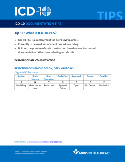


© Copyright 2026