
samples is certainly underestimated. For ex- provides a platform for readers of
Microbiology Comment provides a
platform for readers of Microbiology to
communicate their personal observations
and opinions in a more informal way than
through the submission of papers.
Most of us feel, from time to time, that
other authors have not acknowledged the
work of our own or other groups or have
omitted to interpret important aspects of
their own data. Perhaps we have
observations that, although not sufficient
to merit a full paper, add a further
dimension to one published by others. In
other instances we may have a useful piece
of methodology that we would like to
share.
The Editors hope that readers will take full
advantage of this section and use it to raise
matters that hitherto have been confined to
a limited audience.
Christopher M. Thomas, Editor-in-chief
A new chlamydia-like 16S
rDNA sequence from a
clinical sample
Chlamydiae constitute an important group of
obligate intracellular parasites, causing a variety of diseases in mammals and birds. Among
them Chlamydophila pneumoniae has been
recognized as a common respiratory pathogen
in man and it has been associated to atherosclerosis. Recently, new chlamydia-like organisms have been described, most being responsible for human respiratory infections. In
Israel, Simkania negevensis is frequent in
infants with bronchiolitis (11) and in adults
with community-acquired pneumonia (5, 13).
Parachlamydia acanthamoebae, identified as
an endosymbiont of an Acanthamoeba sp.
isolated from a healthy human nasal mucosa
(1), might be a cause of atypical pneumonia
(2). These organisms form new families within the Chlamydiales (4), and they are not
recognizable by conventional diagnostic procedures for classic chlamydiae, i.e. Cph.
pneumoniae, Chlamydophila psittaci complex or Chlamydia trachomatis. Therefore
their prevalence and diversity in clinical
Microbiology 147, March 2001
samples is certainly underestimated. For example, using PCR, Ossewaarde & Meijer (14)
detected several DNA sequences related to
either Simkania or Parachlamydia in respiratory samples, peripheral blood monocytes
and in a vessel-wall specimen.
Starting with a human respiratory sample
(from broncho-alveolar washings) sent to the
laboratory for the diagnosis of viral or
chlamydial infection, we detected a new 16S
ribosomal DNA sequence belonging to the
parachlamydiae. We have named this corvenA4 (GenBank accession no. AF308693).
DNA was extracted from a 300 µl aliquot
of the sample by the classic phenol\
chloroform method after proteinase K digestion, and the 16S rDNA was amplified by
PCR using a pan-chlamydia primer set amplifying almost all the gene (nucleotide positions
40–1485, P. acanthamoebae 16S rDNA numbering). Manipulations were carried out according to recommended guidelines (12) and
included negative controls starting from the
DNA extraction step. Both strands of the
PCR product (" 1400 bp) were sequenced
(three repetitions) using a series of inner
primers. The resulting complete sequence was
compared to the available corresponding
sequences obtained from GenBank using the
server. Sequences were aligned, gaps
and ambiguous sites excluded, for a total of
1354 nt. A similarity of 91n7–93n8 % was
found with the sequences of P. acanthamoebae and two other related amoebal
symbionts of Acanthamoeba spp. strains
(UWE1 and UWE25), 92n3–92n5 % with that
of Neochlamydia hartmannellae and related
amoebal endosymbionts (Acanthamoeba sp.
strains UWC22 and TUME1), 88 % with that
of Waddlia chondrophila, 86n8 % with that of
S. negevensis, and less than 86n3% with
those of Chlamydophila pneumoniae strain
TW-183, Cph. psittaci strain NJ1 and C.
trachomatis strain Har-13. A matrix of evolutionary distances was derived from the
alignment using , Jukes & Cantor’s
option, and a phylogenetic tree was inferred
using Fitch & Margoliash’s criteria. The
topological stability of the tree was assessed
by bootstrap analysis using to yield
a strict majority-rule consensus tree on 200
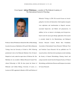
samples. Our sequence, corvenA4, was shown
to be related to, but distinct from, both
Parachlamydia and Neochlamydia lineages
(Fig. 1). Considering a value of 16S
rDNA sequence similarity of at least 95 %
for two organisms belonging to a same
genus (4), it seems probable that corvenA4
was from an organism representing a new
genus within the Parachlamydiaceae. We
failed to isolate such an organism in cell
culture, as evidenced by Giemsa staining
of inoculated Vero and HeLa cells. Amoebae
were not observed under the microscope,
and the sample did not present any evidence
for acanthamoebal DNA by PCR using a
primer set specific for the 18S rDNA.
Therefore a description of this chlamydialike organism was not possible, as the only
evidence of its existence is the 16S rDNA
sequence.
The diversity within the family Parachlamydiaceae is increasing. At present, two species and two genera have been validly
described : P. acanthamoebae (1) and N.
hartmannellae (10). Four other endosymbionts of Acanthamoeba sp. have been identified, probably forming three new species or
genera on the basis of 16S rDNA sequence
similarities (9). UWC22 was from an Acanthamoeba sp. isolated from a case of amoebic
keratitis, while TUME1, UWE1 and UWE25
were from environmental isolates of Acanthamoeba sp. (6, 9). It is interesting to note
that the UWC22 and TUME1 strains are
closely related to Neochlamydia, that infects
M GUIDELINES
Communications should be in the form
of letters and should be brief and to the
point. A single small Table or Figure may
be included, as may a limited number of
references (cited in the text by numbers,
and listed in alphabetical order at the end
of the letter). A short title (fewer than 50
characters) should be provided.
Approval for the publication rests with
the Editor-in-Chief, who reserves the
right to edit letters and\or to make a
brief reply. Other interested persons may
also be invited to reply. The Editors of
Microbiology do not necessarily agree
with the views expressed in
Microbiology Comment.
Contributions should be addressed to the
Editor-in-Chief via the Editorial Office.
515
Microbiology Comment
Parachlamydia
corvenA4
Neochlamydia
UWC22
UWE25
Simkania
Chl. trachomatis
UWE1
Cph. psittaci
Waddlia
Cph. pneumoniae
0·1
Verrucomicrobium
..................................................................................................................................................................................................................
Fig. 1. Unrooted consensus tree obtained by the Fitch–Margoliash method (version 3.573c).
The bar indicates estimated genetic distance. Verrucomicrobium sp. strain VeCb1 was used
as the outgroup.
Hartmannella and Dictyostelium but does not
grow in Acanthamoeba (10).
The possibility of infection of humans by
new chlamydia-like organisms deserves additional investigation. The Simkania-related
and Parachlamydia-related DNA sequences
detected in an abdominal aortic aneurysm
and in the peripheral blood monocytes,
respectively, by Ossewaarde & Meijer (14)
indicate that a variety of such organisms may
be present also in human body sites other than
mucosae. The passage from a putative amoebal host to mammalian cells may be possible,
P. acanthamoebae being cultivated in Vero
cells (1). More extensive studies are necessary
to evaluate their potential pathogenic role,
and might allow demonstration of the reality
of such infections, explaining the aetiology of
numerous respiratory infections in which no
conventional pathogens are found. In vitro
studies (7) showed that amoebae infected by
parachlamydiae exhibit an increased cytopathic effect on cell cultures. Amoebae and
other protists host a variety of intracellular
organisms and may act as a reservoir and\or
vector for human infection (3), as is the case in
Legionella infections. Such amoeba\bacterium systems are very interesting as infectious
sources and symbioses in general, and the
recent discovery of the ‘ pararickettsiae ’ endosymbionts of acanthamoebae isolated from
human ocular samples (8) illustrates the
extreme variety existing in nature. The search
for these systems in clinical samples might
help in estimating their prevalence and diversity.
Daniele Corsaro1, Danielle Venditti2, Alain
Le Faou1, Paolo Guglielmetti3 and Marcello
Valassina4
1
Laboratoire de Virologie-Microbiologie,
Centre Hospitalier Universitaire de Nancy,
Hopital de Brabois – Adultes, Route de
Neufcha# teau, 54511 Vandoeuvre-les-Nancy
cedex, France
516
TREDI De! partement Recherche, Technopo# le
de Nancy-Brabois, France
3
Laboratory of Parasitology, Le Scotte
Hospital, Siena, Italy
4
Department of Molecular Biology,
Microbiology Section, University of Siena,
Italy
endosymbionts recovered from clinical and
environmental isolates of Acanthamoeba spp. Appl
Environ Microbiol 66, 2613–2619.
10. Horn, M., Wagner, M., Mu$ ller, K.-D., Schmid,
E. N., Fritsche, T. R., Schleifer, K.-H. & Michel, R.
(2000). Neochlamydia hartmannellae gen. nov., sp. nov.
(Parachlamydiaceae), an endoparasite of the amoeba
Hartmannella vermiformis. Microbiology 146,
1231–1239.
11. Kahane, S., Greenberg, D., Friedman, M. G., Haikin,
H. & Dagan, R. (1998). High prevalence of ‘ Simkania
Z ’, a novel Chlamydia-like bacterium, in infants with
acute bronchiolitis. J Infect Dis 177, 1425–1429.
12. Kwok, S. (1990). Procedures to minimize PCRproduct carry-over. In PCR Protocols : A Guide to
Methods and Applications, pp. 142–145. Edited by M. A.
Innis, D. H. Gelfand, J. J. Sninsky & T. J. White. San
Diego, CA : Academic Press.
13. Lieberman, D., Kahane, S., Lieberman, D. &
Friedman, M. G. (1997). Pneumonia with serological
evidence of acute infection with the Chlamydia-like
microorganism ‘ Z ’. Am J Respir Crit Care Med
156, 578–582.
14. Ossewaarde, J. M. & Meijer, A. (1999). Molecular
evidence for the existence of additional members of the
order Chlamydiales. Microbiology 145, 411–417.
2
Author for correspondence : Daniele
Corsaro. Tel : j33 3 83 15 34 68.
Fax : j33 3 83 15 34 74.
e-mail : tredi.recherche!wanadoo.fr
1. Amann, R., Springer, N., Scho$ nhuber, W., Ludwig,
W., Schmid, E. N., Mu$ ller, K. & Michel, R. (1997).
Obligate intracellular bacterial parasites of
acanthamoebae related to Chlamydia spp. Appl Environ
Microbiol 63, 115–121.
2. Birtles, R. J., Rowbotham, T. J., Storey, C., Marrie,
T. J. & Raoult, D. (1997). Chlamydia-like obligate
parasite of free-living amoebae. Lancet 349, 925–926.
3. Corsaro, D., Venditti, D., Padula, M. & Valassina, M.
(1999). Intracellular life. Crit Rev Microbiol 25, 39–79.
4. Everett, K. D. E., Bush, R. M. & Andersen, A. A.
(1999). Emended description of the order Chlamydiales,
proposal of Parachlamydiaceae fam. nov. and
Simkaniaceae fam. nov., each containing one monotypic
genus, revised taxonomy of the family Chlamydiaceae,
including a new genus and five new species, and
standards for the identification of organisms. Int J Syst
Bacteriol 49, 415–440.
5. Friedman, M. G., Galig, A., Greenberg, S. & Kahane,
S. (1999). Seroprevalence of IgG antibodies to the
chlamydia-like microorganism ‘ Simkania Z ’ by ELISA.
Epidemiol Infect 122, 117–123.
6. Fritsche, T. R., Gautom, R. K., Seyedirashti, S.,
Bergeron, D. L. & Lindquist, T. D. (1993). Occurrence
of bacterial endosymbionts in Acanthamoeba spp.
isolated from corneal and environmental specimens and
contact lenses. J Clin Microbiol 31, 1122–1126.
7. Fritsche, T. R., Sobek, D. & Gautom, R. K. (1998).
Enhancement of in vitro cytopathogenicity by
Acanthamoeba spp. following acquisition of bacterial
endosymbionts. FEMS Microbiol Lett 166, 231–236.
8. Fritsche, T. R., Horn, M., Seyedirashti, S., Gautom,
R. K., Schleifer, K.-H. & Wagner, M. (1999). In situ
detection of novel bacterial endosymbionts of
Acanthamoeba spp. phylogenetically related to members
of the order Rickettsiales. Appl Environ Microbiol 65,
206–212.
9. Fritsche, T. R., Horn, M., Wagner, M., Herwig, R. P.,
Schleifer, K.-H. & Gautom, R. K. (2000). Phylogenetic
diversity among geographically dispersed Chlamydiales
Bacterial cell division
protein FtsZ is a specific
substrate for the AAA
family protease FtsH
The role of AAA (ATPases Associated to a
variety of cellular Activities) family protease
FtsH in bacterial cell division is not known,
although mutations in ftsH were found to
inhibit cell growth and division (1, 6, 13).
Overexpression of heterologous FtsH in Escherichia coli results in the formation of
multinucleate filamentous cells due to the
abolition of cell septation (8). Further, independent studies on FtsH (15) and FtsZ (2),
which is the key regulator of bacterial cell
division, have shown that FtsH protease and
FtsZ protein are localized to the mid-cell site
during septation. FtsZ protein is the prokaryotic homologue of tubulin (5, 10, 12),
possessing GTP-dependent polymerization
activity (4, 11). Significantly, the AAA family
ATPase member katanin disassembles tubulin
polymers in an ATP-dependent manner (7).
Based on these observations, we reasoned that
an interaction similar to that between katanin
and tubulin might hold true for FtsH and FtsZ
in prokaryotes as well. To verify this hypothesis, we examined whether the FtsH
protease of Escherichia coli (FtsHEc) could
degrade FtsZ of E. coli (FtsZEc) in vitro.
Incubation of FtsZEc with FtsHEc showed
degradation of the FtsZEc protein in an ATPand Zn#+-dependent manner (Fig. 1, lane 2).
Absence of ATP or presence of ortho-phenanthroline (OP ; chelator of zinc) abolished
protease activity (Fig. 1, lanes 3 and 4).
Similarly, substitution of ATP with its unhydrolysable analogue, ATP-γS, also inhibited FtsZ degradation (Fig. 1, lane 5).
Identical observations were made upon incubation of the FtsZ protein of Myco-
Microbiology 147, March 2001
Microbiology Comment
Lane
FtsH
ATP
ATP-γS
OP
1
–
+
–
–
2
+
+
–
–
3
+
–
–
–
4
+
+
–
+
5
+
–
+
–
FtsZEc
FtsZMt
σ32
.....................................................................................................
Fig. 1. SDS-PAGE profile of the in vitro
degradation of FtsZEc, FtsZMt and σ32
proteins by FtsHEc. The FtsHEc, FtsZEc, FtsZMt
and σ32 proteins were expressed as
6ihistidine-tagged fusion proteins from
the respective expression vectors, namely
pSTD113 (a generous gift from Dr Yoshinori
Akiyama, Institute for Virus Research, Kyoto
University, Japan), pQE30-ECZ, pET20-MTZ
and pUEH211 (a kind gift from Dr Bukau,
Institut fur Biochemie und Molekularbiologie, Universitat Freiberg, Germany),
and purified by Ni2+-NTA affinity chromatography. The protease assay was carried
out with the purified proteins essentially
as described (14). The results shown are
representative of at least six independent
experiments.
bacterium tuberculosis H37Rv (FtsZMt) with
FtsHEc (Fig. 1). These features of the protease
activity of FtsHEc enzyme on the FtsZ proteins
from two highly divergent bacterial genera
were identical to its activity on a biologically
relevant and specific substrate, namely σ$#
protein of E. coli, as found by us (Fig. 1) and
reported by others (14). About 90 % of FtsZEc
and 100 % of FtsZMt were degraded by FtsHEc
protease (Fig. 1). Neither heat-denatured FtsZ
nor green fluorescent protein (a heterologous
Microbiology 147, March 2001
protein) was degraded by FtsH (data not
shown), indicating that the recognition and
degradation of FtsZ by FtsH is specific.
Here we demonstrate for the first time that
FtsH protease degrades the cell-division-initiation protein FtsZ in vitro. The implication
of our in vitro studies is that the AAA family
protease FtsH could be a proteolytic regulator
of the cell-division-initiation protein FtsZ in
vivo. The regulation of FtsZ activity has so far
been shown to involve only protein–protein
interactions, which prevent mid-cell localization or polymerization of the protein (3, 9),
whereas our finding is suggestive of the
existence of a proteolytic regulatory mechanism also. Secondly, the fact that FtsHEc
protease degraded FtsZ molecules from two
divergent bacterial genera implies that FtsZ
could be a substrate for FtsH protease across
the bacterial kingdom. Finally, since FtsH is a
stress-responsive protease and its mid-cell
localization occurs only in a fraction of
dividing cells (15), it is logical to presume that
the proteolytic regulation of FtsZ by FtsH protease, if it occurs in vivo, might be restricted
to specific conditions of bacterial growth.
Acknowledgements
This work was supported by a research grant
from the Council of Scientific and Industrial
Research, New Delhi, India, to P. A. The
authors G. A., R. S. and S. P. A. thank the
Indian Institute of Science, Bangalore, for
fellowships.
Gopalakrishnapillai Anilkumar, Ramanujam
Srinivasan, Syam Prasad Anand and
Parthasarathi Ajitkumar
Department of Microbiology and Cell
Biology, Indian Institute of Science,
Bangalore-560012, India
Author for correspondence : Parthasarathi
Ajitkumar. Tel : j91 80 309 2344.
Fax : j91 80 360 2697/0683/0085.
e-mail : ajit!mcbl.iisc.ernet.in
1. Begg, K. J., Tomoyasu, T., Donachie, W. D., Khattar,
M., Niki, H., Yamanaka, K., Hiraga, S. & Ogura, T.
(1992). Escherichia coli mutant Y16 is a double mutant
carrying thermosensitive ftsH and ftsI mutations. J
Bacteriol 174, 2416–2417.
2. Bi, E. & Lutkenhaus, J. (1991). FtsZ ring structure
associated with division in Escherichia coli. Nature 354,
161–164.
3. Bi, E. & Lutkenhaus, J. (1993). Cell division
inhibitors, SulA and MinCD, prevent formation of the
FtsZ ring. J Bacteriol 175, 1118–1125.
4. Bramhill, D. & Thompson, C. M. (1994). GTPdependent polymerization of Escherichia coli FtsZ
protein to form tubules. Proc Natl Acad Sci U S A 91,
5813–5817.
5. Erickson, H. P. (1995). FtsZ, a prokaryotic homolog
of tubulin ? Cell 80, 367–370.
6. Ferreira, L. C. S., Keck, W., Betzner, A. & Schwarz,
U. (1987). In vivo cell division gene product interactions
in Escherichia coli K-12. J Bacteriol 169, 5776–5781.
7. Hartman, J. J. & Vale, R. D. (1999). Microtubule
disassembly by ATP-dependent oligomerization of the
AAA enzyme katanin. Science 286, 782–785.
8. Itoh, R., Takano, H., Ohta, N., Miyagishima, S.-y.,
Kuroiwa, H. & Kuroiwa, T. (1999). Two ftsH family
genes encoded in the nuclear and chloroplast genomes of
the primitive red alga Cyanidioschyzon merolae. Plant
Mol Biol 41, 321–337.
9. Justice, S. S., Garcia-Lara, J. & Rothfield, L. I. (2000).
Cell division inhibitors SulA and MinC\MinD block
septum formation at different steps in the assembly of
the Escherichia coli division machinery. Mol Microbiol
37, 410–423.
10. Lowe, J. & Amos, L. A. (1998). Crystal structure of
the bacterial cell division protein FtsZ. Nature 391,
203–206.
11. Mukherjee, A. & Lutkenhaus, J. (1994). Guanine
nucleotide dependent assembly of FtsZ into filaments. J
Bacteriol 176, 2754–2758.
12. Nogales, E., Wolf, S. G. & Downing, K. H. (1998).
Structure of the αβ tubulin dimer by electron
crystallography. Nature 391, 199–203.
13. Santos, D. & De Almeida, D. F. (1975). Isolation and
characterization of a new temperature sensitive cell
division mutant of Escherichia coli K-12. J Bacteriol 124,
1502–1507.
14. Tomoyasu, T., Gamer, J., Bukau, B. & 9 other
authors (1995). Escherichia coli FtsH is a membranebound, ATP-dependent protease which degrades the heat
shock transcription factor σ$#. EMBO J 14, 2551–2560.
15. Wehrl, W., Niederweis, M. & Schumann, W. (2000).
The FtsH protein accumulates at the septum of Bacillus
subtilis during cell division and sporulation. J Bacteriol
182, 3870–3873.
517


© Copyright 2026