
GEOTRACES Sampling Protocols Manual
Sampling and Sample-handling Protocols for GEOTRACES Cruises Edited by the 2010 GEOTRACES Standards and Intercalibration Committee: Gregory Cutter (Chair), Department of Ocean, Earth and Atmospheric Sciences, Old Dominion University, Norfolk, Virginia 23529 USA; [email protected] Per Andersson, Laboratory for Isotope Geology, Swedish Museum of Natural History, Stockholm Sweden; [email protected] Lou Codispoti, University of Maryland Center for Environmental Science, P.O. Box 775, Cambridge, Maryland 21613; [email protected] Peter Croot, Plymouth Marine Laboratory, Prospect Place, The Hoe, Plymouth PL1 3DH, Devon, United Kingdom; [email protected] Roger Francois, Department of Earth and Ocean Sciences, The University of British Columbia, Vancouver, British Columbia Canada V6T 1Z4; [email protected] Maeve Lohan, School of Geography, Earth, and Environmental Sciences, University of Plymouth, Drake Circus, Plymouth PL4 8AA, United Kingdom; [email protected] Hajime Obata, Atmosphere and Ocean Research Institute, University of Tokyo, Chiba 277-8564 Japan; [email protected] Michiel Rutgers van der Loeff, AWI-Geochemistry, D 27515 Bremerhaven Germany; [email protected] Version 1.0; December 2010 Table of Contents I. Introduction ..................................................................................................................... 5 II. General Considerations .................................................................................................. 5 III. Hydrography and Ancillary Parameters........................................................................ 6 IV. Radioactive Isotopes ..................................................................................................... 8 A. Protocols for 230Th and 231Pa...................................................................................... 8 1. Analytical instrument .................................................................................................. 8 2. Volumes required ........................................................................................................ 8 3. Sampling ..................................................................................................................... 8 4. Sample Processing ...................................................................................................... 9 5. Spike calibrations ...................................................................................................... 11 6. Precision of measurements ....................................................................................... 11 7. References ................................................................................................................. 11 B. Protocols for 234Th.................................................................................................... 12 1. Particulate 234Th Sampling........................................................................................ 12 2. Total 234Th sampling ................................................................................................. 13 3. General Considerations for 234Th .............................................................................. 13 4. References ................................................................................................................. 14 C. Protocols for 226Ra and 228Ra Measurements in Sea Water ..................................... 15 1. Alpha scintillation measurement of 226Ra and 222Rn ............................................... 16 2. Measurements of 226Ra and 228Ra by Ba(Ra)SO4 precipitation from small volume (20 – 40 L) samples ...................................................................................................... 17 3. Measurement of 228Ra via 228Th in-growth .............................................................. 19 4. Gamma spectrometry measurement of 226Ra and 228Ra........................................... 21 5. Protocols for short-lived radium isotopes: 223Ra, 224Ra ............................................ 23 6. Notes on 223Ra and 224Ra measurements................................................................... 24 7. References ................................................................................................................. 24 D. Protocols for 210Po and 210Pb.................................................................................... 27 1. Analytical instrument ............................................................................................... 27 2. Volume required ....................................................................................................... 27 3. Sampling ................................................................................................................... 28 4. Sample acidification and spiking .............................................................................. 29 5. Pre-concentration and onboard preliminary analysis................................................ 29 6. 210Pb yield determination .......................................................................................... 30 7. Digestion of filters containing particulate matter ..................................................... 30 8. Model calculations of final activities of 210Po and 210Pb in seawater samples ......... 31 9. Some issues that need to be considered ................................................................... 33 10. References ............................................................................................................... 34 V. Radiogenic Isotopes ..................................................................................................... 35 1. Analytical instrument ................................................................................................ 35 2. Volume required ....................................................................................................... 35 3. Sampling ................................................................................................................... 36 4. Sample Processing .................................................................................................... 36 5. Spike calibrations and blanks ................................................................................... 37 6. Evaluation of analytical uncertainties ....................................................................... 38 2 7. References ................................................................................................................. 38 VI. Trace Elements ........................................................................................................... 39 1. Pre-cruise Preparations ............................................................................................. 40 1.1 Sampling bottles for collecting clean seawater ...................................................... 40 1.2 Sample Bottle Types for sample storage ................................................................ 41 1.3 Sample Bottle Cleaning .......................................................................................... 41 2. Sample Collection ..................................................................................................... 42 2.1 Surface Sampling .................................................................................................... 42 2.2 Depth Profiles ......................................................................................................... 43 3. Sample Handling....................................................................................................... 44 3.1 Total Dissolvable (unfiltered) Samples................................................................... 46 3.2 Total Dissolved (filtered) Samples .......................................................................... 46 3.3 Speciation samples .................................................................................................. 47 3.4 Sample Acidification ............................................................................................... 49 4. Shipboard Determinations of Selected Dissolved Trace Metals .............................. 49 5. Chemicals and Reagents ........................................................................................... 50 6. Analytical Considerations: Precision and Accuracy ................................................. 50 7. References ................................................................................................................ 51 8. Protocols for Sampling and Determinations of Mercury and its Speciation ............ 52 8.1 Sample Bottle Selection and Cleaning .................................................................... 52 8.2 Sample Collection and Handling ............................................................................ 53 8.3 Sample Analysis ...................................................................................................... 54 8.4 Calibration and Comparability .............................................................................. 58 8.5 Reagents .................................................................................................................. 58 8.6 References ............................................................................................................... 59 9. Collection of particulate samples from GO-Flo sampling bottles ........................... 61 9.1 Filter Type............................................................................................................... 61 9.2 Filter holders .......................................................................................................... 63 9.3 Cleaning Filters and filter holders ......................................................................... 64 9.4 Attachment and use of filter holders on GO-Flo bottles ......................................... 64 9.5 Filtration time and particle settling artifacts ......................................................... 65 9.6 Pressurizing water sampling bottles for filtration .................................................. 66 9.7 Process blanks ........................................................................................................ 66 9.8 Storing Sampled Filters .......................................................................................... 67 9.9 Clean Up and Preparations After Sampling ........................................................... 67 9.10 Off-line Filtration ................................................................................................. 68 9.11 Processing and analysis of particulate samples on filters ................................... 69 9.12 References ............................................................................................................ 73 10. In-situ Pumping Sampling Protocols For Particulate Trace Metals ...................... 73 10.1 Cast documentation .............................................................................................. 73 10.2 Protocols for deployment and recovery ............................................................... 74 10.3 Preparation and configuration of in-situ filtration systems ................................. 76 10.4 Filter type selection: quartz (QMA) and plastic (PES) ....................................... 79 10.5 Filter cleaning procedure ..................................................................................... 81 10.6 Particle Sample Handling ..................................................................................... 83 10.7 List of materials (and example U.S. suppliers) ..................................................... 84 3 10.8 References ............................................................................................................. 85 VII. Nitrate and Silicon Isotopes ...................................................................................... 86 A. Protocols for Nitrate Isotopes .................................................................................. 86 1. Sampling ................................................................................................................... 86 2. Storage ...................................................................................................................... 86 3. Analysis .................................................................................................................... 86 4. Calibration ................................................................................................................ 87 5. References ................................................................................................................. 88 B. Protocols for Silicon Isotopes .................................................................................. 88 1. Sampling ................................................................................................................... 88 2. Storage ...................................................................................................................... 89 3. Analysis .................................................................................................................... 89 4. Calibration ................................................................................................................ 89 5. References ................................................................................................................. 90 VIII. Protocols for Optics: Transmissometer and Scattering Sensors .............................. 90 1. Transmissometers and Scattering sensors................................................................. 91 2. Avoiding optical data dropouts ................................................................................. 93 3. Elimination of optics contamination and cast-to-cast offsets ................................... 93 4. Compensation for Transmissometer Drift and CTD Digitizing Electronics ............ 94 5. References ................................................................................................................. 96 IX. Glossary of Terms....................................................................................................... 97 Appendix 1. Contributors to the GEOTRACES Cruise Protocols, Version 1.0 Appendix 2. GEOTRACES-recommended modifications to JGOFS 19 protocols Appendix 3. PICES Report 34, Determinations of DOC and DON, for GEOTRACES Cruises 4 I. Introduction The GEOTRACES Standards and Intercalibration (S&I) Committee is charged with ensuring that the data generated during GEOTRACES are as precise and accurate as possible, which includes all the steps from sampling to analysis. Thus, sampling methods for dissolved and particulate constituents must take a representative (of the water depth/water mass) and uncontaminated sample, the samples must be stored (or immediately analyzed) in a fashion that preserves the concentrations (activities) and chemical speciation, and the analyses of these samples must yield accurate data (concentration, activity, isotopic composition, and chemical speciation). To this end, experiences from the 2008-2010 GEOTRACES Intercalibration Program, and other related intercalibration efforts, helped to create the protocols in this document. However, methods continually evolve and the GEOTRACES S&I Committee will monitor these advances as validated by intercalibrations and modify the methods as warranted. The protocols here are divided into trace element and isotope groups: Hydrography and Ancillary Parameters, Radioactive Isotopes, Radiogenic Isotopes, Trace Elements, and Nutrient Isotopes. Those who contributed to preparing these protocols are listed in Appendix 1 and are sincerely thanked for their efforts in helping GEOTRACES and the worldwide TEI community. II. General Considerations The following items should be included as a part of a standard intercalibration effort during all GEOTRACES cruises: A. Every cruise should occupy at least one GEOTRACES Baseline Station (where previous intercalibration cruises have established the concentrations, activities, and/or speciation of at least the key GEOTRACES TEIs), or an overlap/cross-over station with a previous GEOTRACES cruise, to affect an intercalibration for sampling through analyses. B. If there is no GEOTRACES Baseline Stations or crossover stations to occupy, we strongly recommend that an intercalibration be conducted via replicate sampling during each cruise. In particular, a minimum of 3 depths (e.g., near surface, mid-water, and deep) at 2 stations should be sampled with duplicate hydrocasts, and samples from these replicates sent to several labs for the determination of trace elements and isotopes. The results from this effort can be examined later for data integrity and coherence. C. Nutrient and salinity samples should be taken along with all trace element samples in order to verify proper bottle and rosette operation and sampling depths (i.e., compare to the hydrography established with the conventional CTD/rosette). D. We will not recommend specific analytical methods for most variables (except for the ancillary parameters and several methods for some TEIs are suggested in the sections to follow). However, during analyses (at sea or in a shore-based lab) appropriate certified reference materials (See IX. Glossary of Terms), or SAFe or GEOTRACES 5 Intercalibration samples as described in the Trace Element Section (VI), should be processed to assess analytical accuracy. E. All aspects of meta data related to sampling, sample logging, and resulting data should follow the guidelines found on the International GEOTRACES Data Assembly Centre (http://www.bodc.ac.uk/geotraces/) web site. Except where activities are reported (e.g., radionuclides), we recommend concentration units be in fractions of a mole per unit mass (kilogram) or volume (liter; most appropriately when shipboard analyses are used) - µmol l-1 or nmol kg-1 as examples. III. Hydrography and Ancillary Parameters Although GEOTRACES is focused on trace elements and their isotopes (TEIs), to achieve the overarching goal of understanding the biogeochemical processes controlling them, the suite of TEIs must be examined in the context of the oceans’ hydrography, including nutrient (C, N, P, Si) cycling. Therefore, the same care in sampling and sample processing of ancillary parameters must be included in GEOTRACES protocols to ensure the best possible precision and accuracy. In addition to the basic water column hydrographic parameters of salinity, temperature, and depth, as well as in situ measurements of fluorescence, transmissometry (See Optics Section VIII), and oxygen concentrations. Table 1 lists GEOTRACES ancillary parameters (and suggested methods of determination) for discrete (depth profile) samples. 6 Table 1. Ancillary Parameters and Recommended Methods for GEOTRACES Cruises Parameter Method Detection Limit Reference Salinity Conductivity NA (not applicable) Hood et al., 2010 Oxygen Manual or automated Winkler 1 µmol l-1 Hood et al., 2010 Ammonium Automated colorimetric 0.1 µmol l-1 Parsons et al., 1984 Nitrite Automated colorimetric 0.1 µmol l-1 Hood et al., 2010 Nitrate Automated colorimetric 0.1 µmol l-1 Hood et al., 2010 Phosphate Automated colorimetric 0.03 µmol l-1 Hood et al., 2010 Silicate Automated colorimetric 0.4 µmol l-1 Hood et al., 2010 Pigments Fluorometry and HPLC NA JGOFS Report 19 DOC/DON Oxidative Combustion NA PICES Report 34 POC/PON Oxidative Combustion NA JGOFS Report 19 Hood, E.M., C.L. Sabine, and B.M. Sloyan, eds. 2010. The GO-SHIP Repeat Hydrography Manual: A Collection of Expert Reports and Guidelines. IOCCP Report Number 14, ICPO Publication Series Number 134. Available online at http://www.go-ship.org/HydroMan.html Parsons, T.R., Y. Maita, and C.M. Lalli. 1984. A Manual of Chemical and Biological Methods for Seawater Analysis. Pergamon, Oxford, 173 pp. JGOFS Report 19, amended to cover the GEOTRACES-relevant parameters (Appendix 2), and the PICES Report 34, DOC/DON section (Appendix 3), are included at the end of this document. Modified Report 19, Report 34, and the publications by Hood et al. (2010) and Parsons et al. (1984) cover all recommended procedures for sampling, sample processing/storage, and analyses for hydrography and ancillary data for GEOTRACES cruises. The GO-SHIP collection (cited as Hood et al.) is particularly relevant to GEOTRACES in that it contains all the recommended procedures used in the CLIVAR Repeat Hydrography Program. However, more accurate and precise determinations of ancillary parameters are encouraged; the methods in Table 1 are capable of the best performance at the time of writing (2010). 7 IV. Radioactive Isotopes A. Protocols for 230Th and 231Pa There is not a unique sampling and analytical procedure that can be recommended, so a range of qualified options is presented. 1. Analytical instrument The most widely used instruments for seawater analysis are sector-field ICP-MS (multi or single collector; Choi et al., 2001; Shen et al., 2002) and TIMS (Shen et al., 2003). ICPMS is increasingly the instrument of choice because of higher sample throughput. 2. Volumes required The volume required for analysis of dissolved 230Th and 231Pa range from a few liters (Shen et al., 2003) to 15-20 liters (Choi et al., 2001). As a rule of thumb, the volume required to analyze suspended particles is 5 times larger for 230Th (10-100L) and 20x larger for 231Pa (40-400L). The volume required for analysis bears significantly on sampling methods (for particles) and sample processing (for dissolved). There are several options at each step of the procedure. This provides flexibility, but will necessitate careful intercalibrations. 3. Sampling 3.1 Dissolved 3.1.1 Sampling Niskin bottles with epoxy-coated stainless steel springs are applicable for radioisotopes (Th and Pa). If the volume required is 10-20 L, dedicated radionuclide hydrocasts may be necessary. 3.1.2 Sample Filtration Samples for operationally-defined dissolved Th and Pa should be filtered. Filtration using capsule filters, preferably 0.8 µm/0.45 µm Acropak® 500 filters, is most feasible for large-volume samples. Different groups use different pre-cleaning methods for these capsules and there are a variety of protocols available. The capsules can be cleaned with HCl, 1.2 M, and rinsed with and stored in Milli-Q water. In the field it is recommended that the capsules be flushed with 1 L seawater prior to first use, and then 10 capsule volumes between casts. This is experience derived from the Intercalibration Cruises 1 and 2. In general, all seawater samples should be processed as quickly as possible to avoid loss of dissolved Th and Pa by absorption on sampling bottle (e.g., Niskin) walls. If membrane filtration (i.e., to keep the particles) is being used, at the time this document was written there is no evidence that one type of membrane filter is preferable to another. 8 However, quartz/glass fiber filters are not recommended as dissolved Th and Pa are likely to adsorb to these materials. 3.1.3 Sample container rinses There is no evidence that dissolved Th and Pa concentrations are compromised by filling acid-cleaned sample containers directly, without rinsing. Nevertheless, rinsing of each sample bottle with sample water is preferable. 3.2 Particles Results from the GEOTRACES Intercalibration exercise indicate that most labs are unable to measure particulate 230Th and 231Pa concentrations in particles filtered from standard sample bottles (e.g., volumes of 10 to 20 liters). Analytical sensitivity of current instrumentation is such that larger samples are generally required, thus necessitating the use of in situ pumps to collect samples for particulate 230Th and 231Pa concentrations (see Section IV.B.1). Ideally, membrane filters used with in situ pumps to collect samples for particulate Th and Pa will be matched with the membrane filters used to collect samples for analysis of dissolved Th and Pa. 4. Sample Processing Filtered seawater samples must be stored in acid-cleaned high/low density polyethylene (HDPE or LDPE) or polycarbonate containers. The GEOTRACES Intercalibration exercise showed that bottle blanks can be a problem for Th and Pa, and these blanks must be quantified for each isotope. In previous studies, filtered seawater samples have either been acidified, spiked and pre-concentrated at sea, or acidified and shipped to the home laboratory for spiking and pre-concentration. For larger volumes, “at sea” processing is often the method of choice. Smaller samples can more easily be shipped to home institutions. The advantages of “at sea” processing are: (1) lower risk of 230Th and 231Pa loss by absorption on the walls of the storage container, and (2) avoids shipping of large quantity of seawater. The advantages of “on land” processing are: (1) avoids shipping and handling of radioisotopes at sea; (2) requires less space and personnel on-board; (3) allows more accurate determination of the sample volume; and (4) loss of 233Pa spike by decay during the cruise/shipping and storing the samples prior to measurement is not a problem. 4.1 Acidification As soon as possible after collection, samples for dissolved Th and Pa should be acidified with HCl to a pH < 2.0 (target 1.7 to 2.0). It is recommended that 6M Hydrochloric Acid is used for sample acidification. It is much easier to commercially transport seawater acidified with Hydrochloric Acid than Nitric Acid. Seawater acidified with Hydrochloric Acid to pH~2 is not considered “hazardous materials”, while the same samples acidified with Nitric Acid are considered “hazardous materials”. Dilution of the Hydrochloric Acid to 6M reduces irritating fumes from the reagent bottle, which, in turn, allows sample acidification without the need for a fume hood. Following acidification, sample integrity 9 should be protected by covering the cap and thread with Parafilm® or similar plastic wrap. Double plastic bags around each bottle/container are recommended. Labeling of samples should be made with a specific GEOTRACES # for each sample and depth. 4.2 Sample volume or weight A variety of approaches have been used to record sample weight or volume, and the literature should be consulted for the best one to use in a particular cruise (e.g., open water vs. in the ice). Some labs use an electronic balance to weigh samples at sea, using a simple computer algorithm to average weights on the moving ship until a stable reading is obtained. Other labs weigh samples after they are returned to the home institution. 4.3 Spiking If spiking is done on board it should be done by pre-weighed spikes and thorough careful rinsing of the spike vial, disposing multiple rinses into the sample container. 4.3.1 233Pa spike preparation There are two ways for producing 233Pa: (1) by milking 237Np (2) by neutron activation of 232 Th. 237 Np milking: the 233Pa spike must be checked for 237Np bleeding. Preferentially by Mass spectrometry (2nd cleaning step may be needed). Advantages: Lower 231 Pa blank; Lower 232Th contamination 232 Th irradiation: Advantages: Large quantities (1mCi) can be easily produced Disadvantages: 232Th contamination precludes its measurement in the same sample. 231Pa is produced by neutron activation of 230Th traces in the 232Th target. 231 Pa contamination can be kept low by preparing a new spike before the cruise to minimize the 231Pa/233Pa in the spike. It can also be precisely quantified by measuring 231Pa/233Pa in the spike before 233Pa decay. Typically, 231Pa blanks range from ~10% in surface water to ~1% in deep water 4.4 Pre-concentration Pre-concentration of 230Th and 231Pa is done by adsorption on a precipitate formed in seawater (scavenging), which is then recovered by decantation and centrifugation and returned to the home laboratory for 230Th and 231Pa purification by ion-exchange. Several scavenging methods have been used: (1) Fe hydroxide; (2) Mg hydroxide; (3) MnO2. Fe hydroxide: 0.05 ml FeCl3 (50 mg Fe/ml; cleaned by extraction in isopropyl ether) is added per liter of acidified seawater with the 229Th and 233Pa spikes. The spiked seawater is left to equilibrate for at least 24 hours. Thereafter, ammonium hydroxide (ultraclean) is added to bring the pH to 8.5-9 and precipitate Fe(OH)3. After 12-24 hours of settling, most of the supernatant is removed and the precipitate is centrifuged. 10 Mg hydroxide: Seawater is acidified, spiked and left to equilibrate for 24 hours. Thereafter, concentrated NH4OH (ultraclean) is added to precipitate Mg(OH)2. The precipitate is decanted and transferred into 250ml polyethylene bottles. 7M HNO3 is then slowly added to reduce the volume of precipitate. Mn dioxide: Seawater is spiked and left to equilibrate for 12 hours. Thereafter, a few drops of ultraclean, concentrated ammonium hydroxide are added, with 0.75 mg/L KMnO4 and 2mg/L MnCl2 (Rutgers van der Loeff and Moore, 1999). After 24 hours, the MnO2 is filtered on 1µm polycarbonate filter. Sample storage: We are not yet sure how long we can store filtered acidified samples for subsequent spiking, pre-concentration and analysis without losing 230 Th or 231Pa on the walls of the containers. Samples collected during the first GEOTRACES intercalibration cruise (July, 2008), acidified to pH 1.7, and analyzed over a period of 1.5 years showed no drift in concentrations of dissolved Th or Pa. NOTE: For samples stored this long it is necessary to make corrections for ingrowth of dissolved 230Th and 231Pa due to radioactive decay of dissolved uranium. The different scavenging methods (Fe(OH)3 vs. Mg(OH)2 vs. MnO2) still have to be compared. 5. Spike calibrations GEOTRACES should agree on a primary Th standard (e.g. NIST SRM 3159) to calibrate the 229Th spikes used by different laboratories. In the meantime, 229Th spikes used in GEOTRACES cruises should be archived for future intercalibrations. Calibration of 233Pa is best done by measuring the ingrowth of 233U by isotope dilution with a 236U standard. GEOTRACES should agree on a primary U standard (e.g. NIST CRM-145) to calibrate the 236U standards used by different laboratories. In the meantime, the 236U standards used to calibrate 233Pa spikes for GEOTRACES cruises should be archived for future intercalibrations. 6. Precision of measurements Precision of measurements conducted on each cruise are best documented by analyzing a set of replicate seawater samples (3 to 6) in the mid-concentration range during each cruise (see Section IIA. above). 7. References Choi, M.-S., R. Francois, K. Sims, M. P. Bacon, S. Brown-Leger, A. P. Fleer, L. Ball, D. Schneider, and S. Pichat. 2001. Rapid determination of 230Th and 231Pa in seawater by desolvated-micronebulization Inductively-Coupled Magnetic Sector Mass Spectrometry. Mar. Chem., 76, 99-112. 11 Shen, C.-C., Edwards, R. L., Cheng, H., Dorale, J. A., Thomas, R. B., Moran, S. B., Weinstein, S. E., Edmonds, H. N. 2002. Uranium and thorium isotopic and concentration measurements by magnetic sector inductively coupled plasma mass spectrometry. Chem. Geol., 185, 165-178. Shen, C.-C., Cheng, H., Edwards, R. L., Moran, S. B., Edmonds, H. N., Hoff, J. A., Thomas, R. B. 2003. Measurement of attogram quantities of 231Pa in dissolved and particulate fractions of seawater by isotope dilution thermal ionization mass spectroscopy. Anal. Chem., 75, 1075-1079. B. Protocols for 234Th 1. Particulate 234Th Sampling In-situ filtration allows the collection of large volume size-fractionated marine particles from the water column. Commercially available battery-operated in-situ pumping systems (e.g., McLane, Challenger) can be deployed simultaneously at multiple depths to collect particulate 234Th samples. 1.1 Filter Type No single filter type can accommodate all the different measurements needed during GEOTRACES. Quartz fiber filters (Whatman QMA) and polyethersulfone (Pall Supor) filters were extensively tested during the Intercalibration Cruises. QMA filters have a nominal pore size of 1μm, have a long track record of use in in-situ filtration, have the best flow characteristics, and result in even particle distribution. QMA filters can be precombusted for particulate organic carbon (POC) analyses. Paired filters (two back to back filters) can be used so that the bottom filter can act as a flow-through blank. QMA filters are found to have significant flow-through blanks due to adsorption especially when low sample volumes are filtered. If sampling constraints makes it necessary to use a plastic filter, then hydrophilic polyethersulfone (PES) membrane filters (e.g., Pall Supor) have the best blank and flow characteristics of the available plastic filters, and are thus currently the plastic filter of choice. The biggest drawbacks for this type of filter is the poor (heterogeneous) particle distribution observed on deep (>500 m) samples. The particle distribution on the filter worsens with depth. However the 234Th absorption blanks for this filter type is negligible. For large (>51 μm) particle collection, 51μm polyester mesh (e.g., 07-51/33 from Sefar Filtration) is a good option. For 234Th analysis of this size fraction, we recommend rinsing the prefilter onto a 25 mm silver membrane filter using filtered seawater. 12 1.2 Pump deployment and handling The preliminary results from the US GEOTRACES intercalibration cruises indicate particle loss from the >51 µm size fraction with increasing flow-rate. We recommend using an initial flow rate of around 0.04 L/cm2/min (equivalent to 6 L/min on a McLane pump) so as to strike a balance between deployment time and particle loss. However if other pumping systems do not allow user to control the initial flow rate, care should be taken to maintain the same initial flow rate during all their deployments. During recovery the pumps should be kept vertical as much as possible. Once the pump is on board, disconnect the filter holders from the pump and attach vacuum lines to filter holders to evacuate residual seawater in the filter holder headspace. 2. Total 234Th sampling Comparison of small volume 234Th method between 12 different labs produced consistent results. The total sample volume used varied between 2L to 8L depending on individual labs. All the labs followed their own version of the analytical method similar to those outlined in Pike et al. (2005) and Rutgers van der Loeff et al. (2006). The addition of a thorium spike to each sample makes it easier to quantity 234Th loss due to leakage, filter breakage or bad precipitation chemistry. So, it is important to add a recovery spike to each sample, however care should be taken to add a precise amount using a well calibrated pipette (we recommend an electronic repeater pipette) and giving the samples adequate time to equilibrate with the spike. No comparison was made between large volume MnO2 impregnated cartridge method and small volume technique, but given the fact that the majority of the labs worldwide have adopted the small volume technique with great success, we would recommend this method. 3. General Considerations for 234Th The method of choice for sampling and analysis of 234Th will depend on the environment and on the questions to be answered. We refer to the recent review of Rutgers van der Loeff et al. (2006) and the methodological papers on which this is based (Buesseler et al., 2001; Buesseler et al., 1992; Cai et al., 2006; Pike et al., 2005; Rutgers van der Loeff and Moore, 1999). For direction in choosing the appropriate 234Th procedure, a decision flow chart was developed by Rutgers van der Loeff et al. (2006). Here are some additional recommendations from that paper for the measurement of dissolved, particulate, and total 234 Th: 1. The validity of the U–Salinity relationship is only appropriate for estimating dissolved 238 U in the open ocean, where waters are well oxygenated and removed from freshwater input. In other regimes, i.e. continental shelves, estuaries, marginal or semi-closed seas, and suboxic/anoxic basins, the U concentration must be measured. 2. Beta counting of filters can be well calibrated only if a) the loading is small enough that self-absorption of 234mPa is absent or b) the loading is constant and can be reproduced 13 with a standard or c) the filter can be prepared to form a homogeneous source of radiation (as in the case of a multiply folded filter) which allows the correction technique described in Section 3.2 of Rutgers van der Loeff (2006). In other cases there is no way to correct for self-absorption of the sample and non-destructive beta counting is not a viable option. 3. Calibration of detectors for various sample types remains a complex issue. In order to standardize the use of “home-made” standards (such as the examples described in section 3.5 of the paper), it would be extremely useful to provide the scientific community with a standard operational procedure. A relatively easy method that can be followed by any lab is to process a natural sample of aged acidified filtered (sea)water in which 234Th and 238 U have reached secular equilibrium and 238U activity has been determined (by alpha spectrometry or ICP-MS). Alternatively, one of the best standards for the inter-calibration of 234Th techniques is to use filtered aged deep-ocean water where the activity of 238U is precisely known and the colloidal 234Th significantly lower than that found in surface waters. Care must be taken in storing that water, e.g. by acidifying it immediately after collection, to prevent Th absorption onto container walls. Aliquots of this water would then be neutralized to seawater pH prior to use. 4. References Buesseler, K.O. et al., 2001. An intercomparison of small- and large-volume techniques for thorium-234 in seawater. Marine Chemistry, 74(1): 15-28. Buesseler, K.O. et al., 1992. Determination of thorium isotopes in seawater by nondestructive and radiochemical procedures. Deep-Sea Res., 39(7/8): 1103-1114. Cai, P., Dai, M., Lv, D. and Chen, W., 2006. How accurate are the 234Th measurements in seawater based on MnO2-impregnated cartridge technique? Geochemistry, Geophysics, Geosystems, 7: Q03020 DOI 10.1029/2005GC001104. Pike, S.M., Buesseler, K.O., Andrews, J. and Savoye, N., 2005. Quantification of Th-234 recovery in small volume seawater samples by inductively coupled plasma-mass spectrometry. Journal of Radioanalytical and Nuclear Chemistry, 263(2): 355-360. Rutgers van der Loeff, M. et al., 2006. A review of present techniques and methodological advances in analyzing 234Th in aquatic systems. Marine Chemistry, 100(3-4): 190-212. Rutgers van der Loeff, M.M. and Moore, W.S., 1999. Determination of natural radioactive tracers. Chapter 13. In: K. Grasshoff, M. Ehrhardt and K. Kremling (Editors), Methods of Seawater Analysis, third Edition. Verlag Chemie, Weinheim, pp. 365-397. 14 C. Protocols for 226Ra and 228Ra Measurements in Sea Water Because of the wide range of activities present in the ocean and the different uses that will be made of the data, each procedure should be researched adequately before its adoption. The procedures we report are not rigid, but are intended as a guide to the methods that are available. In most cases the procedure adopted may be somewhat modified from the specific procedures outlined here. Historically, 226Ra in seawater has been measured by capturing its decay product, 222Rn, and measuring this by alpha scintillation (Broecker, 1965). On GEOSECS (1971-1976) 20 L water samples were returned to shore labs, where 222Rn was allowed to partially equilibrate with 226Ra in a glass bottle. The 222Rn was extracted and measured. This technique was plagued by variable “bottle blanks” which varied with the type or lot of glass bottles used for the extraction and caused inconsistent results among labs. On TTO (Transient Tracers in the Ocean, 1981-1989), 226Ra was extracted from 20 L water samples at sea by passing the water through a column containing MnO2-coated fiber (Mn-fiber; Moore 1976). This eliminated shipping large volumes of water and considerably reduced the bottle blank (Moore et al., 1985). During the Atlantic GEOSECS cruise 228Ra was measured by extracting radium from large volume (200-600 L) sea water samples by Ba(Ra)SO4 precipitation followed by sample cleanup and extraction of partially equilibrated 228Th using alpha spectrometry (Li et al., 1980). This large volume sample was used to measure the 228Ra/226Ra activity ratio. This ratio was multiplied by the 226Ra activity to determine 228Ra activity. On Pacific and Indian Ocean GEOSECS cruises, large volume samples were extracted onto Mn-fiber either on deck or in situ followed by sample cleanup and measurement of partially equilibrated 228Th (Moore 1976). On TTO water samples (270 L) were first stripped of CO2 for 14C measurements and after pH adjustment, radium was extracted onto Mn-fiber (Moore et al., 1985). More recently workers have demonstrated that radium may be recovered essentially quantitatively (97±3%) from 200 – 800 L sea water samples by passing the water through a column of Mn-fiber at a flow rate of <1 L/min (Moore, 2007), so a single sample can be used for both isotopes. Recently, there have been efforts to measure 226Ra and 228Ra by ICP-MS and TIMS (Foster et al., 2004; Olivier et al., 2008). These techniques offer the promise of smaller sample size and increased precision. Currently only a few labs are working with open ocean samples. We encourage additional labs to take the challenge and develop reliable techniques. There is a fundamental trade-off in selecting a method for the analysis of radium in seawater: sample volume vs. time (i.e., the larger the sample volume, the less time is required for an analysis). The procedure requiring the smallest volume (2-5 L) samples is alpha spectrometry, but considerable time for sample preparation and counting is required. Alpha scintillation counting of 20 L samples is the standard procedure for 226Ra measurement in seawater, but other Ra isotopes cannot be measured by this technique. Larger volume samples (100-1000 L) and patience are required to measure 228Ra in open 15 ocean samples via 228Th in-growth. For high activity estuarine or coastal samples, gamma spectrometry offers an easy method of measuring 226Ra and 228Ra and delayed coincidence scintillation counting can be used to measure 223Ra and 224Ra in the same sample. 1. Alpha scintillation measurement of 226Ra and 222Rn The most commonly used method for measuring 226Ra and 222Rn in seawater was first developed by Broecker (1965). This procedure begins with a 15-20 L sample collected in a 30 L Niskin bottle. If 222Rn is to be measured, the water is drawn into an evacuated 20 L glass bottle (wrapped with tape or enclosed in an appropriate container in case of breakage). Containers made from 20 cm diameter plastic pipe are also used (Key et al., 1979). Helium is used to transfer the Rn from the sample to a glass or stainless steel trap cooled with liquid nitrogen or a charcoal-filled trap cooled with dry ice (Broecker, 1965; Key et al., 1979; Mathieu et al., 1988). The helium may be repeatedly circulated through the sample and trap using a diaphragm pump, or passed through once and vented. Traps to remove water vapor and CO2 are usually incorporated into the system. The Rn is transferred from the trap to a scintillation cell by warming the glass trap to room temperature or warming the charcoal-filled trap to 450°C. The scintillation or Lucas cell (Lucas 1957) is made by coating the inside of a Plexiglas, quartz or metal cell with silver-activated zinc sulfide (ZnS[Ag]). After transferring the Rn to the cell, it is stored for 1-2 hours to allow 222Rn daughters, 218Po, 214Pb, 214Bi, and 214Po to partially equilibrate. Alpha decays from 222Rn, 218Po, and 214Po cause emissions of photons from the ZnS[Ag]. These are converted to electrical signals using a photomultiplier tube (PMT) attached to the cell and routed to a counter. After the 222Rn measurement, the sample in the same container may be used for 226Ra measurement by 222Rn emanation. In this case the container is sealed for several days to several weeks to allow 226Ra to generate a known activity of 222Rn. Then 222Rn is again stripped from the sample and measured using the procedure outlined above. In addition to the factors considered in the excess 222Rn calculation, the fraction of equilibrium between 222 Rn and 226Ra must be included to calculate the 226Ra activity. Schlosser et al. (1984) modified this technique to make high precision measurements of 226 Ra in seawater. They degassed the sample by boiling 14 L for 45 minutes and transferred the 222Rn to an activated charcoal trap at -78°C. The charcoal trap was warmed to 450°C and the 222Rn transferred to a proportional counter with a mixture of 90% argon and 10% methane. Details of the proportional counter and associated electronics are given in Schlosser et al. (1983). The calculation of the excess Rn activity of the sample must include (1) a decay correction from the time the sample was collected until the mid-point of the counting time, (2) the fraction of equilibrium attained with the Rn daughters before counting, (3) the efficiency of the detector, (4) the background of the detector, (5) the blank associated with the sample container and extraction system. These calculations and the errors 16 associated with the measurements have been discussed by Lucas and Woodward (1964), Sarmiento et al. (1976), and Key et al. (1979). The best precision obtained for the scintillation counting procedures is approximately ±3%. Schlosser et al. (1984) claim a precision of ±1% for the proportional counting technique. In some cases it is more practical to concentrate 226Ra from the sample at sea to reduce the blank and avoid the problem of shipping large samples of water. In this case 226Ra may be quantitatively removed using a small column (2 cm diameter x 10 cm long) containing a few grams of Mn-fiber (Moore 1976). If the pH of the sample was lowered for other purposes, e. g. 14C extraction, it must first be readjusted to ~7. The sample is passed through the fiber at a flow rate of 0.1-0.3 L/min and discarded after the volume is recorded. In the lab the 226Ra may be removed from the Mn-fiber using HCl, or the 222Rn may be determined by direct emanation from the Mn-fiber. In either case a gas system is used to transfer the Rn to a scintillation cell as described above. Moore et al. (1985) determined that the precision of the Mn-fiber extraction technique followed by alpha scintillation counting of 222Rn is ±3%. A variation on the scintillation technique for 226Ra measurement was suggested by Butts et al. (1988). After concentrating the 226Ra on Mn-fiber, the fiber was partially dried, placed in a glass equilibrator, flushed with nitrogen and sealed to allow 222Rn to partially equilibrate. The equilibrator was connected directly to an evacuated Lucas cell to transfer a fraction of the 222Rn to the cell. The fraction of 222Rn transferred was calculated by measuring the volumes of the equilibrator and Lucas cell and applying the gas law. Butts et al. (1988) demonstrated that this passive technique was much simpler and faster than quantitatively transferring the 222Rn, and gave comparable results for samples containing 8-75 dpm 226Ra. Alternatively, 226Ra collected on Mn-fiber can be measured via its daughters, 222Rn and Po by a radon-in-air monitor, RAD7 (Kim et al., 2001). The Mn-fiber is sealed in a column for several days to weeks and then connected to a closed loop with the RAD7. The circulating air carries 222Rn and 220Rn to the detector chamber where their polonium daughters are measured by alpha-spectrometry. 218 Obviously, great care must be taken to assess the blank associated with any Ra measurement. Glass containers are a source of Rn contamination that can be difficult to assess accurately when low levels of 226Ra are being determined by 222Rn in-growth. Ba salts used to precipitate Ra from solution (discussed later) can contribute significant 226Ra and 228Ra blanks. We suggest screening kg lots of Ba salts by gamma-ray spectrometry to help select the ones with lowest Ra contamination. 2. Measurements of 226Ra and 228Ra by Ba(Ra)SO4 precipitation from small volume (20 – 40 L) samples The precipitation of radium as Ba(Ra)SO4 is a quantitative method for the determination of 226Ra and 228Ra by gamma-spectrometry. Prerequisite to this is the slow and complete precipitation of radium in the presence of a barium carrier solution from a known volume 17 of water, thereby making use of the natural sulfate content. BaCl2 solutions are prepared prior to a cruise/campaign as pre-weighed 100ml aliquots, following the method described by Rutgers van der Loeff and Moore (1999). This method takes advantage of the low solubility product of BaSO4 and the chemical similarity of barium and radium. Efficiency is determined gravimetrically through BaSO4 recovery. 2.1 Sampling procedures • • • • • • • • • • • • • • • • Use a pre-weighed container, note empty weight in log sheet to work out sample volume Rinse container twice with sample water Fill 20-40 L of sea water in container Weigh the container, note total weight in log sheet Place a magnetic stirring bar (about 5 cm in length) on the bottom of the container and put container on magnetic stirrer Place a syringe or small column, equipped with a tip at the end, over the container, fill with deionised water and check dripping velocity; adjust by squeezing tip more or less; 100 ml should roughly take 20 min to percolate through Fill one pre-weighed BaCl2 aliquot in syringe and let drip into sample Rinse bottle of aliquot, including lid, several times and add to syringe Rinse syringe several times after aliquot has passed through Let the sample on the stirrer for another 60-90 min; white clouds of BaSO4 should start forming after 15 min Stop magnetic stirrer, remove and rinse magnetic stirring bar Close container and set aside for 2-3 days to allow BaSO4 crystals to settle; knock on container walls after about a day to remove air bubbles Concentrate crystals by repeated decantation and transfer to smaller containers (20 L -> 5 L, maybe 1 L), allow time for crystals to settle in-between, remove air bubbles from container walls; finally concentrate crystals in falcon tube by centrifugation Clean containers, syringe and magnetic stirring bar mechanically with sponge or paper; take especially care of corners and taps, give rinse with diluted HCl and deionised water Store syringe in plastic bag between precipitations To be done in the home lab: o Wash precipitate with deionised water and centrifuge; repeat this step 3-5 times until all interfering ions are washed out o Dry crystals in glass beakers o Weigh crystals into vials or plastic tubes suitable for gamma spectrometry; samples should be sealed with for example Parafilm. 18 2.2 Additional remarks • • • • • • • • • • • The use of clear containers (polycarbonate) facilitates recovery of the white crystals and subsequent cleaning. Empty weight of the containers should be known and marked on lid before the cruise. Weighing on a moving ship can introduce an error; yet even under rough conditions it rarely exceeds 100 g for 20 L when carefully carried out. Surface water should be pre-filtered before precipitation as the particulate matter will alter the recovery which is determined gravimetrically. Sampling can be done either on station or on a sailing ship. In the latter case, it is recommended to split the sampling in 3 x 7 L, evenly distributed over the sampling transect. Note sample points in log sheet. Addition of extra SO42- ions might become necessary for samples of lower salinity (Baltic Sea, estuaries). Use e.g. diluted sulphuric acid. Water profiles: three 12 L Niskin bottles are necessary for one depth. If station time is restricted, less water can be used (which must be compensated by longer gamma-counting times). Add extra SO42- ions when using only 12 L of water. If samples cannot be precipitated straight after sampling, immediately acidify sample to pH <2 with 6M HCl. When filling the dried precipitates into counting tubes, care should be taken to apply the same pressure for all samples. Similarity in density and geometry is one prerequisite for the successful calibration of the samples. Sealing of the dried BaSO4 precipitates is more important to prevent the loss of sample material than the escape of Radon. Radium is tightly bound in the crystal lattice of BaSO4. If any, only a small fraction of 222Rn will be able to leave the sample within its short half-life (<2%; Michel et al., 1981). Care should be applied to the preparation of a calibration source with a certified 226 Ra and 228Ra activity. This is best done by precipitation of a spike solution of known activity with a BaCl2 aliquot. This will result in a calibration source of same matrix, geometry and density as the samples (Reyss et al.. 1995). Ideally, three to five sources are prepared and the samples calibrated against the mean of them. 3. Measurement of 228Ra via 228Th in-growth Open ocean waters have low activities of 228Ra (<2 dpm/100 L). To measure 228Ra in these waters, large volume samples and sensitive counting techniques are required. Most measurements are made by concentrating the Ra from 100-400 L samples, separating and purifying the Ra, allowing 228Th to partially equilibrate with 228Ra, extracting the 228Th, and measuring its activity in an alpha spectrometer using 230Th as a yield tracer. A separate sample of the same water is measured for 226Ra activity using the 222Rn emanation technique. Water samples are obtained from a large volume collector such as a 270 L Gerard barrel, by tripping multiple Niskin bottles per depth on a CTD rosette, by pumping the sample 19 into a processing tank on the ship, or by concentrating Ra in situ on Mn-fiber or Mncartridges. The in situ extraction may utilize a submersible pumping system to force water through an extraction column containing the Mn-coated media, or by sealing Mnfiber in a mesh bag and exposing it to water at a certain depth (Moore, 1976; Bourquin et al., 2008). This large volume sample is used to determine the 228Ra/226Ra AR of the water. Radium is removed from Mn-fiber by leaching with a mixture of hot hydroxylamine hydrochloride and HCl. This may be done in a suitable beaker on a hotplate followed by vacuum filtration of the solution and thorough washing of the fiber. Leaching may also be accomplished in a Soxhlet extraction apparatus. The Mn-fiber is packed into a glass thimble in the extraction vessel and covered with concentrated HCl for several hours. The HCl reduces Mn4+ to Mn2+ and releases the adsorbed Ra. Dilute (6M) HCl is added to the extraction vessel to induce siphoning to the boiling flask and the system is refluxed until the fiber in the extraction vessel is clear (2-4 hours). During the extraction the solution should stabilize at close to 20% HCl at 108°C. The extract containing Ra and Mn is filtered and mixed with 10 mL of saturated Ba(NO3)2 followed by 25 mL of 7M H2SO4 to coprecipitate Ra with BaSO4. Warming the extract to near boiling produces larger particles of the precipitate and facilitates its separation. After precipitating Ba(Ra)SO4, the precipitant is washed with 3M HCl and water to remove all remaining Mn and dried. The Ba(Ra)SO4 is converted to Ba(Ra)CO3 by fusing it with a mixture of K2CO3 and Na2CO3. The solid is washed with water to remove all traces of sulfate and dissolved in HCl. Fe carrier is added and precipitated with ammonia to remove Th. After removing all traces of Fe(OH)3 from the solution, Ba and Ra are coprecipitated with K2CO3 solution and the precipitate stored for 5-20 months to allow 228 Th to partially equilibrate. Approximately 30% equilibration is attained in 1 year. The Ba(Ra)CO3 precipitate is dissolved in HCl and the solution is spiked with 230Th. After adjusting the pH to 1.5, Th is extracted into a TTA-benzene solution and this solution is mounted on a stainless steel disk. The 228Th/230Th AR is determined by alpha spectrometry and 228Th is calculated from the activity of the spike. The initial 228Ra activity of the sample is calculated by multiplying the measured 228Th activity by the reciprocal of the fraction of 228Th/228Ra equilibrium and this result is decay corrected for the time elapsed from sample collection to the initial purification and precipitation of Ba(Ra)CO3. The solution containing the Ra is measured for 226Ra using the 222Rn scintillation technique to calculate the 228Ra/226Ra AR of the water sample. The activity of 228Ra in the water is obtained by multiplying this AR by the 226Ra activity determined from a separate sample of the same water. The overall precision of this technique, which includes a ±3% error on the 226Ra measurement is ±5% (Moore et al., 1985). Orr (1988) evaluated various methods of measuring 228Ra in open ocean samples and concluded that results could probably be obtained more quickly and with equal precision using beta-gamma coincidence spectrometry (McCurdy and Mellor 1981) or liquid 20 scintillation alpha spectrometry (McKlveen and McDowell 1984). However, these techniques have not been applied to open ocean samples. Procedures for preparing Mn-fiber are detailed in Moore (1976) and Rutgers van der Loeff and Moore (1999). Currently several groups are exploring new media for extracting Ra from seawater. These include wound acrylic and cellulose cartridges with coatings of MnO2. The aim is to provide a larger surface area for Ra adsorption, thus allowing higher flow rates. After tests of these media are complete, the results will be added to the protocols. 4. Gamma spectrometry measurement of 226Ra and 228Ra This technique is applicable to samples containing relatively high activities of 226Ra and 228 Ra (>5 dpm) due to the low detection efficiency of most germanium detectors (Moore 1984). Generally, 100 L samples are required for 226Ra measurements. However, recent advancements in the production of large, high efficiency detectors has extended the technique to 20 L open ocean samples (Reyss et al., 1995; Schmidt and Reyss, 1996). 228 Ra in estuarine, coastal and large volume surface ocean samples is also measured using this technique; however, it is not applicable to 228Ra measurements in the ocean interior unless a high efficiency detector is available or Ra is preconcentrated from a suitably large (>500 L) volume of seawater. The Ra may be quantitatively extracted from a known sample volume on Mn-fiber or simply concentrated on Mn-fiber from an unknown volume. In the latter case the gamma technique is used to establish the 228Ra/226Ra AR and a separate small volume sample is processed to quantitatively measure 226Ra. Alternatively, the Ra may be coprecipitated with BaSO4. In this case the recovery may be determined gravimetrically (Reyss et al., 1995). If the Mn-fiber sample is to be used to quantitatively determine Ra activity, all extractions and purification must be quantitative. This can be accomplished by extracting the Ra on a column of Mn-fiber at a flow rate of 1 L min-1 followed by the Soxhlet extraction apparatus described above. This procedure ensures the complete removal of the radium from the fiber into a relatively small volume of acid. After precipitating the Ba(Ra)SO4, the precipitant is washed and concentrated into a small vial. The vial is stored for 3-4 weeks to allow 228Ac to equilibrate with 228Ra and 222Rn and daughters to equilibrate with 226Ra. An alternative to leaching is ashing the sample to provide a sufficiently small amount of ash to be counted in a bore-hole gamma detector. Ashing is done at 820° C for 16 hours in a covered 250 mL ceramic crucible (Charette et al., 2001). Thirty grams (dry wt.) fiber is reduced to ~3-4 g of ash. The ash is then homogenized with a spatula, placed in a counting vial, and sealed with epoxy for >3 weeks prior to counting to allow for ingrowth of the 214Pb daughter. Alternatively, the ashing can be accomplished in a crucible of stainless steel foil. After ashing the foil is compressed into a small pellet to seal against 222 Rn loss (Dulaiova and Burnett, 2004). 21 The 226Ra and 228Ra activities of the sample are measured using a germanium gamma ray spectrometer. The detector actually measures gamma ray emissions that accompany the decay of 214Bi and 214Pb (226Ra daughters) and 228Ac (228Ra daughter). There are three prominent gamma emissions commonly used for each Ra isotope. For 214Pb emissions occur at 295 and 352 keV; 214Bi has an emission at 609 keV. For 228Ac emissions at 338, 911 and 968 keV are commonly used. These are not the only peaks that can be used for measurement of these isotopes, but they are the most prominent for most detectors. However if a planar or low energy detector is being used, the 209 keV peak from 228Ac and the 186 keV emission from 226Ra may be more useful than the higher energy peaks, but note that the 186 keV peak overlaps a 235U peak. A problem often encountered in samples with relatively high 226Ra but low 228Ra activities is the shielding of the 228Ra peaks by the increased Compton scattering. To quantify the signal from the gamma detector, the detector must be calibrated with respect to its efficiency (E) for detecting each gamma emission and the intensity (I) or probability of gamma emission for each decay must be known. In laboratories that measure a variety of gamma-emitting radionuclides, detectors are usually calibrated for detection efficiency with respect to energy using a set of standards of known activity. This E vs. energy calibration curve can be used to determine the E at each energy of interest. The intensity of gamma emission for each peak can be ascertained from the literature. However there are problems with this method for radium measurements. The literature values for I may include a component derived from coincidence summations. The fraction of the summation component measured by the detector is a function of the counting geometry. Differences are observed when the sample is placed near or far from the detector. When germanium crystals with wells are used to measure samples, the literature values for some emission intensities are considerably different from measured values (Moore 1984). Also, the lower energy gamma rays are preferentially absorbed by the sample matrix. The BaSO4 is a strong gamma ray absorber. Therefore, the best way to calibrate a germanium detector for Ra measurement is to prepare standards containing 228 Ra and 226Ra in the same matrix and geometry as will be used for samples (including the ashing method described above). For each gamma emission that will be used to calculate the Ra activity, determine a factor that converts counts per minute (cpm) to decays per minute (dpm) or Bq (60 dpm = 1 Bq). This factor is the reciprocal of E x I for each peak of interest. Peaks of interest in the signal from the germanium detector must be separated from (1) other peaks in the spectrum, (2) background due to impurities in the detector housing and shielding, and (3) scattering of higher energy emissions (Compton scattering). There are a number of computer programs that perform these functions, but they are often not flexible enough to allow the operator to enter individual factors for each peak. For Ra measurement it is best to use two programs, one that only identifies and quantifies the peaks by separating them from other peaks and Compton scattering and another that converts the peaks to Ra activities using the factors and detector backgrounds for each peak. If activities are determined for each of three peaks, a weighted means assessment 22 can be used to obtain a final result. An excellent program for resolving low activity peaks is HYPERMET (Phillips and Marlow, 1976) 5. Protocols for short-lived radium isotopes: 223Ra, 224Ra The method of choice for the analysis of 223Ra (half life = 11.4 days) and 224Ra (half life = 3.66 days) is the delayed coincidence technique of Moore and Arnold (1996). Samples are collected in 100-1000 liter tanks. In turbid waters samples are filtered (e.g., 1 µm Hytrex II cartridge). The filtrate is then passed through a column of MnO2-coated acrylic fiber (“Mn-fiber”) at <1 l/min to quantitatively remove radium (Moore, submitted; Moore et al., 1985). The amount of fiber needed should be adapted to the volume of water sampled, about 15-25 g dry MnO2-coated fiber (Moore, 1976; Sun and Torgersen, 1998). It is advised to occasionally employ two fiber packages (A and B) in series to check the adsorption efficiency of each fiber package. Preparation of the Mn-fiber is described in Rutgers van der Loeff and Moore (1999). Each Mn-fiber sample containing adsorbed Ra is washed with fresh water and partially dried by passing compressed air through a vertical tube containing the fiber for 1-3 min, which should then have a water-to-fiber weight ratio of 0.7 to 1.5 (Sun and Torgersen, 1998). The damp fiber is fluffed and placed in a tube connected to the closed loop circulation system described by Moore and Arnold (1996). Helium is circulated over the Mn fiber to sweep the 219Rn and 220Rn generated by 223Ra and 224Ra decay through a 1 L Lucas cell where alpha particles from the decay of Rn and daughters are recorded by a photomultiplier tube (PMT) attached to the scintillation cell. Signals from the PMT are routed to a delayed coincidence system pioneered by Giffin et al. (1963) and adapted for Ra measurements by Moore and Arnold (1996). The delayed coincidence system utilizes the difference in decay constants of the short-lived Po daughters of 219Rn and 220Rn to identify alpha particles derived from 219Rn or 220Rn decay and hence to determine activities of 223Ra and 224Ra on the Mn fiber. The system is calibrated using 232Th and 227 Ac standards that are known to have their daughters in radioactive equilibrium and are adsorbed onto a MnO2-coated fiber. The expected error of the short-lived Ra measurements is 8-14% (Garcia-Solsona et al., 2008). After the 223Ra and 224Ra measurements are complete, the Mn fiber samples are aged for 2-6 weeks to allow initial excess 224Ra to equilibrate with 228Th adsorbed to the Mn fiber. The samples are measured again to determine 228Th and thus to correct for supported 224 Ra. Another measurement after 3 months may be used to determine the 227Ac, which will have equilibrated with 223Ra (Shaw and Moore, 2002). An alternate technique for measuring 224Ra on the fiber utilizes a commercially available radon-in-air monitor (RAD-7, Durridge) to count 220Rn released from the fiber. This has been described by Kim et al. (2001). After the short-lived measurements are complete, the Mn fibers may be leached and used for long-lived Ra isotope measurements. 23 6. Notes on 223Ra and 224Ra measurements 1. Surface seawater supply. When collecting large sample volumes for short-lived radium isotopes the ships' seawater intake may not be appropriate if the pipes have scale containing Mn and Fe precipitates that sorb Th and 228Ra, since all these may be a source of 224Ra and 223Ra. One should test the water from the pipes before relying on its use. A towed fish system such as described in Section 6.2.1 would eliminate this problem. 2. Standards. For the short-lived radium isotope counting via the delayed coincidence counter special care should be taken while preparing the standards from 232Th and 227Ac. Some issues have been described in Dimova et al. (2008) and Scholten et al. (2010). These studies found nearly quantitative adsorption of Th and Ac on Mn-fibers if standards were prepared from seawater. 3. Rinsing. Rinsing the Mn-fiber is very important both before and after sample collection. Since we do not have a very efficient way of rinsing the Mn-fiber after cooking, it has some residual Mn on it that can be washed out before passing the sample through. Ensure that the Mn-fiber is washed especially well before standard preparation. 4. For large volume samples use at least 25 g dry weight (~ 250 ml fluffed Mn-fiber). The Mn-fiber should be prewashed to remove unbound MnO2 particles. 5. Column clogging. The outlet of the Mn-fiber column may become clogged with strings of Mn-fiber. Avoid this by putting a small plug of raw acrylic fiber at the base of the Mnfiber. 7. References Bourquin M., van Beek P., Reyss J. L., Souhaut M., Charette M. A., and Jeandel C. (2008) Comparison of techniques for pre-concentrating radium from seawater. Mar. Chem., 109, 226. Broecker, W. S. (1965) An application of natural radon to problems in ocean circulation., in T. Ichiye, ed., Symposium on Diffusion in Oceans and Fresh Waters, Palisades, New York, Lamont Geological Obs., p. 116-145. Butts, J.L., J.F. Todd, I. Lerche, W.S. Moore, and D.G. Moore (1988) A simplified method for 226Ra determination in natural waters., Mar. Chem., 25, 349-357. Charette, M.A., K.O. Buesseler, and J.E. Andrews. (2001) Utility of radium isotopes for evaluating the input and transport of groundwater-derived nitrogen to a Cape Cod estuary. Limnol. Oceanogr., 46, 465-470. Dimova, N., H. Dulaiova, G. Kim, W. C. Burnett (2008) Uncertainties in the preparation of 224Ra Mn fiber standards. Mar. Chem., 109, 220-225. 24 Dulaiova, H. and Burnett, W.C. (2004) An efficient method for γ-spectrometric determination of radium-226,228 via manganese fibers. Limnol. Oceanogr. Methods, 2, 256–261. Foster D. A., Staubwasser M., and Henderson G. M. (2004) 226Ra and Ba concentrations in the Ross Sea measured with multicollector ICP mass spectrometry. Mar. Chem., 87, 59-71. Garcia-Solsona E., Garcia-Orellana J., Masqué P., and Dulaiova H. (2008) Uncertainties associated with 223Ra and 224Ra measurements in water via a Delayed Coincidence Counter (RaDeCC). Mar. Chem., 109, 198. Giffin, C., A. Kaufman, and W. S. Broecker (1963) Delayed coincidence counter for the assay of actinon and thoron. J. Geophys. Res., 68, 1749-1757. Key, R. M., R. L. Brewer, J. H. Stockwell, J. N.L. Guinasso, and D. R. Schink (1979) Some improved techniques for measuring radon and radium in marine sediments and in seawater. Mar. Chem., 7, 251-264. Kim, G., Burnett, W.C., Dulaiova, H., Swarzenski, P.W. and Moore, W.S. (2001) Measurement of 224Ra and 226Ra activities in natural waters using a radon-in-air monitor. Environ. Sci. Technol., 35, 4680-4683. Li, Y.-H., H.W. Feely, R. Toggweiler (1980) 228Ra and 228Th concentrations in GEOSECS Atlantic surface waters. Deep-Sea Res. 27A, 545. Lucas, H.F. (1957) Improved low level alpha scintillation cell for radon, Rev. Sci. Inst., 28, 680-683. Lucas, H.F., and D.A. Woodward (1964) Effect of long decay chains on the counting statistics in the analysis of Ra-224 and Rn-222, J. Appl. Phys., 35. Mathieu, G. G., P. E. Biscaye, and R. A. Lupton (1988) System for the measurement of Rn-222 at low levels in natural waters: Health Phys., 55, 989-943. McCurdy, D. E., and R. A. Mellon (1981) Determination of radium-224, radium-226, and radium-228 by coincidence spectrometry. Anal. Chem., 53, 2212-2216. McKlveen, J.W., and W.J. McDowell (1984) Liquid scintillation alpha spectrometry techniques, Nucl. Instru. Methods Phys. Res., 223, 372-376. Michel, J., Moore, W.S., King, P.T. (1981) γ-ray Spectrometry for Determination of Radium-228 and Radium-226 in Natural Waters. Anal. Chem., 53, 1885-1889. Moore, W.S. (1976) Sampling 228Ra in the deep ocean. Deep-Sea Res., 23, 647-651. 25 Moore, W. S. (1984) Radium Isotope Measurements Using Germanium Detectors, Nucl. Inst. Methods, 223, 407-41. Moore, W.S. (2000) Determining coastal mixing rates using radium isotopes. Cont. Shelf Res., 20, 1993. Moore, W.S. (2007) Seasonal Distribution and Flux of Radium Isotopes on the Southeastern U.S. Continental Shelf, J. Geophys. Res., Oceans, 112, C10013, doi:10.1029/2007JC004199 Moore, W.S. (2008) Fifteen years experience in measuring 224Ra and 223Ra by delayedcoincidence counting. Mar. Chem., 109, 188-197. Moore, W.S., Key, R.M. and Sarmiento, J.L. (1985) Techniques for precise mapping of 226 Ra and 228Ra in the ocean. J. Geophys. Res., 90, 6983-6994. Moore, W.S. and Arnold, R. (1996) Measurement of 223Ra and 224Ra in coastal waters using a delayed coincidence counter. J. Geophys. Res., 101, 1321-1329. Ollivier, P., Claude, C., Radakovitch, O., and Hamelin, B. (2008) TIMS measurements of 226 Ra and 228Ra in the Gulf of Lion, an attempt to quantify submarine groundwater discharge. Mar. Chem. 109, 337-354. Orr, J. C. (1988) Evaluation of counting methods for oceanic radium-228. J. Geophys Res., 93, 8265-8278. Phillips, G.W., and K.W. Marlow (1976) Automatic analysis of gamma-ray spectra from germanium detectors, Nucl. Inst. Methods, 137, 525-536. Reyss, J.-L., Schmidt, S., Légeleux, F., Bonté, P. (1995) Large, low background welltype detectors for measurements of environmental radioactivity. Nuclear Instruments and Methods in Physics Research, A357, 391-397. Rutgers van der Loeff, M.M. and Moore, W.S., 1999. Determination of natural radioactive tracers. Chapter 13. In: K. Grasshoff, M. Ehrhardt and K. Kremling (Editors), Methods of Seawater Analysis, third Edition. Verlag Chemie, Weinheim, pp. 365-397. Sarmiento, J. L., D. E. Hammond, and W. S. Broecker (1976) The calculation of the statistical counting error for 222Rn scintillation counting: Earth Planet. Sci. Lett., 32, 351356. Schlosser, P., B. Kromer, and W. Roether (1983) Electronics for low-level counting using a microcomputer, Nucl. Instru. Methods, 216, 155-160. Schlosser, P., M. Rhein, W. Roether, and B. Kromer (1984) High-precision measurement of oceanic 226Ra: Mar. Chem., 15, 203-216. 26 Schmidt, S., and J.-L. Reyss (1996) Radium as internal tracer of Mediterranean outflow water, J. Geophys. Res., 101, 3589-3596. Scholten J. C., Pham M. K., Blinova O., Charette M., Dulaiova H., and Eriksson M. (2010) Preparation of Mn-fiber standards for the efficiency calibration of the delayed coincidence counting system (RaDeCC). Mar. Chem.,121, 206-214. Shaw, T.J. and W.S. Moore (2002) Analysis of 227Ac in seawater by delayed coincidence counting, Mar. Chem., 78, 197-203. Sun, Y. and Torgersen, T. (1998) The effects of water content and Mn-fiber surface conditions on 224Ra measurement by 220Rn emanation. Mar. Chem., 62, 299-306. D. Protocols for 210Po and 210Pb The determination of 210Po and 210Pb in particulate and dissolved water samples is routinely conducted in the same sample, first by measuring 210Po (called ‘in-situ’ 210Po) and then keeping the same sample for a period of 6 months to 2 years for the in-growth of 210 Po from 210Pb. The second 210Po (called ‘parent-supported’) measurement provides the data on the concentration of 210Pb. There is a number of important decay and in-growth corrections that need to be applied in the calculation of the final activities of in-situ 210Po and 210Pb activities. 1. Analytical instrument The most widely used instrument for seawater (both dissolved and particulate) 210Po and 210 Pb analysis is alpha spectroscopy (Fleer and Bacon, 1984; Sarin et al., 1992; Radakovitch et al., 1998; Hong et al., 1999; Kim et al., 1999; Rutgers van der Loeff and Moore, 1999; Friedrich and Rutgers van der Loeff, 2002; Masque et al., 2002; Stewart et al., 2007; Baskaran et al., 2009). 2. Volume required The volume required for analysis of dissolved and particulate 210Po and 210Pb ranges from a few liters (Hong et al., 1999) to 20-30 L (Sarin et al., 1992; Kim et al., 1999; Friedrich and Rutgers van der Loeff, 2002; Masque et al., 2002; Stewart et al., 2007; Baskaran et al., 2009). Due to finite blank corrections (reagents and spikes), we recommend water volume of at least 10 L for the dissolved 210Po and 210Pb measurements. As a general rule, the required volume for particulate 210Po and 210Pb measurements is 5 times the volume used for dissolved 210Po and 210Pb. 27 3. Sampling 3.1 Dissolved It has been established that Niskin bottles with Teflon coated springs are suitable for collection of sea water for 210Po and 210Pb. For operationally-defined dissolved Po and Pb, the water samples should be filtered through the membrane or cartridge filters with a pore size of 0.4 to 0.8 µm. Since both Po and Pb are particle-reactive it is strongly recommended to filter the samples as soon as possible after collection. From the intercalibration results, it was found that there was no significant difference between the particulate 210Po and 210Pb concentrations using 0.4 or 0.8 µm filters. It was also found that the composition of the filter material (e.g., QMA) affects the particulate 210Po and 210 Pb activity. It is not clear, however, if such differences are due to amounts of dissolved or colloidal Po or Pb sorbed or the differences in the retention of particulate Po and Pb. Based on the Intercalibration results, it is recommended to use Supor 0.4 to 0.8 µm filter cartridges (e.g., Acropak 500) to obtain dissolved water samples. Filtered sea water samples should be stored in acid-cleaned polyethylene (LDPE or HDPE) cubitainers or polycarbonate containers, and acidified as soon as possible (details given later). The container cap should be sealed with plastic wrap (e.g., Parafilm) and stored doubled bagged in black plastic to minimize any light sensitive storage processes. The samples should be properly labeled with the GEOTRACES specific number ID according to sample station, date and depth. The date is requisite in the radionuclide decay and in-growth equations. 3.1.1 Sample weight or volume When water sample from the Niskin bottle is collected in a pre-cleaned cubitainer, the total weight can be measured on a balance (precision ± 1 g). At sea, it may be difficult to obtain ± 1 g, but even ± 10 g error will only result in an error of ± 0.10-1.0% on a 10-L sample. Some labs use an electronic balance to weigh samples at sea, using a simple computer algorithm to average weights on the moving ship until a stable reading is obtained. Other labs weigh samples after they are returned to the home institution. Alternatively, sample containers can be pre-calibrated by volume. 3.2 Particles For particulate 210Po and 210Pb, filtering through standard filtration, such as passing the requisite volume (10’s liters) through 0.45 µm Supor membrane filters is very time consuming. Also longer contact time of the water with the material also could result in the removal of dissolved 210Po and/or 210Pb. Although capsule filters are more efficient, quantitative removal of particulate matter from such filter cartridges is likely to be quite difficult. Results from the GEOTRACES Intercalibration exercise indicate 10-20 L water samples have a relatively high error on the particulate activities of 210Po and 210Pb (>20%). Hence it is recommended to collect at least 50 L for particulate 210Po and 210Pb measurements. In-situ pumps with Supor filters appear to be superior for collecting particulate matter from larger volumes of water. If in-situ pumps are not readily 28 available, it is recommended to use a 50 L volume composited from multiple Niskin bottles and 0.45 µm, 142 mm diameter Supor filters. 4. Sample acidification and spiking The water samples should be acidified immediately after filtration with reagent grade 6M HCl to pH < 2. It is highly desirable to spike the water sample with pre-weighed 209Po, at a suggested activity of ~ 2 dpm for 10-L water sample, preferably using 209Po (Eα = 4.881 MeV) US-NIST Standard Reference Material. The use of 208Po (Eα = 5.115 MeV) as the primary tracer is generally discouraged, as the resolution with 210 Po (Eα = 5.304 MeV) becomes problematic by alpha spectrometry, if the source is thick. However with good plates where the resolution can be corrected using peak overlapping equations (Fleer and Bacon, 1984), there may be an advantage of using both spikes. In this case 209Po is used for the original 210Po and 208Po for that ingrown from 210Pb that eliminates spike carry over in the absence of a separation procedure after the initial plate (Sec. 5). Both 209Po and 208Po are licensed radioactive material and hence require that proper protocol is followed for use onboard the ship. If the samples were not spiked onboard, it is recommended that the spikes are added to the acidified samples at the shore-based laboratory and equilibrated for at least 24 hours with regular mixing. It is assumed that there is no loss of 210Po and 210Pb to walls of the container during acidified storage period. Differences in the activities between the samples spiked onboard and the ones spiked in the shore-based laboratory have not been evaluated. Stable Pb carrier (2 mg Pb/L of water) is added as PbCl2, preferably from an ancient historical or mineral source. Note that some of the Pb carriers obtained commercially have a finite amount of 210Pb (in equilibrium with 210Po) and hence the blank level in Pb carrier should be quantified before use. Iron carrier (5 mg Fe/L of water), in the form of FeCl3 is also added and should be tested for blank levels of 210Po and 210Pb before its use. 5. Pre-concentration and onboard preliminary analysis The acidified and spiked sample with Pb and Fe carriers should be allowed to equilibrate for about 24 hours. After equilibration, Pb and Po are simultaneously co-precipitated with Fe(OH)3 by adding ammonium hydroxide to a pH of 8.5-9 maximum. Note some labs adjust the pH first to 4 and add 1 ml of 10% sodium chromate to enhance the yield by coprecipitation of lead chromate. The precipitate and the solution can be separated either by successive decanting, followed by centrifugation or filtration. The precipitate is dissolved by adding a few milliliters of 6 M HCl followed by washing of the centrifuged tube or filter paper with deionized water to bring the volume for plating to 0.2-0.5 N HCl. To this solution, 200 mg of ascorbic acid are added to yield a colorless solution and adjusted to pH ~2. Note plating also has been done at lower pH (1M HCl), although a set of laboratory experiments shows that plating solutions with pH of 1.5 were found to have the highest plating efficiencies (Jweda, 2007). The Po isotopes are separated by spontaneous electroplating onto a polished silver disc, where the reverse side is covered 29 by a neutral cement or plastic film/spray (Flynn, 1968). This residual solution is dried completely and the residue is taken in 5 ml of 9M HCl for the separation of Po and Pb using an anion-exchange column such as AG1-X8 (Sarin et al., 1992). The purified Pb fraction should be spiked again with 209Po and stored in a clean plastic bottle for at least 6 – 12 months and 210Pb activity measured by the ingrown activity of its granddaughter, 210 Po. One can avoid the column separation of Pb and Po provided 208Po is added at the end of first plating and the correction for residual 210Po is applied from the 210Po/209Po ratios in the first and second plating counting. The 210Po/208Po ratio will be used to determine the activity on the background-corrected counts. Note there is generally some amount of 209Po in the 208Po spike and hence a correction also may have to be applied, as well as possible peak overlap as described above. Note that some or all of the above procedures can be conducted onboard, depending on permission to use of reagents (e.g. ammonia) and radio tracer spikes. If taken through the iron co-precipitation step, it eliminates the need to transport large volume samples. If taken through the plating stage, it insures separation of 210 Po in-growth from the 210 Pb grandparent over prolonged periods of time at sea (weeks to months). It is also noted that if a suitable sample cannot be plated with adequate resolution of the alpha nuclides due to the thickness of the source, the Ag planchet can be totally redissolved. Once diluted back to pH 2 with HCl, a new Ag planchet can be plated. 6. 210Pb yield determination An aliquot of the stored solution (5%) is taken in a 25 ml acid cleaned polyethylene bottle and stored for stable Pb determination (either AAS, ICP-MS, or any other suitable instrument) to obtain the chemical recovery of Pb. The remaining solution is utilized for the electroplating of ingrown 210 Po as described above. The final activity of 210Pb calculation will involve the in-growth factor during 210Po, decay of 210Pb from collection to the second 210Po plating, chemical recovery of Pb, as described in detail in Section 8. 7. Digestion of filters containing particulate matter A number of procedures have been followed in the digestion of the filter material. Since the particulate matter is adsorbed on the filter paper, digestion with a combination of HF (to break the Si matrix), HNO3 (to break the organic matrix) and HCl (to convert to chloride medium) should be sufficient. However, most of the intercalibration groups could not dissolve the Supor filter completely. It is not assessed if there is any difference in the particulate activity between complete dissolution of the Supor filter (three times digestion with ~5 ml HClO4) and partial dissolution (with 5 ml each of conc. HF-HNO3HCl, repeated three times). Since most of the particulate matter is biogenic, we do not recommend that the total dissolution with HClO4 since a special fume hood is needed and may not be available. 30 8. Model calculations of final activities of 210Po and 210Pb in seawater samples 8.1 In-situ 210Po Generally, it is important to correct the in situ 210Po for both its decay and in-growth from in situ 210Pb via 210Bi. This occurs during the time elapsed between sampling and that of first initial separation by plating. Specifically, the corrections need to be made for the time elapsed between: i) first plating on Ag planchet and mid-counting; ii) sample collection and first plating on Ag planchet; iii) in-growth correction from the time of collection to first plating; and iv) decay correction for the spike, from the time it was assayed to the time of counting. Calculation of the in-situ 210Po activity involves the following corrections: i.) Background subtraction of the alpha spectrum for each detector and chamber geometry for each 208Po, 209Po and 210Po regions being used. ii.) Decay of 210Po from the time of plating on Ag planchets to mid-counting time iii.) Decay of 210Po from the time of collection to plated separation on Ag planchets iv.) In-growth correction from the decay of assayed in situ 210Pb via 210Bi v.) Decay correction for 208Po or 209Po tracer spikes from the time of prior standardization to the time of plating. A detailed outline of these steps is presented. A set of model equations are offered that shows step-by-step calculation. Finally, a spread sheet will be posted that link these equations to explicit decay/in-growth corrections, blank/background subtractions and error propagation. This should provide an accurate assay of in situ 210Po and the 210Pb grandparent. 1) The alpha spectrometer background should be obtained for every detector and chamber geometry being used for a particular sample. The Ag planchets should be made from a pure reliable source, and checked for blank/background in each batch. The background is conducted by analyzing an unused cleaned Ag planchet, and subtracting the counting rate from the Po isotope regions of interest. It is also worth checking the detector chamber backgrounds without the Ag planchet to inspect for any spurious Po contamination, such that the two backgrounds are the same within the counting uncertainty. 2) The in-situ 210Po activity is then calculated: 210 Ai’ (dpm) = 210APo-210 where: t3 is the time between collection and mid-counting. 31 3) Decay of the net background corrected 210Po from (1) over the time of sample plating to the mid-counting time is calculated (210APo-210): 210 APo-210 (dpm) = (210Nn/209Nn) Aspike where 210Nn and 209Nn are the net counts of 210Po and 209Po respectively, t1 and t2 are the time elapsed between the first plating and mid-counting and time elapsed between spike polonium (either 209Po or 208Po) was assayed and mid-counting, respectively; λPo210 and λPo are decay constants of 210Po and the spike (either 209Po or 208Po), respectively. 4) The in-growth of 210Po from the in situ 210Pb activity (210Ain-growth) can be calculated using the Bateman’s equation as: Ain-growth = λPb λBi λPo N10 e−λPbt / (λBi - λPb) (λPo - λPb) + λPb λBi λPo N10 e−λBit / (λPb - λBi) (λPo - λBi) + λPb λBi λPo N10 e−λPot / (λPb - λPo) (λBi - λPo) 210 Thus the equation to calculate the in-situ 210Po activity is given by: The in-situ activity 210Af (dpm) = 210Ai’ (dpm) - 210Ain-growth 8.2 Calculation of 210 Pb activity The in situ 210Pb activity involves the following corrections: i.) Background subtraction of the alpha spectrum for each detector and chamber geometry for each 208Po, 209Po and 210Po regions being used; ii.) Decay of 210Po from the time of second plating to mid-counting; iii.) In-growth factor for 210Po from 210Pb; iv.) Chemical yield efficiency for Pb; v.) Decay correction for 210Pb from time of collection to 2nd plating. vi.) Correction factor for the decay of 209Po (or 208Po) from the last time it was assayed to the time of plating Steps (1) and (2) are as outlined for in-situ 210 Po 3) The in-growth factor is calculated using: 210 APb’in-situ = 210A’Pb-i/[1- ] where: 210A’Pb-i is the 210Po activity measured in the second plating and corrected for the time-elapsed between plating and mid-counting; t4 is the time elapsed between Po-Pb column separation and the second plating. 32 4) The chemical yield efficiency of 210Pb’in-situ is corrected by: 210 APb’’in-situ = 210APb’in-situ/ chemical yield where: the chemical yield (ηc) = stable Pb carrier assayed/stable Pb carrier added 5) 210 Pb decay factor is calculated: 210 APb in-situ = 210APb’’in-situ ] where t5 is the time elapsed between collection and 2nd plating. Thus the equation to calculate the in situ 210Pb activity is given by: 210 APb in-situ = (210Pom) [1- ] ] /ηc where: t1 is the time elapsed between plating and mid-counting, t4 is the time elapsed between Po-Pb column separation and second plating, t5 is the time elapsed between collection and 2nd plating and ηc is chemical efficiency, and 210 Pom is the measured 210Po activity. 9. Some issues that need to be considered 1) It has not been verified that dissolved sea water samples acidified and not spiked for prolonged periods after collection will retain their integrity to surface absorption. Indeed, prolonged periods of months without onboard separation only further compromise correction for the in-growth of unsupported 210Po 2) Note that some groups do not separate Pb and Po after the first electroplating of 210Po although some amount of residual Po is left behind. For example leaving the solution for about a year will result in 84% of residual 210Po to decay away, but only <1% of 209Po will decay and hence the residual 209Po will affect the calculation of 210Pb. Neither does additional plating with strips of Ag quantitatively remove residual Po from the solution. Hence it is highly recommended that the ion-exchange separation of Po and Pb be performed. If not, use of a double spike approach can be followed, first plating with 209 Po spike and second plating with 208Po spike. 3) There are alternative methods that have been reported for the separation of 210Po and 210 Pb, such as co-precipitation with Co-ADPC also used successfully during GEOSECS (Boyle and Edmond, 1975). This method while chemically more complex, does allow for co-precipitation of the nuclides under more neutral conditions. Two other methods are reported for the assay of 210Po in fresh water samples published in an IAEA report (2009). It uses an initial separation by manganese co-precipitation followed either by DDTC complexation and solvent extraction into chloroform, or separation by Sr-resin before plating. These methods should be explored further for their efficacy in sea water. 33 10. References Baskaran, M., G.-H. Hong, and P.H. Santschi, 2009. Radionuclide Analysis of Seawater. In: Practical Guidelines for the Analysis of Seawater, Ed. Oliver Wurl, CRC Press, 259304. Boyle, E. A. and J.M. Edmond, 1975. Determination of trace metals in aqueous solution by APDC chelate coprecipitation. In Analytical Methods in Oceanography, Gibb, T. R. P.(Ed), ACS, Adv. Chem. Ser. No. 147: 44-45. Fleer, A.P. and M.P., Bacon, 1984. Determination of Pb-210 and Po-210 in seawater and marine particulate matter. Nucl. Instru. Meth. Phys. Res., A, 223, 243-249. Flynn, W.W., 1968. The determination of low levels of polonium-210 in environmental materials. Anal. Chem. Acta 43, 221-227. 210 234 Friedrich, J. and M.M., Rutgers van der Loeff, 2002. A two-tracer ( Po- Th) approach to distinguish organic carbon and biogenic silica export flux in the Antarctic Circumpolar Current. Deep-Sea Res., I, 49: 101-120. Hong, G.-H., S.-K. Park, M. Baskaran, S.-H. Kim, C.-S. Chung, and S.-H. Lee, 1999. Lead-210 and polonium-210 in the winter well-mixed turbid waters in the mouth of the Yellow Sea. Conti. Shelf Res., 19: 1049-1064. IAEA, 2009. A procedure for the determination of Po-210 in water samples by alpha spectrometry. IAEA Analytical Quality in Nuclear Applications No. IAEA/AQ/12, International Atomic Energy Agency, Vienna. Jweda, J., 2007. Short-lived radionuclides (210Pb, 7Be and 137Cs) as tracers of particle dynamics and chronometers for sediment accumulation and mixing rates in river system in Southeast Michigan. M.S. Thesis submitted Department of Geology, Wayne State University, Detroit, MI, August 2007, pp. 167. Kim, G., N. Hussain, T.M. Church, and H.S. Yang, 1999. A practical and accurate 234 210 210 method for the determination of Th simultaneously with Po and Pb in seawater. Talanta 49:851-858. Masque, P., J.A. Sanchez-Cabeza, J.M. Bruach, E. Palacios and M. Canals, 2002. 210 210 Balance and residence times of Pb and Po in surface waters of the Northwestern Mediterranean Sea. Conti. Shelf Res. 22: 2127-2146. Radakovitch, O., R.D. Cherry, M. Heyraud, and S. Heussner, 1998. Unusual Po-210/Pb210 ratios in the surface water of the Gulf of Lions. Oceanologica Acta 21, 459-468. 34 Sarin, M.M., R. Bhushan, R. Rengarajan, and D.N. Yadav, 1992. The simultaneous determination of 238U series nuclides in seawater: results from the Arabian Sea and Bay of Bengal. Indian Jour. Mar. Sci. 21: 121-127. Stewart, G., J.K. Cochran, J. Xue, C. Lee, S.G. Wakeham, R.A. Armstrong, P. Masque, 210 and J.C. Miquel, 2007. Exploring the connection between Po and organic matter in the northwestern Mediterranean. Deep-Sea Res. I 54: 415-427. V. Radiogenic Isotopes Protocols for Nd isotopes 143Nd/144Nd (εNd(0)) Samples for Nd isotopes (as well as for REE analysis) should be collected using GO-Flo bottles (General Oceanics) or Niskin bottles with epoxy-coated stainless steel springs for trace elements, ideally Niskin-X bottles that have external springs. The samples should be filtered (0.4 to 0.8 μm pore size) to measure dissolved Nd. 1. Analytical instrument The most widely used instrument for analysis of dissolved 143Nd/144Nd in seawater analysis is Thermal Ionization Mass Spectrometry, TIMS (Dahlqvist et al. 2005; Lacan and Jeandel, 2005; Shimizu et al., 1994; Piepgras and Wasserburg, 1987), but Multiple Collector Inductively Coupled Mass Spectrometry, MC-ICPMS, has also become an important method (e.g., Vance et al., 2004) and its importance will likely increase in the future. 2. Volume required The volume of water required for analysis of dissolved 143Nd/144Nd depends on the sensitivity of the TIMS or MC-ICPMS instruments and methods. The amount of required Nd ranges from 1 to 30 ng with the lower range requiring either analysis of Nd on TIMS using NdO+ beam or analysis with very sensitive MC-ICP-MS instruments, while the higher range allows analysis of Nd as metal by TIMS or analysis of Nd by less sensitive MC-ICPMS instruments. The concentration of Nd in most open ocean water generally ranges from 0.5 to 6 ng/kg (Nozaki, 2001) and thus a 10L sample will yield between 5 to 60 ng of total Nd. Analysis of particulate Nd isotopes requires filtration of larger volumes of water in most parts of the oceans (e.g., filtration with in-situ pumps). For example, Nd concentrations of particles in the Sargasso Sea vary between 2.9 to 12 µg/g, dependent on particle size (Jeandel et al., 1995). Assuming a minimum particle concentration in the sub-thermocline water column of about 10 µg/L, filtration of 400 liters would provide between 12 and 48 ng of Nd, comparable to 10L seawater samples. 35 3. Sampling Five to 10 L (up to 20 L in the surface waters of the oligotrophic gyres) volumes are recommended. All seawater samples for operationally defined dissolved Nd should be filtered as soon as possible through membrane or depth filters with a pore diameter between 0.4 and 0.8 µm. At the time this document was written, there was no evidence that one type of filter is preferable to another (i.e., membrane filters, depth filters, and QMA filters gave the same result in open ocean conditions). Filtered seawater samples must be stored in acid-cleaned high or low density polyethylene (HDPE or LDPE) containers and must be acidified with HCl to a pH of 1.7 to 2.0 as soon as possible. 4. Sample Processing Spiking is required if the goal is to measure Nd concentrations (using Isotope Dilution method) on the same aliquot as the one used for Nd isotope analysis. Some users prefer to determine the whole REE patterns (among them Nd) on a separate aliquot; in such cases, spiking the 10 L necessary for Nd isotopes is not required. Samples can be: i) spiked and pre-concentrated on the ship after sampling and filtration (reduces the volumes of water that needs to be shipped to land-based laboratories), or ii) acidified onboard and shipped to the laboratory where spiking, precipitation, separation chemistry and analysis take place. Given the amount of water necessary to perform all suggested analyses within the GEOTRACES program, ideally, several isotope systems should be analyzed on the same samples (e.g., Be, Nd, Pa, Th and even 226Ra, depending on the reagent used to preconcentrate). This last approach has the advantage of saving cable time, and therefore improving the sampling resolution. 4.1 Acidification Add 1 mL concentrated HCl (ultraclean) per L of filtered seawater (pH 1.7-2). Following acidification, sample integrity should be protected by covering the cap and thread with Parafilm® or similar plastic wrap. Double plastic bags around each bottle/container are recommended. Labeling of samples should be made with a specific GEOTRACES # for each sample and depth. 4.2 Spiking If the Nd concentration is measured on the same sample as Nd isotope ratios, an enriched isotope such as a 150Nd spike can be used for determination of the Nd concentration in the filtered water. The spike addition is optimized to achieve a 150Nd/144Nd ratio in the spike sample mixture that introduces the smallest error on the Nd isotopic ratio measurement. The spiked seawater is left to equilibrate for at least 48 hours. If a small aliquot of ca. 500 ml or 1 L has been collected in order to measure all the REE including Nd on the same sample, only the aliquot will be spiked for ICP-MS concentration determination (Lacan and Jeandel, 2001). 36 4.3 Pre-concentration Pre-concentration of Nd and REE could be done by adsorption on a Fe hydroxide precipitate (and/or Mn oxides) formed in seawater (scavenging), which is then recovered by decantation and centrifugation, or by pre-concentration onto C18 cartridges preconditioned with HDEHP/H2MEHP (see below). 4.3.1 Fe hydroxide 2-5mg of ultra-pure Fe (as FeCl3) is added per liter of acidified and spiked seawater, stirred (e.g., by a magnetic stirrer for 2h or manual shaking) for complete mixing and left to equilibrate overnight. Thereafter, ~2-5 mL ammonium hydroxide (ultraclean) is added per L of sample to bring the pH to 7.5-8.5 and precipitate Fe(OH)3. The sample is stirred (e.g., by a magnetic stirrer or manual shaking of the sample container) during ammonium addition. After 12-48 hours of settling, most of the supernatant is removed and the precipitate is centrifuged (or filtered). 4.3.2 C18 cartridges Nd is sometimes pre-concentrated by adsorption onto C18 SepPak cartridges, which are loaded with a mixture of the strong REE complexants di(2-ethyl)hydrogen-phosphate and 2-ethylhexyldihydrogen-phosphate (HDEHP/H2MEHP) based on a method described by Shabani et al. (1992). This method has been applied extensively by Jeandel and coworkers (e.g., Jeandel et al., 1998; Lacan and Jeandel, 2005) and can be carried out at sea or in the home laboratory. Both of the above methods have been compared during the intercalibration of Nd isotopes and were found to yield the same isotopic results. 4.3.3 Mn oxides Other works suggest to co-precipitate using 375 µl of 60 g/L KMnO4 and 150 µl of 400 g/L MnCl2, are successively added to the acidified/spiked sample and then pH is raised to 8 by addition of NH4OH (Rutgers van der Loeff and Moore, 1999). Then, samples are shaken and left at least 24h for equilibration. The co-precipitated samples are then centrifuged or filtered. Mn oxides have been selected as the best scavenger for the simultaneous extraction of Ra, Nd, Th, Pa and U from the same sample (Jeandel et al., 2010). While spiking and pre-concentration can be done aboard, dissolution of the recovered precipitate and subsequent separation of Nd by ion exchange column chemistry is always carried out in the home laboratory. Purification of Nd has to be as rigorous as possible during this stage; for TIMS analysis, traces of Ba will inhibit the Nd emission whereas traces of Sm will result in mass interferences. For MC/ICPMS (or NdO+) analysis, critical interferences are expected from Ce and Pr. 5. Spike calibrations and blanks Any spike used should be calibrated using a gravimetric Nd standard. Measuring different amounts of a calibrated standard solution mixed with the spike solution, and verifying the accuracy and reproducibility of the determined isotopic composition is also 37 a good way to assess the quality and value of the spike. Laboratories participating in 143 Nd/144Nd measurements in seawater should strive towards intercalibrations of their used spikes. Blanks should be determined by isotope dilution and recorded for all batches of reagents and resins used in Nd chemistry. The total chemical procedure should be monitored for blank levels on a frequent basis. 6. Evaluation of analytical uncertainties The reproducibility and precision of the mass spectrometric methods, TIMS or MCICPMS, should regularly be determined by analyzing international Nd standards (e.g., La Jolla Nd, Caltech nNdβ, or JNdi-1). The amount of standard used for the reproducibility runs should be comparable to the Nd amount extracted from seawater samples. Precision of measurements and inter-laboratory accuracy for Nd concentrations and Nd/144Nd ratios have been determined during the GEOTRACES Intercalibration, and should be repeated at least at one cross-over or GEOTRACES Baseline Stations per GEOTRACES cruise. 143 7. References Dahlqvist R., Andersson P.S. and Ingri J. (2005) The concentration and isotopic composition of diffusible Nd in fresh and marine waters. Earth Planet. Sci. Lett. 233: 916. Jeandel C., Bishop J.K. and Zindler A. (1995) Exchange of neodymium and its isotopes between seawater and small and large particles in the Sargasso Sea, Geochim. Cosmochim. Acta, 59: 535-547. Jeandel C., Thouron D. and Fieux M. (1998) Concentrations and isotopic compositions of neodymium in the eastern Indian Ocean and Indonesian Straits. Geochim. Cosmochim. Acta, 62: 2597-2607. Jeandel, C., Venchiarutti, C., Bourquin, M., Pradoux, C., Lacan, F., can Beek, P., Riotte, J. (2010) Using a unique column to sequentially extract Re, Nd, Th, Pa and U from a single natural sample. Geostandards and Geoanalytical Research (in press). Lacan F. and Jeandel C. (2005) Neodymium isotopes as a new tool for quantifying exchange fluxes at the continent-ocean interface. Earth Planet. Sci. Lett. 232: 245-257. Nozaki Y. (2001) Vertical Profiles of Elements in the North Pacific Ocean EOS Transactions AGU. 38 Piepgras D.J. and Wasserburg G.J. (1987) Rare earth element transport in the western North Atlantic inferred from Nd isotopic observations. Geochim. Cosmochim. Acta, 51: 1257-1271. Rutgers van der Loeff M. and Moore W.S. (1999) Determination of natural radioactive tracers: In Grasshoff, K., Ehrardet M., Kremling K. (Eds.) Methods of Seawater Analysis. Verlag Chemie, Weinheim, Chap 13. Shabani M.B., Akagi T. and Masuda A. (1992) Preconcentration of Trace Rare Earth Elements in Seawater by Complexation with Bis(2-ethylexyl) hydrogen phosphate and 2Ethylexyl Dihydrogen Phosphate Adsorbed on a C18 Cartridge and Determination Inductively Coupled Plasma Mass Spectrometry. Anal Chem. 64: 737-743. Shimizu H., Tachikawa K., Masuda A. and Nozaki Y. (1994) Cerium and neodymium isotope ratios and REE patterns in seawater from the North Pacific Ocean. Geochim. Cosmochim. Acta, 58: 323-333. Vance D., Scrivner A. E., Beney P., Staubwasser M., Henderson G.M., and Slowey N.C. (2004) The use of foraminifera as a record of the past neodymium isotope composition of seawater, Paleoceanography, 19, doi:10.1029/2003PA000957. VI. Trace Elements Foreword The collection of dissolved and particulate trace elements is complicated by the issues of contamination, the existence of multiple chemical forms (speciation), differing protocols for the collection and handling of dissolved and particulate phases, and specialized procedures for different elements due to contamination and speciation effects. To simplify this section, the focus will first be on the collection and handling of dissolved trace elements, followed by protocols for mercury, and then two protocols for particulate trace elements. Linkages between these protocols is done as much as possible for continuity, but to also allow the users to navigate through the protocols. Acknowledgments This set of protocols has benefited greatly from the generosity of the trace metal community to willingly share their experiences and information on oceanographic trace metal sampling. There is a caveat here: some of the vital information that was shared in the preparation of this cookbook section was about what not to do, and this knowledge had been gained through a combination of long term experience and common sense. However you will not find this information repeated here, as this cookbook is concerned only with working protocols. 39 1. Pre-cruise Preparations 1.1 Sampling bottles for collecting clean seawater GO-Flo bottles (General Oceanics) are the generally-accepted device for collecting trace element depth profiles. Their interior surfaces should be Teflon-coated, the top air-bleed valve replaced with a Swagelok fitting to allow pressurization with clean nitrogen or filtered air, and the sample valve replaced with a Teflon plug valve. In addition, all the orings should be replaced with silicone (red) or Viton ones. In addition to GO-Flo bottles, Niskin-X (External spring water sampler) bottles have also been used successfully for water sampling, and should be modified in the same manner as the GO-Flos (e.g., Tefloncoated). 1.1.1 Requirements for deploying the Sampling Bottles The GO-Flo or Niskin-X bottles should be deployed via one of the following methods (see also section 2.2): (a) Individual Teflon-coated GO-Flo bottles hung manually on a Kevlar (or similar trace metal clean) wire, this is the standard method used successfully for the past three decades (Bruland et al., 1979). (b) Teflon-coated GO-Flo bottles mounted on a trace metal-clean rosette system which uses a suitable trace-metal clean cable (Kevlar/polyester conducting cable or similar). Weights to provide negative buoyancy for the Kevlar line or rosette should be provided by lead encased in epoxy. Information on the construction of these weights can be found in Measures et al. (2008). It is recommended for the rosette systems that they incorporate a titanium housing and examples of this include the US GEOTRACES system, the CLIVAR system (Measures et al., 2008) and the TITAN system (de Baar et al., 2008). Zn anodes should be removed to prevent contamination. A short description is given here of the US GEOTRACES system: The carousel is a Seabird aluminum frame with polyurethane powder coating that holds twenty four, 12 L GO-Flo bottles capable of firing up to 3 at once. The bottles are mounted onto pivoting polyethylene blocks with titanium pins to facilitate easy removal. The carousel has a Seabird 9+ CTD with dual temperature and conductivity sensors, SBE 43 oxygen sensor, a Seapoint fluorometer, and a Wet Labs transmissometer; all of the pressure housings and pylon are titanium, eliminating the need for zinc anodes and resulting contamination. 1.1.2 Cleaning procedure for sampling bottles (Note: There is some disagreement about whether cleaning these bottles is needed or desirable, but if GO-Flo bottles are cleaned; no acid should contact the outside of the bottle, the nylon components in particular.) 40 1. Fill bottles with detergent for one day. 2. Rinse 7x with deionized water (DIW) thoroughly until there is no trace of detergent 3. Rinse 3x with ultra high purity water (UHPW such as Milli-Q) 4. Fill bottles with 0.1M HCl (analytical grade) for one day, and empty out through the spigot to rinse these. 5. Rinse 5x with UHPW 6. Fill bottles with UHPW for more than one day before use 7. After discarding UHPW from bottles, deploy and trigger the bottles in open ocean water. 8. After discarding seawater from Teflon spigot, use bottles for sampling Note: It is imperative that the Teflon spigots are cleaned during this process also, not just the inside of the bottles. 1.2 Sample Bottle Types for sample storage For both total dissolvable and total dissolved trace metal analysis it is recommended that Low Density Polyethylene (LDPE) or High Density Polyethylene (HPDE) bottles be used. It is important to know whether the sample bottle manufacturers are using high quality resins and that there is little variation between batches. Good results have been found in the past (SAFE, GEOTRACES intercalibration) with bottles manufactured by both Nalgene; BelArt and HUB, though other bottles manufactured by other companies may also be suitable. Bottle caps with inserts are not reliable; caps made with PP are in general suitable for most metals. Aluminum must be sampled in bottles and caps made of 100% LDPE. Bottles for speciation samples and their cleaning are discussed below in Section 3.3. Polyethylene bottles are not recommended for Hg or metalloids (see Hg Section 5 for bottle types and cleaning). 1.3 Sample Bottle Cleaning Please note this is a rigorous protocol, one of many that are currently employed by research groups with a long history of successful trace metal clean sampling. For more details on the cleaning procedure used by individual laboratories please contact the authors of this report or the labs themselves directly. 1.3.1 For LDPE and HDPE bottles (dissolved and dissolvable trace elements): 1. The bottles may need to be rinsed with methanol or acetone to release oils from manufacturing. 2. Soak bottles for one week in an alkaline detergent (e.g. Micro, Decon). This process can be sped up by soaking at 60°C for one day 3. Rinse 4x with ROW/DIW 4. Rinse 3x with UHPW under clean air. 5. Fill bottles with 6M HCl (reagent grade) and submerge in a 2M HCl (reagent grade) bath for one month. Again this can be sped up by heating for one week. Make sure threads and caps are leached! These acids don’t need to be fresh each 41 time; they can be reused several times (e.g. typically most groups replace the acid in the acid baths after every 4-6 cycles of bottles through the baths). 6. Rinse 4x with UHPW under clean air. 7. Fill bottles with 0.7 M HNO3 (trace metal grade) or 1 M HCl (trace metal grade) for at least one month (i.e., transport on cruise filled with this). Should be stored doubled bagged. Note that you shouldn’t use HNO3 if you are intending to perform redox sensitive analysis. HNO3 can also result in adsorption at neutral pH. 8. Rinse with UHPW, and ship the bottles empty and double bagged. 1.3.2 For PFA Teflon bottles: Groups using Nalgene PFA bottles typically use the same cleaning protocol as for FEP Teflon found above (section 1.3.2. The following protocol was developed by Japanese colleagues for bottles manufactured by other companies, due to the variability in the quality of the PFA Teflon. 1. Soak bottles for one day in an alkaline detergent 2. Rinse 7x with DIW thoroughly until there is no trace of detergent 3. Rinse 3x with UHPW 4. Soak in 6 M reagent grade HCl bath for 1 day 5. Rinse 5x with UHPW 6. Fill bottles with a mixture of 1M (each) nitric acid, sulfuric acid and perchloric acid (analytical grade) and keep them at 100°C for 5 hours in a fume hood 7. Rinse 5x with UHPW water inside an ISO Class-5 laminar flow hood 8. Fill bottles with UHPW water and keep them at 80oC for 5 hours 9. Rinse 5x with UHPW water inside an ISO Class-5 laminar flow hood. Should be stored doubled bagged 2. Sample Collection 2.1 Surface Sampling It is recommended that a clean surface pump sipper/tow fish system which consists of (see also photo below): a. A PTFE Teflon diaphragm pump (e.g. Almatec A-15TTT; or large peristaltic pump with silicone pump tubing (e.g., Vink et al. Deep-Sea Res. I, 47: 1141-1156, 2000)). Note: That there are still some issues with the use of these systems as not all metals have been tested at present. Diaphram pumps are in general preferred over peristaltic pumps, as the latter may disrupt or break zooplankton or phytoplankton cells. b. PFA Teflon sample tubing; Bev-a-Line IV or Tygon 2275 may also be used, although Hg contamination may be an issue. Recommend a minimum 0.5’’ OD, 3/8’’ ID. c. PVC depressor vane 1 m above a 20 kg weight enclosed in a PVC fish, alternatively a several groups have deployed a 50 kg stainless steel fish which does not require a separate depressor. d. Polyester braided line connecting the fish to the depressor (if required) and then to the ship; the Teflon sampling tubing is run along this line. 42 e. PFA Teflon tubing is used on the other side of the pump to deliver seawater directly into a clean area for sampling. For underway surface sampling at speeds from 1 to 12 knots, the sipper system is deployed off the side of the ship using the ship’s crane to suspend the fish outside of the bow wake with the intake at approximately 2-m deep. Faster speeds are possible with this sipper design if there is little or no swell and the sipper remains outside of any breaking bow waves (Note: slight design changes to the fish and towing at 4-5 m allow sampling up to 15 knots). The sipper design also allows near-stationary sampling (moving forward into clean water at 0.5 to 1 knots) in order to collect large volumes of trace metal–clean seawater at depths up to 25 m. A YSI Sonde (or equivalent) can also be attached to the bottom of the vane that allows accurate depth samples to be collected as well as providing T and S data. This system pumps water at ca. 5 L min-1 and is excellent for large volume collection. It should be noted that there are currently several groups worldwide that operate systems capable of clean surface sampling for Fe similar to the one described in detail above. It is highly recommended that researchers wishing to develop their own system contact the existing groups directly for more information. 2.2 Depth Profiles See Section VI.1.1.1 above on the pre-cruise preparations required for making trace metal depth profiles. The following description is based on the US GEOTRACES program as information on this system is readily available (contact: Greg Cutter, ODU). The US GEOTRACES system consists of an epoxy powder-coated, aluminum rosette (Seabird) that holds 12-24 x 12 L GO-Flo bottles (or Niskin-X) and deployed on a Kevlar conducting cable allow rapid and contamination-free sampling. The bottles are sent down open, but when on-deck the open bottles are covered with plastic shower caps and the spigots have a sealed 3cm long piece of 3/8” Bev-a-line 4 tubing inserted into them. The shower caps are removed at the last minute before deployment and minimize contamination while on the deck. Sample bottles are triggered using Seabird software on the ascending cast (at 1-3 m min-1). 43 Previously, the deployment of individual GO-Flo bottles (12-30 L) attached to a Kevlar cable and triggered with plastic messengers has served the community well in this respect. There are other rosette options (CLIVAR & TITAN) that have been successfully deployed in the past, the main criteria for any new rosette system is the demonstration of results identical to, or comparable to, data obtained by existing verified protocols from GEOTRACES baseline stations. Once onboard the GO-Flo bottle ends are covered with the plastic shower caps and transported to a clean area (Either a specialized lab container or a ‘bubble’ constructed from plastic sheeting) where sample handling is performed in clean HEPA filtered air. It should be noted that the GO-Flo bottles themselves can be placed outside the container and connected by tubing to the clean air zone inside the container. If the GO-Flo is pressurized then the entire bottle must be under clean air at all times. The critical point is that the sample water itself is only exposed to clean air. 3. Sample Handling All sample handling should take place in a clean area, preferably an ISO Class-5 area (See Table 1). To minimize contamination, it is best to use two people for sampling handling. One person will open up the outside sample bottle bag and the other person can then open the inside bag and remove the previously labeled bottle and rinse/fill the bottle in the clean area. The GO-Flo is pressurized using a low overpressure (<50 kPA, or <7 psi, maximum) of filtered (0.2 μm PTFE) high-quality nitrogen gas or compressed air to obtain a sufficient flow across the filters, while minimizing cell rupture or lysis. The GO-Flo is pressurized by connecting the polyethylene gas line to the Swagelok fitting on the GO-Flo. For filtered waters a capsule filter or membrane filter holder (see below) is connected to the GO-Flo’s Teflon plug valve with Teflon PFA tubing (or clean equivalent) and the sample bottles are filled as above with the effluent from this filter (capsule filters should be rinsed with ca. 0.5 L of sample water prior to collection of the filtrate). PE gloves are the cleanest for all metals and are recommended here if available. Gloves made from other materials (e.g., latex, nitrile) can be used but should be powder free and the users should ensure before use at sea that the gloves do not contaminate for any of the elements under investigation. If using nitrile gloves, rinse with clean water prior to use. 44 Table 1. N e w C l e a n R o o m S t a n d a r d s OLD Federal Standard 209E Airborne Particulate Cleanliness Classes Class Limits Class Name SI English M1 M1.5 1 M2 M2.5 10 M3 M3.5 100 M4 M4.5 1,000 M5 M5.5 10,000 M6 M6.5 100,00 0 M7 0.1μm Volume units 3 m ft3 0.2μm Volume units m3 ft3 2.14 7.50 21.4 75.0 214 750 2,140 –– –– –– –– 0.3μm Volume units 0.5μm Volume units m3 ft3 m3 ft3 30.9 106 309 1,060 3,090 10,600 30,900 –– –– –– –– 0.875 3.00 8.75 30.0 87.5 300 875 –– –– –– –– 10.0 35.3 100 353 1,000 3,530 10,000 35,300 100,000 353,000 1,000,000 0.283 1.00 2.83 10.0 28.3 100 283 1,000 2,830 10,000 28,300 350 1,240 3,500 12,400 35,000 –– –– –– –– –– –– 9.91 35.0 99.1 350 991 –– –– –– –– –– –– 75.7 265 757 2,650 7,570 26,500 75,500 –– –– –– –– –– –– –– –– –– –– 3,530,000 –– –– –– –– –– –– 10,000,000 100,00 0 283,00 0 5μm Volume units m3 ft3 –– –– –– –– –– –– –– 247 618 2,470 6,180 –– –– –– –– –– –– –– 7.00 17.5 70.0 175 24,700 700 61,800 1,750 NEW ISO/TC209 14644-1 Airborne Particulate Cleanliness Classes Concentration Limits (particles/m3) 0.1μm ISO Class 1 ISO Class 2 ISO Class 3 ISO Class 4 ISO Class 5 ISO Class 6 ISO Class 7 ISO Class 8 ISO Class 9 10 100 1,000 10,000 100,000 1,000,000 0.2μm 2 24 237 2,370 23,700 237,000 0.3μm 10 102 1,020 10,200 102,000 0.5μm 4 35 352 3,520 35,200 352,000 3,520,000 35,200,000 1μm 8 83 832 8,320 83,200 832,000 8,320,000 Important Note: If using a waste bucket to collect water used in rinsing the sample bottles or otherwise, it is recommended to place a plastic mesh over the bucket to minimize aerosol generation and splash back. 45 5μm 29 293 2,930 29,300 293,000 3.1 Total Dissolvable (unfiltered) Samples Prior to sampling, the sample bottles should be already empty of any solutions used in transport. The bottles should be rinsed at least three times with unfiltered samples from the GO-Flo bottles. Ensure that the caps are also rinsed by placing sample water in the bottle, screwing the lid back on, shaking, and then pouring the sample into the lid and then over the bottle threads. The sample should be filled to the bottle’s shoulder. It is important that all bottles are filled to the same amount so that acidification of samples is equal (i.e., same pH in all bottles). Samples should then be acidified to pH 1.8 using Sea Star hydrochloric acid or 6M sub-boiled distilled trace metal grade HCl (4 mL per L sample), capped tightly, and resealed in the bags. 3.2 Total Dissolved (filtered) Samples 3.2.1 No particle collection The first consideration is whether only the dissolved sample is being taken (no particle collection), or particle samples are being collected along with the dissolved sample (i.e., the filter and the filtrate will be analyzed). If only the filtered water sample is needed, then the use of a Pall Acropak capsule (or similar) is recommended (see below) in combination with a slightly pressurized GO-Flo (see above for details on this). Gravity filtration is not recommended for 0.2 µm filters due to the slow flow rates. For cartridge-type filters where only the filtered water is sought, it is recommended from the results of the SAFe and CLIVAR programs, and from the GEOTRACES intercalibration, to use the Pall Acropak Supor capsule filter (0.8/0.2 µm). Equivalent filters such as the Sartorius Sartobran have been found to perform similarly, though have not been tested as thoroughly as the Acropak to date. These filters were shown to be excellent for the following trace metals: Fe, Zn, Co, Cd, Mn, Pb, Cu and Ni. The following description of use is based on experiences with the Acropak or Sartobran capsule filters: Clean tubing (Teflon or clean alternative) should be used to connect the filter cartridge to the pump outlet. The filters are not acid cleaned, but instead they are rinsed for 10 L with filtered open ocean seawater (either surface sipper/tow fish water or seawater from a near surface GO-Flo), and then stored in a refrigerator until use (Note: Make sure they do not freeze). One filter capsule can be used for multiple depth profiles, working from surface to deep. Some groups use one for deep, and one for shallow, over several casts. When the filtration rate begins to noticeably slow down, the capsule is changed for a new clean one. As noted above the filters are rinsed with ca. 0.5 L of sample water before final collection into the sampling bottle. Cleaning method for capsule-type polysulfone filter (see also particle section): 1. Fill capsules with 0.1M HCl (trace metal grade) and keep them heated one day (Higher than 80o C acid will damage the filters). 2. Rinse capsules with UHPW thoroughly (more than 5x) until there is no residual acid 46 3. Fill capsules with UHPW and heat at about 70° C for one day 4. Rinse capsules 5x with UHPW 5. Fill and store capsules with UHPW Some researchers have reported getting good data for some elements without any precleaning. It is not recommended to use nitric acid for this type of filter due to the risk of nitrate contamination. 3.2.2 Particle collection Particle collection from GO-Flo samples is thoroughly discussed in Section IV.9 below. For the collection of water from samples from which particles are also being collected, the same method as above is used, but a 25, 47 or 142 mm polycarbonate or TFE Teflon filter holder and filter are used in place of the filter cartridge (filters discussed below in Section 8). The dissolved sample is collected as above, but the total volume of water passing through the filter must be recorded (e.g., (5) 2 L bottles filled + rinses = 12 L, etc. It is important to note that leaking membrane filter holders have been identified as a major source of contamination. Please see the Section IV.9 on GO-Flo particle collection for more details. 3.3 Speciation samples Many of the trace elements in GEOTRACES that are core parameters exist as multiple species in the water column, in some instances in multiple redox states. Characterization of the speciation of these elements is often fundamental to understanding their properties, and several investigators have been funded to participate in GEOTRACES cruises in Japan, Europe and North America. The incorporation of speciation measurements into a large, multi-national section-based program like GEOTRACES poses important challenges: (1) For many measurements, sampling must be carried out on board, particularly for species which are highly reactive, such as Fe(II). (2) For some parameters, many measurements must be made on a single sample, such as complexometric titrations. Such measurements are labor intensive and require specialized instrumentation on board. (3) Some measurements can be carried out ashore with frozen samples, but this requires large freezer capacity and careful attention to the conditions of freezing. (4) Some methodologies are operationally defined, which can confound intercomparisons between different methods which are ostensibly determining the same parameter. The protocols here apply to the determination of transition metal complexation by organic matter, and the determination of Fe(II) in seawater, since these parameters were examined as a part of the GEOTRACES Intercalibration program, but the protocols probably apply to other dissolved phase speciation measurements. This document does not cover particulate speciation protocols (for example selective leaching) that are covered elsewhere. Sampling in low oxygen environments requires special considerations and is discussed separately. 47 3.3.1 Sampling Trace metal speciation should be carried out under the same rigorously clean conditions used for the determination of total dissolved metals. Contamination can completely alter the results, for example when metal-complexing ligands become saturated by a contaminant. Speciation samples should be collected from the same Go-Flo cast/depth and, preferably, bottle as the total dissolved metal samples, so that separate total analyses do not have to be performed on every speciation sample. Results from the Intercalibration cruises revealed that all of the filter capsules used were acceptable for metal complexation measurements and the determination of Fe(II). The results also indicated that these samples can be collected directly from the pressurized Go-Flos through capsule filters as for other samples, without a need for specialized plumbing. Therefore, complete integration of speciation sampling with other TM sampling is acceptable. 3.3.2 Sample handling Two types of container are recommended for handling speciation samples: Teflon (FEP) and fluorinated linear polyethylene (FLPE). LDPE is not recommended because organic material leaches into the sample and interferes with many assays. These bottles should be cleaned using the same protocols for total dissolved metals, but special care must be taken to ensure there is no residual acid in the bottles. Even traces of acid might lead to pH-generated artifacts in species distribution. Samples for metal complexation can be refrigerated for several days, but must be frozen after that. Samples for metal complexation measurements can be frozen in FLPE or FEP, but FLPE is recommended because of cost and because Teflon requires significant conditioning in seawater before routine use. The bottle should be filled to about 80% of capacity and stored upright in a -20° C freezer. Rapid freezing in a -80° C freezer is not recommended for FLPE bottles; samples in FLPE were contaminated for Fe and Cu when frozen at 80° C. It is possible that such rapid freezing leads the bottle to become very brittle while the sample is still undergoing expansion during the freezing process. 3.3.3 Sampling Protocols for Fe(II) Intercalibration results suggest that samples for Fe(II) can be collected from Go-Flos in the same way as other samples, and transferred to another location on the ship for immediate analysis. If many samples are taken at once (i.e., if a complete profile is compiled) then it has been suggested by J. Moffett (USC) to acidify the sample with MOPS buffer (3-(N-morpholino) propane sulfonate; contact Jim Moffett at [email protected]) at pH 7.2 so decay is slowed while all 24 samples are run. However, other groups do not acidify, but maintain the samples at 2-4° C using a water bath. Acidification to lower pH values is not recommend as it may lead to artificially high values over time. Freezing samples is not an acceptable preservation method for Fe(II). 48 3.3.4 Special consideration for samples collected from anoxic or suboxic zones The top priority is to ensure that chemistry does not change significantly between bottle tripping and sample drawing. Concentrations of many TM, especially Fe and Mn are much higher in suboxic zones. It is important to exclude oxygen from these bottles and/or sample them quickly. Oxidation will compromise speciation data and also total data, since Fe(III) is more particle reactive and may adsorb onto the walls of the bottle, compromising total data and leading to memory effects on the next cast. One recommendation is to pressure bottles from these depths with nitrogen, rather than compressed air. A secondary consideration is that waters from these depths are supersaturated in CO2. Outgassing will lead to an elevation of pH which can influence speciation and exacerbate wall-loss artifacts, as observed for Fe on the SAFe cruise in 2004. Rapid sampling and capping bottles with no headspace is recommended. 3.3.5 Speciation Methodologies Description of specific methodologies is beyond the scope of the proposed work. However, given that many techniques yield results that are operationally defined, thorough, detailed metadata is critical, including parameters such as reagents and their concentrations, pH, buffers used, and so forth. 3.4 Sample Acidification Samples for total metal analysis should be acidified using HCl to below pH 1.8 (0.024M). HCl is preferred for a number of reasons over HNO3, with a key reason being transport issues for samples containing a strong oxidizing agent. Important Note: Some researchers prefer not to have their samples acidified at sea, but to receive unacidified samples that they then acidify later in their home laboratories. Thus, it is important that when samples are being exchanged between groups that this preference is indicated at the earliest possible opportunity to avoid confusion and/or duplicate acid additions. 4. Shipboard Determinations of Selected Dissolved Trace Metals We recommend that shipboard analysis of Fe, Zn and Al is carried out onboard to check for contamination. This should be carried out on all sampling bottles (GO-Flo or NiskinX) at the start of the cruise and periodically throughout the cruise. The shipboard methods should be checked for accuracy using GEOTRACES and SAFe reference samples. It is strongly recommended that for onboard analysis samples are acidified to 0.024 M HCl (pH 1.7 – 1.8), as it was discovered during the SAFe cruise (Johnson et al., 2007) that total dissolved Fe was not rendered "reactive" to methods that only acidify to pH 3 for short exposure times prior to analysis. Alternatively microwaving to 60° C rendered the total dissolved Fe "reactive" within a few minutes; however acidification to 0.024M HCl (pH 1.7-1.8) was more effective overall. Presently it is suggested a combination of acidification and microwaving may be the best approach if the samples are to be 49 measured immediately onboard, though there is currently no published study comparing these approaches. Flow Injection techniques have been successfully used onboard ship for Fe and Al (e.g., Measures et al., 1995; Obata et al., 1993; Lohan et al., 2006; Brown & Bruland, 2008; and many others.. For Zn, analysis at sea has typically been carried out using voltammetric analysis via either anodic or cathodic stripping voltammetry (e.g., Jakuba et al., 2008; Lohan et al., 2003). 5. Chemicals and Reagents All chemicals and reagents used in sample analyses should obviously be of the highest quality possible. Researchers are encouraged to exchange information on their findings on the quality of the same chemical from different suppliers or different batches from the same supplier. Information on the shelf life and storage of analytical chemicals is also of use. When primary standards are prepared from solids, the preparation method should be well described. Where possible, primary standards for TEIs should be exchanged between researchers to ensure analytical intercalibration. 6. Analytical Considerations: Precision and Accuracy The precision and accuracy of each analytical procedure should always be reported. Accuracy is a measure of how close an analysed value is to the true value. In general, the accuracy of an analytical method is determined by the use of calibrated, traceable reference standards. However, it is important to bear in mind that the assessment of accuracy based upon primary standards can be misleading if the standards are not prepared in seawater because of matrix (i.e., salt) effects. In addition, it must be recognized that for many of the TEIs there are no readily available reference materials. Precision is a measure of the variability of individual measurements (i.e., the analytical reproducibility) and for GEOTRACES two categories of replicates should be measured; field and analytical replicates. Analytical replication is the repeated analysis of a single sample and is a measure of the greatest precision possible for a particular analysis. Field replication is the analysis of two or more samples taken from a single sampling bottle and has an added component of variance due to sub-sampling, storage, and natural within sample variability. The variance of field and analytical replicates should be equal when sampling and storage have no effect on the analysis (assuming the analyte is homogenously distributed within the sampling bottle). Therefore, the difference between field and analytical replicates provides a first order evaluation of the field sampling procedure. It should easily be apparent from these definitions that precision and accuracy are not necessarily coupled. An analysis may be precise yet inaccurate, whereas the mean of a 50 variable result may be quite accurate. Therefore, precision and accuracy must be evaluated independently. It is recommended that the SAFe samples should be used as a Reference Material (RM) to test of the accuracy of the methods used. Currently there is some consensus that the SAFe samples are valid RMs for the following elements: Fe, Zn, Cd, Cu, Mn, Co, and Pb. Presently there is also tentative agreement for Al, pending further analyses. SAFe samples can be obtained by e-mailing: [email protected] and providing a shipping FED-Ex number. These samples are in LDPE bottles and have an individual sample number. Two types of samples are available; a surface sample and a deep water sample. 7. References Brown, M.T. and Bruland, K.W. 2008. An improved flow-injection analysis method for the determination of dissolved aluminium in seawater. Limnol. Oceanogr. Methods, 6: 87-95. Bruland, K.W., Franks, R.P., Knauer, G.A. and Martin, J.H., 1979. Sampling and analytical methods for the determination of copper, cadmium, zinc and nickel at the nanogram per liter level in seawater. Anal. Chim. Acta, 105: 233-245. de Baar, H.J.W. et al., 2008. Titan: A new facility for ultraclean sampling of trace elements and isotopes in the deep oceans in the international Geotraces program. Mar. Chem., 111(1-2): 4-21. Jakuba, R.W., Moffett, J.W. and Saito, M.A. 2008. Use of a modified, high-sensitivity, anodic stripping voltammetry method for determination of zinc speciation in the North Atlantic Ocean. Anal. Chim. Acta, 614: 143-152. Johnson, K.S. et al., 2007. Developing standards for dissolved iron in seawater. EOS, Trans., 88: 131-132. Lohan, M.C., Aguilar-Islas, A.M. and Bruland, K.W. 2006. Direct determination of iron in acidified (pH 1.7) seawater samples by flow injection analysis with catalytic spectrophotometric detection: Application and intercomparison. Limnol. Oceanogr. Methods, 4: 164-171. Lohan, M.C, Crawford, D.W., Purdie, D.A. and Statham, P.J., 2003. Iron and zinc enrichments in the northeastern subarctic Pacific: Ligand production and zinc availability in response to phytoplankton growth. Limnol. Oceanogr., 50: 14271437. Measures, C.I., Landing, W.M., Brown, M.T. and Buck, C.S., 2008. A commercially available rosette system for trace metal clean sampling. Limnol. Oceanogr. Methods 6: 384-394. Measures, C.I., Yuan, J. and Resing, J.A. 2005. Determination of iron in seawater by flow injection analysis using in-line preconcentration and spectrophotometric detection. Mar. Chem., 50: 3-12. Obata, H., Karatani, H. and Nakayama, E. 1993. Automated determination of iron in seawater by chelating resin concentration and chemiluminescence detection. Anal. Chem., 65: 1524-1528. 51 8. Protocols for Sampling and Determinations of Mercury and its Speciation The intent of this document is to summarize the results of a recent NSF-sponsored international intercalibration/comparison exercise into the accurate and efficacious collection and analysis of open ocean seawater samples for total mercury (Hg) determinations as well as Hg speciation within the context of a GEOTRACES cruise. This report is not meant to be a standalone description of all aspects of on board collection activity during a GEOTRACES cruise, but rather those aspects that we have come to view as the “recommended practice” with regard to Hg determinations. These activities include bottle selection and cleaning, sample collection and handling on board, sample filtration, the recommended analytical procedures for both on board or on shore analyses and the latest view of optimal storage/preservation approaches if immediate analysis is not possible. 8.1 Sample Bottle Selection and Cleaning As part of the Intercalibration Program, we revisited some of the most fundamental analytical considerations regarding bottle selection and cleaning. Particular care was taken to examine the susceptibility of sample bottles to the diffusion of elemental Hg (Hgo) through the walls. Consideration of this potential contamination pathway is unique to mercury and is particularly important because many GEOTRACES cruises are likely to have large amounts of Hg0 on board for electrochemical-based speciation analyses of Zn, Co, Pb and Fe. In addition, mercury is often used to preserve biological samples and there may be legacy Hg0 in the ships laboratories from broken Hg thermometers. The potential for significantly elevated Hg0 levels in shipboard laboratory spaces may result in airborne Hg concentrations that are highly elevated with respect to ambient air (ca. 1.5 ng m-3). For example, on the two US GEOTRACES Intercalibration cruises, we found Hg0 concentrations in the Hg Group work spaces that ranged from 20 to 50 ng m-3. Given this range in ship-board air mercury concentrations, capturing Hg0 from the shipboard laboratory air in a half-filled 500 mL sample bottle would result in a contamination increase ranging from 0.1-0.25 pM. Since the range of total Hg anticipated in open ocean seawater is around 0.25 to 2.5 pM, the potential impact from airborne contamination is quite significant. While there are methods to fix this contamination (see below), every effort should be made to minimize work space Hgo concentrations, including the use of activated charcoal scrubbers in laminar flow benches and the requisition of a separate laboratory van so that analyses may be performed outside of ship’s lab spaces. With Hgo concentrations present in work spaces a potential problem, gas impermeability is an important consideration when selecting bottles to receive samples, especially for long term storage aboard ship. We found that glass and impermeable plastics (like polycarbonate) are the best for long-term (months) storage of seawater for Hg analysis. Our recommended bottle cleaning procedure is shown below, and was found to be effective for the very low-level seawater concentrations, and resulted in low blanks for bottles made of almost any material. The key ingredient seemed to be BrCl, which is the commonly used wet chemical oxidant for digesting aqueous samples prior to total Hg 52 analyses. The BrCl concentration used during cleaning should be greater than that used in subsequent sample digestion to ensure best results. Bottles used for minority species analyses (Hgo, (CH3)2Hg and CH3Hg(I)) should be thoroughly cleaned of BrCl prior to use, to avoid destruction of these forms. For example, a rinse with low Hg NH2OH (see below) following the BrCl cleaning could be useful; however, we have found that copious rinses with high-purity water are equally effective. In our recommended workflow described below, we also segregate the analysis of total Hg (which uses BrCl) and the minority species into different bottles, to avoid accidental oxidation. 6 day Citranox soak >6 day 10% HCl 1 day 0.5% BrCl pH 2 water rinse We recommend that GEOTRACES samples for Hg be collected into those bottles that best fit the individual workflow of the cruise. For example, Table 1. Recommended cleaning Teflon is recommended for short-term storage procedure for new bottles for Hg when samples will be analyzed within a few days species in seawater. as they are unquestionably clean, highly durable and less gas permeable than polyethylene. If longer term storage is intended, then collection in either polycarbonate or glass is recommended to provide the best protection against Hgo diffusion. It should be noted that polycarbonate does not fare well when exposed to strong oxidizing acid (>4N HNO3) or strong base for extended periods. Thus, if the cleaning regimen includes either of these solutions, polycarbonate is not recommended. 8.2 Sample Collection and Handling We found that the collection of Hg is relatively insensitive to the sampling platform used (e.g., CLIVAR clean rosette, GEOTRACES rosette or GO-Flo bottle hung sequentially on a non-metallic hydrographic line, such as Kevlar). Thus, as long as the collection bottle (GO-Flo, X-Niskin or the equivalent) has been shown to be appropriately cleaned for other metals (e.g. Zn and Pb), it should be suitable for the collection of total Hg and Hg species. Furthermore, a number of different filtering strategies were tested, including the use of pressurized GO-Flos and in-line capsule filters (Osmonics 0.2 µm Teflon and the Acropak 0.2 µm Polyethersulfone) and as well as vacuum-assisted membrane filtration. The most commonly used membrane (0.45 µm pore size Nuclepore) and the capsule filters all seemed to compare well, suggesting that the particular filtering medium used is not critical, as long it has been previously tested to ensure a low blank. Results from the highly oligotrophic Sargasso Sea (Bergquist and Lamborg, unpublished) suggest that there is essentially no “colloidal” Hg or CH3Hg(I) present in open ocean seawater, where colloidal was defined as particles between 0.02 – 0.45 µm effective size. Thus, we should not be surprised that different filtering media, assuming that they do not contribute a Hg blank or absorb Hg, should provide similar “dissolved” Hg results. Colloidal Hg is significant in coastal ocean environments, however, so that near-shore sampling should include a pore size-dependent definition of “dissolved” (e.g., Stordal et al., 1996; Choe et al., 2003). 53 8.3 Sample Analysis A major advancement in the determination of CH3Hg(I) in seawater was made during this project, which has lowered the detection limit, increased accuracy and facilitated a further streamlining of Hg species determinations (Bowman and Hammerschmidt, in preparation). We now recommend this method and describe it below, as well as its integration into the general workflow. During the Intercalibration Program, all but two of the participating laboratories used cold vapor atomic fluorescence spectroscopic (CVAFS) determination of Hg (as Hgo). The other two laboratories employed the other commonly used analytical approaches, inductively coupled plasma-mass spectrometry (ICP-MS) (with isotope dilution) and cold vapor atomic absorption spectrometry (CVAAS). Both CVAFS and ICP-MS compared well, while the CVAAS did not exhibit adequate sensitivity to detect total Hg on the Intercalibration samples (250 mL). Thus, we recommend CVAFS or ICP-MS for Hg determinations. The CVAFS approach has the distinct advantage of being field employable allowing rapid determination of Hgo and (CH3)2Hg at sea. ICP-MS, especially when employed with isotope dilution, has the potential for a lower absolute detection limit. Thus, we recommend CVAFS for at sea determinations, but feel that either approach is appropriate for on shore analyses. Our recommended workflow is illustrated in Figure 1. Details of instrument use are documented elsewhere (e.g., Fitzgerald and Gill, 1979; Gill and Fitzgerald, 1985; Gill and Fitzgerald, 1987; Horvat, 1991; Hintelmann and Wilken, 1993; Horvat et al., 1993; Hintelmann et al., 1997; Hintelmann, 1998; Hintelmann and Simmons, 2003; Bowman and Hammerschmidt, in preparation). The workflow presented is oriented toward at-sea, multi-species determinations by CVAFS, but could be easily adapted for use with ICPMS back on shore. A ready supply of high quality water (18 MΩ-cm resistivity) will be necessary for at sea or on shore cleaning, standard and reagent making. Most commercially available “ultrapure” water systems are adequate for Hg analyses, but a check of the ship’s system should be done immediately, and it may be prudent to bring a back-up system. Though not shown in the workflow, laboratories need to also do a very careful determination of analytical, bottle, and reagent blanks to assure that they are working at levels appropriate to the determination of open ocean seawater. If possible, this should be done on shore prior to a cruise as well as during the cruise. Replicate analyses on several samples to demonstrate precision is also a highly desirable when adequate sample is available. Standard spikes recoveries, especially for the CH3Hg(I) determination, should also be performed. These QA results should be reported along with the Hg results to demonstrate capability, reproducibility and accuracy. 54 Figure 1. Our recommended workflow. All four analyses could be performed on one 2-L sample, but the reagents associated with analysis of CH3Hg(I) have a larger blank than those associated with total Hg determination. Therefore, for at-sea measurements, we recommend two separate aliquots be collected: one 250-mL sample for total Hg and one 2-L sample for Hgo, (CH3)2Hg and CH3Hg(I). 8.3.1 Total Hg During recent cruises, we have documented concentrations of total Hg in surface waters that are often highly depleted due to biological uptake and particle scavenging. Thus, GEOTRACES analysts should be prepared to deal with samples containing as little as 0.1 pM total Hg. As typical CVAFS arrangements have absolute detection limits on the order of 10 fmole, analyses performed on sample volumes of ca. 250 mL is recommended to ensure a resolvable signal. Filtered aliquots of seawater should be pre-treated prior to analysis as follows: oxidize the sample with 0.05% (w/v) bromine monochloride (BrCl) solution or equivalent for at least 1 hour, removal of excess halogens with 0.05% v/v hydroxylamine hydrochloride (NH2OH·HCl) solution for at least 5 minutes, and final reduction with 0.05% v/v stannous chloride (SnCl2) solution followed by purging of Hgo and trapping on gold or gold-coated sand (or the equivalent). Purging should progress until a volume of gas of at least 15 times the volume of liquid has been sparged, and at a volumetric flow rate of no more than 1 L min-1 (we recommend 0.5 L min-1). 55 The sparging step should be conducted in a manner that minimizes introduction of shipboard laboratory air to the bubbler system. A closed sample introduction system is ideal, or a procedure that allows complete flushing of the headspace above the sample with Hg0-free air (achieved using a Au trap column on the air inlet) prior to initiation of sample sparging. For samples less than about 300 mL in size, we recommend either a custom Fitzgerald Bubbler (diagram in Figure 2), or a 3 port bottle top sparging adaptor (e.g., Bio-Chem Omnifit #00945Q-3; fits any glass bottle with a GL45 thread) that can be fitted with a simple three-way manual Figure 2. The sparging design developed at the valve (e.g. Cole-Parmer EW-30600University of Connecticut. It allows samples to be 23) and attached to the sample bottle. poured in at the top through the standard taper joint, Expelling the room air from the while simultaneously allowing clean gas to vent the headspace of the Fitzgerald Bubbler is headspace. Emptying of the bubbler in preparation for another sample is achieve through the stopcock at the accomplished by having the purge gas bottom, which will allow the bubbler to again fill with flowing through the headspace and offclean gas instead of room air. The three-way stopcock line with the collection gold trap for allows for the direction of sparging gas either through enough time to affect at least 5 volume the headspace or the sparging frit at the bottom. exchanges. Entrainment of room air bubbles in the sample should also be avoided by decanting samples slowly and avoiding turbulent mixing after reagents have been added. 8.3.2 Hg0 and (CH3)2Hg Although these two dissolved gaseous mercury species are minor components (typically sub-pM concentrations) of the total mercury present in seawater, they are nonetheless highly important to measure as they are involved in air-sea exchange of Hg and probably in the formation of CH3Hg(I). Given the extremely low concentrations of these species, we recommend using 2 L sample sizes for analysis, with determination of Hgo, (CH3)2Hg and CH3Hg(I) all performed on the same aliquot. Procedurally, Hgo and (CH3)2Hg are the easiest of the species to measure, requiring only that a volume of stripping gas of at least 15x the volume of liquid be sparged through the fluid without further amendment. We have successfully used two sorption media in series to discriminate between these two gaseous mercury species. The gas exiting the sparger should pass first through a moisture trap (e.g., soda lime), then either Tenax or Carbotrap (or the equivalent) for (CH3)2Hg collection, followed by Au or Au-coated sand for Hgo collection (e.g., Bloom and Fitzgerald, 1988; Tseng et al., 2004; Conaway et al., 2009; Lamborg et al., in preparation). Following sparging, the traps are analyzed separately using a CVAFS 56 system that is equipped with a gas flow train. The Hg0 collected on the gold trap is liberated for detection by simply heating (600-800 °C) in an argon gas-flow train connected to the CVAFS detector. The (CH3)2Hg retained on the chromatography material trap is liberated under low heat (90-250 °C) and is passed first through a low temperature, isothermal chromatographic column (see in CH3Hg(I) section below) and then through a high temperature (600-800 °C) column packed with quartz wool to pyrolyze the (CH3)2Hg to Hgo and make it available for detection by CVAFS (Bloom and Fitzgerald, 1988). Tenax and Carbotrap columns should be rigorously preconditioned prior to use by sparging and heating them several times. Furthermore, they should be tested to ensure that they do not retain Hg0 to a large degree. We recommend the use of Tenax rather than Carbotrap as it retains much less moisture and Hg0. Fresh soda lime drying agent should be used on each sample, and can be recycled through baking. 8.3.3 CH3Hg(I) Following the sparging of Hgo and (CH3)2Hg, the 2 L sample can be processed for CH3Hg(I) determination. The sample must first be “digested” for > 12 h, through addition of 40 mL of conc. H2SO4. Following digestion, the sample is first neutralized with ca. 60 mL of 50% KOH, and then buffered to ca. pH=5 with 30 mL of 2 M Na-Acetate/Acetic Acid buffer. The pH should be checked and adjusted as necessary with small additions of strong acid (H2SO4) or strong base (KOH). To sparge the CH3Hg(I) from solution, it must first be derivatized or converted into a more volatile compound. Both alkylation (ethylation or propylation) and hydride generation have been used for this purpose. The new method described here, and in more detail in Bowman and Hammerschmidt (in preparation), makes use of a direct ethylation reaction applied to the seawater matrix. They have found that with the digestion step, close attention to pH and the use of fresh and cold ethylating agent (Na-tetraethylborate; NaTEB), quantitative ethylation in seawater can be achieved. This new proposed method eliminates the common practice currently employed of including a sample distillation step in the analysis to isolate the CH3Hg(I) from the matrix prior to the ethylation step. As noted below, the ethylating agent is made up in small batches, but which often are not completely consumed within one week. After a week, even when kept frozen, the ethylating agent loses its potency and should be discarded. The thawed, working aliquot of 1% (wt:vol) NaTEB will also unavoidably lose potency throughout the course of the day, which can be slowed by keeping the solution cold. We recommend working samples in batches of four, by adding 1.5 mL of NaTEB directly to the buffered 2 L sample, allowing each sample to react for at least 15 minutes, and then sparging the methylethyl mercury (CH3CH2HgCH3) from the sample using a bottle top sparging adaptor as mentioned above. The purge gas should first pass through a soda lime trap to remove moisture and then the CH3CH2HgCH3 is collected on a Tenax trap column. Determination of CH3CH2HgCH3 is conducted in an analogous way to (CH3)2Hg. The chromatographic separation is accomplished with a packed column (~0.5 cm diameter; ~60 cm length) of OV-3 on Chromosorb, held at about 60 °C. 57 8.4 Calibration and Comparability One of the findings of the Intercalibration was that interlaboratory comparability was on the order of 50%. This lack of interlaboratory accuracy is unacceptable, as basin-to-basin variation in Hg concentrations (when comparing regions of similar productivity) can be expected to be considerably less. If datasets from cruises where different groups were involved are to be comparable, then overall accuracy must be improved. We therefore recommend that traceable Standard Reference Materials be included at numerous times during analyses. A list of Certified and Standard Reference Materials relevant to marine research is included below in Table 2. However, reasonably-sized seawater reference materials are not readily available for Hg determinations in the range that analysts will face in the open ocean. Therefore, we have set aside a large number of coastal seawater samples (125 mL), stored in BrCl cleaned glass vials for both total Hg and CH3Hg(I), where analysis of ca. 50 mL should provide similar absolute Hg species amounts as those in larger open ocean samples. These are available free of charge for use on any GEOTRACES cruise as a Consensus Value Reference Material. Participating laboratories should trace their analyses of this CVRM to a CRM in their laboratories prior to analysis. Analysis of the CVRM will ensure consistency across cruises, should the labs working Hg and CH3Hg(I) standards suffer from inaccuracy associated with dilution or handling. Contact Carl Lamborg to receive CVRM aliquots. In order to achieve the most accurate results, we recommend analysts use the combination of both saturated vapor standard and aqueous standard calibrations. The combination of two working standards will aid in identification of gas leaks, column inefficiencies, standard degradation and low process yields. These processes can result in both random and systematic errors for individual samples as well as high- and low-biased calibrations. 8.5 Reagents Hydroxylamine hydrochloride – dissolve 300 g of NH2OH·HCl in 18 MΩ-cm water and bring to 1.0 L. Stannous chloride – Bring 200 g of SnCl2·2H2O and 100 mL conc. HCl to 1.0 L with 18 MΩ-cm water. Purge with N2 to lower blank. Store cold and tightly capped. Bromine monochloride – In a fume hood, dissolve 27 g of reagent grade KBr in 2.5 L of low-Hg HCl. Stir on stir plate if available. Slowly add 38 g KBrO3 to the acid while stirring. Acetate Buffer – Add 11.8 mL of glacial acetic acid and 2.2 g reagent grade sodium acetate trihydrate to ca. 50 mL 18 MΩ-cm water and shake until dissolved. Test pH, and adjust with acetic acid or sodium acetate to equal 5.5. Add more water to make up to 100 mL. 58 Sodium tetraethylborate – add 1 g of NaTEB (Strem 11-0575 or equivalent) to 100 mL of reagent-grade water. Divide the solution equally among plastic vials that then are capped and frozen. This solution should be kept frozen until used and made fresh every week or earlier. Working Standards –We recommend making working standards from a stock solution of CH3HgCl (Strem 80-2250 or equivalent) and HgNO3 (reference solution; Fisher Scientific SM114-100 or equivalent). For CH3Hg(I), we have found that preservation with either 1) 2% glacial acetic acid and 0.2% concentrated HCl or 0.5% HCl to be useful. For Hg(II), preservation with 0.1% BrCl (see above) is sufficient. Nitric Acid (for sample acidification) – J.T Baker Instra-analyzed trace metal grade. The acid blank should be determined prior to use (<0.01 ng/mL). Argon – ultra-high purity grade with in-line gold and organic vapor removal traps Soda Lime – ACS grade, 4-8 mesh, non-indicating, Alfa Aesar (stock number 36596). Approximately 5 cm length of soda lime is packed into ~0.5 cm (ID) by ~10 cm Teflon tubing and held in place with quartz or borosilicate glass wool. The columns are purged in a bubbler system for 10-15 minutes prior to use. Prepurging of soda lime columns is not necessary for trapping of methyl mercury. Ultra-Pure Water – Obtained from a multi-column mixed-bed deionzing water system (e.g. Millipore Milli-Q Element system) that can produce 18 MΩ-cm water with a Hg content <0.1 ng/L. 8.6 References Bloom, N. and W.F. Fitzgerald (1988) Determination of volatile mercury species at the picogram level by low-temperature gas chromatography with cold-vapor atomic fluorescence detection, Analytica Chimica Acta, 208, 151-161. Bowman, K. and C.R. Hammerschmidt (in preparation) Direct ethylation of seawater suitable for monomethylmercury determination, Conaway, C.H., F.J. Black, M. Gault-Ringold, J.T. Pennington, F.P. Chavez and A.R. Flegal (2009) Dimethylmercury in Coastal Upwelling Waters, Monterey Bay, California, Environmental Science & Technology, 43, 1305-1309. Fitzgerald, W.F. and G.A. Gill (1979) Subnanogram determination of mercury by twostage gold amalgamation applied to atmospheric analysis, Analytical Chemistry, 51, 1714-1720. Gill, G.A. and W.F. Fitzgerald (1985) Mercury sampling of open ocean waters at the picomolar level, Deep-Sea Research, 32, 287-297. 59 Gill, G.A. and W.F. Fitzgerald (1987) Picomolar mercury measurements in sea water and other materials using stannous chloride reduction and two-stage gold amalgamation with gas phase detection, Marine Chemistry, 20, 227-243. Hintelmann, H. and R.D. Wilken (1993) The Analysis of Organic Mercury-Compounds Using Liquid-Chromatography with Online Atomic Fluorescence Spectrometric Detection, Applied Organometallic Chemistry, 7, 173-180. Hintelmann, H., R. Falter, G. Ilgen and R.D. Evans (1997) Determination of artifactual formation of monomethylmercury (CH3Hg+) in environmental samples using stable Hg2+ isotopes with ICP-MS detection: Calculation of contents applying species specific isotope addition, Fresenius Journal of Analytical Chemistry, 358, 363-370. Hintelmann, H. (1998) Distillation of methylmercury using a microdistillation technique, Canadian Journal of Analytical Sciences and Spectroscopy, 43, 182-188. Hintelmann, H. and D.A. Simmons (2003) Determination of aqueous methylmercury species using electrospray mass spectrometry, Canadian Journal of Analytical Sciences and Spectroscopy, 48, 244-249. Horvat, M. (1991) Determination of methylmercury in biological certified reference materials, Water Air and Soil Pollution, 56, 95-102. Horvat, M., L. Liang and N.S. Bloom (1993) Comparison of Distillation with Other Current Isolation Methods for the Determination of Methyl Mercury-Compounds in LowLevel Environmental-Samples. 2. Water, Analytica Chimica Acta, 282, 153-168. Lamborg, C.H., C.R. Hammerschmidt, M.A. Saito, P.J. Lam and T.J. Goepfert (in preparation) A full depth zonal section of Hg species across tropical South Atlantic Ocean, Stordal, M.C., G.A. Gill, L.S. Wen and P.H. Santschi (1996) Mercury phase speciation in the surface waters of three Texas estuaries: Importance of colloidal forms, Limnology and Oceanography, 41, 52-61. Tseng, C.M., C. Lamborg, W.F. Fitzgerald and D.R. Engstrom (2004) Cycling of dissolved elemental mercury in Arctic Alaskan lakes, Geochimica Et Cosmochimica Acta, 68, 1173-1184. 60 Agency Item Description Certified for: Amount IAEA IRMM IRMM IRMM IRMM IRMM IRMM IRMM IRMM IRMM IRMM IRMM IRMM IRMM IRMM NIST NIST NIST NIST NIST NIST NIST NIST NIST NRCCNRC NRCCNRC NRCCNRC NRCCNRC NRCCNRC NRCCNRC WHOI IAEA-SL-1 BCR-060 BCR-142R BCR-143R BCR-145R BCR-145R BCR-277R BCR-280R BCR-320R BCR-414 BCR-463 BCR-579 ERM-CC580 ERM-CE278 ERM-CE464 SRM-1944 SRM-1946 SRM-1947 SRM-1974b SRM-2702 SRM-2703 SRM-2781 SRM-2782 SRM-2976 Lake sediment Aquatic plant Light sandy soil Sludge amended soil Sewage sludge Sewage sludge Estuarine sediment Lake sediment Channel sediment Plankton Tuna fish Coastal sea water Estuarine sediment Mussel Tissue Tuna fish Harbor Sediment Lake Superior Fish Tissue Lake Michigan Fish Tissue Mussel Tissue Marine sediment Sediment Domestic sludge Industrial sludge Mussel Tissue T T T T T T T T T T T/M T T/M T T/M T T/M T/M T/M T T T T T/M T/M 0.13 0.34 0.067 1.1 2.01 8.6 0.128 1.46 0.85 0.276 2.85/3.04 1.9 ng/kg 132/0.0755 0.196 5.24/5.50 3.4 0.433/0.394 mg/kg wet 0.254/0.233 167/69.6 µg/kg dry 0.4474 0.474 3.64 1.10 61.0/28.09 µg/kg 2.58/1.33 DOLT-4 Dogfish liver T/M 0.382/0.355 DORM-3 Fish protein homogenate T 0.091 MESS-3 Marine sediment T 22.0 pg/g ORMS-4 River water T 3.04 PACS-2 Marine sediment T/M 0.27/0.152 TORT-2 WBW-1-2010 Lobster hepatopancreas Coastal seawater T/M TBA /TBA Table 2. Compilation of various marine relevant reference materials for total Hg and CH3Hg(I). All concentrations are mg/kg unless otherwise noted. CH3Hg(I) concentrations are as mass of Hg. T=total Hg, T/M=total and CH3Hg(I). IAEA: International Atomic Energy Agency. 9. Collection of particulate samples from GO-Flo sampling bottles The goal of sampling suspended particles from water sampling bottles mounted on a trace metal-clean rosette (e.g. GO-Flo bottles) is to allow analysis of particulate TMs if large volume in situ pumps are not available, and to complement pumping approaches to increase spatial resolution with minimal additional ship time expenditure. Hence these methods are the recommended for filtration of suspended particles from 5-12 L volumes, for purposes of analyzing for the key GEOTRACES trace elements, as well as additional elements as desired. Filtration may be done directly on-line from pressurized GO-Flo bottles, or off-line using a separate apparatus; recommendations for on-line filtration are given first, followed by procedural modifications for off-line filtration, and finally by analytical considerations. 9.1 Filter Type We recommend Pall Gelman Supor 0.45 µm polyethersulfone filters. This recommendation is made after testing the properties of several candidate filter types. The factors that favored Supor filters were low metal blank in cleaned unused filters, mechanical strength and ease of handling, relatively high particle load capacity, low 61 tendency to clog completely, and good filtration flow rate. A filter diameter of 25 mm works well for ~10 L volumes from most depths at open ocean stations, while 47 mm is preferred for shelf-slope stations where particle concentrations are higher, and may be used as well for upper euphotic zone samples at open ocean stations, as 25 mm filters may effectively clog before entire volume is filtered. Filter diameter should be minimized in general so that loading per area of filter is maximized in order that sample element concentrations exceed filter blank to the greatest degree possible. An alternative filter type is mixed cellulose ester (we found MF-Millipore type HAW to work well), which is close in filtration performance to the Supor filters, but has higher blanks for some elements (e.g., Al, Th, Mn). Cellulose filters have the advantage that they will digest completely in nitric acid, which is not the case for Supor filters, though comparison of these filter types during GEOTRACES Intercalibration cruises suggests that this difference has no effect on completeness of dissolution of natural particles, using the digestion methods outlined below. However, we saw clear evidence that the type of filter used can affect the measured particulate TE concentrations, presumably due to differences in the effective size fractions and particle subpopulations sampled by each filter type. Clearly, particulate metal concentrations are operationally defined, and consistent filtration methods should be used for this reason. Filter choice should be consistent as the GEOTRACES program progresses and results are compared among various sampling programs. Prefilter screens may be used upstream of main filters if size-fractionated sampling is desired, for example to provide samples comparable to size-fractionated sample collection by in situ pumping on the same cruise. In this case, prefilters can be mounted in separate filter holders connected to main filter holders. One convenient property of prefilters is that they pass air bubbles readily, and do not normally need inversion or other treatments to clear trapped head-space air. We recommend the use of 51 µm square weave polyester screens (#07-51/33 from Sefar Filtration) since they are also recommended for in situ pumping. Filter material can be punched to make circular filters before acid leaching as for other filter types. The use of prefilter diameters smaller than the main filter (e.g., 13 mm prefilters for 25 mm main filters) will increase particle loading per filter area on the larger size fraction and thus increase sample to filter blank ratio, a significant concern given relatively high prefilter blanks for some elements (Cullen and Sherrell, 1999). Resultant higher flow rates, however, can also disaggregate larger particles deposited on the prefilter, altering the apparent size fractionation in favor of small particles. Because filter blanks can be very large on these recommended filters for some elements (e.g., Cd, Cu; Cullen and Sherrell, 1999), we recommend collecting only one size fraction (>0.45 µm) as a default for the GEOTRACES program for cruises during which particle sampling will be done exclusively from GO-Flo bottles, with no in situ pump sampling. 62 9.2 Filter holders Filter holders should be compatible with trace metal clean procedures so that filtrate may be used for analysis of dissolved TMs if desired. Many types are available but none is ideal in design. We used Advantec-MFS 47 mm polypropylene inline filter holders (type PP47; www.advantecmfs.com) and Millipore Swinnex polypropylene 25 mm filter holders (http://www.millipore.com/catalogue/module/C160). These filter Figure 1. Advantec-MFS polypropylene 47 mm filter holders. holders are shown in Figures 1 and 2. Any filter support screen on the upstream side of filter should be removed as it could act as an inadvertent prefilter. The MFS filter holders have the advantage of closing by locking collar, so that filter is not subjected to twisting motion upon tightening, has convenient connectors for plumbing fittings and pressure applications, and is made of clean materials (e.g., red silicone o-rings). However, some effort is necessary to ensure proper sealing upon tightening, the blue polypropylene body is not transparent so headspace bubbles cannot be seen, and there is no air vent, requiring removal of headspace air by loosening the filter holder during initial flow (see “Attaching filter holders to GO-Flo bottles”, below). Some other filter holder designs had some of these features, but had other disadvantages. The 25 mm Swinnex filter holders have no screen on the inlet side (not true of some other 25 mm inline filter holders), but have imperfect sealing capabilities under pressure with the supplied white silicone gaskets, causing occasional slow drips to escape through the closure. Purchase extra silicone gaskets as these become easily distorted to imperfect circle shapes. Again, these choices were the best compromise we found, but other filter holders should be considered by future users. It is recommended that each filter holder be marked with a unique number, so that samples can be kept organized while held in filter holders, and that persistent problems (e.g., blank, poor sealing) can be recorded and traced as necessary to particular filter holders. Advice in selection and operation is available from Rob Sherrell ([email protected]). 63 9.3 Cleaning Filters and filter holders Filters are cleaned by the following protocol: 1. Pre-clean a 1000 mL LDPE pre-cleaned bottle by filling with 10% (v/v, or 0.12M) of TM Grade HCl, double bagging in 4mil Ziploc polyethylene bags, and placing in oven at 60°C for 4 hrs to overnight. Remove to fume hood and place inverted so that lid is acid-leached while acid cools. Pour out acid and rinse thoroughly at least 3 times with TM-clean deionized water (e.g., MilliQ). 2. Fill the clean bottle 90% full with TM-clean deionized water. 3. Remove filters from original box using metal-free forceps (e.g., Bel-Art #379220000 Tefzel forceps, Product number 22-261826 from Fisher Scientific), grasping filters only on the edge so that sample region is not damaged, and carefully drop them in bottle. Make sure any separator papers from original packaging are not included. When 100 filters have been immersed in the water, fill last 10% of bottle volume with concentrated TM Grade HCl, cap tightly, mix gently so that filters do not crease, and place double bagged bottle in 60°,C oven overnight, as for bottle cleaning. 4. When bottle of filters is cool, slowly pour off acid to waste, retaining filters with cap held against bottle mouth. Keep filters in suspension by gentle handagitation while pouring off acid, to minimize folding and creasing when all solution is removed. Fill bottle slowly with DI water running gently down the inside wall, swirl gently, and pour out water, retaining filters with cap. Repeat 5 times. Leave last rinse in bottle and allow to sit at room temperature overnight so that any residual acid diffuses from pore spaces of filter. Repeat 3 more rinses the next day. Filters can be left in DI water suspension for use on ship from this supply, or can be loaded into individual Petri-slides for easy use, sampling, and replacing in Petri-slide. Use caution to avoid getting doubled filters, as they tend to stick to each other. 9.4 Attachment and use of filter holders on GO-Flo bottles Filter holders require tight, metal-clean connections to GO-Flo bottles that can also be rotated so that filter holder can be inverted for clearing air from head space. Since the stopcocks on the US GEOTRACES GO-Flo bottles have 3/8” compression fittings, we used a ~4” length of 3/8” OD polyethylene or Bev-A-Line (Cole-Parmer) tubing, which was inserted into the stopcock fitting at one end and into a 90° elbow (white polypropylene) with 3/8” compression at one end and ¼” female NPT fitting at the other. This fitting can screw directly onto the inlet fitting of the MFS 47 mm filter holder, or can mate to a Luer-lock adapter that attaches to the inlet of the Swinnex 25 mm filter holder (Fig. 2). It is recommended to minimize the length of small diameter tubing or Luer fittings, as they may cause flow restriction in early stages of filtration. The 90° fitting allows the filter holder to sit approximately horizontal during filtration, and also allows the 3/8” poly tube to be twisted in the stopcock fitting in order to allow clearance 64 of air bubbles. Clearance of trapped air is accomplished by opening stopcock with filter holder inverted, then unscrewing filter holder about ½ turn to allow a small volume of water to flow around filter, sweeping out trapped air. Filter holder is then tightened securely, the 3/8” tube twisted again so that filter holder is right-side up, and filtrate flows normally with no seeping detected at threads of filter holder. Other solutions to the air-lock problem may be found, for example modifying the filter holder by making a larger ID inlet, but this was not thoroughly investigated. A clean outlet tubing (e.g., Bev-A-Line, C-Flex) can now be attached to the outlet of the filter holder if filtrate water is being retained in a sample bottle. Otherwise filtrate can flow to waste into a rectangular plastic waste bucket (ours were 11 L capacity). This allows filtered volume to be retained and measured later by repeated pouring into 2 L graduated cylinder. Alternatively, if volume in GO-Flo bottle is known, and volume is completely filtered, then volume measurement is not necessary. If the filter clogs, filtration should be stopped and either filtrate or residual water in GO-Flo bottle can be measured. Figure 2. Swinnex 25 mm filter holders showing 3/8” OD tubing, 90° compressionNPT adapter, and NPT-Luer lock adapter. Note 11 L waste baskets for filtrate volume measurements. 9.5 Filtration time and particle settling artifacts In order to optimize the ratio of particulate elemental concentrations to filter blank contributions, filters should be loaded as well as possible with sample. In practice, this means filtering to the flow rate of about one drop per second through 0.45 µm Supor 65 filters, if possible. In our experience, this could be achieved within a 1-2 hour filtration period. Generally, at open ocean stations below 200 m, the full bottle volume of 10-11 L could be filtered through a 25 mm filter before this clogging point was reached, with the result of sufficient loading of the filter. In very clean deep water, two GO-Flo bottles (20-22 L) could be filtered through a single 25mm filter before clogging. However, volumes greater than 10 L were not deemed necessary for sufficient sample/blank ratio when filtering deep particulate matter. Sample bias due to particulate sedimentation in water bottles prior to filtration has been a long recognized problem (Bishop and Edmond, 1976; Gardner 1977) and biases can be a factor of two or more. Allowing filtration times longer than 1-2 hours can lead to significant artifacts due to particle settling within the GO-Flo bottle. Settled particles tend to be larger aggregates, of course, and their loss by accumulation below the stopcock will affect measured particulate concentrations of elements differentially. Since particle settling can occur continuously during the period between GO-Flo closing at depth and initiation of filtration, we recommend gentle mixing of GO-Flo bottles just before filtration, but after a small (0.5-1.0 L) volume is removed for oxygen, salinity, etc. This small headspace allows effective mixing and homogenization of suspended particles. We recommend mixing by supporting the GO-Flo bottle horizontally and tilting slowly about 20° both directions, repeated three times, to achieve complete homogenization without unnecessary turbulence. Commence filtration immediately afterward. Alternative bottle designs with the stopcock at lowest point in bottle may alleviate this artifact, but users should be aware that at the low flow rates through these small filters, water movement near the bottom of the bottle is likely insufficient to resuspend and transport settled particles to the stopcock inlet. It is not clear that curved tubes attached to the inside of the stopcock and leading to the lowest point in the bottle are effective at re-entraining settled particles and aggregates. Demonstration that particle settling artifacts do not lead to inaccurate particulate elemental concentrations requires comparison to a collection method that is not vulnerable to this artifact, most notably in situ filtration. 9.6 Pressurizing water sampling bottles for filtration Gas pressure applied to GO-Flo bottle is necessary to achieve acceptable filtration flow rates. Recommended gas is clean air, provided to a plastic tubing manifold by an oilfree compressor and filtered (0.22 µm) at entrance to each sampling bottle. We recommend < 7 psi (50 kPa) for filtration, a good compromise between high rate of filtration and minimization of cell lysis or other pressure-related artifacts. Nitrogen should be considered as a substitute when sampling suboxic waters. 9.7 Process blanks It is highly recommended to collect filtration process (e.g., adsorption) blanks for comparison to unused filter blanks, in order to subtract an appropriate blank from concentrations measured on particulate samples. In our experiments, process filter blanks increase for some elements and decrease for others, to a significant degree, 66 relative to blanks on unused, pre-cleaned, filters. We recommend using a 0.2µm pore size capsule filter (same Acropak as described in VI.3.2.1) on the outlet of the GO-Flo bottle, attaching the loaded filter holder to the capsule filter outlet, and filtering normally to a default volume of 2 L, so that TM-clean 0.2 µm filtered seawater passes through the particle sampling filter. Treat this filter thereafter as for normal samples. Such process blanks should be taken frequently enough during a sampling cruise that process blanks are representative of major water types (euphotic zone, thermocline, deep water column) and oceanic regimes being sampled (open ocean, slope water, shelf water), with some replication. This is necessary so that appropriate blanks can be compared to sample filters. 9.8 Storing Sampled Filters When filtration is complete, residual headspace seawater may not flow through the nearly clogged filter. We recommend attaching an all-polypropylene syringe, filled with air within a laminar flow bench, to the top of the filter holder and forcing residual seawater through the filter under pressure. This will avoid spillage and loss of particulate material from face of filter when filter holder is opened, and will remove as much seawater as possible in order to reduce the residual sea salt matrix for analytical simplicity after the sample is digested. This method works well for key GEOTRACES trace metals, but may not be sufficient to reduce sea salt to a level where salt corrections are small enough for accurate determination of particulate Ca. In this case, a method for misting filters with DI water will need to be devised, as for in situ pumped samples (Section VI.9). In a laminar flow clean bench, filter holders can be disassembled and filters carefully removed using Tefzel forceps. If filters are still quite wet with seawater, they may be blotted by placing sample-face-up for a few seconds on an acid-cleaned quartz fiber filter, which will act as a wicking agent, further reducing the sea salt matrix. Filters should be stored in a Petri-slide or similar suitable container and frozen at 20° C. Freezing is recommended mainly as a way to physically stabilize the sample. Samples left at room temperature may allow residual seawater on the filter to slough off, leading to sample loss. Drying in a TM-clean oven at 60° C is also acceptable to prepare samples for storage and shipping. One group has noted that placing a wet filter in contact with a plastic surface and air-drying, oven-drying or freezing can lead to differential fractionation of major sea salt ions to the plastic surface when the filter is removed for later processing, such that Na, Ca, or Mg concentrations, used to correct particulate composition for sea salt contributions, are biased. This may be an issue for any particulate element with a substantial sea salt correction due to residual dried seawater on the filter. 9.9 Clean Up and Preparations After Sampling All manipulations involving opening the filter holders should be done in a laminar flow clean bench. Once filters are removed to storage containers, filter holders should be rinsed on internal surfaces with a squirt bottle containing TM-clean DI water. In highly productive waters in particular, particles may adhere to the filter holder, and to the top headspace surfaces in particular. After shaking filter holder dry, new filters can be 67 loaded into the filter holders in preparation for the next cast. Pre-sampling storage of the loaded filters in this manner is not problematic, as long as filter holders are stored in a metal-clean location (e.g., multiple layers of plastic bag or box). 9.10 Off-line Filtration Filtration of seawater off-line, after collection from the GO-Flo sampling bottles into a secondary transfer container, has been shown to work as well, without large obvious artifacts (Experiments by R. Sherrell and J. Bishop). Off-line filtration allows rapid removal of seawater from the sampling bottle, decreasing between-cast turnaround time, and has the potential to minimize the particle settling loss artifact, which is a concern with on-line filtration. Off-line filtration may be the only practical alternative for some kinds of sampling systems a. Removing volume for filtration: It is recommended to mix the GO-Flo bottle, as described above, immediately before aliquoting volume for filtration. Volume to filter is suggested to be 5-10 L, as practical. These volumes will load filters sufficiently to exceed filter blanks for nearly all samples and all analytes. Seawater should be drained cleanly and quickly into the transfer bottle or jug, which is then removed to a separate clean area for filtration. b. Filtration Method: A sample receiving bottle may be modified for direct filtration by inversion, with an air vent on bottom and a custom fabricated filter holder adapter that replaces the normal cap. If the face of the filter is open to the bottle volume, without the normal constriction of typical in-line filter holders, then there will be no concerns with air lock or bubbles during filtration. If receiving bottle has tapered shoulders, this will be advantageous as particles will have reduced tendency to settle on shoulders during filtration. For this inversion method, a custom rack is recommended that supports the inverted bottles while still allowing them to be swirled periodically as filtration proceeds so that particles do not settle on bottom walls or shoulders. If bottle is not strong enough to be pressurized at 7 psi as for GO-Flo bottles (many plastic bottles are not sufficiently strong, or pose an explosion hazard), then vacuum can be applied to the filtrate outlet plumping (though difficult to integrate a vacuum method with clean collection of 5-10 L of filtrate), or the outlet flow can be passed through a clean peristaltic pump to provide suction. Alternatively, the inversion method can be abandoned, and the unfiltered seawater in the receiving bottle could be poured in sequential aliquots into a conventional TM-clean filter funnel apparatus placed within a clean bench; this requires much more attention, whereas the bottle inversion methods should be largely self-tending. In either case, it is expected that the entire 510 L volume will be filtered through the filter types and sizes recommended above, so that the off-line method results in filters that are loaded to within a 68 factor of 2 of those resulting from the on-line method, allowing reasonably large sample to filter blank ratios for all GEOTRACES key trace elements. If filtrate is needed for other analyses, secondary filtrate receiving bottles will be necessary. In this case, the entire procedure should be checked for freedom from procedural contamination. c. Small volume off-line filtration method: A smaller volume version of the offline inverted bottle filtration method may be employed if available volumes are limited. A 1 liter sample receiving bottle may be modified for direct filtration by inversion, with an air vent on bottom and a custom fabricated filter holder adapter that replaces the normal cap (Fig. 3). This method has been used routinely on CLIVAR A16N, A16S, VERTIGO, and GEOTRACES IC expeditions, although not all key GEOTRACES TEs have been analyzed. In theory, if filter diameter is scaled down (e.g. 13mm) so that particle loading overcomes filter blank, this method could be used for all GEOTRACES key TEs. This method does not permit filtrate collection. Figure 3. An example of a 1 L offline filtration method as used routinely on CLIVAR A16N, A16S, VERTIGO, and GEOTRACES IC expeditions. Pre-cleaned 1L LDPE bottles are modified with closing air vents at bottom. Sample is quickly transferred from the GO-Flo into the 1 L LDPE bottle which is then capped conventionally. Once returned to a Laminar Flow bench environment, the top is substituted for a tapered adaptor which has a mated 47 mm MFS filter holder with preloaded 0.45 µm Supor filter. The upstream orifice of the filter holder has been drilled out to twice standard diameter to minimize air-lock effects. Once samples are filtered under 25 to 40 mm Hg vacuum, they are transferred directly to sample bottles for further processing. Primary sample bottles and filter holders are reused after TM-clean DI water rinsing. More information available from J. Bishop ([email protected]) or Todd Wood ([email protected]). 9.11 Processing and analysis of particulate samples on filters If the object is to achieve complete digestion of all particle types and therefore a total suspended matter analysis, we recommend below a procedure for the acid digestion of particulate samples, making the distinction between methods appropriate for Supor® (polysulfone) and MF-Millipore® (mixed cellulose ester) filters. Other methods may achieve comparable results for some or all key trace elements, but will need to be checked using appropriate certified reference materials and/or intercomparison with this 69 method. The methodology for analysis of the resulting solution is the choice of the analyst, but guidelines are given, based on the ICP-MS methods developed during the GEOTRACES Intercalibration Program. 9.11.1 Digestion vial cleaning procedure Savillex® 15 mL flat-bottom Teflon vials or equivalent are recommended. • • • • • • • New Teflon vials and caps are cleaned in 1-3% solution of P-free lab detergent (e.g. Micro®). Teflon vials and caps are rinsed with Milli-Q water 3 times. Boiled in 50% TM grade HCl approximately 2 hours, in glass beakers on hot plate. Bulk rinsed with Milli-Q water and rinsed individually 3 times. Refluxed with cap tightened using 1-2 mL a solution of approximately 50% nitric acid, 10% hydrofluoric acid (this solution is recycled) for approximately 4 hours at 120˚C. Rinsed with Milli-Q water before reuse 3 times. Blank digest (no filter) should then be performed to determine metal blanks derived from Teflon vial walls. These should be compared to determined filter blanks and are expected to be at least several times lower. If they are not, vial cleaning procedure should be repeated until all vials meet digest blank criteria. 9.11.2 Cleaning of 15 mL archiving tubes For storing digest solutions prior to analysis and for archiving, Corning® 15 mL clear polypropylene (PP) centrifuge tubes or equivalent are recommended. • • • • • Filled with 1.2M TM grade HCl (this solution is recycled), capped tightly and placed in a plastic or polystyrene foam tube rack. Double-bagged in 4 mil plastic zip-lock bags, then heated in a 60˚ C oven for 4 hours to overnight. Turned upside down to cool in fume hood and leach caps. Rinsed with Milli-Q water 3 times, including careful rinsing of cap and tube threads. Shaken dry, and allowed to dry briefly but thoroughly in laminar flow clean bench. 9.11.3 Filter Digestion procedure Ultrapure grade acids (e.g., Fisher Optima or equivalent) are recommended in this protocol. • • • Digestion procedure is based on that developed by Sherrell (1991) and Cullen and Sherrell (1999). Ideally, 1 filter is to be digested per digestion vial. 10% HF/50% HNO3 (v/v) digest solution is recommended in order to achieve complete dissolution of all particle types, and in particular to bring all lithogenic 70 • • • • • • • • material in solution. Higher concentrations of HNO3 have no effect on particle digestion effectiveness, but can increase filter blank. Polyethersulfone filters (Supor®) are placed against the wall of the vial, close enough to the top edge to avoid submerging any part of the filter in the digestion medium. This is done to allow refluxing, whereby the acid droplets to collect on the top of the vial (inside of cap), slide down the side of the vial over the sampled face of the filter and continue refluxing. Filters that are damp with residual seawater, or are dampened during the addition of digest acid, stick closely to the wall, so that refluxing acid passes over the face of the filter, not under it. The filter material stays relatively intact against the side of the vial but is never immersed fully in hot acid. Supor® filters do not fully dissolve in any case in this acid mixture, and hot immersion can increase the organic matter matrix of the digest solution, or occlude undigested particles in the resulting shrunken and distorted filter matrix. MF-Millipore filters are placed in the bottom of the vial because a complete digestion of the cellulose filter is achieved in under these conditions. 47 mm filters are cleanly cut in half using a ceramic blade scalpel, and the halves placed on opposite sides of the vial for refluxing. Typically, for a 25 mm diameter filter, add 1 mL of 50% HNO3/10% HF solution to each vial. Roll acid around inside vial to ensure full contact with filter. Close the caps tightly and place vials on a Teflon or silicone surface hot plate at 130° C for 4 hours. After a cool down period, collect all the droplets from the cap and inside of the vials down to the bottom of the vial by either tapping the sealed vials or rolling the solution around. Dry down the solution on the hot plate at 130° C. Watch it until near dryness, reducing heat as necessary. Remove when droplet is reduced to <5 µL volume. This step reduces the HF in the sample, and allows the matrix to be switched to dilute nitric acid for analysis. Heat lamps cleanly mounted above the hot plate may help prevent condensation on vial walls. If desired, add 100 µL concentrated HNO3, directly onto residual droplet, and dry down again to same size droplet. This ensures sufficient HF removal so that glass and quartz components of the introduction system of the analytical instrument are not etched or degraded. 9.11.4 Blanks Vial blanks should be assessed, following the same protocol as described above, but deleting the filter. These are to be compared to digestions of unused filters and sampling process blank filters, in order to determine overall blank contributions and their sources. 9.11.5 Archiving procedure The nearly dried residues are brought back into solution with 5% HNO3 (for ICP-MS) or another acid mixture as required by the analytical method to be followed. The completeness of this redissolution can be checked with tracer elements and analysis of CRMs. This solution is referred to as the archiving solution hereafter. 71 • After the dry down step, add 3 mL of archiving solution to the Teflon vial, seal cap, and heat gently for 1 hour at 60° C to ensure a complete redissolution. This volume results in a solution for analysis (without further dilution) that contains relatively high concentrations of trace metals, minimizing effort expended to achieve extremely low instrument blanks during analysis. Roll the hot solution up on the walls of the vial to ensure that any digest solution dried to the surface of the filter is completely redissolved and quantitatively taken up. • Pour or cleanly pipet this solution into precleaned 15 mL tubes and store them at 4° C to minimize evaporative loss. 9.11.6 Analysis procedures The following in provided as an analytical guideline, not a rigid protocol; analysts may follow a variety of equally valid approaches. The procedure will also vary according to the type of mass spectrometric or other method to be used for analysis. However, the ideal procedure should consider the following aspects: reproducibility, precision, accuracy, and drift. We describe procedures used in the lab of R. Sherrell (Rutgers University) below, in order to show an example of the aspects of a successful analytical approach: • • • • • • Each sample should be spiked with a drift monitor (In, Sc) in order to make an accurate correction for drift and matrix-dependent sensitivity variations of the instrument. These element spikes can be added directly to the bottle of 5% HNO3 archiving solution before adding 3 mL volumes to vials. External standard curves should be made in the archiving solution matrix, containing all elements of interest in appropriate ratios for typical expected sample composition. Since element concentrations may differ by many orders of magnitude (e.g., Ca vs. Co), single-element standards should be checked for cross-contamination before mixing. To be safe, two standard mixtures (high and low) are recommended. Standard curves of ~8 points should be constructed because element concentrations can vary greatly in natural samples (e.g., surface water vs. deep water), and curves used should contain points bracketing all sample concentrations encountered. Every 10 samples, a replicate analysis of a selected sample digest solution should be made. Spike recovery should be also assessed every 10 samples by spiking one additional sample aliquot with a known volume of a known composition solution. An aliquot of a representative large sample digestion solution should be run each analytical day as an internal laboratory consistency standard to check the inter-run long-term precision of the measurements. Since there is no certified reference material (CRM) for suspended oceanic particulate matter, a combination of CRMs like the ones specified here may be used instead: BCR-414a (http://irmm.jrc.ec.europa.eu/html/reference_materials_catalogue/) One or two of several available marine sediment CRMs (e.g., MESS, HISS). 72 The mass of certified standard used should be sufficient to be a representative subsample and its digestion volume should be scaled to mass as per oceanic particulate samples. 9.12 References Bishop J. K. B. and J. M. Edmond (1976) A new large volume filtration system for the sampling of oceanic particulate matter. J. Marine Res. 34, 181-198. Cullen, J.T. and R.M. Sherrell (1999) Techniques for determination of trace metals in small samples of size-fractionated particulate matter: Phytoplankton metals off central California, Mar. Chem. 67, 233-247. Gardner W. D. (1977) Incomplete extraction of rapidly settling particles from water samples, Limnol. Oceanogr. 22, 764-768. Sherrell, R.M. (1991) Collection of suspended oceanic particulate matter for trace metal analysis using a new in-situ pump, in Marine Particles: Analysis and Characterization, ed. D.C. Hurd and D.W. Spencer, Amer. Geophys. Union, pp. 285-294. 10. In-situ Pumping Sampling Protocols For Particulate Trace Metals In-situ filtration allows the collection of large volume size-fractionated samples of marine particulate matter from the water column. The ship-electricity powered Multiple Unit Large Volume in-situ Filtration System (MULVFS; Bishop et al., 1985) was designed to sample particle populations from 1000’s to 10,000 L plus volumes of seawater accurately and without sampling bias or contamination in calm to harsh sea conditions including strong current regimes such as in the Gulf Stream. Its current depth capability is 1000 m. Commercially available battery-operated in-situ pumping systems (e.g., McLane, Challenger) can operate at any depth, and although scaled down in terms of volume filtered, can be used to achieve the same performance goals with modifications as detailed below. In addition to discussions below, please refer to the GO-Flo filtration section (VI.8) for further details on filter blanks and analytical details and the Particle Optics Protocols (Section VIII) for trouble-free transmissometer deployment. In the discussion that follows, we identify protocols applicable to all in-situ filtration systems (MULVFS, McLane, and Challenger) with specific call outs where appropriate. 10.1 Cast documentation Casts are identified by standard operation number, date, time of start of cast, filtration starting (time, lat., long.), filtration ending (time. lat., long.), and time of end of cast. Samples in each cast are identified by wire out depth, pump depth, electrical breakout number (for MULVFS), pump number/name, filter holder ID (especially for multiple filter holders per pump), filter type, and volume(s) of water filtered. 73 Volume(s) of water filtered is determined by flow meter readings before and after deployment. Electronic calculations of volumes filtered (as on McLane pumps) should not be trusted. Flow meters must be read twice prior to first deployment and must be verified against final readings from the previous deployment prior to each new deployment. 10.2 Protocols for deployment and recovery As for any contamination-prone sampling, the bridge should be asked to stop grey water discharge for the duration of pump deployments. Needle gunning, sweeping, or hosing on deck should also be suspended for the entire duration of sampling on station. 10.2.1 Cable for deploying pumps A metal-free line should be used to deploy McLane battery powered pumps. McLane pumps attach to a wire via 2 book-style stainless steel clamps (Figure 1). This requires a wire that does not compress very much when squeezed. Many braided metal-free lines (e.g., Amsteel, Kevlar) are unsuitable because they compress and prevent secure attachment of the pump onto the line. We have successfully used 3/8” OD Hytrel-coated Vectran, a liquid crystal polymer, for deploying up to 11 McLane pumps at once. The Hytrel jacket is a thermoplastic polyester elastomer that is extruded over the Vectran strength member, and provides a rigid surface to which to clamp. Figure 1. McLane pump deployed on Vectran cable. Figure 2. MULVFS pump showing nylon strap tether and main and 142 mm auxiliary “mini-MULVFS” filter holders. 47 mm filter holders also shown. 74 The MULVFS uses a dedicated 1000 m long Hytrel-jacketed electromechanical cable with 18 tapered electrical breakouts spaced along its length. MULVFS pumps are lifted onto and off of the wire with winch assist using a nylon-strap-tether that attaches to a nylon strap loop integrated into the cable above each connection point (Figure 2). 10.2.2 Deployment Pumps are best deployed off the side of the ship to minimize vertical motion in high sea states and minimize particle contamination from ship propulsion systems. Wire angle must be maintained vertical to less than 5 degrees at all times during operations. It is often easier for the bridge to monitor wire angle if the pumps are deployed over the side. If deployment must take place from the stern, the bridge must understand that propeller wash is to be avoided during deployment and recovery operations. A self-recording CTD (e.g., SBE 19-plus) can be shackled to the end of the line to monitor depth and collect profile data during deployment and recovery to provide a hydrographic context (T, S, density) for the samples and ideally particle optics (transmissometer, scattering, fluorescence) data. At minimum, a self-recording depth sensor (e.g., Vemco Minilog, available to a maximum depth rating of 680 m, or RBR depth loggers, available to full ocean depth) should be attached to a pump or directly to the line to monitor deviations from expected depths during pumping. Pumps are attached at the appropriate wire-out readings (or breakout numbers in the case of MULVFS) that correspond to desired pumping depth. After attaching a pump to the line, the pump should sit just below the surface for ~30 s to allow for bubbles to escape. In rough weather, a depth of 5 meters may be more practical. Alternatively, the pumps can be lowered at low (10 m/min) speed until 10 meters down. Winch speed should be ~30-45 m/min for deployment. Slower winch speeds must be used in high sea states. 10.2.3 During pumping It is imperative to keep in good communication with the bridge to maintain a wire angle of less than 5 degrees during pumping, and especially to maintain a vertical wire angle during recovery of pumps to maintain an even distribution of particles on the filter to allow representative sub-sampling. Pumping times will depend on the requirements for the types of analyses to be performed. McLane pumps are typically programmed to pump at 7-8 L/min for 2-4 hours (~1000-2000 L), depending on wire-time constraints, analytical requirements, and particle concentrations in the water column. McLane pumps slow down as filters are loaded, and shut off automatically once the pump rate reaches a minimum threshold (4 L/min for an 8 L/min pump head), regardless of whether the programmed pumping time has elapsed. This automatic shut off can occur using Supor filters after only 100-200 L are pumped through because of clogging. The automatic shut-off does not affect sample quality, but may limit the volume of water that can be pumped through, particularly in the euphotic zone. Thus far, the dual-flow prototype (see section 10.3.4 below) loaded with paired QMA filters in one head and paired 0.8 μm Supor filters in the other head has not shut-off before the elapsed programmed pump times, as the effective filter area is doubled. For deep samples (>500 m), the particle concentrations are so low that clogging 75 does not occur, and we expect a full 4 hours of pumping at 8 L/min (~2000 L) to be required to maximize particle loading for many analyses. MULVFS pumping times are typically 4 hours (2-3 hours in particle rich waters) and 10,000 L and 2000 L volumes of water are typically processed through main and auxiliary filter holders (Figure 2) below the euphotic layer. 10.2.4 Recovery Winch speed should not exceed 30 m/min upon recovery. Filter holders should be covered with clean plastic bags as soon as pumps are out of water and stable. Pumps must remain vertical as they are being taken off the wire. In the case of battery pumps, a good way to facilitate this is to have one person use a block and tackle to take the weight of the pump (Figure 1, foreground) while two additional people take the pump off the wire. In the case of MULVFS, the pumps are lighter, and one of the recovery personnel can steady the pump as it is being detached from the electrical cable and lowered with winch assist to the deck. Once the battery pump is on board, the quick release connectors from the bottom of filter holders should be disconnected from the plumbing and attached to vacuum lines to evacuate residual seawater in filter holder headspace. After headspace is evacuated, the filter holder should be disconnected from the pump and put into a clean container to bring into the lab. The pump can then be secured. Always keep filter holder upright to prevent particle redistribution on filter surface in the event that residual water remains in the filter holder. For MULVFS pumps, a vacuum hose is attached to a side port on the main filter holder (and to the bottom of the auxiliary holder) while the pump is still on the wire and kept in place while the pump is unclamped and lowered to the deck (Figure 3). Pumps are secured as soon as possible to their mounting plate. 10.3 Preparation and configuration of in-situ filtration systems 10.3.1 Filter Holder Design to prevent large particle loss Commercially available (e.g. McLane) and “home made” single-baffle 142mm filter holders were found to lose major quantities of large particles during the two US GEOTRACES intercalibration cruises (Wood et al., 2010; Bishop and Lam, in prep). There is no doubt that particles are collected during operation of pumps; the loss of large particles clearly occurs from single baffle filter holders after the pumps shut down prior to and during the recovery process. It must be stressed that GEOTRACES IC work was undertaken in near waveless and windless conditions. We thus strongly and urgently recommend use of filter holders that have multiple baffle systems similar to that used in the MULVFS system. A “mini-MULVFS” design was tested and shown to be effective 76 Figure 3. MULVFS pump during initial recovery operations. Vacuum hose connects to main and auxiliary filter holders. GEOTRACES IC 2. at retaining large particulates during the 2009 intercalibration cruise (Figures 2 and 4). A skilled machinist can manufacture “Mini MULVFS” holders at low cost; please contact Phoebe J. Lam ([email protected]) for drawings and list of materials. 10.3.2 Filter Holder Pre-Cruise Preparation and Handling Prior to a cruise, plastic filter holder components should be leached overnight in 1.2M HCl (trace metal grade) at room temperature and well rinsed with Milli-Q (or similar ultrapure) water. Porous polyethylene frits retain acid, so rinse water pH should be monitored to ensure all acid is removed. Note: Most PVC and acrylic components of the filter holders can be leached in 1.2M HCl, but acetal (Delrin) components and silicone O-rings (if present) are not acidresistant and should be soaked for at most a few hours in 0.1M HCl. Filter holders should be rinsed with Milli-Q water after each deployment and stored in plastic boxes/bags between uses. Figure 4a. Prototype dual-flow battery operated in-situ pump (WHOI modification of McLane Research, Inc. Large Volume Water Transfer System sampler). Numbers mark the direction of flow during pumping, with flow entering the two “miniMULVFS” style 142mm filter holders (1a, 1b) independently metered through two flowmeters (2a, 2b), then joining (3) to pass through the elevated Mn cartridge (6), pump head (8), and through a final flow meter (10). A restriction valve between 1b and 2b (not visible in picture) allows restriction of flow from second filter holder. A 1-way check valve (4) is placed between the filter holders and Mn cartridge to prevent backflow from the Mn cartridge, and another 1-way check valve is placed immediately upstream of the Mn cartridge as a debubbler. A priming port facilitates the introduction of distilled water to expel trapped air from the first two flowmeters. Figure 4b. Schematic representation of multi-baffle “mini-MULVFS” holder design. A-C, anti washout baffles designed to eliminate effects of horizontal flows on collected large particle samples when pump is no longer running. Between C and D. 51 µm prefilter supported by 149 µm mesh. D prefilter support. Between D and E, paired filters QMA or Supor 0.8 µm (if QMA, supported by 149 µm mesh). E porous polyethylene frit as main filter support. From Bishop and Wood (2008). 77 10.3.3 System configuration: debubblers and backflow check valves Based on extensive experience with MULVFS, we highly recommend incorporating a one-way check valve (e.g., PVC ball check valve) as a debubbler to allow escape of air bubbles trapped in pump components when the pumps are first submerged in the water. All in-situ pumps induce water flow by inducing suction below the filter holder. Pumps operated in shallow water (depths less than 50 m) will separate significant quantities of dissolved gases from water as samples are filtered. Failure to allow this air to escape can result in filter tearing as expanding bubbles force their way through the filter during recovery. The debubbler should be located at the highest point in the plumbing (Figures 4a and b – McLane setup; 5 and 2 – MULVFS) and thus provide an escape route for air bubbles (e.g., Bishop and Wood, 2008). Winch speeds on recovery should be <30 m/min within 50 m of the surface to permit air sufficient time to escape. Additional one-way check valves are recommended between the base of the filter holder and pump to prevent backflow and loss of particles and to isolate sources of contamination (e.g., rusty pump components, MnO2-coated cartridges, see below) from the underside of the filter (Figure 4a). PVC Y-check valves or ball check valves can be used for this purpose. If the latter, the valve may need to be retrofitted with a buoyant ball (e.g., ¾” polypropylene ball for a ½” NPT PVC ball check valve) to allow for a seal if the valve is oriented “upside down” (downflow). 10.3.4 Dual-flow modification for McLane pumps Based on successful multipath filtration achieved by MULVFS, dual flow battery operated pumps were developed and tested by the US group for deployment on the US GEOTRACES North Atlantic Transect to allow the simultaneous use of quartz fiber filters (Whatman QMA) and hydrophilic polyethersulfone (Pall Supor) filters and MnO2coated adsorption cartridges (Figure 4, above). Main modifications include two additional flow meters to separately measure the flow through each filter holder, and a final flowmeter to measure total outflow for a total of three flowmeters (Figure 4a). Because of the higher flow rates through QMA compared to Supor filters, we have added a restrictor (ball) valve below the QMA filter holder to permit flow balancing between the two filter types. Using paired QMA filters in one holder and paired 0.8μm Supor filters in the other holder (see section 4) typically results in a 2:1 volume ratio filtered between the QMA and Supor holders. With these plumbing modifications, it is necessary to prime the pump before first use to expel trapped air from the initial 2 flowmeters. We have added a priming port (Figure 4) to facilitate this. Milli-Q water (or similar) should be used to prime the pump before attaching the filter holders and should flood both initial flowmeters. After the first deployment, seawater is retained in the plumbing lines and subsequent deployments do not require priming. 10.3.5 Mn cartridge Samples for short-lived radionuclides are often collected using a Mn-coated cartridge plumbed in line or into a separate flow path of an in-situ pump (e.g., Charette et al., 1999; Hancock et al., 2006; Luo et al., 1995). Simultaneous collection of particulates for trace 78 metal analysis and with a MnO2-coated cartridge downstream is possible (e.g., Bishop and Wood, 2008), but plumbing modifications (debubblers, check valves) mentioned above become essential. Since the Mn cartridge is downstream of the filters, contamination is not an issue during pumping. The biggest opportunity for contamination is when the pump is first submerged and seawater floods the plumbing to displace air, potentially backflushing through the Mn cartridge and up into the filter holder. Placement of the Mn cartridge must be higher than the filter holder to minimize contamination of filters due to backflow (Figures 4 and 5). Figure 5: schematic of Mn cartridge placement on MULVFS. From Bishop and Wood, 2008. The placement of the Mn cartridge above the filter holder minimizes the backflushing through the Mn cartridge and into the filter holder as air is forced out of the system through the debubbler. The placement of a debubbler at the highest point in the plumbing and next to the Mn cartridge further allows excess Mn to escape as the plumbing floods with seawater. A one-way check valve is placed just upstream of the Mn cartridge as an additional safeguard from contamination from the Mn cartridge (Figure 4a). Finally, the outflow from the pump should point downward and be vertically separated from expected intake for the filter holders. We have found that an outflow separated by ~1m from the filter holder is sufficient for horizontal currents to carry the Mn-rich effluent away. 10.4 Filter type selection: quartz (QMA) and plastic (PES) No single filter type can accommodate the needs of all desired measurements. Ideally, a combination of quartz and plastic filters are deployed on a multiple flow path pump. 10.4.1 Quartz fiber filters QMA filters have a nominal pore size of 1μm for seawater filtration, have a long track record of use in in-situ filtration, have the best flow characteristics, and result in even particle distribution. QMA filters can be pre-combusted for particulate organic carbon (POC) concentration and isotopic analyses, and are suitable for analyses of most trace metals when using leaches (e.g., hot 0.6M HCl; HNO3:HCl) which leave the filter matrix intact. Some elements (documented for Al and U (Bishop; Geotraces – unpublished data); suspected for Pa (M. Fleisher pers. communication, 2009) and possibly Th, do adsorb significantly to QMA filters, and appropriate flow-dependent blanks must be collected to determine these (see below). QMA filters may be unsuitable for total digests using hydrofluoric acid (HF), as blanks for some elements are high (Cullen and Sherrell, 1999). 79 We recommend deploying paired QMA filters (e.g., Whatman) supported by a ~150 μm (or 149 µm) polyester mesh (e.g. 07-150/41 from Sefar Filtration) as a physical support for the fragile QMA filters during pumping and for ease of handling post sampling. QMA filters should be loaded in the filter holder one on top of the other with the small gridded mesh pattern (visible on some batches of QMA filters) down, and on top of the ~150 μm mesh support filter. Paired filters (2 filters sandwiched together) increase particle collection efficiency to capture a portion of the sub-micron particle population (Bishop and Wood, 2008; Bishop et al., 1985), important for some biologically associated elements (e.g., P and Cd, where the sub-micron contribution would be expected to scale with picoplankton abundance). For other elements, the bottom filter can act as a flow-through blank (e.g., Al, which exhibits significant flow-dependent adsorption to QMA). In a worst-case scenario in which all plumbing safeguards detailed in section 3 above fail, the bottom filter can act as a barrier to unexpected contamination (e.g., from Mn cartridge or Fe from rusty pump components downstream), allowing the top filter to still be analyzed. The top and bottom filters should thus by analyzed separately. 10.4.2 Hydrophilic polyethersulfone (PES) membrane filters Hydrophilic polyethersulfone (PES) membrane filters (e.g., Pall Supor) have low unused filter blanks and have the best flow characteristics of the available plastic filters, and are thus currently the plastic filter of choice (see GO-Flo filtration cookbook). Mixed cellulose ester filters (e.g., MF-Millipore type HAW), which may be a suitable alternative for GO-Flo filtration, become very brittle upon drying and are thus more difficult to handle for the larger sizes used for in-situ filtration. Supors are suitable for digestions that use HF, although the filters are difficult to get completely into solution unless perchloric acid is used. In-situ adsorption blanks are still being investigated and are significant for some elements (including P). Supors and plastic filters in general do have serious drawbacks, however, the greatest of which is the poor (heterogeneous) particle distribution beginning to be observed on deep (>200 m) samples. The particle distribution on the filter worsens with depth and with decreasing pore size. This issue may not be resolvable when using Supors, as it may have to do with the manufacturing process and be inherent to the filter medium itself. Other U.S. manufacturers of PES membrane filters either do not make a 142 mm diameter filter size or do not have pore sizes greater than 0.2 μm. For in-situ filtration, we currently recommend paired 0.8 μm PES filters (e.g., Supor 800) as the best compromise. As with the QMA, paired 0.8 μm Supor filters increase particle collection efficiency and collect in total a similar particle population to a single 0.45 μm Supor filter, while having better flow characteristics and better particle distribution compared to a single 0.45 μm Supor (Wood et al., 2010). Flow rates achieved are approximately 40% of that through QMA filter pairs (Wood et al., 2010; Bishop and Lam, in prep.). Also like the QMA, the bottom Supor can act as a cross check for adsorption blanks and acts as a barrier to particulate contamination if necessary. Supors 80 should not be supported with a 150 μm mesh filter, as this prevents an adequate seal in the filter holder stage. 10.4.3 Prefilter Mesh For large (>51μm) particle collection, 51μm polyester square weave mesh (e.g., 07-51/33 from Sefar Filtration) loaded upstream of QMA or Supor filters is the best known option, supported by a 150 or 149 μm polyester mesh as for the QMA for ease of handling (51μm filter should be loaded directly on top of the 150 μm support filter in the filter holder). Polyester has acceptable blanks for typical particle composition and filter loading for leach conditions that do not destroy the filters (e.g., 0.6M HCl), but it has known high concentrations of Mn, Ti, and P (Cullen and Sherrell, 1999; Lam et al., 2006), making this filter unsuitable for total digestion when these elements are low in the samples. For total digestion of the >51 µm size fraction, we recommend rinsing freshly collected particles from a pie slice subsample of the prefilter of known area onto a 25mm Supor filter using trace-metal clean filtered (0.2-0.45 μm) seawater (such as from a towed fish). 10.4.4 Filter Blanks Filter blanks are determined using cleaned unused and process blank filters. A process blank filter is one that is deployed at depth on a pump but has no water actively pumped through it. This filter is processed in an identical way to samples. Process blanks should be obtained at least once every other station. One unused filter set should be retained for blank purposes at least once every 30 samples. 10.5 Filter cleaning procedure All filter cleaning and handling should be done in a HEPA-filtered environment. 10.5.1 Preparation and cleaning of QMA filters Cleaning procedures for QMA filters generally follow those described in (Bishop et al., 2008). The protocol that follows has been demonstrated effective during GEOTRACES IC expeditions. QMA filters are typically sold as 8”x10” sheets in the U.S. 142 mm diameter circles are punched using a sharpened 142mm-diameter template (made of stainless steel, if possible). 293 mm QMA filters for MULVFS are available by special order from Whatman and have been cut from bulk roll material in the past. Briefly, ~5 stacks of 10 cut filters, each separated by a polystyrene grid (see Materials List), are leached at room temperature in a recirculating bath system in two overnight batches of 1.2M trace metal grade HCl in series and rinsed copiously (over 2-3 days) with Milli-Q water until the pH of the rinse water indicates that all the acid is rinsed out. 81 Figure 6: Schematic of filter rinsing For the acid-leaching steps, the filters can be submerged in the acid with a peristaltic pump to aid in recirculation although elevating the stack of filters above the level of the acid solution – specifically required for larger 293 mm filters (Bishop et al., 1985), guarantees that acid flows through all filters (Figure 6). For rinsing, it is important to elevate the filter stack above the level of the rinse water, and to pump water from the bottom of the tub using a peristaltic or similar pump and to dispense it onto the top of the filter stack to allow Milli-Q water to gravitationally drip through the stack to rinse out residual acid (Figure 6). The pump rate should exceed the ability of the filters to absorb the liquid (~600 mL/min for 142 mm filters). The rinse water should be changed periodically until the pH of the rinse water indicates that all acid has been rinsed out (pH~5). Simply soaking filters in Milli-Q water will not get residual acid out, and pH of rinse water must be monitored to determine when rinsing is complete. Filter stacks are then dried in a laminar flow hood (~2 days). After drying, the 5 filter stacks are placed into a clean Pyrex baking dish, each stack of 10 separated by 2 Pyrex rods, and the entire stack covered with an inverted Pyrex dish to guard against contamination, and combusted at 450° C for 4 hrs in a clean muffle furnace that is dedicated to combusting unused filters. When cool, the topmost and bottommost QMA filters in the entire stack are discarded after combustion, and the remaining QMA filters are packaged in polyethylene clean room bags. If a clean muffle furnace is not available, QMA filters can be combusted before acid leaching, although DOC contribution from the acid-leaching process has not been tested. 10.5.2 Supor filters Supor filters are leached overnight in a non-recirculating 1.2M HCl (trace metal grade) bath at 60° C, then rinsed copiously with Milli-Q water until the pH of rinse water indicates that all acid has been rinsed out. An elevated recirculating system as for the QMA filters (Figure 6) accelerates the rinsing process, though is not as crucial as for the QMA filters. Regardless, the pH of the rinse water must be monitored to determine when acid has been rinsed out. 82 Use in pumps: The manufacturer (Pall) indicates that slightly better flow rates may be obtained by retaining the filter side facing up in the package as the upstream side. It is important to keep track of which side is up during the cleaning process, as there are no visual cues once the filters are out of the box. 10.5.3 Polyester filters 51 μm and 150 μm polyester mesh filters are leached overnight at room temperature in 1.2M HCl (trace metal grade) in a non-recirculating bath, soaked overnight in Milli-Q water, then rinsed with Milli-Q water. They are air dried in a laminar flow bench. 10.6 Particle Sample Handling All filter handling should be done in a HEPA filtered environment (flow hood or bubble) wearing powder-free nitrile or vinyl gloves. Filter samples should be transferred to a filter stand in the lab that is pulling a 0.25 – 0.5 atm vacuum to remove as much residual seawater as possible from filter pores to reduce sea salt on the sample. A slightly modified extra base plate with vacuum line can be used as a filter stand. If rinsing with Milli-Q water, this should also be done under vacuum, and we recommend using an aerosol mister to minimize the volume of water used (e.g., Figure 7). We find that ICP-MS results are more stable with reduced salt. Isotonic rinses (e.g. ammonium formate) are to be avoided since weakly associated metals are easily lost. Figure 7. Nalgene Aerosol Spray Bottle has no metal parts, and can be acid cleaned 10.6.1 Photo documentation of filters Filter samples should be photographed under fixed lighting and camera geometry to document particle distribution (Figure 8). A white target photographed at varying camera shutter speeds is used for image calibration. Digital photographs or dried filters can be quantitatively processed to achieve accurate representation of particle profiles (Lam and Bishop, 2007). 10.6.2 Filter drying Filter samples for particulate trace metal analysis are typically dried on square [15 cm (for 142 mm) or 30 cm (for 293 mm)] acid-leached polystyrene grids (see materials list) in a clean oven at 60° C. This grid material is the same as used for prefilter support in MULVFS and mini-MULVFS filter holders. The low surface area contact of the filter on the grids promotes drying and minimizes fractionation of elements. Drying is complete in 1-2 days for QMA filters, and ~1 day for prefilter or Supor filters, depending on filter loading. 83 We have shown that a regular gravity-flow stainless steel oven dedicated to filter drying, with stainless steel oven racks replaced with polystyrene grids, is suitable for sample drying. The oven ideally should be situated in a HEPA-filtered clean bubble, and vented outside of the bubble to prevent overheating. Dried samples are stored in polyethylene clean room bags or acid leached plastic containers. Storage of wet samples in plastic containers is to be avoided because of (1) sample degradation, and (2) fractionation of salt-associated elements to the dish. 10.6.3 Particle subsampling QMA filters are easily subsampled using a sharpened and acid-leached acrylic or polycarbonate tube of any Figure 8. Laminar Flow bench diameter. Round punches are not practical with Supor setup used for MULVFS sample processing (rinsing/suction stand filters, and we recommend cutting straight-edge slices at left). Remotely controlled using a stainless steel scalpel or ceramic blade, either in digital camera and lighting for pie-wedges (e.g., ¼, 1/8, 1/16) or by tracing around a photo-documentation. straight-edged hard-plastic template. Subsampling can be done when filters are wet or dry, depending on the analytical needs. We are still assessing the best way to deal with heterogeneity in particle distribution on Supor and Polyester filters. At minimum, we recommend photo documentation of the filter using fixed lighting and camera geometry (Figure 8) before and after subsampling to document heterogeneity. Details of the procedures are described in Lam and Bishop (2007). Great care should be taken to sample as representatively as possible, including taking multiple smaller subsamples across heterogeneous areas, or subsampling larger pie-slices (quarters or eighths) to average out the heterogeneity. Final subsampling shape and size will depend on particle distribution and analysis needs. We are investigating if the measurement of a common element (e.g. Mn, P, Ba) can be used to normalize for heterogeneity. 10.7 List of materials (and example U.S. suppliers) • • • 51 μm polyester prefilter: precision woven open mesh polyester fabric. Sefar PETEX 07-51/33 from Sefar filtration ([email protected]): available in the U.S. per meter on a large roll, or Sefar will laser-cut discs to specified diameters for a minimum order of 250 pieces (~US$1/142mm disc in 2009). ~150 μm support: Sefar PETEX 07-150/41 from Sefar Filtration; otherwise as above Quartz fiber filter: Whatman QMA available in the U.S. as 8”x10” sheets from Fisher Scientific, and must be cut manually. Larger 293 mm Filters for MULVFS must be custom ordered. 84 • • • • • • • Hydrophilic polyethersulfone (PES) membrane filters: available in 142 and 293 mm diameter from Pall Corporation (“Supor800 PES Membrane Disc Filters”) Plastic (poly)styrene grids: called “egg crate louvers” or “(poly)styrene fluorescent light diffusing panels”. 2’x4’x~3/8” sheets available at U.S. hardware stores in the lighting/electrical section or online (e.g. www.edee.com/eggcrate.htm). Very versatile—used as anti-washout baffles in filter holders, stack separators during filter cleaning, oven racks, and filter support grids during oven drying. Vemco Minilog (www.vemco.com). Recording pressure logger. Debubbler: e.g. ¼” NPT trim check valve (PVC ball check valve) from Hayward™ Check valves below filter holders: e.g. 1/2” NPT true union design ball check valve from Hayward™ Flowmeters: e.g. Elster AMCO Water, Inc. Polyethylene clean room bags: e.g. KNF FLEXPAK Clear Polyethylene Clean room bags 10.8 References Bishop, J.K.B., Schupack, D., Sherrell, R.M., Conte, M., 1985. A Multiple-Unit LargeVolume In-situ Filtration System for Sampling Oceanic Particulate Matter in Mesoscale Environments. Advances in Chemistry Series (209), 155-175. Bishop, J.K.B., Wood, T.J., 2008. Particulate matter chemistry and dynamics in the twilight zone at VERTIGO ALOHA and K2 sites. Deep Sea Research Part I: Oceanographic Research Papers 55 (12), 1684-1706. Charette, M.A., Moran, S.B., Bishop, J.K.B., 1999. Th-234 as a tracer of particulate organic carbon export in the subarctic northeast Pacific Ocean. Deep-Sea Research Part Ii-Topical Studies in Oceanography 46 (11-12), 2833-2861. Cullen, J.T., Sherrell, R.M., 1999. Techniques for determination of trace metals in small samples of size-fractionated particulate matter: phytoplankton metals off central California. Marine Chemistry 67 (3-4), 233-247. Hancock, G.J., Webster, I.T., Stieglitz, T.C., 2006. Horizontal mixing of Great Barrier Reef waters: Offshore diffusivity determined from radium isotope distribution. J. Geophys. Res. 111 (C12), C12019. Lam, P.J., Bishop, J.K.B., 2007. High Biomass Low Export regimes in the Southern Ocean. Deep Sea Research Part II: Topical Studies in Oceanography 54, 601-638. Lam, P.J., Bishop, J.K.B., Henning, C.C., Marcus, M.A., Waychunas, G.A., Fung, I.Y., 2006. Wintertime phytoplankton bloom in the subarctic Pacific supported by continental margin iron. Global Biogeochemical Cycles 20 (1), doi:10.1029/2005GB002557. 85 Luo, S.D., Ku, T.L., Kusakabe, M., Bishop, J.K.B., Yang, Y.L., 1995. Tracing Particle Cycling in the Upper Ocean with Th-230 and Th-228 - an Investigation in the Equatorial Pacific Along 140-Degrees-W. Deep-Sea Research Part Ii-Topical Studies in Oceanography 42 (2-3), 805-829. VII. Nitrate and Silicon Isotopes A. Protocols for Nitrate Isotopes 1. Sampling • • • • Given that nitrate is not contamination-prone, sample collection via the ship’s rosette is adequate. Water volumes of approximately ~250 mL per depth are needed for triplicate 50 mL samples, plus bottle rinses. Samples for nitrate isotope analysis should be filtered then frozen at -20 ˚C (see below for more details on filtration and sample storage). Sample containers (60 mL square wide-mouth HDPE bottles, Thermo Scientific No. 2114-0006) need not be precleaned, but should be triple-rinsed with seawater prior to sample collection. 2. Storage • • It is recommended that samples be filtered and stored frozen at -20˚ C. Filtration on Intercalibration Cruises 1 and 2 (IC1 and IC2) was achieved via pressure filtration through 0.22 µm Sterivex filter capsules. However, on section cruises, we plan to switch to gravity filtration through stacked 0.8/0.45 µm polyethersulfone membrane filters (e.g., Acropak 500) to coordinate sampling with other (e.g., radioisotope) groups. It is not known whether this will have an adverse effect on nitrate isotope storage. However, storage tests during IC1 showed no difference between filtered (0.2 µm) and unfiltered seawater stored at 20 ˚C for up to 18 months in waters collected at BATS from 150 m, 500 m, and 800 m with nitrate concentrations ranging from 2-22 µM. Filtration is still recommended, however, as it adds an extra layer of protection against biological activity altering nitrate isotope ratios during freezing and thawing in samples collected from more highly productive waters or in samples with lower nitrate concentrations. 3. Analysis • The nitrate isotope intercalibration included analyses via the denitrifier method (Sigman et al. 2001; Casciotti et al. 2002) and the Cd/azide method (McIlvin and Altabet 2005). According to the published protocols, the precision should be similar between the methods, or approximately 0.2‰ for δ15NNO3 and 0.5‰ for 86 δ18ONO3. Either method should provide the necessary sensitivity and throughput • • for nitrate isotope analyses in GEOTRACES. Regardless of analytical technique, it is recommended that each sample be analyzed in duplicate. Given that replicate analyses run on different days show more variability than replicates within a given day’s run (especially for δ18ONO3), it is recommended that replicate analyses be performed on separate days to capture the day-to-day variability. During the intercalibration exercises, several procedural modifications were tested that can be used to minimize sample drift and therefore improve analytical precision. Grey butyl vial septa (MicroLiter part #20-0025) were found to be gastight (for up to six months), yet adequately pliable to use in an autosampler (McIlvin et al., in prep). In addition, we found that backflushing a portion of the GC column between samples kept backgrounds low for m/z 44, 45, and 46 and increased analytical precision (McIlvin et al., in prep). 4. Calibration • • • • • International reference materials available for nitrate isotopes (δ15NNO3 and δ18ONO3) should be used to calibrate measured δ15NNO3 and δ18ONO3 (Table 1; Sigman et al., 2001; Casciotti et al., 2002; Böhlke et al., 2003). It is recommended that at least two bracketing standards be chosen to calibrate δ15NNO3 and δ18ONO3. Note that due to a 17O anomaly ( Böhlke et al. 2003), USGS-35 should not be used to calibrate δ15NNO3 via N2O-based methods. The number of standard analyses per run and their distribution over the run may vary; however, standards should each be analyzed at least in triplicate with a given batch of samples, and the standard deviation of these standard analyses should be less than 0.2‰ for δ15NNO3 and less than 0.5‰ for δ18ONO3. Internal laboratory standards can be used to ensure day-to-day consistency of sample calibration. Standards should be made up in high purity water (> 18 MΩ - cm) or in nitratefree seawater. To ensure proper blank correction (Casciotti et al., 2002), standard injections should closely match the nmole amounts and volumes (where possible) of the samples in the run. If more than one laboratory is involved in analyzing nitrate isotopes from a given oceanographic section, it is recommended that some profiles be measured by both laboratories to ensure that proper intercalibration is maintained. Table 1: Nitrate isotope reference materials (Böhlke et al., 2003) Standard δ15N (‰ vs. AIR) δ18O (‰ vs. VSMOW) USGS-32 +180.0 +25.7 USGS-34 -1.8 -27.9 USGS-35 +2.7 +57.5 IAEA NO3 +4.7 +25.6 87 5. References Böhlke, J. K., S. J. Mroczkowski, and T. B. Coplen. 2003. Oxygen isotopes in nitrate: new reference materials for O-18 : O-17 : O-16 measurements and observations on nitrate-water equilibration. Rapid Communications in Mass Spectrometry 17: 1835-1846. Casciotti, K. L., D. M. Sigman, M. Galanter Hastings, J. K. Böhlke, and A. Hilkert. 2002. Measurement of the oxygen isotopic composition of nitrate in seawater and freshwater using the denitrifier method. Analytical Chemistry 74: 4905-4912. Granger, J., and D. M. Sigman. 2009. Removal of nitrite with sulfamic acid for nitrate N and O isotope analysis with the denitrifier method. Rapid Communications in Mass Spectrometry 23: 3753-3762. McIlvin, M. R., and M. A. Altabet. 2005. Chemical conversion of nitrate and nitrite to nitrous oxide for nitrogen and oxygen isotopic analysis in freshwater and seawater. Analytical Chemistry 77: 5589-5595. B. Protocols for Silicon Isotopes 1. Sampling • • • • • Water samples for silicic acid and biogenic silica isotope analysis should be gravity filtered through 0.45 µm, polycarbonate or polyethersulfone membrane filter cartridges using silicone tubing and then stored at room temperature in the dark. For larger sample volumes a peristaltic pump can be inserted on the silicone tubing between the Rosette sampling bottle and the filter cartridge. Water volumes of between 1.0 and 4.5 L per depth are required for triplicate analysis, plus bottle rinses. Sample volume will depend upon the needs of the sample preparation and analytical method employed. Triethylamine silico molybdate purification coupled to MC-ICP-MS (Abraham et al., 2008) and IRMS methods (Brzezinski et al. 2006) have higher mass requirements (~2-3 µmol Si) and 4 L samples are recommended in oligotrophic surface waters. The sample mass requirements for cationic chromatography followed by MC-ICPMS (Georg et al. 2006) are lower and a 1 L sample is recommended. For deeper waters with higher [Si(OH)4] (> 10 µM) a sample volumes of 1.0 L is sufficient for both methods. Suggested seawater sample containers are HDPE or PP bottles. Sample containers should be pre-cleaned by soaking overnight in 10% HCl, followed by triple rinsing with high purity water (> 18 MΩ - cm). Bottles should be triple-rinsed with seawater prior to sample collection. For particulate biogenic silica, samples are collected onto polycarbonate or polyethersulfone filters using in-situ pumping devices. In oligotrophic or deep waters 100-400 L of water should be filtered to obtain sufficient mass for analysis. Membranes should be dried in a clean environment overnight at 60° C. 88 2. Storage • • It is recommended that filtered water samples be stored in the dark at room temperature. There is no need to acidify samples. Dried filters containing particulate Si can be stored in polypropylene tubes. 3. Analysis • • • • The silicon isotope intercalibration included analyses via MC-ICPMS (Abraham et al. 2008; Georg et al. 2006) and IRMS (Brzezinski et al. 2006). For silicic acid in low Si seawater, magnesium co-precipitation (Reynolds et al. 2006a) proved to be an effective means of concentrating Si however recovery should be checked and the addition of base adjusted to ensure quantitative recovery of Si. Purification can then be processed using either cationic chromatography (Georg et al., 2006) or reaction of silicic acid to silicomolybdic acid and precipitation with triethylamine (De La Rocha et al. 1996), providing residual Mo and major elements are checked to be negligible to avoid matrix effect when using MC-ICPMS. For biogenic silica, a 1-step leaching (0.2M NaOH, 40 mins., 100° C) adapted from Ragueneau et al. (2005) or Varela et al. (2004) should be applied first. Potential lithogenic contamination can be monitored by measuring Al content in the leachate. Regardless of analytical technique, it is recommended that each sample be analyzed at least in duplicate. Given that replicate analyses run on different days show more variability than replicates within a given day’s run it is recommended that replicate analyses be performed on separate days to capture the day-to-day variability. 4. Calibration • • • • NBS 28 silica sand (NIST RM 8546) is the preferred primary reference material for silicon isotopes, i.e. δ30Si = 0 ‰ (Reynolds et al. 2006b). Unfortunately, despite a huge stock, this reference material is currently no longer being distributed by NIST. It is required to calibrate any in-house standard or secondary reference material. Two well characterized in house standards are “diatomite” and “Big Batch” (Reynolds et al. 2007). Laboratory in-house standards can be used to ensure dayto-day consistency of sample calibration. The number of in-house standard analyses per run and their distribution over the run may vary; however, standards should each be analyzed at least in triplicate with a given batch of samples, and the standard deviation of these standard analyses should be less than 0.1‰ for δ30Si. If more than one laboratory is involved in analyzing Si isotopes from a given section, it is recommended that some profiles be measured by both laboratories to ensure that proper intercalibration is maintained. 89 5. References Abraham, K. and others 2008. δ30Si and δ29Si Determinations on USGS BHVO-1 and BHVO-2 Reference Materials with a New Configuration on a Nu Plasma Multi-Collector ICP-MS. Geostandards and Geoanalytical Research 32: 193-202. Brzezinski, M. A., J. L. Jones, C. P. Beucher, and M. S. Demarest. 2006. Automated determination of silicon isotope natural abundance by the acid decomposition of cesium hexafluosilicate. Anal. Chem. 78: 6109-6114. De La Rocha, C. L., M. A. Brzezinski, and M. J. Deniro. 1996. Purification, recovery, and laser-driven fluorination of silicon from dissolved and particulate silica for measurement of natural stable isotope abundances. Anal. Chem. 68: 3746-3750. Georg, R. B., B. C. Reynolds, M. Frank, and A. N. Halliday. 2006. New sample preparation techniques for the determination of Si isotopic compositions using MCICPMS. Chemical Geology (Isotope Geoscience Section) 235: 95-104. Ragueneau O., N. Savoye, Y. Del Amo, J. Cotten, B. Tardiveau and A. Leynaert, 2005. A new method for the measurement of biogenic silica in suspended matter of coastal waters: using Si:Al ratios to correct for the mineral interference. Continental Shelf Research, 25, 697-710. Reynolds, B. C. and others 2007. An inter-laboratory comparison of Si isotope reference materials. J. Anal. At. Spectrom 22: 561-568. Reynolds, B. C., M. Frank, and A. N. Halliday. 2006a. Silicon isotope fractionation during nutrient utilization in the North Pacific. Earth Plan. Sci. Let. 244: 431-443. Reynolds, B. C., R. B. Georg, F. Oberli, U. Wiechert, and A. N. Halliday. 2006b. Reassessment of silicon isotope reference materials using high-resolution multicolletor ICPMS. J. Anal. At. Spectrom 21: 266-269. Varela, D. E., C. J. Pride, and M. A. Brzezinski. 2004. Biological fractionation of silicon isotopes in Southern Ocean surface waters. Global Biogeochem. Cycles 18: GB1047, doi:1010.1029/2003GB002140. VIII. Protocols for Optics: Transmissometer and Scattering Sensors In this document we present the methodology for optical characterization of particles using transmissometer and scattering sensors during CTD casts. The examples cited apply to WETLabs, Inc. C-STAR red (660 nm) transmissometers and Seapoint Inc. turbidity (810 nm) sensors but apply to all similar instruments. The treatment of data from similar optical sensors should follow recommendations outlined below. Methodology closely follows Bishop and Wood (2008). 90 1. Transmissometers and Scattering sensors Transmissometers are the most sensitive sensors for particle distributions in seawater and track closely the variations of POC in the water column (e.g. Bishop 1999; Bishop and Wood, 2008). They have had 3 decades of development and have found worldwide deployment. With the protocols below, it is possible to achieve an absolutely calibrated data set on particle abundance, not only in surface waters, but also throughout the entire water column. Scattering sensors are often deployed together with transmissometers and are more sensitive to variations of particle size and refractive index. The physically meaningful parameter derived from a transmissometer is beam attenuation coefficient, c, which is the light loss from a collimated* beam due to combined effects of absorption and scattering by particles and absorption by water. Effects of light absorption by water are assumed constant at 660 nm and are eliminated by defining 100% transmission as the transmissometer reading in particle-free water. * In practice, transmissometer beams are usually divergent, and the detector view of the beam is also divergent (e.g. 1.5° in C-Star transmissometers; 0.92° in C-Rover transmissometers; 0.5° in old Sea Tech instruments) and thus at wider view angles, the increased detection of forward scattered light by particles can lower sensitivity (Bishop and Wood, 2008). For additional discussion consult (Boss et al. 2009). Accurate determination of particle beam attenuation coefficient, cp, requires (1) care in mounting sensors, (2) elimination of optics contamination while the sensor is not in the water, (3) compensation for sensor drift, and compensation for the specific analogue to digital conversion electronics of the equipment being used to read the sensor. 1.1 Sensor mounting Transmissometer sensors are best mounted horizontally with the water path unimpeded to water flow during down and up casts (Figure 1). The sensor must be supported, but not stressed by mounting clamps/hardware. Mounting is facilitated by use of all-stainless steel hose clamps and backing the sensor with 2 – 3 mm thick silicone rubber. Use black electrical tape to cover any shiny band material in proximity to the light path of the instrument. The CTD and sensors should be covered to prevent baking in strong sunlight between stations. 91 Figure 1. Mounting of 2 transmissometers and PIC sensor on the GEOTRACES rosette system during the 2008 and 2009 Intercalibration Expeditions. Plastic caps prevent optics contamination see section 3.0. Methodology from Bishop and Wood (2008). For Rosette/Carousel Systems: It is not recommended to mount transmissometers vertically clamped to the CTD (Figure 2, left). This arrangement makes it extremely difficult to service/clean optical windows and to place or remove plastic caps (to prevent optics contamination) when the rosette is populated with bottles. The use of bulky clamps close to the optical path further results in flow separation during up and down casts and can lead to biased profiles. For logging CTD packages deployed during in-situ pump casts, transmissometer sensors must be mounted vertically due to smaller frame dimensions. Note: clamping is away from the optical path of the C-Rover instrument. Figure 2. Vertical mounting of transmissometers close to the CTD unit (SBE 911 shown) at the center pylon of rosette/carousel frames (left) results in cleaning access difficulty with bottles emplaced and possible flow separation from optics during casts. Vertical mounting of transmissometers on autonomous logging CTD’s (right) is sometimes unavoidable due to geometric constraints. Unit shown on right is the SBE 19plus, WETLabs Inc. CROVER transmissometer, Seapoint scattering sensor package deployed with MULVFS during GEOTRACES IC expeditions. Scattering sensors. Scattering sensors must be mounted in a way where water flows past the sensor windows tangentially and in a way where the sensor is not influenced by 92 structures on the frame to which it is mounted. In the case of Seapoint sensors, structures (Rosette frame, bottles, etc.) must be at a distance of 50 cm or more otherwise profiles are offset high. The signal from scattering sensors is ‘bottom up’ and thus the major concern when deploying scattering sensors on CTD’s is the accurate determination of the signal when ‘zero’ particles are present. This can be assessed by pressing a strip of black rubber sheeting onto the source and detector windows and reading recording 10 sec averaged 24 Hz data. Seapoint sensors must be operated at 100x gain to be useful in the ocean. 2. Avoiding optical data dropouts When optical sensors are mounted on CTD’s at the beginning of an expedition, it is important to carefully inspect cables, clean all connector contacts, and to avoid any stress on the wiring harness from the CTD at the point where the connector mates with the transmissometer. In other words, there should be no bending stress of the connector at the point where it is connected. Data dropouts during a cast will lead to unexpectedly low transmissometer voltage readings even in parts of the profile where data are not interrupted. If dropouts develop during an expedition, cabling stress is almost always the primary cause. 3. Elimination of optics contamination and cast-to-cast offsets Contamination of transmissometer optics while the CTD-rosette system is on deck has been a major and recurring problem preventing absolute measures of light transmission in the water column (Bishop, 1999). In many cases, an assumption of constant and low cp is assumed for deep (2000 m) waters (e.g. Gardner et al., 2006) and cast data can be offset to superimpose in deep water. This offsetting protocol will not work close to continental margins. 3.1 Preinstallation Cleaning and Cap Protocol Prior to installation of the transmissometer on the CTD, optical windows must be cleaned thoroughly with Milli-Q (or other clean deionized) water and dried with lint-free wipes. We found that monitoring transmission output with a 4.5 (4 or 5) digit voltmeter to be a useful guide to cleanliness. We aim for readings that are stable to better than 1 mV. Once clean, plastic bottle caps (from 125 mL Nalgene polyethylene bottles) are installed to isolate the transmissometer windows from further contamination. Caps remain in place to protect the transmissometer while it is being mounted on the CTD, and until CTD deployment. Figure 3. CSTAR transmissometer with plastic bottle caps installed on optical windows. Shown effective at preventing optics contamination while not deployed. 93 If the transmissometer is already mounted to a CTD / Rosette system, then the entire package must be clean and dry in a dry low humidity environment and digitizing software for the CTD can be used for pre-cruise calibration; one will need to digitally record 10 second averages of 24 Hz data to gain sufficient precision to follow cleaning progress and the CTD computer display should be conveniently located near to the rosette. 3.2 Deployment Just prior to each CTD cast (at the same time when salinity sensors are serviced) caps are removed and transmissometer source and detector windows are rinsed with MilliQ water. When the rosette cast returns (before water sampling from the rosette begins), windows are rerinsed with MilliQ water and plastic bottle caps are reinstalled to seal the transmissometer windows from the deck environment. Windows can remain wet with MilliQ water. The MilliQ water quenches any biofouling of the optics between casts. 4. Compensation for Transmissometer Drift and CTD Digitizing Electronics Manufacturers (e.g., WETLabs, Inc.) provides calibration readings of transmissometer voltages in air, in particle-free water, and with beam-blocked, referred to specifically as VairCAL, VrefCAL, and VzeroCAL. Ideally, these numbers should be provided at millivolt (or better) accuracy/precision. 4.1. On CTD Calibration Assuming that the transmissometer is already cleans and ‘lab’ calibrated on the ship (section 2.1), ‘On-CTD’ air and beam-blocked measurements, VairCTD and VzeroCTD (after careful cleaning of optics) must be performed before the first and after the final CTD deployment of a specific GEOTRACES leg. We note that VairCTD values can often be over 1 percent lower than VairCAL (the manufacturer’s air calibration data) even for fresh out-of-the-box instruments when they are attached to low input impedance CTDs such as the SeaBird 911. VzeroCTD will often be different from VzeroCAL. VzeroCTD is measured with plastic caps in place with CTD in acquire mode (collecting 24 Hz data). Provided that the transmissometer windows are dry and the environment on deck is sheltered from salt spray, rain etc., VairCTD, can be determined at the same time by removing the plastic caps from the transmissometer for 1 minute while recording CTD data at 24 Hz. This procedure should be repeated at the end of the expedition after rinsing and drying the windows. 4.2 Compensation for drift Loss of transmissometer beam intensity over a cruise is significant and must be corrected for. For example during the VERTIGO ALOHA expedition (2004), VairCTD showed a 94 0.76% loss of transmission over 56 hours of CTD use and 103 casts; for the VERTIGO K2 expedition (2005), transmission loss was -0.29% over 95 hours and 86 casts in the colder waters. Drift may be temperature dependent. The drift of VairCTD for any expedition should be interpolated over the accumulated CTD operation time to provide VairCTD-n, where n is the cast number. Scaling by elapsed sensor “on” time is reasonable based on known aging properties of LED light sources; we have found VzeroCTD to be invariant during any one expedition. VairCTD-n = VairCTD-cal1 – R(VairCTD-cal1 - VairCTD-cal2) (1) Here VairCTD-cal1 and VairCTD-cal2 are the pre and post expedition on-CTD air calibrations and R is the fraction of CTD “on” time elapsed at the time of the cast-n. Transmissometers deployed with logging CTDs (such as those deployed with pumping systems) should be cleaned and air calibrated (VairCTD-n determined for each cast) in the dry environment of the ship’s laboratory every time they are deployed. In this case cp may be calculated accurately after each cast. VrefCTD-n, the voltage the sensor would read in particle free water at the time of the specific CTD cast, is derived according to Equation 2. VrefCTD-n = (VairCTD-n —VzeroCTD)/(VairCAL—VzeroCAL)*(VrefCAL—VzeroCAL) + VzeroCTD (2) Transmission (T) is calculated using Equation 3: T = (Vread-n —VzeroCTD)/(VrefCTD-n — VzeroCTD) (3), where Vread-n is the instantaneous voltage reading of the transmissometer at different depths during the specific cast. Particle beam attenuation coefficient, cp, is calculated: -1 cp = -(1/0.25)*ln(T) m (4), where the 0.25 is the path length of the transmissometer in meters. Given the requirement for pre and post expedition “on CTD” calibrations, The CTD data must be post-processed after completion of each leg in order to arrive at accurate values for cp. Other NOTES: Raw data profiles should reproduce on up and down casts by better than 1 mV (the precision of CTD digitization) except when thermal structure of the water column is highly variable (Figure 4, below). 95 Figure 4. Examples of good (left) and poor (right) reproducibility of transmissometer data during GEOTRACES IC1 – Cast 070708a near the Bermuda Time Series Station. The profile on the right shows moderate thermal hysteresis of the C-STAR (1035DR) response during down and up (shifted to higher voltage) profiles. Profile on the left (CST 391DR) shows profile repeatability to better than 1 mV – the digitizing precision of the CTD. Profile data are raw 24Hz transmission voltages with 10 second averaging. 5. References Bishop, J.K.B., Wood, T.J., 2008. Particulate matter chemistry and dynamics in the twilight zone at VERTIGO ALOHA and K2 sites. Deep Sea Research Part I: Oceanographic Research Papers 55 (12), 1684-1706. Bishop, J.K.B. (1999) Transmissometer Measurement of POC. Deep-Sea Research I. 46(2) 353-369. Boss, E., W.H. Slade, M. Behrenfeld, G. Dall’Olmo (2009) Acceptance angle effects on the beam attenuation in the ocean. Optics Express 17 (3) 1535-1550. . 96 Gardner, W.D., Mishonov, A.V., Richardson, M.J., 2006. Global POC concentrations from in-situ and satellite data. Deep-Sea Research II 53, 718–740. IX. Glossary of Terms Terminology relevant to GEOTRACES Standards and Intercalibration Activities (not in alphabetical order, but by category) Accuracy – The degree of agreement of a measured value with the true or expected value of the quantity of concern (Taylor, J.K. 1987. Quality Assurance of Chemical Measurements. Lewis Publishers, Michigan, 328 pp.). Accuracy therefore includes random and systematic errors. Precision – The degree of mutual agreement characteristic of independent measurements as the result of repeated application of the process under specified conditions. It is concerned with the closeness of results (Taylor, 1987). Precision therefore is a measure of random errors in a method or procedure. Standard (also, measurement standard or étalon) – Material measure, measuring instrument, reference material or measuring system intended to define, realize, conserve or reproduce a unit or one or more values of a quantity to serve as a reference (ISO. 1993. International Vocabulary of Basic and General Terms in Metrology, Second Edition. International Organization of Standardization, Switzerland, 59 pp.). See Primary Standard for a definition more relevant to GEOTRACES. Primary Standard – Standard that is designated or widely acknowledged as having the highest metrological qualities and whose value is accepted without reference to others standards of the same quantity (ISO, 1993). Reference Material – Material or substance one or more of whose property values are sufficiently homogeneous and well established to be used for the calibration of an apparatus, the assessment of a measurement method, or for assigning values to materials (ISO, 1993). Certified Reference Material – Reference material, accompanied by a certificate, one or more of whose property values are certified by a procedure which establishes traceability to an accurate realization of the unit in which the property values are expressed, and for which each certified value is accompanied by an uncertainty at a stated level of confidence (ISO, 1993). Standard Reference Material – Reference material which by community agreement can be used as an intercomparison sample for stated TEIs. Validation of the SRM is carried out by repeated analysis during an intercalibration exercise. 97 Intercalibration – The process, procedures, and activities used to ensure that the several laboratories engaged in a monitoring program can produce compatible data. When compatible data outputs are achieved and this situation is maintained, the laboratories can be said to be intercalibrated (Taylor, 1987). Intercalibration therefore is an active process between laboratories that includes all steps from sampling to analyses, with the goal of achieving the same accurate results regardless of the method or lab. Intercomparison – This is not well defined in the literature, but by implication is the comparison of results between laboratories, but is not the active process of ensuring that the same results are achieved as in an Intercalibration. It also may not include all steps, for example, sampling, sample handling, and analyses. 98 Appendix 1 Contributors to the GEOTRACES Cruise Protocols, Version 1.0 Robert Anderson Lamont-Doherty Earth Observatory, USA [email protected] Per Andersson Swedish Museum of Natural History, Sweden [email protected] Mark Baskaran Wayne State University, USA [email protected] James Bishop University of California, Berkeley, USA [email protected] Edward Boyle Massachusetts Institute of Technology, USA [email protected] Mark Brzezinski University of California, Santa Barbara, USA [email protected] Kenneth Bruland University of California, Santa Cruz, USA [email protected] Kristen Buck Bermuda Institute of Ocean Sciences [email protected] Ken Buesseler Woods Hole Oceanographic Institution, USA [email protected] Randelle Bundy University of California, San Diego, USA [email protected] 1 Karen Casciotti Woods Hole Oceanographic Institution, USA [email protected] Matthew Charette Woods Hole Oceanographic Institution, USA [email protected] Thomas Church University of Delaware, USA [email protected] Peter Croot Plymouth Marine Laboratory, UK [email protected] Gregory Cutter Old Dominion University, USA [email protected] Paul Field Rutgers University, USA [email protected] Jessica Fitzsimmons Massachusetts Institute of Technology, USA [email protected] Marty Fleisher Lamont-Doherty Earth Observatory, USA [email protected] Celine Gallon University of California, Santa Cruz, USA [email protected] Gary Gill Pacific Northwest National Laboratory, USA [email protected] Steven Goldstein Lamont Doherty Earth Observatory, USA [email protected] 2 Chad Hammerschmidt Wright State University, USA [email protected] Gideon Henderson Oxford University, UK [email protected] Timothy Kenna Lamont-Doherty Earth Observatory, USA [email protected] Yoshiko Kondo University of Southern California, USA [email protected] Phoebe Lam Wood Hole Oceanographic Institution, USA [email protected] Carl Lamborg Woods Hole Oceanographic Institution, USA [email protected] William Landing Florida State University, USA [email protected] Maeve Lohan Plymouth University, UK [email protected] Kanchan Maiti Louisiana State University, USA [email protected] Robert Mason University of Connecticut, USA [email protected] Angie Milne Florida State University, USA [email protected] 3 James Moffett University of Southern California, USA [email protected] Willard Moore University of South Carolina, USA [email protected] Paul Morris Woods Hole Oceanographic Institution, USA [email protected] Peter Morton Florida State University, USA [email protected] Abigail Noble Woods Hole Oceanographic Institution, USA [email protected] Kazuhiro Norisuye University of Kyoto, Japan [email protected] Hajime Obata University of Tokyo, Japan [email protected] Daniel Ohnemus Woods Hole Oceanographic Institution, USA [email protected] Katharina Pahnke University of Hawaii, USA [email protected] Helene Planquette Rutgers University, USA [email protected] Mak Saito Woods Hole Oceanographic Institution, USA [email protected] 4 Robert Sherrell Rutgers University, USA [email protected] Geoffrey Smith University of California, Santa Cruz, USA [email protected] Benjamin Twining Bigelow Laboratory for Ocean Sciences, USA [email protected] Michiel Rutgers van der Loeff Alfred Wegner Institute, Bremerhaven, Germany [email protected] Todd Wood Lawrence Berkeley Laboratory, USA [email protected] Jingfeng Wu University of Miami, USA [email protected] 5 Appendix 2 GEOTRACES-recommended modifications to JGOFS 19 protocols (November 2007) Comment on Chapter 2, Section 2.0. Hydrocasts. Often the fact that it takes time for a sampling bottle to flush and return water from the depth sampled rather than a “smear “ of water from a range of depths is not sufficiently recognized. There is no documented time for how long a bottle must be maintained at depth before “tripping” (closing) in order to obtain a representative sample from the selected depth depends on rosette size, ship motion, etc., but the necessary time is probably on the order of one minute. Comparison of salinities from the sample bottle with CTD salinities can be employed to assess bottle flushing, and experiments in which multiple bottles are tripped at ~ 10s intervals at the same depth may also help. Modifications to Chapter 3, CTD and Related Measurements Present text: 2.3 Beam Transmission: Sea Tech, 25 cm path-length. Light source wavelength = 670 nm. Depth range 0–5000 m. The SeaTech instrument has not been manufactured for 5-8 years, though some of the instruments still exist. SeaTech was bought by Wetlabs and they no longer service SeaTech transmissometers. The Wetlabs units do not have the same problems that began occurring in the SeaTech units. Replacement text: 2.3 Beam Transmission: WetLabs C-Star, 25 cm path-length. Light source wavelength = 660 nm. Depth range 0–6000 m. http://www.wetlabs.com/iopdescript/attenintro.htm Present text: 4.4 Transmissometer Calibration. The transmissometer shows frequent offsets in deep water which indicate variations in its performance. The theoretical clear water minimum beam attenuation coefficient is 0.364 (Bishop, 1986). We assume that the minimum beam ‘C’ value observed at the BATS site in the depth range 3000-4000 m is representative of a clear water minimum. We equate this minimum value with the theoretical minimum to determine an offset correction. The correction is given by: offset = 0.364 - BACmin where BACmin = minimum beam ‘C’ for 3000 m<depth<4000 m. This offset is applied to the entire profile. The Sea Tech transmissometers used on these cruises have had a series of problems, some of them associated with component failures on the deeper casts. Other problems are associated with the temperature compensation unit in the transmissometer. These temperature related problems give rise to a variety of suspect behaviors: 1) high surface values (well beyond normal) that correlate with the time of day (highest at noon), 2) exponential decay within and below the mixed layer, 3) linear or exponential decays in the permanent thermocline, and 4) high cast to cast variability, even in deep water. The ability to distinguish between genuine patterns and instrument problems can be difficult. Replacement text: 4.4 Transmissometer Calibration. Complete protocols for the WetLabs transmissometer can be obtained at http://www.wetlabs.com/products/cstar/cstar.htm. Transmissometers are factory calibrated in particle free water. Published values for minimum beam attenuation coefficients in particle-free vary. The widely used (but no longer manufactured or supported) SeaTech transmissometers had their clear-water values set to yield a value of 0.364 in particle-free water (Bartz et al., 1978). WetLabs transmissometers are similarly factory calibrated and provide a Vref for the voltage of the unit in particle-free water. If the transmissometer shows any inter-cast offsets in deep water it is probably due to windows not being properly cleaned before each cast, or to a “dimming” of the LED. To correct for “dirty” windows, one can assume that the minimum beam c value observed in a profile of water >1000 m is representative of a clear water minimum, or one can take the minimum value for an entire cruise. We equate this minimum value with the factory-set minimum to determine an offset correction. The correction is given by: offset = 0.364 - BACmin for SeaTech transmissometers offset = Vref - BACmin for WetLabs transmissometers where BACmin = minimum beam ‘c’ for the cast or cruise. This offset is applied to the voltages of the entire profile before the voltages are converted to beam attenuation. If there appears to be a “dimming” of the LED, one may subtract a trend line from the minimum beam c (Gardner et al., 2006). Additional References: Bartz, R., Zaneveld, J.R.V. and Pak, H. (1978). A transmissometer for profiling and moored observations in water. Soc. Photo Opt. Instrum. Eng., 160,: 102-108. Gardner, W. D. A.V. Mishonov, and M. J. Richardson, 2006, Global POC Concentrations from in-situ and satellite data. Deep-Sea Research II, 53 (5-7): 718-740 Modifications to Chapter 6, Determination of Dissolved Oxygen by the Winkler Procedure. 1. The Winkler Dissolved Oxygen Procedure in the JGOFS protocols assumes that the deionized water (DIW) contains insignificant quantities of oxidizing or reducing agents. This may not always be the case. To check on the quality of the DIW, one may compare blanks as described in the JGOFS protocols with blanks determined on samples that contain twice the normal amount of reagents. The blank procedure described in the protocols determines positive or negative blanks by titrating 1 ml of standard in DIW to which the reagents have been added in reverse order, and then adding another ml of standard and titrating again. Subtracting the second reading from the first gives a positive (oxidant) or negative (reductant) blank that is then subtracted from the sample titration readings. As suggested by Bob Williams of SIO/ODF, by subtracting the blank determined using the normal amount of reagents from a blank determined with twice the normal amount of reagents, the true reagent blank can be estimated. Using the true blank value, and the results from the blank determined in the normal manner, one can determine the blank (if any) arising from the DIW. For example, suppose the true reagent blank is + 0.001 ml, but the deionized water contains an opposite but equal reagent blank of –0.001 ml, then the blank value resulting from the method described in the JGOFS protocols would be zero when in fact the reagents contain oxidants that cause the sample and standard titration readings to be too high by 0.001 ml. Running a “double reagent” blank would return a difference between the first and second blank titrations of + 0.001 ml. Thus, by subtracting the normal reagent blank reading (1x reag + DIW) from the 2x reagent blank (2x reag + DIW), one obtains the true reagent blank. With the true reagent blank in hand, one can then determine the blank (if any) arising from the DIW (i.e. 0.001 +DIW = 0, therefore, in this case, the DIW blank = - 0.001 ml). Also, note that blank values of ~0.001 ml using a 1 ml automated pipette are higher than we normally see with high quality reagents and DIW. Of course, the amounts of DIW during these experiments, should be kept approximately constant. 2. Although reagent additions during Winkler standardizations and blank determinations are properly described in the JGOFS protocols, the need to ensure that the contents of the titration flask are completely mixed, the walls washed down etc. such that every portion of the contents are acidic before adding the Manganous reagent cannot be overemphasized. Comments on Chapters 8-12, Nutrients. It is often useful to collect a large volume of abyssal water about 500 m off the bottom, to use as a check sample during nutrient analyses. This water often has ammonium and nitrite values close to zero, and, even at room temperature, nutrient concentrations often drift quite slowly. Thus, if one sees significant changes in the values from this water, or negative nitrite or ammonium values indications of problems with baselines, standards, etc. are almost immediate. Protocols for the Joint Global Ocean Flux Study (JGOFS) Core Measurements Knap, A., A. Michaels, A. Close, H. Ducklow and A. Dickson (eds.). 1996. Protocols for the Joint Global Ocean Flux Study (JGOFS) Core Measurements. JGOFS Report Nr. 19, vi+170 pp. Reprint of the IOC Manuals and Guides No. 29, UNESCO 1994. Preface 1 Chapter 1 Introduction by Dr. A.H. Knap 3 Chapter 1.0 2.0 3.0 4.0 5.0 6.0 2 Shipboard Sampling Procedures Introduction Hydrocasts Water Sampling Primary Production Sediment Trap Deployment and Recovery Shipboard Sample Processing 5 5 6 6 7 7 Chapter 1.0 2.0 3.0 4.0 5.0 3 CTD and Related Measurements Scope and field of application Apparatus Data Collection Data Processing References 9 9 11 12 16 Chapter 4 Quality Evaluation and Intercalibration 1.0 Introduction 19 Chapter 1.0 2.0 3.0 4.0 5.0 6.0 7.0 8.0 9.0 10.0 5 Salinity Determination Scope and field of application Definition Principle of Analysis Apparatus Reagents Sampling Procedures Calculation and expression of results Quality assurance References 21 21 21 21 21 22 22 22 23 23 Chapter 1.0 2.0 3.0 4.0 5.0 6.0 7.0 8.0 9.0 10.0 6 Determination of Dissolved Oxygen by the Winkler Procedure Scope and field of application Definition Principle of Analysis Apparatus Reagents Sampling Tiration Procedures Calculation and expression of results Quality assurance References 25 25 25 26 27 28 29 31 32 33 Chapter 7 The Determination of Total Inorganic Carbon by the Coulometeric Procedure 1.0 Scope and field of application 2.0 Definition 3.0 Principle of Analysis 4.0 Apparatus 5.0 Reagents 6.0 Sampling 7.0 Procedures 8.0 Calculation and expression of results 9.0 Quality assurance 10.0 References 35 35 35 36 37 38 39 41 41 42 Chapter 8 The Determination of Nitrite, Nitrate +Nitrite, Orthophosphate and Reactive Silicate in Sewater using continuous Flow Analysis 1.0 Scope and field of application 2.0 Definition 3.0 Principle of Analysis 4.0 Apparatus 5.0 Reagents 6.0 Sampling 7.0 Procedures and Standardization 8.0 Analytical Methods 9.0 Calculations 10.0 Quality Assurance 11.0 References 43 43 45 46 51 54 57 65 81 88 90 Chapter 1.0 2.0 3.0 4.0 5.0 6.0 7.0 8.0 9.0 10.0 9 The Determination of Nitrate in Sea Water Scope and field of application Definition Principle of Analysis Apparatus Reagents Sampling Procedures Calculation and expression of results Notes References 93 93 93 94 94 94 95 97 97 98 Chapter 1.0 2.0 3.0 4.0 5.0 6.0 7.0 8.0 9.0 10 The Determination of Nitrite in Sea Water Scope and field of application Definition Principle of Analysis Apparatus Reagents Sampling Procedures Calculation and expression of results References 99 99 99 99 99 100 100 101 102 Chapter 1.0 2.0 3.0 4.0 5.0 6.0 7.0 8.0 9.0 11 The Determination of Phosphorus in Sea Water Scope and field of application Definition Principle of Analysis Apparatus Reagents Sampling Procedures Calculation and expression of results References 103 103 103 103 103 104 104 105 106 Chapter 1.0 2.0 3.0 4.0 5.0 6.0 7.0 8.0 9.0 10.0 12 The Determination of Reactive Silicate in Sea Water Scope and field of application Definition Principle of Analysis Apparatus Reagents Sampling Procedures Calculation and expression of results Notes References 107 107 107 107 107 108 108 109 110 110 Chapter 13 Measurement of Algal Chlorophylls and Carotenoids by HPLC 1.0 Scope and field of application 2.0 Definition 3.0 Principle of Analysis 4.0 Apparatus and Reagents 5.0 Eluants 6.0 Sample Collection and Storage 7.0 Procedure 8.0 Calculation and expression of results 9.0 References 111 111 111 112 113 113 113 115 115 Chapter 14 Measurement of Chlorophyll a and Phaeopigments by Fluorometric Analysis 1.0 Scope and field of application 2.0 Definition 3.0 Principle of Analysis 4.0 Apparatus 5.0 Reagents 6.0 Sample Collection and Storage 7.0 Procedure 8.0 Calculation and expression of results 9.0 References 119 119 119 119 120 120 120 122 122 Chapter 15 Determination of Particulate Organic Carbon and Particulate Nitrogen 1.0 Scope and field of application 2.0 Definition 3.0 Principle of Analysis 4.0 Apparatus 5.0 Reagents 6.0 Sampling 7.0 Procedures 8.0 Calculation and expression of results 9.0 References 123 123 123 123 124 124 124 125 125 Chapter 16 Determination of Dissolved Organic Carbon by a High Temperature Combustion/Direct Injection Technique 1.0 Scope and field of application 2.0 Definition 3.0 Principle of Analysis 4.0 Apparatus 5.0 Reagents 6.0 Sampling 7.0 Procedures 8.0 Calculation and expression of results 9.0 Quality control/quality assessment 10.0 Notes 11.0 Intercomparison 12.0 References 127 127 127 128 129 130 131 134 137 139 141 142 Chapter 1.0 2.0 3.0 4.0 5.0 6.0 7.0 8.0 9.0 10.0 11.0 12.0 17 Determination of New Production by 15 N Scope and field of application Definition Principle of Analysis Apparatus Reagents Sampling Procedures Calculation and expression of results Quality Control Intercomparison Parameters References 145 145 145 145 146 146 146 147 148 149 149 149 Chapter 1.0 2.0 3.0 4.0 5.0 6.0 7.0 8.0 18 Determination of Bacterioplankton Abundance Scope and field of application Definition Principle of Analysis Apparatus Reagents Sampling Procedures Calculation and expression of results 151 151 151 151 152 152 152 153 9.0 10.0 Chapter 1.0 2.0 3.0 4.0 5.0 6.0 7.0 8.0 9.0 10.0 11.0 Quality Control References 19 Primary Production by 14 C Scope and field of application Definition Principle of Analysis Apparatus Reagents and Supplies Sampling Procedures Calculation and expression of results Quality Control Notes References 153 154 155 155 155 156 156 158 160 160 161 162 162 Chapter 20 Determination of Bacterial Production using Methyltritiated Thymidine 1.0 Scope and field of application 2.0 Definition 3.0 Principle of Analysis 4.0 Apparatus 5.0 Reagents 6.0 Sampling and incubation 7.0 Procedures 8.0 Calculation and expression of results 9.0 Quality Control 10.0 Interpretation of results 11.0 References 163 163 163 163 164 165 166 166 167 168 168 Chapter 1.0 2.0 3.0 4.0 5.0 6.0 7.0 8.0 9.0 10.0 21 Determination of Bacterial Production using Tritiated Leucine Scope and field of application Definition Principle of Analysis Apparatus Reagents Sampling and incubation Procedures Calculation and expression of results Other Remarks References 171 171 171 172 172 173 173 174 175 176 Chapter 1.0 2.0 3.0 4.0 5.0 6.0 7.0 22 Microzooplankton Biomass Scope and field of application Definition Principle Apparatus Reagents Sampling Procedures 179 179 179 179 180 180 180 8.0 9.0 10.0 11.0 12.0 Calculation and expression of results Quality control and Assessment Notes Intercomparison References &JGOFS papers published using these techniques 182 183 183 183 183 Chapter 1.0 2.0 3.0 4.0 5.0 6.0 7.0 8.0 9.0 10.0 11.0 12.0 23 Microzooplankton Herbivory Scope and field of application Definition Principle Apparatus Reagents Sampling Procedures Calculation and expression of results Quality control and assessment Notes Intercomparison References & JGOFS papers published using these techniques 185 185 185 186 186 186 187 189 190 190 190 190 Chapter 1.0 2.0 3.0 4.0 5.0 6.0 7.0 8.0 9.0 10.0 11.0 12.0 13.0 24 JGOFS Sediment Trap Methods Introduction Scope and field of application Definition Principle of Analysis Apparatus Reagents Sampling Post-collection Procedures Calculation and expression of results Quality Control/Quality Assessment Intercomparison Notes References 193 193 193 194 194 194 194 196 199 199 200 200 200 Chapter 1.0 2.0 3.0 4.0 5.0 6.0 7.0 8.0 9.0 10.0 25 Trap-Collected Particle Flux with Surface-Tethered Traps Scope and field of application Definition Principle of Analysis Apparatus Reagents Sampling Sample Processing Procedures Calculation and expression of results Quality Control and Assessment References 203 203 204 204 205 205 206 207 207 208 Preface The Joint Global Ocean Flux Study relies on a variety of techniques and measurement strategies to characterize the biogeochemical state of the ocean, and to gain a better mechanistic understanding required for predictive capability. Early in the program, a list of Core Measurements was defined as the minimum set of properties and variables JGOFS needed to achieve these goals. Even at the time of the North Atlantic Bloom Experiment (NABE), in which just a few nations and a relatively small number of laboratories contributed most of the measurements, there was a general understanding that experience, capability and personal preferences about particular methods varied significantly within the program. An attempt to reach consensus about the best available techniques to use is documented in JGOFS Report 6, “Core Measurement Protocols: Reports of the Core Measurement Working Groups”. As JGOFS has grown and diversified, the need for standardization has intensified. The present volume, edited by Dr. Anthony Knap and his colleagues at the Bermuda Biological Station for Research, is JGOFS’ most recent attempt to catalog the core measurements and define the current state of the art. More importantly, the measurement protocols are presented in a standardized format which is intended to help new investigators to perform these measurements with some understanding of the procedures needed to obtain reliable, repeatable and precise results. The job is not finished. For many of the present techniques, the analytical precision is poorly quantified, and calibration standards do not exist. Some of the protocols represent compromises among competing approaches, where none seems clearly superior. The key to further advances lies in wider application of these methods within and beyond the JGOFS community, and greater involvement in modification and perfection of the techniques, or development of new approaches. Readers and users of this manual are encouraged to send comments, suggestions and criticisms to the JGOFS Core Project Office. A second edition will be published in about two years. JGOFS is most grateful to Dr. Knap and his colleagues at BBSR for the great labor involved in creating this manual. Many scientists besides the Bermuda group also contributed to these protocols, by providing protocols of their own, serving on experts’ working groups, or reviewing the draft chapters of this manual. We thank all those who contributed time and expertise toward this important aspect of JGOFS. Finally, we note the pivotal role played by Dr. Neil Andersen, US National Science Foundation and Intergovernmental Oceanographic Commission, in motivating JGOFS to complete this effort. His insistence on the need for a rigorous, analytical approach employing the best available techniques and standards helped to build the foundation on which the scientific integrity of JGOFS must ultimately rest. Hugh Ducklow Andrew Dickson January 1994 JGOFS Protocols—June 1994 1 2 JGOFS Protocols—June 1994 Chapter 1. Introduction The Joint Global Ocean Flux Study (JGOFS) is an international and multi-disciplinary study with the goal of understanding the role of the oceans in global carbon and nutrient cycles. The Scientific Council on Ocean Research describes this goal for the international program: “To determine and understand the time-varying fluxes of carbon and associated biogenic elements in the ocean, and to evaluate the related exchanges with the atmosphere, sea floor and continental boundaries.” As part of this effort in the United States, the National Science Foundation has funded two time-series stations, one in Bermuda and the second in Hawaii and a series of large process-oriented field investigations. This document is a methods manual describing many of the current measurements used by scientists involved in JGOFS. It was originally based on a methods manual produced by the staff of the US JGOFS Bermuda Atlantic Time-series Study (BATS) as part of their efforts to document the methods used at the time-series station. It has been modified through the comments of many JGOFS scientists and in its present form is designed as an aid in training new scientists and technicians in JGOFS style methods. An attempt was made to include many JGOFS scientists in the review of these methods. However, total agreement on the specifics of some procedures could not be reached. This manual is not intended to be the definitive statement on these methods, rather to serve as a high quality reference point for comparison with the diversity of acceptable measurements currently in use. Presented in this manual are a set of accepted methods for most of the core JGOFS parameters. We also include comments on variations to the methods and in some cases, make note of alternative procedures for the same measurement. Careful use of these methods will allow scientists to meet JGOFS and WOCE standards for most measurements. The manual is designed for scientists with some previous experience in the techniques. In most sections, reference is made to both more complete detailed methods and to some of the authorities on the controversial aspects of the methods. The organization and editing of this manual has been largely the effort of the scientists and technicians of the BATS program as administered by the Bermuda Biological Station For Research, Inc. (Dr. Anthony H. Knap as principal investigator). A large number of scientists from around the world submitted valuable comments on the earlier drafts. We acknowledge the considerable input from our colleagues at the Hawaii Ocean Time-series (HOT) and members of the methods groups of the international JGOFS community. The Group of Experts on Methods, Standards and Intercalibration (GEMSI), jointly sponsored by the Intergovernmental Oceanographic Commission and the United Nations Environment Programme, have also reviewed this document. The support for compilation of this work was provided in part by funds from the United States National Science Foundation OCE-8613904; OCE-880189. Dr. Anthony H. Knap Chairman, IOC/UNEP - GEMSI JGOFS Protocols—June 1994 3 4 JGOFS Protocols—June 1994 Chapter 2. Shipboard Sampling Procedures 1.0 Introduction Described here is a model sampling scheme that uses the methods in this manual. It is based on the core monthly time-series cruises of the Bermuda Atlantic Time-series Study (BATS). This sequence is described for illustrative purposes. The actual cruise plan for a specific experiment is determined by the scientific objectives and logistical constraints. The order of sampling from each CTD cast may vary, but some of the general patterns (i.e. sampling gases immediately after retrieval of the cast) will hold for all programs. Each BATS cruise is four to five days duration and occur at biweekly to monthly intervals. The core set of measurements are collected on two hydrocasts, one measurement of integrated primary production and a sediment trap deployment of three days duration. These cruises usually follow a regular schedule for the sequence and timing of events. Weather, equipment problems and other activities occasionally cause this schedule to be interrupted or rearranged. In the data report for each cruise, the exact schedule actually used should be reported, including the timing and nature of other activities. The schedule described below represents a summary of all the core activities on each cruise in the order that they would be performed barring any other factors. Immediately after arrival near the station (31° 50' N, 64° 10' W), the sediment traps are deployed. This trap array has Multi-traps at 150, 200, and 300 m depths. The trap is freefloating and equipped with a strobe, radio beacon and an ARGOS satellite transmitter. The ship remains near the trap for the rest of the sampling period (see production section below) resulting in a quasi-Lagrangian sampling plan. The locations of each cast are reported with the data reports. The decision to keep the ship near the drifting trap is done for logistical reasons only. In other studies, casts at a fixed location may be preferred. 2.0 Hydrocasts The core measurements require 2 hydrocasts using the 24 place rosette system. The deeper of the two casts is usually done first. 24 discrete water samples are taken on each cast with the 12 l Niskin bottles. The cast order is as follows: Cast 1: 0–4200 m. Bottle samples (24) are collected at 4200, 4000, 3800, 3400, 3000 (duplicates), 2600, then at 200 m intervals until 1400 m, and at 100 m intervals from 300-1400 m. Cast 2: 0–250 m. 2 bottles are closed at each of 12 depths of 250, 200, 160, 140, 120, 100, 80, 60, 40, 20 and the surface. The extra pair of bottles are closed at the subsurface chlorophyll a maximum as JGOFS Protocols—June 1994 5 determined by the fluorescence profile on the downcast. Gases, nutrients and dissolved organic matter samples are taken from this cast, as well as water samples for particulate organic carbon and particulate nitrogen, pigments and bacterial abundance. 3.0 4.0 Water Sampling 3.1 Sampling begins immediately after the rosette is brought on board and secured. Care should be taken to protect the rosette sampling operation from rain, wind, smoke or other variables which may effect the samples. Oxygen samples are drawn first (if freon and/or helium is sampled, they should be drawn before the oxygen samples). Two 115 ml BOD bottles are filled from each Niskin and the order of the two samples is recorded. One set of BOD bottles is for the first oxygen sample, termed O2-1 and a different and distinct set is for the second oxygen sample which is termed the replicate oxygen sample or O2-2 in all data records. After the oxygens, samples for total CO2 and alkalinity (only taken on cast 2) are drawn, followed by a single salinity sample. This sampling order is common to all the bottles in the two casts. The remainder of the sampling differs depending on the depth. 3.2 The next step in the sampling is drawing particulate organic carbon and nitrogen samples, followed by nutrient samples. Samples for bacterial enumeration are drawn at 3000 and 4000 m and most of the shallow depths. The replicate depths in cast 2 are used for chlorophyll determination, bacterial enumeration and samples for HPLC determination of pigments. 3.3 Deckboard water-processing activities are usually divided into specific tasks. Two or three people draw the water, while one person adds reagents to the oxygen samples and keeps track of the sampling operation. Bottle numbers for each sample at each depth are determined before the cast. All of the sampling people are informed of the sampling scheme and the oversight person ensures that it is being carried out accurately. Primary Production The primary production cast is generally performed on the second day, depending on the weather, time of arrival at station, etc. The dawn to dusk in situ production measurement involves the pre-dawn collection of water samples at 8 depths using trace-metal clean sampling techniques. A length of Kevlar hydrowire has been mounted on one of the winches. The bottles are 12 liter Go-Flos with Viton O-rings. These Go-Flos are acid cleaned with 10% HCl between cruises. The bottles are mounted on the Kevlar line and depths are measured with a metered block, or premeasured before the cast, and marked with tape. These samples are brought back on deck, transferred in the dark to 250 ml 6 JGOFS Protocols—June 1994 incubation flasks, 14C added and the flasks attached to a length of polypropylene line at each depth of collection.This array is deployed with surface flotation which includes a radio beacon and a flasher. The ship follows this production array during the 12–15 hour period that it is deployed, occasionally shuttling back to the sediment trap location. This array is recovered at sunset and processed immediately. 5.0 Sediment Trap Deployment and Recovery Upon arrival at the BATS station, the sediment trap array is deployed and allowed to drift free for a 72 hour period. The array’s location is monitored via the ARGOS transponder and by regular relocation by the ship. Twice daily, the trap position is radioed to the ship by BBSR personnel. The rate of drift can be considerable, as much as 100 km in three days. 6.0 Shipboard Sample Processing Most of the actual sample analysis for the short BATS cruises is done ashore at the Bermuda Biological Station for Research. Oxygen samples are analyzed at sea because of concerns regarding the storage of these samples for periods of two to three days. Oxygen samples collected on the last day are sometimes returned to shore for analysis. All of the other measurements have preservation techniques that enable the analysis to be postponed. See the individual chapters for details. For longer cruises, it is strongly recommended that analytical work be carried out at sea for best results. JGOFS Protocols—June 1994 7 8 JGOFS Protocols—June 1994 Chapter 3. CTD and Related Measurements SEE ATTACHED MODIFICATIONS FOR TRANSMISSOMETER 1.0 Scope and field of application This chapter describes an appropriate method for a SeaBird CTD. The CTD with additional sensors is used to measure continuous profiles of temperature, salinity, dissolved oxygen, downwelling irradiance, beam attenuation and in vivo fluorescence. Other CTD systems are available, the details of which will not be discussed here. Individual research groups have developed a wide variety of methods of handling CTD data, some of which differ significantly from the method presented here. The BATS (Bermuda Atlantic Time-series Study) methods are presented as one example that gives good results in most conditions. As presented, they are specific to the SeaBird CTD and software. Most of the post-cruise processing can easily be modified to the data collected by other CTD systems. JGOFS also recognizes certain protocols and standards adopted by the World Ocean Circulation Experiment (WOCE). In regard to CTD measurements of other hydrographic properties, we note the availability of the WOCE Operations Manual, particularly Volume 3, The Observational Programme; Section 3.1, WOCE Hydrographic Programme; Part 3.1.3, WHP Operations and Methods. This manual contains the reports and recommendations of a group of experts on calibration and standards, water sampling, CTD methods, etc. This report was published by the WOCE WHP Office in Woods Hole as WOCE WHP Office Report WHPO 91-1 (WOCE Report 68/91, July 1991). Copies are available on request from the SCOR Office at the Department of Earth and Planetary Sciences, The Johns Hopkins University, Baltimore, MD, 21201, USA (OMNET: E.GROSS.SCOR, fax +1-410-516-7933), or directly from the WHP Office, WHOI, Woods Hole, MA 02543 USA. 2.0 Apparatus The SeaBird CTD instrument package is mounted on a 12 or 24 position General Oceanics Model 1015 rosette that is typically equipped with 12 l Niskin bottles. The package can be deployed on a single conductor hydrowire. 2.1 The Seabird CTD system consists of an SBE 9 underwater CTD unit and an SBE 11 deck unit. There are four principal components: A pressure sensor, a temperature sensor, a flow-through conductivity sensor and a pump for the conductivity cell and oxygen electrode. The temperature and conductivity sensors are connected through a standard Seabird “TC-Duct”. The duct ensures that the same parcel of water is sampled by both sensors which improves the accuracy of the computed salinity. The pump used in this system ensures constant sensor responses since it maintains a con- JGOFS Protocols—June 1994 9 stant flow through the “TC-Duct”. The pressure sensor is insulated by standard SeaBird methods which reduces thermal errors in this signal. 2.1.1 Pressure: SeaBird model 410K-023 digiquartz pressure sensor with 12-bit A/D temperature compensation. Range: 0–7000 dBar. Depth resolution: 0.004% full scale. Response time: 0.001 s. 2.1.2 Temperature: SBE 3–02/F. Range: -5 to 35°C. Accuracy ±0.003°C over a 6 month period. Resolution: 0.0003°C. Response time: 0.082 s at a drop rate of 0.5 m/sec. 2.1.3 Conductivity: (flow-through cell): SBE 4-02/0. Range 0-7 Siemens/meter. Accuracy ±0.003 S/m per year. Resolution: 5 x 10⁻⁵ S/m. Response time: 0.084 s at a 0.5 m/s drop rate with the pump. 2.1.4 Pump: SBE 5-02. Typical flow rate for the BBSR system is approx. 15 ml/s. (The pump is used to control the flow through the conductivity cell to match the response time to the temperature sensor. It is also used to pull water through the dissolved oxygen sensor.) 2.2 Dissolved Oxygen: (Flow-through cell): SBE 13-02 (Beckman polargraphic type) Range: 0-15 ml/l. Resolution: 0.01 ml/l. Response time: 2 seconds. 2.3 Beam Transmission: Sea Tech, 25 cm path-length. Light source wavelength = 670 nm. Depth range 0–5000 m. 2.4 Downwelling Irradiance (PAR): Biospherical QSP-200L, logarithmic output, irradiance profiling sensor. Uses a spherical irradiance receiver (no cosine collector in use). Spectral response — equal quantum response from 400–700 nm wavelengths. Depth range: 0–1000 m. Used in conjunction with a Biospherical QSP-170 deckboard unit for measuring surface irradiance (PAR). 2.5 Fluorescence: Sea Tech SN/83 (plastic housing). Three sensitivity settings: 0–3 mg/ m3 (used in BATS), 0–10 mg/m3, and 0–30 mg/m3. Excitation: 425 nm peak, 200 nm FWHM. Emission: 685 nm peak, 30 nm FWHM. The fluorescence unit is rated to 500 m depth and is only used on the shallow casts. Connecting the fluorescence unit requires disconnecting and rearranging some of the other instruments. The oxygen sensor is disconnected. The transmissometer is plugged into the dissolved oxygen sensor socket, and the fluorometer plugged into the transmissometer socket. The temperature transducer and conductivity cell are returned to SeaBird approximately once/twice a year for routine calibration by the NWRCC. The dissolved oxygen sensor is 10 JGOFS Protocols—June 1994 returned to SeaBird every six months for calibration; however, if the performance of the cell is found to be suspect, it is returned more frequently. The pressure transducer is calibrated less frequently and it is usual that this calibration is performed during complete CTD maintenance checks or upgrades at SeaBird. 3.0 Data Collection The CTD package is operated as per SeaBird's suggested methods. The data from the package pass through a SeaBird deck unit and a General Oceanics deck unit before being stored on the hard disk of a PC-compatible portable computer. The CTD is powered with a single conducting electro-mechanical cable. This single conductor is unable to maintain power to the CTD during bottle fires. During this time, the CTD is kept at the desired depth for 90-120 seconds, after which time a software bottle marker is created. Following the mark, the bottle is immediately fired, which takes approximately 20 seconds during which time the CTD is depowered. Once power has returned to the CTD, the package is further maintained at depth for 120 seconds. After this period, the CTD sensors are found to be stable which permits the continuation of the upcast. The data acquisition rate is 24 samples per second (Hz). The SeaBird deck unit averages these data to 2 Hz in real time. Averaging in the time-domain helps reduce salinity spiking. The 2 Hz data are subsequently stored on the PC. After each cast, a CTD log sheet is completely filled out (Figure 1). The ship's position is recorded directly from the GPS and Loran system. We use the Loran TD values rather than the Loran unit's calculated position which is not usually current. Relevant information such as weather conditions are added in the notes section. The file naming convention used for BATS CTD data is as follows: GF##C@@ ## is the cruise number (e.g. 08 for the eighth BATS cruise) @@ is the cast number on that cruise (e.g. 04 for the fourth cast) The SeaBird software produces four files for each cast using the above BATS prefix convention. The four files are: GF##C@@.DAT Raw 2 Hz data file, binary GF##C@@.HDR Header file, lat, long, time, etc. GF##C@@.CFG Configuration file, containing instrument configuration and calibrations used by the software GF##C@@.MRK Mark file, a record of all parameters when each bottle is fired JGOFS Protocols—June 1994 11 After the cast is complete, these four files are immediately backed up onto floppy disks. SeaBird data acquisition and processing software are used during the cruise for preliminary observations of raw data. The programs are: SEASAVE: Display, recording and playback of data. SEACON: Entry of calibration coefficients and recording of the configuration. SPLITCTD: Split file into separate up and down casts. BINAVG: Bin averages existing SEASAVE data files and converts to ASCII text. In addition, the matrix manipulation program Matlab (The Math Works, Inc., 21 Elliot Street South Natick, MA 01760 USA) is used for post-cruise calibration of data with the discrete samples. 4.0 Data Processing Data processing can be done on a UNIX workstation or IBM compatible microcomputer using the SeaBird software and Matlab. The raw 2 Hz data are first converted to an ASCII format. At this stage, a pressure filter is applied which effectively eliminates all scans for which the CTD speed through the water column is less than 0.25 ms-1. Each profile is then plotted and visually examined for bad data and spikes which are removed. The salinity and dissolved oxygen data are then passed through a 7 point median filter to systematically eliminate spikes. The oxygen data are further smoothed by the application of a 17 point running mean. The necessary sensor corrections are then applied to obtain a calibrated 2 Hz data stream (see below). Finally, for data submission and distribution, the data are bin averaged to 2 dbar resolution. 4.1 Temperature Corrections: The SeaBird temperature sensors (SBE 3-O2/F) are found to have characteristic drift rates. The drift is a linear function of time with a dependency on temperature. For each cruise the calibration history of the sensor is used to determine an offset and slope value. The corrected temperature measurement is given by: T = Tu + D D = a + b × Tu 12 JGOFS Protocols—June 1994 where: T Tu D a b = = = = = corrected in situ temperature (°C) uncorrected in situ temperature (°C) net drift correction F(t), drift offset correction (°C) F(t), drift slope correction (°C) 4.2 Salt Corrections: The salinity calculated from the conductivity sensor is calibrated using the discrete salinity measurements collected from the Niskin bottles on the rosette. The samples from the entire cruise are combined to give an ensemble of 36 samples in the depth range 0-4200 m. The bottle salinity samples from the upcast are mapped to the downcast CTD salinity trace, at the temperature of the Niskin closure. These matched pairs from all associated casts are grouped together and used to determine a specific salinity correction. The deviation between the bottle salinity and CTD values is regressed against pressure, temperature and the uncorrected CTD salinity using a polynomial relationship: l dS = R 0 + ∑ i=1 P i A i ------------ + 4300 m ∑ i=1 i T B i ------ + 30 n ∑ i=1 Su i C i ------ 37 S = S u + dS where: dS = model (measured bottle salinity - CTD salinity) S = calibrated salinity R0 = offset P = gauge pressure (dbar) T = temperature (°C) Su = uncorrected CTD salinity Ai, Bi, Ci = regression coefficients l, m, n = order of the polynomial functions (usually = 3) The order of each polynomial is modified for each cast to provide the best fit for the lowest order polynomial. The F-test indicates the statistical significance of the model. The r2 value predicts the amount of variance explained by the model. The r2 value and a graphical examination of the model residuals are used to determine the best form of the polynomial expression. The standard deviation of the residuals is JGOFS Protocols—June 1994 13 typically less than 0.003. The consequent regression relationship is used to modify the CTD salinity values from the downcast profile and the regression relationship is reported with the CTD data. 4.3 Oxygen Corrections: In early cruises, the oxygen sensor was calibrated before each cruise. Saturated water was made by bubbling air from a SCUBA tank through tap water for 5–10 hours. Oxygen free water was made by adding 3% sodium sulfite. The current (µA), temperature and barometric pressure were recorded for both solutions and entered into the SeaBird program OXFIT to calculate the calibration factors for the oxygen sensor. Nevertheless, the oxygen sensor gives a very poor fit to the bottle data, probably because of both pressure and temperature hysteresis effects. There are 36 replicate discrete oxygen samples from 0-4200 m. These oxygen samples from the upcast are mapped to the downcast profile at the temperature of the Niskin closure. These matched pairs from all associated casts are grouped together to determine a single equation for the complete depth range. The measured bottle oxygen values are regressed against temperature, pressure, oxygen current, oxygen temperature and oxygen saturation such that the CTD oxygen is directly predicted by the following equation: l MO = R 0 + ∑ i=1 P i A i ------------ + 4300 m ∑ i=1 i T B i ------ + 30 o n ∑ i=1 i C i ( OC ) + OS i -------D ∑ i 300- i=1 where: MO R0 P T OC OS (T,p,S) Ai, Bi, Ci, Di l, m, n, o 14 = = = = = = model CTD oxygen linear offset pressure (dbar) temperature (°C) oxygen sensor current (µA) oxygen saturation value at measured temperature, salinity and pressure (µmolkg) = regression coefficients = order of the polynomial functions (l = 3, rest usually = 2) JGOFS Protocols—June 1994 The order of each polynomial is determined by comparing successive fits until the correlation coefficients stabilize, and the residuals seem randomly distributed. The standard deviation of the residuals is typically less than 1.5 µmol kg-1. 4.4 Transmissometer Calibration. The transmissometer shows frequent offsets in deep water which indicate variations in its performance. The theoretical clear water minimum beam attenuation coefficient is 0.364 (Bishop, 1986). We assume that the minimum beam ‘C’ value observed at the BATS site in the depth range 3000-4000 m is representative of a clear water minimum. We equate this minimum value with the theoretical minimum to determine an offset correction. The correction is given by: offset = 0.364 – BAC min where BACmin=minimum beam ‘C’ for 3000 m<depth<4000 m. This offset is applied to the entire profile. The Sea Tech transmissometers used on these cruises have had a series of problems, some of them associated with component failures on the deeper casts. Other problems are associated with the temperature compensation unit in the transmissometer. These temperature related problems give rise to a variety of suspect behaviors: 1) high surface values (well beyond normal) that correlate with the time of day (highest at noon), 2) exponential decay within and below the mixed layer, 3) linear or exponential decays in the permanent thermocline, and 4) high cast to cast variability, even in deep water. The ability to distinguish between genuine patterns and instrument problems can be difficult. 4.5 Fluorometer Calibration. The fluorometer returns a voltage signal that is processed by the SEASOFT software to a chlorophyll concentration. There is a standard instrument offset which is determined from the voltage reading on deck with the light sensor blocked off. There is a “scale factor” which is determined for each chlorophyll range. The BATS fluorometer is scaled to read chlorophyll from 0 - 1.5 µg l-1. In addition to the standard offset, there is a post cruise offset that is applied considering the measured chlorophyll concentration in the water column. This “field offset” is determined using the data from 250 m depth: Field Offset = Extracted chlorophyll (@ 250 m) in situ fluorometer chlorophyll (@ 250 m) JGOFS Protocols—June 1994 15 This offset procedure is applied to all of the CTD casts on that cruise. Further regression analysis of bottle chlorophyll versus fluorometry or HPLC chlorophyll can also be performed. 5.0 References Bishop, J. (1986). The correction and suspended particulate matter calibration of Sea Tech transmissometer. Deep-Sea Research 91, 7761–7764. SeaBird Electronics, Inc. CTD Data Acquisition Software manual. 16 JGOFS Protocols—June 1994 JGOFS Protocols—June 1994 17 CTD LOG SHEET Cruise: Leg: Station: Cast #: Type: Date: CTD status time(LT) lat (1) long (1) system (1) lat (2) long (2) system (2) in water on deck CTD Model Serial number Sensors (tick) Comments (offsets, performance, etc.) Cond Press Temp Oxy Trans Fluor PAR SPAR Bottles Other Niskin # Time tripped Depth (M or db) Desired depth (m) Comments (misfiring, leaking, etc.) 1 2 3 4 5 6 7 8 9 10 11 12 Software version : Averaging scheme: Raw data Filenmame : Split Files : Plots created : Weather and Sea Conditions wind speed: seastate: sun intensity: air temp: wind dirn: gusts: swell: local wind waves: cloud cover: rainfall: met. synopsis Additional comments Figure 1. Sample BATS CTD Log Sheet. 18 JGOFS Protocols—June 1994 Chapter 4. Quality Evaluation and Intercalibration 1.0 Introduction The measurements described in the next chapters provide part of the core set of data for the scientists of JGOFS and the U.S.JGOFS Bermuda Atlantic Time-series Study (BATS). The continuous CTD data are calibrated by the bottle-collected salinity and oxygen data. Most of the techniques are standard and widely used. However, there are also numerous ways that the data can be inaccurate, from mechanical failure of the Niskin bottles to accidents in the laboratory. Since these kinds of problems are unavoidable, a lab must set up a series of procedures for checking the data both internally (consistency with the other similar data) and externally (consistency with historical data for the area and intercalibrations with other labs). These quality control methods are used primarily to evaluate the salinity, dissolved oxygen, dissolved inorganic carbon, and nutrient data, and to a lesser extent the particulate and rate measurements. The methods used in the BATS program are presented here as an illustration of a procedure that might be applicable to similar datasets. The measures that BATS employs are a combination of formal and informal examinations of the data for inconsistencies and errors. The technicians who are making the measurements are well trained and make the same measurements month to month. They often spot an error in the data set as the number is being generated or as the data are entered into the computer. They know the values that they usually get at each depth and can spot many of the outliers. Such points are not automatically discarded. The identification of an aberrant result, either at this step or in the subsequent examinations, is only cause for rechecking the previous steps in the data generation process (sampling, analysis, data entry and calculation, etc.) for inadvertent errors. If no inadvertent error can be found, then a decision must be made. If the datum is out of the bounds of possibility the datum is likely discarded (see below). The next step in data inspection is to graph the data with depth and visually examine the profile. At this step, aberrant points can also become evident as deviations from the continuity of the profile. These deviations are checked as above. The other analyses of samples from the same Niskin bottle are also examined to see if they all are aberrant, indicating that the bottle misfired or leaked. If a bottle appears to have leaked, all the measurements from that bottle are discarded, even if some of them appear to fall within the correct range. Other graphical methods are also employed to examine the data. T-S diagrams are plotted and compared with historical data. Nutrients are plotted against temperature and density and against each other. Contour plots of a measurement on axes of potential density and time are particularly useful in identifying anomalous data and calibration errors. Nitrate- JGOFS Protocols—June 1994 19 phosphate plots have proved very useful in identifying both individual and systematic problems in those nutrient data. The final examination procedure is the comparison with a carefully selected set of data called our QC windows. In our case, this is a data set compiled by G. Heimerdinger (National Oceanic Data Center) from a number of cruises to within 200 miles of Bermuda between 1975 and 1985. These are data that he believes are of high quality and also reflect the kinds of variation that would be seen at the BATS station. Salinity and oxygen are well represented in this data set, while nutrients are present for only four cruises. G. Heimerdinger is constantly expanding this QC data set. As the BATS data grows, we have compiled a second set of QC windows from BATS data to compliment G. Heimerdinger's. The BATS data are graphically overlaid on both sets of the QC data and both systematic and individual variations noted and checked carefully as above. Similar data can be compiled to construct QC windows for other ocean regions. This may not be helpful in coastal areas with great variability. The most difficult problems to resolve are small systematic deviations from the QC envelopes. We are unwilling to automatically discard every deviation from the existing data, especially when they can find no reason that a previously reliable analysis should show the deviation. If the measurements were meant to come out invariant, there would be no reason to collect new data. Therefore, some of the data that are reported deviate from the QC envelope and it is left to others to decide whether they agree with the values. These deviations are noted in the cruise summaries that accompany each data report. BATS does not flag individual values. In the WOCE program the data reporting system is different. All of the measurements are reported and each is accompanied by a quality flag (see WOCE Manual cited previously). Finally, one must constantly expand the methods used to check data quality. For many measurements, BATS has added internal standards, sample carry-overs between months and other procedures to prevent accuracy and standardization biases from giving false temporal change. They are currently involved in a number of intercalibration/ intercomparison efforts between the BATS lab and other laboratories that regularly make these kinds of analyses. The results of these intercalibrations (and other types of methods checks) are reported in regular data reports. 20 JGOFS Protocols—June 1994 Chapter 5. Salinity Determination 1.0 Scope and field of application This procedure describes the method for the determination of seawater salinity. The method is suitable for the assay of oceanic levels (0.005–42). The method is suitable for the assay of oceanic salinity levels of 2-42. This method is a modification of one published by Guildline Instruments (1978). 2.0 Definition The method determines the practical salinity (S) of seawater samples which is based on electrical conductivity measurements. The Practical Salinity Scale 1978 (PSS 78) defines the practical salinity of a sample of seawater in terms of the conductivity ratio (K15) of the conductivity of the sample at a temperature of 15°C and pressure of one standard atmosphere to that of a potassium chloride (KCl) solution containing 32.4356 g of KCl in a mass of 1 kg of solution. 3.0 Principle A salinometer is used to measure the conductivity ratio of a sample of seawater at a controlled temperature. The sample is continuously pushed through an internal conductivity cell where electrodes initiate signals that are proportional to the conductivity of the sample. Using an internal preset electrical reference, these signals are converted to a conductivity ratio value. The number displayed by the salinometer is twice the conductivity ratio. The internal reference is standardized against the recognized IAPSO standard seawater. 4.0 Apparatus Guildline model 8400A Autosal Salinometer. The Autosal has a 4 electrode cell which measures the conductivity ratio of a sample seawater in less than one minute. The salinity range of the instrument is about 0.005–42 and has a stated accuracy of ± 0.003 by the manufacturer. In practice, accuracies of 0.001 are possible with careful analysis. 5.0 Reagents IAPSO Standard Seawater. Standard seawater for instrument calibration. JGOFS Protocols—June 1994 21 6.0 Sampling Salinity samples are collected from Niskin bottles at all depths. These samples are collected after the oxygen and CO2 samples have been drawn. The bottles used are 125 and 250 ml borosilicate glass bottles with plastic screw caps. A plastic insert is used in the cap to form a better seal. The remaining sample from the previous use is left in the bottles between uses to prevent salt crystal buildup from evaporation and to maintain an equilibrium with the glass. When taking a new sample, the old water is discarded and the bottle is rinsed three times with water from the new sample. It is then filled to the bottle shoulder with sample. The neck of the bottle and inside of the cap are dried with a Kimwipe. The cap is then replaced and firmly tightened. These samples are stored in a temperature controlled laboratory for later analysis (1-5 days after collection). Every six months the bottles are acid washed (1 M HCl), rinsed with deionized and Milli-Q water. After this cleaning they are rinsed five times with copious amounts of sample before filling. 7.0 Procedures The samples are analyzed on a Guildline AutoSal 8400A laboratory salinometer using the manufacturer’s recommended techniques. The salinometer is calibrated with IAPSO standard seawater. Two standards are run prior to running the samples. If those two standards agree, the samples are run. At the end of the run, two new standards are run to check for instrument drift. The drifts are generally found to be zero. Using this procedure, the instrument can give a salinity precision of ± 0.0010.002. 8.0 Calculation and expression of results The calculation of salinity is based on the 1978 definition of practical salinity (UNESCO, 1978). The following gives the necessary computation to calculate a salinity (S) given a conductivity ratio determined by the salinometer: S = a0 + 1 --2 a1 RT + a2 RT + 1 3 --2 a3 RT + 2 a4 RT 3 + 5 --2 a5 RT 5 ------2 2 2 2 T – 15 + ---------------------------- b 0 + b 1 R T + b 2 R T + b 3 R T + b 4 R T + b 5 R T 1 + kT – 15 22 JGOFS Protocols—June 1994 where: a0 a1 a2 a3 a4 a5 k RT T = 0.0080 b0 = 0.0005 = -0.1692 b1 = -0.0056 = 25.3851 b2 = -0.0066 =14.0941 b3 = -0.0375 = -7.0261 b4 = 0.0636 = 2.7081 b5 = -0.0144 = 0.0162 = conductivity ratio of sample (=0.5 salinometer reading) = bath temperature of salinometer (°C) 5 ∑ ai = 35.0000 i=0 5 ∑ bi = 0.0000 i=0 for: -2°C 2 9.0 10.0 ≤ ≤ T S ≤ ≤ 35°C 42 Quality assurance 9.1 Quality control: The bottle salinities are compared with the downcast CTD profiles to search for possible outliers. The bottle salinities are plotted against potential temperature and overlaid with the CTD data. Historical envelopes from the time-series station are further overlaid to check for calibration problems or anomalous behavior. 9.2 Quality assessment: Deep water samples (>3000 m) are duplicated. These replicate samples are found to agree in salinity of ±0.001. 9.3 Regular intercalibration exercises should be preformed with other laboratories. References Guildline Instruments. (1981). Technical Manual for ‘Autosal’ Laboratory Salinometer Model 8400. UNESCO. (1978). Technical Papers in Marine Science, 28, 35pp. JGOFS Protocols—June 1994 23 24 JGOFS Protocols—June 1994 Chapter 6. Determination of Dissolved Oxygen by the Winkler Procedure SEE ATTACHED MODIFICATIONS FOR OXYGEN 1.0 Scope and field of application This procedure describes a method for the determination of dissolved oxygen in seawater, expressed as µmol kg-1. The method is suitable for the assay of oceanic levels, e.g. 0.5 to 350 µmol kg-1 of oxygen in uncontaminated seawater and is based on the Carpenter (1965) modification of the traditional Winkler titration. As described it is somewhat specific to an automated titration system. A manual titration method is also described. There are currently alternative methods of assessing the endpoint (e.g., potentiometric) that give comparable precision, but these are not described here. This method is unsuitable for seawater containing hydrogen sulfide. 2.0 Definition The dissolved oxygen concentration of seawater is defined as the number of micromoles of dioxygen gas (O2) per kilogram of seawater (µmol kg-1). 3.0 Principle of Analysis The chemical determination of oxygen concentrations in seawater is based on the method first proposed by Winkler (1888) and modified by Strickland and Parsons (1968). The basis of the method is that the oxygen in the seawater sample is made to oxidize iodine ion to iodine quantitatively; the amount of iodine generated is determined by titration with a standard thiosulfate solution. The endpoint is determined either by the absorption of ultraviolet light by the tri-iodide ion in the automated method, or using a starch indicator as a visual indicator in the manual method. The amount of oxygen can then be computed from the titer: one mole of O2 reacts with four moles of thiosulfate. More specifically, dissolved oxygen is chemically bound to Mn(II)OH in a strongly alkaline medium which results in a brown precipitate, manganic hydroxide (MnO(OH)2). After complete fixation of oxygen and precipitation of the mixed manganese (II) and (III) hydroxides, the sample is acidified to a pH between 2.5 and 1.0. This causes the precipitated hydroxides to dissolve, liberating the Mn(III) ions. The Mn(III) ions oxidize previously added iodide ions to iodine. Iodine forms a complex with surplus iodide ions. The complex formation is desirable because of its low vapor pressure, yet it decomposes rapidly when iodine is removed from the system. The iodine is then titrated with thiosulfate; iodine is reduced to iodide and the thiosulfate is oxidized to tetrathionate. The stoichiometric equations for the reaction described above are: Mn2+ + 2OH- → Mn(OH)2 2Mn(OH)2 + 1/2O2 + H2O → 2MnO(OH)2 JGOFS Protocols—June 1994 25 2Mn(OH)3 + 2I- + 6H+ → 2Mn2++ I2 + 6H20 I2 + I- ↔ I3- I3- + 2S2O32- → 3I- + S4O62- The thiosulfate can change its composition and therefore must be standardized with a primary standard, typically potassium iodate. Standardization is based on the coproportionation reaction of iodide with iodate, thereby forming iodine. As described above, the iodine binds with excess iodide, and the complex is titrated with thiosulfate. One mole of iodate produces three moles iodine, and amount consumed by six moles of thiosulfate. 4.0 → 3I3- + 3H2O I3- + 2S2O32- → 3I- + S4O62- Apparatus 4.1 Sampling apparatus 4.1.1 Sample flasks: custom made BOD flasks of 115 ml nominal capacity with ground glass stoppers. The precise volume of each stopper-flask pair is determined gravimetrically by weighing with water. It is essential that each individual flask/stopper pair be marked to identify them and that they be kept together for subsequent use. 4.1.2 Pickling reagent dispensers: two dispensers capable of dispensing 1 ml aliquots of the pickling reagents. The accuracy of these dispensers should be 1% (i.e. 10 µl). 4.1.3 4.1.4 4.2 Tygon tubing: long enough to reach from spigot to the bottom of the sample bottle. Thermometers: one thermometer is used to measure the water temperature at sampling to within 0.5°C. Two platinum resistance temperature sensors are used to monitor the temperatures of the titrating solutions in the laboratory. Manual titration apparatus 4.2.1 26 IO3- + 8I-+ 6H+ Titration box: a three-sided box containing the titration apparatus. The walls should be painted white to aid in end point detection. JGOFS Protocols—June 1994 4.3 5.0 4.2.2 Dispenser: capable of delivering 1 ml aliquots of the sulfuric acid solution. 4.2.3 Burette: a piston burette capable of dispensing 1 ml and 10 ml of KIO3 for blank determination and thiosulfate standardization. An alternate, precisely calibrated dispenser may be used for these steps. 4.2.4 Magnetic stirrer and stir bars. 4.2.5 Burette: a piston burette with a one milliliter capacity and anti diffusion tip for dispensing thiosulfate. Automated titration apparatus 4.3.1 Metrohm 655 Dosimat burette: a piston burette capable of dispensing 1 to 10 ml of KIO3 for blank determination and standardization. 4.3.2 Metrohm 665 Dosimat Oxygen Auto-titrator. The apparatus used for this technique consists of a thiosulfate delivery system (the Dosimat) and a detector that measures UV transmission through the sample in a custom designed BOD bottle. 4.3.3 AST computer. The burette, endpoint detector and A/D convertor are controlled by an IBM compatible PC, in a system designed by R. Williams (SIO). 4.3.4 Dispenser: capable of delivering 1 ml aliquots of the sulfuric acid solution. 4.3.5 Magnetic stirrer and stir bars. Reagents 5.1 Manganese (II) chloride (3M: reagent grade): Dissolve 600 g of MnC12•4H2O in 600 ml distilled water. After complete dissolution, make the solution up to a final volume of 1 liter with distilled water and then filtered into an amber plastic bottle for storage. 5.2 Sodium Iodide (4M: reagent grade) and sodium hydroxide (8M: reagent grade): Dissolve 600 g of NaI in 600 ml of distilled water. If the color of solution becomes yellowish-brown, discard and repeat preparation with fresh reagent. While cooling the mixture, add 320 g of NaOH to the solution, and make up the volume to 1 liter with distilled water. The solution is then filtered and stored in an amber glass bottle. JGOFS Protocols—June 1994 27 6.0 28 5.3 Sulfuric Acid (50% v/v): Slowly add 500 ml of reagent grade concentrated H2SO4 to 500 ml of distilled water. Cool the mixture during addition of acid. 5.4 Starch Indicator (manual titration only): Place 1.0 g of soluble starch in a 100 ml beaker, and add a little distilled water to make a thick paste. Pour this paste into 1000 ml of boiling distilled water and stir for 1 minute. The indicator is freshly prepared for each cruise and stored in a refrigerator until use. 5.5 Sodium Thiosulfate (0.18 M: reagent grade): Dissolve 45 g of Na2S2O3•5H2O and 2.5 g of sodium borate, Na2B4O7 (reagent grade) for a preservative, in 1 liter of distilled water. This solution is stored in a refrigerator for titrator use. 5.6 Potassium Iodate Standard (0.00167M: analytical grade): Dry the reagent in a desiccator under vacuum. Weigh out exactly 0.3567 g of KIO3 and make up to 1.0 liter with distilled water. Commercially prepared standards can also be used. One ampule of Baker’s DILUT-IT KIO3 analytical concentrate solution is diluted 1:10 to create a 0.0167M stock solution. This solution is diluted 1:10 for titration use, 0.00167M. It is important to note the temperature of the solution so that a precise molarity can be calculated. Sampling 6.1 Collection of water at sea, from the Niskin bottle or other sampler, must be done soon after opening the Niskin, preferably before any other samples have been drawn. This is necessary to minimize exchange of oxygen with the head space in the Niskin which typically results in contamination by atmospheric oxygen. 6.2 Sampling procedure: 6.2.1 Before the oxygen sample is drawn the spigot on the sampling bottle is opened while keeping the breather valve closed. If no water flows from the spigot it is unlikely that the bottle has leaked. If water does leak from the bottle it is likely that the Niskin has been contaminated with water from shallower depths. The sample therefore may be contaminated, and this should be noted on the cast sheet. 6.2.2 The oxygen samples are drawn into the individually numbered BOD bottles. It is imperative that the bottle and stopper are a matched pair. Two samples are drawn from each Niskin and the order of sampling is recorded. JGOFS Protocols—June 1994 7.0 6.2.3 When obtaining the water sample, great care is taken to avoid introducing air bubbles into the sample. A 30–50 cm length of Tygon tubing is connected to the Niskin bottle spout. The end of the tube is elevated before the spout is opened to prevent the trapping of bubbles in the tube. With the water flowing, the tube is placed in the bottom of the horizontally held BOD bottle in order to rinse the sides of the flask and the stopper. The bottle is turned upright and the side of the bottle tapped to ensure that no air bubbles adhere to the bottle walls. Four-five volumes of water are allowed to overflow from the bottle. The tube is then slowly withdrawn from the bottle while water is still flowing. 6.2.4 Immediately after obtaining the seawater sample, the following reagents are introduced into the filled BOD bottles by submerging the tip of a pipette or automatic dispenser well into the sample: 1 ml of manganous chloride, followed by 1 ml of sodium iodide-sodium hydroxide solution. 6.2.5 The stopper is carefully placed in the bottle ensuring that no bubbles are trapped inside. The bottle is vigorously shaken, then reshaken roughly 20 minutes later when the precipitate has settled to the bottom of the bottle. 6.2.6 After the second oxygen sample is drawn, the temperature of the water from each Niskin is measured and recorded. 6.2.7 Sample bottles are stored upright in a cool, dark location and the necks water sealed with saltwater. These samples are analysed after a period of at least 68 hours but within 24 hours. The samples are stable at this stage. Titration Procedures The basic steps in titrating oxygen samples differ little regardless of whether one uses the manual or the automated procedure. First the precise concentration of the thiosulfate must be determined. Next the blank, impurities in the reagents which participate in the series of oxidation-reduction reactions involved in the analysis, is calculated. Once the standard titer and blank have been determined, the samples can be titrated. The fundamental differences between the manual and automated titration methods are the means of endpoint detection (visual versus a UV detector) and the method of thiosulfate delivery. The auto-titrator rapidly dispenses thiosulfate. As the changes in UV absorption are noted, the rate is slowed, and finally the continuous addition is stopped. The endpoint is approached by adding ever-smaller increments of thiosulfate until no further change in absorption is detected, indicating that the endpoint has been passed. Standardization, blank determination, and sample analysis are described generically below for both methods, with specifics where warranted. JGOFS Protocols—June 1994 29 7.1 7.2 Standardization: 7.1.1 To one BOD bottle add approximately 15 ml of deionized water and a stir bar. 7.1.2 Carefully add 10 ml of standard potassium iodate (0.00167 M) from an “A” grade pipette or equivalent or the Metrohm 655 Dosimat. Swirl to mix. Immediately add 1 ml of the 50% sulfuric acid solution. Rinse down sides of flask, swirling to mix, thus ensuring an acidic solution before the addition of reagents. 7.1.3 Add 1 ml of sodium iodide-sodium hydroxide reagent, swirl, then add 1 ml of manganese chloride reagent. Mix thoroughly after each addition. Once solution has been mixed, fill to the neck with deionized water. 7.1.4 Titrate the liberated iodine with thiosulfate immediately. In the manual method, use the 1 ml burette to titrate the standard with sodium thiosulfate (approximately 0.18 M) until the yellow color has almost disappeared. Add 1–2 ml of the starch indicator, which should turn the solution deep blue to purple in color. Titrate until this solution is just colorless and then record room temperature. This titration should be reproducible to within ± 0.03 ml, once the varying BOD bottle volumes have been accounted for. 7.1.5 The automated titrator system delivers 0.2 N thiosulfate to the acidified standard solution and reads the change in UV light absorption in the solution. As the endpoint is approached, it delivers progressively smaller aliquots of thiosulfate until no further change in absorption shows that the endpoint has been reached.The endpoint is determined by a least squares linear fit using a group of data points just prior to the endpoint, where the slope of the titration curve is steep, and a group of points after the endpoint, where the slope of the curve is close to zero. The intersection of the two lines of best fit is taken as the endpoint. Reproducibility should be better than 0.01 ml l-1. 7.1.6 The mean value should be found from at least three and preferably five replicate standards, and standards should be run at the beginning, end, and periodically throughout the time that samples are being titrated. Blank determination: 7.2.1 30 Place approximately 15 ml of deionized water in a BOD bottle with a stir bar. Add 1 ml of the potassium iodate standard, mix thoroughly, then add 1 ml of 50% sulfuric acid, again mixing the solution thoroughly. JGOFS Protocols—June 1994 7.3 8.0 7.2.2 Before beginning the titration add the reagents in reverse order: 1 ml of sodium iodide-sodium hydroxide reagent, rinse, mix, then 1 ml of manganese chloride reagent. Fill the BOD bottle to just below the neck with deionized water. Titrate to the endpoint as described for the standardization procedure. 7.2.3 Pipette a second 1 ml of the standard into the same solution and again titrate to the end point. 7.2.4 The difference between the first and second titration is the reagent blank. Either positive or negative blanks may be found. Sample analysis: 7.3.1 After the precipitate has settled (at least 6-8 hours for the automated method), carefully remove the sealing water taking care to minimize disturbance of the precipitate. Wipe the top of the flask to remove any remaining moisture and carefully remove the stopper. 7.3.2 Immediately add 1 ml of 50% sulfuric acid. Carefully slide a stir bar down the edge of the bottle so as not to disturb the precipitate. 7.3.3 Titrate as described in the standardization procedure. Calculation and expression of results The calculation of oxygen concentration (µmol l-1) from this analysis follows in principle the procedure outlined by Carpenter (1965). ( R – R b ⁄ k )V IO3 ⋅ M IO3 ⋅ E – DOreg O 2 ( ml/l ) = ------------------------------------------------------------( R Std – R b ⁄ k ) ( V b – V reg ) R = Rb/k = VIO = 3 Vb = Vreg = 8.1 Sample titration (ml) Blank as measured above (ml) Volume of KIO3 standard (ml) Volume of sample bottle (ml) Volume of reagents (2 ml) RStd = Volume used to titrate standard (ml) MIO3= Molarity of standard KIO3 (mol/l) E = 5,598 ml O2/equivalent DOreg= oxygen added in reagents The additional correction for DOreg of 0.0017 ml oxygen added in 1 ml manganese chloride and 1 ml of alkaline iodide has been suggested by Murray, Riley and Wilson (1968). JGOFS Protocols—June 1994 31 8.2 9.0 32 Conversion to µmol/kg: To make an accurate conversion to µmoles/kg, two corrections are needed: (1) to correct for the actual amount of thiosulfate delivered by the burette (which is temperature dependent); and (2) to correct for the volume of the sample at its drawing temperature. Both calculations are undertaken automatically in many versions of software driven titration. Two pieces of information are required: (a) the temperature of the sample (and bottle) at the time of fixing; the reasonable assumption being that the two are the same; (b) the temperature of the thiosulfate at the time of dispensing. Some versions of the automatic titration may also call for in situ temperature, as well as salinity, which allow for the calculation of oxygen solubility and thus the percentage saturation and AOU. Quality assurance 9.1 Quality Control: For best results, oxygen samples should be collected in duplicate from all sample bottles. This allows for a real measure of the precision of the analysis on every profile. A mean squared difference (equivalent to a standard deviation of repeated sampling) is the measure of precision for these profiles. As this replication takes into account all sources of variability (e.g. sampling, storage, analysis) it gives a slightly larger imprecision than indicated by the analytical precision of the titration (e.g. repeated measures of standards in the lab). In addition, periodic precision tests are done by collection and analysis of 5–10 samples from the same Niskin bottle. This precision should be better than 0.01 ml l-1. Field precision can vary from 0.005 to 0.03 depending on the sea conditions and the performance of the auto-titrator. Samples are reduced to oxygen concentrations prior to the next cruise to identify degradation of the precision, before too many additional profiles have been collected. 9.2 Quality assessment: No absolute standard exists for oxygen analysis. Standards are made by gravimetric and volumetric measurements of reagent grade chemicals.Commercially prepared standards such as DILUT-IT can be used for comparison with the freshly made up standard in the lab. Standard solutions are relatively stable and provide an early warning of errors by changes in their titer. Profiles of oxygen are examined visually and numerically. At any depth where the replicates differ by 0.04 ml/l or greater, the samples are carefully scrutinized. The profile is compared with the historical profiles for consistency, particularly in the deep water. These profiles are also compared with the CTD oxygen sensor. Although CTD oxygen sensors are very imprecise and inaccurate, they provide a continuous record. Deviations from the general shape of the profile by a single oxygen sample is evidence of inaccuracy in the wet oxygen measurement. JGOFS Protocols—June 1994 10.0 References Carpenter, J.H. (1965). The Chesapeake Bay Institute. Technique for the Winkler oxygen method. Limnol. Oceanogr., 10, 141–143. Grasshoff, K. Ehrhardt, M, and K. Kremling (1983). Methods of Seawater Analysis. Grasshoff, Ehrhardt and Kremling, eds. Verlag Chemie GmbH. 419 pp. Murray J.N., Riley, J.P. and Wilson, T.R.S. (1968). The solubility of oxygen in Winkler reagents used for the determination of dissolved oxygen. Deep-Sea Res., 15, 237–238. Strickland, J.D.H., and Parsons, T.R. (1968). Determination of dissolved oxygen. in A Practical Handbook of Seawater Analysis. Fisheries Research Board of Canada, Bulletin, 167, 71–75. Williams, P.J.leB., and Jenkinson, N.W. (1982). A transportable microprocessor-controlled precise Winkler titration suitable for field station and shipboard use. Limnol. Oceanogr., 27 (3), 576–584. Winkler, L.W. (1888). Die Bestimmung des in Wasser gelösten Sauerstoffen. Berichte der Deutschen Chemischen Gesellschaft, 21: 2843–2855. JGOFS Protocols—June 1994 33 Chapter 8. The Determination of Nitrite, Nitrate + Nitrite, Orthophosphate and Reactive Silicate in Sea Water using Continuous Flow Analysis 1.0 Scope and field of application The following protocol for nutrient analysis is taken from the WOCE (World Ocean Circulation Experiment) Methods Manual WHPO 91-1, “A Suggested Protocol for Continuous Flow Automated Analysis of Seawater Nutrients (Phosphate, Nitrate, Nitrite and Silicic Acid) in the WOCE Hydrographic Program and the Joint Global Ocean Flux Study” (Gordon et al. 1993). This suggested protocol provides a description of procedures which, when implemented by a competent analytical chemist, can provide high quality measurements of the concentrations of the nutrients, silicic acid, phosphate, nitrate plus nitrite, and nitrite in seawater samples. These procedures are not necessarily the only procedures which will meet this claim. Nor are they necessarily the best procedures to use for all oceanographic studies. They have been optimized to provide data to be used in open ocean, deep water, descriptive and modelling studies. Careful adherence to the protocol and methods outlined can facilitate obtaining data which can meet U.S. WOCE specifications (U.S. WOCE Office, 1989). However, to accomplish this requires a great deal of attention to detail and scrupulous monitoring of the performance of the CFA system. Although it only addresses four of the nutrients being measured in the Joint Global Ocean Flux Studies (JGOFS) program, it can serve as a basis for these analyses in part of that program. The JGOFS program primarily addresses euphotic zone experiments and observations. But it treats deep water column issues and sediment-water situations as well. For near-surface waters the concentration ranges of the nutrients are usually much lower than in most of the WOCE study areas. By adjusting experimental parameters the methods of this Protocol can be made considerably more sensitive for the near-surface work. For JGOFS work in deeper and near-bottom waters and in the Southern Ocean these methods are quite serviceable as they are presented. 2.0 Definition Several conventions are used for denoting the nutrients discussed here: Silicic acid, phosphate, nitrate plus nitrite, and nitrite. Although some of these conventions are more precise than the abbreviated terms used in this suggested protocol, the authors beg the readers' sympathy with the need to be concise. A glossary of terms follows: JGOFS Protocols—June 1994 43 Aerosol-22 ASW BPM Brij-35 CFA DIW F/C, f/c I.D. I/F IPH LNSW M Nitrate Nitrite O.D. OSU OTCR Phosphate psi Silicic acid ≡ ≡ ≡ ≡ ≡ ≡ ≡ ≡ ≡ ≡ ≡ ≡ ≡ ≡ ≡ ≡ ≡ ≡ ≡ ≡ ≠ 44 SIO-ODF ≡ SLS µM ≡ ≡ a proprietary surfactant, widely sold under this name artificial seawater bubbles per minute a proprietary surfactant, widely sold under this name continuous flow analysis (or analyzer) deionized water flowcell inside diameter (in reference to pump tubing) interference filter inches per hour (1 IPH = 7.06 x 10⁻⁴ cm•sec-1) low-nutrient natural seawater molar (1 gram mole of solute / liter of solution) M or M dissolved reactive nitrate ion, NO-3 dissolved reactive nitrite ion, NO-2 outside diameter (refers to glass or plastic tubing) Oregon State University open tube cadmium reductor dissolved, reactive, inorganic ortho-phosphate ion, HPO-24 pounds in-2 (1 psi = 6.895 x 10³ Pa) dissolved reactive ortho-silicic acid, Si(OH)4. This undissociated acid is probably the most abundant species of silicic acid and its dissociation products present in seawater. Theoretically it accounts for approximately 80-90% of the silicic acid present in seawater with its first dissociation product constituting most of the remainder. A very small fraction might be present in low molecular weight polymers; however dimers, and probably, trimers are recovered by the method given. Silicate, dissolved silica, or sometimes “silica” (Used in this sense, “silica” is not correct chemical nomenclature. Silica denotes solid SiO2!) Scripps Institution of Oceanography, Oceanographic Data Facility sodium lauryl sulfate, C12H25NaO4S micromolar (10-6 moles of solute/liter of solution) JGOFS Protocols—June 1994 3.0 Principle of Analysis A Continuous Flow Analyzer (CFA) uses a multichannel peristaltic pump to mix samples and chemical reagents in a continuously flowing stream to automate colorimetric analysis. CFA's reduce technician error principally by treating samples and standards exactly alike and by precision in timing and proportioning of reagent addition. Segmenting the sample stream with air bubbles reduces mixing of adjacent samples and enhances mixing of the reagents within the sample stream. The segmented stream passes through a system of glass coils where mixing and time delays are accomplished. The sample-reagent mixture reacts chemically to produce a colored compound whose light absorbance is approximately proportional to the concentration of nutrient in the sample. Finally the absorbance is measured by a flow-through colorimeter located at the end of the flow path. The colorimeter output is an analog voltage proportional to absorbance. A fundamental difference between manual and CFA procedures is that complete color development is not required with CFA. Since all standards and samples are pumped through the system at the same rate and in constant proportion to the color developing reagents, all samples and standards achieve virtually identical degrees of color development. This saves considerable time and is one reason for the higher speeds attainable with CFA systems. However, this aspect can introduce errors from any factor affecting the kinetics of color development, e.g. laboratory temperature. Laboratory temperature fluctuation historically has caused serious problems with the silicic acid analysis in particular. The modification described in this protocol greatly reduces the effect of ambient laboratory temperature. In the Oregon State University (OSU) and Scripps Institution of Oceanography Oceanographic Data Facility (SIO-ODF) programs, the Technicon_ AutoAnalyzer_ II (AA-II) and Alpkem Rapid Flow Analyzer_ (RFA_) systems have been used to determine the seawater concentrations of silicic acid, phosphate, nitrate + nitrite and nitrite since the early 197O's. The principles of these methods are only briefly described here. Operational details for each method are given in Section 8. The phosphate analysis is a modification of the procedure of Bernhardt and Wilhelms (1967). Molybdic acid is added to the seawater sample to form phosphomolybdic acid which is in turn reduced to phosphomolybdous acid using hydrazine as the reductant. Heating of the sample stream is used to speed the rate of color development. Nitrate + nitrite and nitrite are analyzed according to the method of Armstrong et al. (1967). At a buffered, alkaline pH the sample nitrate is reduced to nitrite in a column of copperized cadmium. The sample stream with its equivalent nitrite is treated with an acidic sulfanilamide reagent and the nitrite forms nitrous acid which reacts with the sulfanilamide to produce a diazonium ion. N-Naphthylethylene-diamine added to the sample stream then couples with the diazonium ion to produce a red azo dye. With JGOFS Protocols—June 1994 45 reduction of the nitrate to nitrite, both nitrate and nitrite react and are measured; without reduction, only nitrite reacts. Thus, for the nitrite analysis no reduction is performed and the alkaline buffer is not necessary. Nitrate is computed by difference. The silicic method is analogous to that described for phosphate. The method used is essentially that of Armstrong et al. (1967), wherein silicomolybdic acid is first formed from the silicic acid in the sample and added molybdic acid; then the silicomolybdic acid is reduced to silicomolybdous acid, or “molybdenum blue,” using stannous chloride as the reductant. This method is quite sensitive to laboratory temperature. The method is also nonlinear at high silicate concentrations, necessitating on-line dilution of samples from deep and high latitude waters and/or correcting for the nonlinearity during data processing. The OSU choice has been to dilute high concentration samples on-line by using larger flow of a diluted molybdic acid reagent, while the ODF choice has been to correct for the nonlinearity during data processing. An adaptation of the Armstrong et al. method by Gordon et al. (in preparation) greatly reduces the effect of laboratory temperature and improves linearity. This adaptation is presented here. 4.0 Apparatus 4.1 Continuous Flow Analyzers: This protocol covers use of either the Technicon AutoAnalyzer -II or the newer Alpkem_ RFA-300 or Alpkem RFA-2 systems. In this protocol, the abbreviation “CFA” refers to continuous flow analyzer systems including both the Technicon and Alpkem systems. “AA-II” denotes the Technicon Instruments Industrial AutoAnalyzer II systems and “RFA” denotes both the RFA-300 and RFA-2 systems collectively or separately. All operational and chemical considerations apply equally to both RFA's. The AA-II and RFA systems tested gave comparable results for the same natural seawater samples to which known additions of nutrients had been made. This remained valid upon comparison of contemporary deep-water data obtained with the RFA systems with historical data of modern quality obtained in the same area using the AA-II. The criterion for “comparable results” is agreement within routinely achieved precision, namely the WOCE specifications for nutrient precision. The Alpkem systems have the advantage of speed (ca. a factor of two), lower consumption rate of reagents and seawater samples (ca. a factor of four or more) and somewhat lower space requirements for the RFA-II. However the Technicon AA-II hardware is somewhat more reliable and robust and permits longer pathlengths for greater sensitivity for phosphate. Unfortunately, the longer pathlengths and more primitive flowcell designs of the AA-II add to the magnitudes of the corrections for refractive index differences between pure water and seawater. Both lines of equipment include an automated sampler that introduces the seawater samples into the analytical system at precise intervals. It separates the samples by 46 JGOFS Protocols—June 1994 introducing for short periods of time a “wash” consisting of low nutrient seawater or artificial seawater having low nutrient content. The effect of the wash is to provide a low-concentration marker (generally a negative-going “spike”) between samples and between standards. It serves little useful purpose as an actual “wash” of the system. The next major component is a peristaltic pump that simultaneously pumps samples, reagents and air bubbles through the system. The pump is the analog of the chemist who pipets reagents into samples in manual methods. The analytical “cartridges” are systems of injection fittings, helical mixing coils and heating baths. Figure 8.1 schematically illustrates the general components of a CFA. Figure 8.1. A generalized continuous flow analyzer, schematic picture. For satisfactory results the components must be arranged with several ideas in mind. First, the pathlengths between sampler and pump, pump and analytical “cartridges,” etc. must be kept as short as possible. This is especially true of parts of the flow streams that are not segmented by air bubbles, e.g. the lines between the sample “sipper” and the pump.1 Otherwise excessive mixing between adjacent samples and between samples and wash water results. Second, all components should be arranged in a near horizontal plane. This is especially true of the relationships between the sample sipper tube, the flow stream “waste” outlets and the levels of reagents in the reagent reservoirs. Thus, it is not good practice to locate reagent reservoirs on shelves over the CFA, or drain waste tubes of small diameter into receptacles on the floor. The objective is to avoid large hydraulic pressure heads along the flow stream. 1. The “sipper” is a ca. 1 mm I.D. stainless steel tube that dips into the successive sample containers on the sampler tray under control of the sampler timing circuit. JGOFS Protocols—June 1994 47 Large hydraulic heads promote noisy output signals. A third point is to avoid “dead volumes” in the flow channels. These can be introduced by debubblers, voids in butt joints between ends of tubes, and unnecessarily large inside diameter tubing. The solutions are to avoid debubblers if not absolutely required, to cut the ends of pieces of connecting tubing square and make certain they are tightly butted together (and stay that way) and tight in their sleeves, and to use no longer connecting tubing than necessary. Voids at joints between connecting tubing and glass fittings are notorious for disrupting bubble patterns. Regular bubble patterns are necessary for noise-free output signals. Achieving good bubble patterns primarily depends upon maintaining a clean system. Appropriate wetting agents at proper concentrations are also vitally important in most of the analyses. Excessively high temperatures of heating baths can also seriously disrupt bubble patterns. 4.2 Volumetric Laboratory Ware: All volumetric glass- and plastic-ware used must be gravimetrically calibrated. Plastic volumetric flasks must be gravimetrically calibrated at the temperature of use within 2-3K. Temperature effects upon volumes contained by borosilicate glass volumetric ware are well documented and volumes at normally encountered ship and shore laboratory temperatures can easily be computed from any usual calibration temperature (e.g. Kolthoff et al., 1969; Weast, 1985). A note about the use of glass volumetric ware and contamination of standard solutions by dissolution of the glass is in order. In response to reviewers' comments to an earlier draft of this manual the OSU group has collected data on dissolution rates of Pyrex_ volumetric flasks. This group of flasks gave initial dissolution rates of 0.03 to 0.045 µM silicic acid per minute into LNSW and virtually no dissolution into DIW. Note that these data apply to the set of flasks tested and these flasks have had a varied history of prior use in the OSU laboratories. Prior leaching by acid solutions, for example might profoundly influence the dissolution rate. Because of the marked superiority of Pyrex flasks to plastic with respect to thermal expansion and because of the very slow attack by DIW, Pyrex is recommended for preparation of the concentrated “A” and “B” standard solutions (the OSU “ABC” standard solution nomenclature is explained in Section 7). Exposure time to the Pyrex is kept to minimum. The details of use of glass and plastic ware for standard preparation are given in Section 7. 4.2.1 48 Volumetric flasks. Volumetric flasks of NIST Class A quality, or the equivalent, should be used because their nominal tolerances are 0.05% or less over the size ranges likely to be used in this work. Class A flasks are made of borosilicate glass and as just noted, the standard solutions are transferred to plastic bottles as quickly as possible after they are made up to volume and JGOFS Protocols—June 1994 well mixed in order to prevent excessive dissolution of silicic acid from the glass. High quality plastic (polymethylpentene, PMP, or polypropylene) volumetric flasks must be gravimetrically calibrated and used only within 2-3K of the calibration temperature. Plastic volumetric flasks must be of ISO class 384 tolerance. N.B. All volumetric flasks, including Class A, must be weight calibrated before use! Occasional calibration errors are made by manufacturers. Handbook tables make the computation of volume contained by glass flasks at various temperatures other than the calibration temperatures quite easy (e.g. Weast, 1985). Because of their larger temperature coefficients of cubical expansion and lack of tables constructed for these materials, the plastic volumetric flasks must be gravimetrically calibrated over the temperature range of intended use and used at the temperature of calibration within 2°C. The weights obtained in the calibration weighings must be corrected for the density of water and air buoyancy. The gravimetrically calibrated volumes must be used in computing concentrations of standard solutions. The volumes of plastic volumetric flasks calibrated in the OSU laboratory have been stable over several years' time. However, it is recommended that each volumetric flask be recalibrated once after an interval of ca. six months and annually after that in order to accumulate good replicate calibration data. Use of uncalibrated plastic volumetric ware and lack of attention to solution temperature at the time of making up standards can lead to aggregate errors on order of three percent or even more. 4.2.2 Pipets and pipettors. All pipets should have nominal calibration tolerances of 0.1% or better. These too must be gravimetrically calibrated in order to verify and improve upon this nominal tolerance. Up to this time two commercial pipettors have proven to provide adequate precision for WOCE nutrient work in the experience of the OSU group. The first is the U.S.-made Lab Industries Standard REPIPET_ which dependably provides 0.1% precision. To achieve 0.1% accuracy the REPIPET must be gravimetrically calibrated; because its volume adjustment has been known to shift slightly it must be regularly recalibrated during and after a cruise. Considerable skill which can be attained with practice is required to achieve the 0.1% precision. Because REPIPETs employ a glass syringe they contaminate with silicic acid unless certain precautions are taken. A plastic reservoir prevents contamination from that source. Flushing the syringe three or four times by dispensing to a waste receptacle immediately before use removes contaminated solution from the syringe. The second high precision pipettor readily available in the U.S.A. is the Eppendorf Maxipettor. Its specifications claim 0.05 to 0.1% precision and accuracy in delivery volumes ranging from 10 to 1cc, respectively. These JGOFS Protocols—June 1994 49 specifications apply to use with special, “positive displacement” tips individually calibrated with a matched pipettor. The pipettors and tips must be serially numbered and correct matching maintained during use. Gravimetric calibrations performed by five analysts and technicians of varying skill levels and with four different pipettors and dozens of tips have shown that these specifications are credible. These pipettors should nevertheless be gravimetrically calibrated by each analyst who will use them to verify accuracy for each new pipettor and set of tips and to ensure that each analysts skill with the pipettor is adequate. Because the wetted parts of the Maxipettor are plastic, contamination with silicic acid is not a problem. There are undoubtedly other commercially available pipettors that have sufficiently high precision and accuracy for this work. However we have not certified any others as of the time of this writing. Other nominations are welcome, particularly when accompanied by qualifying data. Volumetric, borosilicate glass transfer pipets of the Mohr type are no longer recommended for preparation of reference or calibration standards in the WOCE Hydrographic Program (WHP). There are several reasons for this. Their accuracy and precision, with the most skillful use and gravimetric calibration, do not match those of the Eppendorf Maxipettor. Under marginal conditions of sea state it becomes difficult to maintain the attention to detail in their use required for acceptable accuracy and precision. Being glass and of awkward dimensions they are susceptible to breakage. Breakage at sea makes it impossible to recalibrate them should an error in their calibration be suspected. Maxipettors appear to be remarkably insensitive to operator technique and are quite robust. 4.2.3 Calibration of pipets at sea. This is dependent upon the particular volumetric ware being used. Because their delivery volume settings can slip, REPIPETs must be calibrated once every week to ten days to detect possible changes in delivery volume. At-sea “calibration” is done by dispensing replicate deliveries into glass ampules and sealing the ampules with a oxygen-gas torch. Care must be taken not to evaporate any of the water delivered, for instance from a drop deposited in the neck of the ampule. The ampules are returned to the shore lab where the volumes delivered are weighed and the delivery volumes calculated and checked. This is done as quickly as possible after the end of the cruise. Note that during this step it is not important that glass drawn off from the ampule neck be saved. It may be discarded. However, when the final opening, rinsing and drying of the ampules is performed after obtaining their gross weights considerable care must be taken. One must not only not lose any fragments of glass when cracking off the necks but must keep each paired broken-off neck and parent ampule together. This can be done by 50 JGOFS Protocols—June 1994 assigning each ampule and broken-off neck to their own numbered and tared container such as a borosilicate glass Petri dish. The opened and rinsed (DIW) ampules, necks and their Petri dishes are dried in an oven at 105-110 °C overnight, cooled to room temperature and reweighed. 4.3 Other Laboratory Ware. For the remaining laboratory ware the main requirements are convenience, scrupulous cleanliness, and guarding against exposure of either standard solutions or silicic acid reagents to contamination by glass dissolution. Unpublished results of work here at OSU and at the U.S. Geological Survey in Menlo Park, California, indicates that an effective method for cleaning and maintenance of standard and sample bottles is by use of acetone (Gordon et al., unpublished results; S.W. Hager, personal communication) or 10% HCl (Gordon et al., unpublished results). The acetone procedure consists of rinsing once or twice with DIW to remove most dissolved salts, rinsing once with acetone, rinsing with DIW two or more times and finally storage until next use, “shaken dry” and capped. For the HCl procedure simply rinsing with the HCl followed by thorough rinsing with DIW and storage as for acetone treatment suffices. The HCl procedure avoids the fire and toxicity hazard of acetone use. Regular cleaning of storage containers reduces variance in the analytical results, i.e., samples degenerate more slowly in well maintained bottles than in dirty ones. Similar cleaning procedures using isopropyl alcohol or DIW instead of acetone or dilute acid did not maintain low variance after storage. 5.0 Reagents In general all reagents must be of very high purity. Terms denoting adequate purity in the U.S.A. include “C.P. (Chemically Pure) Reagent Grade,” “Analytical Grade,” “Analyzed Reagent Grade” and others. N.B. When weighing and packaging “preweighed” reagents or “preweighs” for work at sea it is imperative that the label of each preweighed container contain the name of the manufacturer and lot number from the label of the original container. Further, when making up the actual reagent solutions, it is imperative that all of the information contained on the label of the preweighed package be copied into the laboratory notebook. The analyst must also note the time and date of reagent preparation and the time and date when its use is begun. Such information can be invaluable for tracing sources of problems arising from “bad batches” of reagents or improperly formulated or weighed reagents. Special considerations apply for chemical reagents to be used for standard materials because some candidate materials are not available in sufficient or known purity or they may be unstable with time. For example, assays of nitrite salts given by reagent manufacturers are commonly in the range of 95-96%. The assays are often given to 0.1% JGOFS Protocols—June 1994 51 but the figure is really a minimum guaranteed value and not necessarily precise or accurate; nitrites are unstable salts. Fortunately, nitrite concentrations in the oceans are generally low and the required analytical precision is usually only on order of 2-5% of water column maxima at best. When an assay is given on the reagent bottle one may use that value to adjust the weights taken. Reported nitrite concentrations using this procedure therefore might be biased by ca. one percent, a figure we regard as acceptable for nitrite. If one could assure that the reduction efficiency of the cadmium reductor of the nitrate channel were nearly 100%, the nitrite assay could be checked by passing the nitrite standard through the nitrate channel. Unfortunately, the efficiency of the cadmium reductor is usually checked by comparing the responses of the nitrate channel to nitrite and to nitrate standards, making this difficult. Note that no precision or accuracy specification has been adopted for nitrite concentration in the U.S. WOCE hydrographic program (U.S. WOCE Office, 1989, p.30). In the WOCE Hydrographic Program the objective for silicic acid precision is much stricter. Although the specified objective is only 3% precision and accuracy, several laboratories routinely achieve short-term, within-laboratory precision of a few tenths percent (Weiss et al., 1983). Hence it would seem desirable to achieve accuracy in preparation of standards to this level. The goal of the protocols and methods set forth in this Suggested Protocol is on order of 0.1% for accuracy and precision of standard preparation. Even though sodium fluosilicate is a convenient and reproducible material for producing working standards to calibrate the CFA, it is not available in sufficient purity to function as a calibration standard on its own. Individual batches from the same or different manufacturers differ in equivalent silicic acid content by as much as 3% or more. Therefore, although fluorosilicate may be used as a routine calibration standard, its composition must be assayed by comparison with standards prepared by fusion of very pure silicon dioxide. Sufficient replicate comparisons of pure silicon dioxide (SiO2) with replicate standards prepared from sodium fluorosilicate must be made to assure adequate confidence in the assay. Extremely high purity SiO2 is available from suppliers to the semiconductor industry; more than 99.9% purity is readily available at modest cost. (It must be dried by ignition at high temperature following manufacturers' specifications in order to meet this purity criterion.) A suitable procedure is given by Kolthoff et al. (1969, p. 651). This procedure is followed as far as the dissolution of the fusion cake. At that point the solution is diluted to a precise volume and a suitable aliquot is diluted to a working concentration. This concentration should be similar to that of a fluorosilicate working standard made from the fluorosilicate reagent to be assayed. Finally, the solutions are compared using the method given in this Protocol. Once a bottle of silicofluoride has been so assayed it may be used for years if care is taken to prevent contamination. N.B. At the outset of the assay process the 52 JGOFS Protocols—June 1994 fluorosilicate should be mixed thoroughly using a scrupulously clean metal spatula to assure homogeneity. 5.1 Deionized Water. Dependable, pure water is an absolute necessity for the nutrient work. It may be double distilled water (DDW) or deionized water (DIW). In the case of DDW, the analyst must be careful to avoid contamination with silicic acid from dissolution of quartz or glass stills, connecting tubing or reservoirs. There are several high quality, commercially available systems that consistently deliver high purity DIW having 18.0 Megohm-cm specific resistance or better (American Society for Testing and Materials, or ASTM, Type I). These systems generally employ four steps including a prefilter, a high capacity resin cartridge and two tandem, ultrahigh purity, mixed-bed cartridges. This water suffices for preparation of reagents, higher concentration standards and for measurement of reagent and system blanks. To be certain of an adequate supply of DIW or DDW at installation time in the shipboard laboratory it may be necessary to obtain reliable DIW or DDW supply from a local laboratory or vendor, perhaps 50 l or more. This supply may have to last through the first few days at sea while purer water from the ship's evaporator (distilling system) flushes shore water out of ship's storage tanks. In port water supplies are notoriously impure and can rapidly exhaust the very expensive cartridges in a demineralizer system. Furthermore, the high concentrations of silicic acid present in many coastal fresh waters cause some silicic acid to pass through many commercial water purification systems. Often it is best to obtain feed water for the laboratory deionizer system directly from the ship's evaporator if possible. The analyst must check the water immediately for possible contamination by phosphate and/or silicic acid. These are common ingredients in formulations for cleaning and eliminating boiler scale in evaporators. 5.2 Low-Nutrient Seawater (LNSW): Final, working, or calibration standards are best prepared using natural seawater of low nutrient content as the matrix. Given the complex composition of seawater, there are manifold possibilities of interferences by exotic constituents. An inherently dependable way of compensating such errors is to make the working standards in a matrix as close in composition to the unknown samples as possible. Fortunately, low nutrient seawater is abundantly available in open ocean, central gyres in the late spring and summer. Ideally, it should be collected and filtered through a filter having a pore size of 10 µm or smaller and then be stored in the dark for several months to stabilize. Filtration and storage are not absolutely necessary, but more consistent day-to-day results will result from use of filtered and aged seawater. The accuracy and precision of working standards will not suffer markedly using fresh, unfiltered seawater if the time between preparation and use of the standards is kept short, less than two or three hours, to avoid significant change. JGOFS Protocols—June 1994 53 The nitrate and silicic acid concentrations of the LNSW should be less than ca. 5 µM to avoid driving the total concentrations of these nutrients significantly out of the concentration range for which the nonlinearity has been measured. 6.0 Sampling Two factors dictate nutrient sampling procedures; the range of concentrations of nutrients present in the oceans, from extremely low to only moderate concentrations, and the biochemical and chemical reactivity of the nutrients present in seawater. The extremely low concentrations present in oligotrophic surface waters of central gyres in spring and summer can be contaminated seriously during sampling and sample storage. Microbial films form on sampler and sample bottle walls in very short times, hours to a few days. Such films can take up or release nutrients significantly. The nutrients vary widely in biochemical and in vitro reactivity. Nitrite and phosphate are the most labile while silicic acid appears to be the least reactive. Nitrite concentrations in seawater samples and standard solutions often change markedly in a few hours under common storage conditions. Yet silicic acid samples and standards can often be stored at room temperature (in the dark) for days with little detectable change. At the beginning of every cruise leg and at approximately weekly intervals or more often if indicated, the water samplers (usually 10L Niskin samplers in the WHP) must be inspected for evidence of biological or inorganic films on the interior walls, valves or end caps. A powerful flashlight or work light is necessary for this. Watch especially for iron rust staining on walls near the points where sampler handles are installed and on the end caps where coatings on springs may have worn through allowing the spring to corrode. If present the rust stains must be removed with 8M, or stronger, HCl. Springs whose coatings have worn through must be replaced and any other sources of rust must be eliminated or adequately protected from corrosion. Check with the hydrographic technicians for components and assistance. Accumulated microbial films should be removed using suitable brushes, scouring agents and detergent solutions. The scouring agents and/or detergents used must be checked to be certain they are nutrient-free. 6.1 Nutrient Sample Containers. These may be made of any of several plastics. Glass of any kind including “resistance glass” or “borosilicate glass” is not acceptable. Any glass contaminates the samples with silicic acid by easily measurable dissolution. 30cc (1oz.) high density polyethylene or polypropylene small mouth bottles (“Boston Rounds”) serve very well. These bottles, when filled ca. 2/3 full, contain ample water for either the AA-II or the RFA. Many laboratories have shown these bottle materials to be acceptable; 54 JGOFS Protocols—June 1994 they neither add nor remove nutrients from seawater samples. Before using them for the first time they are easily cleaned with warm detergent solutions but again, one must avoid nutrient-containing detergents. Some workers find 50cc screw-capped, plastic centrifuge tubes more useful. The particular plastics in these tubes should be checked for possible interferences such as adsorption of phosphate from the samples. The sample bottles or other containers must be cleaned frequently to prevent nutrient uptake or release from microorganisms that colonize the inside surfaces. Experiments were conducted at sea, aimed at reducing variance in the data that arise from this source particularly if samples have to be held for a time before analysis, with or without refrigeration. Cleaning at least once every four days with acetone or dilute acid following a procedure such as that in Section 4.3 significantly reduced variance in replicate samples. The experiments also showed that rinsing with DIW or isopropanol is not effective in stopping the activities of these microorganisms. After cleaning the bottles may be stored filled with DIW or shaken nearly dry and stored in that condition. They must not be stored filled or partially filled with seawater! At the very least the seawater remaining after analysis should be poured out and the bottles shaken dry. 6.2 Sampling Order, Procedure and Precautions. In the WOCE Hydrographic Program the nutrient samples are to be drawn immediately following the tritium samples and just before the salinity samples for CTD calibration (Joyce et al., 1991) making them the ninth set of samples drawn. In general, drawing the nutrient subsamples immediately after the samplers arrive on deck is not critically important. It is certainly less so than for some of the dissolved gases (e.g. dissolved oxygen, CFC's and other trace gases such as nitrous oxide and carbon monoxide). The nutrients should be sampled before the tritium samples if possible. This can save up to one hour of nutrient decomposition time. In any case, the analyst should not waste any more time at this stage than is necessary especially because perhaps an hour will have already been lost while the other preceding samples have been drawn. One should try to keep the interval between arrival on deck and start of analysis to less than an hour and a half if possible. When no other gas or tracer samples than dissolved oxygen are to be taken, the nutrients immediately follow oxygen sampling. When practical, preliminary start-up of the CFA should be done before actually beginning the nutrient sampling in order to keep the delays to a minimum. The sampling procedure is important. Sample containers must be rinsed three times with approximately 10-15cc of sample, shaking with the cap loosely in place after drawing each rinse. Pour the rinse water into the cap to dissolve and rinse away any salt crusts remaining from earlier sampling and trapped in the threads of the cap. Finally, fill the sample container ca. 2/3 to 3/4 full and screw the cap on firmly. JGOFS Protocols—June 1994 55 During sampling care must be taken not to contaminate the nutrient samples with fingerprints. Fingerprints contain measurable amounts of phosphate. Thus one should not handle the end of the sample draw tube, touch the inside of the sample bottle cap or any place on the sample bottle neck. Another point to watch while sampling is not to let the nutrient samples be contaminated with seawater, rainwater or other spurious material dripping off the rosette or water samplers. Immediately upon completion of the nutrient sampling take the samples to the analytical laboratory and begin the analyses as quickly as possible. Again, if possible, have the CFA running with reagents flowing before going to collect the samples. Often the preliminary blank and standard sequences can be programmed into the analyzer during waiting periods while sampling. In a series of observations, phosphate concentrations changed by 0.005 µM/hr for Antarctic waters while sitting in the sampler tubes on the analyzer sampler (Gordon and Dickinson, unpublished data). 6.3 Sample Storage. Nutrient samples must be analyzed immediately after sampling if at all possible! The only exception is if the CFA is not functioning correctly. Refrigeration of nutrient samples is not effective for more than an hour or two. Refrigerator temperatures are not low enough to stop growth of many marine organisms, those which grow optimally at typical deep-sea temperatures of 1-4°C. To be sure, growth is slower at lower temperatures but it is in general not stopped. This problem may or may not appear with some water samples from particular regions of the oceans and with varying degrees of cleanliness of the nutrient sample bottles. There has not been a great deal of quantitative data published on this subject (but see Gilmartin, 1967; Grasshoff et al., 1983; Macdonald et al., 1986; Chapman and Mostert, 1990). However most analysts agree that whenever possible natural seawater samples should be analyzed for nutrients as quickly as possible after collection. Sample storage is to be avoided in the WOCE hydrographic program where accuracy and precision are of highest priority (Group of Technical Experts on Nutrient Analysis, 1988). As a last resort, if the CFA is not operable and it appears that it can be repaired within less than eight or perhaps up to 12 hours, the samples can be refrigerated in the dark at 4 °C or less. Should this happen, it must be noted in the laboratory notebook and/or on the sample log sheets. In general, the resulting variance and accuracy will suffer. If longer storage is necessary samples should be frozen as soon after collection and as rapidly as possible. Before freezing ensure that no sample bottles are filled more than 3/4 full and all caps are firmly screwed on because loss of brine can cause extreme systematic errors. If a freezer is used, it should be a deep freezer (t ≤ -20°C). Good air circulation around the bottles in the freezer is important. An open wire rack is preferable to wooden trays. Ensure that the sample bottles remain upright while 56 JGOFS Protocols—June 1994 freezing and while in storage. Again, loss of unfrozen brine will be fatal to good results. Errors on order of 100% can result! Often, when a low temperature freezer is not available, a better freezing method is to use an ice-salt bath and later to transfer the samples to the storage freezer. Another expedient is to use an anti-freeze solution in a bath in the ordinary freezer to improve heat transfer rates during the freezing step. Nutrient samples continuously degrade during frozen storage. Analyze them as soon as possible. Keep a maximum-minimum recording thermometer in the storage freezer to detect otherwise unnoticed, thawing temperatures that might occur before analysis. As a final note, samples should be frozen only as a last resort, when they cannot be analyzed within 8-10 hours of collection. Important: To thaw frozen samples for analysis use a tepid water bath (ca. 40°C) and thaw the samples in less than 15 minutes; no more at a time than can be accommodated by the CFA, perhaps 5-10 at a time. A running (cold) water bath is also satisfactory if the samples can be thawed within 15 minutes. In either case take care not to contaminate the samples with the water used for thawing; make certain the caps are screwed on firmly and try to keep the bottles upright with the caps above the water line in the bath. Also important—be certain to mix the samples thoroughly after thawing in order to mix the supernatant, fresher water completely with the concentrated, underlying brine that was formed by the freezing. Otherwise, errors can exceed 300% depending upon vagaries of geometry of the CFA sampler, ship motion and other conditions. If silicic acid concentrations exceed ca. 40µM the samples will have to be saved after the first pass through the CFA and re-analyzed after standing for 24 hr. Silicic acid numbers will be biased low for the first pass. Store the samples in the dark at room temperature to allow polymerized silicic acid to depolymerize. Then, mix the samples thoroughly again before analysis. 7.0 Procedures and Standardization: 7.1 Calibration Protocol. This protocol is designed for calibration of the continuous flow analyzer (CFA) systems to be used for nutrient analyses in WOCE and JGOFS. It assumes that working standard solutions for calibration of the analyzers will be prepared by dissolution at sea of pure, crystalline standard materials, pre-weighed ashore, followed by dilution to appropriate, working concentrations (described in Sections 7.2-7.4). Efforts have been made in the OSU laboratory to prepare stable working calibration standards at oceanic concentrations that can be prepared ashore prior to an expedition, shipped to the expedition ports and stored with integrity for several months. These efforts have not been successful. Therefore this protocol continues the scheme of preweighing and packaging the dry, crystalline standard materials and making the working standard solutions at sea. JGOFS Protocols—June 1994 57 The procedure given here consists of first preparing a set of “A” standards using precisely weighed (to ±0.1 mg) primary standard materials (phosphate, nitrate, nitrite) dissolved in DIW and made up to accurately known volumes. The weights taken must be corrected to in vacuo. The nominal weights given here for standard preparation are NOT in vacuo weights. The correction is approximately 0.1%. The buoyancy correction should be calculated for the laboratory conditions of atmospheric pressure, temperature and humidity occurring at a given institution. It will be essentially constant and one value for the correction factor can probably be used at all times. However, this should be checked for each set of laboratory conditions. For all WOCE work and deep-water work in JGOFS, standard concentrations must be calculated for the exact weights taken, not the nominal weights. Nitrite A standards are made separately but phosphate and nitrate may be made up as a single, mixed A standard. A “B” standard is next prepared by dissolving a preweighed silicic acid standard material in DIW, adding an aliquot of mixed or aliquots of single phosphate and nitrate A standard(s) and making the solution up to an accurately known volume. Finally, an aliquot of the B standard together with an aliquot of the nitrite A standard is added and the solution is made up to working, calibrationstandard concentrations, or “C” standards, at typical, oceanic concentrations using LNSW. The working standards are thus mixed standards containing all four nutrients. Note that whether or not nitrite is present in the mixed standard appreciable systematic errors in the nitrate results can occur under certain conditions. These conditions are discussed in the section on nitrate analysis. The proportions of the different nutrients in the standards may need to be adjusted to approximate ca. 80 ± 10% of their maximum concentrations in the ocean basin to be studied. This may be done by adjusting the weights of primary standard materials taken or the volumes of A standards pipetted into the B or working C standards, as appropriate. The proportions to be used must be decided before beginning a cruise leg and not changed during the leg. To summarize the standard solution nomenclature: A standard: stock standard solution containing primary standard nitrate, phosphate, or nitrite prepared in DIW. It may contain both nitrate and phosphate. B standard: stock standard solution containing aliquots of the phosphate and nitrate A standards plus the primary standard for silicic acid (also prepared in DIW). C standard: the calibration standard or working standard that is actually introduced into the analyzer for calibration (prepared in low-nutrient seawater). The timing and frequency of standard preparations, comparisons and analyzer calibrations given here represent minimum guidelines. Individual laboratories and analysts may have more stringent protocols that will match or improve the accuracy and 58 JGOFS Protocols—June 1994 precision of their work beyond that attainable with these minimum guidelines. Other protocols are acceptable only insofar as they result in achieving the WOCE and JGOFS specifications of precision and accuracy. The protocols given here, if carefully followed, will assure achievement of the WOCE and JGOFS specifications. N.B. It is imperative the analyst keep a complete and detailed record in the laboratory notebook of all pipet, pipet tip and volumetric flask identities used for preparation of each standard. Further, the label information for each preweighed standard used must also be recorded in the notebook. Record the date and time of preparation and date and time placed in use. 7.1.1 Scheduling of preparation of A standards. Prepare three sets of A standards at the beginning of a cruise or cruise leg. One will be used for preparation of working, calibration standards. The others will be used for preparation of reference standards to be used to check the integrity of the working A standard. Whenever possible, the first check should be carried out before the first station of the cruise or leg and certainly before the end of the first week. The absorbances of working standards prepared from the A standards must agree within 0.2, 0.3 and 0.4% for silicic acid, nitrate and phosphate, respectively. Nitrite must agree within an absorbance difference corresponding to 0.05 µM. If the standards do not agree within these specifications, a fourth A standard is to be prepared and another check conducted immediately. Usually the standard will agree within specifications with two of the first three and any of them may be used to prepare the working standards. If not, a fifth must be prepared, checked and the preparations repeated until satisfactory results are obtained. If this requires more than three preparations something is likely to be seriously wrong with homogeneity of the standard reagent material, the weighings or the volumetric work. Any wildly discordant A standard preparations may be discarded after complete and appropriate notes have been entered in the field notebook. Thus, a sufficient number of dark, plastic storage bottles must be provided to save up to four A standards. Retain all concordant A standard preparations throughout a cruise leg, or until used up. Prepare a fresh A standard at least once a month and immediately check against the previously prepared standards. If possible, the working A standards should be compared with an A check standard once per week, the comparison data processed and examined that day and results of the comparison noted in the seagoing lab notebook. 7.1.2 Scheduling of preparation of B standards: Prepare B standards at least once per week. This frequency must be monitored for the particular shipboard laboratory conditions by following this scheduling protocol. More frequent checking may be necessary under some conditions. Lack of agreement within the specifications noted earlier is an indication that more frequent JGOFS Protocols—June 1994 59 comparisons are required. Note that each B standard preparation requires a new, preweighed silicic acid standard. Provision must be made for a sufficient number of B standard preparations to meet the worst-case number of preparations for the duration of the cruise. 7.1.3 Scheduling of preparation of C standards: These are, in general, stable for no longer than four to six hours. They must be prepared just before each station unless the stations are separated by no more than three hours. Lack of agreement between results from deep water samples from adjacent stations may indicate storage of working, calibration standards for too long. 7.1.4 Frequency of calibration of the nutrient analyzer. The drifts of the nutrient analyzer sensitivities for all the methods, colorimeters and laboratory conditions checked at OSU appear almost always to be monotonic and approximately linear with time. This seems valid for periods of about one to one and a half hours, approximately the time required to analyze one station's set of samples. It also assumes use of the low temperature drift modification of the silicic acid method described here (Gordon et al., in preparation). Therefore the protocol presented here consists of running a complete set of reagent blank (DIW) samples, working standard matrix (MAT) and upscale concentration (STD) calibration standards only at the beginning and end of each station's set of samples. If the time lapse between standard sets exceeds one and a half hours, sample degradation can become a problem. Possible remedies include dividing the samples into batches with standards and blanks at beginning and end of each, or the station sample sequence can be interrupted to allow a mid- batch standard and blank set. If the OSU nutrient data processing software is being used, it must be modified to correctly process the data. At present it cannot handle mid-batch standards and blanks. 7.1.5 Linearity (“Beer's Law”) checks. Although all of the analytical methods described in this Suggested Protocol are sufficiently linear for the WHP (when corrected as necessary), linearity must be checked at the beginning of the cruise or leg, before any samples are analyzed. The checks must be repeated once a week thereafter and again at the very end of the station work, just after or together with the last station's samples. There are several reasons for this. One is that performing a linearity check provides a good test of system performance. It helps assure that all of analytical parameters are correctly set up. The data from the first linearity test can be used to evaluate the “carryover correction” for each channel, an excellent quality control check. If the data originating group chooses this approach the linearity data are used to correct for nonlinearity. This approach won't be discussed here. Perhaps most importantly, if an operating parameter has inad- 60 JGOFS Protocols—June 1994 vertently been changed, thereby making a method excessively nonlinear, the existence of the nonlinearity measurements permits post-cruise correction. All of the methods presented here are linear within experimental error on averaging of several linearity checks. This should be true with a mid-scale offset from a straight line of less than 0.2%. If not, something is wrong and troubleshooting must be started before any samples (or any more samples) are analyzed. For the previous CFA methods for silicic acid from ca. 1973 to the present there was a mid-scale non-linearity of ca. 0.4 to 0.7%. This is a sensitive function of the extent of dilution of the sample to acceptable, maximal concentrations. The new silicic acid method described in this Suggested Protocol, optimized to reduce lab temperature sensitivity, also meets this nonlinearity specification. 7.2 Materials for Preparation of Calibration Standards, General Considerations. We now give a detailed set of instructions for preparation of the working, or calibration standards. The reference A standards to be used for checking the working A standards are prepared according to the same instructions and using the same highaccuracy volumetric techniques as for the calibration standards. Again, the working, or calibration, standards are used for calibrating the CFA; the reference A standards are used for checking the integrity of the calibration standards. 7.2.1 The primary standard materials: These must be chemically pure, reagent grade or primary standard grade chemicals, crushed and dried at 105°C for ≥ 2 hours and stored in a desiccator over BaO or MgSO4 (P2O5 also may be used but with care to avoid contamination). NB. The chemicals are finely crushed using a carefully cleaned mortar and pestle; they must not be ground! There is a difference.1 Again, weights must be corrected to in vacuo in order to achieve 0.1% accuracy which is desirable given the reproducibility attainable with CFA. The weights given below are nominal. If, for efficiency, exact weights are not taken, careful track must be kept of the exact weights placed in each “preweighed” container, air buoyancy corrections made, and actual concentrations used in subsequent computations of concentrations. 7.2.2 Deionized water (DIW): This is prepared by passing fresh water through two or more research grade, mixed-bed, ion exchange columns. See Section 5.1 1. Crushing is accomplished with use of minimum force, rocking the pestle back and forth over a small amount of the material to be crushed. Grinding is defined here as a vigorous circular movement of the pestle against the mortar, with maximum or strong force. Grinding can impart considerable energy to the material being ground, sufficient to cause chemical change in some cases. The need for crushing is to fracture coarsely crystalline material into a rather fine, fairly uniform powder so that water trapped in coarse crystals can evaporate during the drying process. JGOFS Protocols—June 1994 61 for more details on commercially available systems capable of producing acceptable deionized water. 7.2.3 Artificial seawater (ASW). ASW of salinity ca. 34.7 is prepared by dissolving 128.5 g sodium chloride (NaCl); 28.5 g magnesium sulfate heptahydrate (MgSO4.7H2O); and 0.672 g sodium bicarbonate (NaHCO3) in four liters of DIW. These reagents must be high quality reagent grade, to avoid excessive nutrient or trace metal contamination. ASW is used for wash solution between seawater samples and in an emergency for making up the C standards (and, in that case, it also substitutes for the LNSW). Some laboratories have been more or less successful in making “zero nutrient” artificial seawater for measuring reagent blanks. Usually the constituent salts are too contaminated with nutrients to make this feasible, particularly with respect to phosphate and silicic acid. With the advent of commercially ultra-high purity materials this might now be possible. If so it would be nice to have an artificial seawater of essentially zero nutrient concentration with which to measure reagent blanks without having to worry about refraction errors. There appear to be two drawbacks to this approach but it should be pursued. First, it is likely to be quite expensive to make ASW in the necessary quantities. Second, it is possible that interfering substances in natural seawater but not present in the usual recipes for ASW might be quantitatively significant. This places a burden of responsibility upon a laboratory using that approach to guard carefully against this possibility. 62 7.2.4 Low-nutrient seawater (LNSW): Natural seawater containing low concentrations of nutrients should be filtered upon collection and stored in the dark for three or four months to stabilize (see Section 5.2). This water is used for preparation of the C standards. It need not contain “zero” nutrient concentrations because it is not used for reagent blank measurements. Also, it is usually too precious to be used for “baseline checks.” OSU requirements are usually ca. 100L for a typical one-month WOCE-type expedition leg. 7.2.5 Volumetric glassware: For reagent preparation it is not necessary to calibrate the volumetric ware used. For standard preparation it must be gravimetrically calibrated! (See Section 4.2) JGOFS Protocols—June 1994 7.3 7.4 Preparation of A Standards. 7.3.1 Phosphate and nitrate A standards: 2,500 µM HPO4-2 and 37,500 µM NO3-. Quantitatively transfer 0.3402 g potassium di-hydrogen phosphate (KH2PO4) and 3.7912 g potassium nitrate (KNO3) to a calibrated 1000 ml volumetric flask and dissolve in DIW, bring exactly to the mark with DIW. If using a gravimetrically calibrated plastic volumetric flask, the temperature of the DIW must be within 2°C of its calibration temperature. This A standard may be made up as two individual phosphate and nitrate solutions with subsequent aliquots in Table 8.1 adjusted accordingly. 7.3.2 Nitrite A standard: 2,000 µM NO2-. In a 1000 ml volumetric flask dissolve 0.1380 g sodium nitrite (NaNO2) in DIW and dilute exactly to the mark with DIW. Pure NaNO2 is difficult to obtain; one should check the manufacturer's assay (e.g. Kolthoff et al., 1969, p. 821). The typical purities of 97-98% are usually adequate for oceanographic purposes (see Section 5.0). B Standard: 2500 µM in silicic acid, 50 µM in phosphate, 750 µM in nitrate. 7.4.1 Quantitatively transfer 0.4701 g sodium silicofluoride (Na2SiF6) to a 1000 ml polypropylene or PMP Erlenmeyer flask containing ca. 800 ml of DIW, cover with plastic film and dissolve on an electric reciprocating shaker at moderate speed. Alternatively, the solution can be stirred with a shaft stirrer using a plastic stirrer. Complete dissolution usually requires 2-24 hours. Gentle warming can be used to speed dissolution of the fluorosilicate. Again, note that sodium fluorosilicate cannot easily be obtained in purities greater than 99%. Hence it must be assayed against pure SiO2 (available in ultra-high purity grades, see Section 5.0). 7.4.2 Inspect the solution for undissolved material and record the observation in the notebook. Quantitatively transfer the solution to a 1000 ml Pyrex_ volumetric flask. Add: 20ml HPO4-2 + NO3- mixed A standard or 20 ml each of the separate HPO4-2 NO3- A standards if so formulated. The actual 20 ml volumes dispensed must be known to ±0.02 ml. 7.4.3 Dilute to the 1000 ml mark exactly with DIW. Mix thoroughly. 7.4.4 Store in a polyethylene bottle previously well-rinsed with acetone, DIW, then with three 15-20 ml portions of this B standard. Do not forget to rinse the bottle cap also. JGOFS Protocols—June 1994 63 _____________________________________________________________________ Table 8.1 Concentrations of nutrients in the B standard _____________________________________________________________________ HPO4-2 NO3 50 µM 750 µM - NO2- 0 µM Si(OH)4 2,500 µM _____________________________________________________________________ 7.4.5 7.5 B Matrix Solution: Save approximately 500 ml of the DIW used for preparation of the B standard and store as for B standard. This solution is taken as the “MAT” in the third column of Table 8.2. Working Standards: Of various nominal concentrations. Nominal concentrations, given in Table 8.2, are obtained by diluting the given volumes of B standard and Secondary Matrix Solution to 500 ml with LNSW. These proportions between nutrient concentrations have been found convenient for Pacific and Antarctic work. As noted earlier, they may be, and should be, adjusted for other ocean basins. This may be done by adjusting weights of solid primary standard materials and/or the volumes of aliquots taken at suitable points in the preparations. All working standard concentrations are nominal and must be corrected according to the gravimetrically calibrated volumes contained by all the volumetric flasks and deliveries of all the pipets employed, corrected to the temperatures at which the flasks and pipets are used. For the best work, the calibrations must be checked before and after each cruise and no less often than every six months. Possible changes in nutrient concentrations of the B standard over time must be monitored by comparing freshly prepared B standard with B standard that has been stored one day or more. In general, HPO4-2, NO3- and Si(OH)4 concentrations are stable for several days in the B standard (if NO2- and/or NH3 were also present in the B standard formulation their concentrations commonly would change appreciably after only 1 or 2 days). However, this is only a guideline. The B standards must be monitored and the guideline confirmed or adjusted for each expedition because the 64 JGOFS Protocols—June 1994 stability of the B standard may change as a function of the particular conditions prevailing during any given time. Table 8.2: calibration standard recipes and concentrations Volume (cc) STD NO. B STD MAT Concentration added (µmol) NO2–A HPO4–2 NO3– NO3– +NO2– NO2– SiO2 0 (LNSW) 0 30 0.00 0.0 0.0 0.0 0.0 0.0 1 5 25 0.05 0.5 7.5 7.7 0.2 25.0 2 10 20 0.10 1.0 15.0 15.4 0.4 50.0 3 15 15 0.15 1.5 22.5 23.1 0.6 75.0 4 20 10 0.20 2.0 30.0 30.8 0.8 100.0 5 25 5 0.25 2.5 37.5 38.5 1.0 125.0 6 30 0 0.30 3.0 45.0 46.2 1.2 150.0 8.0 Analytical Methods This section presents the details of each of the analytical methods for use with either the AA- II or RFA systems. The chemistry of the methods is the same for each. Of course the pump tube volumes and details of plumbing will differ. Flow schematics, reagent formulations and special notes where appropriate are given for both systems. The reader will observe that analytical wavelengths for the analyses in general differ somewhat for the AA-II and RFA methods. This is mainly historical, having to do with availability of interference filters at optimum wavelengths in the early years. In some cases it was the result of the wavelengths having been specified by previous authors or by the instrument vendors. The wavelengths given here are all satisfactory if not always maximally optimum. To assure optimal wavelength selection, it is good analytical practice to measure the absorption spectrum of the colored species for each analysis as produced by the particular method used. This is done by collecting the effluent from the flowcell, preferably directly into a microflowcell, and measuring the spectrum as quickly as possible. Modern, linear diode array spectrophotometers help immensely in this regard. It is also good technique to regularly measure the band pass spectrum of each and every interference filter to be used in all of the analyses; this includes measurement of the spare filters as well. The interlayer metal films of interference filters are subject to corrosion with resultant loss of transmission and widening of bandwidth. JGOFS Protocols—June 1994 65 In order to maintain regular bubble patterns, necessary for clean signals, the flow channels must be frequently cleaned. This should be done at least daily using 1.2M HCl followed by thorough rinsing by flowing DIW through all reagent and sample tubes. Occasional washes with 2.5M NaOH are very helpful. Care must be taken to have thoroughly flushed reagents out of their tubes and out of the system before the acid or base wash. Some of the reagents will precipitate or decompose in strong acid or base solutions and cause minor to major havoc in the system tubing. Related to cleanliness and regular bubble patterns is the issue of wetting agents (surfactants). Consistency in use of particular wetting agents is an important consideration for long term consistency in results. Substitution of one surfactant for another without careful checking on many analytical factors is dangerous. If bubble patterns break up it is often wiser to clean the system rather than trying to add more wetting agent or change to another, especially at sea. NB. When preparing reagents is imperative that the analyst carefully record all of the label information for all preweighed reagents in the laboratory notebook. The analyst must also record the date and time of preparation, her or his initials as preparer and when each new batch of reagent is placed in use. At the beginning of the expedition leg the analyst should enter his or her full name and initials to be used to annotate each reagent preparation and the time of coming on watch. 8.1 Phosphate: The phosphate method is a modification of the procedure of Bernhardt and Wilhelms (1967) employing hydrazine as the reductant. This method provides ca. 15% increased sensitivity over the ascorbic acid method often used and at the same time seems to reduce coating of the flowcell window. Because of reduced flowcell coating it also exhibits less drift than does the stannous chloride method previously reported (Hager et al., 1968). Slow coating of the flowcell windows does occur with hydrazine over a period of a few weeks. The coating can be removed by treatment with 5.4 M (30%) sulfuric acid approximately once a week. The manifolds for the analysis are shown in Figure 8.2. For the AA-II a 5cm flowcell and Technicon_ wide range S-1 phototubes (also designated as CE-25V) are used. Historically, 830 nm interference filters were used but because the absorbance maximum is rather broad, 820 nm is equally acceptable. 820 nm is routinely used with the RFA. This phosphate method characteristically exhibits a linear response up to 5.0 µM HPO42- with a worst-case deviation from a linear regression through the BeersLaw check data of less than 0.1% of full scale. This was the highest concentration tested. At the wavelengths indicated the analytical sensitivity is 0.071 AU/µM phos- 66 JGOFS Protocols—June 1994 phate in the seawater sample stream. Maximum absorbance for the highest open ocean concentrations is ca. 0.25 AU. Figure 8.2.a Flow diagrams for the phosphate method. a. OSU AA-II method. See text for wavelength considerations. BPM signifies bubbles per minute. Figure 8.2.b Flow diagrams for the phosphate method. b. OSU RFA method. See text for wavelength considerations. BPM signifies bubbles per minute. JGOFS Protocols—June 1994 67 Note that the SIO-ODF analytical group uses an insulated air bath for the accelerated color development instead of a water bath. Also, in that modification, there is no water jacketed cooling coil between the heating bath and the colorimeter. Drafty conditions in some shorebased and shipboard labs might cause the sample stream entering the colorimeter to fluctuate in temperature and cause noisy colorimeter output. This needs to be checked for individual installations and conditions. At this writing, the AA-II appears to give more dependable performance with the phosphate analysis. The RFA tends to be somewhat noisier and exhibits drift more frequently. When, as usual, it is functioning correctly it matches the AA-II in performance. 8.1.1 Reagent Preparation: Molybdic acid reagent, 0.186 M in 6.3 M sulfuric acid. 8.1.1.1.1 Ammonium molybdate, 0.088 M; 109 g (NH4)6Mo7O24•4H2O, diluted to 1000 ml with DIW. NB. Seven moles Mo/mole ammonium molybdate enters the calculation of the concentration of the final reagent.) 8.1.1.1.2 Sulfuric acid, 8.8 M; carefully add 1280 ml concentrated H2SO4 to 1620 ml DIW. Allow to cool between partial additions. Cool to room temperature. 8.1.1.1.3 Molybdic acid. Mix above and allow to cool. If the reagent has a bluish tinge or a precipitate develops, discard it and prepare a new solution. Store in a dark polyethylene bottle. This is usually stable for three to four months. Requirement: AA-II, 150 ml/24 hours; RFA, 54 ml/24 hours. Note: A molybdic acid reagent using 224 g ammonium molybdate instead of 109 g gave an increase in absorbance of approximately 15% at the level of 2.5 µM HPO42-. However, this reagent caused very high reagent blanks and excessive baseline drift. All linearity and other tests were performed with the reagent concentration listed above. 8.1.1.2 Hydrazine sulfate, 0.062 M (1% w/v). 2.5 g dihydrazine sulfate, (NH2)2SO4, are dissolved and diluted to 250 ml with DIW. This reagent is usually consumed before any sign of instability is noticed; no particular storage requirements. Requirement: AA-II, 150 ml/24 hours; RFA 54 ml/24 hours. 0.5ml Aerosol-22 per 250ml may be added to this reagent. 8.1.1.3 Wash Water: Artificial seawater should be used to wash between samples. This will greatly reduce noise in the recorder trace caused by refractive effects of switching between seawater and distilled 8.1.1.1 68 JGOFS Protocols—June 1994 8.1.1.4 8.2 water. Natural seawater having a very low concentration of nutrients also can be used if a plentiful and cheap source is available. Wetting agents: The methods presented here do not use wetting agents, relying instead upon keeping the flow system scrupulously clean. Some workers have reported problems with interferences and erratic baselines when using wetting agents with the phosphate analysis. Experience at OSU is consistent with these observations. Nitrate: The nitrate + nitrite analysis uses the basic method of Armstrong et al. (1967) with modifications to improve the precision and ease of operation. The original method is unacceptably non-linear at concentrations above ca. 15 µM. To achieve a more linear response in the AA-II system we dilute the sample. One scheme requires one sample tube (0.23 ml/min) and a DIW dilution tube (1.20 ml/min), an arrangement which provides linearity up to 40.0 µM and adequate sensitivity for deep water nitrate samples. Alternatively the buffer solution may be diluted and its pump tube size increased to provide the necessary dilution while keeping the sample tube size constant. A similar procedure may be employed with the RFA. The methods shown here include the latter modification. Conversely, at low concentrations, higher sensitivity can be had by concentrating the buffer solutions and using higher sample to buffer flow rate ratios. The manifolds for the analysis are shown in Figure 8.3. For the AA-II a 15 mm flowcell, selenium photocells and 520 nm interference filters are used in the colorimeter. For the RFA the standard Alpkem phototubes and 540 nm filters are used. At the wavelengths shown the analytical sensitivity is ca. 0.0048 AU/µM nitrate (and/or nitrite) in the sample stream. Maximum absorbance for the highest open ocean concentrations is ca. 0.25 AU. Note that the two small circles at the ends of the “U”-shaped cadmium column in the AA-II diagram denote two, three-way valves used to switch the column in and out of the flow system without having to shut off the pump. One four-way valve can also be used. Care must be used in selecting and using a suitable valve to minimize bubble breakup or introduction of unwanted dead volume into the sample stream. Care must be exercised in turning an otherwise satisfactory valve to the proper position to avoid bubble breakup. In some installations only one three-way valve is used, the downstream one being replaced by a tee. This diverts the flow around the column but does not completely isolate it from the sample stream. The end left open can allow slow diffusion of unbuffered rinse water into the column, as the pressure in the system oscillates during the channel shutdown and start-up periods. With attention paid to this possibility, the system can be operated without undesirable column degradation. JGOFS Protocols—June 1994 69 Figure 8.3. Flow diagrams for the nitrate method. a. AA-II. b. RFA See text for discussion of wavelengths. “ul/min” signifies µl/min. “Copperized” cadmium reduces nitrate to nitrite in both the AA-II and RFA methods. (The methods actually measure this nitrite.) The AA-II uses a packed column, the RFA an open tube cadmium reductor (OTCR). The latter has the advantage of being more convenient to use, lower toxicity hazard in handling and no requirement 70 JGOFS Protocols—June 1994 for debubbling the flow stream prior to its entry into the reductor. It is purchased completely fabricated for conditioning and insertion into the system. Its main disadvantage is its high cost. From time to time vendors have supplied defective columns which required (no-cost) replacement at some inconvenience to the user. Directions for preparing packed columns are given in section 8.2.2. For instructions on activating and maintaining the OTCR, see the Alpkem manual for the RFA-300 or RFA-II. We find their instructions complete and reliable. Although the OSU RFA method employs the OTCR, either reductor type can be used with good results. N.B. Take very seriously the Alpkem instructions for storage of the OTCR between measurement sessions. OTCR's can be irreparably destroyed by improper storage. If, for analytical efficiency, as is recommended in this Protocol, the nitrate and nitrite channels are calibrated using mixed nitrate and nitrite working standards, reductor efficiency must be carefully monitored. This is done by comparing the response of the nitrate channel alternately to nitrate and nitrite standards at nearly full-scale nitrate concentrations. As an example one may place ten each, alternate 30 µM standard nitrate and nitrite solutions in the sampler. Note that nitrite salts are commonly less than 100% pure while nitrate reagent grade salts are typically 99.9% pure or better. This means that if the reductor were 100% efficient in reducing nitrate and also did not further reduce any nitrite it would be possible to observe 100% or greater efficiencies, that is, higher response to nitrate than to nitrite solutions of the same concentration. This rarely happens. Reductors usually gradually degrade yielding reduction efficiencies that can drop below 90%, 80% or less. Although the final degradation of the column can be rapid, the early stages of gradual degradation can be insidious. Garside (1993) has shown that for low column efficiencies (85%) and some combinations of nitrate and nitrite standard and sample concentration ranges serious systematic errors in observed nitrate concentration of more than 1 µM (up to 3% of deep water values) can occur. To prevent this from happening, the analyst must regularly measure the reductor efficiency and monitor the magnitude of the nitrate sensitivity factor. The reductor efficiency should be checked at least once a week and the sensitivity factor should be checked as quickly as possible at or even before the end of every set of analyses. For the WOCE program the reductor should be reactivated if the efficiency drops below 95% and replaced if reactivation cannot bring the efficiency above 95%. To minimize the adverse impact of low reductor efficiency, the nitrite calibration standard concentration should be kept as low as possible for the oceanic region of study. For open ocean studies, away from intense upwelling systems (e.g. northern Indian Ocean bays, Peruvian upwelling system) or open ocean locations like the Costa Rica Dome where high nitrite concentrations can be expected, nitrite calibration standards should be limited to at most ca. 1.0 µM. JGOFS Protocols—June 1994 71 8.2.1 Nitrate Reagents: 8.2.1.1 8.2.1.2 8.2.1.3 8.2.1.4 8.2.1.5 8.2.1.6 8.2.2 Ammonium Chloride buffer/complexing agent, NH4Cl, 0.71 M (3.8% w/v) for the AA-II method. Dissolve 38 g NH4Cl and 1 ml BRIJ-35 per liter in DIW. It's convenient to make this in 4 L batches because of the high consumption rates. Requirement: AA-II, 1400 ml/24 hours. This is the historical Technicon buffer but the RFA imidazole buffer which follows may also be used, with excellent results. Imidazole buffer/complexing agent, 0.05 M, containing copper (3 µM), for the RFA method. Dissolve 6.8 g imidazole, C3H4N2, in ca. 1500 ml DIW; add 30 ml ammonium chloride- copper sulfate stock solution (described below) and 2ml BRIJ-35; make up to 2000 ml with DIW. Adjust the pH to 7.8-7.85 with concentrated HCl (ca. 2 ml). This reagent is usually consumed before showing any signs of instability; no particular storage requirement. Requirement: RFA, 820 ml/24 hours. Sulfanilamide, 0.06 M (1% w/v) in 1.2 M HCl. Dissolve 10g sulfanilamide, 4-NH2C6H4SO3H, in 1 L of 1.2 M (10%) HCl. Stable at room temperature. Requirement: AA-II, 150 ml/24 hours; RFA, 106 ml/24 hours. N-1-Napthylethylene-diamine dihydrochloride, NEDA, 0.004 M. Dissolve 1 g NEDA, C10H7NHCH2CH2NH2•2HCl, in 1 L of DIW. Refrigerate in an airtight, dark bottle; discard if colored. Requirement: AA-II, 150 ml/24 hours; RFA, 106 ml/24 hours. Ammonium chloride-copper sulfate stock solution, 4.7 M NH4Cl 0.2 mM CuSO4. Dissolve 250 g ammonium chloride, NH4Cl, in 1 L DIW, add 2.5 ml copper sulfate stock solution. Requirement: One liter lasts for more than one month-long cruise. Copper sulfate stock solution, 0.08 M. Dissolve 20 g cupric sulfate pentahydrate, CuSO4•5H2O, in 1 L DIW. Stable at room temperature. Requirement: One liter lasts for much more than a month-long cruise. Cadmium Column Preparation and Maintenance: For the AA-II. Figure 8.4 shows a Cd-Cu packed column. (Packed columns are used in all AA-II work and may also be used for the RFA. More on this later.) Note that SIO-ODF uses unwaxed dental floss rather than glass wool for item 5. 72 JGOFS Protocols—June 1994 Figure 8.4. Packed Cd-Cu reduction column for use in AA-II nitrate analysis: 1. Teflon tubingˆ(1 mm I.D.); 2. Tygon sleeving; 3. Technicon N-6 nipple; 4. Tygon tubing (1/4"); 5. Glass wool plug; 6. Copperized cadmium granules; 7. Glass tubing (~1/4" O.D., 3" long) or vinyl tubing of similar dimensions formed into a “U”. 8.2.3 Prepare the column as follows: 8.2.3.1 8.2.3.2 8.2.3.3 8.2.3.4 Sieve 250 g of E. Merck1 granulated cadmium (Product No. 2001) keeping the 20-50 mesh size fraction. Wash sieved granules several times with isopropyl alcohol, DIW and 1.2 M HCl; rinse well with DIW. Wash granules with 75-100 ml of 2% w/v copper sulfate stock solution. Repeat, allowing the blue color of the solution to disappear before decanting and adding fresh solution. After treating the granules with about 500 ml of 2% copper sulfate solution they should appear bright again. Wash the “copperized” granules with DIW several times to remove all colloidal Cu. From this point on, it is very important to avoid any further exposure of the cadmium granules to the air. They must be kept covered with DIW or buffer/ complexing agent at all times. For this reason, some workers prefer to pack the column before copperizing and to then copperize the packed column either using syringes for the copperizing, washing and conditioning solutions or doing it online using the system pump (e.g. Mostert, 1988). However, great care must be taken to prevent passing fine copper particles into the flowcell! Therefore one must disconnect the outlet of the column from the rest of the system and pass its effluent to waste during online copperizing and washing steps. Failure to observe this precaution may cause noisy traces during analysis because of copper particles trapped in the flowcell. Transfer the granules in suspension to the column (see Wood et al., 1967). To prevent trapping of air bubbles, the column should be filled with water and the lower connecting tubing sealed off. The full column should be tightly packed with the granules, tapping while filling to assure this. Carefully add the other end fitting without adding air bubbles. In this form, the column can be stored air 1. Can be purchased through E. M. Laboratories, 500 Exec. Blvd., Elmsford, N. Y. 10523. JGOFS Protocols—June 1994 73 free for several weeks. The column body may be either glass or PVC tubing. PVC is less fragile. Segmenting the flow stream with nitrogen instead of air in the AA-II method, as is done with the RFA, will give longer column life. 8.2.3.5 The column is conditioned on stream. Before introducing the column to the sample stream, start the buffer through and allow sufficient time for it to flush the system beyond the column inlet. Momentarily stop the pump. Add the column to the sample stream keeping it free of air bubbles. Restart the pump. Stopping the pump is not necessary if a single three-way or a four-way valve is used to isolate the column. The column is then conditioned by running 30 ml of 2.5% w/v Na2EDTA and 10 ml of 60-100 µM nitrate standard through it. (SIO-ODF finds the EDTA conditioning step unnecessary.) Be sure to remove and flush any sulfanilamide reagent from the system before this conditioning. The acidic sulfanilamide reagent can precipitate the EDTA and clog the flowcell or a transmission line. Columns prepared and conditioned in this way remain effective for hundreds to thousands of samples. For the RFA. Either a packed column or an open column tubular reductor (OCTR) may be used. The OTCR has the advantage that the flow stream does not require debubbling before passage through the reductor. The presence of a debubbler in the system increases carryover as noted earlier in this Protocol. The useful lifetime of an OTCR seems to be comparable to that of a packed column reductor. Reduction efficiency is also comparable. However, some workers have chosen to use packed columns with the RFA and have accepted the need for debubbling, claiming better performance or column life. If a packed column reductor is used for the RFA its inside diameter should be reduced by a factor of ca. 2 from the AA-II, and debubbling will be necessary. It may also be necessary to use a finer size fraction for the cadmium granules. The OSU group has no experience using packed column reductors with the RFA systems. Preparation of the OTCR is similar to the preparation of the packed column reductor with obvious differences such as not having to transfer cadmium granules to the reductor. The OTCR is particularly convenient and easy to clean, copperize and condition. Most operations are easily performed using 5 or 10 ml plastic syringes to hold the successive reagents. The detailed instructions for preparing and maintaining the OTCR that come with the RFA systems are clearly written and should be followed carefully to assure proper operation and long life of the OTCR. Imidazole is the usual buffer/Cd complexing agent for the OTCR and may be used quite successfully with packed columns as well. 74 JGOFS Protocols—June 1994 8.2.4 Factors Affecting the Success of the Methods: 8.2.4.1 8.2.4.2 8.2.4.3 8.2.4.4 8.2.4.5 8.2.4.6 8.2.4.7 8.3 The sample/dilution mixture must be thoroughly mixed prior to entering the debubbler in the AA-II method. Bubbles must be rigorously excluded from the reducing column in the AA-II method. The column should be well packed but not so densely that flow is impeded. Good packing minimizes dead space and greatly improves resolution. Colloidal copper formed during the “copperizing” step causes serious problems and must be removed from the cadmium by thorough washing. Whenever transmitting an unsegmented stream (e.g. the output tubes from packed Cd-Cu columns and debubblers) use small bore (1 mm I.D.) tubing. This decreases transmission time and minimizes carryover of samples. Both packed columns and OTCR's should be kept filled with buffered sample or buffered DIW stream at all times; never with unbuffered DIW or sample. Before introducing the column into the flow stream, make certain that buffer has reached the reductor inlet point. When shutting down the system be sure to isolate the reductor before moving the buffer tube from the buffer reservoir to DIW. A microbore, four-way valve at this point in the system works very well for this as does a three-way valve (cf. section 8.2). Linearity checks are important in the nitrate method. Nitrite: Nitrite analysis is performed on a separate channel, omitting the cadmium reductor and the buffer. The volume flow of the buffer is compensated by using a correspondingly larger sample pump tube; this also increases sensitivity. Nitrate concentrations never become high enough in the open oceans for the system response to become unacceptably nonlinear. The colorimeter sensitivity may also be increased by resetting the “Standard Cal” potentiometer and or using a longer flow cell. The resultant flow system is shown in Figure 8.5. All reagents required are described in Section 8.2.1. At the wavelengths indicated the analytical sensitivity is 0.056AU/µM nitrite in the sample stream. Maximum absorbance for the highest open ocean concentrations is 0.25 AU. JGOFS Protocols—June 1994 75 Figure 8.5. Flow diagrams for the nitrite method. a. AA-II. b. RFA. See text for discussion of wavelength. “ul/min” signifies µl/min. 76 JGOFS Protocols—June 1994 8.4 Silicic Acid: The method is based on that of Armstrong et al. (1967) as adapted by Atlas et al. (1971). The modifications presented here reduce its sensitivity to laboratory temperature (Gordon et al., in preparation). The rationale is explained in a later paragraph. The Armstrong et al. silicic acid method is excessively nonlinear at deep-water concentrations. The modified method shown also reduces the nonlinearity to an acceptable degree over the oceanic concentration range of 0-200 µM silicic acid. However, considerable nonlinearity can also be corrected in the data processing stage as is done by the SIO-ODF. At the wavelengths indicated the sensitivity is ca. 0.006 AU/ µM silicic acid in the sample stream. Maximum absorbance for the highest, open ocean concentrations is ca. 1.0 AU. Figure 8.6 shows our flow diagrams and operational parameters for the silicic acid analysis. The colorimeter for the AA-II uses a 15 mm flowcell pathlength, 660 or 820 nm interference filters and Technicon S-10 phototubes. The interference filters for the RFA are either 815, 820 or 660 nm and the flowcell pathlength is 10 mm. The 660 nm choice for the AA-II reduces the degree of nonlinearity. Although sensitivity is less at this wavelength, the method is sufficiently sensitive for deep, “blue-water” work. The absorbance maximum lies at ca. 813 nm and at that wavelength somewhat better sensitivity and, to some extent, linearity result. Unfortunately filters close to this wavelength have not been available until recently. They are now available at 815 nm for the RFA but not for the AA-II. Results at this wavelength have been favorable so far. Some methods call for work at 880 nm. Spectra for blanks taken at OSU have shown considerable blank absorbance and this wavelength also lies well down the side of the absorbance maximum, not an ideal analytical condition. The marked temperature sensitivity of the Armstrong et al. method is caused by the very short time allowed for production of silicomolybdic acid by reaction of the molybdic acid and the silicic acid in the sample. The kinetics of this reaction are, of course, temperature dependent. The initial rate of increase in silicomolybdic acid, and hence the ultimate absorbance, is quite fast. By allowing the reaction to go closer to completion the temperature-dependent kinetics become less important. The laboratory temperature effect is ca. 20 times less than the Armstrong et al. method formerly used at OSU. A reviewer of an earlier draft of this manual stated that methods using ascorbic acid or metol as the reductant to silicomolybdic acid are not dependent upon laboratory temperature. Because the effect appears to be caused by the formation of silicomolybdic acid prior to reduction, it would be difficult to understand how this could be. This has not been checked at OSU. Some workers choose to heat the sample stream after addition of molybdic acid. This should also solve the temperature dependence problem but at the cost of more added complexity to the system. The SIO-ODF method for the AA-II uses somewhat different analytical parameters from OSU's. Typical SIO-ODF flow rates are, in µl/min: sample, 420; stannous JGOFS Protocols—June 1994 77 Figure 8.6.a. Flow diagrams for the silicic acid method. (a)AA-II. See text fot wavelength considerations, also for differing paramets at OSU and SIO-ODF. chloride, 100; tartaric acid, 320; DIW, 1200; molybdic acid, 160; air injection, 320; and waste draw, 1400. Also the molybdic acid reagents differ. Because the sample stream is diluted less, the SIO-ODF method is more nonlinear. 8.4.1 Reagent Preparation: Molybdic acid reagent for AA-II, SIO-ODF, 0.113 M in 0.74 M HCl. 8.4.1.1 Ammonium molybdate stock solution, 0.0405 M (5% w/v). Dissolve 50 g (NH4)6Mo7O24•4H2O in 1000 ml DIW. This 5% molybdate stock solution is stable for several months if stored in a dark, air-tight bottle. If a white precipitate forms, the solution should be discarded and a fresh batch prepared. 8.4.1.2 Hydrochloric acid, 1.24M (10% v/v). Add 100 ml concentrated HCl to 800 ml DIW, mix, bring to 1000 ml, mix. 8.4.1.3 Molybdic acid reagent Mix 200 ml 5% ammonium molybdate stock solution with 300 ml 1.24 M HCl. Requirement: For AA-II, 230ml/ 24 hours. 78 JGOFS Protocols—June 1994 Figure 8.6.b. Flow diagrams for the silicic acid method. (b) RFA. See text for wavelength considerations, also for differing parameters at OSU and SIO-ODF. 8.4.1.4 Molybdic acid reagent for RFA and OSU AA-II, 0.061 M in 0.03 M sulfuric acid. Dissolve 10.8g ammonium molybdate, (NH4)6Mo7O24•4H2O, in 1000 ml DIW containing 2.8ml concentrated H2SO4 and 2.0ml 15% SLS per liter. Requirement: For AA-II, 900 ml; RFA, 555 ml/ 24 hours. 8.4.1.5 Tartaric Acid, 1.25 M (20% w/v) in DIW for both AA-II and RFA. Dissolve 200 g tartaric acid, HOCO(CHOH)2COOH, in 950 ml DIW. Filter every ten days. Add one ml of reagent grade chloroform per 4 liters for preservation, refrigerate. Do not add too much chloroform; its solubility limit is ca. 0.6% in DIW; droplets of undissolved chloroform can cause noisy traces if they find their way to the flowcell. Requirement: AA-II, 470 ml/24 hours; RFA, 170 ml/24 hours. JGOFS Protocols—June 1994 79 8.4.1.6 8.4.2 Reagent Notes. 8.4.2.1 8.4.2.2 8.4.2.3 80 Stannous Chloride 8.4.1.6.1 Hydrochloric acid, 6 M (50% v/v). Dilute 50 ml concentrated HCl to 100 ml with DIW, mix. The resulting concentration is only approximately 6 M but need not be more exact than this. 8.4.1.6.2 Stannous chloride stock solution, ca. 4.4 M (50% w/v) in ca. 6 M HCl. Dissolve 50g SnCl2•2H2O in 6 M HCl and make up to 100 ml with 6 M HCl. Store in a plastic bottle in a freezer at -10 C or below. If no freezer is available, store under mineral oil with a piece of mossy tin added. At freezer temperatures the solution is stable for one to two months. 8.4.1.6.3 Stannous chloride working solution, ca. 0.11 M (ca. 1.1%) in 1.3 M HCl. Dilute 5 ml of stannous chloride stock solution to 200 ml with 1.2 M HCl. Make up fresh daily. Refrigerate whenever possible. A piece of mossy tin may be added. Requirement: AA-II, 150 ml/24 hours; RFA, 107 ml/24 hours. 8.4.1.6.4 SLS, 0.5M (15% w/v). Dissolve 15g sodium lauryl sulfate (C12H25NaO4S) in 87ml DIW. The stannous chloride reagent deterioration can be very rapid and may cause an unstable baselines, poor peak shapes and, in case of total deterioration, no response at all. When experiencing these problems with the silicate analysis, this is the first place to look for the remedy. Stannous chloride as purchased, or sometimes after prolonged storage, does not always dissolve completely. An insoluble white residue remains and the reagent is unfit for use. Therefore, all new batches or batches that have been stored for some time since last being used should be tested! SIO-ODF recommends use of anhydrous stannous chloride finding that it stores better than the dihydrate. This hasn't been checked at OSU. Tin is not an environmentally friendly pollutant. Some in the nutrient analyzing community use more benign reagents. Ascorbic acid is used by some groups, metol by others; work at OSU indicates there are some disadvantages to using ascorbic acid and further work continues. JGOFS Protocols—June 1994 8.4.2.4 9.0 Again, care must be taken to monitor the silicic acid concentration of the DIW used for measuring the reagent blank for several days after leaving port (see Section 5.1). Calculations. The data processing described in this section consists of converting a set of voltage readings to concentrations of nutrients in the samples analyzed. The voltages read are analogs of optical absorbance of the sample streams flowing through the colorimeters. The two main steps are to correct the absorbance (voltage) data for a number of zero-offset errors, and to multiply the corrected absorbances by appropriate response factors, or “sensitivities,” for the various analyzer channels. The zero-offset corrections include: (a) correction for nutrient impurities in the reagents and impurities in the reagents that behave like the nutrients in generating measurable color in the flow stream. This correction is termed the “reagent blank,” (b) errors in the optics arising from the difference in refractive index between deionized water and seawater. This correction is the “refraction correction,” (Atlas et al., 1971), and (c) the electronic and/or optical zero offset of the colorimeter/recorder system. This correction is made manually when adjusting the CFA colorimeters at the start of analysis and does not appear explicitly in the computations. (d) An error having a similar behavior arises from the contamination of a sample in the flow stream by a residuum of the previous sample. This is commonly called the “washout” or “carryover” error. This affects all sample, standard and blank measurements, to a greater or lesser degree depending upon the differences in concentrations of successive samples entering the flow stream. It is highly dependent upon the presence of poorly flushed “dead volumes” in the flow stream and upon the sheer length and complexity of the flow stream. Unfortunately this error is time dependent, often having characteristic times on the order on the residence time of one or a few samples in the flow colorimeter. 9.1 Reagent Blank Estimation. Correction for the reagent blank depends upon a reliable source of a nutrient-free solution. Ideally, this would be nutrient-free natural seawater. However it is extremely difficult in practice to obtain or prepare nutrient-free seawater. Deionized water (DIW) is used instead. Sufficiently nutrient-free DIW is quite easy to prepare routinely and reliably at sea (see Section 4.2). NB. DIW prepared by ion exchange techniques can become contaminated by high levels of silicic acid in the fresh water JGOFS Protocols—June 1994 81 supply. This can happen, and has too often happened, when ships take on fresh water in ports of call. One measures the reagent blank by introducing two or more samples of DIW at the beginning and at the end of each batch of samples analyzed. In principle, the absorbance developed by these samples will result only from: a) the presence of nutrient impurities in the reagents, b) from the colorimeter's optics and electronics (instrumental zero) and, c) nutrients present in the wash water introduced between samples. Thus, one can subtract the reagent blank absorbance from all the remaining samples and standards and arrive at the absorbance arising just from the nutrients contained in those standards and samples. But note that the instrument zero can drift measurably; experience shows that this drift is generally monotonic and linear with time. Therefore the combined instrument zero and reagent blank absorbances (readings for DIW) are regressed upon position number in the batch being analyzed and interpolated values subtracted from all sample absorbances. They may also be subtracted from standard and standard matrix absorbances; in that case they cancel out upon taking differences to calculate response (or “sensitivity”) factors as will be explained later. Note that it is not necessary to bring the output signals down to the reagent blank level between each pair of samples by prolonged “wash times!” When operating properly a CFA should not drift enough to make this necessary for the nutrient methods described here. Operation in this mode approximately doubles the analysis time; the result is more or less degeneration of the samples by bacterial activity and loss of operational efficiency. The only purpose of the intersample wash is to provide an easily detected mark between the output signals of adjacent samples! 9.2 Refraction Error Estimation. The use of DIW to measure the reagent blank corrections introduces a new source of error, the refraction error (Atlas et al., 1971). It derives from the difference in refractive indices of pure water and seawater and the imperfect optics of the AutoAnalyzer or RFA flow cell. (Were the end windows of the flow cell planar and parallel to each other, the light beam perfectly collimated and the flow cell's inside diameter sufficiently larger than the diameter of the light beam there would be no error from this cause.) The measured “reagent blank” therefore includes both the true reagent blank and this refractive error. To measure the refractive error itself one first removes a critical reagent from each analyzer flow stream, replacing the reagent with DIW. The critical reagent selected is the one contributing least to the total ionic strength of the stream and its total flow rate and whose absence assures complete elimination of color development at the wavelength of absorbance measurement. Then, one passes a series of alternating 82 JGOFS Protocols—June 1994 DIW and natural seawater samples through the system, records the absorbances and computes the refraction error, d, as the average difference with regard to sign. At least ten differences should be obtained. Σ ( A SW – A DW ) d = --------------------------------n where: d = refraction error A = absorbance n = number of differences between seawater and DIW peaks DW = SW DIW = seawater This procedure is followed for all analytical channels and the resulting average refractive corrections are subtracted from the signals of all samples, working calibration standards (including standard matrices). The refractive correction is sensitive to reagent and sea salt concentrations in the flow cell, colorimeter “Standard Cal.” or range settings, and recorder gain settings. Therefore it must be remeasured after any change in pump tubes, even if no pump tube sizes have been changed, and any change in any of these colorimeter or recorder settings! Note that the sign of the refraction may be negative. Given CFA system optics, this is a possible and acceptable case and attention must be paid to the sign of the correction. Typical refraction errors range from zero for silicic acid to one or two percent of full scale concentration for phosphate. The error, with the AA-II optics, can be as much as three percent of deep-water phosphate concentrations. Fortunately these errors are quite constant and measurable with good precision. Thus, the variability is less than 0.1% for silicic acid, ranging to ca. 0.3% at most for phosphate, with respect to deep water concentrations. 9.3 Computation of Carryover Correction. The carryover results from the finite and more or less incomplete flushing of the flow system between samples. Thus an error is present in any given absorbance reading. Angelova and Holy (1983) have shown that the carryover signal can be approxi- JGOFS Protocols—June 1994 83 mated as linearly dependent upon the difference between the absorbance of a given sample and that of the preceding sample for a linear system: o = k ( A i – A i –1 ) where: o = carryover correction k = carryover coefficient i = sample position number Ai = absorbance of the first full-scale standard Ai–1 = absorbance of the near-zero standard preceding the first fullscale standard To correct a given absorbance reading, Ai, one then adds the carryover correction: A i, c = A i + o where: Ai,c = corrected absorbance The carryover coefficient, k, is obtained for each channel by measuring the difference between the absorbances of the second and first full-scale standards following a near-zero standard or sample, all having the same, natural seawater matrix composition. It can equally well be calculated from the difference between the first two nearzero standards following a full-scale standard or sample. Measurement of the carryover is done in triplicate at the beginning of a cruise in order to obtain a statistically significant number. It must be checked carefully every time any change in plumbing of a channel is done, including simple pump tube or coil replacement. The formula for k is: Ai + 1 – Ai k = -------------------A i – A i –1 where: Ai 84 = Absorbance of the first full-scale standard JGOFS Protocols—June 1994 Ai+1 = Absorbance of the second full-scale standard Ai–1 = Absorbance of the near-zero standard preceding the first fullscale standard Note that k is also valuable for monitoring system performance. Its value depends strongly upon several operational conditions such as constant timing of the pump and minimal dead volume in the flow system. Mechanical wear in the pump or pump tubes or dead volume accidentally introduced when maintaining the flow system can often be detected very quickly by monitoring k. To monitor for these effects, one should carefully record values of k and, if possible, accumulate them in a data quality control file and frequently and regularly plot k against time. Carryover corrections for well designed and maintained channels are usually less than 0.3%. The worst cases are for systems with large volumes such as those containing heating baths (phosphate) or debubblers (AA-II channels) or packed bed columns (the nitrate reduction column). 9.4 Calibration of analyzer response The response of each analytical channel per unit nutrient concentration is obtained by addition of known nutrient concentrations to natural seawater and measuring the resultant increases in absorbances. Using natural seawater assures that systematic effects (possible, unknown interferences) derived from natural seawater constituents will be present in both the calibration standards and seawater samples. However the natural seawater used for this purpose will, in general, contain finite concentrations of nutrients. It is not necessary that these concentrations be zero, only low, thus, “low-nutrient seawater” (LNSW). If the concentration were high to begin with, adding sufficient additional nutrients to obtain a usefully large signal might increase the total nutrient concentration enough that the analyzer response becomes nonlinear. In particular, this must be avoided if linear formulae for data processing are used. Even when nonlinear responses are corrected using nonlinear data processing techniques application of the corrections can become complicated if the matrix seawater contains appreciable nutrient concentrations. (OSU protocols strive for a mid-range nonlinearity of no more than 0.4% in all analyses and use a linear algorithm for data processing. The SIO-ODF employs a nonlinear algorithm.) In general, LNSW is acceptable if it contains less than ca. five percent of full-scale concentrations of all the nutrients. Given this condition the calibration procedure then consists of measuring both the LNSW and the LNSW with known additions of nutrients. The system response to nutrient addition is computed from the slope of the “Beer's Law” plot of measured absorbance versus standard additions to the matrix LNSW. Again, a nonlinear fit to this plot may be used. JGOFS Protocols—June 1994 85 Other than to correct the responses to the working standards for the nutrient content of the matrix LNSW the signals from the LNSW alone are of no intrinsic value. In some situations they're of value to monitor the DIW used for reagent blank measurement, for example when contamination of shipboard DIW occurs. Calibration standards (at least in duplicate, preferably triplicate) must be placed at the beginning and end of each and every set of samples analyzed. Insert standards more often if the time required for a set exceeds one and a half hours. This time was selected on the basis of observed instrument response drift rates. Drifts in CFA response are usually linear and monotonic with time, similar to the situation with the zero offsets. The OSU data processing protocol regresses the observed beginning and ending response or “sensitivity factors” on sample number (counting blanks and standards as samples in this instance) and applies linearly interpolated “response factors” when computing concentrations. (Strictly speaking, the response factor as defined in the following equation is the reciprocal of sensitivity, hence the quotation marks.) The response factors are computed from: Ca f = -------------------( As – Am ) where: ƒ = response factor (or “sensitivity”) Ca = added concentration of nutrient in the calibration standard As = absorbance of calibration standard Am = absorbance of standard matrix seawater (LNSW) 9.5 86 Summary of Steps for Computing Concentration: To summarize, the data processing involves both additive corrections to the absorbances and multiplication of the fully corrected absorbances by the response factor to obtain the sample concentrations. The additive corrections can be made in the following sequence: 9.5.1 Correct all absorbances for carryover. 9.5.2 Regress the reagent blank absorbances against position number in the sample set and subtract the interpolated reagent blank from all absorbances. Strictly JGOFS Protocols—June 1994 speaking, there is no need to do this for the calibration standard absorbances and their associated LNSW absorbances but there is no harm in doing so. It is simply easier to do it this way in most computer programs. 9.5.3 Subtract the refraction correction from all seawater sample absorbances. Again, there is no need to do this for the calibration standard and LNSW absorbances but it does no harm if done. This step produces fully corrected absorbances for all seawater samples. 9.5.4 Calculate the beginning and ending response factors, regress them against position number in the set and multiply sample absorbances by the interpolated values, giving the desired seawater concentrations. Some of these computations can be carried out in orders other than what is given here. Three important points to note here are, a) that this procedure gives correct results, b) that the analyst must thoroughly understand the concepts involved before making any changes in the procedure and c) that the analyst must compare the results obtained by the changed procedure with those resulting from this one and be certain they agree over a variety of conditions and concentration levels before accepting the new procedure. 9.6 Units for Expression of the Final Results and Conversion Factors: The concentrations resulting from the preceding calculations are micromolar, that is, micromoles per liter (µM or µmol•dm-3) of the nutrient ion. Expressing nutrient concentrations in these volumetric units makes them numerically dependent upon the ambient pressure experienced by the seawater sample. In order to be free of this pressure dependence many workers, chiefly those in geochemistry, choose the pressure independent units, µmol•kg-1. To accomplish the numerical conversion it is necessary to know the density of the seawater samples at the time they are volumetrically drawn into the CFA pump and compared with the working standards whose concentrations are known in volumetric units. To do this one requires knowledge of the salinity of the samples and their temperature at analysis time. The salinities are generally known from the concomitant hydrographic observations. The sample temperatures closely enough approximate the laboratory temperature at the time the samples are analyzed. Fofonoff and Millard (1983) give a convenient algorithm for computing the density. The volumetric units are simply divided by the density to convert to pressure-independent gravimetric units. 9.7 Computer Software: The OSU group has developed a series of programs for nutrient data acquisition and processing. “DATABEEP,” the first of these is a QuickBASIC program for control of a Keithley Instruments System500 data acquisition system in an IBM-PC type environment. It controls acquisition and digitization of the analog data from the flow colorimeters. It does this in “background” allowing the analyst to JGOFS Protocols—June 1994 87 interact with DATABEEP's operational parameters in “foreground” to accomplish tasks like adjusting peak window delays and widths. DATABEEP's output is a raw, absorbance data file that can be edited and processed by the second program “NUTCALC.” NUTCALC, is also a QuickBASIC program. It carries out the computations described in this section in a menu-driven environment, operating upon an array of blank, standard and sample absorbances or voltage analogs. These can have been constructed by any digital data acquisition system including DATABEEP or by manually digitizing the data. It takes the raw data file through editing and processing steps to a new data file in concentration units. NUTCALC applies baseline and sensitivity drift (assumed linear and monotonic), applies carryover corrections, computes sensitivities (or calibration factors) and computes concentrations in micromolar units. Hydrographic and other bottle data can be entered into the nutrient data file, replicate samples averaged, sample depths entered, etc. Output from the program is in ASCII format. NUTCALC and its companion programs including a multivariable plotting program are available on request from the authors at no cost. 10.0 Quality Assurance: Quality assurance in nutrient analyses as with any analytical procedure begins with well designed and meticulously executed sampling methods. These have already been described. The same must be said for the execution of the actual analyses themselves. The analyst must carefully monitor the performance of the CFA at all times, correcting and noting any deviations from normal and acceptable performance. It is imperative that the analyst not continue operation of the CFA should its performance not guarantee acceptably high quality data. In such a case operation must be halted and the problem corrected. It's obvious that a CFA can generate a distressingly large amount of bad data in a short time if not properly maintained and operated. A gap in a data set is far less objectionable than a spate of bad data! We'll go through a plan of quality assurance steps that can facilitate producing a good data set. This will include a program of replicate sampling to provide a measure of short-term, within laboratory precision, both for sampling from the water column and for analysis of homogeneous water samples by the CFA. Somewhat longer term precision can be evaluated by examining consecutive station agreement of deep samples and more rigorously by examination of variance along isopycnal surfaces over not-too-long horizontal distances. 88 JGOFS Protocols—June 1994 10.1 Replicate sampling: Draw duplicate samples from two water samplers at each station. One pair is to be drawn from one of the deepest depths, another pair from the nitrate/phosphate maximum. Alternate the first with a mixed layer duplicate so that there is a good mix between low nutrient and high nutrient duplicates. The duplicates should be well separated in the sample tray and not placed in consecutive positions. As the cruise proceeds, maintain a cumulative log file of these replicate measurements. 10.2 Replicate analysis: For each station's set of samples, analyze two pairs of samples, selected in a manner similar to that in Section 10.1. Put seawater from each of the two sample bottles in two positions in the sampler tray. Again, the duplicates should be well separated in the sample tray and not placed in consecutive positions. 10.3 Quality checks during operation: Peak checking, offset detection. There are two general areas where the analyst must be diligent in maintaining quality assurance while operating the CFA and in the first steps of processing the data. First, the analyst must be conscientious to almost an extreme in constantly watching the flow characteristics of all channels of the CFA and monitoring the quality of the strip chart recorder traces. Second, if the data logging software implements an on-line computation and printout or display of root-mean-square noise on the sample peaks, the analyst must pay particular attention to abnormal variance and to correction of the cause. 10.4 Multivariate plotting of vertical profiles: As soon as possible following analysis of each station the analyst should construct a composite vertical profile plot of the nutrient data. Abnormal performance of water samplers and/or the CFA often show up as “flyers” in one or more of the nutrients. The nutrient analyst can often be the first person to notice the effects of a particular water sampler that habitually or often leaks. Sudden jumps in deep water concentrations observed upon overlaying subsequent vertical profiles can alert the analyst to a problem with preparation of a working or earlier stage calibration standard or with an unstable standard. 10.5 Use of the WHPEDIT program: The WHPEDIT program developed by the WOCE Hydrographic Program (WHP) Office serves as a highly sensitive device for the detection of flyers and offsets in the nutrient and other data. We heartily endorse its use. Further, the data originators in the WHP program, including the nutrient data originators, are responsible for the first round of assigning data quality flags to the data. WHPEDIT has been expressly designed to assist with this process and makes the process much easier for the analyst than entering data quality flags into the WHP data format by hand. JGOFS Protocols—June 1994 89 10.6 Comparison with historical data: If the analysts have time at sea and if adequate historical data are available overlaying plots of the current data with the historical data is an excellent quality assurance technique. Care must be taken that the historical data are, in fact, of quality adequate for the purpose! 11.0 References Angelova, S., and H.W. Holy. 1983. Optimal speed as a function of system performance for continuous flow analyzers. Analytica Chimica Acta 145: 51-58. Armstrong, F. A. J., C. R. Stearns, and J. D. H. Strickland. 1967. The measurement of upwelling and subsequent biological processes by means of the Technicon AutoAnalyzer and associated equipment. Deep-Sea Res. 14(3): 381-389. Atlas, E. L., S. W. Hager, L. I. Gordon and P. K. Park. 1971. A practical manual for use of the Technicon Autoanalyzer in seawater nutrient analyses; revised. Technical Report 215. Oregon State University, Dept. of Oceanography, Ref. No. 71-22. 48 pp. Bernhardt, H. and A. Wilhelms. 1967. The continuous determination of low level iron, soluble phosphate and total phosphate with the AutoAnalyzer. Technicon Symp., 1967, Vol. I, p. 386. Chapman, P. and S.A. Mostert. 1990. Does freezing of nutrient samples cause analytical errors? S. Afr. J. Mar. Sci. 9:239-247. Fofonoff, N.P., and R.C. Millard, Jr. 1983. Algorithms for computation of fundamental properties of seawater. UNESCO technical papers in marine science 44, UNESCO. 53 pp. Gilmartin, M. 1967. Changes in inorganic phosphate concentration occurring during seawater sample storage. Limnol. Oceanogr. 12: 325-328. Garside, C. 1993. Nitrate reductor efficiency as a source of analytical error. Marine Chemistry, submitted. Gordon, L.I., J.M. Krest and A.A. Ross. in preparation. Continuous Flow Analysis of silicic acid in seawater: Reducing sensitivity to laboratory temperature fluctuations. Grasshoff, K., M. Ehrhardt, K. Kremling. 1983. Methods of seawater analysis. 2nd. ref. ed. Verlag Chemie GmbH, Weinheim. 419 pp. Group of Technical Experts on Nutrient Analysis for the WOCE Hydrographic Program. 1989. Unpublished minutes. Hager, S. W., L. I. Gordon and P. K. Park. 1968. A practical manual for use of the Technicon AutoAnalyzer in seawater nutrient analyses. Final report to Bureau of Commercial Fisheries, Contract 14-17-0001-1759. Oregon State University, Dept. of Oceanography, Ref. No. 68-33. 31 pp. 90 JGOFS Protocols—June 1994 Joyce, T., C. Corry and M. Stalcup, eds. 1991. Requirements for WOCE Hydrographic Programme data reporting. WHPO Publication 90-1 Revision 1. Woods Hole Programme Office, Woods Hole Oceanographic Institution. WOCE Report 67/91. UNPUBLISHED MANUSCRIPT. 71 pp. Kolthoff, I.M., E.B. Sandell, E.J. Meehan and S. Bruckenstein. 1969. Quantitative Chemical Analysis, 4th ed., Macmillan, London. 1199 pp. Macdonald, R.W., F.A. McLaughlin and C.S. Wong. 1986. The storage of reactive silicate samples by freezing. Limnol. Oceanogr. 31(5):1139-1142. Mostert, S.A. 1988. Notes on improvements and modifications to the automatic methods for determining dissolved micronutrients in seawater. S. Afr. J. Mar. Sci. 7:295-298. U.S. WOCE Office. 1989. U.S. WOCE Implementation Plan, U.S. WOCE Implementation Report Number 1. College Station, TX. 176 pp. Weast, R.C., M.J. Astle and W.H. Beyer, eds. 1985. CRC Handbook of Chemistry and Physics. 66th. edition. Weiss, R.F., W.S. Broecker, H. Craig and D. Spencer. 1983. GEOSECS Indian Ocean Expedition, Vol. 5, Hydrographic Data, 1977-1978. 48 pp. Wood, E. D., F. A. J. Armstrong, and F. A. Richards. 1967. Determination of nitrate in seawater by cadmium-copper reduction to nitrite. J. Mar. Biol. Assoc. U. K. 47(1): 2331. JGOFS Protocols—June 1994 91 Chapter 13. Measurement of Algal Chlorophylls and Carotenoids by HPLC 1.0 Scope and field of application Many individual algal pigments or pigment combinations and ratios are taxon-specific. Therefore, analysis of the chlorophylls and carotenoids present in a seawater sample can reveal the taxonomic composition of natural algal populations. This technique allows for the rapid separation of important phytoplankton pigments with detection limits for chlorophylls and carotenoids (using absorbance spectroscopy as analyzed by HPLC) on the order of 1 ng (Bidigare, 1991). The HPLC method described here is a modified version of Wright et al. (1991), provided by Bidigare (in press). Scientists who employ this or other methods to measure pigments should make themselves aware of the current and historical issues that surround these techniques and make appropriate decisions about specific methodologies for their application based on the scientific requirements and constraints of their individual programs. 2.0 Definition The concentration of all pigments is given as ng kg-1 in seawater. 3.0 Principle of Analysis The reverse phase high performance liquid chromatography method described here separates all the phytoplankton pigments listed below in order of polarity upon passage through a column. The most polar pigments are removed earlier than the less polar pigments. Chlorophyllide b Chlorophyllide a Chlorophyll c3 Chlorophyll c1+ c2 and Chlorophyll Mg 3,8DVP a5 Peridinin 19' - Butanoyloxyfucoxanthin Fucoxanthin 19' - Hexanoyloxyfucoxanthin Prasinoxanthin Pyrophaeophorbide a Diadinoxanthin Alloxanthin Diatoxanthin JGOFS Protocols—June 1994 111 Lutein Zeaxanthin Chlorophyll b Chlorophyll a Phaeophytin b Phaeophytin a α Carotene β Carotene Picoplanktonic prochlorophytes are abundant in tropical and subtropical seas and oceans. They contain divinyl-chlorophyll a and divinyl-chlorophyll b (more appropriately called 8-desethyl, 8-vinyl Chlorophyll), both co-eluting with “normal” chlorophyll a and b with this reverse phase liquid chromatography technique. 4.0 112 Apparatus and Reagents 4.1 Filtration System and Whatman 47 mm GF/F filters 4.2 Liquid nitrogen and freezer for storage and extraction 4.3 Glass centrifuge tubes for extraction, 15 ml 4.4 High pressure liquid chromatograph capable of delivering three different solvents at a rate of 1 ml/minute. 4.5 High-pressure injector valve equipped with a 200 µL sample loop. 4.6 Guard Column (50 x 4.6 mm, ODS-2 C18 packing material, 5 µm particle size) for extending life of primary column. 4.7 Reverse phase HPLC Column (250 x 4.6 mm, 5 µm particle size, ODS-2 Spherisorb column). 4.8 Absorbance detector capable of monitoring ar 436 nm, or preferably, an on-line diode array spectrophotometer. 4.9 Data recording device: strip chart recorder or, preferably, an electronic integrator or computer equipped with hardware and software for chromatographic data analysis. JGOFS Protocols—June 1994 4.10 Glass syringe, 500 µl 5.0 Eluants Eluant A (80:20, v:v, methanol: 0.5 M ammonium acetate, aq., pH=7.2), eluant B (90:10, v:v, acetonitrile:water), and eluant C (ethyl acetate). Use HPLC-grade solvents, measure volumes before mixing. Filter eluents through a solvent-resistant 0.4 µm filter before use and de-gas with helium. The gradient program is listed in Table 13-1. 6.0 Sample Collection and Storage Water samples are collected from niskins into clean polyethylene bottles with Tygon tubing. Samples are immediately filtered through 47 mm GF/F filters using polycarbonate in-line filter holders (Gelman) and a vacuum of less than 100 mm Hg. Filters are folded in half twice and wrapped in aluminum foil, labeled, and stored in liquid nitrogen (to avoid formation of degradation products) until on-shore analysis.Alternatively, filters can be immediately placed in acetone for pigment extraction if analysis is to be carried out onboard ship. Samples collected for HPLC analysis can also be used in the measurement of chlorophyll a and phaeopigments by fluorometric analysis. Filtration volume will vary with sampling location. For oligotrophic waters, 4 liters are filtered, whereas in coastal regions a smaller volume (0.5-1.0 liters) may be appropriate. In this case, a 25 mm GF/F filter is recommended. 7.0 Procedure 7.1 After removal from liquid nitrogen, the pigments are extracted by placing the filters in 5.0 ml 100% acetone. For 47 mm GF/F filters, 0.8 ml of water is retained on the filter, adjusting the final extraction solution to 86% acetone and the final extraction volume to 5.8 ml. In order to correct for any errors introduced by evaporation or experimental losses, 100 µl of an internal standard (canthaxanthin in acetone, Fluka) is added to each sample which elutes after zeaxanthin and before chlorophyll b. The samples are covered with Parafilm to reduce evaporation, sonicated (0°C, subdued light) and allowed to extract for 4 hours in the dark at -20oC. Following extraction samples are vortexed, filters are pressed to the bottom of the tube with a stainless steel spatula, and centrifuged for 5 minutes to remove cellular debris. External standards are also run before each sample set for daily HPLC calibration. JGOFS Protocols—June 1994 113 The addition of 5.0 ml acetone for pigment extraction is necessary to completely submerge 47 mm GF/F filters in 15 ml centrifuge tubes. However, this volume can be altered depending on the sizes of the filter and the extraction tube. 7.2 The HPLC system is setup and equilibrated with solvent system A at a flow rate of 1 ml/min. 7.3 Samples and standards are prepared for injection by mixing a 1 ml aliquot of the pigment extract with 300 µl of distilled water in a 2 ml amber vial. Shake and allow to equilibrate for 5 minutes prior to injection. 7.4 Approximately 500 µl of a sample is injected into the 200 µl sample loop and the three-step solvent program initiated is on closure of the injection valve. The chromatogram is then collected on a recording device. 7.5 The identities of the peaks from the sample extracts are determined by comparing their retention times with those of pure standards and algal extracts of known pigment composition. Peak identities can be confirmed spectrophotometrically by collecting eluting peaks from the column outlet. 7.6 Calibration: The HPLC system is calibrated with pigment standards obtained commercially (chlorophylls a and b, and ß-carotene can be purchased from Sigma Chemical Co., and zeaxanthin and lutein from Roth Chemical Co.) and/or by preparative scale HPLC (collecting and purifying HPLC fractions and placing in standard solvents) standards. Concentrations of pigment standards should be determined using a monochromator-based spectrophotometer in the appropriate solvents prior to the calibration of the HPLC system. The recommended extinction coefficients for most of the common algal pigments are provided in Table 13-2 (Bidigare 1991). Pigment standard concentrations are calculated as follows: ( A max – A 750nm ) 1000mg C s = ------------------------------------------ × -------------------1g E×l where: Cs Amax A 750 nm E l 114 = = = = = pigment concentration (mg l-1) absorbance maximum (Table 2) absorbance at 750 nm to correct for light scattering extinction coefficient (L g-1 cm -1, Table 2) cuvette path length (cm) JGOFS Protocols—June 1994 Standards stored under nitrogen in the dark at -20°C are stable for approximately one month. After determining the concentrations of the pigment standard they are injected onto an equilibrated HPLC system to calculate standard response factors (RF). Response factors are calculated as weight of standard injected (determined spectrophotometrically) divided by the area of the pigment standard plus areas of structurally related isomers, if present. 8.0 Calculation and expression of results Concentration of the individual pigments in the sample are calculated using the following formula: 1 1 C i = ( A ) × ( RF ) × ------ × ( EV ) × ------- IV SV where: Ci A RF IV EV SV = = = = = = individual pigment concentration (ng per liter) integrated peak area standard response factor injection volume extraction volume with internal standard correction sample volume The units of ng kg-1 can be obtained by dividing this result by the density of the seawater. 9.0 References Bidigare, R. (1991). in Spencer and Hurd (eds.). The analysis and characterization of marine particles. American Geophysical Union, Washington D.C. Herbland, A., A. Le Bouteiller, and P. Raimbault. (1985). Size structure of phytoplankton biomass in the equatorial Atlantic Ocean. Deep-Sea Res. 32:819-836. Holm-Hansen, O., and B. Riemann. (1978). Chlorophyll a determination: improvements in methodology. Oikos, 30: 438-447. Wright, S.W., S.W. Jeffrey, F.C. Mantoura, C.A. Llewellyn, T. Bjørnland, D. Repeta, and N. Welschmeyer (1991). Improved HPLC method for the analysis of chlorophylls and carotenoids from marine phytoplankton. Mar. Ecol. Prog. Ser. 77:183-196. JGOFS Protocols—June 1994 115 Table 13-1. HPLC solvent system program. Time 0.0 2.0 2.6 13.6 18.0 23.0 25.0 26.0 34.0 116 Flow Rate 1.0 1.0 1.0 1.0 1.0 1.0 1.0 1.0 1.0 %A %B %C Conditions 100 0 0 0 0 0 0 100 100 0 100 90 65 31 31 100 0 0 0 0 10 35 69 69 0 0 0 Linear gradient Linear gradient Linear gradient Linear gradient Hold Linear gradient Linear gradient Hold Inject JGOFS Protocols—June 1994 Table 13-2: Pigment Chlorophyll a Chlorophyll b Chlorophyll c1+c2 Chlorophyllide a Fucoxanthin 19’ - Hexanoyloxyfucoxanthin 19’ - Butanoyloxyfucoxanthin Lutein Zeaxanthin Prasinoxanthin Alloxanthin Peridinin Diadinoxanthin Diatoxanthin β Carotene Phaeophorbide a Phaeophytin a Wavelength (solvent) 664 nm (90% acetone) 647 nm (90% acetone) 631 nm (90% acetone) 664 nm (90% acetone) 449 nm (EtOH) 447 nm (EtOH) 446 nm (EtOH) 445 nm (EtOH) 450 nm (EtOH) 454 nm (EtOH) 453 nm (EtOH) 472 nm (EtOH) 446 nm (EtOH) 449 nm (EtOH) 453 nm (EtOH) 665 nm (90% acetone) 665 nm (90% acetone) JGOFS Protocols—June 1994 E 1cm(L g-1cm-1) 87.67 51.36 42.6 128.0 160.0 160.0 160.0 255.0 254.0 160.0 262.0 132.5 262.0 262.0 262.0 69.8 49.5 117 118 JGOFS Protocols—June 1994 Chapter 14. Measurement of Chlorophyll a and Phaeopigments by Fluorometric Analysis 1.0 Scope and field of application Chlorophyll a measurements have historically provided a useful estimate of algal biomass and its spatial and temporal variability. The fluorometric method is extensively used for the quantitative analysis of chlorophyll a and phaeopigments. However, errors can be introduced into the results when chlorophylls b and/or chlorophylls c are present. Chlorophyll b is the main source of error in this method. While generally not abundant in surface waters, chlorophyll b can be as high as 0.5 times the chlorophyll a concentration in the deep chlorophyll maximum, causing slight underestimations of the chlorophyll a concentration, and drastic overestimations of the phaeopigment concentrations. Divinylchlorophyll a also interferes and is taken as chlorophyll a by this method. The procedure described here is appropriate for all levels of chlorophyll a concentration in the marine environment. Filtration volumes should be modified for the different environments. Scientists who employ this or other methods to measure pigments should make themselves aware of the current and historical issues that surround these techniques and make appropriate decisions about specific methodologies for their application based on the scientific requirements and constraints of their individual programs. 2.0 Definition The concentrations of chlorophyll a and phaeopigments in seawater are given as µg kg-1. 3.0 Principle of Analysis Algal pigments, particularly chlorophyll a, fluoresce in the red wavelengths after extraction in acetone when they are excited by blue wavelengths of light. The fluorometer excites the extracted sample with a broadband blue light and the resulting fluorescence in the red is detected by a photomultiplier. The significant fluorescence by phaeopigments is corrected for by acidifying the sample which converts all of the chlorophyll a to phaeopigments. By applying a measured conversion for the relative strength of chlorophyll and phaeopigment fluorescence, the two values can be used to calculate both the chlorophyll a and phaeopigment concentrations. 4.0 Apparatus 4.1 Filtration system and Whatman GF/F filters 4.2 Liquid nitrogen and freezer for storage and extraction JGOFS Protocols—June 1994 119 5.0 6.0 4.3 Glass centrifuge tubes for extraction, 15 ml 4.4 Turner fluorometer, fitted with a red sensitive photomultiplier, a blue lamp, 5-60 blue filter and 2-64 red filter. Reagents 5.1 100% acetone 5.2 90% acetone 5.3 1.2M HCl (100 ml HCl in 900 ml de-ionized water) Sample Collection and Storage Water samples are collected from niskins into clean polyethylene bottles with Tygon tubing. Samples are immediately filtered through 47 mm GF/F filters using polycarbonate in-line filters (Gelman) and a vacuum of less than 100 mm Hg. Filters are folded in half twice and wrapped in aluminum foil, labeled, and stored in liquid nitrogen (to avoid formation of degradation products) until shore analysis. Alternatively, filters can be placed immediately in acetone for pigment extraction if analysis is to be carried out onboard ship. In oligotrophic waters, for this measurement coupled with HPLC determined pigments, 4 liters are filtered. For fluorometric analysis alone, a smaller volume (0.5 -1.0 l) may be sufficient. In coastal regions, a volume of 0.1-0.5 l may be adequate. In this case, use of 25 mm GF/F filters may be appropriate. 7.0 Procedure 7.1 120 After removal from liquid nitrogen or freezer), the pigments are extracted by placing the filters in 5.0 ml 100% acetone. For 47 mm GF/F filters, 0.8 ml of water is retained adjusting the final extraction solution to 86% acetone and the final extraction volume to 5.8 ml. The samples are covered with Parafilm to reduce evaporation, sonicated (0°C, subdued light) and allowed to extract for 4 hours in the dark at -20°C. Following extraction, samples are vortexed, filters are pressed to the bottom of the tube with a stainless steel spatula and spun down in a centrifuge for 5 minutes to remove cellular debris. For fluorometric analysis (not HPLC), decantation can replace centrifuging. JGOFS Protocols—June 1994 7.1.1 The addition of 5.0 ml acetone for pigment extraction is necessary to completely submerge 47 mm GF/F filters in 15 ml centrifuge tubes. This volume may be altered depending on the size of the filter and volume of the extraction tube. 7.2 The fluorometer is allowed to warm up and stabilize for 30 minutes prior to use. 7.3 The fluorometer is zeroed with 90% acetone. 7.4 1.0 ml of pigment extract is mixed with 4.0 ml 90% acetone in a cuvette and read on the appropriate door to give a reading between 30 and 100. The sample is then acidified with 2 drops of 1.2 M HCl. Further dilutions may be necessary for higher chlorophyll a concentrations. 7.5 Standardization 7.5.1 For laboratory use, the fluorometer is calibrated every 6 months with a commercially available chlorophyll a standard (Anacystis nidulans, Sigma Chemical Company). If the fluorometer is taken to sea, it is recommended that the fluorometer be calibrated before and after each cruise. 7.5.2 The standard is dissolved in 90% acetone for at least 2 hours and it’s concentration (mg l-1) is calculated spectrophotometrically as follows: ( A max – A 750nm ) 1000mg chla = --------------------------------------- × -------------------E×l 1gram where: Amax = absorption maximum (664 nm) A750 nm = absorbance at 750 nm to correct for light scattering E = extinction coefficient for chl a in 90% acetone at 664 nm (87.67 L g-1 cm-1) l = cuvette path length (cm) 7.5.3 From the standard, a minimum of five dilutions are prepared for each door. Fluorometer readings are taken before and after acidification with 2 drops 1.2 M HCl. 7.5.4 Linear calibration factor (Kx) are calculated for each door (x) as the slope of the unacidified fluorometric reading vs. chlorophyll a concentration calculated spectrophotometrically. JGOFS Protocols—June 1994 121 8.0 7.5.5 The acidification coefficient (Fm) is calculated by averaging the ratio of the unacidified and acidified readings (Fo/Fa) of pure chlorophyll a. 7.5.6 Samples are read using a door setting that produces a dial reading between 30 and 100. The fluorometer is zeroed with 90% acetone each time the door setting is changed. Calculation and expression of results The concentrations of chlorophyll a and phaeopigments in the sample are calculated using the following equations: vol ex Fm Chl (µg/l) = ------------- × ( F o – F a ) × K x × ------------- F m – 1 vol filt Fm Phaeo (chl equiv. weights) = ------------- × [ ( F m • F a ) – F o ] K x – vol ex F m – 1 where: Fm Fo Fa Kx volex volfilt 9.0 = = = = = = acidification coefficient (Fo/Fa) for pure Chl a (usually 2.2). reading before acidification reading after acidification door factor from calibration calculations extraction volume sample volume References Herbland, A., A. Le Bouteiller, and P. Raimbault. (1985). Size structure of phytoplankton biomass in the equatorial Atlantic Ocean. Deep-Sea Res., 32: 819-836. Holm-Hansen, O., and B. Riemann. (1978). Chlorophyll a determination: improvements in methodology. Oikos, 30: 438-447. 122 JGOFS Protocols—June 1994 Chapter 15. Determination of Particulate Organic Carbon and Particulate Nitrogen 1.0 Scope and field of application This procedure describes a method for the determination of particulate organic carbon and particulate nitrogen in seawater. The assay is appropriate for measuring oceanic levels of particulate organic carbon (5.0 - 500.0 µg C/kg) and particulate nitrogen (0.5 - 100.0 µg N/kg).The principles for this method were first described by Gordon (1969) and Kerambrun and Szekielda (1969). Sharp (1974) describes a number of useful modifications to the existing method applied here. Detailed description of the analytical procedure is given by the manufacturer (Control Equipment Corporation 1988). Some of the details of the actual measurement of carbon and nitrogen in this method are specific to the Control Equipment Corporation (CEC) 240-XA Elemental Analyzer hardware used at the Bermuda Atlantic Time-series Study. Scientists who employ this or other methods to measure POC and PN should make themselves aware of the current and historical issues that surround these techniques and make appropriate decisions about specific methodologies for their application based on the scientific requirements and constraints of their individual programs. 2.0 3.0 Definition 2.1 The concentration of particulate organic carbon is given in µg C/kg seawater. 2.2 The concentration of particulate nitrogen is given in µg N/kg seawater. Principle of Analysis A dried, acidified sample of particulate matter is combusted at 960°C. The organic carbon is converted to CO2 and the nitrogen oxides are subsequently reduced to N2 gas. Both gases are measured by thermal conductivity. 4.0 Apparatus 4.1 Control Equipment Corporation (CEC) 240-XA Elemental Analyzer (Leeman Labs, Inc.) 4.2 CAHN Model 4400 Electrobalance 4.3 Hewlett Packard (HP-150) Analytical Software JGOFS Protocols—June 1994 123 5.0 6.0 Reagents 5.1 Hydrochloric acid (concentrated HCl: reagent grade) 5.2 Acetanilide (reagent grade): Acetanilide has 0.7109 g C and 0.1036 g N per g total mass. Sampling The POC/PN samples are taken after oxygen, CO2, salinity and nutrient samples have been removed, approximately 30–60 minutes after the CTD/rosette reaches the surface. Settling of large particles in the Niskin bottles will create a non-uniform distribution of the particles within this period of time. For best results, the Niskin bottle should therefore be shaken before sampling or the entire volume filtered (including the volume below the spigot). Samples are collected in 4 liter polypropylene bottles equipped with a 1/4” outlet at the base. The filtration is “in-line” with the filter mounted in a Delrin filter holder. The holder is connected to the outlet at the bottom of the 4 liter bottle on one end and a vacuum system (liquid container and pump) on the other. Two liters are normally filtered at all depths (although this volume may not be adequate for all systems) from surface to 1000 m onto precombusted (450°C, 5 hours) 25 mm Whatman GF/F filters (nominal pore size 0.7 µm). The filter is removed, wrapped in precombusted aluminum foil and stored frozen in a deep freezer (-20°C) until processed. 7.0 Procedures 7.1 124 Sample Analysis 7.1.1 Prior to analysis, the filters are thawed, allowed to dry overnight at 65°C in acid washed and precombusted (450°C, 2 hours) scintillation vials and then placed overnight in a desiccator saturated with HCl fumes. The air in the desiccator is kept saturated by leaving concentrated HCl in an open container in the lower compartment of the desiccator. Thereafter, the filters are dried again at 65°C and packed in precombusted (850°C, 1 hour) nickel sleeves. 7.1.2 The samples are analyzed on a Control Equipment Corporation (CEC) 240XA Elemental Analyzer following the guidelines given by the manufacturer. Sixty-four samples are run at a time on the auto-sampler, of which one is a standard (see below) and approximately nine are Ni sleeve blanks. The machine operator checks on the machine regularly to ensure that problems JGOFS Protocols—June 1994 have not developed. Data are collected and stored by a microcomputer automatically. 7.2 8.0 Standardization and blank determination: Acetanilide standard and blanks (empty Ni sleeves) are measured prior to each batch run of samples (64 samples). A minimum of three empty filters are processed as an ordinary sample and analysed for each cruise as filter blanks. The acetanilide standard is weighed in acetone washed tin capsules on a CAHN Electrobalance. Standard weights are usually between 0 and 2.0 mg. The tin capsule with the standard is put into a nickel sleeve and run on the Elemental Analyzer. The empty filter blanks should be treated exactly like sample filters except that no sample water is passed through them. Calculation and expression of results The POC and PN weights of each of the samples are integrated and estimated automatically by the Hewlett Packard (HP-150) Analytical Software, supplied with the CEC instrument. The program automatically includes the latest Ni sleeve blank into its calculations. The in-situ concentration is estimated: µg/kg = S-B _______ Vρ Where: S B V ρ 9.0 = = = = the result for the filtered sample the measured filter blank volume filtered (liters) density (a function of T, S and P, where T = model temperature at filtration, S = salinity of the sample, and P = atmospheric pressure) References Control Equipment Corporation. (1988). The automated and advanced Model 240-XA Elemental Analyzer. Lowell, MA. Gordon, Jr. D.C. (1969). Examination of methods of particulate organic carbon analysis. Deep-Sea Research 16:661-665. Kerambrun, P. and K.H. Szekielda. (1969). Note technique. Tethys 1:581-584. Sharp, J.H. (1974). Improved analysis for “particulate” organic carbon and nitrogen from seawater. Limnology and Oceanography 19:984-989. JGOFS Protocols—June 1994 125 126 JGOFS Protocols—June 1994 Chapter 16. Determination of Dissolved Organic Carbon by a High Temperature Combustion/Direct Injection Technique 1.0 Scope and field of application This protocol describes a high temperature combustion/direct injection (HTC/DI) technique for the determination of dissolved organic carbon (DOC) in seawater, suitable for the assay of concentrations of DOC (30-300 µmol C/kg). 2.0 Definition The DOC content of seawater is defined as the total concentration of all non-volatile organic substances expressed as moles of C per kilogram of seawater. An alternate and equivalent definition for the DOC content of seawater is the number of moles of carbon dioxide produced when all of the non-volatile organic substances are fully oxidized. For example, if a sample contained 60 µmol of glucose per kilogram, then the DOC content would be 360 µmol C/kg. 3.0 Principle of analysis This method of analysis is based upon the complete oxidation of organic compounds to carbon dioxide followed by quantitative measurement of the CO2 produced by nondispersive infra-red (NDIR) analysis. This technique was first attempted for seawater by Sharp (1973) upon modification of a procedure developed by Van Hall et al. (1963) for fresh water. Interferences from the particulate carbon and inorganic carbon in seawater are first removed by filtration through glass fiber filters and sparging with CO2-free gas after acidification of the sample (Sharp and Peltzer, 1993). The instrument response is calibrated by the method of standard additions. Known amounts of organic compounds are added to produce a series of solutions with consistently increasing concentrations of organic carbon. The slope of the regression line obtained when peak area is plotted against the amount of carbon added is the instrument response factor. Both distilled water and seawater solutions have been used for this calibration. The principle is the same although the calculations are slightly different. (See section 8.3 below). The instrument blank is determined by injecting the identical volume used during sample analysis and measuring the peak area. The peak area represents the amount of CO2 liberated from the catalyst/combustion tube upon injection of a liquid sample and so each injection must be corrected by subtraction of this amount. It is important that the water used for this purpose be as carbon-free as possible (otherwise over-correction will occur and the DOC concentration will be under-estimated) and that this measurement be JGOFS Protocols—June 1994 127 repeated throughout the analytical sequence to closely monitor the instrument blank which may vary over time and use. Until a universally available source of carbon-free seawater (CFSW) is developed, carbon-free distilled water (CFDW) is recommended. 4.0 128 Apparatus 4.1 Filtration apparatus: In cases where POC levels are high (>2 µmol C/kg), the samples need to be filtered to avoid interference with the DOC determination. Samples are filtered through a Whatman GF/F glass fiber filter using an in-line filter holder. Samples can be either gravity filtered directly from the Niskin bottle or pressured filtered at < 3 psig. Samples should not be vacuum filtered as this often results in low level contamination. 4.2 Sparging apparatus: After filtration and acidification, samples are sparged to remove > 99.95% of the inorganic carbon. For small volume samples (< 40 mL) samples can be sparged by bubbling CO2 free gas (oxygen or nitrogen) through a fine teflon line (spaghetti tubing) placed directly in the sample to almost the vessel bottom. A flowrate of 100-20 mL/min for 6-8 minutes is usually sufficient to remove all inorganic carbon. For larger samples, a polyethylene frit on the end of a 3mm diameter teflon tube aids in the production of fine bubbles. For 80-100 mL samples a flowrate of 500 mL/min for 5-6 minutes is usually sufficient. Each investigator should check the efficiency of their sparging system by re-sparging several samples. A consistent decrease of > 1 µmol C/kg after re-sparging indicates insufficient sparging during the first time period. 4.3 DOC analyzer: Several versions of HTC/DI analyzers have been built, either commercially or “homemade”. Each of these consists of a furnace and gas processing stream containing the following essential components: 4.3.1 Source of CO2-free carrier gas (preferably oxygen although nitrogen has been used) delivered through a pressure regulator with a stainless steel diaphragm. 4.3.2 High temperature combustion furnace. 4.3.3 Syringe to inject the seawater sample. 4.3.4 Trap to remove HCl and SO2. 4.3.5 Aerosol filter. 4.3.6 NDIR CO2 analyzer. JGOFS Protocols—June 1994 4.3.7 5.0 Peak area integrator Reagents 5.1 5.2 5.3 Gases 5.1.1 Oxygen: Ultra-high purity or zero-grade oxygen may be used for sparging and as the carrier gas for the DOC analyzer. The gas may contain at most 1 ppm total hydrocarbons and 1 ppm CO2. Typically, the UHP gas is listed as >99.993%, the zero-grade gas as >99.6%—it contains some nitrogen. Both gases should be passed through a drying trap filled with ascarite for final removal of CO2 immediately prior to use. 5.1.2 Nitrogen: Ultra-high purity or zero-grade nitrogen may be used for pressure filtration. The gas should contain at most 1 ppm total hydrocarbons and 1 ppm CO2. Typically, the UHP gas is listed as > 99.998%. The gas is passed through a drying trap filled with ascarite for final removal of CO2 immediately prior to use. Dry chemicals 5.2.1 Ascarite: Thomas Scientific, Swedesboro, NJ. 5.2.2 Magnesium perchlorate (anhydrous): Fisher Chemical Co., Pittsburgh, PA. 5.2.3 Soda lime (4-8 mesh). Fisher Chemical Co. Solutions 5.3.1 50% (w/w) phosphoric acid: Prepared by diluting the nominally 85% (w/w) concentrated acid (Fisher Chemical Co.) with CFDW. 5.3.2 AgNO3/H3PO4: Mix 5 g of AgNO3 (Fisher Chemical Co.) with 95 g 10% H3PO4. 5.3.3 KHP stock solution: 4 mM potassium hydrogen phthalate (Aldrich Chemical Company, Milwaukee, WI) in CFDW. 5.3.4 30% (w/w) hydrogen peroxide: Fisher Chemical Co. 5.3.5 10% (w/v) sodium hydroxide: Mallinckrodt Specialty Chemicals Co., Paris, Kentucky. JGOFS Protocols—June 1994 129 5.3.6 6.0 Sampling 6.1 130 0.1N hydrochloric acid: prepared from doubly distilled azeotrope. Sample bottle preparation 6.1.1 100 mL “Boston rounds”: Soak bottles overnight in room-temperature 10% NaOH. Drain, rinse three times with distilled water, three more times with 0.1N HCl and finally three times with distilled water. Oven dry overnight at 150°C. The green caps with integral teflon liners are cleaned by soaking for one hour or more in distilled water, rinsed with same then air dried. The removable teflon liners (which are added to the caps when dry) are cleaned by rinsing with distilled water, sonicating three times with acetone for fifteen minutes followed by three more ultra-sonic treatments with dichloromethane. The liners are then rinsed with dichloromethane and oven dried at 150°C overnight. 6.1.2 40 mL “EPA vials”: Rinse each 40 mL vial three times with distilled water to remove dust and other fine particles. After air-drying, place in muffle furnace at 500°C overnight (12-16 hrs) then cool. Cap with green caps when cool. The green caps with integral teflon liners are cleaned by soaking for one hour or more in distilled water, rinsed with same then air dried. The removable teflon liners (which are added to the caps when dry) are cleaned by rinsing with distilled water, sonicating three times with acetone for fifteen minutes followed by three more ultra-sonic treatments with dichloromethane. The liners are then rinsed with dichloromethane and oven dried at 150°C overnight. 6.2 Niskin bottles: Use of “well-aged” Niskin bottles is recommended. Replace all Orings with silicone ones and use either teflon coated stainless steel springs or heavywalled silicone tubing. The stopcocks may be nylon, polypropylene or teflon but not PVC. The bottles should be free of oil and dirt and rinsed thoroughly with fresh water before the ship leaves port. At a test station or at the first station, the bottles should be well rinsed with seawater. Repeated lowerings and firings at 1-2000 m is recommended. 6.3 Drawing of samples: DOC samples are easily contaminated with organic compounds adsorbed from the air, from fingerprints or on the sampling ports. In order to keep the sampling ports as clean as possible for these samples, no Tygon or phthalate containing tubing may be used in connection with the sampling ports prior to drawing the DOC samples. Ideally, DOC samples should be drawn first, and if not first, then immediately following the gas samples. The sample should be allowed to flow freely from the Niskin bottle for a few seconds to clean the port. No transfer tubing JGOFS Protocols—June 1994 is necessary. The sample bottle should not be allowed to contact the sampling port, rather the sample should flow through a few cms of air before entering the bottle. The bottles and caps are rinsed three times with a small volume of sample and then the bottle is immediately filled. Allow a sufficient headspace for sparging the sample. 7.0 6.4 Sample acidification: For open ocean seawater samples of 35ppt salinity, 5 µL of 50% H3PO4 should be added per mL of sample. The acid may be added immediately after the sample is drawn (if a clean environment for this work is available) or one may wait the 20-30 minutes required to sample the whole hydrocast, then acidify all the samples at the same time in the lab. Unless drawing the sample or acidifying, the bottles should be tightly capped at all times to avoid contamination of the samples from the ship's stack gases or fuel vapors. 6.5 Sample storage: 6.5.1 Refrigeration for short-term: Unless the samples will be analyzed immediately, they should be refrigerated at 2-4°C until analyzed immediately after acidification. This type of storage is acceptable for time periods ranging from a few hours to several months. 6.5.2 Freezing for long-term: If the samples are not to be analyzed during the course of the cruise, they should be frozen until time of analysis for best keeping. Immediately after acidifying, the samples should be placed in an aluminum block (specifically bored-out to maintain a tight fit with the sample vials) cooled to -20°C to achieve a rapid cooling of the samples. After one hour, the samples should be checked to see if they are frozen. Super-cooling often occurs. In this case a quick twist of the vial often encourages immediate solidification of the sample with little or no brine formation. Once frozen, samples may be moved to a cardboard container for storage at -20°C. Samples should be kept frozen until analysis. Avoid thawing and slow re-freezing of the samples as this encourages fractionation of the samples and brine formation. Procedures 7.1 CFDW preparation: Carbon-free distilled water (CFDW) can be prepared by a variety of methods. However, no method is refined to the point that guarantees a DOC level below a certain limit. Thus it is imperative that the analyst continually check the quality of his blank water, maintain suitable quality control charts, and crosscheck with other sources and analysts. JGOFS Protocols—June 1994 131 7.2 7.1.1 UV-H2O2 method: Low DOC water (<20 µMC)—either distilled, Milli-Q or reverse osmosis— is placed inside a one liter Quartz flask. One mL of 30% H2O2 is added and the solution tightly capped with a quartz stopper. The flask is then placed in direct sunlight on a cloudless day for 8-10 hours. This process is repeated 3-4 times, or until the instrument blank “levels-off”. Then the irradiation process is repeated once more without the additional H2O2. After several days this solution becomes saturated with oxygen so one must be careful not to vigorously shake the solution. It is also a good idea to relieve the internal pressure from time to time. 7.1.2 Redistillation from persulfate: Very low DOC water (< 4 µMC, comparable to the UV-H2O2 oxidized CFDW) can be prepared by redistillation from persulfate. Milli-Q water is further purified by reverse osmosis then distilled in an all-glass still. This water is then re-distilled in 1L batches after addition of 1g K2S2O8 and 1 mL 85% H3PO4 per liter of water (see Benner and Strom, 1993). 7.1.3 Milli-Q. Some Milli-Q systems are capable of achieving comparable quality water straight-away. However, this can only be verified by comparison against other sources and long-term reference solutions. Continual quality control is a must when this source of CFDW is used. Standard preparation: 7.2.1 Distilled water standards: A series of reference solutions with a step-interval of approximately 32 µMC are prepared by sequential addition of the 4 mM KHP standard stock solution to 100 mL of distilled water. Add 0, 100, 200, 300, 400 and 500 µL of the standard stock solution to six 100 mL volumetrics. Fill to volume with the same CFDW used to make the reference water. To each add 500 µL of 50% H3PO4. Seal and store at 4°C. The exact concentration of the standards can be calculated directly from the concentration of the stock solution: DOC(µMC) = ( ( vol std × con. stock solution ) ⁄ 100ml ) 7.2.2 132 A series of seawater based reference solutions with a step-interval of approximately 32 µMC are prepared by sequential addition of the 4 mM KHP standard stock solution to 100 mL aliquots of seawater water. It is best to use deep ocean seawater (> 1000m) or well filtered and aged surface water. Weigh out the equivalent of 100 mL of seawater (mass = 100 mL * density at lab temperature—calculate density from measured salinity) into six 100 mL bottles. Add 0, 100, 200, 300, 400 and 500 µL of the standard stock solution JGOFS Protocols—June 1994 to the bottles in order. To each add 500 µL of 50% H3PO4. Seal and store at 4°C. The exact concentration of the standards can be calculated from the concentration of the stock solution and the background DOC concentration as described below in section 8.3.2. 7.3 Blank determination: It is essential that all peak area measurements are corrected for the instrument blank. In order to do this, a CFDW sample is injected at regular intervals throughout the day's analysis run (see section 7.5). Typically, three injections of the blank water sample are made at a regular time interval (usually 4-5 mins). This water is acidified and sparged in the same fashion as the samples. 7.4 Response factor determination: There are two ways to determine the instrument response factor. The first involves running the complete set of standard solutions. Generally, this method is used only when a few or no samples are to be run that day due to its time-consuming nature. The second involves running only two standards (high and low) spanning the range of concentrations expected for that days run. Typically, this method is used when a large number of unknown samples are to be run that day. The standards are then run both at the beginning and the end of the days run (see section 7.5). 7.4.1 Standard addition series: After running 3 or 4 warm-up samples (three injections of each) and a CFDW blank, the complete set of the standard addition series is run—again, three injections of each. Finally, a CFDW blank is run. The response factor is calculated as per the method in section 8.3.1 for distilled water or 8.3.2 for seawater based standards. 7.4.2 Two-point determination: When a large number of samples are to be run, a two-point calibration is practical. The two standards should bracket the extremes of that day’s runs. There should be a difference in concentration between the two of 60-120 µMC for typical open ocean samples. The two standards should be bracketed by CFDW samples to observe and correct for any change in instrument blank. This calibration is done twice: Once at the beginning of the day’s run and once at the end. By repeating the calibration at both the beginning and end of the day’s run it is possible to tell if the instrument response factor was drifting during the day and to correct for any drift observed. 7.4.3 CO2 gas standard calibration: Both of the proceeding methods assume that complete oxidation of the added standard is occurring. In order to verify this, one can by-pass the uncertainty of the oxidation step by injecting CO2 in air standards. These should be obtained from a reliable source (e.g. in the U.S., NIST) with the concentration known to a precision of ±< 1 ppm. Calibrate the instrument response by injecting (in triplicate) a series of volumes then JGOFS Protocols—June 1994 133 plot mean peak areas versus moles of CO2 injected divided by your nominal injection volume. Remember that CO2 is not an ideal gas so the Van der Waals equation of state must be used to calculate the number of moles injected from the observed volume and room temperature and pressure. The slope of this line should be identical with your normal calibration. 7.5 Analytical protocol: A typical day’s run consists of 3-4 warm-up seawater samples, a CFDW blank, a calibration set, a series of samples run in groups of 4-6 with CFDW blanks interdispersed, a CFDW blank, a second calibration set and a CFDW blank. The warm-up seawater samples are run to minimize and stabilize the instrument background/blank. The same sample is run repeatedly so it will be possible to see if the instrument blank has stabilized. If the instrument is still drifting after 4 samples, run a few more until a repeatable signal is obtained for the warm-up sample before beginning the high-low calibration set. 7.6 Sample injection: All samples (warm-up, CFDW, calibration, or unknown) are injected in triplicate. Samples are first sparged with CO2-free gas (see section 4.2) then the syringe is filled. First, rinse the syringe three times with sample, discarding each rinse, then over-fill the syringe. Invert to expel all air bubbles and express excess sample. The sample is then injected into the furnace. Different instruments have different procedures but a common thread is the injection of samples at regular time intervals to minimize instrument background/blank variation. While making one run, sparge the sample for the next analysis. All NDIR data is digitized and recorded by computer. 7.7 Post-Analysis: Following the sample analysis runs, a recalibration sequence and CFDW blanks must be done. Finally, the CFDW used for the day’s run is compared with the long-term standard to check for drift/contamination. The data are reprocessed according to the equations in section 8. 8.0 Calculation and Expression of Results 8.1 Peak Screening: Before calculating the mean corrected peak area for each sample, it is imperative that the peak integration be verified. Check that the integration baseline is correct—intercepting the middle of the baseline noise at both the beginning and end of each peak. Reject peak areas (or re-integrate peaks) where improper baseline is observed, poor or irregular peak shape is observed or there is other indications of a bad injection. Average all acceptable peaks for each sample or blank run. 8.2 Blank Correction: Early in the lifetime of the combustion tube, the instrument blank tends to slowly decrease. In these cases, interpolate the instrument blank between 134 JGOFS Protocols—June 1994 CFDW runs to blank correct the sample runs. Use a simple linear interpolation. Later in the combustion tube lifetime, the instrument blank can be stable. On these days, average the instrument blank over the course of the days run. Calculate the mean corrected peak area by subtracting the appropriate instrument blank. 8.3 Response factor determination 8.3.1 Distilled water standard addition series: Plot the mean corrected peak area as a function of the concentration of the distilled water standard. Fit a linear regression to the points. The slope of the line is the instrument response factor in area units per micromole. 8.3.2 Seawater based standard addition series: Because the seawater used to make the seawater-based standard addition series contains DOC, one must do the calculation twice. The first pass determines the background DOC level, the second pass to determine the concentration of each standard. First plot the mean corrected peak areas vs. the amount of DOC added calculated by the following formula: ( vol. std × conc. stock solution ) DOC – add ( µMC ) = --------------------------------------------------------------------------------------------------------------------------( ( mass of seawater ⁄ density ) + vol std. + vol. acid ) Fit a linear regression to the points. The slope of the line is the instrument response factor in area units per micromole. The DOC background can be calculated from the y-intercept: Background DOC = y – intercept ⁄ slope Now the exact concentration of each standard can be calculated taking into account the DOC background and the acid+std. dilution effect: ( vol. std. × conc. stock solution ) + ( bkgrd × mass of seawater ⁄ density ) DOC ( µMC ) = --------------------------------------------------------------------------------------------------------------------------------------------------------------------------( ( mass of seawater ⁄ density ) + vol. std. + vol. acid ) Now re-plot the mean corrected peak areas vs. the actual concentration of the standard solutions. Fit a linear regression to the points. The slope of the line JGOFS Protocols—June 1994 135 is the instrument response factor in area units per micromole. Note that this slope includes an adjustment for the amount of acid added. To accurately determine the sample concentrations, they will need to be corrected for the amount of acid added (see section 8.4.4). 8.3.3 Two-point determination: After running the two standards, correct their mean areas for the instrument blank, then calculate the instrument response factor: mean net area ( high – standard ) – mean net area ( low – standard ) slope = ---------------------------------------------------------------------------------------------------------------------------------------------------conc ( high – standard ) – conc ( low – standard ) This calibration is done twice daily. Differences between the morning and afternoon calibrations greater than 3% of the mean calibration mean that the instrument calibration is drifting and the response factor must be interpolated for that day’s run (Section 8.4.2, below). Differences of less than 3% are most likely due to noise. Calculate the average of the two response factors. 8.4 136 Sample analysis 8.4.1 Blank determination: Plot the mean area for each of the day’s CFDW runs (in area units) versus run number. If no trend is apparent, then the mean of that day’s CFDW runs should be calculated. Otherwise, to determine the blank, a simple linear interpolation is generally sufficient. For example, find the difference between two successive blanks, count the number of runs in between and divide the difference by this count plus one. The quotient is the step difference in the blank for successive runs. 8.4.2 Response factor interpolation: When the difference between the morning and afternoon calibrations is greater than 3% of the mean response factor, it is necessary to interpolate the response factor for calculation of sample concentrations measured during the day. A simple linear interpolation is used. To find the step difference in the calibration factor, find the difference between the two calibrations and divide by the number of intervening runs plus one. 8.4.3 Zero water adjustment: The CFDW used to make instrument blank measurements throughout the day is often > 0 µMC. When this area is subtracted from the sample peak areas, it results in an over-correction and an under-estimation of the DOC concentration. Thus it is important to adjust the blank correction. This is done by adding the concentration of DOC in the CFDW back to the sample. (For example see sections 8.4.4 and 8.4.5.) The DOC concentration of the CFDW is measured by comparing it to a “primary” DOC free distilled water which has very low DOC and has been set aside for JGOFS Protocols—June 1994 this purpose. It is (by definition) the CFDW that gives the smallest apparent instrument blank. 8.4.4 DOC calculation: Use the following formula to calculate the DOC concentration of a sample: mean sample area – blank ( CFDW ) DOC = ------------------------------------------------------------------------------------ + DOC( CFDW ) × dil.factor response factor Where: mean area sample = blank (CFDW) = response factor = DOC (CFDW) = dil. factor = 8.4.5 Sample spreadsheet calculation: Sample CFDW SSW-1 SSW-2 SSW-3 SSW-4 CFDW mean peak area (in mV-secs) for three injections of the sample peak area (in mV-secs) for instrument blank, either the daily mean or the interpolated value instrument slope as appropriate - either the daily mean or the interpolated value (mV-secs/µMC) apparent DOC concentration of the CFDW used to measure the instrument blank that day dilution factor: Vol (sample)/[Vol (sample) + Vol (acid)]; use only if seawater standards are used to calibrate slope Area (mV-sec) 15.3 187.5 186.2 183.4 191.4 12.3 Blank (mV-sec) 14.7 14.1 13.5 12.9 Net RF (mV-sec) (mV s/µMC) 172.8 172.1 169.9 178.5 2.059 2.059 2.059 2.059 CFDW (µMC) DOC (µMC) 1.2 1.2 1.2 1.2 85.1 84.8 83.7 87.9 Note: In this example, the instrument blank has decreased over the course of the set of samples but the response factor has stayed constant. The CFDW DOC correction is also constant or it would not be useful as a measure of the instrument blank. No correction for the dilution factor was made because distilled water standards were used to calibrate the instrument. 9.0 Quality control/quality assessment JGOFS Protocols—June 1994 137 9.1 9.2 138 QC charts: In order to have tight quality control over the analyses, plot the following on a daily basis. Instrument drift or bad blanks will be readily apparent from any trends in the data. 9.1.1 Daily blanks (mean with range in µMC units): Each day plot the mean and the range of all CFDW blanks. A spurious blank will be readily apparent as an anomalously high value; the range should decrease as the combustion tube ages. Note that range = high and low not ± one standard deviation. Also plot the value of the reference CFDW used to check the bottle of CFDW prepared/used daily. 9.1.2 Daily response factors: Each day plot the mean and the range of both calibration tests. Units = area per unit concentration = milli-volt-secs/micro-molar carbon. Quality assurance: Although the HTC/DI-DOC analytical method has begun to develop some acceptance within the marine chemical community, it is imperative that each investigator demonstrate the validity of their own analyses. This may be accomplished via several mechanisms: (1) oxidation of recalcitrant compounds, (2) CRM analyses, (3) comparison with a referee method, and (4) shipboard reference material. 9.2.1 Hard-to-oxidize standards: The simplest means of determining the “completeness” of oxidation of any particular technique is to analyze a set of seawater samples spiked with a variety of “recalcitrant” organic compounds. Percent yield of CO2 based on the amount of each standard added is a direct measure of the efficiency of oxidation of the particular method. Suitable test compounds are: alginic acid, caffeine, EDTA, fulvic and humic acids, soluble starch, urea, 2,2'-dipyridyl, and oxalic acid. 9.2.2 Certified Reference Material (CRM) analysis: Alternatively, if a certified reference seawater were available, then one could check for completeness of oxidation directly. Unfortunately, such a material is not available at this time but may become available in the future. 9.2.3 Referee analysis: Two mechanisms exist for comparison with a “referee” method. First, is the often tried inter-lab comparison exercises. While these are useful in determining relative accuracy, they often fail to demonstrate whether any of the methods involved achieved truly complete combustion. The second method is to compare the HTC/DI-DOC technique to sealed-tube combustion. Wangersky (1975, 1993) and others have cited this technique as being the most likely candidate for achieving complete oxidation of all the organic carbon in a sample. A direct comparison of samples analyzed by both JGOFS Protocols—June 1994 methods will give an estimate of the “completeness of oxidation” of an individual technique. 9.2.4 10.0 Shipboard reference analysis: In the absence of a CRM-seawater standard, it is possible to simulate one over the course of a cruise. Collect a large volume (>1L) sample at the test station or the first hydro-station from >2000m. The DOC in this sample should be old and relatively stable and recalcitrant. Careful storage at 4°C should preserve it for the course of most normal cruises. Analysis of this sample from time-to-time throughout the cruise will serve as a reliable reference material. Notes 10.1 General precautions: DOC is the most easy to contaminate substance to be measured in oceanographic samples. As such, stringent anti-contamination protocols must be adhered to at all times. Most important to observe is what others around you may be doing which could adversely affect your samples. A general rule of thumb for DOC contamination is: if you can smell it, then it is probably trouble. 10.1.1 Sampling: No amount of post-analysis mathematical manipulation can salvage poorly drawn or contaminated samples. Every precaution should be taken to collect samples in the cleanest environment possible. DOC samples should be drawn first to avoid contamination from the tubing used as transfer lines in the collection of most gas samples. Tygon is especially troublesome. Most troublesome is the rosette interloper. Watch-out for someone who wants to just hop ahead for one sample. Their technique is generally poor and their presence is especially erratic making any problems they cause intermittent. Above all else, keep you fingers out of the samples. Do not trust rubber/plastic gloves to do anything except keep your hands from getting salty. 10.1.2 Sample storage: DOC samples are prone to contamination at this stage as well. Avoid storing samples in refrigerator/freezers which contain copious amounts of organic material, especially fresh (and not-so-fresh) fish. Checkout the reliability of the sample storage bottles carefully and well in advance of when the samples are to be collected. Caps and cap liners are often the cause of inadvertent and highly variable contamination. Do not ever ship sample containers filled with strong acids or bases to clean them while in transport. 10.1.3 Lab-space requirements: Just as sample storage space must be odor free, so must the analytical space be free of organic vapors and heavy dust loads. JGOFS Protocols—June 1994 139 Good ventilation with clean outside air free of organic solvent vapors is a must. 10.2 Possible modifications: 10.2.1 Blank water: Presently, CFDW serves as an adequate instrument blank checking material. However, in terms of good laboratory practices and a rigorous analytical chemical approach, carbon-free seawater is the unquestionably superior material for measuring the instrument blank. Development of a process to produce this material quickly, reliably, easily and cheaply is a priority. 10.2.2 Standard solutions: Several standard compounds (glucose, KHP, etc.) are used as a calibration material as well as both distilled and seawater. Ideally, a single organic compound in a single matrix should be used by the entire community. This protocol recommends KHP in seawater—either deep (>2000m) ocean water or filtered and well-aged coastal seawater. Analytically speaking, one should use the same matrix for blanks and standards as in the samples. 10.3 Backward compatibility: It is now apparent that a fair degree of correspondence exists between the historical analyses and the newer HTC/DI-DOC methods. Although there is some evidence that the HTC/DI-DOC technique achieves a higher degree of oxidation efficiency, this increase appears to be small: 10-20%. Three obstacles to a direct comparison of present analyses to the data in the literature exist: Temporal variability, spatial variability and precision of analysis. There is little the analysts can do to avoid the first two; indeed, studying these is one of the objectives of oceanography. However, the third needs considerable attention. 10.3.1 Precision problems: Historically, DOC concentrations were regarded as both relatively uniform and invariant, in part, due to the relatively poor precision of the analyses. The uncertainties in these older methods were on the order of 10-25% of the DOC and 10-50% of the gradients. Thus much of the oceanographic information was lost to the imprecision of the methods. By achieving a precision of ±1 µMC, this situation can be greatly improved and a much more adequate picture of the oceanic organic carbon cycle will be revealed. This level of precision (± 1-2%) can be achieved and should be the goal of each and every analyst. 10.3.2 Deepwater reference: One of the more analytically useful features of DOC is that the deep oceanic concentrations of DOC are relatively low, virtually invariant in time and with extremely shallow gradients. The deep water DOC 140 JGOFS Protocols—June 1994 serves as a natural CRM for controlling the quality of the DOC analyses. Thus, each and every cruise where DOC is measured an effort should be made to collect and analyze samples from >2-3000m as a check against consistency. It will be on the basis of these analyses that we can best compare the results of the newer analytical techniques to the historical database. 10.4 Volatile organic carbon: By virtue of the nature of the analytical protocol there is little that a DOC analyst can say regarding the presence or distribution of volatile organic compounds as these were stripped from the samples during the sparging step. For most of the oceanic samples this is of little consequence as these compounds comprise only a tiny fraction of the total DOC pool. However, in certain environments (e.g., sediments, trapped bottom water/fjords, arctic basins, coastal waters and estuaries), this may not be the case and analysts using this technique in these areas should be aware of the potential possibility for analytical artifacts due to the presence or variable distribution of volatiles. 11.0 Intercomparison 11.1 Other methods: MacKinnon (1978) and Gershey et al. (1979) were among the first to try a direct comparison between methods. Although their sealed-tube measurements were not as easy to perform as the newer HTC/DI-DOC technique, they do provide a similar picture when compared with both the UV and persulfate techniques. The slightly higher yields of the sealed-tube analyses preceded the current HTC/DI-DOC revolution by many years, but the lower precision of the competing analyses did not warrant a significant investment of time nor resources due to the limited statistical reliability regarding this difference. 11.2 Recent HTC comparisons: 11.2.1 Seattle Workshop: In the late spring of 1991, a community-wide international workshop on the analysis of DOC by various methods —principally by HTC/ DI-DOC — was held in Seattle. The results of this workshop are now published (Mar. Chem., 41(1-3) (1993). The reader is referred to this report for essential reading regarding the development of the method. While the community failed to achieve an acceptable level of agreement between analyses on common samples, considerable progress to resolving these differences was made and many recommendations for future modifications and improvements are included. 11.2.2 Bermuda paper: Sharp et al. (1994) have published a comparison of several of the commercially available HTC/DI-DOC analyzers. While the data contained in this report is somewhat limited due to the time and logistical con- JGOFS Protocols—June 1994 141 straints imposed, there is some useful information in this report regarding modifications (both realized and potential) to these various instruments. 11.2.3 EqPac comparison: Sharp et al (submitted) have compared several HTC/DIDOC methods with the modified persulfate technique on a large suite of samples collected during two of the U.S. JGOFS EqPac cruises in 1992. This comparison is unique in the large number of samples involved and the high degree of correlation between several of the analysts. The greater precision of the HTC/DI-DOC analysis versus the modified persulfate technique is also apparent. This paper stands in direct contrast to the Seattle Workshop where values of 30- >300 µMC were reported for a single sample. In this report, typical variations between analysts were on the order of a few µMC. 11.2.4 2nd community-wide comparison: A second, community-wide, international DOC comparison is in progress (see Sharp et al., 1994). The first stage involved the shipping of blank water, low DOC seawater and spiked seawater to the analysts. The samples were identified to the analysts so they could see how well they were doing relative to a given standard. The second stage will consist of a set of blank water, known standards and several unknown samples. Results will be reported with the analysts identified at a future date. 12.0 References Benner, R. and M. Strom. (1993). A critical evaluation of the analytical blank associated with DOC measurements by high-temperature catalytic oxidation. In: Measurement of Dissolved Organic Carbon and Nitrogen in Natural Waters (eds. Hedges and Lee). 41(1-3):153-160. Gershey, R.M., M.D. MacKinnon, P.J. Le B. Williams and R.M. Moore (1979). Comparison of three oxidation methods used for the analysis of the dissolved organic carbon in seawater. Mar. Chem., 7:298-306. MacKinnon, M.D. (1978). A dry oxidation method for the analysis of the TOC in seawater. Mar. Chem. 7:17-37. Sharp, J. (1973). Total organic carbon in seawater—Comparison of measurements using persulfate oxidation and high temperature combustion. Mar. Chem., 1:211-229. Sharp, J. and E.T. Peltzer (1993). Procedures subgroup report. Mar. Chem. 41:37-49 Sharp et al. (1994). Bermuda instrument comparison. Jour. Mar. Res. Sharp, J.H., R. Benner, L. Bennett, C.A. Carlson, S.E. Fitzwater, E.T. Peltzer and L. Tupas (submitted). Dissolved organic carbon: Intercalibration of analyses with Equatorial Pacific samples. Van Hall, C.E., J. Safranko, and V.A. Stenger (1963). Rapid combustion method for the determination of organic substances in aqueous solution. Anal. Chem. 35:315-319. 142 JGOFS Protocols—June 1994 Wangersky, P. J. (1975). Measurement of organic carbon in seawater. pp. 148-162, In: Analytical Methods In Oceanography, T.R.P. GIBB (ed.), American Chemical Society, Washington, DC. Wangersky, P. J. (1993). Dissolved organic carbon methods: A critical review. In: Measurement of Dissolved Organic Carbon and Nitrogen in Natural Waters (eds. Hedges and Lee). 41(1-3):61-74. JGOFS Protocols—June 1994 143 144 JGOFS Protocols—June 1994 ERRATUM In the equation for phaeopigments in section 8.0 of Chapter 14 (page 122) the last term: − volex should be substituted by the following: vol ex vol filt The correct version of the phaeopigments equation is then: Phaeo (chl equiv. weights) vol ex Fm × [(Fm ⋅ Fa ) − Fo ]K x × = vol Fm − 1 filt Appendix 3 PICES Report 34, Determinations of DOC and DON, for GEOTRACES Cruises Page 1 of 7 Determination of dissolved organic carbon and total dissolved nitrogen in sea water 1. Scope and field of application This procedure describes a method for the determination of dissolved organic carbon (DOC) and total dissolved nitrogen (TDN) in sea water, expressed as micromoles of carbon (nitrogen) per liter of sea water. The method is suitable for the assay of oceanic levels of dissolved organic carbon (<400 µmol·L-1) and total dissolved nitrogen (<50 µmol·L-1). The instrument discussed and procedures described are those specific to the instrument employed in the Hansell Laboratory at the University of Miami. Instruments produced by other manufacturers should be evaluated for suitability. 2. Definition The dissolved organic carbon content of seawater is defined as: The concentration of carbon remaining in a seawater sample after all particulate carbon has been removed by filtration and all inorganic carbon has been removed by acidification and sparging. The total dissolved nitrogen content of seawater is defined as: The concentration of nitrogen remaining in a seawater sample after all particulate nitrogen has been removed by filtration. 3. Principle A filtered and acidified water sample is sparged with oxygen to remove inorganic carbon. The water is then injected onto a combustion column packed with platinum-coated alumina beads held at 680°C. Non-purgeable organic carbon compounds are combusted and converted to CO2, which is detected by a nondispersive infrared detector (NDIR). Non-purgeable dissolved nitrogen compounds are combusted and converted to NO, which when mixed with ozone chemiluminesces for detection by a photomultiplier. Page 2 of 7 4. Dissolved organic carbon Apparatus Shimadzu TOC-VCSH with ASI-V auto sampler and TNM-1 Total Nitrogen detector (or equivalent). 5. Reagents 5.1. Compressed gas Ultra High Purity (UHP 99.995%) oxygen is used as the carrier gas for the Shimadzu TOC-V. High quality carrier gas is required to obtain low background levels in the detector. Oxygen is used to ensure complete combustion of all organic material. 5.2. Combustion Column Catalyst The carrier gas passes through a column packed with 2 mm platinum-coated alumina beads (Shimadzu P/N 017-42801-01), held at 680°C. 5.3. Platinum Gauze Pure platinum wire gauze (52 mesh woven from 0.1 mm diameter wire) is roughly formed into cubes (≈0.5 cm to a side) and several (3-5) are placed on top of the combustion column bed. The platinum gauze improves analytical reproducibility and retains injected salt. 5.4. Acidification of Sample Trace-impurity analyzed concentrated hydrochloric acid is used to acidify samples prior to analysis. Approximately 0.1% by volume of the concentrated acid is added to each sample prior to analysis to lower the pH of the sample to <pH 2. At this pH and with sparging, all inorganic carbon species are converted to CO2 and removed from the sample. Automated acidification by the TOC-V is not used as with time the blank using this acid solution increases. By manually acidifying the sample with acid freshly taken from a sealed bottle, the increase in blank has not been observed. 6. Sampling Proper sampling techniques and handling are essential to good quality data. Care must be taken to minimize contamination of the sample. Sampling from the rosette should be done using clean silicone tubing. Gloves should be worn during sampling. It is recommended that anyone sampling from the rosette prior to collection of the samples (e.g., gases) also wear gloves. If that it not possible, every effort must be made not to touch the sample nipple (the path of the water stream, from Niskin to sample bottle, must be kept very clean). Grease (whether mechanical grease from ship operations or sealing grease as employed for some gas sampling) should never be allowed to come in contact with the sample nipple. 6.1 Sample preparation Prior to sampling, 60 ml High Density Polyethylene (HDPE) bottles are cleaned, first by rinsing with distilled water, followed by a 4 hour soak in 10% hydrochloric acid, and then copiously rinsed with distilled water, inverted onto a clean surface and allowed to air dry. All tubing and the polycarbonate inline filter holder should be acid washed and rinsed with copious quantities of distilled water prior to use. Tubing should be silicone; under no circumstances should Tygon® tubing be used as it is a source of contamination. GF/F filters should be combusted at 450°C for at least 4 hours prior to use and stored in a glass airtight container. 6.2 Sample Collection Whether or not a sample is filtered prior to analysis depends on the goal of the measurement. If DOC(N) is the variable of interest, then ideally all samples must be filtered. However, the handling of water required for filtration can introduce contaminants, so in some cases filtration may be bypassed. In oligotrophic waters, for example, where particulate organic carbon concentrations may be a very small fraction of the total organic carbon, filtering may not be necessary. Since the particles are generally small and homogeneously distributed in a sample, the analysis of unfiltered water results in a good measure of total organic carbon (TOC). Likewise, samples collected at depths >250 meters may be left unfiltered as water from these depths normally have low particulate organic carbon loads (<1 μmole/liter). Page 4 of 7 Dissolved organic carbon In high productivity areas (nutrient rich zones), a substantial portion of the total carbon may be present in particulate form, and many of those particles may be large and so not homogeneously and representatively assessed in the DOC analyzer. In those situations, samples collected between the surface and 250 m are filtered through a precombusted GF/F filter. For consistency, when sampling in both oligotrophic and eutrophic environments as part of a study, prefiltering is recommended for all upper layer waters. The GF/F filters are housed in a polycarbonate inline filter holder connected to the Niskin bottle sample nipple with silicone tubing, with collection of filtrate into a precleaned 60ml HDPE bottle. HDPE sample bottles should be labeled with sample-specific information, such as the cruise designation, cast number, and Niskin bottle number. The filter holder, with filter in place, must be well flushed with sample prior to collection into the bottles. The sample bottles should be rinsed 3 times with sample prior to filling. Bottles should be filled to between 75 and 90%, or 45 to 55 ml into the 60 ml bottle. This volume provides room for expansion of the water on freezing. The sample bottles are then capped tightly and frozen upright. 7. Procedures Water samples are collected from the rosette. Water taken from the surface to 250 m is filtered using precombusted (450°C) GF/F inline filters as they are being collected from the Niskin bottle. At depths >250 meters, the samples are collected without filtration. After collection, samples are frozen upright in 60 ml acid-cleaned HDPE bottles, and remain cold until analysis. Prior to analysis, samples are returned to room temperature and acidified to pH <2 with concentrated hydrochloric acid. Analysis is performed using a Shimadzu TOCVCSH Total Organic Carbon Analyzer with the TNM-1 Total Nitrogen detector. Instrument conditions are as follows: Combustion temperature Carrier gas Carrier flow rate Ozone generation gas Ozone flow rate Sample sparge time Minimum number of injections Maximum number of injections 680°C UHP Oxygen 150 ml/min Zero Air from Whatman TOC Gas Generator 500 ml/min 2.0 minutes 3 5 Number of washes Standard deviation maximum CV maximum Injection volume 2 0.1000 2.00% 100 μl Each detector functions independently with respect to the acceptance values above. If DOC meets the required specifications, but TDN does not, the instrument will continue making injections until either the criteria are met or the maximum number of injections has been reached. The same is true for the situation where TDN has met the criteria and the DOC has not. The DOC system is calibrated using potassium hydrogen phthalate and the TDN system using potassium nitrate, both in Milli-Q water. System performance is verified daily using Consensus Reference Water (http://www.rsmas.miami.edu/groups/biogeochem/CRM.html). This reference water is deep Sargasso Sea water (DSR) that has been acidified and sealed in 10 ml ampoules, the concentrations of which (of DOC and TDN) has been determined by the consensus of up to six expert and independent laboratories. Low Carbon Water (LCW) that has gone through the same acidification, sealing process, and consensus verification program as the DSR and has an agreed upon carbon concentration of 1 to 2 μmoles C/L is also analyzed and used to determine the instrument blank. After verifying proper operation of the TOC/TN instrument, samples are placed on an auto sampler for analysis. The run starts with a QW (Q Water) blank and a reference seawater analysis. Then six samples are analyzed, followed by another QW blank and reference seawater. This sequence is repeated until all samples for that run are analyzed. The run ends with a QW blank, reference water, and a QW blank that had not been acidified. This last blank verifies that the hydrochloric acid used to acidify the samples is not contaminated. QW blanks and reference water samples are used to evaluate system performance during the analytical run. If a problem is detected with the blanks or reference waters, the samples are reanalyzed. 8. Calculation and expression of results The Shimadzu TOC-V is calibrated for carbon using a 4 to 5 point analysis of potassium hydrogen phthalate in Milli-Q water. Since the instrument performs using units of parts per million (ppm), the concentration of the sample in μM (micromolar or micromoles per liter), and correction for the instrument blank, is calculated as: [(Sample (ppm) – LCW (ppm)) X 83.33333] + LCW value (μM) where Sample and LCW are the concentrations determined by the TOC-V, 83.33333 is a conversion factor converting ppm to μM and LCW is the carbon Page 6 of 7 Dissolved organic carbon concentration of the Low Carbon Water CRM. Subtracting the LCW (ppm) from the sample removes both instrument blank and carbon content of the LCW. The carbon content of the LCW is added again (final term in equation) to calculate the correct sample concentration. For total dissolved nitrogen, the instrument is calibrated using a similar method to that used for calibrating total carbon. The standard is potassium nitrate in Milli-Q water. Again the instrument is calibrated in ppm and the following calculation is used to convert from ppm to μM: Sample (ppm) X 71.43 where sample is the concentration determined by the TOC-V and 71.43 is a conversion factor from ppm to μM. An instrument blank has not been detected for the nitrogen system. Dissolved Organic Nitrogen (DON) is calculated by subtracting inorganic nitrogen (NO2, NO3, etc) from the total dissolved nitrogen determined by the TOC-V. 9. Quality assurance On a daily basis, Consensus Reference Water (CRM) is analyzed to verify system performance. If the value of the CRM does not fall within the expected range, samples are not analyzed until the expected performance has been established. The QW blanks and reference seawater samples analyzed with the samples are used for quality assurance and quality control (QA/QC). By evaluating the performance of these reference waters, instrument drift and performance can be evaluated. If a problem is detected with either drift or performance, the samples are reanalyzed. Citation: Dickson, A.G., Sabine, C.L. and Christian, J.R. (Eds.) 2007. Guide to best practices for ocean CO2 measurements. PICES Science Report No. 34.
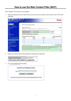


© Copyright 2026