
CLASS- III - MALOCCLUSION: A CEPHALOMETRIC STUDY IN A
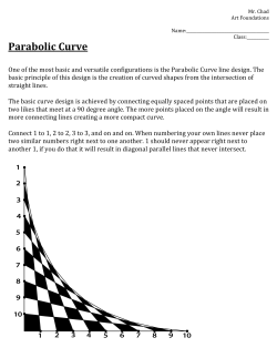
Pakistan Oral & Dent. Jr. 26 (2) Dec. 2006
CLASS- III - MALOCCLUSION: A CEPHALOMETRIC STUDY IN A
PAKISTANI POPULATION SAMPLE
*ARFAN-UL-HAQ, BDS, MCPS (Operative Dentistry), FCPS (Orth)
**M.WAHEED-UL-HAMID, BDS, M.S, MCPS (Orthodontics), M.Orth
ABSTRACT
This study was carried out to evaluate the Cephalometric characteristics of class-III malocclusion in
a sample of Pakistani population with age range 18-25 years visiting Orthodontics department,
de'Montmorency college of Dentistry / Punjab dental hospital, Lahore. Lateral Cephalometric
radiographs, Orthopentomograms, Study casts, extra oral and intra oral photographs were taken for
every patient. Results indicate that regarding the Cephalometric characteristics of Pakistani patients
with class III malocclusion, the Maxilla showed retrognathism in relation to cranial base both for
angular and linear parameters in antero-posterior relationship while the Mandible showed prognathism
in relation to cranial base both for angular and linear parameters in antero-posterior relationship.
However the Dento-alveolar measurements showed maxillary incisors proclination and mandibular
incisors retroclination, suggestive of dental compensation to skeletal discrepancy. Males showed
comparatively horizontal while females exhibited vertical growth tendency.
Key words: Class 111 malocclusion, cepahlomatric characteristics, Pakistani sample.
INTRODUCTION
Class III malocclusions are considered one of the
most complex and difficult orthodontic problems to
Skeletal Class III malocclusion is usually characdiagnose and treat10, 11,12 It is now well established that
terized by a steep mandibular plane angle, obtuse gonial
this malocclusion is not limited to dental discrepancies
angle, overdeveloped mandible, underdeveloped
but is often related to an underlying skeletal problem13.
maxilla, and a small cranial base angle, which may Studies conducted to identify the etiologic features of a
displace the glenoid fossa anteriorly to cause a forward Class III malocclusion showed that the deformity is not
positioning of the mandible1. This type of facial dyspla- restricted to the jaws but involve the total craniofacial
sia is produced by excessive growth disharmony of the complex 14,15,16.
mandible in size, form, and position with respect to
The previous investigators have described the
maxilla and / or the cranial base2, 3
morphologic characteristics of Pakistani population with
The prevalence of class III malocclusions varies respect to class I17 and class 116,18 relationships.
among the races. The percentage of this type of maloc- Although the reported prevalence of class III maloccluclusion in white population is less than 5 %. On the sion is different among races, no such study has been
other hand, the prevalence of skeletal class III maloc- previously conducted in Pakistan to examine the morclusion has been reported to be 0.5 % to 1.6 % in phologic characteristics of this type of malocclusion.
Caucasians4,5, 14.5 % in Chinese6, 4.0 % to 13. 0% in
The objective of this study was to evaluate the
Japanese', 9.4 % to 19 % in Koreans8and 2.9% in Indian
Cephalometric characteristics of class III malocclusion
children with age range of 9-14 years9.
* Senior Demonstrator, Department of Orthodontics, de,Montmorency College of Dentistry, Lahore
** Head, Department of Orthodontics, de,Montmorency, College of Dentistry, Lahore — Pakistan, E-mail:
[email protected].
Correspondence: Dr Arfan-ul-Haq, 385/F-II, Johar Town, Lahore, E-mail: [email protected].
191
in a sample of Pakistani population visiting Orthodontic
department, de' Montmorency college of Dentistry /
Punjab dental hospital, Lahore. This study will serve as
a useful tool in the diagnosis and treatment planning of
class III malocclusion.
MATERIALS AND METHODS
Fifty patients (25 males and 25 females) with age
range 18-25 years, having Angle's class III molar
relationship with no previous orthodontic treatment
27
were selected from orthodontics department of
de,Montmorency College of Dentistry/ Punjab Dental
Hospital, Lahore. Complete set of diagnostic records
28
including:
Lateral
Cephalometric
radiographs,
Orthopentomogram, Study casts, Extra oral and intra
oral photographs were taken for every patient. However patients with craniofacial skeletal deformities (cleft
lip and palate, facial asymmetry, syndromes), any
Fig. 1. Cephalometric Landmarks and Planes
previous trauma to dento facial structures, TMJ ankylosis or dislocation and missing or cariously damaged 1. S-Sella, 2. N-Nasion, 3. Ba-Basion, 4. Ar-Articulare,
dentition were excluded from the study.
5. Co-Condylion, 6. Pt.A, 7. ANS-Anterior nasal spine, 8.
Lateral Cephalometric radiographs for all the pa- PNS-Posterior nasal spine, 9. Upper incisor tip, 10.
tients were taken with the teeth in centric occlusion Upper incisor apex, 11. Lower incisor tip, 12. Lower
and relaxed lips. All the radiographs were taken in the incisor apex, 13. Pt.B, 14. Pog-Pogonion, 15. GnDepartment of Radiology, de'Montmorency College of Gnathion, 16. Me-Menton, 17. Go-Gonion, 18.
Pronasale, 19. Subnasale, 20. Upper lip, 21. Lower lip,
Dentistry/ Punjab Dental Hospital, Lahore.
22. Soft tissue Pogonion, 23. S-N Plane, 24.
All Lateral Cephalometric radiographs were traced
Palatal/Maxillary Plane, 25. Mandibular Plane, 26.
on 0.003 inches thick and 8x10 inches acetate sheet
Facial plane, 27. Rickett's E-Plane, 28. S-Line
with 4H lead pencil. All the radiographs were traced at
the same sitting to minimize tracing error. Cephalometric landmarks and planes were determined means, standard deviation, standard error of means
manually (Fig 1). The cranio facial relationships were and range for all the quantitative variables were anadivided in to following categories for analysis: Cranial lyzed (Table.2). A paired t-test was applied to
base, Maxillary, Mandibular, Dento-Alveolar and Soft calculate statistical significance level of any difference
tissue relationships (Table 1). The linear and angular between males and females (Table.3).
measurements were adopted from Downs, Steiner's,
To evaluate the method error, 15 radiographs
McNamara and Rickett's cephalometric analysis (Figs
were
randomly selected and manually retraced for all
2 and 3).
the variables. The retraced values were compared
The data was compared with Downs26, Steiner's19,
with first readings of the same radiographs, using
McNamara20, Rickett's25 and Tajik,s17"normal values for
paired t-test. There was statistically insignificant
class I skeletal relationships. Male and female samples
difference between first and second tracing on
were also compared to evaluate any sexual
applying paired t-test.
dimorphism.
The levels of significance used were a probability
Statistical analysis
value ofless than 0.001 (highly significant), a value
The database of all the measurements was devel- less than 0.01 (significant) and a value less than 0.05
oped in SPSS info version 8.0 software. The arithmetic (insignificant).
192
TABLE 1: CEPHALOMETRIC MEASUREMENTS USED IN PRESENT STUDY
Cranial
base
Maxillary
Mandibular
Intermaxillary
Facial
Heights
Dentoalveolar
Soft tissue
S-N (mm)
N-S-Ba (°)
SNA (°)
SN-PP (°)
Co-Pt.A (mm)
SNB (°)
SNPg (°)
SN-MP (°)
Y-axis (°)
S-Ar-Go (°)
Ar-Go-Gn (°)
Co-Gn (mm)
Go-Gn (mm)
ANB (°)
MMA (°)
PFH (mm)
AFH (mm
LAFH (mm)
SN-UI (°)
PP-UI (°)
IMPA (°)
IIA(e)
UL-E.line (mm)
UL-S.Line (mm)
LL-Sline (mm)
LL-S.line (mm)
NLA (°)
Fig. 2. Cephalometric Angular Measurements
1. N-S-Ba, 2. SNA, 3. SNB, 4. SN-Pg, 5. SN-PP, 6. SNMP, 7. Y.axis (N-S-Gn) , 8. MMA (Maxillomandibular
plane angle), 9. S-Ar-Go, 10. Ar-Go-Gn, 11. SN-UI, 12.
PP-UI, 13. IMPA, 14. IIA, 15. NLA (Nasolabial angle).
RESULTS
Over all skeletal and dentoalveolar relations
including cranial base, maxillary, mandibular,
Intermaxillary, dentoalveolar and soft tissue
measured in 50 patients of both gender were reviewed. The mean, standard deviation, standard
error of means and range for all cephalometric
variables of class III malocclusion are presented in
Table 2. The comparative means, standard deviation
Fig. 3. Cephalometric Linear Measurements
1. S-N, 2. Co-Pt A, 3. Co-Gn, 4 Gn-Gn, 5. PFH (S-Go),
6. AFH (N-Me), 7. LAFH (ANS-Me), 8. UL-E. Line, 9.
UL-S.line, 10. LL-E.line, 11. LL-S.line
and significance for males and females are summarized
in Table 3.
Cranial Base Relationships
It is observed that the mean measurement of S-N
was 72.08 ±4.59 mm. On the other hand, the mean
value of N-IIA(°for entire sample was 130.16°± 6.16.
The mean value for cranial base length (S-N) in males
193
TABLE 2: CEPHALOMETRIC MEASUREMENTS FOR ENTIRE SAMPLE
(Cranial base, Maxillary, Mandibular, Intermaxillary, Dentoalveolar and Soft tissue)
n=50
Variable
Mean
SD
SEM
MIN
Max
S-N (mm)
N-S-Ba (°)
SNA (°)
SN-PP (°)
Co-Pt.A (mm)
SNB (°)
SN-Pog (°)
SN-MP (°)
Y-Axis (°)
S-Ar-Go (°)
Ar-Go-Gn (°)
Co-Gn(°)
Go-Gn (mm)
ANB (°)
MMA (°)
PFH/TAFH (%)
LAFH/TAF'H(%)
UI-SN (°)
UI-PP (°)
IMPA (°)
HA (°)
UL-E (mm)
LL-E (mm)
UL-S (mm)
LL-S (mm)
NLA (°)
72.08
130.16
78.94
7.86
87.46
82.46
83.48
33.66
66.98
140.60
125.90
126.32
86.24
-3.52
26.56
65.29
57.96
112.56
119.68
85.68
127.90
-7.64
-1.40
-2.76
1.54
94.48
4.59
6.16
4.09
2.91
5.42
3.91
3.88
6.58
3.45
7.45
5.61
10.43
6.27
2.42
5.92
4.81
3.76
8.54
8.94
6.01
8.53
3.24
3.57
2.55
2.77
8.27
0.65
0.87
0.58
0.41
0.77
0.55
0.55
0.93
0.49
1.05
0.79
1.48
0.89
0.34
0.84
0.68
0.53
1.21
1.26
0.85
1.21
0.46
0.51
0.36
0.39
7.17
65
118
70
0
78
78
76
18
60
128
115
109
78
0
12
55.3
52.55
98
109
76
111
0
+5
+2
+6
81
82
142
88
13
97
92
93
48
73
154
140
155
107
-9
40
78.8
76.60
132
143
103
140
-15
-10
-9
-5
122
sample. The maxillary length Co-Pt A was also less as
compared to normal value for class I subjects. The
vertical relation of maxilla to cranial base shown by
angle SN-PP was within normal range. The mean value
for
maxillary
length
(Co-Pt.A)
in
males
(90.28±5.88mm) was 5.64 mm larger thLAFH/TAFH
Maxillary Relationships
(84.64±2.98mm) at significance level of p<0.001. The
The mean values for angular relationships for SNA difference between male and females for SNA and SNand SN-PP were 78.94U± 4.09 and 7.86U± 2.91 respec- PP remained insignificant (Tables 2 and 3).
tively. Mean linear measurement of overall sample for
Mandibular Relationships
Co-Pt.A was 87.46±5.42 mm. The anteroposterior relaThe mean angular degrees of SNB, SN-Pog, SNMP,
tion of maxilla to cranial base was determined by angle
SNA with decreased value as compared to norms for Y-Axis, S-Ar-Go and S-Ar-Gn were 82.46°±3.91,
83.48°±3.48, 33.66°±6.58, 66.98°±3.45, 140.60°±7.45 and
class I relations suggestive of deficient maxilla in this
(74.56±4.20 mm) was higher than females (69.60±3.54
mm) at highly significant level of p<0.001. The average
values regarding N-S-Ba for male and female subjects
were 128.76°±5.93 and 131.56°±6.19 with statistically
insignificant difference (Tables 2 and 3).
194
TABLE 3: CEPHALOMETRIC MEASUREMENTS FOR MALE AND FEMALE SAMPLE
(Cranial base, Maxillary, Mandibular, Intermaxillary, Dentoalveolar and Soft tissue)
n=50
Variable
S-N (mm)
N-S-Ba (°)
SNA (°)
SN-PP (°)
Co-Pt.A (mm)
SNB (°)
SN-Pog (°)
SN-MP (°)
Y-Axis (°)
S-Ar-Go (°)
Ar-Go-Gn (°)
Co-Gn (°)
Go-Gn (mm)
ANB (0)
MMA(°)
PFH/TAFH(%)
LAFH/TAFH(%)
UI-SN (°)
UI-PP (°)
IMPA (°)
IIA (°)
UL-E (mm)
LL-E (mm)
UL-S (mm)
LL-S (mm)
NLA (°)
MALE (25)
Mean
SD
74.56
128.76
79.84
8.48
90.28
83.40
84.40
32.48
66.76
139.28
126.20
130.04
83.40
-3.56
24.60
66.32
57.91
114.92
122.32
86.44
126.52
-7.92
-1.20
-2.72
1.96
94.52
4.20
5.93
3.70
2.52
5.88
4.12
4.19
6.62
3.46
8.64
5.80
11.76
6.66
2.20
6.06
4.63
4.61
8.55
8.58
5.99
9.85
3.53
3.63
2.97
2.73
9.79
FEMALE (25)
Mean
SD
Diff
Diff
Significance
P-value
69.60
131.56
78.04
7.24
84.64
81.52
82.56
34.84
67.20
141.92
125.60
122.60
84.28
-3.48
28.52
64.25
58.01
110.20
117.04
84.92
129.28
-7.36
-1.60
-2.80
1.12
94.44
4.96
2.80
1.80
1.24
5.64
1.88
1.84
2.36
0.44
2.64
0.60
7.44
0.88
0.08
3.92
2.07
0.10
4.72
5.28
1.52
2.76
0.56
0.40
0.08
0.84
0.08
P<0.001
NS
NS
NS
P<0.001
NS
NS
NS
NS
NS
NS
P<0.01
P<0.01
NS
P<0.01
NS
NS
P<0.01
P<0.01
NS
NS
NS
NS
NS
NS
NS
125.90°±5.61. The average measurements of Co-Gn and
Go-Gn were 126.32±10.43 mm and 86.24±6.27 mm (Table
2). The Comparison of males and females for Mandibular
Skeletal Relationships was carried out. Mandibular Skeletal
Relationships including SNB, SNPog, SN-MP, Y-Axis, SAr-Go and Ar-Go-Gn for males were 83.40°±4.12,
84.40°±4.19, 32.48°±6.62, 66.76°±3.46, 139.28°±8.64,
126.20°±5.80 degrees and those for females were
81.52°±3.51, 82.56°±3.37, 34.84°±6.46, 67.20°±3.50,
141.92°±5.92, 125.60°±5.53. It was observed that the angle
of SNB, SN-Pog, and Y-Axis were more or less similar in
both sexes. However the angle of SNMP showed variation
among male and female patients
3.54
6.19
4.33
3.19
2.98
3.51
3.37
6.46
3.50
5.92
5.53
7.42
5.21
2.66
5.17
4.86
2.75
8.02
8.66
6.06
6.90
2.96
3.58
2.12
2.80
6.62
at significance level ofp< 0.01. The mean values for
CoGn and Go-Gn of males were 130.04±11.76mm,
83.40±6.66 mm and those for females were
122.60±7.42mm and 84.28±5.21mm. There was statistically significant difference at p<0.01 between male
and female samples for Co-Gn and Go-Gn (Table 3).
Intermaxillary Relationships
The Inter-Maxillary Relationships include ANB
(difference of SNA and SNB angles) and MMA (maxillary to mandibular plane angle). It is observed that
mean angle of ANB and MMA was -3.52°±2.42 and
26.56°±5.92. The mean percentages of PFH/TAFH and
195
LAFH/TAFH of total subjects were 65.29±4.81% and
57.96±3.76% (Table 2). A Comparison of males and
females for Inter-Maxillary Relationships was also
carried out. It is observed that mean angle of ANB is
quite similar in both sexes (-3.56°±2.20 and-3.48°±2.66)
and their comparison shows no significant difference.
On the other, the angle of MMA was significantly high
(P<0.01) in females (28.52°±5.17) as compared to male
patients (24.60°±6.06) (Table 3). The mean ratio of
PFH/AFH in males and females was 66.32%±2.07 and
64.25%±4.86 with statistically insignificant difference.
The mean ratio of LAFH/AFH in males and females was
57.91%±4.617% and 58.01%±2.75 with statistically
insignificant difference (Table 3).
Dentoalveolar Relationship
Dento-Alveolar relationships include the angles of
UI-SN (Upper incisor-Sella Nasion plane), UI-PP (Upper incisor-Palatal/maxillary plane), IMPA (Lower incisor to mandibular plan angle) and IIA (inter incisor
angle). It is observed that the mean angles of UI-SN and
UI-PP were 112.56°±8.54 and 119.68°±8.94. The mean
angles of IMPA and of IIA were 85.68°±6.01 and
127.90±8.53° (Table 2). It is also observed that the
angle of UI-SN and UI-PP were smaller in female
patients (110.92°±8.02 and 117.04°±8.66) as compared
to angles for male patients (114.92°±8.55
and122.32°±8.58) with significant difference (P<0.01).
The values for IMPA and IIA were also different in
female (84.92°±6.06 and 129.28±6.90°) as compared to
the angles of male (86.44°±5.99 and 126.52°±9.85) but
there was no significant difference (Table 3).
94.44U±6.62) was also figured and found quite similar
relationship in both sexes (Table3).
DISCUSSION
The review of literature indicated that there was a
need to evaluate Cephalometric characteristics of class
III malocclusion in Pakistani population. So, this study
was undertaken to disclose the morphologic characteristics of the craniofacial complex of Pakistani adults
with class III malocclusion.
The antero-posterior relationship of maxilla to
cranial base as shown by angle SNA for this sample
was 78.94t, and this value is suggestive of class III
relationship as compared to normal values of
Steiner19(82Ú) for the Caucasian population, Tajik17
(81.25Ú) for Pakistani population. Using angular
analysis SNA; 54 % showed retrusive maxilla, 32%
normally positioned while 14% prognathic maxillary
relationship to cranial base. This confirmed that a
retrusive maxilla in relation to cranial base is one of the
etiologic factors for class III malocclusion. In linear
analysis using Co-Pt.A, this sample showed 3.24 mm
decreased length as compared to normal value for class
I sample from Burlington Orthodontic research Centre.
This further confirmed short maxillary length a feature
of class III malocclusion.
The antero-posterior relationship of mandible to
cranial base as shown by angle SNB for this sample was
82.46U, and this value is suggestive of class III relationship as compared to normal values of Steiner19(80Ú for
the Caucasian population, Tajik17 (78.97U) for Pakistani
population. Using angular analysis SNB; 62 % showed
Soft tissues Relationships
protrusive mandible, 34% normally positioned while
4% retrusive mandibular relationship to cranial base.
Mean soft tissue relationship (UL-E, LL-E, UL-S
This confirmed that a protrusive mandible in relation to
and LL-S) in total patients was —7.64±3.24, -1.40±3.57,
cranial base is one of the etiologic factors for class III
-2.76±2.55 and 1.54±2.77 mm. However, the mean
malocclusion.
nasolabial angle in whole subjects was 94.48°±8.27
(Table 2).
The effective Mandibular length (Co-Gn) in males
(130.04 mm) was 7.44 mm greater than females (122.60
Soft tissue relationships (UL-E, LL-E, UL-S and
mm). These values of effective Mandibular length for
LL-S) in male / female subjects were figured (- 7 .92m
males and females of this sample are comparatively
m ±3 .53 , -1.20mm±3.63, -2.72mm±2.97, 1.96mm±2.73
larger than Ann Arbor norms for normal well-balanced
and —7.36mm±2.96, -1.60mm±3.58, - 2.80mm±2.12,
faces and good occlusion 20.
1.12mm±2.80). An insignificantly higher soft tissue
The Mandibular corpus length (86.24 mm) in this
relationship was observed in female patients as
class
III sample was 7.00 mm greater than X+7 that
compared to the male patients. A comparison of
showed an increased length of the body of mandible as
Nasolabial angle in both sexes (94.52°±9.79 and
196
compared to anterior cranial base length. As a compariThe mean value of maxillary-mandibular
son with Tajik's17 Norms (78.31), the body of mandible plane (MMA) angle in males/females showed that
in our sample is 8 mm large suggestive of enlarged body females have more vertical pattern of growth than
of mandible.
males. This also confirmed the tendency toward
vertical growth in females exhibited by ManIn linear analysis, total Mandibular length and
dibular angular measurements for vertical relationMandibular corpus length showed much greater values
ships.
than normal, which means that prognathic mandible act
as a feature of class III malocclusion. These findings
The dento-alveolar measurements were taken for
are similar to those observed by previous investigators UI-SN plane, UI to PP, LI to MP (IMPA), and
3,14,15,21,22.
The males of this sample exhibited larger Interincisal angle (IIA). These values are more than
values for SNB, SN-Pog, Co-Gn, and Go-Gn than normal range, which means that upper incisors are
females.
proclined due to dental compensation in class III
subjects. These results are in consistent with other
Using angular measurements (SNB, SN-Pog), the
studies9, 22,23,24. The mean values for UI to SN plane in
males showed insignificantly larger values for anteromales and females showed that females of this sample
posterior relation of mandible to cranial base. But there
have significantly less proclination of upper incisors.
were significantly (p<0.01) larger values of linear
Similarly the mean value of UI-PP angle for the males
measurements (Co-Gn, and Go-Gn) in males of this
and females confirmed that females have less incisor
sample than females, which meant that males with class
proclination as compared to males with a significant
III malocclusion had larger antero-posterior size of
difference.
mandible than females.
The mean value for the lower incisor to mandibular
The mean value for SN-plane to mandibular plane
plane angle showed a lower value. This lower value is
angle, for goni al angle and for the Articulare angle
suggestive of mandibular incisor's retrusi on as a result
was compared with normal occlusion values of a
of dental compensation to underlying skeletal discrepstudy", all these values fall in the normal range. This
ancy. The mean value for interincisal angle (HA) of
means that all the patients have normal growth pattern
entire sample is slightly more than normal occlusion
and vertical relationship to cranial base. The females of
value ofinvestigator17" and less than the study
this sample showed higher values for SN-MP, Y-axis,
ofnormal value of another investigator19. This showed
Gonial angle, and Articulare angle than males. This
that there is less change in IIA due to upper and lower
means that females with class III malocclusion have
incisors compensation.
slightly vertical growth pattern while males shows
normal growth pattern.
CONCLUSIONS
The mean value of ANB angle for entire sample
It is therefore concluded that regarding the Cephawas less than normal value of a study19, and also
lometric characteristics of Pakistani patients with class
significantly less than that value for ANB in the normal
III malocclusion;
occlusion sample of a pollster". This value is suggestive of class III relationship between maxillary and 1. The maxilla showed retrusive relation to cranial
mandibular apical bases. The analysis of ANB angle
base antero posteriorly for both angular and linear
revealed that 96% of the sample exhibited class III
measurements
relationship. A retrusive maxilla and an overdeveloped
2. The Mandible showed prognathism in relation to
and prognathic mandible caused this significant undercranial base both for angular and linear parameters
lying skeletal discrepancy. The mean value for the
in antero-posterior relationship.
ANB angle in males/females showed an insignificant
difference of 0.08°. However the mean value of maxil- 3. The Dento-alveolar measurements showed maxillary-mandibular plane angle shows normal relationship
lary incisors proclination and mandibular incisors
of maxilla and mandible in vertical plane for this
retroclination, suggestive of dental compensation
sample.
to skeletal discrepancy.
197
4. Males exhibited more dominant anteroposterior
dysplasia while females showed more vertical
growth tendency.
REFERENCES
1. Sato S. Developmental characterization of
malocclusion; Angle Orthod 1994; 2:105-12.
Class
III
2. Mitani H, Sato K, Sagawara J. Growth of mandibular prognathism after pubertal growth peak. Am J Orthod
Dentofac Orthop 1993; 104:330-6.
3. Jacobson A, Evans WG, Preston
PL.Mandibular prognathism. Am J
Orthop 1974; 66:141-71.
CB, Sandowsky
Orthod Dentofac
4. Ast DB, Carlos JP, Cons NC, The prevalence and characteristics of malocclusion among senior high school students in
upstate New York. Am J Orthod 1965; 51:437-445.
5. Barret MJ, Brown T, Macdonald MR. Dental observation on
Australian Aborigines: A roentgenographic study of prognathism. J Dent Aust 1963; 8:418-427.
6. Qamar R. Cephalometric characteristics of class II malocclusion in a Pakistani population sample. FCPS Dissertation
2002, Karachi: College of Physicians and Surgeons Pakistan.
7. Lew KKK, FoongWC. Horizontal skeletal Typing in an ethnic
Chinese Population with true Class III malocclusion. Br J
Orthod 1993; 20:19-23.
8. Lim HH, Yoon Yi, Kim KW. A study of the characteristics of
craniofacial skeleton on orthognathic surgical cases with
skeletal class III malocclusion. Korean J Orthod 1998; 28:
189-208.
9. Grew JM, Carvenka J, Shapro BL, witkop CJ. Prevalence of
malocclusion in Chippewa Indian children. J dent Res
1968;47:302-5.
10. Mouakeh M. Cephalometric evaluation of craniofacial
pattern of Syrian children with class III malocclusion. Am
J Orthod Dentofac Orthop 2001; 119:640-49.
11. Eisenhauer AS, Lux CJ, Schuster G. Treatment decision in adult
patients with class III malocclusioin:Orthodontic therapy or
orthodontic surgery ? Am J Orthod Dentofac Orthop 2002;
122:27-38.
12. McNamara JA Jr. What works, what doesn't, and Why. (Ed.),
Craniofacial Growth Series 35, Center for Human Growth
198
and Development, The university of Michigan, Ann Arbor,
1999.
13. Kapust AJ, Sinclair PM, Tirley PK Cephalomatric effects of
face mask/expansion therapy in class 111: a comparison
of three age groups. Am J Orthod Dentofac Orthop. 1998,
113:204-12
14. Guyer EC, Ellis E, McNamara JA Jr, Behrents RG. Components of class III malocclusion in juveniles and adolescents.
Angle Orthod 1986; 56:7-31.
15. Chang HP, Kinoshita Z, Kawamoto T. Craniofacial pattern
of class III deciduous dentition. Angle Orthod 1992;62:
139-44.
16. Battegal JM. The aetiological factors in class III malocclusion.
Eur J Orthod 1993; 15:247-70.
17. Tajik I. Cephalometric pattern of a Pakistani population.
FCPS Dissertation, Karachi: College of Physicians and
Surgeons Pakistan, 2000: 5-6.
18. Firdos T. Investigation of Class II malocclusion-A study conducted at Khyber College of Dentistry Peshawar-Pakistan.
Pakistan Oral and Dental Journal 2000; 20:158-64.
19. Steiner CC. Cephalometrics for you and me. Am J Orthod
1953; 39:729.
20. McNamara JA Jr. A method of cephalometric evaluation.
Am J Orthod 1984; 86:449-69.
21. Dietrich UC. Morphologic variability of skeletal class III
relationships as revealed by cephalometric analysis.
Trans Europ Orthod Soc 1970; 46:131-43.
22. Mayajima K, McNamara JA Jr, Sana M, Murata S. An estimation of craniofacial growth in untreated class III female with
anterior cross bite. Am J Orthod Dentofac Orthop 1997;
112:425-34.
23. Rakosi T. Treatment of class III malocclusion. In; Graber TM.
Rakosi T, Petrovie A, editors. Dentofacial Orthopedics with
functional appliances. St. Louis (MO): Mosby; 1997.
24. Ellis E, McNamara JA Jr. Components of adult class III
malocclusion. Am J Oral Maxillofacial Surg 1984; 42:
295-305.
25. Ricketts RM. Planning treatments on the basis of the facial
pattern and an estimate of its growth. Angle Orthod
1957; 27: 14
26. Downs WB. Variations in facial relationships: their significance
in treatment prognosis. Am J Orthod 1948; 34:812-40.


© Copyright 2026