
Document 276181
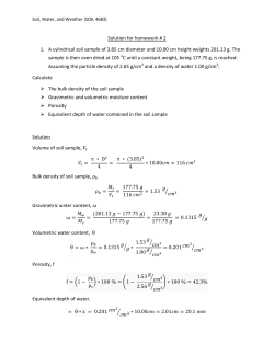
Method Development and Sample Processing of
Water, Soil, and Tissue for the Analysis of Total
and Organic Mercury by Cold Vapor Atomic
Fluorescence Spectrometry
R. b. Jones TM, M.E. Jacgbson 1, R. Jaffe 2,3, J. West-Thomas 1, C. Arfstrom ~
and A. Alli ~'2
1 Southeast Environmental Research Program, 2 Department of Chemistry,
3 Drinking Water Research Center, 4 Department of Biological Sciences.
Florida International University, University Park, Miami, Florida 3 3 1 9 9 , USA.
Atomic Fluorescence-basedmethodshave beendevelopedfor measuringultratrace levels
of mercury (Hg) in environmental (water, soil) and biological (fish tissue) samples. In addition,
methods for preparationof water, soil, and tissue samples have been developed.For the analysis
of total Hg in soil, sediment and fish the samples are digested with concentrated nitric acid in
sealed glass ampules, and subsequently autoclaved. Water samples are diges~tedusing standard
brominating procedures. A Merlin Plus, PS Analytical atomic fluorescence spectFometer(AFS)
system equipped with an autosampler, vapor generator, fluorescence detector and a PC based
integrator package is used in the determination of total Hg. The determination of Hg mercury
species in water, without pre-derivatization, involves adsorbent pre-eoncentration of the
organomercurialsonto sulfydryl-cotton fibers. The organic Hg compounds are elutad with a small
volume of acidic KBr and CuSO4and extracted into dichloromethane. Sediment, soil and tissue
samples are homogenizedand the organomercurialsfirst releasedfrom the sampleby the combined
action of acidic KBr and CuSO4 and extracted into dichloromethane. The initial extracts are
subjected to thiosulfate clean-up and the organomercury species are isolated as their chloride
derivatives by cupric chloride addition and subsequent extraction into a small volume of
dichloromethane. Analysis of organic Hg compounds is accomplished by capillary column
chromatography coupled with atomic fluorescence detection.
Abstract.
1. Introduction
Mercury is a w i d e l y distributed pollutant in the environment and has gained
considerable toxicological concern in recent years. In some cases, the desired
quantitation levels of this metal challenge the detection limits of the
instrumentation and methods in current use (Swift and Campbell, 1993;
Kammin and Knox, 1992). This has certainly encouraged the development of
sensitive, reliable and precise methods for the analysis of Hg. Further, the
organic forms of Hg, particularly m e t h y l m e r c u r y (CH~Hg*), are far more toxic
than the inorganic forms (Hg 2§ Hg ~ of the pollutant (Rubi et al., 1992; Bryan
and Langston, 1992). In efforts to project long-term health risks and the
ecological impact associated w i t h trace amounts of Hg in the environment,
reliable quantitation and accurate speciation at increasing lower levels are
necessary.
The open-vessel digestion procedures and detection methods for total
Hg analysis in w a t e r samples that are c o m m o n l y used are based on acid
leaching and permanganate/persulfate oxidation followed by cold vapor atomic
absorption (CVAAS), (Szak,~cs et al., 1980; Van Delft and Vos, 1988). One of
the most c o m m o n l y used analytical technique for the determination of
organomercurials is gas chromatography w i t h electron capture detection (GCECD), (Rubi et al., 1992; O'Reilly, 1982) with or w i t h o u t pre-derivatization of
the organic mercury compounds. The instrumentation and sample preparation
Water, Air, and Soil Pollution 80: 1285-1294, 1995.
9 1995 Kluwer Academic Publishers. Printed in the Netherlands.
1286
R.D. JONES ET AL.
of the existing methods strongly limit the ultimate sensitivity and efforts to
lower the detection limits have not been entirely successful (Swift and
Campbell, 1993). In addition, the ECD is an unselective detector and the
column has to be tediously conditioned with large injections of Hg (11)chloride
to alleviate poor chromatographic response to organomercurials (Hight and
Capar, 1984; Rubi et al., 1992; Uthe et al., 1972; Bryan and Langston, 1992;
Bulska et al., 1992). These disadvantages demonstrate the need for the
development of new methods in this field.
This paper describes atomic fluorescence-based methods for analyzing
total H g and organic Hg compounds at low part-per-trillion levels in
environmental and biological samples. The atomic fluorescence (AFS) method
(Bloom, 1989; Alli et al., 1994) has become increasingly important compared
to CVAAS, since the instrumental detection limit of this method is about 1
picogram or less and at least one order of magnitude better than for CVAAS
(Lindqvist, 1993). Total Hg analysis involves three stages: sample digestion,
cold vapor generation and atomic fluorescence detection.
In water samples, the difficulty in measuring MeHg and other
organomercurials lies in concentrating these compounds from solution. This
work employs a sulfydryl cotton fibre medium (Lee and Mowrer, 1989) which
effectively adsorbs and preconcentrates trace levels of organomercurials. The
organic Hg compounds are eluted with acidic potassium bromide and extracted
into dichloromethane and subjected to GC analysis with AFS detection. Soil,
sediment and tissue samples are treated with acidic potassium bromide and
copper sulfate, and extracted with dichloromethane. The initial extracts are
subjected to sodium thiosulfate clean-up subsequent to capillary gas
chromatography with atomic fluorescence detection (Alli et al., 1994).
2. Materials and Methods
2.1 SAMPLE COLLECTION AND PREPARATION
Surface water samples are collected in 2 L Teflon (Nalgene) bottles using a
vacuum system. Samples are screened (105 pm Nytex netting) to prevent the
collection of large particles with the water samples. All tubings and fittings
used in the sampling system are constructed of teflon (SERP, internal SOP,
1994). The samples are collected by a "clean person" using double gloves
(short vinyl gloves under shoulder length polyethylene gloves, OakTech) and
a double bagging technique. All samples are placed in zip-lock polyethylene
bags (Fisher Scientific), then in an additional plastic sample bag and placed in
an icechest/cooler. In the clean room, concentrated hydrochloric acid, (trace
metal grade, Fisher Scientific) is added to the water samples for preservation.
Surface, soil and sediment samples are collected using either a stainless
steel spade, trowel or Eckman dredge. These samples are placed into wide
mouth polyethylene specimen cups (125 ml, Fisher Scientific). Subsurface soil
or sediment samples are collected in polycarbonate core tubes. Upon arrival to
the laboratory the samples are immediately frozen to preserve their chemical
integrity.
Fish samples are collected using a dip net, with the sampler wearing
two pairs of gloves. The fish are placed in zip-lock sample bags, labelled, and
stored in a cooler with ice for transport to the laboratory. Fish samples remain
frozen until ready fo r analysis.
2.2 SAMPLE DIGESTION FOR TOTAL MERCURY DETERMINATION
Water Samples. Water samples are digested in a 125 mL teflon bottle with 1
mL HCI and 2.5 mL potassium bromate (KBrOs)/potassium bromide (KBrO)
mixture overnight (Szak~cs et. al., 1980; Bloom and Fitzgerald, 1988}. These
COLD VAPOR ATOMATIC FLUORESCENCE SPECTROMETRY
1287
samples are prepared and remain (in capped bottles) in the Hg-clean room.
Prior to analysis, 500 pL hydroxylamine hydrochloride is added to destroy
excess bromine and the samples thoroughly shaken.
Soil and Sediment Samples. Soils (such as peat, marls and marly peat) are first
homogenized by adding 30 to 50 mL of deionized water and blended for 3
minutes to a uniform consistency with a blender (Osterizer). From the
homogenized slurry 5 mL is diluted into 45 mL of 0.6N HCI to neutralize any
carbonates, in a clean specimen cup. Of this mixture 1 mLis placed in a 10 mL
ampule with 2 mL of concentrated nitric acid (HN03), (trace metal grade, Fisher
Scientific), Digested soil and sediment samples are left to stand under a fume
hood for 20 minutes. The ampules are subsequently sealed and autoclaved for
1 h at 151~
Before analysis the digestates are diluted with 0.12N HCI
solution in a 20 mL polyethylene vial.
Fish Samples. To quantify total Hg in small fish (< 0.4 g, < 30 mm in length)
the entire fish is weighed and placed in 10 mL ampules and digested using 1
mL deionized water and 2 mL concentrated HN03. After standing 20 minutes
under a fume hood, the ampules are sealed and autoclaved as described above.
For the analysis of larger fish (approximately 30 cm or longer), 3 tissue plugs
(stainless steel core tube, 4 mm in diameter) are taken from the left side (using
only muscle tissue), and combined to obtain a representative sample
(approximately 0.4 g). The samples are then processed as indicated above for
soil and sediment.
These digestion procedures result in the conversion of organic forms
of Hg to inorganic mercury (Hg2*). The digested samples are introduced to the
cold vapor generator, at which point tin (11) chloride is used to effectively
reduce inorganic mercury (Hg 2§ to its elemental gaseous form (Hg~ prior to
detection by atomic fluorescence.
2.3 INSTRUMENTATION AND ANALYSIS
A PS Analytical Merlin Mercury Fluorescence Detector System used in this
study was supplied by P.S. Analytical Ltd. (UK). This system incorporates an
Autosampler, Vapor Generator, Fluorescence Monitor and an IBM compatible
Computer System. Instrument operating conditions for ultratrace and high
levels of Hg concentrations are given in Table I.
Table L O/Jb'rnized o/~erab'n.q condib'ons of the AFS System for totaI-Hq
concentrations.
Ultratrace levels
Carrier gas mL/min
Sheath gas mL/min
Calibration range
Fine grain
Damping Switch
125
150
1000
4.0
On
High levels
Carrier gas mL/min
Sheath gas mL/min
Calibration range
Fine grain
Damping Switch
200
350
100
2.5
On
1288
R.D. JONES
ETAL.
Cold Vapor Generation. In the continuous flow vapor generator system, Hg(ll)
is reduced to Hg ~ following the addition of tin(ll) chloride. The volatile Hg is
stripped from the solution (in the gas liquid separator) by a carrier gas (argon).
The rate of argon flow depends on whether the analysis is for ultratrace or high
levels of Hg determination (P.S. Analytical, 1991).
Atomic Fluorescence Detection. A sheath gas (also argon) is used to channel
the Hg vapor through a chimney past a light source and a photomultiplier tube
that are at right angles to each other. With a specific high intensity Hg lamp
source (Cathodeon Ltd., Cambridge) and a fixed 254 nm filter, efficient
isolation Qf the required excitation and emission wavelengths is achieved (P.S.
Analytical, 1992).
Reagents. All reagents used in total Hg analysis are of certified ACS grade and
obtained commercially from Fisher Scientific, unless otherwise stated. A
Barnstead B-pure system (located in the Hg-clean room) produces all deionized
water used in making up reagents, sample digestates, calibration solutions,
stock solution and quality control standards. This water is first filtered through
a Culligan system consisting of activated charcoal and two mixed bed ion
exchange cartridges before being piped to the Hg-clean room. 0.1 N KBrO3, 0.2
N KBrO and 1.7 M hydroxylamine hydrochloride solutions are made up by
dissolving the appropriate amounts of the salts in deionized water. The KBrO3
and KBrO salts are heated overnight in a glass vial at 250~ to remove
adsorbed Hg. The digesting solution is made up daily by mixing equal volumes
(100 mL) of 0.1N KBrO3 and 0.2N KBrO solutions. All solutions are prepared
weekly and stored in borosilicate bottles with teflon lined caps.
Standards. Working standards are prepared daily from a Hg stock solution (100
ng/mL) and diluted to the desired concentration. The stock solution is also
made up daily from a commercially available mercury standard ( 1000 pg/mL,
SPEX Industries, Edison, NJ). Calibration solutions are made up in 500 mL
teflon bottles and stabilized by adding 5 mL concentrated HCI. No certified
material exists for quality control of Hg in water near ultratrace levels. Soil
(NIST sediment nominal value 60 ng/g, 8406) and tissue (NBS 1566a Oyster
Tissue, 64 ng/g) quality control standards are obtained from the National
Institute of Standards and Technology (Gaithersburg, MD).
2.4 ORGANIC MERCURY DETERMINATION
Sediment, Soil and Tissue Samples. A 1.0-5.0 g portion of the homogenized
sample (as prepared above) is placed in a 20 mL borosilicate glass scintillation
vial (Kimble, #74511 ). To the vial 5 mL distilled water, 3.0 mL of 1.0 M copper
sulfate and 3.0 mL of acidic potassium bromide solution are added. The
mixture is shaken for 1 hr at 330 rpm (Gyrotory Shaker Model G2).
Dichloromethane (5 mL) is added and the mixture is shaken for 24 h at 330
rpm and then centrifuged for 10 min at 5000 x g in a Sorvall Model RC-5
refrigerated centrifuge (Dupont). An exactly known volume of the
dichloromethane layer (3.5-4.0 mL) is transferred to a 7.0 mL borosilicate glass
scintillation vial (Kimble, #0333726) and 1.0 mL of 0.01 M sodium thiosulfate
is added. The mixture is shaken for 20 min at 330 rpm and centrifuged at high
speed in a IEC clinical centrifuge. The aqueous layer (0.9 mL) is placed in a 2;0
mL microcentrifuge tube (Fisherbrand, Fisher Scientific), and 0.3 mL of 0.5 M
copper chloride and 0.3 mL dichloromethane are added. The contents are
mixed for 1 rain on a Vortex Genie mixer and centrifuged for 2 min at high
speed (16,749 x g) in a Hermle centrifuge. The dichloromethane is transferred
to a 2.0 mL glass sampling vial containing a few crystals of anhydrous sodium
sulphate and subjected to GC analysis. Injections of 5.0 pL are used. Samples
spiked with known concentrations of methyl - and ethlymercury chloride are
COLD VAPOR ATOMATIC FLUORESCENCE SPECTROMETRY
1289
extracted to evaluate the recovery factor used for quantification.
Water Samples. The sulfydryl-cotton (SFC) fibre columns are made of 1 mL
disposable pipette tips containing 0.1 g of SFC fibre, packed loosely and as
evenly as possible. Two SFC columns are connected in series and the water
sample is passed through these by vacuum. One mL of acidic potassium
bromide and 0.5 mL of 1.0 M copper sulfate are then pipetted on the surface
of the adsorbent and the eluate is collected in a 2 mL micro-centrifuge tube
(Fisher Scientific). This is extracted with 0.2 mL dichloromethane on a Vortex
Genie mixer for 1.5 min and centrifuged as described above. The
dichloromethane layer is then transferred to a 2 mL glass sampling vial
containing a few crystals of anhydrous sodium sulfate and subjected to GC
analysis.
2.5 INSTRUMENTATION AND ANALYSIS
A schematic diagram of the GC-AFS system used in this work is shown in
Figure I and the optimum operating conditions are summarized in Table II. A
E15
A
B
IA
I
I
0
Figure I. Gas'Chromatographic-Atomic Fluorescence Spectrometric System.
1A: Helium, 1 B: Argon, 2: Oxygen trap, 3: Mercury trap, 4: Moisture trap, 5:
Automatic sampler, 6: Injector, 7: Column, 8: Press-fit union, 9: Pyrolyser, 10:
Deactivated fused-silica 0.53mm i.d., 11: Teflon unions, 12: Teflon transfer
line 0.Smm i.d., 13: Atomic Fluorescence detector, 14: E-Lab chromatographic
control and data acquisition system, 15: Mass flow controller-Channel A makeup, Channel B sheath gas.
Hewlett-Packard (Model 5890 Series II) gas chromatograph coupled with an HP
(Model 7673) automatic sampler is used. A fused-silica, bonded phase
megabore column (15 m x 0.53 mm i.d., 1 pm non-polar DB-1 coating, J & W
Scientific) and the splitless injection mode is employed. The effluent from the
1290
R.D. JONES ET AL.
column is led through a pyrolyser (P.S. Analytical Ltd., UK), positioned inside
the GC oven via a piece of 65 cm length of deactivated fused-silica (0.53 mm
i.d., J & W Scientific), which is connected to the column with a glass "press
fit" union (J & W Scientific). The Hg atoms formed in the pyrolysis unit are
transferred from the outlet end of the deactivated fused-silica tubing to the
fluorescence detector (teflon transfer line, 0.5 mm i.d., AIItech Associates).
The transfer line is passed through a small hole on the top of the GC oven to
a Merlin Mercury Fluorescence Detector, and the connections are made via
teflon unions.
Table II. Optimized operating conditions of GC-AFS.
Gas chromatograph.
Injector temperature
250~
1 min at 40~ 60~
Temperature program
3 min at 140~ 50~
10 min at 200~
Pyrolyser temperature
800~
Column flow
4.0 mL/min
Make-up flow
60 mL/min
Atomic fluorescence system
Sheath gas flow
300 mL/min
Integrate time
0.25s
Calibration range
1000 (most sensitive)
Fine gain
10 (maximum)
Recorder output voltage
1V
Damping switch
On
(for signal smoothing)
to 140~
to 200~
A real time chromatographic control and data acquisition system (E-Lab,
Version 4.10R, OMS TECH, INC.) is interfaced with the GC and AFS detector
system. In this work, the detection limit is defined as the amount of Hg
necessary to give a peak area equal to three times the standard deviation of
the background signal.
Gases. All gases are supplied by Liquid Carbonic Speciality Gases and are of
zero grade quality. Helium (99.995%) is used as the carrier gas (GC), passed
first through an oxygen trap, then through a Hg trap (gold-activated carbon)
and a moisture trap prior to the GC. Argon (99.998%) is employed as the
make-up gas and the sheath gas for the GC-AFS system and is also passed
through moisture and Hg traps before use. Its flow is regulated by a mass flow
controller (Omega) equipped with two channels, channel A (make-up flow) and
channel B (sheath gas flow, see Figure I).
Reagents. Double deionized water produced by a Barnstead B-Pure system is
used in all solutions. Certified ACS grade potassium bromide, copper(ll) sulfate,
copper(ll) chloride and sodium thiosulfate (Fisher Scientific) are used
throughout this work. The acidic potassium bromide solution is prepared by
dissolving 180 g in 200 mL water. Trace metal grade concentrated sulphuric
acid (50 mL, Fisher Scientific) is added to 100 mL of water. After cooling to
room temperature the solutions are mixed and made up to 1 L with water.
Copper sulfate (1.0 M), copper chloride (0.5 M) and sodium thiosulfate (0.01
M) solutions are prepared by dissolving appropriate amounts of the salts in
water. All solutions are extracted with dichloromethane prior to use.
Standards. All Hg standards are purchased from Ultra Scientific. Stock standard
solutions of methyl- and ethylmercury chloride are prepared by dissolving
appropriate amounts of the standards in optima grade methanol (Fisher
COLD VAPOR ATOMATIC FLUORESCENCE SPECTROMETRY
I291
Scientific). These solutions are stored in dark brown bottles and diluted with
dichloromethane to give working standards of the desired concentrations when
required.
Synthesis of Sulfydryl-cotton (SHC) fiber adsorbent. This synthesis follows the
procedure used by Lee and Mowrer (1989). A mixture is first prepared by
adding the following reagents in sequence to round bottom flask: 100 mL
thioglycolic acid, 60 mL acetic anhydride, 40 mL acetic acid (36%) and 0.30
mL concentrated sulfuric acid. The mixture is allowed to cool to 45~ then 30
g of cotton wool are added and allowed to soak thoroughly in the mixture. The
reaction bottle is placed in an oven for 3 to 4 days at 40~ then the product
is placed in a filter-funnel with suction filtration and washed thoroughly with
deionized water to remove traces of thioglycolic acid. The SHC fiber obtained
is dried at 40~ for 24 h and stored in the refrigerator.
3. Results and Discussion
Internal standard operating procedures (SERP, Internal SOP, 1994) for quality
assurance purposes have been developed for ultratrace levels of Hg
determination. The method detection limit (MDL, ppt), accuracy (%R) and
precision (%RSD) for the various matrices and analytes considered in this study
are shown in Table III. An MDL of 0.3 ng/L is achieved which is based on the
US EPA method used for the calculation of this parameter. This number can
also be viewed as the instrument detection limit, since the matrix of
determination is a spiked blank that did not undergo the standard Hg digestion
procedure. The method detection limit obtained is based on the analysis of
seven replicate samples of spiked reagent blank water stabilized with
concentrated HCI conducted on 3 nonconsecutive days. The standard deviation
for each set of analyses is multiplied by the students' t value for a 99%
confidence level and a standard deviation estimate with n- 1 degrees of freedom
is, t = 3. 14 for seven replicates (US EPA, 1993). As shown in Table Ill, the
MDL for water samples has a precision of about 5% relative standard deviation
(%RSD) and recovery between 90 and 110%. The concentrations of Hg in
sediment and tissue samples are significantly higher than water samples and
can bd determined at better precision ( < 5 %RSD) and accuracy (95 to 105
%R). The MDL is reevaluated every 6 months (SERP, internal SOP, 1994).
In totaI-Hg determination, samples are prepared and analyzed according
to the internal SOP established. The optimized operating conditions of the AFS
System are listed in Table I, and as indicated, these parameters vary markedly
depending on whether ultratrace levels or high levels of Hg are to be measured.
In addition, the calibration levels used in the generation of daily calibration
curves also depend on the level of Hg to be monitored in the sample. For
ultratrace levels of Hg determination, calibration levels are 0, 10, 20 and 30
ng/L. Calibration levels for soil, sediment and fish samples are 0, 100, 250 and
500 ng/L. The linear correlation coefficient met EPA Contract Laboratory
Program requirements of < 0.995 (Inorganic USEPA CLP SOW 3/90).
Quality control checks are performed on NIST soil and tissue standards that are
digested and autoclaved. Subsequent analysis of the digestates (1:20 dilution)
yielded recovery values of 90 to 110% (58 to 70 ng/g) for the tissue standard
and 95 to 105 % (57 to 63 ng/g) for soil standard. These values are within the
acceptance criteria window for soil and tissue standards of + 1 0 % .
1292
R.D. JONES ET AL.
Table III. Precision, recovery and method detection limits for inorqanic, total
and organic Hg.
Analyte
Matrix
Inorganic Hg
Water
Total Hg
Water
Total Hg
Total Hg
Tissue (NBS
oyster tissue
1566a 64
ng/g)
Soils,
sediments
(NIST
sediment
8406 60
ng/g)
Precision
(%RSD)
Recovery
(%)R
MDL
--5
90 - 110
0.3 ng/L
--5
90 - 110
0.3 ng/L
<5
90 - 110
<5
95 - 105
Organic Hg
(MeHg*,
EtHg §
Water
<5
98 - 110
0.O2 ng/L
Organic Hg
(MeHg §
EtHg*)
Soils,
sediments,
tissue
<5
67 - 80
0.2 pg
In the analysis of organomercurials, the mercuric chloride conditioning of the
GC column is associated with many 9
(Rubi et al., 1992) and this
procedure is a major limitation of the analytical technique. The aqueous phase
ethylation technique derivatizes both inorganic mercury (Hg 2§ and
ethylmercury (C2H6Hg*) to diethylmercury [(C2H6)2Hg] and thus the
quantification of these species inherent in the sample can become difficult.
These disadvantages indicate the need for the development of more straightforward methods in the analysis of organic Hg compounds.
This work employed a capillary column for higher efficiency separation
and a mercury fluorescence detector which affords better selectivity and
sensitivity compared to the ECD. The configuration of the GC-AFS System is
outlined in Figure I and the optimized operating conditions is shown in Table I1.
The extraction of soil, sediment and tissue samples involve a thiosulfate cleanup step and with this procedure, no mercuric chloride conditioning is necessary
(Alli et al., 1994). In addition, the thiosulfate back-extraction step effectively
removes sample matrix interferences (high-molecular-weight compounds,
possibly containing sulfur), which cause rapid stationary phase deterioration
(Alli et aL, 1994; Lansens et al., 1991; O'Reilly, 1982). A typical
chromatogram of a sediment sample is shown in Figure II. Note that both
methyl- and ethylmercury are efficiently separated.
In natural waters, organomercurials are present in very low
concentrations and this is one of the major limitations in analyzing these
compounds. The SHC fiber lends efficiently to solid phase extraction (SPE) and
COLD VAPOR ATOMATIC FLUORESCENCE SPECTROMETRY
1293
allows for the analysis of trace levels of organomercurials. Further, since the
SHC fiber has a high selectivity for organic mercury compounds, it avoids the
extraction of extraneous compounds which causes severe column problems.
Quantitative data are obtained using the calibration curves generated
daily. The chlorides of methyl- and ethylmercury are used to create the
standard calibration curves expressed in terms of peak area vs organomercury
chloride concentration (pg Hg/5 #L injection). The relative standard deviation
of the signal for a 2 pg Hg/5 pL standard was 1.5% for peak area
measurements (n =3). The linear range used for the generation of calibration
m
C
j
MeHQ*
._J
l
g
l
l
~
|
l
i
l
l
i
l
l
lg
Ratsntion l~me
Figure II. Chromatogram of sediment sample after thiosulfate clean-up (MM:
1945 pg Hg/g, EM: 1236 pg Hg/g)
curves is 0 and 4 pg Hg/pL and the linear correlation coefficients are typically
0.998 and 0.999 for methylmercury chloride and ethylmercury chloride
respectively.
Quality control is maintained by determination of % recoveries for each
sample. The recovery factor (%R) varies between 67 and 80% for soil,
sediment and tissue samples, compared to 98 and 110% for water samples
(Table III). This establishes the importance for determining a recovery factor for
each sample since this value is influenced by differences in sample matrices
which affect the partitioning of organic Hg compounds. Further support for this
determination (%R) is not possible due to the lack of official standard materials
(for organic Hg analysis) and also demonstrates the current need for internal
standard(s).
1294
R.D. JONES ET AL.
4. Conclusion
Sealed ampule digestion of environmental and biological samples for total Hg
determination described in this article is a relatively new method which
provides accuracy of 95 - 105% recovery of Hg~ Digestion of soil, sediment
and fish samples in sealed 10 mL ampules is a clean and straightforward
method for Hg determination. When these samples are autoclaved they liquify
which makes it very easy to dilute samples suitable for AES detection. Closed
vessel digestion followed by cold vapor generation and atomic fluorescence
detection has yielded detection limits that allow the quantification of ultratrace
levels of Hg in water samples. The preparation techniques and use of an Hgclean room have made it possible to reduce significantly contamination of
samples.
In the speciation of organic Hg by GC, sample matrix interferences
become adsorbed or bound to the stationary phase of the column after various
injections, exerting a negative effect on the efficiency of the analysis. With a
thiosulfate back-extraction step, these interferences can be effectively
removed, allowing efficient analysis of the organomercurials. In water samples,
the organic Hg compounds can be efficiently preconcentrated onto the SHC
fiber, which also provide a sample clean-up. The column life becomes
considerably longer with the clean-up step and can be used routinely for the
analysis of organomercurials with no apparent loss in efficiency.
Acknowledgements
This study was supported by the National Park Service (Everglades National
Park) and the United States Environmental Protection Agency through
cooperative agreement (CA5280-1-9016).
References
Alli, A., Jaffe, R. and Jones R.: 1994, (in press) J. High Res. Chrom.
Bloom, N.S.: 1989 Can. J. Fish. Aquat. Sci. 46, 1131-1140.
Bloom, N. S. and Fitzgerald W. F.: 1988 Anal Chim. Acta 208, 151-161.
Bryan, G. W. and Langston, W. J.: 1992, Environ. Pollut. 76, 89-131.
Bulska, E., Emteborg, H. and Baxter, D. C. etaL: 1992, Analyst 117, 657-663
Hight, S. C. and Caper, S. G.:1984, J. Off. Anal. Chem. 67, 715-717
Kammin, W. R. and Knox, R.: 1992, Environ. Lab. Aug~Sept. 32-34
Lansens, P., Casals, C. and Meuleman, C., et el.: 1991, J. Chromatogr. 586, 329-340.
Lee, Y. H. and Mowrsr, J.: 1989, Anal. Chim. Acta 221, 259-268.
Lindqvist O.,: 1993, Water, Air and Soil Pollution (Ed.), Kluwer Academic Publishers, Netherlands.
O'Reilly, J.: 1982, J. Chromatogr. 239, 433-444.
P S Analytical, Medin Plus Manual, 1991. P S Analytical Ltd., Arthur House, B4 Chaucer Business
Park, Watery Lane, Kemsing, Sevenoeks, Kent TN156QY, UK.
Rubi, E., Lorenzo, R.A. end Casals, C., etal.: 1992, J. Chromatogr. 605, 69-80.
SERP, Internal Standard Operating Procedures (SOP) 1994. F.I.U., University Park, Miami, FI.
33199
Swift, R. P. and Campbell, J. E.: 1993, Spectroscopy 8(2), 38-47.
Szek&cs, O., L&sztlty, A. and Horv&th, ZS.: 1980, Anal. Chim. Acta 209, 147-156.
US EPA. 40 CFR Ch. 1, Part 136, Appendix B, revision 1.11. 1993, Office of Water Regulations
and Standards, Washington. 569-570.
Uthe, J. F., Solomon, J. and Grift, B.: 1972, J. Off. Aria~. Chem. 55, 83-589.
Van Delft, W. and Vos, G.: 1988, Anal. Chim. Acta 209, 147-156.


© Copyright 2026