
Document 277921
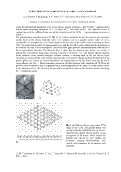
Journal of Microscopy, Vol. 218, Pt 3 June 2005, pp. 199– 207
Received 7 January 2005; accepted 28 February 2005
INVITED REVIEW
Blackwell Publishing, Ltd.
Sample preparation procedures for biological atomic
force microscopy
K . E L K I R AT , I . B U RT O N , V. D U P R E S & Y. F. D U F R E N E
Unité de chimie des interfaces, Université catholique de Louvain, Croix du Sud 2/18, B-1348
Louvain-la-Neuve, Belgium
Key words. Atomic force microscopy, biological samples, substrates,
immobilization, imaging.
Received 7 January 2005; accepted 28 February 2005
Summary
Since the late 1980s, atomic force microscopy (AFM) has been
increasingly used in biological sciences and it is now established
as a versatile tool to address the structure, properties and functions of biological specimens. AFM is unique in that it provides
three-dimensional images of biological structures, including
biomolecules, lipid films, 2D protein crystals and cells, under
physiological conditions and with unprecedented resolution.
A crucial prerequisite for successful, reliable biological AFM is
that the samples need to be well attached to a solid substrate
using appropriate, nondestructive methods. In this review, we
discuss common techniques for immobilizing biological specimens for AFM studies.
1. Introduction
During the past two decades, the atomic force microscope (AFM)
(Binnig et al., 1986) has provided new avenues for microscopists
to study biological specimens. AFM yields three-dimensional
images of biosystems (single molecules adsorbed on surfaces,
lipid membranes, 2D protein crystals, living cells) in aqueous
solutions and with (sub)nanometre resolution. In addition, it
can also measure forces with remarkable sensitivity and positional precision. In this mode, known as force spectroscopy, the
cantilever deflection is recorded as the tip is pushed towards
the sample and retracted from it. Force spectroscopy can be
used to probe quantitatively physical properties such as local
elasticity, surface forces, surface charge and hydrophobicity
and to measure inter- and intramolecular interactions, providing new insights into the molecular bases of processes such
as protein folding and receptor–ligand interactions (for an
overview of imaging and force spectroscopy applications, see
Jena & Hörber, 2002).
Correspondence: Y. F. Dufrêne. Tel.: + 32 10 47 36 00; fax: +32 10 47 20 05;
e-mail: [email protected]
© 2005 The Royal Microscopical Society
In this paper, we provide a survey of the various methods used
for preparing samples for biological AFM. We first describe how
to select and prepare appropriate substrates and then discuss
immobilization protocols available for isolated molecules,
supported lipid films, two-dimensional protein crystals and cells.
Although not the primary goal of this review, we also briefly
highlight some important imaging applications in biosciences
(for detailed information on methodologies and applications,
see Ikai, 1996; Shao et al., 1996; Colton et al., 1998; Morris
et al., 1999; Jena & Hörber, 2002).
2. Substrates
After the first AFM images of biological structures were obtained,
it was soon realized that a crucial factor for successful imaging
is the specimen preparation. The first issue is to select an
appropriate substrate. Indeed, to observe biological structures
in their native state, these must be well attached to a smooth
solid substrate to resist the lateral forces exerted by the scanning
tip. In that respect, mica, glass and silicon oxide have proved to
be excellent substrates. Muscovite mica, KAl2(OH)2AlSi3O10, is
a nonconducting layered mineral composed of multiple 1 nm
thick layers (Bailey, 1984). It can be cleaved easily with the
help of adhesive tape to produce clean, atomically flat surfaces
that are negatively charged. Today, mica is the most frequently
used substrate for imaging biological specimens, including
double-stranded DNA, DNA–protein complexes, protein arrays,
densely packed proteins, supported lipid films and animal cells.
Interestingly, the mica surface can be modified with silanes
either to promote adsorption or to allow covalent binding of
the biomolecules (Lyubchenko et al., 1992).
Glass represents another suitable substrate for biological
AFM. Glass cover slips are flat enough for imaging cells and
other large structures, but are usually too rough for imaging
adsorbed molecules. The glass surface is always coated with
organic contaminants and particles that should be removed
before use, e.g. by washing in concentrated acidic solution
200
K . E L K I R AT E T A L .
followed by ultrasonication in water solutions. As for mica, glass
can also be modified with silane molecules, bringing new
chemical functions at the surface for further covalent modifications (Karrasch et al., 1993). In some cases, silicon oxide wafers
initially developed for the semiconductor industry can be used
instead of glass. Although they are more expensive and more
difficult to handle, they have a much smoother surface than
glass.
Hydrophobic substrates may sometimes be preferred. A wellknown example is highly orientated pyrolytic graphite (HOPG),
which is atomically flat over large areas (Cullen & Lowe, 1994).
Coating the mica surface with carbon yields a hydrophobic
surface that is well suited for immobilizing DNA (Yang et al.,
1992). Spin-coating of polymers [polystyrene, poly(methyl
methacrylate)] is another convenient method for creating
flat hydrophobic substrates for biological AFM (Dupont-Gillain
et al., 1999). For some applications requiring specific surface
chemistries, gold surfaces may be useful. Thin layers of gold
can be prepared by thermal evaporation onto mica, glass or
silicon oxide substrates. To obtain very smooth gold surfaces
(roughness ∼0.1 nm), various methods, referred to as template
stripped gold (TSG) methods, have been developed (Wagner
et al., 1995). The typical procedure involves depositing gold
onto a smooth substrate such as silicon, supporting the free
gold surface by gluing it to a glass slide using epoxy glue and
stripping the gold film from the silicon substrate. The advantage
of using gold is that it is chemically inert against oxygen and
stable against radicals and can be easily modified with selfassembled monolayers (SAMs) of organic alkanethiols that
can be used further for adsorbing or attaching biomolecules.
Finally, as we shall see below, three-dimensional systems such
as porous polymer membranes and macromolecular gels can
sometimes be used to immobilize large objects such as cells.
3. Biomolecules
Several approaches may be used for preparing biomolecules
for AFM in air. A simple immobilization procedure consists
of depositing a drop of an aqueous solution containing the
macromolecule of interest on the substrate, and then allowing
the drop to evaporate or blowing the sample dry in nitrogen.
Alternatively, the substrate can be immersed in the solution to
allow adsorption of the biomolecules for a given period of time,
rinsed and then air-dried. Finally, the molecules can also be
sprayed with an aqueous solution, or in the presence of glycerol,
onto the substrate. DNA alone or in association with proteins
has be deposited using these protocols, and then imaged in air
(Vesenka et al., 1992; Rees et al., 1993), under alcohols (Hansma
et al., 1992) or in aqueous solution (Bezanilla et al., 1993).
Similar protocols have been applied to proteins such as fibronectin
(Emch et al., 1992) or collagen (Chernoff & Chernoff, 1992),
and to polysaccharides (Kirby et al., 1996).
Physical adsorption in the presence of appropriate electrolytes
is another approach which offers the advantage of allowing
the sample to be directly imaged under liquid without the need
for air-drying. A wide range of biomolecules, primarily proteins
and DNA, have been imaged by AFM while being adsorbed on
mica (Hoh et al., 1993; Radmacher et al., 1994; Yang et al., 1994;
Fritz et al., 1995; Hansma et al., 1995). For soluble proteins, the
very first in situ images were generally of poor resolution (Lin
et al., 1990). However, since then there has been continuous
progress in improving preparation methods and imaging
conditions for these specimens (for reviews, see, e.g. Shao et al.,
1996; Wagner, 1998). For instance, in an early high-resolution
study, pertussis toxin could be imaged down to a resolution of
0.5 nm following simple adsorption on mica (Yang et al., 1994).
As discussed below (Section 5), proteins that form stable twodimensional crystals are actually very well-suited for high
resolution imaging.
Various approaches may be used to promote the attachment of adsorbed biomolecules. For DNA, pretreating the
substrate with silanes bearing an amino group such as 3aminopropyltriethoxysilane favours attachment (Lyubchenko
et al., 1992). DNA molecules were also found to bind tightly
enough on mica when the solution contains 1 mm concentrations of certain divalent cations, i.e. Ni2+, Co2+, Zn2+ or Mn2+
(Hansma & Laney, 1996). Interestingly, by adjusting the concentration of Zn2+, it is possible to bind DNA sufficiently so that
it can be imaged but loosely enough to be translocated by the
RNA polymerase (Kasas et al., 1997; Fig. 1). For proteins
(lysozyme, streptavidin, the cholera toxin B-subunit, GroES and
GroEL), it has recently been shown that monovalent cations
(K+, Na+, Li+) can inhibit the adsorption to mica, suggesting that
reducing this inhibition may be a generally useful procedure to
maximize the amount of proteins on this surface (Czajkowsky
& Shao, 2003).
Covalent attachment of macromolecules may sometimes be
desired. Immobilization of proteins on glass may be achieved
using a photoreactive cross-linker (Karrasch et al., 1993;
see Section 5). Another elegant approach is to form Nhydroxysuccinimide-terminated monolayers on gold-coated
substrates, using, e.g. dithio-bis-succinimidylundecanoate, and
then binding the proteins covalently in aqueous buffers under
mild conditions (Wagner et al., 1996; Wagner, 1998). Finally,
it is worth noting that covalent binding of molecules on AFM tips
and/or substrates is also used for measuring specific biomolecular
interactions by force spectroscopy. Here, several flexible spacer
molecules have been introduced, including poly(ethylene
glycol) (Hinterdorfer et al., 1996) and carboxymethyl-amylose
(Grandbois et al., 2000), in order to provide enough mobility
to the attached biomolecules so that they can freely interact
with complementary molecules.
4. Supported lipid films
Thin lipid films supported on solid substrates are valuable model
systems for mimicking biological surfaces. In particular, these
systems are widely used in biophysical research to investigate
© 2005 The Royal Microscopical Society, Journal of Microscopy, 218, 199–207
P R E PA R I N G S A M P L E S F O R B I O L O G I CA L A F M
201
Fig. 1. Adsorption in the presence of appropriate ions allows to observe biomolecules at work. In this example, immobilization of linear double-stranded
DNA templates on mica in the presence of Zn2+ allowed imaging of transcription by Escherichia coli RNA polymerase (RNAP). The transcription process was
detected by observing the translocation of the DNA template by RNAP on addition of ribonucleoside 5′-triphosphates (NTPs) in sequential AFM images.
The first two images show that before the NTPs enter, the DNA has mobility on the mica. The six images after NTP addition, from time 0:00 onwards are
sequential and show that one arm of the DNA template becomes progressively shorter until it is released (02:38). Reprinted with permission from Kasas
et al. (1997). © 1997, American Chemical Society.
the properties of biological membranes and processes such as
molecular recognition, enzymatic catalysis, cell adhesion and
membrane fusion (McConnell et al., 1986; Sackmann, 1996).
Transferring lipid films onto solid substrata offers the possibility
of applying a range of surface analytical techniques, and more
particularly AFM.
For AFM imaging applications, supported lipid films may
be prepared either by the Langmuir–Blodgett (LB) technique
or by fusion of lipid vesicles (Czajkowsky & Shao, 2002). In the
first method, a Langmuir trough consisting of a rectangular
Teflon bath equipped with moveable barriers is used to compress the lipid molecules at the air–water interface (Ulman,
1991). Lipids are usually spread at the air–water interface in
hexane/ethanol or chloroform/methanol mixtures, and then
compressed after letting the solvent to evaporate for 15 min. A
sensor records the surface pressure at the interface, which can
be expressed as a function of the interfacial area. The obtained
surface pressure vs. area isotherms can provide useful information on the packing and organization of the lipid molecules.
In the LB technique, the monolayer of amphiphilic molecules
is transferred at constant surface pressure and constant speed
onto a solid substrate, usually mica. Careful control of surface
pressure and lifting speed is essential to avoid artefacts such as
defect formation or feature alignment of the deposited structures. Lipid monolayers interact with mica through the polar
heads, thus exposing the hydrophobic tails to the environment. These systems are stable in air, not in water, and should
therefore be examined in air by AFM. Transferring a second
lipid layer onto a mica-supported lipid monolayer yields a supported bilayer, which best mimics cellular membranes. These
© 2005 The Royal Microscopical Society, Journal of Microscopy, 218, 199–207
supported bilayers should always be kept and analysed in
aqueous solution, as they are not stable in air.
Fusion of lipid vesicles on solid substrates is another approach
to obtain supported lipid bilayers (Brian & McConnell, 1984;
Horn, 1984). Typically, the lipids are first solubilized in organic
solvent. After solvent evaporation under nitrogen and subsequent
desiccation under vacuum, the dried lipid film is resuspended
in aqueous buffer solution (usually Tris or PBS) yielding a
multilamellar vesicles (MLVs) suspension. From this suspension, small unilamellar vesicles (SUVs) can be obtained using
various approaches, sonication being the most popular one.
The suspension is sonicated to clarity (e.g. five cycles of 2 min)
using a titanium probe sonicator while keeping the suspension
in an ice bath, after which the suspension is filtered on 200 nm
nylon filters to eliminate titanium particles. Then fusion is
achieved by heating the SUV suspension in contact with freshly
cleaved mica for 1 h at a temperature between 45 °C and
60 °C. The supported bilayers are finally gently cooled to room
temperature and rinsed abundantly with the appropriate
imaging buffer. Compared to LB deposition, the drawbacks of the
fusion method are the impossibility of preparing asymmetric
bilayers composed of two layers of different nature as can be
easily obtained with the LB technique, and the lack of control
of the lateral pressure in the lipid layers. However, because the
fusion approach is much simpler and permits lipid diffusion as
in free-standing bilayers, it is the most widely used method in
AFM lipid bilayer research.
Since the first AFM images revealing the molecular structure
of supported phospholipid films were published (Egger et al.,
1990), a broad spectrum of AFM applications has emerged in
202
K . E L K I R AT E T A L .
Fig. 2. Imaging supported lipid monolayers prepared
by the Langmuir–Blodgett (LB) technique. AFM friction
images in air of mixed fengycin/ceramide monolayers
supported on mica prepared at 20 °C/pH 2 (A; 2 µm ×
2 µm) and 37 °C/pH 5 (B; 15 µm × 15 µm). The images
reveal phase-separation in the form of two-dimensional
hexagonal or circular domains of ceramide surrounded
by a fengycin-enriched fluid phase and demonstrate the
dramatic influence of the environmental conditions
(i.e. temperature, pH) on the molecular organization
of the films. Reprinted with permission from Eeman
et al. (2005). © 2005, American Chemical Society.
lipid film research (for reviews see, e.g. Dufrêne & Lee, 2000;
Czajkowsky & Shao, 2002), among which are visualizing the
molecular structure and organization of phase-separated films
(Dufrêne et al., 1997; Vié et al., 1998; Eeman et al., 2005; Fig. 2),
probing the effect of external agents such as solvents (Mou
et al., 1994a, 1994b), peptides (Rinia et al., 2002; El Kirat
et al., 2005), proteins (Milhiet et al., 2002) and antibiotics
(Berquand et al., 2004; Fig. 3) and using lipid films as substrates
for anchoring biomolecules such as DNA (Mou et al., 1995a)
and crystalline protein arrays (Reviakine et al., 1998).
5. Two-dimensional protein crystals
Owing to their stability and structural regularity, two dimensional
(2D) bacterial protein crystals (Sleytr & Beveridge, 1999) have
proved to be excellent systems for high resolution AFM imaging.
AFM yields structural information with a lateral resolution
better than 1 nm, directly under physiological conditions, which
makes it a complementary tool to X-ray and electron crystallography (for an overview of applications in the field, see Müller &
Engel, 2002).
Several immobilization strategies have been established
for 2D protein crystals, the most frequently used procedures
being based on physical adsorption in aqueous solution (for
reviews, see Müller et al., 1997a,b). A well-known example is
the purple membrane from Halobacterium salinarum, which
readily adsorbs on mica. The typical procedure for adsorption
is as follows (Müller et al., 1997a): 150 mm KCl is added with
10 mm Mes, Hepes or Tris for adjusting the pH; the purple
membranes are diluted in the buffer to a concentration of
about 50 µg mL−1 and a drop of this solution is deposited onto
freshly cleaved mica; after adsorption for 10–30 min, samples
are gently washed with buffer to remove membranes that are
not firmly attached. Using this procedure, individual bacteriorhodopsin molecules in the membrane can be imaged at
subnanometre resolution (Fig. 4). Furthermore, force-induced
conformational changes can be directly visualized.
Another example is the channel-forming protein OmpF porin
from Escherichia coli that forms 2D crystals when reconstituted
in the presence of lipids. Immobilization onto freshly cleaved
mica is achieved in the presence of small amounts of divalent
ions (2 mm MgCl2) and monovalent ions (NaCl) between 100
and 150 mm (Schabert & Engel, 1994; Schabert et al., 1995).
The presence of Mg2+ is essential to compensate the negative
surface charges of both sample and substrate. This protocol
allowed high-resolution imaging of porin OmpF 2D crystals,
with a lateral resolution of 1 nm and a vertical resolution of
0.1 nm (Schabert et al., 1995).
To enhance the adsorption of negatively charged samples,
the surface properties of the substrate can be modified by
coating with a polycation such as poly l-lysine (Müller et al.,
1997b). Crosslinkers can also be used to covalently anchor
membrane proteins on substrates. An original procedure has
been developed for the hexagonally packed intermediate
(HPI) layer of Deinococcus radiodurans. It involves the chemical
modification of glass with the photoactivatable cross-linker
N-5-azido-2-nitrobenzoyloxysuccinimide, followed by crosslinking the samples by irradiation with ultraviolet light
(Karrasch et al., 1993; Karrasch & Engel, 1998). This procedure allowed the HPI layer proteins to be imaged in buffer
solution with a lateral resolution of 1 nm. Covalent immobilization has the disadvantage of requiring more preparation steps and may alter the native structure of the biological
specimen.
Lipid monolayers and bilayers have also proved to be a valuable
preparation method for immobilizing recrystallized protein
arrays. A first approach, used for instance for Bacillus S-layers,
consists in recrystallizing the proteins on a lipid monolayer in
a LB trough and then transferring the composite structure
on a flat substrate for AFM examination (Pum et al., 1993).
Alternatively, supported lipid bilayers containing various
ligand-linked lipids may be used to specifically bind the proteins.
Examples of such protein crystallization procedures have been
reported for streptavidin (Weisenhorn et al., 1992; Scheuring
et al., 1999), cholera toxin (Mou et al., 1995b) and annexin V
(Reviakine et al., 1998), which, respectively, bind to biotinylated
lipids, GM1 ganglioside and negatively-charged lipids such as
phosphatidylserine.
© 2005 The Royal Microscopical Society, Journal of Microscopy, 218, 199–207
P R E PA R I N G S A M P L E S F O R B I O L O G I CA L A F M
203
Fig. 3. Real-time imaging of supported lipid bilayers prepared by vesicle fusion. AFM height images (7.5 µm × 7.5 µm; z-scale: 10 nm) of a mixed dioleoylphosphatidylcholine (DOPC)/dipalmitoyl-phosphatidylcholine (DPPC) bilayer recorded in solution at increasing incubation times with the antibiotic
azithromycin. The antibiotic causes progressive erosion of the gel DPPC domains, an effect attributed to the intercalation of the drug at the domain
boundary. Reprinted with permission from Berquand et al. (2004). © 2004, Elsevier Science Ltd.
6. Cells
The potential of AFM for probing cells was recognized quite early
(Butt et al., 1990). Here, a key challenge has been to develop
procedures allowing imaging of the cells in their native state. For
animal cells, a simple preparation method is to exploit the ability
© 2005 The Royal Microscopical Society, Journal of Microscopy, 218, 199–207
of the cells to spread and adhere to solid surfaces (Radmacher
et al., 1992; Matzke et al., 2001). Coating the substrate with
adhesion proteins such as collagen may be used to enhance
immobilization, a procedure which allowed the observation of
actin filament dynamics under the cell membrane of glial cells
(Henderson et al., 1992). A rather different approach is to image
204
K . E L K I R AT E T A L .
Fig. 4. Adsorption of 2-D protein crystals on mica, here the purple
membrane, allows to record high-resolution AFM images of their surface
in aqueous solution. A fully reversible force-induced conformational
change is observed: at the top of the image the force applied to the AFM
stylus is 100 pN; while scanning the surface line by line the force is
increased until it reaches 150 pN at the bottom of the image. Reprinted
with permission from Müller et al. (2000). © 2000, Elsevier Science Ltd.
living cells fixed only by a suction pipette, using an AFM combined
with an optical microscope (Hörber et al., 1992). Cells can be kept
alive for days in growth medium while being examined, making
it possible to study cell activities and dynamics. Finally, in some cases
chemical fixation of the cells using cross-linking agents such as
glutaraldehyde may be required either to prevent cell damage
or detachment by the scanning tip or to obtain high resolution
images (Le Grimellec et al., 2002). Owing to these different preparation methods, various cell types have been investigated, such
as CV-1 kidney cells, fibroblasts, MDCK, platelets and cardiomyocytes (Jena & Hörber, 2002). An important achievement in
cell biology has been the possibility of following dynamic processes at cell surfaces, as was shown for instance for the plasma
membrane of pancreatic acinar cells, where depressions attributed
to fusion pores could be observed (Schneider et al., 1997).
As opposed to animal cells, microbial cells (bacteria, yeast and
fungi) cannot spread over solid substrates, meaning immobilization by means of simple adsorption procedures is often
inappropriate, as this most often leads to cell detachment by the
scanning probe. Stronger attachment may be achieved either by
pretreating the substrate with polycations (Schaer-Zammaretti
& Ubbink, 2003) or by bonding the cells covalently to the substrate (Camesano et al., 2000). The ability of microorganisms
to attach to solid substrates by means of extracellular polymeric
substances may also be exploited for AFM studies, but the
resolution remains limited (Beech et al., 1996). Another possibility is to immobilize the cells mechanically in an agar gel or
in a porous membrane. In the first approach, the agar gel is used
as a soft, deformable immobilization matrix, thereby allowing
direct visualization of growth processes (Gad & Ikai, 1995). In
the second method, spherical cells are trapped in a polymer
membrane with a pore size comparable to the dimensions of
the cell, allowing repeated imaging without cell detachment
or cell damage (Kasas & Ikai, 1995; Dufrêne et al., 1999; Fig. 5).
In a recent comparative study, it was concluded that, in general,
mechanical trapping of bacterial cells in filters appears to be the
most reliable method for immobilization (Vadillo-Rodriguez
et al., 2004). Physical adsorption on modified substrata was
suggested to promote structural rearrangements in bacterial
cell surface structures, whereas glutaraldehyde treatment was
shown to induce changes on cell surface properties.
Another advantage of the mechanical trapping approach is
that it allows observing dynamic processes. In one such study,
Touhami et al. (2004) combined AFM imaging in aqueous
solution and thin-section transmission electron microscopy
(TEM) to investigate the changes in the cell wall of Staphylococcus
aureus cells as they grow and divide. A good correlation of the
structural events of division was found using the two techniques
and the AFM was shown to provide new information.
7. Perspectives
Since the birth of AFM, tremendous advances have been made in
developing sample preparation methods for biological specimens.
Today, simple, reliable protocols are available for most specimens,
meaning that, in general, sample preparation is no longer a
bottleneck for biological AFM. However, there are still many
technical challenges to address in the field. In particular, highresolution imaging on soft samples such as animal cells remains
often very difficult due to the damaging effects of the scanning
tip. To solve this issue, several approaches are being developed
(Hörber & Miles, 2003). For instance, tip–specimen interaction
forces can be greatly reduced using tapping mode AFM with an
active resonance control (Tamayo et al., 2001). Another promising approach is the photonic force microscope, in which the AFM
cantilever is replaced by the 3D trapping potential of a laser focus,
allowing 3D imaging of biological structures with extremely
small loading forces (Pralle et al., 2000). Time resolution is
another problem in current biological AFM studies, as 30–60 s
are usually needed to record an image. In this context, new ultrafast instruments provide exciting avenues to explore dynamic
processes associated with biomolecules and cells (Viani et al.,
2000; Ando et al., 2001; Hörber & Miles, 2003).
Acknowledgements
The support of the FNRS, of the Université catholique de
Louvain (Special Fund for Research), of the Région wallonne,
of the Federal Office for Scientific, Technical and Cultural Affairs
© 2005 The Royal Microscopical Society, Journal of Microscopy, 218, 199–207
P R E PA R I N G S A M P L E S F O R B I O L O G I CA L A F M
205
Fig. 5. Imaging living microbial cells immobilized in
porous polymer membranes. Low (A; 3 µm × 1.5 µm)
and high (B; 500 nm × 285 nm) resolution AFM
images obtained for the bacterium Lactococcus lactis.
The cell surface is quite smooth in agreement with
the presence of peptidoglycan, which is the major
determinant of cell shape and rigidity.
(Inter-university Poles of Attraction Programme) and of the
Research Department of Communauté Française de Belgique
(Concerted Research Action) is gratefully acknowledged.
I.B. and Y.F.D. are, respectively, Research Fellow and Research
Associate of the Belgian National Foundation for Scientific
Research (FNRS).
References
Ando, T., Kodera, N., Takai, E., Maruyama, D., Saito, K. & Toda, A. (2001)
A high-speed atomic force microscope for studying biological macromolecules. Proc. Natl. Acad. Sci. USA, 98, 12468–12472.
Bailey, S.W. (1984) Micas. Mineralogical Society of America, Washington,
DC.
Beech, I.B., Cheung, C.W.S., Johnson, D.B. & Smith, J.R. (1996) Comparative studies of bacterial biofilms on steel surfaces using atomic force
microscopy and environmental scanning electron microscopy. Biofouling,
10, 65–77.
Berquand, A., Mingeot-Leclercq, M.-P. & Dufrêne, Y.F. (2004) Real-time
imaging of drug–membrane interactions by atomic force microscopy.
Biochim. Biophys. Acta – Biomembranes, 1664, 198–205.
Bezanilla, M., Bustamante, C.J. & Hansma, H.G. (1993) Improved visualization of DNA in aqueous buffer with the atomic force microscope.
Scan. Microsc. 7, 1145–1148.
© 2005 The Royal Microscopical Society, Journal of Microscopy, 218, 199–207
Binnig, G., Quate, C.F. & Gerber, C. (1986) Atomic Force Microscope.
Phys. Rev. Lett. 56, 930 –933.
Brian, A.A. & McConnell, H.M. (1984) Allogeneic stimulation of cytotoxic
T cells by supported membranes. Proc. Natl. Acad. Sci. USA, 81, 6159–
6163.
Butt, H.-J., Wolff, E.K., Gould, S.A.C., Dixon Northern, B., Peterson, C.M.
& Hansma, P.K. (1990) Imaging cells with the atomic force microscope.
J. Struct. Biol. 105, 54–61.
Camesano, T.A., Natan, M.J. & Logan, B.E. (2000) Observation of changes
in bacterial cell morphology using tapping mode atomic force microscopy. Langmuir, 16, 4563 – 4572.
Chernoff, E.A.G. & Chernoff, D.A. (1992) Atomic force microscope images
of collagen fibers. J. Vac. Sci. Technol. A, 10, 596 –599.
Colton, R.J., Engel, A., Frommer, J.E., et al. (1998) Procedures in Scanning
Probe Microscopies. John Wiley & Sons Ltd, Chichester.
Cullen, D.C. & Lowe, C.R. (1994) AFM studies of protein adsorption. 1.
Time-resolved protein adsorption to highly oriented pyrolytic-graphite.
J. Colloid Interf. Sci. 166, 102–108.
Czajkowsky, D.M. & Shao, Z. (2002) Supported lipid bilayers as effective
substrates for atomic force microscopy. Atomic Force Microscopy in Cell
Biology, Methods in Cell Biology, Vol. 68 (ed. by B. P. Jena and J. K. H.
Hörber), pp. 231–241. Academic Press, San Diego.
Czajkowsky, D.M. & Shao, Z. (2003) Inhibition of protein adsorption to
muscovite mica by monovalent cations. J. Microsc. 211, 1–7.
Dufrêne, Y.F., Barger, W.R., Green, J.-B.D. & Lee, G.U. (1997) Nanometer-scale
206
K . E L K I R AT E T A L .
surface properties of mixed phospholipid monolayers and bilayers.
Langmuir, 13, 4779–4784.
Dufrêne, Y.F., Boonaert, C.J.P., Gerin, P.A., Asther, M. & Rouxhet, P.G.
(1999) Direct probing of the surface ultrastructure and molecular
interactions of dormant and germinating spores of Phanerochaete chrysosporium. J. Bacteriol. 181, 5350 –5354.
Dufrêne, Y.F. & Lee, G.U. (2000) Advances in the characterization of
supported lipid films with the atomic force microscope. Biochim. Biophys.
Acta, 1509, 14–41.
Dupont-Gillain, C.C., Nysten, B. & Rouxhet, P.G. (1999) Collagen adsorption on poly (methyl methacrylate): net-like structure formation upon
drying. Polymer Int. 48, 271–276.
Eeman, M., Deleu, M., Paquot, M., Thonart, P. & Dufrêne, Y.F. (2005)
Nanoscale properties of mixed Fengycin/Ceramide monolayers explored
using atomic force microscopy. Langmuir, 21, 2505 –2511.
Egger, M., Ohnesorge, F., Weisenhorn, A.L., et al. (1990) Wet lipid protein
membranes imaged at submolecular resolution by atomic force microscopy. J. Struct. Biol. 103, 89–94.
El Kirat, K., Lins, L., Brasseur, R. & Dufrêne, Y.F. (2005) Nanoscale modification of supported lipid membranes: synergetic effect of Phospholipase
D and viral fusion peptides. J. Biomed Nanotechnol. in press.
Emch, R., Zenhausern, F., Jobin, M., Taborelli, M. & Descouts, P. (1992)
Morphological difference between fibronectin sprayed on mica and on
PMMA. Ultramicroscopy, 42– 44, 1155–1160.
Fritz, M., Radmacher, M., Cleveland, J.P., et al. (1995) Imaging globular
and filamentous proteins in physiological buffer solutions with tapping
mode atomic force microscopy. Langmuir, 11, 3529–3535.
Gad, M. & Ikai, A. (1995) Method for immobilizing microbial cells on gel
surface for dynamic AFM studies. Biophys. J. 69, 2226–2233.
Grandbois, M., Dettmann, W., Benoit, M. & Gaub, H.E. (2000) Affinity
imaging of red blood cells using an atomic force microscope. J. Histochem. Cytochem. 48, 719–724.
Hansma, H.G. & Laney, D.E. (1996) DNA binding to mica correlates with
cationic radius: assay by atomic force microscopy. Biophys. J. 70,
1933–1939.
Hansma, H.G., Laney, D.E., Bezanilla, M., Sinsheimer, R.L. & Hansma,
P.K. (1995) Applications for atomic force microscopy of DNA. Biophys.
J. 68, 1672–1677.
Hansma, H.G., Vesenka, J., Siegerist, C., et al. (1992) Reproducible imaging and dissection of plasmid DNA under liquid with the atomic force
microscope. Science, 256, 1180–1184.
Henderson, E., Haydon, P.G. & Sakaguchi, D.S. (1992) Actin filament
dynamics in living glial-cells imaged by atomic force microscopy. Science, 257, 1944–1946.
Hinterdorfer, P., Baumgartner, W., Gruber, H.J., Schilcher, K. & Schindler,
H. (1996) Detection and localization of individual antibody-antigen
recognition events by atomic force microscopy. Proc. Natl Acad. Sci.
USA, 93, 3477–3481.
Hoh, J.H., Sosinsky, G.E., Revel, J.-P. & Hansma, P.K. (1993) Structure of
the extracellular surface of the gap junction by atomic force microscopy. Biophys. J. 65, 149–163.
Hörber, J.K.H., Häberle, W., Ohnesorge, F., et al. (1992) Investigation of
living cells in the nanometer regime with the scanning force microscope. Scan. Microsc. 6, 919–930.
Hörber, J.K.H. & Miles, M.J. (2003) Scanning probe evolution in biology.
Science, 302, 1002–1005.
Horn, R.G. (1984) Direct measurement of the force between two lipid
bilayers and observation of their fusion. Biochim. Biophys. Acta – Biomembranes, 778, 224–228.
Ikai, A. (1996) STM and AFM of bio/organic molecules and structures.
Surf. Sci. Report, 26, 261–332.
Jena, B.P. & Hörber, J.K.H. (2002) Atomic Force Microscopy in Cell Biology,
Methods in Cell Biology, Vol. 68. Academic Press, San Diego.
Karrasch, S., Dolder, M., Schabert, F., Ramsden, J. & Engel, A. (1993)
Covalent binding of biological samples to solid supports for scanning
probe microscopy in buffer solution. Biophys. J. 65, 2437–2446.
Karrasch, S. & Engel, A. (1998) AFM imaging of HPI layers in buffer solution.
Procedures in Scanning Probe Microscopies (ed. by R. J. Colton, A. Engel,
J. E. Frommer, et al.), pp. 433–439. John Wiley & Sons Ltd, Chichester.
Kasas, S. & Ikai, A. (1995) A method for anchoring round shaped cells for
atomic force microscope imaging. Biophys. J. 68, 1678–1680.
Kasas, S., Thomson, N.H., Smith, B.L., et al. (1997) Escherichia coli
RNA polymerase activity observed using atomic force microscopy. Biochemistry, 36, 461–468.
Kirby, A.R., Gunning, A.P. & Morris, V.J. (1996) Imaging polysaccharides
by atomic force microscopy. Biopolymers, 38, 355–366.
Le Grimellec, C., Giocondi, M.C., Lenoir, M., Vater, M., Sposito, G. & Pujol,
R. (2002) High-resolution three-dimensional imaging of the lateral
plasma membrane of cochlear outer hair cells by atomic force microscopy. J. Comp. Neurol. 451, 62–69.
Lin, J.N., Drake, B., Lea, A.S., Hansma, P.K. & Andrade, J.D. (1990) Direct
observation of immunoglobulin adsorption dynamics using the atomic
force microscope. Langmuir, 6, 509–511.
Lyubchenko, Y.L., Gall, A.A., Shlyakhtenko, L.S., Harrington, R.E., Oden,
P.I., Jacobs, B.L. & Lindsay, S.M. (1992) Atomic force microscopy imaging of double stranded DNA and RNA. J. Biomol. Struct. Dyn. 9, 589–
606.
Matzke, R., Jacobson, K. & Radmacher, M. (2001) Direct, high-resolution
measurement of furrow stiffening during division of adherent cells.
Nature Cell Biol. 3, 607–610.
McConnell, H.M., Watts, T.H., Weis, R.M. & Brian, A.A. (1986) A supported planar membranes in studies of cell-cell recognition in the
immune system. Biochim. Biophys. Acta, 864, 95–106.
Milhiet, P.E., Giocondi, M.C., Baghdadi, O., Ronzon, F., Le Grimellec, C. &
Roux, B. (2002) AFM detection of GPI protein insertion into DOPC/
DPPC model membranes. Single Molecules, 3, 135–140.
Morris, V.J., Kirby, A.R. & Gunning, A.P. (1999) Atomic Force Microscopy
for Biologists. Imperial College Press, London.
Mou, J., Czajkowsky, D.M., Zhang, Y. & Shao, Z. (1995a) High-resolution
atomic force microscopy of DNA – The pitch of the double helix. FEBS
Lett. 371, 279–282.
Mou, J., Yang, J., Huang, C. & Shao, Z. (1994a) Alcohol induces interdigitated domains in unilamellar phosphatidylcholine bilayers. Biochemistry, 33, 9981–9985.
Mou, J., Yang, J. & Shao, Z. (1994b) Tris (hydroxymethyl) aminomethane
(C4H11NO3) induced ripple phase in supported unilamellar phospholipid bilayers. Biochemistry, 33, 4439– 4443.
Mou, J., Yang, J. & Shao, Z. (1995b) Atomic force microscopy of cholera
toxin B-oligomers bound to bilayers of biologically relevant lipids. J.
Mol. Biol. 248, 507–512.
Müller, D.J., Amrein, M. & Engel, A. (1997a) Adsorption of biological
molecules to a solid support for scanning probe microscopy. J. Struct.
Biol. 119, 172–188.
Müller, D.J. & Engel, A. (2002) Conformation, flexibility, and interactions
observed on individual membrane proteins by atomic force microscopy. Atomic Force Microscopy in Cell Biology, Methods in Cell Biology,
Vol. 68 (ed. by B. P. Jena and J. K. H. Hörber), pp. 257–299. Academic
Press, San Diego.
© 2005 The Royal Microscopical Society, Journal of Microscopy, 218, 199–207
P R E PA R I N G S A M P L E S F O R B I O L O G I CA L A F M
Müller, D.J., Engel, A. & Amrein, M. (1997b) Preparation techniques for
the observation of native biological systems with the atomic force
microscope. Biosensors Bioelectronics, 12, 867–877.
Müller, D.J., Heymann, J.B., Oesterhelt, F., Möller, C., Gaub, H., Büldt, G. &
Engel, A. (2000) Atomic force microscopy of native purple membrane.
Biochim. Biophys. Acta, 1460, 27–38.
Pralle, A., Keller, P., Florin, E.L., Simons, K. & Hörber, J.K.H. (2000)
Sphingolipid-cholesterol rafts diffuse as small entities in the plasma
membrane of mammalian cells. J. Cell Biol. 148, 997–1008.
Pum, D., Weinhandl, M., Hödl, C. & Sleytr, U.B. (1993) Large-scale recrystallization of the S-layer of Bacillus coagulans E38–66 at the air/water
interface and on lipid films. J. Bacteriol. 175, 2762–2766.
Radmacher, M., Fritz, M., Hansma, H.G. & Hansma, P.K. (1994) Direct
observation of enzyme activity with the atomic force microscope.
Science, 265, 1577–1579.
Radmacher, M., Tillmann, R.W., Fritz, M. & Gaub, H.E. (1992) From
molecules to cells: imaging soft samples with the atomic force microscope. Science, 257, 1900–1905.
Rees, W.A., Keller, R.W., Vesenka, J.P., Yang, G. & Bustamante, C. (1993)
Evidence of DNA bending in transcription complexes imaged by scanning
force microscopy. Science, 260, 1646–1649.
Reviakine, I., Bergsma-Schutter, W. & Brisson, A. (1998) Growth of protein
2-D crystals on supported planar lipid bilayers imaged in situ by AFM. J.
Struct. Biol. 121, 356–361.
Rinia, H.A., Boots, J.W., Rijkers, D.T., et al. (2002) Domain formation in
phosphatidylcholine bilayers containing transmembrane peptides:
specific effects of flanking residues. Biochemistry, 41, 2814–2824.
Sackmann, E. (1996) Supported membranes: scientific and practical
applications. Science, 271, 43–48.
Schabert, F.A. & Engel, A. (1994) Reproducible acquisition of Escherichia
coli porin surface topographs by atomic force microscopy. Biophys. J.
67, 2394–2403.
Schabert, F.A., Henn, C. & Engel, A. (1995) Native Escherichia coli OmpF
porin surfaces probed by atomic force microscopy. Science, 268, 92–94.
Schaer-Zammaretti, P. & Ubbink, J. (2003) Imaging of lactic acid bacteria
with AFM – elasticity and adhesion maps and their relationship to biological and structural data. Ultramicroscopy, 97, 199–208.
Scheuring, S., Müller, D.J., Ringler, P., Heymann, J.B. & Engel, A. (1999)
Imaging streptavidin 2D-crystals on biotinylated lipid monolayers at high
resolution with the atomic force microscope. J. Microsc. 193, 28–35.
Schneider, S.W., Sritharan, K.C., Geibel, J.P., Oberleithner, H. & Jena, B.P.
(1997) Surface dynamics in living acinar cells imaged by atomic force
microscopy: identification of plasma membrane structures involved in
exocytosis. Proc. Natl. Acad. Sci. USA, 94, 316–321.
© 2005 The Royal Microscopical Society, Journal of Microscopy, 218, 199–207
207
Shao, Z., Mou, J., Czajkowsky, D.M., Yang, J. & Yuan, J.-Y. (1996) Biological
atomic force microscopy: what is achieved and what is needed. Adv.
Physics, 45, 1–86.
Sleytr, U.B. & Beveridge, T.J. (1999) Bacterial S-layers. Trends Microbiol. 7,
253–260.
Tamayo, J., Humphris, A.D.L., Owen, R.J. & Miles, M.J. (2001) High-Q
dynamic force microscopy in liquid and its application to living cells.
Biophys. J. 81, 526–537.
Touhami, A., Jericho, M.H. & Beveridge, T.J. (2004) Atomic force microscopy of cell growth and division in Staphylococcus aureus. J. Bacteriol.
186, 3286–3295.
Ulman, A. (1991) Ultrathin Organic Films. Academic Press, San Diego.
Vadillo-Rodriguez, V., Busscher, H.J., Norde, W., de Vries, J., Dijkstra,
R.J.B., Stokroos, I. & van der Mei, H.C. (2004) Comparison of atomic
force microscopy interaction forces between bacteria and silicon nitride
substrata for three commonly used immobilization methods. Appl.
Envir. Microbiol. 70, 5541–5546.
Vesenka, J., Guthold, M., Tang, C.L., Keller, D., Delaine, E. & Bustamante, C.
(1992) Substrate preparation for reliable imaging of DNA molecules
with the scanning force microscope. Ultramicroscopy, 42–44, 1243–
1249.
Viani, M.B., Pietrasanta, L.I., Thompson, J.B., et al. (2000) Probing
protein–protein interactions in real time. Nat. Struct. Biol. 7, 644–647.
Vié, V., Van Mau, N., Lesniewska, E., Goudonnet, J.P., Heitz, F. & Le
Grimellec, C. (1998) Distribution of ganglioside G (M1) between twocomponent, two-phase phosphatidylcholine monolayers. Langmuir,
14, 4574–4583.
Wagner, P. (1998) Immobilization strategies for biological scanning
probe microscopy. FEBS Lett. 430, 112–115.
Wagner, P., Hegner, M., Guntherodt, H.-J. & Semenza, G. (1995) Formation and in situ modification of monolayers chemisorbed on ultraflat
template-stripped gold surfaces. Langmuir, 11, 3867–3875.
Wagner, P., Hegner, M., Kernen, P., Zaugg, F. & Semenza, G. (1996)
Covalent immobilization of native biomolecules onto Au (111) via Nhydroxysuccinimide ester functionalized self-assembled monolayers
for scanning probe microscopy. Biophys. J. 70, 2052–2066.
Weisenhorn, A.L., Schmitt, F.-J., Knoll, W. & Hansma, P.K. (1992) Streptavidin
binding observed with an atomic force microscope. Ultramicroscopy,
42– 44, 1125–1132.
Yang, J., Mou, J. & Shao, Z. (1994) Structure and stability of pertussis
toxin studied by in situ atomic force microscopy. FEBS Lett. 338, 89–
92.
Yang, J., Takeyasu, K. & Shao, Z. (1992) Atomic force microscopy of DNA
molecules. FEBS Lett. 301, 173–176.


© Copyright 2026