
The high-throughput protein sample production platform of the Northeast
Journal of Structural Biology 172 (2010) 21–33
Contents lists available at ScienceDirect
Journal of Structural Biology
journal homepage: www.elsevier.com/locate/yjsbi
The high-throughput protein sample production platform of the Northeast
Structural Genomics Consortium
Rong Xiao, Stephen Anderson, James Aramini, Rachel Belote, William A. Buchwald, Colleen Ciccosanti,
Ken Conover, John K. Everett, Keith Hamilton, Yuanpeng Janet Huang, Haleema Janjua, Mei Jiang,
Gregory J. Kornhaber, Dong Yup Lee, Jessica Y. Locke, Li-Chung Ma, Melissa Maglaqui, Lei Mao, Saheli Mitra,
Dayaban Patel, Paolo Rossi, Seema Sahdev, Seema Sharma, Ritu Shastry, G.V.T. Swapna, Saichu N. Tong,
Dongyan Wang, Huang Wang, Li Zhao, Gaetano T. Montelione *, Thomas B. Acton **
Center for Advanced Biotechnology and Medicine, Department of Molecular Biology and Biochemistry, Rutgers, The State University of New Jersey and Robert Wood Johnson
Medical School, and Northeast Structural Genomics Consortium, 679 Hoes Lane, Piscataway, NJ 08854, United States
a r t i c l e
i n f o
Article history:
Received 4 April 2010
Received in revised form 24 July 2010
Accepted 28 July 2010
Available online 3 August 2010
Keywords:
Structural genomics
High-throughput protein production
Construct optimization
Disorder prediction
Ligation-independent cloning
Multiple Displacement Amplification
Laboratory Information Management
System
Protein Structure Initiative
NMR
X-ray crystallography
T7 Escherichia coli expression system
Wheat germ cell-free
NMR microprobe screening
Parallel protein purification
6X-His tag
HDX-MS
a b s t r a c t
We describe the core Protein Production Platform of the Northeast Structural Genomics Consortium
(NESG) and outline the strategies used for producing high-quality protein samples. The platform is centered on the cloning, expression and purification of 6X-His-tagged proteins using T7-based Escherichia
coli systems. The 6X-His tag allows for similar purification procedures for most targets and implementation of high-throughput (HTP) parallel methods. In most cases, the 6X-His-tagged proteins are sufficiently
purified (>97% homogeneity) using a HTP two-step purification protocol for most structural studies.
Using this platform, the open reading frames of over 16,000 different targeted proteins (or domains) have
been cloned as >26,000 constructs. Over the past 10 years, more than 16,000 of these expressed protein,
and more than 4400 proteins (or domains) have been purified to homogeneity in tens of milligram quantities (see Summary Statistics, http://nesg.org/statistics.html). Using these samples, the NESG has deposited more than 900 new protein structures to the Protein Data Bank (PDB). The methods described here
are effective in producing eukaryotic and prokaryotic protein samples in E. coli. This paper summarizes
some of the updates made to the protein production pipeline in the last 5 years, corresponding to phase
2 of the NIGMS Protein Structure Initiative (PSI-2) project. The NESG Protein Production Platform is suitable for implementation in a large individual laboratory or by a small group of collaborating investigators.
These advanced automated and/or parallel cloning, expression, purification, and biophysical screening
technologies are of broad value to the structural biology, functional proteomics, and structural genomics
communities.
Ó 2010 Elsevier Inc. All rights reserved.
1. Introduction
The Northeast Structural Genomics Consortium (NESG)1 project
(http://www.nesg.org), is one of the four National Institutes of
Health (NIH)-funded structural genomics Large Scale Centers
(LSC) of the National Institute of General Medical Sciences (NIGMS)
Protein Structure Initiative (PSI). The primary goal of these structure production centers is to determine the three-dimensional
* Corresponding author.
** Corresponding author.
E-mail addresses: [email protected] (G.T. Montelione), [email protected] (T.B. Acton).
1
Abbreviations used: 6X-His, hexa-histidine polypeptide sequence tag; 3D, three-dimensional; HCPIN, Human Cancer Pathway Protein Interaction Network; HDX-MS, amide
hydrogen deuterium exchange with mass spectrometry detection; HMM, hidden Markov model; HTP, high throughput; LIC, ligation independent cloning; LSC, Large Scale
Centers; MALDI-TOF, matrix-assisted laser-desorption-induced time-of-flight; MCS, multiple cloning site; MDA, Multiple Displacement Amplification; MMLV, Moloney Mouse
Leukemia Virus; NESG, Northeast Structural Genomics Consortium; NIGMS, National Institute of General Medical Sciences; NMR, nuclear magnetic resonance spectroscopy; PCR,
polymerase chain reaction; PDB, Protein Data Bank; PLIMS, Protein Laboratory Information Management System; PSI-2, Protein Structure Initiative-2; SDS–PAGE, sodium
dodecylsulfate–polyacrylamide electrophoresis; WGA, Whole Genome Amplification.
1047-8477/$ - see front matter Ó 2010 Elsevier Inc. All rights reserved.
doi:10.1016/j.jsb.2010.07.011
22
R. Xiao et al. / Journal of Structural Biology 172 (2010) 21–33
(3D) atomic-level structures of hundreds of novel proteins and protein domains. The novel structural information generated can then
be utilized in modeling thousands of additional proteins (or protein
domains). In addition, these centers have a major focus on development and refinement of new technologies for high-throughput (HTP)
protein production, X-ray crystallography, NMR spectroscopy, structural bioinformatics, and related supporting infrastructure. Overall,
these centers aim to enrich the biological community by disseminating 3D structural information on important protein domain families,
providing access to protein expression systems and protocols for
protein sample preparation, and further enabling research by providing improved technology for the preparation of protein samples.
Nucleic acid-based genomic efforts have the advantage that the
biophysical properties of the macromolecules studied are rather
homogenous, allowing sample preparation that is highly standardized and amenable to high-throughput methods. By contrast, proteins often have diverse biophysical properties, making the
preparation of suitable samples more difficult, especially when
considering parallel HTP methods. Not surprisingly, one of the
most critical issues facing structural genomics is the requirement
to provide tens of milligram quantities of soluble, high purity, correctly folded, monodisperse protein samples. Adding additional
complexity to this issue is the fact that the NESG Consortium utilizes both nuclear magnetic resonance (NMR) and X-ray crystallographic methods for protein structure determination (Montelione
and Anderson, 1999), producing a similar number of structures
by each method. Protein samples suitable for rapid three-dimensional (3D) structure determination by NMR generally require
13
C, 15N, and/or 2H isotope enrichment, while for X-ray crystallography we generally require selenomethionine labeling. Therefore
the NESG Protein Production Platform must be flexible enough to
handle preparation of protein samples for both crystallization/
crystallography and for heteronuclear NMR studies. Considering
these challenges, one of the major contributions of the NESG is
the development of new technologies in the areas of protein
expression and purification to deliver protein samples suitable
for both NMR and X-ray crystallography.
Here we describe our HTP cloning, protein expression and protein purification pipeline. This article emphasizes the recent technological advances that have been made during PSI-2, and builds
on previous work describing our pipeline (Acton et al., 2005). This
system is primarily based on Escherichia coli T7 expression systems,
which has to date proven to be the most productive, most efficient,
and least expensive method to produce the quantities of protein
required for structural studies. The description of this platform includes target selection, construct optimization, ligation-independent cloning (LIC), analytical scale expression and solubility
screening, Midi-scale expression, purification and biophysical
characterization and large-scale protein sample production
(Fig. 1). Protein targets of the NESG project are either full-length
proteins or domain constructs. Currently, each week over one hundred protein targets are cloned and screened for expression, 50–75
expression constructs are fermented on a preparative (1–2 liter)
scale, and roughly 30–40 targets are purified in tens of milligram
quantities for biophysical characterization, including NMR and/or
crystallization screening. This platform is both scalable and portable, and can be readily implemented by traditional structural biology laboratories, biotechnology industry, and various proteomics
and functional genomics projects.
2. Bioinformatics infrastructure and target curation
Protein targets, either full-length proteins or domain constructs,
for structure determination are derived from three sources. The
bulk of targets for the PSI LSCs are selected by a centralized PSI bio-
Fig. 1. Protein Sample Production Platform currently used at the NESG. This
diagram presents a schematic representation of the bioinformatics (purple);
cloning, expression, purification, characterization, and sample preparation (green);
structure determination (blue); and salvage strategies (yellow) used by the NESG
Protein Sample Production Platform. A.S. – aggregation screening, ES – metric of
Expression and Solubility level.
informatics committee, including bioinformatics scientists nominated by each of the LCSs, and distributed among the four centers
(Dessailly et al., 2009). These generally constitute large protein domain families with numerous members that have not been structurally characterized (BIG families), very large protein families
with limited structural coverage (MEGA families), and domain families selected from metagenomic projects (META-families) such as
the human gut microbiome project (Gill et al., 2006). The overall
goal of targeting large protein domain families is to provide the
greatest novel leverage of structure space per target (Nair et al.,
2009). Consequently this allows for pan-genomic targeting, taking
advantage of the sequence differences and their concomitant biophysical characteristics within a domain family to isolate family
members amenable for structure determination (Liu et al., 2004;
Acton et al., 2005; Punta et al., 2009). Each LSC also is responsible
for targets from a biomedical theme. The NESG pursues proteins
from the Human Cancer Pathway Protein Interaction Network
(HCPIN) (Huang et al., 2008) that we develop and curate (http://
nesg.org:9090/HCPIN/). This is a collection of proteins involved in
cancer associated signaling pathways and biological processes, together with their associated protein–protein interaction partners.
Finally, the biomedical community nominates targets to the central
committee, which distributes these Community Nominated Targets
to the various PSI LSCs. Although protein target families are derived
from these many sources, the focus of the NESG is on domain families represented in eukaryotic proteomes, including families that
have exclusively eukaryotic members (e.g. the Ubiquitin Domain
Mega family) and families that have both eukaryotic and prokaryotic members (e.g. the Start Domain Mega family).
One of the major goals of structural genomics is to increase the
efficiency of structure production. More specifically, in the area of
protein production, both experimental and bioinformatics studies
have been published describing efforts to identify parameters
and procedures that correlate with success, such as high levels of
protein solubility or clone to PDB deposition rates (Dyson et al.,
2004; Goh et al., 2004; Graslund et al., 2008a; Slabinski et al.,
2007). We have developed numerous bioinformatics tools for the
purpose of identifying the members of a protein domain family
R. Xiao et al. / Journal of Structural Biology 172 (2010) 21–33
that are most amenable to protein production and structure determination. It is clear that the variation in protein sequence within a
family can have great effects on its behavior with respect to protein
production and biophysical properties. Using our extensive data
set of proteins prepared in a similar fashion, we have identified primary sequence traits that correlate with (i) high levels of protein
expression (E) and solubility (S) in our bacterial expression systems (PES) (unpublished results), (ii) greater probability of crystal
structure determination based on protein sequence (PXS) (Price
et al., 2009) and (iii) greater probability of amenability to NMR
structure determination (PNMR) (unpublished results). These tools,
together with our pan-genomic targeting strategy using our extensive list of over 175 Reagent Genomes (fully sequenced archeal,
bacterial, and eukaryotic genomes and the corresponding genetic
material for cloning) allows us to select several (4–6) proteins from
each family for protein production by identifying those that are
most likely to succeed.
Although we have made great efforts to enrich our protein production pipeline with amenable targets, one of the greatest
enhancements to our pipeline in PSI-2 is our NESG Construct Optimization Software. A highly homogeneous protein sample with
minimal numbers of disordered nonnative residues is generally required for successful protein crystallization and structure determination by X-ray crystallography (Sharma et al., 2009). While NMR
can often be used successfully to study even fully disordered proteins, disordered segments of proteins can cause them to aggregate, and can be deleterious to NMR spectral quality. In addition,
many targets are within multidomain proteins, which often
misfold in prokaryotic systems (Netzer and Hartl, 1997), many
multidomain proteins are also beyond the size limitations for
high-throughput NMR structural determination techniques. To
circumvent these problems domain parsing is often required.
Obtaining soluble well-behaved domains with minimal disordered
regions is challenging, and often cannot be accurately predicted.
The NESG and others have taken an approach of producing several
alternative constructs varying the termini of a targeted domain to
identify the most amenable sequence (Graslund et al., 2008b;
Chikayama et al., 2010). Briefly, the construct optimization software uses reports from the NESG DisMeta server, a metaserver
providing a consensus analysis of sequence-based disorder predictors to predict disordered regions (www-nmr.cabm.rutgers.edu/
bioinformatics/disorder), identify predicted secretion signal
peptides, trans-membrane segments, possible metal binding sites,
secondary structure, and interdomain disordered linkers. These
structural bioinformatics data, together with multiple sequence
alignments of homologous proteins and hidden Markov models
(HMMs) characteristic of the targeted protein domain families
(Dessailly et al., 2009) are used to identify possible structural
domain boundaries. Based on this information, the software generates nested sets of alternative constructs, for full-length proteins,
multidomain constructs, and single domain constructs. Thus for a
single targeted region, we generally design multiple open reading
frames varying the N and/or C-terminal sequences. Compared to
only pursuing full-length proteins, these alternative constructs
often possess significantly better expression, solubility and biophysical behavior, increasing the likelihood of success in crystallization and the efficiency of structure production.
Each of the proposed constructs are reviewed by a bioinformatics expert, and targets that pass this review are entered into our
Protein Laboratory Information Management System (PLIMS). This
JAVA-based Oracle database provides a detailed protein production
data model, integrating closely with activities in the lab. A webbased application, PLIMS consists of four main modules: (i) target
registration and management, (ii) molecular biology and protein
expression, (iii) large-scale fermentation, and (iv) protein purification. It is designed to capture all the information needed to com-
23
pletely reproduce the protein sample production process,
interfacing where possible with robotics, and utilizing bar codes,
PDAs, and wireless technology. Data from PLIMS is then uploaded
to the internet-accessible NESG SPINE Structure Production Database (Bertone et al., 2001; Goh et al., 2003) to be shared across
the consortium and with public databases.
Alternative construct DNA sequences are generated in the PLIMS
database in a 96-well format. These sequences are then entered into
the NESG Primer Prim’er software for automated primer design
(Everett et al., 2004). This freely available web-based software
(http://www.nesg.org/primer_primer) generates vector specific
PCR primer sets designed to amplify and insert DNA targets into a
vector of choice. Usually this vector is part of our ‘‘Multiplex Vector
Kit’’, a series of vectors with a common multiple cloning site designed to minimize the number of nonnative residues while adding
a 6X-His tag (Acton et al., 2005). Affinity tags are generally required
for high-throughput purification protocols (Sheibani, 1999; Crowe
et al., 1994) however, large disordered tags found with many commercial vector systems can interfere with structural determination
efforts. Although both restriction endonuclease and viral recombination cloning strategies are supported in Primer Prim’er, we design
ORF-specific primers with vector overlap regions for use with InFusion (Clonetech) ligation-independent cloning (LIC). Predominantly
we clone into NESG-modified pET15 or pET21 T7 expression vector
derivatives with N- (MGHHHHHHSH–) or C- (–LEHHHHHH) 6X-His
affinity purification tags, respectively. The primer information in
96-well format is then entered into PLIMS, which produces the order forms for our oligonucleotide vendor.
3. High-throughput cloning for E. coli expression
3.1. Methods for the production of PCR template
The first step in the cloning of our structural genomics targets
involves PCR amplification of gene regions targeted in the construct design process described above. Oligonucleotide primers designed with Primer Prim’er are easily procured from a variety of
vendors at inexpensive rates. However, PCR templates are not easily procured and are often expensive, and in the case of genomic
DNA preparations from prokaryotic targets, of limited quantity.
Further, our focus on eukaryotic protein families has the added
complication that we must use cDNA in most cases in order to
clone eukaryotic targets. Here we outline two alternative methods
for generating template DNA for HTP 96-well PCR reactions.
The number of fully sequenced prokaryotes has increased at a
rapid rate resulting in the elucidation of the genomic sequences
of over 1000 organisms, with even more in progress. As methods
to predict success in expression, solubility, crystallization, and
NMR spectral quality, based on primary sequence, are refined
(Price et al., 2009), it becomes possible to increase efficiency by
selecting target proteins and domains from large domain families
that are most likely to be successful. The protein sequences that
arise from these prokaryotic sequencing projects are a rich source
of targets that may be amenable to structural determination. However, genomic DNA preparations are commercially available for
only a small fraction (10%) of the sequenced prokaryotic strains.
It is possible to produce genomic DNA by direct extraction from
cultures, however, many strains require specialized media and
growth conditions which make such a strategy difficult and expensive. To circumvent these problems we have implemented Whole
Genome Amplification (WGA) by Multiple Displacement Amplification (MDA) utilizing phi29 DNA polymerase (Lasken, 2007,
2009; Kvist et al., 2007), to produce microgram quantities of genomic DNA suitable for use as cloning template (Dean et al., 2002).
WGA by MDA is routinely used in metagenomic/environmental
24
R. Xiao et al. / Journal of Structural Biology 172 (2010) 21–33
genome sequencing projects to prepare DNA templates from minute quantities of cells (Kvist et al., 2007; Lasken, 2009). As the vast
majority of sequenced bacterial strains are available as inexpensive
lyophilized cultures from ATCC (American Type Culture Collection), we routinely perform MDA on a small aliquot of freeze-dried
cells to provide genomic DNA suitable for use as PCR template in
cloning NESG target genes. This high-fidelity technique has proven
to be extremely robust, and has successfully generated genomic
DNA for more than 30 new bacterial and archaeal Reagent Genomes, including a number of human gut metagenome species,
greatly expanding the range of proteomes that we can target (Acton et al., in preparation).
One major advantage of bacterial targets for HTP cloning is the
fact that they do not contain introns in their coding sequences.
Therefore, genomic DNA can be used as template for PCR amplifying the coding region of a target for subsequent cloning into a bacterial expression vector. In a high-throughput setting, this also has
the added advantage that a PCR ‘‘master mix”, containing the genomic DNA as template, can be added to multiple reactions, saving
time and robotic liquid handling tips.
Conversely, eukaryotic organisms often have introns in the coding regions of their genes. E. coli does not have the ability to splice
mRNA transcripts prohibiting the use of genomic DNA as PCR template for eukaryotic targets, a cDNA source is then necessary for
amplification. There are numerous commercially available sources
for cDNA. However, many have significant problems with the
majority lacking full-length sequence verification, as such they
can often contain polymorphisms. While there are full-length, fully
sequenced cDNA sources such as the ORFeome collaboration clones
(Open Biosystems) (Rual et al., 2004; Lamesch et al., 2007), these
reagents are quite costly and these collections are not complete.
Further, these individual clone libraries have logistical issues, they
must be archived and rearrayed, before use as PCR template. In order to circumvent these problems, we have taken the approach of
producing cDNA pools from various cell types using commercially
available mRNA preparations (Clontech). In this strategy we use
polyadenylated mRNA from various tissues, cell types, and developmental stages, including a considerable number of tumor cells
and human cell lines, together with oligo dT or random primers,
to carry out MMLV-mediated reverse transcriptase reactions (Acton et al., 2005). These cDNA pools are then mixed together and
used as a common template that is added to each PCR reaction
with target specific primers much like using bacterial genomic
DNA. This greatly increases throughput and allows us to target
genes that may not be in the available cDNA libraries. This strategy
is quite effective. Analysis of our PLIMS database indicates that 88%
of our GC-rich (>59% GC content) and 96% of lower GC content RTPCR amplification products are of the correct size. Although this
approach may also generate clones with polymorphisms, we find
it to be cost effective and more amenable to HTP in comparison
to cloning from commercially available cDNA sources. Recently
we have expanded this strategy from mainly human targets to include Bos taurus, Mus musculus, Rattus norvegicus and Aribidopsis
thaliana among others.
3.2. Ligation-independent cloning (LIC) and automated vector
construction with the Qiagen BioRobot 8000
The first step in the HTP production of proteins is the construction of vectors for expression of the target proteins. The NESG initially developed HTP approaches to cloning utilizing classical
restriction endonuclease/ligase-dependent methods in combination with our Multiplex Cloning Vector Set implemented in 96-well
format using a BioRobot 8000 (Acton et al., 2005). The vector system we created was designed to minimize the number of nonnative residues in the open reading frame while adding a 6X-His
tag. Using this robust strategy we have cloned nearly 7000 target
protein (or domains). Ligation-independent cloning systems (LIC)
are generally far more efficient, less time consuming and require
less technical skill than ligase-dependent cloning (Aslanidis and
de Jong, 1990; Haun and Moss, 1992). However, our view at the
start of the project was that although the LIC technologies were
promising, the technology was not developed enough in 2000 to
meet the needs of a structural genomics project. For example, most
of the early technologies resulted in the addition of a large number
of nonnative residues to a protein coding sequence, which is not
desirable for crystallization or NMR studies.
Although we have had great success with our classical system of
cloning, LIC systems always held promise for our HTP applications.
Recent advances, such as the InFusion cloning system (Clonetech)
have negated the previous drawbacks. During PSI-2 we adapted
the InFusion strategy to our HTP cloning pipeline. InFusion cloning
only requires the addition of a 15 base pair tail to each of the gene
specific PCR primers for a given target ORF; these base pairs are
complimentary to the 50 and 30 regions of the vector multicloning
site, respectively (Zhu et al., 2007). After PCR amplification, the
ORF DNA fragment now containing the region of vector overlap
is incubated with the vector and the InFusion enzyme for 30 min
and directly transformed into bacterial competent cells, resulting
in a protein expression clone. LIC competent vector is produced
by restriction endonuclease digestion in a nearly identical manner
as described for ligase-dependent cloning (Acton et al., 2005).
Briefly digestion with NdeI is followed by XhoI digestion, agarose
gel purification, gel extraction and finally the concentration is normalized to 8 ng/ll. Further vector treatment against self-ligation is
not necessary since the Infusion enzyme does not have ligase activity, and ligation by host enzymes appears inefficient with the minute overhangs produced by restriction digest. The greatest
advantage of the InFusion method is the substantial decrease in
the number of cloning steps and the overall high efficiency of cloning. The restriction digest steps, long overnight ligation reactions,
and several purification steps are no longer necessary. This results
in a dramatic time savings, allowing the same number of technicians to nearly double their cloning output. In addition, this strategy is completely compatible with our Multiplex Cloning Vector
Set (Acton et al., 2005), using the same exact vectors and the same
strategy for minimizing nonnative residues. The removal of the
restriction digestions steps also allows those ORFs with the most
favored restriction sites internal to their coding sequence to be
cloned while minimizing nonnative residues. Our modifications
to the InFusion cloning system also allows this strategy to be cost
efficient and actually below the cost of our ligase-dependent system. Using this new method we have cloned over 20,000 constructs of some 9000 unique protein targets (multiple alternative
constructs per target) into pET expression vectors, a dramatic increase in our previous rate of cloning.
We have automated each step of our vector construction strategy using a BioRobot 8000 to allow high-throughput cloning in a
96-well manner. Fig. 2 outlines each of these steps of vector construction. Steps shown in blue typeface are automated while those
in red are semiautomated, requiring some manual manipulations.
A detailed protocol of the entire process can be downloaded
(http://www-nmr.cabm.rutgers.edu/labdocuments/proteinprod/
index.htm) and has been previously described (Acton et al., 2005).
Each automated step is controlled by a custom Qiasoft 4.1 program
developed in house. Initially, 50 lM concentrations for forward
and reverse primers for each specific ORF (identical wells on two
separate 96-well blocks) are placed on the BioRobot. From a separate position, the eight-channel pipette head transfers an appropriate PCR reaction mix to each well in a 96-well PCR plate [dNTPs,
Advantage HF2 high-fidelity polymerase and buffer (Clontech),
template DNA, and nuclease free H2O]. The BioRobot then transfers
R. Xiao et al. / Journal of Structural Biology 172 (2010) 21–33
Colo
n
PCR y
PCR Reaction
Bind
Vacuum
Vacuum
Liga
tion
In
Clo depend
ning
ent
Wash
T ra
nsfo
rma
t
PCR samples
ion
PCR Purification
Overnight cultures
TurboFilter 96
QIAprep 96
Resuspend
Lyse Transfer
Filter
Bind
Wash
Elute
Vacuum
Pure PCR products
25
transformation procedure is then carried out on the robot deck
keeping the PCR plate at 0 °C until a manual heat shock. SOC
(100 ll) is added to each well, and the plate is incubated at 37 °C
for 1 h. The entire content of each well is transferred to a corresponding well in one of four 24-well blocks. The robot’s platform
shaker spreads the mix via the 5–10 (3-mm-diameter) glass beads
over the 2 ml of Luria broth (LB) medium/Agar with ampicillin in
each well. Following overnight incubation at 37 °C, two colonies
per ORF are harvested for colony PCR, using primers flanking the
multiple cloning site (MCS). The results are visualized by agarose
gel electrophoresis, documented into PLIMS, and the correct clones
are subcultured overnight. Plasmid DNA is isolated using a completely automated Qiagen 96-well DNA mini-prep procedure and
both the cultures and DNA constructs are archived in an NESG Reagent Repository.
Elute
96 Expression Vectors
Fig. 2. Schematic of the cloning process using the Qiagen BioRobot 8000. Each step
in the cloning strategy is indicated. Blue type denotes steps that are completely
automated, and red type indicates steps that require some manual input.
Procedures of Qiagen-based protocols were modified, including Qiaquick Purification and DNA Mini-Prep protocols. However, most have been designed in the NESG
Protein Production laboratory. A more detailed description of the robotic cloning
procedure, as well as the automated protocols are provided elsewhere (Acton et al.,
2005).
100 pmol of the appropriate forward and reverse primers from the
primer blocks into the corresponding well for each target in the
PCR plate. A variety of Applied Biosystems thermocyclers are used
for amplification with 35 total cycles. Each cycle contains a 10 s
94 °C melting step, a 20 s annealing step (50–55 °C), and a 3 min
68 °C elongation step. An annealing temperature step increase
after 10 rounds of amplification is included taking advantage of
the increased stability derived from the added recombination sites
base pairs (Acton et al., 2005).
Our expansion of Reagent Genomes during PSI-2 has also increased the number of GC-rich genomes. As the GC content increases, PCR amplification becomes more problematic. In order to
circumvent this problem, alternative thermostable polymerases
and buffer conditions (such as the addition of DMSO) must be utilized. Care must be taken to adjust buffer and annealing temperature conditions to maximize fidelity while increasing the likelihood
of obtaining amplification product. GC-rich templates are often a
problem with eukaryotic genes and although we have great success with the Advantage GC 2 Polymerase (Clontech), higher error
rates will occur.
PCR products are visualized and separated on a 2% agarose gel,
followed by Alpha Imager (Cell Biosciences) documentation, and
entry into the PLIMS data management system. DNA fragments
of the correct size are excised from the gel with a SafeXtractor
and relocated into the appropriate well of a 96-well S-Block (Qiagen). Using reagents from the Qiagen Gel Extraction Kit and a QIAquick 96-well column PCR Cleanup plate, an automated 96-well gel
extraction is performed on the BioRobot 8000. The resulting purified PCR products are then subjected to LIC cloning into pET
expression vectors, as described above. Following the InFusion enzyme activity, the resected and paired DNA fragments (vector and
insert) are transformed into E. coli cells, using a 24-well format robotic transformation procedure. Briefly, a single microliter LIC
product is transferred to the corresponding well of a fresh 96-well
PCR plate prechilled at 0 °C on the robot deck. Each well of this
plate contains 10 ll of XL-10 ultracompetent cells (Agilent). A
4. Protein expression, solubility, and biophysical
characterization
4.1. Analytical scale expression
The goal of analytical scale expression is to measure the
expression and solubility level of each construct. Depending on
the source of the protein target, roughly 30–50% of the clones will
express soluble protein at the level needed for large-scale (1–3
liters) fermentation in shake flasks, and purification. With this
attrition rate, preparative-scale fermentation of each clone is
not feasible. We have therefore developed a plate-based strategy
to evaluate expression (E) and solubility (S) in a HTP fashion,
while maintaining the highly aerated growth conditions found
in later fermentation efforts. Fig. 3 outlines this process starting
with transformation into the codon-enhanced BL21(DE3)pMgK
strain using a robotic transformation protocol and 24-well plates.
Following overnight growth, individual colonies are inoculated
into the corresponding well of a 96-well block containing 0.5 ml
of LB per well. The pre-culture is incubated for 6 h at 37 °C, and
preserving well assignment, subcultured robotically into a fresh
96-well block containing 0.5 ml of MJ9 minimal media (Jansson
et al., 1996) for overnight growth. Growth in this same minimal
media will be utilized in preparative-scale fermentations for isotope or selenomethionine enrichment. We have found that
growth under minimal media conditions differs significantly from
rich media, often affecting expression and solubility behavior. The
BioRobot performs a 1:20 dilution of the saturated growth into
one of four 24-square-well blocks (10 ml maximum volume/well)
containing 2 ml of MJ9 media, preserving well assignment. Each
block is sealed, covered with Airpore tape (Qiagen), and grown
to mid-log phase (2–3 h growth, 0.5–1.0 OD600 units) with vigorous shaking at 37 °C. Expression is then induced with 1 mM IPTG,
the temperature is shifted to 17 °C, and the cultures are grown
overnight with vigorous shaking. The low temperature incubation
often aids in producing soluble proteins (Shirano and Shibata,
1990), while the vigorous shaking with gas permeable tape allows
for greater aeration rates like those that we obtain in our Midiscale fermentor (described below) or large-scale fermentation in
baffled flasks. Following overnight induction, cells are harvested
by centrifugation, the pellets are resuspended in lysis buffer
(50 mM NaH2PO4, 300 mM NaCl, 10 mM 2-mercaptoethanol)
and robotically transferred to a 96-well PCR plate. A 96-probe
sonicator (Misonix) is used for cell disruption. Total and soluble
portions of the cell lysate are visualized by SDS–PAGE. Expression
(E) and solubility (S) are scored, each on a scale of 0 (none) to 5
(max); i.e. the E S (or ES value) ranges from 0 to 25. All data is
documented in the PLIMS system.
26
R. Xiao et al. / Journal of Structural Biology 172 (2010) 21–33
96 Probe Sonicator
Transformation
96-Well Plate
fug
e
Ha
r
e C ve
en
st
t ri
BL21
24-Well Block
2.2 ml S-Block
LB
C
37o C
Pla
t
96-Well Culture
@1
7o
Overnight Culture
37o C
Plate Centrifuge
O/N
96 Well Transfer (MJ9)
2.2 ml S-Block
Tot
Sol
Tot
Sol
Tot
Sol
Tot
Sol
Tot
Sol
24-well blocks
37 o C
96 to 24 Well Transfer
Induction
SR62
SR63
SR64
SR65
SR68
OD600 ~0.4-0.8 IPTG
GNF Midi-Scale/Large-Scale Fermentation
Fig. 3. High-throughput analytical scale protein expression screening using robotic methods. This schematic shows the step-by-step procedure used for small-scale
expression screening. Completely automated steps are shown in blue, and partially automated steps are shown in red. Briefly, initial cultures are grown in 2.2 ml 96-well Sblocks (Qiagen), followed by subculturing in 24-well blocks (Qiagen). Following overnight incubation the cultures are transferred into two separate S-blocks (1 ml per
respective well) and harvested by centrifugation (3000 g, 10 min). The media is discarded and the cell pellet is resuspended in 100 ll of lysis buffer and transferred to a 96well round bottom plate (Greiner). Following sonication a 30 ll aliquot of the total cellular lysate (Tot) is transferred to a new plate. The remainder is centrifuged for 10 min at
3000 g, and a 30 ll aliquot of the supernatant (Sol) is transferred to a new plate. Equal amounts of Tot and Sol are added to adjacent wells for SDS–PAGE analysis.
4.2. Midi-scale protein production and characterization
Although all expression constructs with high expression and
solubility levels (e.g. ES > 11) can be scaled-up on a preparative
scale, a large fraction of the resulting samples turn out to be aggregated or even unfolded following preparative purification. As
shown in Table 1, retrospective analysis of our earlier extensive
data set (>1500 purified proteins) demonstrates that crystallization success rates are dramatically increased more than 10-fold
for monodisperse protein samples in comparison with those polydisperse or aggregated (Price et al., 2009). Based on these results,
and in order to maximize efficiency, we have developed a HTP
Midi-scale Protein Production Pipeline, allowing production of hundreds-of-microgram quantities of protein, sufficient to characterize
the biophysical properties of protein constructs before investing in
large-scale expression and purification (Fig. 4). This system utilizes
(i) a 96-tube GNF fermentor (Genomics Institute of the Novartis
Research Foundation) with O2 aeration allowing for high cell density protein expression at 60 ml scale; (ii) a His MultiTrap HP 96-
well plate (GE Healthcare) for Ni-affinity protein purification;
and (iii) Zeba™ 96-well desalting spin plate (Thermo Scientific)
for buffer exchange. Typical yields of 0.2–1.0 mg of protein per
60 ml fermentation are achieved, with 96 fermentations carried
out in parallel. These quantities of purified protein are sufficient
for a series of analytical protein chemistry steps including: aggregation screening by analytical gel filtration with static light scattering (Acton et al., 2005), homogeneity analysis using Caliper
microfluidics, target validation by MALDI-TOF mass spectrometry,
concentration determination by a NanoDrop ND-8000 spectrophotometer, and NMR screening using a 1.7-mm micro cryo NMR
probe (35 lL sample volume). Identification of aggregated/polydisperse proteins prior to scale-up allows us to screen multiple constructs of a target in order to find those most likely to succeed in
crystallization and/or NMR experiments. NMR screening (Rossi
et al., 2010), requiring 100–300 lg samples of protein, allows spectral evaluation by 1D 1H NMR prior to isotopic enrichment. Further,
the Midi-scale process avoids scale-up of intractable protein targets, greatly increasing project efficiency.
Table 1
Analysis of crystal hits from NESG protein samples with various monodispersity (2001–2007).
Year
Monodisperse
Predominantly monodisperse
Mostly polydisperse
Polydisperse
2001
2002
2003
2004
2005
2006
2007
1/2 (50.0%)
24/82 (29.3%)
14/52 (26.9%)
42/112 (37.5%)
37/148 (25.0%)
37/223 (16.6%)
47/277 (17.0%)
0/4 (0.0%)
1/3 (33.3%)
6/19 (31.6%)
2/31 (6.5%)
14/41 (34.1%)
7/57 (12.3%)
1/1 (100.0%)
0/11 (0.0%)
1/9 (11.1%)
2/32 (6.3%)
0/26 (0.0%)
Total
202/896 (22.5%)
30/155 (19.4%)
4/79 (5.1%)
Proteins with crystal hits/proteins provided for crystallization screening (% crystal hits).
Monodisperse: >90% monodispersity.
Predominantly monodisperse: >80% but <90% monodispersity.
Mostly polydisperse: >50% but <80% monodispersity and <3 peaks.
Polydisperse: <50% monodispersity with >3 peaks.
Indeterminate: protein not in void volume (Vo), but obscured by ring-down from Vo.
Aggregated: protein in Vo.
Indeterminate
Aggregated
0/110 (0.0%)
0/24 (0.0%)
2/30 (6.7%)
0/19 (0.0%)
0/20 (0.0%)
1/46 (2.2%)
1/69 (1.4%)
0/1 (0.0%)
0/35 (0.0%)
0/27 (0.0%)
1/25 (4.0%)
1/70 (1.4%)
2/183 (1.1%)
2/135 (1.5%)
2/158 (1.3%)
27
R. Xiao et al. / Journal of Structural Biology 172 (2010) 21–33
GNF system
(60 ml)
Supernatant in
96-well plate
Purification using
96-well plate with
Ni-NTA superflow
buffer exchange
using 96-well
desalting plate
(0.2—1.0 mg, 4-10 µg/µl)
35 µl
NMR
Microprobe
Screening
1 µl
2 µl
ND 8000
(NanoDrop)
Lab Chip 90
(Caliper)
Concentration
Purity
15 µl
1 µl
MALDI-TOF
(Appl. Biosys)
MW
HPLC 1200 Series (Agilent)
MiniDawn+Optilab (Wyatt)
Aggregation
Screening
Fig. 4. Midi-scale 96 sample protein expression, purification, and characterization. This system utilizes (i) a 96-tube GNF fermentor (Genomics Institute of the Novartis
Research Foundation) with O2 aeration at 60 ml scale; (ii) a His MultiTrap HP 96-well plate (GE Healthcare) for Ni-affinity protein purification; and (iii) Zeba™ 96-well
desalting spin plate (Thermo Scientific) for buffer exchange. Analytical protein chemistry steps include aggregation screening by analytical gel filtration with static light
scattering, homogeneity analysis using Caliper LabChipÒ 90 system, target validation by MALDI-TOF mass spectrometry, concentration determination by a NanoDrop ND8000 Spectrophotometer, and NMR screening using a 1.7-mm micro cryo NMR probe.
4.3. Midi-scale fermentation with GNF fermentor system
To produce sufficient quantities of protein for biophysical
characterization we have recently adapted a GNF 96-well fermentor (Genomics Institute of the Novartis Research Foundation)
to our Midi-scale pipeline. Using rich TB media (Peti et al., 2005),
we routinely reach cell densities in the range of 15–20 OD600
units. This correlates to a quarter of the final cell mass obtained
from 1 l of our large-scale protein expression in minimal media,
which roughly produces to 3–5 OD600 units. Briefly this procedure starts with placing 500 ll of TB media with ampicillin
and kanamycin into each well of a 96-well block. Expression
clones scored with high (>11) ES values are robotically transferred from their plate-based glycerol stocks into a PLIMS-directed unique well. Following an overnight incubation, the entire
contents of each well are then transferred to a 100 ml test tube
in the corresponding position of the GNF fermentor. Each tube
contains 57 ml of TB and anti-foam, the air intake manifold is inserted into the rack of 96-tubes and placed in a water bath preheated to 37 °C. Using the manifold and its canulae, 100% oxygen
is distributed to each well at a flow rate of 3.5 cfm. This provides oxygen for growth as well as agitation for mixing the culture. We have found that the dual functioning canulae
necessitate a high percentage of oxygen addition for the greatest
yield, in turn requiring adequate system ventilation for safety.
When OD600 reaches 5–6 units, IPTG is added for a final concentration of 1 mM. Concurrently, the water bath temperature is decreased to 17 °C using a refrigerated water circulator (VWR
Scientific). Following 16 h of incubation at this temperature
and aeration with 100% oxygen, an aliquot is taken from each
well to assay final cell density and for SDS–PAGE analysis of
expression and solubility levels, and each is transferred to a labeled 50 ml conical tube, and centrifuged. The resulting data is
documented in the PLIMS database.
4.4. Ni-affinity protein purification using His MultiTrap HP 96-well
plate
Cell pellets from each culture are resuspended in lysis buffer
containing 1 cell lytic B, 500 mg/ml lysozyme (freshly prepared),
100 units/ml RNAse, 100 units/ml DNAse, and 40 mM imidazole.
Following a shaking incubation at 37 °C for 30 min, cell debris is
cleared by centrifugation at 3000 rpm for 20 min. Two milliliters
of each resulting supernatant is transferred to an empty 2.2-ml
deep-well plate (Qiagen S-block). A Liquidator96 (Rainin) is used
to transfer 400 ll from each well to the corresponding well of a
His MultiTrap HP 96-well plate (GE Healthcare) for Ni-affinity protein purification. The plate is centrifuged for 4 min at 100 g, the
flow through is discarded, and the process repeated four more
times to load the entire contents of each well. Each well in the
Ni-IMAC plate is washed three times with 500 lL of lysis buffer
containing 40 mM imidazole (pH 7.5). Proteins are next eluted by
adding 75 ll of lysis buffer containing 300 mM imidazole (pH
7.5) to each well, the plate is then incubated at room temperature
for 5 min and centrifuged at 100 g for 4 min. The Ni-affinity purified proteins are then immediately transferred to a Zeba™ 96-well
desalting spin plate (Thermo Scientific) for buffer exchange into
appropriate buffers for biophysical characterization.
4.5. Biophysical characterization
4.5.1. Homogeneity analysis using Caliper LabChipÒ 90 system
To assay the purity of proteins from the Midi-scale purification
we have incorporated the use of a LabChipÒ 90 system (Caliper).
This microfluidic device uses the same electrophoresis separation
principle as SDS–PAGE (Bousse et al., 2001). However, the LabChipÒ 90 system has higher sensitivity, lower volume (1–2 ll)
requirements, 96-well format compatibility with the BioRobot
8000, and is less time consuming (90 min per plate). These make
28
R. Xiao et al. / Journal of Structural Biology 172 (2010) 21–33
it ideal for our Midi-scale purification and characterization platform. Briefly, samples are prepared in a 96-well plate by mixing
2 ll of protein sample with 7 ll of denaturing buffer. Following
heat denaturation (5 min @ 95 °C), 35 ll of water is added to each
well. The LabChip 90 automation system then loads the Protein Express chip, following separation and detection the software reports
the size, relative concentration and purity of the proteins. Although
this system provides high-quality results, lower molecular weight
proteins (<12 kDa) cannot be accurately analyzed with this system.
All data is archived in our PLIMS database.
4.5.2. Target validation by MALDI-TOF mass spectrometry
Samples are prepared by mixing 1 ll of the protein sample from
each well with 10 ll of sinapinic acid (SA) matrix solution (10 mg/
ml SA in 50% acetonitrile/50% 0.1% TFA). Spectra are collected for
each protein spot, corresponding to a well, on a MALDI-TOF/TOF
(ABI-MDS SCIEX 4800) in single TOF mode. The spectrum of each
well is compared to the expected size of the purified protein; species differing from their expected mass by greater than 500 Da
likely represent invalid targets, and are further investigated in order to validate the protein sequence.
4.5.3. Aggregation screening by analytical gel filtration with static light
scattering
It is now well established that proteins that are monodisperse
in solution are more likely to produce crystals during screening trials than polydisperse or aggregated samples (Klock et al., 2008;
Ferre-D’Amare and Burley, 1994, 1997; Price et al., 2009). Analytical gel filtration followed by multi-angle static light scattering
(SEC-LS) is an extremely sensitive method for detecting the distribution of oligomers and/or aggregates in a protein sample. Briefly,
an Agilent 1200 series HPLC system with an automated 96-well
sample changer is used with a Shodex KW-802.5 HPLC size-exclusion column to separate the protein species in solution. A miniDAWN TREOS detector (Wyatt technologies) simultaneously
measures light scattering at three different angles (45°, 90°, and
135°). Refractive index is also measured using an Optilab rEX
Refractometer (Wyatt Technology). Together, the analysis of this
data provides the shape-independent weight-average molecular
mass of each species in the gel filtration effluent, and their relative
distributions. As shown in the top panel of Fig. 5, the light scatter-
0.6
Detector: 2
4.5.4. Concentration determination by a NanoDrop ND-8000
spectrophotometer
Traditional spectrophotometry requires placing samples into
cuvettes or capillaries. This is impractical due to the limited sample
volumes generated by the Midi-scale system. The NanoDrop™
8000 spectrophotometer enables the quantification of samples in
volumes as low as 0.5–2 ll without dilution. Using an eight-channel pipette to transfer the purified proteins from the 96-well plate
to a linear array (96-well spacing) of pedestals allows the measurement of 96 samples in less than 6 min. The protein concentration in
each well is calculated automatically using its respective extinction
coefficient. The accurate protein concentrations derived from this
assay are used for the light scattering data analysis, sample preparation for NMR screening, and for calculating process yield. All of
the information generated in this step is recorded in the Spine
database.
4.5.5. Microprobe NMR screening
Recent advancements in NMR microprobe technology have
greatly decreased the amount of protein necessary for study. Typically, only 10–200 micrograms of protein in a volume of 35 ll is
sufficient for screening with a Bruker 600 MHz and TXI 1.7 mm
MicroCryoprobe. Our microscale protein NMR sample screening
pipeline has been discussed elsewhere (Zhang et al., 2008; Rossi
et al., 2010). In the context of the Midi-scale expression and purification procedure, there are a few changes. The 96 purified proteins are buffer exchanged into NMR buffer (typically, 20 mM
MES, 200 mM NaCl, 10 mM DTT, 5 mM CaCl2 at pH 6.5) using a
Zeba™ 96-well desalting spin plate (Thermo Scientific) and aliquots are transferred to 1.7 mm SampleJet Tubes (Bruker) using a
Gilson 96 liquid-handler. The rich TB broth does not allow for isotope enrichment, therefore only 1D 1H NMR spectra can be pre-
Strip Chart – HR3580C_001_01
0.7
Detector: AUX1
ing trace for NESG target HR3580C indicates peaks corresponding
to monomer, dimer, higher oligomers, and aggregates of the protein. The bottom panel traces the refractive index and indicates
that the majority of mass is contained as a monomer. Further data
analysis indicates that roughly 75% of the mass is monomeric, with
significant mass in other species. This suggests that further construct optimization or other ‘‘salvage” efforts are required before
promotion to large-scale fermentation and purification.
Monomer
Oligomer
0.5
Dimer
Aggregated
0.4
0.3
0.2
0.1
0.4
0.3
0.2
0.1
0.0
0
5
10
15
20
Valume (mL)
Fig. 5. Aggregation screening using analytical gel filtration with static light scattering. Data was collected on a miniDAWN Light Scattering instrument (Wyatt Technology) at
k = 690 nm and at 30 °C on a sample of target HR3580C. The elution profile as detected by static light scattering at 90° (LS) (red-trace) and refractive index (blue-trace) is
illustrated.
R. Xiao et al. / Journal of Structural Biology 172 (2010) 21–33
formed. However, this screen can detect the dispersion of amide
protons and upfield-shifted methyl protons that are indicative of
aromatic and methyl stacking (folded protein core). Proteins exhibiting these traits, well folded and disperse amide protons, are more
than likely amenable for structure determination by NMR and can
be scaled-up for fermentation and purification with isotope
enrichment.
5. Preparative-scale protein sample production
Proteins with high expression and solubility levels, high monodispersity, and/or good 1D 1H NMR spectra are promoted for largescale expression, purification, and sample preparation.
5.1. Preparative-scale fermentation
Although recent technical advances have in some cases allowed
structural determination with as little as 75 lg of protein (Aramini
et al., 2007), the amount of protein required for crystallization
screening and/or structure determination by NMR is typically 5–
50 mg, with greater than 95% purity. Our process for preparativescale (large-scale) protein expression has been designed to optimize conditions with respect to yield, cost, throughput, and the different structural determination approaches. A strategy based on
fermentation in 2-l baffled Furnbach flasks was chosen because
of its simplicity, the low cost of the associated equipment such
as shakers, and ease of parallelization (Acton et al., 2005). In addition, NMR structural determination requires enrichment with 15N,
13
C, and/or 2H isotopes. Conversely, high-throughput X-ray crystallography of proteins is most efficient using single (SAD) and multiple anomalous diffraction (MAD) methods (Hendrickson, 1991)
requiring selenomethionine substituted protein samples. In order
to achieve this we have developed a fermentation system based
on growth with MJ9 minimal media (Jansson et al., 1996). This allows both isotopic (i.e. 15N, 13C, and/or 2H) enrichment or selenomethionine labeling while achieving adequate cell density and
protein expression levels for structural biology studies.
Briefly, protein expression constructs that pass analytical scale
characterization are identified through the PLIMS database, and
their appropriate glycerol stock plate and well position reported
(each plate has a unique bar code identification). An aliquot is
transferred to 500 ll of LB with ampicillin and kanamycin and
incubated for six hours at 37 °C. This pre-culture (40 ll) is then
used to inoculate a 250 ml flask containing 40 ml of MJ9 minimal
media, and incubated overnight at 37 °C. For producing isotope-enriched proteins for NMR (e.g. U-13C, U-15N-enriched proteins), the
entire volume of overnight culture is then used to inoculate a 2-l
baffled flask containing 1.0 l of MJ9 supplemented with uniformly
(U)-13C glucose and (U)-15NH4 salts as the sole source of carbon
and nitrogen. For X-ray crystallography, non-isotope-enriched carbon and ammonia sources are used. In both cases, the cultures are
incubated at 37 °C until the OD600 reaches of 0.8–1.0 units, equilibrated to 17 °C, and induced with IPTG (1 mM final concentration).
As a slight modification, in selenomethionine labeling, induction is
done 15 min after addition of several amino acids to the medium to
down regulate methionine synthesis (lysine, phenylalanine, and
threonine at 100 mg/l, isoleucine, leucine, and valine at 50 mg/l,
L-selenomethionine at 60 mg/l) (Doublie et al., 1996). The use of
a methionine auxotroph is often used for selenomethionine incorporation (Walden, 2010). However, our strategy, repressing methionine synthesis, routinely results in 75–80% selenomethionine
substitution and allows for the same expression host to be utilized
for both NMR and X-ray crystallography sample production. Incubation with vigorous shaking in a 17 °C room continues overnight
followed by harvesting through centrifugation. An aliquot of cells
29
at harvest is used for determining final cell density and for SDS–
PAGE analysis of expression and solubility. An aliquot is also taken
and sequence analyzed for quality control. During PSI-2, we acquired an Avanti centrifuge (Beckman) with the Harvestliner bag
system, these centrifuge bags allow for storage in minimal space,
as well as ease of cell resuspension in subsequent steps. All data
is uploaded in the PLIMS database; select information useful for
sharing across the NESG consortium and/or with the public databases is transferred to our project-wide SPINE database (Bertone
et al., 2001; Goh et al., 2003).
5.2. Large-scale parallel protein purification using ÄKTAxpress systems
For both X-ray crystallography and NMR structural studies, it is
imperative that the protein samples are highly homogeneous. The
need to produce protein samples of sufficient purity while retaining high throughput is challenging. For preparing samples for
either NMR or X-ray crystallography, the centrifuge bags are
thawed on ice and 30 ml of lysis buffer containing protease inhibitors (Complete, Mini, EDTA-free, Roche) are used to resuspend the
cells. The bag contents are then transferred to a metal sonication
cup and sonicated in an ice water bath for 5 60 s cycles (10 s
on/10 s off). The supernatant is cleared by centrifugation at
27,000 g for 30 min, followed by filtering through a 0.2 lm filter.
The supernatant is then loaded onto an ÄKTAxpress system (GE
Healthcare) and a two-step automated purification protocol is performed, comprised of a Ni-affinity column (HisTrap HP, 5 ml), and a
gel filtration column (Superdex 75 26/60, GE Healthcare) in a linear
series using the preinstalled default settings (AF-GF). Briefly, the
6X-His-tagged proteins are eluted from the HisTrap column using
five column volumes of elution buffer (50 mM Tris–HCl, 500 mM
NaCl, 500 mM imidazole, 1 mM TCEP, pH 7.5) at 4 ml/min. The proteins are automatically detected by monitoring absorbance at
280 nm, and fractions above the designated threshold (major
peaks) are collected into internal storage loops. The major peaks
are then automatically injected onto the Superdex 75 gel filtration
column equilibrated with low salt buffer (20 mM Tris–HCl,
100 mM NaCl, 5 mM DTT, pH 7.5) or Standard NMR Buffers (Table 2). Resulting protein fractions above the designated absorbance
threshold are collected into 2 ml 96-well blocks and the purification trace for each protein is archived into the Spine database.
The ÄKTAxpress system is modular in design with four HisTrap
HP columns and one size-exclusion column per module allowing
four separate two-step purifications in less than twelve hours.
Overall, we have found this system to be extremely robust.
5.3. Sample preparation
The fractions produced on the ÄKTAxpress are analyzed by
SDS–PAGE and pooled followed by concentration using Amicon
ultrafiltration concentrators (Millipore). The preparation is then
subjected to a series of quality control and analytical protein chemistry steps including aggregation screening by analytical gel filtration with static light scattering, homogeneity analysis using SDS–
PAGE, molecular weight validation by MALDI-TOF mass spectrometry, and concentration determination by a NanoDrop ND-8000
Spectrophotometer. This data is then archived into the SPINE
database for use by researchers throughout the NESG.
For NMR sample preparation, the fractions produced on the ÄKTAxpress are analyzed by SDS–PAGE and pooled, followed by concentration using Amicon ultrafiltration concentrators (Millipore).
All samples are spiked with 50 mM DSS (4,4-dimethyl-4-silapentane-1-sulfonic acid) as an internal reference, 1 Complete Protease cocktail (Roche) and 10% 2H2O. For NMR microprobe screening,
aliquots (8 or 35 ll) are then transferred to 1.0-mm or 1.7-mm
SampleJet Tubes (Bruker), respectively, using a Gilson 96-well
30
R. Xiao et al. / Journal of Structural Biology 172 (2010) 21–33
Table 2
Buffers used for NMR buffer optimization screening.
Buffer ID
pH
Recipe
MJ001
MJ002
MJ003
MJ004
MJ005
MJ006
MJ007
MJ008
MJ009
MJ010
MJ011
MJ012
6.5
5.5
4.5
5.0
5.0
6.0
6.0
6.5
6.5
6.5
6.5
6.5
20 mM
20 mM
20 mM
50 mM
50 mM
50 mM
50 mM
25 mM
20 mM
20 mM
20 mM
20 mM
a
b
c
d
e
f
MESb, 100 mM NaCl, 5 mM CaCl2, 10 mM DTTa, 0.02% NaN3, protease inhibitorf 1, 10% D2O
NH4OAc, 100 mM NaCl, 5 mM CaCl2, 10 mM DTT, 0.02% NaN3, protease inhibitor 1, 10% D2O
NH4OAc, 100 mM NaCl, 5 mM CaCl2, 10 mM DTT, 0.02% NaN3, protease inhibitor 1, 10% D2O
NH4OAc, 10 mM DTT, 50 mM arginine, 0.02% NaN3, protease inhibitor 1, 10% D2O
NH4OAc, 10 mM DTT, 5% CH3CN, 0.02% NaN3, protease inhibitor 1, 10% D2O
MES, 10 mM DTT, 50 mM arginine, 0.02% NaN3, protease inhibitor 1, 10% D2O
MES, 10 mM DTT, 5% CH3CN, 0.02% NaN3, protease inhibitor 1, 10% D2O
Na2PO4, 450 mM NaCl, 10 mM DTT, 20 mM ZnSO4, protease inhibitor 1, 0.02% NaN3, 10% D2O
MES, 100 mM NaCl, 5% CH3CN, 10 mM DTT, 0.02% NaN3, protease inhibitor 1, 10% D2O
MES, 100 mM NaCl, 50 mM Arginine, 10 mM DTT, 0.02% NaN3, protease inhibitor 1, 10% D2O
MES, 100 mM NaCl, 1% Zwitterc, 10 mM DTT, 0.02% NaN3, 10% D2O
MES, 100 mM NaCl, 50 mM ZnSO4, 10 mM DTT, 0.02% NaN3, protease inhibitor 1, 10% D2O
DTT: Dithiothreitol.
MES: 2-(N-morpholino)ethanesulfonic acid.
Zwitter: ZWITTERAGENTÒ 3–12 detergent cat. 963015 (CALBIOCHEM).
TCEP: tris(2-carboxyethyl)phosphine.
Tris: tris(hydroxymethyl)aminomethane.
Protease inhibitor: Protease inhibitor cocktail tablets cat. 11836170001 (ROCHE).
liquid-handler. Samples destined for structure determination are
transferred into a 5 mm Shigemi tube (BP50) with a volume of
300 ll, and stored at 4 °C.
For preparation of crystallization screening samples, selenomethionine labeled proteins are concentrated to 10 mg/ml in low
salt buffer (10 mM Tris–HCl, pH 7.5, 100 mM NaCl, 5 mM DTT).
Protein samples are aliquoted in 100 ll portions, flash-frozen in liquid nitrogen, and stored at 80 °C. These samples are then used
for crystallization screening (Luft et al., 2003).
In addition to archiving the data generated during the purification procedure, the SPINE database also serves to direct shipments
of protein samples to NESG researchers outside of the protein production lab. The majority of crystal screening and structure determination is performed outside of the protein production lab and
NMR samples are also shipped for structural determination by various NESG researchers. The SPINE database coordinates this effort
with bar code based registration of shipment tubes and automatically tracks shipments through the FedEx database.
6. Protein salvage strategies
For proteins providing marginal quality (e.g. ‘‘Promising’’) HSQC
spectra or crystal screening hits that cannot be optimized to provide diffraction quality crystals, several ‘‘salvage’’ processes have
been developed to provide more tractable protein samples. Some
of the most effective strategies include sample buffer optimization
using microprobe NMR screening (Rossi et al., 2010) and further
construct optimization using amide hydrogen deuterium exchange
with mass spectrometry detection (HDX-MS) (Sharma et al., 2009).
6.1. Buffer optimization
Proteins are identified for buffer optimization during the initial
screening process carried out with a standard NMR buffer at pH 6.5
(or pH 4.5) (Table 2). If a protein sample is deemed adequate for
structure determination but unstable such that prohibitive precipitation would occur during the data acquisition time period, or provided marginal quality HSQC spectra, then the sample is directed
to buffer optimization. Briefly, a purified protein for a given target
is exchanged into twelve buffer conditions (Table 2) using a Zeba™
96-well desalting spin plate (Thermo Scientific) and loaded into
separate corresponding NMR microprobe tubes. The tubes are
scored for precipitation following a set time interval equal to the
average data acquisition time for an NMR study. This is followed
by NMR screening. This identifies the most stable buffer conditions
and future samples of this protein are prepared in the identified
buffer. A more detailed description of sample preparation for buffer optimization has been previously described (Rossi et al., 2010).
6.2. Construct optimization using amide hydrogen deuterium
exchange with mass spectrometry detection (HDX-MS)
The disorder prediction methods described above using the DisMeta server have improved the efficiency of our production pipeline. In some instances, however, these predictions do not
reliably identify the disordered regions of the protein. In these
cases, NMR screening can identify that some regions of the protein
are disordered, even without determining the resonance assignments and location of the disordered regions. In order to refine
such constructs, we have implemented and automated the HDXMS procedure (Sharma et al., 2009; Englander, 2006; Woods and
Hamuro, 2001) for the experimental identification of the boundaries of disordered protein segments. Once identified, alternate
constructs are designed to delete these disordered regions, and
reintroduced into the protein production pipeline for recloning,
protein purification and attempts for structural determination. As
an example, in a recent pilot study on a small set of targets from
the NESG (Sharma et al., 2009), we demonstrated the value of using
HDX-MS to design truncated constructs yielding NMR spectra that
are more amenable for NMR structure determination and
crystallization.
6.3. Alternative expression systems
6.3.1. Wheat germ cell-free protein expression
A recent analysis of expression and solubility data derived from
the NESG pipeline attests to the difficulty of producing eukaryotic
proteins in E. coli. Whereas 49% of bacterial target domains were
solubly expressed, only 32% of eukaryotic target domains were solubly expressed in bacteria. Previous studies have shown that
eukaryotic cell-free systems may permit successful production of
proteins that undergo proteolysis or accumulate in inclusion
bodies during bacterial expression (Vinarov et al., 2006). To further
explore and develop this technology, we have modified the Promega TnT wheat germ cell-free (WGCF) vector to allow ligase independent cloning of the PCR products used in our normal cloning
pipeline. A pilot study (Zhao et al., 2010) using this system with
66 non-secreted human targets that were problematic in the prokaryotic expression system produced very promising results. Following wheat germ cell-free expression we have found that 9 out
R. Xiao et al. / Journal of Structural Biology 172 (2010) 21–33
of 13 bacterially expressed but insoluble proteins were solubly expressed in WGCF. In total, 34 of the 66 non-secreted human targets
(52%) were solubly expressed in WGCF. The major drawback to this
system is the fact that only small quantities of protein can be produced. However, recent advances in NMR microprobe technology
have made it possible to determine structures with the roughly
100 lg levels of protein routinely produced in this system (Aramini
et al., 2007; Rossi et al., 2010).
6.3.2. Chaperone enhanced E. coli expression strains
The NESG has also developed E. coli strains in which one or
more E. coli chaperones (including GroEL/ES, trigger factor, DnaK/
J, GrpE, and ClpB) can be induced during recombinant protein
expression. These systems exploit the pACYCDuet vector system
(Novagen), in which multiple T7lac promoters control expression
of the chaperones. These vectors are compatible with our modified
pET15 and pET21 vectors, the workhorse plasmids of the NESG,
and can co-exist within BL21(DE3) in a stable fashion. We have
found that these chaperones can aid in producing soluble protein
expression. As a case study, protein target ER58 (COAE_ECOLI)
has limited solubility when expressed in our normal T7 system.
However, co-expression with trigger factor in a pDuet system
greatly enhances solubility. Using this approach resulted in the
structure of protein target ER58 (PBD ID: 1spv).
7. Future perspectives
The attrition rate in protein production of human and other
eukaryotic targets is considerably higher and continues through
each step in the pipeline. However, we view eukaryotic protein targets as very high value in spite of the added difficulty. This will also
be a driving force for technology development going forward.
Although challenges exist, the current NESG Protein Production
Pipeline has proven very robust for producing some eukaryotic
proteins in a form amenable to 3D structure determination. To
date, the NESG has deposited into the Protein Data Bank (PDB)
some 80 eukaryotic protein structures, including some 50 human
protein structures. This represents approximately 10% of the total
NESG structure count of over 900 PDB submissions.
Although eukaryotic targets have higher attrition rates than
prokaryotic targets we have made considerable progress as shown
in Fig. 6. During PSI-2 we have cloned over 1200 human protein
domains as 2850 constructs (2–10 constructs per domain based
on the number of predicted disordered regions in the target).
Roughly 4% of cloned human domains have progressed to structural determination, and many more of these samples will yield
structures in the coming months. This success rate greatly eclipses
the rate we found with over 1000 full-length human proteins (<1%)
in PSI-1. A large factor in this increased efficiency is no doubt a re-
31
sult of the disorder-based construct optimization software. As
shown in the bar graph in Fig. 6, it appears that NMR has a significantly higher success rate than X-ray crystallography in producing
structures from these optimized human constructs. Conversely,
many other protein families such as the meta domains and Tol-B
mega-family among others are more successful as X-ray crystallography targets. This is consistent with previous reports indicating
the complementary nature of the structural determination approaches (Snyder et al., 2005; Yee et al., 2005). Although we have
greatly increased efficiency in producing protein samples of human
domains amenable for structural determination, prokaryotic targets continue to yield higher success rates (6% ‘‘In PDB”/cloned).
Realizing the need for producing eukaryotic proteins in eukaryotic hosts, the NESG is investing in many promising technologies
for eukaryotic protein sample production, such as WGCF coupled
with NMR microprobe technology, as described above. Unfortunately, the yield of proteins from WGCF is often not sufficient for
X-ray crystallography studies in a relatively cost effective manner.
Accordingly, we are also exploring other technologies that produce
the larger amounts of eukaryotic proteins necessary for crystallization studies. NESG researchers have explored a Pichia pastoris
expression system that has been successful in producing soluble
secreted human proteins. Fermentation yields from this organism
can often be in the tens of milligram range. Another promising
eukaryotic expression system we are exploring is a human
HEK293T cell-based expression systems in conjunction with new
bioreactor technology. This technology involves large-scale production of recombinant secreted proteins using the ‘‘BelloCell”
oscillating bioreactor, one-liter the surface area of over 20 1-l roller
bottles (Wang et al., 2006; Ho et al., 2004). Using this technology,
NESG researchers have produce 3–4 mg samples of secreted
glycosylated human proteins, amounts suitable for crystallization
studies, while greatly reducing media cost.
Going forward we will continue to develop the NESG Protein
Production Pipeline with the goal of increased efficiency and
broader applicability to eukaryotic proteins and protein complexes. There are several areas that can be improved as technology
is developed. One area is increased use of total gene synthesis.
Although prohibitively expensive to use as a primary source of
PCR template today, it continues to decline in cost. Gene optimization for expression in E. coli has in our hands resulted in increased
expression levels. In many cases it is the difference between no
expression with the natural sequence to high levels of expression
with the codon optimized synthetic genes. Although we typically
employ rare codon-enhanced strains of BL21, not all genes can be
rescued by this strategy. Total gene synthesis will no doubt play
a large role in biology research going forward. Additionally, we will
develop eukaryotic host systems, more robust vectors, and improved purification procedures for the eukaryotic targets. Often,
Fig. 6. The percentage of PDB depositions relative to cloned targets from various origins and target classes. The graph is further broken down into targets solved by NMR and
X-ray crystallography. Target classes are from left to right Prokaryotic (Prok), Eukaryotic (Euk), Human, Metagenomic, Ubiqutitin, OB-Fold (2.40.50.140), Start Domains
(3.30.530.40), MCSG Salvage (small soluble proteins that failed to crystallize), BIG-Family, P-loop (3.40.50.300), NADP-binding (3.40.50.720), Tol-B (2.120.10.30), Aldolase
(3.20.20.70), Polysaccaride synthesis, (3.90.550.10), Oxidoreductase (3.20.20.70) and VP39 (3.40.50.150). CATH superfamily identification numbers (Greene et al., 2007) are
indicated in parenthesis where appropriate.
32
R. Xiao et al. / Journal of Structural Biology 172 (2010) 21–33
the lower expression rates and more limited solubility of eukaryotic proteins leads to protein purifications that are less homogenous following our current high-throughput procedures, and
various approaches are being explored to improve the purification
process for these targets.
8. Conclusions
This paper illustrates the overall sample production and screening technologies of the NESG. It is both scalable and efficient in regard to cost (initial setup of equipment and supplies) and time. We
have outlined our strategies and platform for HTP production of
high-quality protein samples using E. coli expression systems.
The platform contains powerful bioinformatics tools (software,
database, and servers), HTP ligation-independent cloning using a
BioRobot 8000, HTP Midi-scale fermentation for biophysical characterization, and multi-module large-scale protein purification
system using ÄKTAxpress systems, among other notable technology. Detailed protocols can be found at (http://wwwnmr.cabm.rutgers.edu/labdocuments/proteinprod/index.htm) and
are freely available.
Unlike many other reported protein production pipelines, this
system is easily scalable, adding more equipment or personnel will
easily increase output. Perhaps even more important, this system
was engineered to work with commercially available equipment
and reagents, such that it can be duplicated in any structural biology lab or core facility. Much of the technology designed for this
project will likely prove useful for other structural biologists, such
as the disorder-based construct design, robotic cloning and analytical expression.
With the low cost of primers and relatively inexpensive Qiagen
lab automation the barrier to this strategy is low. One of the main
goals of this paper to inform the community about our technology
and to make it accessible to other researchers. Overall, this pipeline
has allowed us to clone and express nearly 17,000 different protein
targets, purify over 4400 proteins in tens of milligram quantities,
and deposit over 900 new protein structures to the PDB over the
past 10 years (http://nesg.org/statistics.html). Our current production rates are about 30–40 purified protein targets in tens of milligram quantities per week. This enables us to achieve success in
producing not only enough protein samples but also 3D structures.
We hope that these improved automated and/or parallel cloning,
expression, protein production, and biophysical screening technologies will of value to genomic biologists, biochemists, and structural biologists.
Acknowledgments
We thank Profs. C. Arrowsmith, J. Hunt, M. Gerstein, M. Inouye,
J. Marcotrigiano, and L. Tong, along with all the members of the
NESG Consortium, for valuable advice in the development of the
NESG Protein Sample Production Platform. This work was supported by a grant from the National Institute of General Medical
Sciences Protein Structure Initiative U54-GM074958 (to G.T.M.).
References
Acton, T.B., Gunsalus, K.C., Xiao, R., Ma, L.C., Aramini, J., Baran, M.C., Chiang, Y.W.,
Climent, T., Cooper, B., Denissova, N.G., Douglas, S.M., Everett, J.K., Ho, C.K.,
Macapagal, D., Rajan, P.K., Shastry, R., Shih, L.Y., Swapna, G.V., Wilson, M., Wu,
M., Gerstein, M., Inouye, M., Hunt, J.F., Montelione, G.T., 2005. Robotic cloning
and Protein Production Platform of the Northeast Structural Genomics
Consortium. Methods Enzymol. 394, 210–243.
Acton, T.B., Lee, D.Y., Wang, H., Montelione, G.T., in preparation. An alternative
method for generating genomic DNA PCR template and expansion of ‘Reagent
Genomes’.
Aramini, J.M., Rossi, P., Anklin, C., Xiao, R., Montelione, G.T., 2007. Microgram-scale
protein structure determination by NMR. Nat. Methods 4 (6), 491–493.
Aslanidis, C., de Jong, P.J., 1990. Ligation-independent cloning of PCR products (LICPCR). Nucleic Acids Res. 18 (20), 6069–6074.
Bertone, P., Kluger, Y., Lan, N., Zheng, D., Christendat, D., Yee, A., Edwards, A.M.,
Arrowsmith, C.H., Montelione, G.T., Gerstein, M., 2001. SPINE: an integrated
tracking database and data mining approach for identifying feasible targets in
high-throughput structural proteomics. Nucleic Acids Res. 29 (13), 2884–2898.
Bousse, L., Mouradian, S., Minalla, A., Yee, H., Williams, K., Dubrow, R., 2001. Protein
sizing on a microchip. Anal. Chem. 73 (6), 1207–1212.
Chikayama, E., Kurotani, A., Tanaka, T., Yabuki, T., Miyazaki, S., Yokoyama, S.,
Kuroda, Y., 2010. Mathematical model for empirically optimizing large scale
production of soluble protein domains. BMC Bioinformat. 11, 113.
Crowe, J., Dobeli, H., Gentz, R., Hochuli, E., Stuber, D., Henco, K., 1994. 6XHis-Ni-NTA
chromatography as a superior technique in recombinant protein expression/
purification. Methods Mol. Biol. 31, 371–387.
Dean, F.B., Hosono, S., Fang, L., Wu, X., Faruqi, A.F., Bray-Ward, P., Sun, Z., Zong, Q.,
Du, Y., Du, J., Driscoll, M., Song, W., Kingsmore, S.F., Egholm, M., Lasken, R.S.,
2002. Comprehensive human genome amplification using multiple
displacement amplification. Proc. Natl. Acad. Sci. USA 99 (8), 5261–5266.
Dessailly, B.H., Nair, R., Jaroszewski, L., Fajardo, J.E., Kouranov, A., Lee, D., Fiser, A.,
Godzik, A., Rost, B., Orengo, C., 2009. PSI-2: structural genomics to cover protein
domain family space. Structure 17 (6), 869–881.
Doublie, S., Kapp, U., Aberg, A., Brown, K., Strub, K., Cusack, S., 1996. Crystallization
and preliminary X-ray analysis of the 9 kDa protein of the mouse signal
recognition particle and the selenomethionyl-SRP9. FEBS Lett. 384 (3), 219–
221.
Dyson, M.R., Shadbolt, S.P., Vincent, K.J., Perera, R.L., McCafferty, J., 2004. Production
of soluble mammalian proteins in Escherichia coli: identification of protein
features that correlate with successful expression. BMC Biotechnol. 4, 32.
Englander, S.W., 2006. Hydrogen exchange and mass spectrometry: a historical
perspective. J. Am. Soc. Mass Spectrom. 17 (11), 1481–1489.
Everett, J.K., Acton, T.B., Montelione, G.T., 2004. Primer Prim’r: a web based server
for automated primer design. J. Funct. Struct. Genomics 5 (1–2), 13–21.
Ferre-D’Amare, A.R., Burley, S.K., 1994. Use of dynamic light scattering to assess
crystallizability of macromolecules and macromolecular assemblies. Structure
2 (5), 357–359.
Ferre-D’Amare, A.R., Burley, S.K., 1997. Dynamic light scattering in evaluating
crystallizability of macromolecule. Methods in Enzymology, vol. 276. Academic
Press, New York, pp. 157–166.
Gill, S.R., Pop, M., Deboy, R.T., Eckburg, P.B., Turnbaugh, P.J., Samuel, B.S., Gordon, J.I.,
Relman, D.A., Fraser-Liggett, C.M., Nelson, K.E., 2006. Metagenomic analysis of
the human distal gut microbiome. Science 312 (5778), 1355–1359.
Goh, C.S., Lan, N., Douglas, S.M., Wu, B., Echols, N., Smith, A., Milburn, D.,
Montelione, G.T., Zhao, H., Gerstein, M., 2004. Mining the structural genomics
pipeline: identification of protein properties that affect high-throughput
experimental analysis. J. Mol. Biol. 336 (1), 115–130.
Goh, C.S., Lan, N., Echols, N., Douglas, S.M., Milburn, D., Bertone, P., Xiao, R., Ma, L.C.,
Zheng, D., Wunderlich, Z., Acton, T., Montelione, G.T., Gerstein, M., 2003. SPINE
2: a system for collaborative structural proteomics within a federated database
framework. Nucleic Acids Res. 31 (11), 2833–2838.
Graslund, S., Nordlund, P., Weigelt, J., Hallberg, B.M., Bray, J., Gileadi, O., Knapp, S.,
Oppermann, U., Arrowsmith, C., Hui, R., Ming, J., dhe-Paganon, S., Park, H.W.,
Savchenko, A., Yee, A., Edwards, A., Vincentelli, R., Cambillau, C., Kim, R., Kim,
S.H., Rao, Z., Shi, Y., Terwilliger, T.C., Kim, C.Y., Hung, L.W., Waldo, G.S., Peleg, Y.,
Albeck, S., Unger, T., Dym, O., Prilusky, J., Sussman, J.L., Stevens, R.C., Lesley, S.A.,
Wilson, I.A., Joachimiak, A., Collart, F., Dementieva, I., Donnelly, M.I.,
Eschenfeldt, W.H., Kim, Y., Stols, L., Wu, R., Zhou, M., Burley, S.K., Emtage, J.S.,
Sauder, J.M., Thompson, D., Bain, K., Luz, J., Gheyi, T., Zhang, F., Atwell, S., Almo,
S.C., Bonanno, J.B., Fiser, A., Swaminathan, S., Studier, F.W., Chance, M.R., Sali, A.,
Acton, T.B., Xiao, R., Zhao, L., Ma, L.C., Hunt, J.F., Tong, L., Cunningham, K., Inouye,
M., Anderson, S., Janjua, H., Shastry, R., Ho, C.K., Wang, D., Wang, H., Jiang, M.,
Montelione, G.T., Stuart, D.I., Owens, R.J., Daenke, S., Schutz, A., Heinemann, U.,
Yokoyama, S., Bussow, K., Gunsalus, K.C., 2008a. Protein production and
purification. Nat. Methods 5 (2), 135–146.
Graslund, S., Sagemark, J., Berglund, H., Dahlgren, L.G., Flores, A., Hammarstrom, M.,
Johansson, I., Kotenyova, T., Nilsson, M., Nordlund, P., Weigelt, J., 2008b. The use
of systematic N- and C-terminal deletions to promote production and structural
studies of recombinant proteins. Protein Exp. Purif. 58 (2), 210–221.
Greene, L.H., Lewis, T.E., Addou, S., Cuff, A., Dallman, T., Dibley, M., Redfern, O., Pearl,
F., Nambudiry, R., Reid, A., Sillitoe, I., Yeats, C., Thornton, J.M., Orengo, C.A., 2007.
The CATH domain structure database: new protocols and classification levels
give a more comprehensive resource for exploring evolution. Nucleic Acids Res.
35 (Database issue), D291–D297.
Haun, R.S., Moss, J., 1992. Ligation-independent cloning of glutathione S-transferase
fusion genes for expression in Escherichia coli. Gene 112 (1), 37–43.
Hendrickson, W.A., 1991. Determination of macromolecular structures from
anomalous diffraction of synchrotron radiation. Science 254 (5028), 51–58.
Ho, L., Greene, C.L., Schmidt, A.W., Huang, L.H., 2004. Cultivation of HEK 293 cell line
and production of a member of the superfamily of G-protein coupled receptors
for drug discovery applications using a highly efficient novel bioreactor.
Cytotechnology 45 (3), 117–123.
Huang, Y.J., Hang, D., Lu, L.J., Tong, L., Gerstein, M.B., Montelione, G.T., 2008.
Targeting the human cancer pathway protein interaction network by structural
genomics. Mol. Cell Proteomics 7 (10), 2048–2060.
Jansson, M., Li, Y.-C., Jendenberg, L., Anderson, S., Montelione, G.T., 1996. High-level
production of uniformly 15N- and 13C-enriched fusion proteins in Escherichia
coli. J. Biomol. NMR 7, 131–141.
R. Xiao et al. / Journal of Structural Biology 172 (2010) 21–33
Klock, H.E., Koesema, E.J., Knuth, M.W., Lesley, S.A., 2008. Combining the
polymerase incomplete primer extension method for cloning and
mutagenesis with microscreening to accelerate structural genomics efforts.
Proteins 71 (2), 982–994.
Kvist, T., Ahring, B.K., Lasken, R.S., Westermann, P., 2007. Specific single-cell
isolation and genomic amplification of uncultured microorganisms. Appl.
Microbiol. Biotechnol. 74 (4), 926–935.
Lamesch, P., Li, N., Milstein, S., Fan, C., Hao, T., Szabo, G., Hu, Z., Venkatesan, K.,
Bethel, G., Martin, P., Rogers, J., Lawlor, S., McLaren, S., Dricot, A., Borick, H.,
Cusick, M.E., Vandenhaute, J., Dunham, I., Hill, D.E., Vidal, M., 2007. HORFeome
v3.1: a resource of human open reading frames representing over 10,000
human genes. Genomics 89 (3), 307–315.
Lasken, R.S., 2007. Single-cell genomic sequencing using Multiple Displacement
Amplification. Curr. Opin. Microbiol. 10 (5), 510–516.
Lasken, R.S., 2009. Genomic DNA amplification by the multiple displacement
amplification (MDA) method. Biochem. Soc. Trans. 37 (Pt 2), 450–453.
Liu, J., Hegyi, H., Acton, T.B., Montelione, G.T., Rost, B., 2004. Automatic target
selection for structural genomics on eukaryotes. Proteins 56 (2), 188–200.
Luft, J.R., Collins, R.J., Fehrman, N.A., Lauricella, A.M., Veatch, C.K., DeTitta, G.T., 2003.
A deliberate approach to screening for initial crystallization conditions of
biological macromolecules. J. Struct. Biol. 142 (1), 170–179.
Montelione, G.T., Anderson, S., 1999. Structural genomics: keystone for a Human
Proteome Project. Nat. Struct. Biol. 6 (1), 11–12.
Nair, R., Liu, J., Soong, T.T., Acton, T.B., Everett, J.K., Kouranov, A., Fiser, A., Godzik, A.,
Jaroszewski, L., Orengo, C., Montelione, G.T., Rost, B., 2009. Structural genomics
is the largest contributor of novel structural leverage. J. Struct. Funct. Genomics
10 (2), 181–191.
Netzer, W.J., Hartl, F.U., 1997. Recombination of protein domains facilitated by cotranslational folding in eukaryotes. Nature 388 (6640), 343–349.
Peti, W., Page, R., Moy, K., O’Neil-Johnson, M., Wilson, I.A., Stevens, R.C., Wuthrich,
K., 2005. Towards miniaturization of a structural genomics pipeline using
micro-expression and microcoil NMR. J. Struct. Funct. Genomics 6 (4), 259–267.
Price 2nd, W.N., Chen, Y., Handelman, S.K., Neely, H., Manor, P., Karlin, R., Nair, R.,
Liu, J., Baran, M., Everett, J., Tong, S.N., Forouhar, F., Swaminathan, S.S., Acton, T.,
Xiao, R., Luft, J.R., Lauricella, A., DeTitta, G.T., Rost, B., Montelione, G.T., Hunt, J.F.,
2009. Understanding the physical properties that control protein crystallization
by analysis of large-scale experimental data. Nat. Biotechnol. 27 (1), 51–57.
Punta, M., Love, J., Handelman, S., Hunt, J.F., Shapiro, L., Hendrickson, W.A., Rost, B.,
2009. Structural genomics target selection for the New York consortium on
membrane protein structure. J. Struct. Funct. Genomics 10 (4), 255–268.
Rossi, P., Swapna, G.V., Huang, Y.J., Aramini, J.M., Anklin, C., Conover, K., Hamilton,
K., Xiao, R., Acton, T.B., Ertekin, A., Everett, J.K., Montelione, G.T., 2010. A
microscale protein NMR sample screening pipeline. J. Biomol. NMR 46 (1), 11–
22.
Rual, J.F., Hirozane-Kishikawa, T., Hao, T., Bertin, N., Li, S., Dricot, A., Li, N.,
Rosenberg, J., Lamesch, P., Vidalain, P.O., Clingingsmith, T.R., Hartley, J.L.,
Esposito, D., Cheo, D., Moore, T., Simmons, B., Sequerra, R., Bosak, S., DoucetteStamm, L., Le Peuch, C., Vandenhaute, J., Cusick, M.E., Albala, J.S., Hill, D.E., Vidal,
33
M., . Human ORFeome version 1.1: a platform for reverse proteomics. Genome
Res. 14 (10B), 2128–2135.
Sharma, S., Zheng, H., Huang, Y.J., Ertekin, A., Hamuro, Y., Rossi, P., Tejero, R., Acton,
T.B., Xiao, R., Jiang, M., Zhao, L., Ma, L.C., Swapna, G.V., Aramini, J.M., Montelione,
G.T., 2009. Construct optimization for protein NMR structure analysis using
amide hydrogen/deuterium exchange mass spectrometry. Proteins 76 (4), 882–
894.
Sheibani, N., 1999. Prokaryotic gene fusion expression systems and their use in
structural and functional studies of proteins. Prep. Biochem. Biotechnol. 29 (1),
77–90.
Shirano, Y., Shibata, D., 1990. Low temperature cultivation of Escherichia coli
carrying a rice lipoxygenase L-2 cDNA produces a soluble and active enzyme at
a high level. FEBS Lett. 271 (1–2), 128–130.
Slabinski, L., Jaroszewski, L., Rychlewski, L., Wilson, I.A., Lesley, S.A., Godzik, A.,
2007. XtalPred: a web server for prediction of protein crystallizability.
Bioinformatics 23 (24), 3403–3405.
Snyder, D.A., Chen, Y., Denissova, N.G., Acton, T., Aramini, J.M., Ciano, M., Karlin, R.,
Liu, J., Manor, P., Rajan, P.A., Rossi, P., Swapna, G.V., Xiao, R., Rost, B., Hunt, J.,
Montelione, G.T., 2005. Comparisons of NMR spectral quality and success in
crystallization demonstrate that NMR and X-ray crystallography are
complementary methods for small protein structure determination. J. Am.
Chem. Soc. 127 (47), 16505–16511.
Vinarov, D.A., Newman, C.L. Loushin, Markley, J.L., 2006. Wheat germ cell-free
platform for eukaryotic protein production. FEBS J. 273 (18), 4160–4169.
Walden, H., 2010. Selenium incorporation using recombinant techniques. Acta
Crystallogr. D: Biol. Crystallogr. 66 (Pt 4), 352–357.
Wang, I.K., Hsieh, S.Y., Chang, K.M., Wang, Y.C., Chu, A., Shaw, S.Y., Ou, J.J., Ho, L.,
2006. A novel control scheme for inducing angiostatin-human IgG fusion
protein production using recombinant CHO cells in a oscillating bioreactor. J.
Biotechnol. 121 (3), 418–428.
Woods Jr., V.L., Hamuro, Y., 2001. High resolution, high-throughput amide
deuterium exchange-mass spectrometry (DXMS) determination of protein
binding site structure and dynamics: utility in pharmaceutical design. J. Cell.
Biochem. Suppl. 37, 89–98.
Yee, A.A., Savchenko, A., Ignachenko, A., Lukin, J., Xu, X., Skarina, T., Evdokimova, E.,
Liu, C.S., Semesi, A., Guido, V., Edwards, A.M., Arrowsmith, C.H., 2005. NMR and
X-ray crystallography, complementary tools in structural proteomics of small
proteins. J. Am. Chem. Soc. 127 (47), 16512–16517.
Zhang, Q., Horst, R., Geralt, M., Ma, X., Hong, W.X., Finn, M.G., Stevens, R.C.,
Wuthrich, K., 2008. Microscale NMR screening of new detergents for membrane
protein structural biology. J. Am. Chem. Soc. 130 (23), 7357–7363.
Zhao, L., Zhao, K., Hurst, R., Slater, M., Acton, T.B., Swapna, G.V.T., Shastri, R.,
Kornhaber, G.J., Montelione, G.T., 2010. Engineering of a wheat germ expression
system to provide compatibility with a high throughput pET-based cloning
platform. J. Struct. Funct. Genomics. doi:10.1007/s10969-010-9093-8.
Zhu, B., Cai, G., Hall, E.O., Freeman, G.J., 2007. In-fusion assembly: seamless
engineering of multidomain fusion proteins, modular vectors, and mutations.
Biotechniques 43 (3), 354–359.


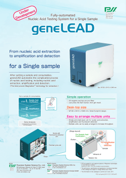


© Copyright 2026