
Program Title Program No. Classification
ENVIRONMENTAL HEALTH AND SAFETY EHS PROGRAM MANUAL Program Title Radiation Safety Manual Program No. 9.1 Classification Radiation Safety 1.0 INTRODUCTION The Radiation Safety Program at the Weill Cornell Medical College (WCMC) and NewYorkPresbyterian Hospital (NYP) Weill Cornell Medical Center is overseen and administered by WCMC Environmental Health and Safety (EHS). This Radiation Safety Program has been established to promote a safe environment for all employees, students, patients, and members of the public who may work with or be exposed to radiation while they are physically located on the WCMC / NYP campus. WCMC EHS oversees environmental and occupational health and safety during the use, storage and disposal of radioactive materials in all research applications. WCMC EHS also provides the following services to the WCMC / NYP community. Diagnostic Imaging Quality Assurance - Performs and documents testing on all diagnostic imaging equipment to assure optimal, safe performance. Central Isotope Laboratory - Controls the ordering, receipt and distribution of radioactive materials. Cyclotron and Radiochemistry Facility - Provides environmental and occupational safety during production and use of positron-emitting radiolabeled pharmaceuticals. 2.0 TABLE OF CONTENTS 1.0 Introduction .......................................................................................................................... 1 2.0 Table of Contents .................................................................................................................. 1 3.0 Applicability ....................................................................................................................... 10 4.0 Administrative Commitment / As Low As Reasonably Achievable (ALARA) ......................... 10 5.0 Contact Information ....................................................................................................................... 11 5.1 Environmental Health and Safety (EHS) ............................................................................................. 11 5.2 Emergency Contact Information ............................................................................................................. 11 6.0 Roles and Responsibilities ............................................................................................................. 12 6.1 Radiation Safety Committee (RSC) ..................................................................................... 12 6.2 Radiation Safety Officer (RSO) ........................................................................................... 12 6.3 WCMC Environmental Health and Safety (EHS)................................................................ 13 6.4 Authorized Users / Principal Investigators / Laboratory managers ..................................... 14 6.4.1 ALARA ......................................................................................................... 14 6.4.2 Accountability ................................................................................................ 14 6.4.3 Compliance with Regulations .......................................................................... 14 6.4.4 Responsibilities .............................................................................................. 15 Date Issued: March 28, 2014 Supersedes: All Programs Prior to the Date Issued Page: Page 1 of 148 Program Title Radiation Safety Manual 6.5 6.6 Program No. 9.1 Classification Radiation Safety Certified users / Researchers ................................................................................................ 16 6.5.1 Compliance with Regulations ................................................................................. 16 6.5.2 Responsibilities ....................................................................................................... 17 Supervised Individuals / Laboratory Personnel ................................................................... 17 6.6.1 Responsibilities ....................................................................................................... 17 7.0 Guide to Becoming a Radioactive Materials Authorized User ...................................................... 18 7.1 Application and Approval for Radioactive Materials Authorized User ............................... 18 7.2 Radioactive Materials License Fee ...................................................................................... 18 7.3 Limits of Authorized Radioactive Material Use .................................................................. 18 8.0 Radiation Doses Investigational Levels ......................................................................................... 18 8.1 Personal Dose Less than the Investigational Level .............................................................. 19 8.2 Personal Dose Equal To or Greater than Investigational Level but Less than Investigational Level II ............................................................................................................................................................ 19 8.3 Personal Dose Equal To or Greater than Investigational Level II ....................................... 19 8.4 Re-Establishment of Investigational Levels ......................................................................... 19 9.0 Occupational Dose Limits.............................................................................................................. 19 10.0 Personal Monitoring / Dosimetry.................................................................................................. 20 10.1 Radiation Dose Sources ....................................................................................................... 20 10.1.1 Internal Doses ......................................................................................................... 20 10.1.2 External Doses ........................................................................................................ 20 10.2 Regulations governing Monitoring ...................................................................................... 21 10.3 Limitations of OSL Dosimeters Used at WCMC / NYP...................................................... 21 10.4 Placement of Dosimeters...................................................................................................... 21 10.4.1 Whole-Body Dosimeter .......................................................................................... 21 10.4.2 Lens of the Eye ....................................................................................................... 21 10.4.3 Embryo/Fetus .......................................................................................................... 22 10.4.4 Multiple dosimeters................................................................................................. 22 10.4.5 Extremities .............................................................................................................. 22 10.5 Frequency of Wearing Dosimeters....................................................................................... 22 10.6 Issuing Dosimeters ............................................................................................................... 22 10.7 Frequency of Reading Dosimeters ....................................................................................... 23 10.8 Determination of Prior Exposure ......................................................................................... 23 10.9 Determination of Lifetime Dose .......................................................................................... 23 10.10 Dosimetry Reports ............................................................................................................... 23 10.11 Bioassay ............................................................................................................................... 23 10.12 Radioiodine Assay ............................................................................................................... 24 10.13 Titritium Assay .................................................................................................................... 25 11.0 Employee Declaration of Pregnancy.............................................................................................. 26 12.0 Protection of the General Public .................................................................................................... 27 12.1 Dose Limits for the General Public ...................................................................................... 27 12.2 ALARA Principal ................................................................................................................ 27 12.3 Radiation and Non-Radiation Workers ................................................................................ 27 Date Issued: March 28, 2014 Supersedes: All Programs Prior to the Date Issued Page: Page 2 of 148 Program Title Radiation Safety Manual Program No. 9.1 Classification Radiation Safety 13.0 Security of Radioactive materials .................................................................................................. 28 13.1 NRC Security Regulations ........................................................................................................................ 28 14.0 Emergency Proceduress for Laboratories ...................................................................................... 29 14.1 Emergency Contact Numbers .................................................................................................................. 29 14.2 Major Spills – Greater Than 100 ml or 10 mCi ................................................................................. 29 14.3 Minor Spills – Less Than 100 ml or 10 mCi ....................................................................................... 29 14.4 Dry Spills........................................................................................................................................................ 30 14.5 Personal Decontamination ........................................................................................................................ 30 14.6 Radioactive Dust, Mists, Fumes, Gases, Etc....................................................................................... 30 14.7 Inuries Involving Radiation Hazards ..................................................................................................... 30 14.8 Fires Involving Possible Radiation hazards......................................................................................... 31 15.0 Radiation Safety Procedures .......................................................................................................... 31 15.1 Emergency Procedures ............................................................................................................................... 31 15.2 Accidental Inhalation, Ingestion, or Injury .......................................................................................... 32 15.3 Eating, Drinking and Smoking ................................................................................................................ 32 15.4 Exiting the Laboratory / Radioactive Materials Area....................................................................... 32 15.5 Contamination Prevention......................................................................................................................... 32 15.6 Housekeeping................................................................................................................................................ 32 15.7 Dress Code in a Radioactive Materials Area ...................................................................................... 33 15.8 Personnel Monitors ..................................................................................................................................... 33 15.9 Mouth Suction and Pipetting .................................................................................................................... 33 15.10 Radioactive Material Storage ................................................................................................................... 33 15.11 Minors ............................................................................................................................................................. 33 15.12 Shielding of Sources ................................................................................................................................... 34 15.13 Aerosols, Dusts and Gaseous Products ................................................................................................. 34 15.14 Chemical Hoods ........................................................................................................................................... 34 15.15 House Vaccum Lines .................................................................................................................................. 34 15.16 Volatile Compounds Work ....................................................................................................................... 34 15.17 Laboratories Using High-Energy Beta or Gamma Radiation ........................................................ 34 16.0 Radioactive Materials Signage ...................................................................................................... 35 16.1 Locations Requiring Radioactive Materials Signage ....................................................................... 35 16.2 Health and Safety Door Sign.................................................................................................................... 35 16.3 Example Radiation Signs .......................................................................................................................... 35 16.3.1 Caution Radioactive Materials ................................................................................ 35 16.3.2 Caution Radiation Area ........................................................................................... 35 16.3.3 Caution High Radiation Area .................................................................................. 36 16.3.4 Caution Very High Radiation Area ......................................................................... 36 16.4 Laboratory Signage ..................................................................................................................................... 36 16.5 Containers and Equipment Labeling ..................................................................................................... 36 16.6 Requesting Signs.......................................................................................................................................... 37 17.0 Radioactive Waste Management .................................................................................................... 37 18.0 Lead Safety .................................................................................................................................... 37 18.1 Permanently Installed Lead ...................................................................................................................... 37 Date Issued: March 28, 2014 Supersedes: All Programs Prior to the Date Issued Page: Page 3 of 148 Program Title Radiation Safety Manual 18.2 18.3 18.4 18.5 18.6 18.7 Program No. 9.1 Classification Radiation Safety Lead Not in Use............................................................................................................................................ 37 Drilling, Milling and Sawing ................................................................................................................... 37 Propping of Doors ....................................................................................................................................... 37 Glove Use ....................................................................................................................................................... 37 Hand Hygiene ............................................................................................................................................... 37 Disposal of Lead and Lead Pigs / Ingots .............................................................................................. 38 19.0 Personal Protective Equipment (PPE)............................................................................................ 38 20.0 Equipment ...................................................................................................................................... 38 20.1 Radioactive Materials in Gas Chromatography Equipment ........................................................... 38 20.2 Liquid Scintillation and Gamma Counting Equipment ................................................................... 39 20.3 Equipment Repair, Maintenance and Disposal .................................................................................. 39 21.0 Centralized Ordering System for Isotopes ..................................................................................... 39 22.0 Handling Packages Containing Radioactive Material.................................................................... 40 22.1 Receiving Packages..................................................................................................................................... 40 22.2 Opening Packages........................................................................................................................................ 40 22.3 Discarding Packaging Materials ............................................................................................................. 41 23.0 Sealed Sources ............................................................................................................................... 41 23.1 Testing Purchased and Fabricated Sealed Sources............................................................................ 41 23.2 New York City DOH Requirements (Article 175.03(E)) ............................................................... 41 23.3 Exceptions to Leak Test Requirements................................................................................................. 42 23.4 Authorized User / Principal Investigator Responsibilities .............................................................. 42 24.0 Inventory Control ........................................................................................................................... 42 24.1 Receipt of Vials ............................................................................................................................................ 43 24.2 Withdrawals by Individuals ...................................................................................................................... 43 24.3 Disposal .......................................................................................................................................................... 43 24.4 Additional Inventory Control Guidelines ............................................................................................. 44 25.0 Transporting and Shipping Radioactive Materials......................................................................... 44 25.1 Transfers within the Workplace .............................................................................................................. 44 25.2 Transfers within WCMC / NYP .............................................................................................................. 44 25.3 Transfers Between WCMC / NYP and Other Institutions within the U.S. ............................... 45 25.4 International Shipments ............................................................................................................................. 46 26.0 Contamination Control................................................................................................................... 46 26.1 Area Classification ...................................................................................................................................... 46 26.1.1 Controlled Area ....................................................................................................... 46 26.1.2 Restricted Area ........................................................................................................ 46 26.1.3 Contaminated Area ................................................................................................. 47 26.1.4 Highly Contaminated Area ..................................................................................... 47 26.1.5 Radioactive Material Area ...................................................................................... 47 26.2 Area Posting .................................................................................................................................................. 47 26.3 Area Preparation .......................................................................................................................................... 47 26.4 Intake Considerations ................................................................................................................................. 48 Date Issued: March 28, 2014 Supersedes: All Programs Prior to the Date Issued Page: Page 4 of 148 Program Title Radiation Safety Manual Program No. 9.1 Classification Radiation Safety 26.4.1 Precautions Prior to Eating, Drinking, or Smoking ................................................ 48 26.4.2 Air Contamination and Inhalation ........................................................................... 48 26.4.3 Incubation................................................................................................................ 49 26.5 Surface Contamination............................................................................................................................... 49 26.6 Hazard Planning ........................................................................................................................................... 50 26.7 Equipment Protection ................................................................................................................................. 50 27.0 Decontamination ............................................................................................................................ 50 27.1 Steps to take Immediately Following Discovery of Contamination ............................................ 51 27.2 Equipment and Surface Decontamination............................................................................................ 51 27.2.1 Determine Contamination Level ............................................................................. 51 27.2.2 Determine whether the Contamination is Fixed or Removable .............................. 51 27.2.3 Scrubbing ................................................................................................................ 51 27.2.4 Unsuccessful Decontamination of Equipment and Surfaces................................... 51 27.3 Personal Decontamination ........................................................................................................................ 51 27.3.1 Removing Radioactive Materials from the Skin ..................................................... 52 28.0 Routine Contamination Surveys .................................................................................................... 52 28.1 Minimum Protection Standard................................................................................................................. 53 28.2 Suspect Surveys............................................................................................................................................ 53 29.0 Portable Survey Instruments .......................................................................................................... 53 29.1 Possession of Geiger-Mueller Detector ................................................................................................ 54 29.2 Calibration of Survey Meters ................................................................................................................... 54 29.3 Geiger Counter Applications.................................................................................................................... 54 29.4 Survey Meter Operational Function Tests ........................................................................................... 54 29.4.1 Battery Check.......................................................................................................... 54 29.4.2 Cable Check ............................................................................................................ 54 29.4.3 Check Source .......................................................................................................... 55 29.4.4 Background Check .................................................................................................. 55 29.5 Performing a Survey Using Geiger-Mueller Instrument ................................................................. 55 29.5.1 Operational Check................................................................................................... 55 29.5.2 Choose Correct Probe ............................................................................................. 55 29.5.3 Probe Motion........................................................................................................... 55 29.5.4 Areas Requiring Survey Before and After Working with Isotopes......................... 55 29.6 Documentation of Geiger-Mueller Survey .......................................................................................... 56 29.6.1 Maintenance of Survey Documentation .................................................................. 56 29.7 Frequency of Geiger-Mueller Surveys .................................................................................................. 56 30.0 Liquid Scintillation Counting (LSC) ............................................................................................. 56 30.1 Wipe Test Methodology ............................................................................................................................ 57 30.2 Wipe Test Frequency .................................................................................................................................. 58 30.3 Wipe Test Documentation ........................................................................................................................ 58 30.3.1 Maintenance of Wipe Test Reports ......................................................................... 58 30.4 Calculating Removable Activity ............................................................................................................. 58 30.5 Removable Activity Action Levels ........................................................................................................ 59 30.6 Liquid Scintillation Fluid .......................................................................................................................... 59 Date Issued: March 28, 2014 Supersedes: All Programs Prior to the Date Issued Page: Page 5 of 148 Program Title Radiation Safety Manual Program No. 9.1 Classification Radiation Safety 30.7 Liquid Scintillation Counting Errors ..................................................................................................... 60 30.8 Avoiding Luminescence ............................................................................................................................ 61 31.0 Electron Microscopes..................................................................................................................... 61 32.0 X-Ray Diffraction and Medical X-Ray Equipment ....................................................................... 61 32.1 X-ray Diffraction ......................................................................................................................................... 61 32.2 Clinical X-ray Equipment ......................................................................................................................... 62 33.0 Gammacell 1000 ............................................................................................................................ 63 33.1 Licensing and Training Requirements .................................................................................................. 63 33.2 Malfunctions and Emergencies ............................................................................................................... 63 33.3 Radioactive Source Information ............................................................................................................. 63 33.4 Dose Rate and Clock Setting.................................................................................................................... 63 33.5 Gammacell 1000 Operating Procedures ............................................................................................... 64 33.5.1 Important Notes....................................................................................................... 65 33.6 Gammacell 1000 Irradiator Emergency Procedures ......................................................................... 65 33.7 Emergency or Unusual Occurrence Procedures ................................................................................. 65 34.0 Laboratory Decomissioning ........................................................................................................... 66 34.1 Notification .................................................................................................................................................... 66 34.2 When all Radioactive Material Use Ceases ......................................................................................... 66 34.3 Equipment ...................................................................................................................................................... 67 34.4 WCMC / NYP Custodial Service / Outside Movers......................................................................... 67 35.0 Reportable Events .......................................................................................................................... 68 35.1 Stolen, Lost or Missing Licensed or Registered Sources of Radioactive Materials ............... 68 35.2 Notifications of Incidents .......................................................................................................................... 68 35.2.1 Immediate Notification ........................................................................................... 68 35.2.2 Twenty-Four Hour Notification .............................................................................. 69 36.0 Physical Properties of Radioactive Materials ................................................................................ 69 36.1 Hydrogen – 3 [H-3] Physical Properties............................................................................................... 69 36.1.1 Physical Data........................................................................................................... 69 36.1.2 Radiological Data .................................................................................................... 70 36.1.3 Shielding ................................................................................................................. 70 36.1.4 Survey Instrumentation ........................................................................................... 70 36.1.5 Personal Radiation Monitoring Dosimeters ............................................................ 70 36.1.6 Radioactive Waste................................................................................................... 71 36.1.7 Regulatory Compliance Information (10 CFR 20, Appendix B) ............................ 71 36.1.8 General Radiological Safety Information ............................................................... 71 36.2 Carbon-14 [C-14] Physical Properties .................................................................................................. 73 36.2.1 Physical Data........................................................................................................... 73 36.2.2 Radiological Data .................................................................................................... 74 36.2.3 Shielding ................................................................................................................. 74 36.2.4 Survey Instrumentation ........................................................................................... 75 36.2.5 Radiation Monitoring Dosimeters ........................................................................... 75 36.2.6 Regulatory Compliance Information (10 CFR 20, Appendix B) ............................ 75 Date Issued: March 28, 2014 Supersedes: All Programs Prior to the Date Issued Page: Page 6 of 148 Program Title Radiation Safety Manual 36.3 36.4 36.5 36.6 36.7 36.8 Program No. 9.1 Classification Radiation Safety 36.2.7 General Radiological Safety Information ............................................................... 76 Fluorine-18 [F-18] Physical Properties ................................................................................................. 77 36.3.1 Physical Data........................................................................................................... 77 36.3.2 Radiological Data .................................................................................................... 77 36.3.3 Shielding ................................................................................................................. 78 36.3.4 Dosimetry Monitoring............................................................................................. 78 36.3.5 Detecting and Measurement .................................................................................... 78 36.3.6 Special Precautions ................................................................................................. 78 36.3.7 General Precautions ................................................................................................ 78 Phosphorus-32 [P-32] Physical Properties .......................................................................................... 79 36.4.1 Physical Data........................................................................................................... 79 36.4.2 Radiological Data .................................................................................................... 80 36.4.3 Shielding ................................................................................................................. 81 36.4.4 Survey Instrumentation ........................................................................................... 81 36.4.5 Dose Rates (From Unshielded 1.0 mCi Isotropic Point Source) ............................. 81 36.4.6 Regulatory Compliance Limits (10 CFR 20, Appendix B) ..................................... 81 36.4.7 General Radiological Safety Information ............................................................... 82 Phosphorus-33 [P-33] Physical Properties .......................................................................................... 83 36.5.1 Physical Data........................................................................................................... 83 36.5.2 Radiological Data .................................................................................................... 84 36.5.3 Shielding ................................................................................................................. 84 36.5.4 Survey Instrumentation ........................................................................................... 84 36.5.5 Personnel Dosimeters .............................................................................................. 85 36.5.6 Regulatory Compliance Limits (10 CFR 20, Appendix B) ..................................... 85 36.5.7 General Radiological Safety Information ............................................................... 85 Sulfur-35 [S-35] Physical Properties ..................................................................................................... 86 36.6.1 Physical Data........................................................................................................... 86 36.6.2 Radiological Data .................................................................................................... 86 36.6.3 Shielding ................................................................................................................. 87 36.6.4 Survey Instrumentation ........................................................................................... 87 36.6.5 Radiation Monitoring Devices ................................................................................ 87 36.6.6 Regulatory Compliance Information (10 CFR 20, Appendix B) ............................ 88 36.6.7 General Radiological Safety Information (S-35) .................................................... 88 Chromium – 51 [Cr-51] Physical Properties ....................................................................................... 90 36.7.1 Physical Data........................................................................................................... 90 36.7.2 Radiological Data .................................................................................................... 91 36.7.3 Shielding ................................................................................................................. 92 36.7.4 Survey Instrumentation ........................................................................................... 92 36.7.5 Personal Radiation Monitoring Dosimeters ............................................................ 92 36.7.6 Regulatory Compliance Information....................................................................... 92 Iron – 59 [FE-59] Physical Properties ................................................................................................... 93 36.8.1 Physical Data........................................................................................................... 93 36.8.2 Radiological Data .................................................................................................... 94 36.8.3 Shielding ................................................................................................................. 94 36.8.4 Survey Instrumentation ........................................................................................... 94 36.8.5 Personal Radiation Monitoring Dosimeters ............................................................ 94 Date Issued: March 28, 2014 Supersedes: All Programs Prior to the Date Issued Page: Page 7 of 148 Program Title Radiation Safety Manual Program No. 9.1 Classification Radiation Safety 36.8.6 Regulatory Compliance Information....................................................................... 94 36.9 Strontium – 90/Yttritum – 90 [Sr-90], [Y-90 IT], [Y-90] ............................................................... 95 36.9.1 Physical Data........................................................................................................... 95 36.9.2 Radiological Data .................................................................................................... 96 36.9.3 Shielding ................................................................................................................. 96 36.9.4 Survey Instrumentation ........................................................................................... 96 36.9.5 Personal Radiation Monitoring Dosimeters ............................................................ 97 36.9.6 Regulatory Compliance Information....................................................................... 97 36.10 Iodine-125 [I-125] Physical Properties ................................................................................................. 97 36.10.1 Physical Data........................................................................................................... 97 36.10.2 Radiological Data .................................................................................................... 97 36.10.3 Shielding ................................................................................................................. 98 36.10.4 Survey Instrumentation ........................................................................................... 98 36.10.5 Dose Rates (From Unshielded 1.0 mCi Isotropic Point Source) ............................. 98 36.10.6 Regulatory Compliance Information (10 CFR 20, Appendix B) ............................ 98 36.10.7 Iodination Procedures ............................................................................................. 99 36.10.8 General Radiological Safety Information ............................................................. 100 36.11 Iodine-131 [I-131] Physical Properties .............................................................................................. 101 36.11.1 Physical Data......................................................................................................... 101 36.11.2 Radiological Data .................................................................................................. 101 36.11.3 Shielding ............................................................................................................... 102 36.11.4 Exposure Rates (From an Unshielded 1.0 mCi Isotropic Point Source I-131) ..... 102 36.11.5 Survey Instrumentation ......................................................................................... 103 36.11.6 Personal Radiation Monitoring Dosimeters .......................................................... 103 36.11.7 Regulatory Compliance Limits (10 CFR 20, Appendix B) ................................... 103 36.11.8 General Radiological Safety Information ............................................................. 104 36.11.9 Iodination Procedures ........................................................................................... 105 36.12 Technetium – 99m [TC-99m] Physical Properties ......................................................................... 106 36.12.1 Physical Data......................................................................................................... 106 36.12.2 Radiological Data .................................................................................................. 107 36.12.3 Shielding ............................................................................................................... 108 36.12.4 Survey Instrumentation ......................................................................................... 108 36.12.5 Personnel Radiation Monitoring Dosimeters ........................................................ 108 36.12.6 Regulatory Compliance Information (10 CFR 20, Appendix B) .......................... 109 36.12.7 General Radiological Safety Information ............................................................. 109 37.0 Radiation Theory and Fundamentals ........................................................................................... 111 37.1 Radioactivity .............................................................................................................................................. 111 37.2 Ionizing Radiation .................................................................................................................................... 111 37.2.1 Alpha Particles ...................................................................................................... 112 37.2.2 Beta Particles......................................................................................................... 112 37.2.3 Gamma Rays ......................................................................................................... 112 37.2.4 X-Rays .................................................................................................................. 112 37.2.5 Neutrons. ............................................................................................................... 113 37.3 Why is Material Radioactive?............................................................................................................... 113 37.4 Production of Radioactive Materials .................................................................................................. 114 Date Issued: March 28, 2014 Supersedes: All Programs Prior to the Date Issued Page: Page 8 of 148 Program Title Radiation Safety Manual Program No. 9.1 Classification Radiation Safety 37.5 Decay of Radioactive Materials ........................................................................................................... 115 37.5.1 Alpha Decay.......................................................................................................... 115 37.5.2 Beta Decay ............................................................................................................ 116 37.5.3 Electron Capture Decay ........................................................................................ 117 37.5.4 Gamma Decay ....................................................................................................... 117 37.5.5 X-Ray Decay ......................................................................................................... 118 37.5.6 Auger Electron Decay ........................................................................................... 118 37.6 Activity of Radioactive Materials........................................................................................................ 118 37.6.1 The Curie............................................................................................................... 118 37.6.2 The Becquerel ....................................................................................................... 118 37.6.3 Specific Activity ................................................................................................... 119 37.7 Radiation Exposure .................................................................................................................................. 119 37.7.1 Radiation Absorbed Dose ..................................................................................... 120 37.7.2 Dose Equivalent .................................................................................................... 120 37.7.3 Quality Factor ....................................................................................................... 121 37.7.4 Effective Dose Equivalent..................................................................................... 122 37.7.5 Committed Dose Equivalent ................................................................................. 123 37.7.6 Committed Effective Dose Equivalent (CEDE).................................................... 123 37.7.7 Total Effective Dose Equivalent (TEDE) ............................................................. 123 37.8 Characteristics of Radioactive material ............................................................................................. 124 37.8.1 Physical Half-Life ................................................................................................. 124 37.8.2 Biological Half-Life .............................................................................................. 124 37.8.3 Effective Half-Life ................................................................................................ 124 37.8.4 Fission and Criticality ........................................................................................... 124 38.0 Occupational Radiation Exposure................................................................................................ 126 38.1 Common Sources of ionizing Radiation............................................................................................ 126 38.2 U.S. Population Exposure ...................................................................................................................... 127 38.2.1 Cosmic Rays ......................................................................................................... 127 38.2.2 Natural Radiation .................................................................................................. 128 38.2.3 Medical Use of radiation ....................................................................................... 128 38.3 Radiation Hazards at WCMC / NYP .................................................................................................. 129 38.3.1 Unsealed Radioactive Sources .............................................................................. 129 38.3.2 Sealed Sources ...................................................................................................... 129 38.3.3 Irradiators .............................................................................................................. 129 38.3.4 Radioactive Waste Storage ................................................................................... 130 38.3.5 Diagnostic Equipment and Procedures ................................................................. 130 38.3.6 Radiation Therapy Sources ................................................................................... 130 38.3.7 Linear Accelerators ............................................................................................... 130 38.3.8 Controlling Radiation Dose: Time ........................................................................ 131 38.3.9 Controlling Radiation Dose: Distance .................................................................. 131 38.3.10 Controlling Radiation Dose: Shielding ................................................................. 132 38.3.10.1 Alpha Shielding.......................................................................... 132 38.3.10.2 Beta Shielding ............................................................................ 133 38.3.10.3 Positron Shielding ...................................................................... 133 38.3.10.4 Cerenkov Radiation Shielding ..................................................... 134 Date Issued: March 28, 2014 Supersedes: All Programs Prior to the Date Issued Page: Page 9 of 148 Program Title Radiation Safety Manual Program No. 9.1 Classification Radiation Safety 38.3.10.5 Gamma Shielding .............................................................................. 134 38.4 Source Exposure, Intake and Ontake Control .................................................................................. 136 39.0 Training........................................................................................................................................ 137 39.1 Training Frequency .................................................................................................................................. 138 39.2 Training Registration ............................................................................................................................... 138 40.0 Recordkeeping and Retention ...................................................................................................... 138 40.1 Radioactive Materials Use and Disposal Records .......................................................................... 138 40.2 Radioactive Waste Disposal Records ................................................................................................. 138 40.3 Contamination Survey Records ............................................................................................................ 138 40.4 Equipment Calibration Records ........................................................................................................... 138 40.5 Dosimetry Records ................................................................................................................................... 138 40.6 Training Records ....................................................................................................................................... 138 41.0 Definitions ................................................................................................................................... 139 42.0 References .................................................................................................................................... 143 Appendix A Declaration of Pregnancy ....................................................................................... 145 Appendix B Laboratory Self-Audit Checklist ............................................................................. 147 3.0 APPLICABILITY This Radiation Safety Program applies to all non-human radioactive materials use at WCMC / NYP. All faculty and staff using radioactive materials must read and implement the practices outlined in this Program. Adherence to good radiation safety practices and the development of specific research protocols will ensure safety and compliance with standards issued by the Occupational Safety and Health Administration (OSHA), the New York City Department of Health (NYDOH), and the New York State Department of Environmental Conservation (NYDEC). 4.0 ADMINISTRATIVE COMMITMENT / AS LOW AS REASONABLY ACHIEVABLE (ALARA) Weill Cornell Medical College (WCMC) and NewYork-Presbyterian Hospital (NYP) are committed to the Radiation Safety Program described herein for keeping individual and collective doses from ionizing radiation ‘As Low As Reasonably Achievable’ (known as the “ALARA Principle”). In accord with this commitment, a Radiation Safety Committee (RSC) and a Radiation Safety Officer (RSO) and have been appointed to develop written policies, procedures, and instructions to foster the ALARA concept within WCMC / NYP. The Radiation Safety Program and ALARA standards will be reviewed annually. The review will include audits of operating procedures, past dose records, inspections, and consultations. Date Issued: March 28, 2014 Supersedes: All Programs Prior to the Date Issued Page: Page 10 of 148 Program Title Radiation Safety Manual Program No. 9.1 Classification Radiation Safety Modifications to research protocols, maintenance procedures, equipment, and facilities will be made if they will reduce exposures, unless the burden of the modifications outweighs the potential for dose reduction. Documentation will be available to demonstrate that improvements have been sought, modifications have been considered, and have been implemented when reasonable. If radiological design modifications have been recommended but not implemented, justifications for not implementing them will be available. The goal of ALARA is to maintain doses to individuals and releases to environment as far below the limits as is reasonably achievable. The sum of the doses received by all exposed individuals will also be maintained at the lowest practicable level consistent with an expanding research program. The normal radiation doses received by radiation workers or other personnel are considered dose limits. The guiding principle of all radiation work is the dose should be As Low As Reasonably Achievable, economic and social factors being taken into account. Most Radiation Users are able to maintain an annual exposure not only well below the legal limit but also well below even the lower limit operating at WCMC / NYP for this category of radiation worker. Any Radiation User whose annual or quarterly dose, as measured by external monitoring or calculated from the results of bioassay procedures, greatly exceeds the normal value either for that individual or for persons carrying out similar work, is subject to investigation by the Permit Holder, in cooperation with EHS. 5.0 CONTACT INFORMATION 5.1 ENVIRONMENTAL HEALTH AND SAFETY (EHS) Contact EHS for all radiation safety related questions or requests for assistance including: isotope ordering, dosimetry services, waste disposal, etc. 5.2 Phone: (646) 962-7233 Fax: (646) 962-0288 Email: [email protected] EMERGENCY CONTACT INFORMATION Radiation Emergency during business hours, contact EHS: (646) 962-7233 Radiation Emergency during off-hours, contact Security: (212) 746-0911 o When contacting Security during off-hours, request that they contact the on-call EHS Emergency Responder. Date Issued: March 28, 2014 Supersedes: All Programs Prior to the Date Issued Page: Page 11 of 148 Program Title Radiation Safety Manual Program No. 9.1 Classification Radiation Safety 6.0 ROLES AND RESPONSIBILITIES 6.1 RADIATION SAFETY COMMITTEE (RSC) The Radiation Safety Committee (RSC) is comprised of physicians, scientists, administrators, and nursing personnel from WCMC and NYP that establishes the policies and regulations regarding the human and non-human use of radiation (both ionizing and non-ionizing) within NYP / WCMC. The Chairman of Radiology, the Dean of the Medical School, and the Dean of the College appoint members. The Committee reports to the NYP WCMC Internal Review Board (IRB) and the Environment of Care Council (ECC). The RSC has the following roles and responsibilities. 6.2 Applicant Qualification Reviews – During the authorization approval process, the RSC will review the qualifications of each applicant with respect to the types and quantities of materials and methods of use for which application has been made to ensure that the applicant will be able to maintain exposure ALARA. Procedure Review – The RSC will ensure that the users document their procedures and will review the efforts of the applicants to maintain exposure ALARA. Incident, Accident, and Hazard Evaluation Reviews – The RSC will review incidents, accidents, and results of hazard evaluations as well as corrective actions taken. Annual Review of Occupational Radiation Exposure – The RSC will perform an annual review of occupational radiation exposures. The principal purpose of this review is to assess trends in occupational exposure as an index of the ALARA program quality. Annual Evaluation of ALARA Efforts – The RSC will evaluate WCMC / NYP's overall efforts for maintaining doses ALARA on an annual basis. This review will include the efforts of the RSO, Authorized Users and ancillary groups as well as those of the administration. RSC Authority – The RSC will support the Radiation Safety Office (RSO), see Section 7.2, when it is necessary for the RSO to assert authority. If the RSC has overruled the RSO, it will record the basis for its action in the minutes of its meetings. RADIATION SAFETY OFFICER (RSO) The Radiation Safety Officer (RSO) is a member of WCMC Environmental Health and Safety (EHS). The RSO has the following roles and responsibilities. Safe Working Conditions – The RSO will ensure that safe radiological working conditions are established and maintained for all WCMC / NYP Date Issued: March 28, 2014 Supersedes: All Programs Prior to the Date Issued Page: Page 12 of 148 Program Title Radiation Safety Manual Program No. 9.1 Classification Radiation Safety faculty, students, patients, staff, visitors, and the general public, and shall ensure compliance with all pertinent federal, state, and local regulations. 6.3 Role in Authorization Approval Process – During the authorization approval process the RSO will encourage all users to review procedures and develop new or revised procedures as appropriate to implement the ALARA concept. Radiation Survey Records Review – The RSO will review radiation surveys to determine that dose rates, amounts of contamination, and releases to the environment were at ALARA levels during the previous quarter. Review of Occupational Exposures – The RSO will review, at least annually, the radiation doses of Authorized Users and workers to determine that their doses are ALARA in accordance with the provisions of Section 5 of this program. Annual Review of the Radiation Safety Program – The RSO will perform an annual review of the radiation safety program for consistency with the ALARA philosophy. Input from Radiation Users – The RSO will establish procedures for receiving and evaluating the suggestions of individual radiation users for improving radiation safety practices and will encourage the use of those procedures when deemed appropriate. Investigation of, and Response to, Deviations from ALARA – The RSO will initiate investigations of all known instances of deviation from the ALARA philosophy and, if possible, will determine the causes. When the cause is known, the RSO will implement changes in the program to maintain doses ALARA. Quarterly Review of Exposure Records – The RSO, or delegated senior staff, will review the exposure records on at least a quarterly basis and initiate investigations where indicated. WCMC ENVIRONMENTAL HEALTH AND SAFETY (EHS) Conduct inspections, hazard evaluations, interviews, and make recommendations that include radiological planning to contribute to dose reduction and promote a safe working environment. Consult with Principal Investigators, researchers and other personnel about laboratory design, appropriateness of methods and alternatives. Perform facility and laboratory radiation surveys and inspect facilities to enhance contamination control and reduction of radiation exposure. Date Issued: March 28, 2014 Supersedes: All Programs Prior to the Date Issued Page: Page 13 of 148 Program Title Radiation Safety Manual 6.4 Program No. 9.1 Classification Radiation Safety Provide Radiation Safety Training to all students, faculty, and staff who are authorized radiation users or ancillary personnel who may be exposed to radiation. Manage radioactive isotope ordering for authorized radiation users at WCMC / NYP. Provide radioactive waste disposal procedures and services Retain all training, dosimetry, and inspection records as required by the appropriate regulatory agencies. Annually review and update this written program. AUTHORIZED USERS / PRINCIPAL INVESTIGATORS / LABORATORY MANAGERS An Authorized User of radioactive materials is a WCMC / NYP Principal Investigator or Laboratory Manager who has been approved by the Radiation Safety Committee (RSC) to order, receive, possess, and use radioactive materials within the confines of WCMC / NYP under the regulatory authority of the New York City Department of Health and Mental Hygiene. 6.4.1 ALARA Authorized Users must: Explain the ALARA concept and the need to maintain exposures ALARA to all supervised individuals. Ensure that supervised individuals who are subject to occupational radiation exposure are trained and educated in good radiation safety practices and in maintaining exposures ALARA. 6.4.2 Accountability Authorized users are accountable for radiation protection policy and practices in their laboratories. 6.4.3 Compliance with Regulations Authorized Users must comply with the regulations governing the use of radioactive materials, as established by Article 175 of the Rules of the City of New York, Title 24 and the WCMC / NYP Radiation Safety Committee which specify: Laboratory air and water concentrations shall be maintained below the levels specified in Article 175.03, Appendix B of the New York City Health Code (same as 10CFR20, “Standards for Protection Against Radiation) and 6 NYCRR Part 380 of New York State Department of Environmental Conservation (DEC). Date Issued: March 28, 2014 Supersedes: All Programs Prior to the Date Issued Page: Page 14 of 148 Program Title Radiation Safety Manual Program No. 9.1 Classification Radiation Safety Disposal of radioactive materials into the sewage system is strictly prohibited. 6.4.4 Responsibilities Complete the Radiation Safety Committee Application for Radioactive Material Non-Human Use Application and receive written approval from the RSC prior to working with or modifying work with radioactive materials. Comply with the regulations governing the use of radioactive materials, as established by Article 175 of the Health Code of the City of New York and the WCMC / NYP Radiation Safety Committee, which include, but are not limited to: Following correct procedures for the procurement of radioactive materials by purchase or transfer. Posting areas where radionuclides are kept or used, or where radiation fields may exist. Confirming that each sign carries the name of the personnel currently responsible for the associated area. Recording the receipt, transfer, and disposal of radioactive materials in the user’s area. This includes sealed sources such as ion sources in gas chromatographs and static eliminators. The Authorized User must be prepared to submit the required inventory data upon request. Follow radioactive ordering procedures and obtain radioactive materials only via the Radioactive Isotope Laboratory. Maintain laboratory security where radioactive materials are stored or used. These areas are restricted areas and are to be kept locked. Report any breaches of radioactive material security to WCMC EHS immediately. Take steps to prevent the transfer of radioactive materials to unauthorized individuals. This includes the proper disposition of radioactive materials in the possession of terminating workers or Authorized Users. Develop and implement policies and procedures in compliance with this Program and ensure that all personnel are aware of and compliant with them. Ensure all radiation users receive appropriate Radiation Safety Training. Ensure all personnel wear appropriate personal protective equipment and dosimetry badges as required. Date Issued: March 28, 2014 Supersedes: All Programs Prior to the Date Issued Page: Page 15 of 148 Program Title Radiation Safety Manual Program No. 9.1 Classification Radiation Safety Minimize stocks of stored radioactive materials within laboratory areas, including radioactive waste. Provide EHS with a list of laboratory workers and their activities in laboratories. Notify EHS of any personnel changes. Contact the EHS whenever major changes in operational procedures, new techniques, alterations in the physical plant (e.g., the shutdown or removal of a radiochemical fume hood), or when new operations, which might lead to personnel exposure, are anticipated. Assure that all radioactive waste materials are consigned to EHS for proper disposal. Comply with proper procedures for closing down or moving a laboratory, including: Prior notification to EHS. Proper removal and disposition of all radioactive waste and sources. Complete a final contamination survey. Notify EHS of the location of the new facility Submission of a drawing or schematic of the new area for inclusion with the user’s license on file in the EHS Office. 6.5 CERTIFIED USERS / RESEARCHERS Certified Users are WCMC / NYP researchers who have completed WCMC Radiation Safety Training but are not Authorized Users. They have been approved by EHS to receive, use, and possess radionuclides in a safe, scientific manner. Certified Users may include physicians, scientists, and other personnel and are ultimately responsible for the safe and appropriate usage of all radionuclides in their possession. 6.5.1 Compliance with Regulations Certified Users must comply with the regulations governing the use of radioactive materials, as established by Article 175 of the Health Code of the City of New York and the WCMC / NYP Radiation Safety Committee. Laboratory air and water concentrations shall be maintained below the levels specified in Article 175.03, Appendix B of the New York City Health Code (same as 10CFR20, “Standards for Protection Against Radiation) and 6 NYCRR Part 380 of New York State Department of Environmental Conservation (DEC). Disposal of radioactive materials into the sewage system is strictly prohibited. Date Issued: March 28, 2014 Supersedes: All Programs Prior to the Date Issued Page: Page 16 of 148 Program Title Radiation Safety Manual 6.5.2 Program No. 9.1 Classification Radiation Safety Responsibilities Adequately plan before an experiment is performed to ascertain the level of protection required. Thoroughly outline and rehearse procedures to preclude mistakes or unexpected circumstances. Consult with EHS before proceeding in any situation where there may be an appreciable radiation hazard. Take all precautions to maintain exposure to radiation as low as possible below the maximum permissible exposures as listed in in Table 8-1. Wear issued dosimetry equipment at all times in a controlled radiation work area. A variety of monitors are available including ring, wrist, whole body, and neutron badges. Personnel who work only with pure beta emitters having a maximum energy of 0.2 MeV (200 keV) or less are not required to wear film badges. See Section 10 for additional information on dosimetry equipment. 6.6 SUPERVISED INDIVIDUALS / LABORATORY PERSONNEL A Supervised Individual is a researcher who is neither an Authorized User nor a Certified User. Personnel in this category must work under the direct supervision of an Authorized User or Certified User for a limited time or until the earliest time WCMC Radiation Safety Training can be completed. 6.6.1 Responsibilities Ensure all work with radioactive materials is performed under the direct supervision of an Authorized or Certified User. Adequately plan before an experiment is performed to ascertain the level of protection required. Thoroughly outline and rehearse procedures to preclude mistakes or unexpected circumstances. Consult with EHS before proceeding in any situation where there may be an appreciable radiation hazard. Take all precautions to maintain exposure to radiation as low as possible below the maximum permissible exposures as listed in Table 8-1. Wear issued dosimetry equipment at all times in a controlled radiation work area. A variety of monitors are available including ring, wrist, whole body, and neutron badges. Personnel who work only with pure beta emitters having a maximum energy of 0.2 MeV (200 keV) or less Date Issued: March 28, 2014 Supersedes: All Programs Prior to the Date Issued Page: Page 17 of 148 Program Title Radiation Safety Manual Program No. 9.1 Classification Radiation Safety are not required to wear film badges. See Section 10 for additional information on dosimetry equipment. 7.0 GUIDE TO BECOMING A RADIOACTIVE MATERIALS AUTHORIZED USER 7.1 APPLICATION AND APPROVAL FOR RADIOACTIVE MATERIALS AUTHORIZED USER All researchers who wish to use radioactivity must apply to the Radiation Safety Committee (RSC). Only the RSC can approve the use of radioactive materials at WCMC / NYP. Approved researchers are prohibited from obtaining isotopes for non-approved researchers. Follow the steps below to become an Authorized User of Radioactive Materials. Complete and the Radioactive Materials User Form and submit to EHS. This form is available on the EHS website here: http://weill.cornell.edu/ehs/static_local/pdfs/NonHuman_Isotope_Authorization_Application.pdf. Upon approval by WCMC / NYP Radiation Safety Committee, provide payment for the annual fee ($500) to EHS. Designate employees in the laboratory who will be ordering and/or working with radioactivity under the Authorized User. Ensure all staff members working with radioactivity complete WCMC Radiation Safety Training and obtain a certificate. 7.2 RADIOACTIVE MATERIALS LICENSE FEE Maintaining the status of being an Authorized User carries a $500 annual fee. The initial fee is due upon application approval. Thereafter, Authorized Users will be invoiced annually. 7.3 LIMITS OF AUTHORIZED RADIOACTIVE MATERIAL USE The limits of authorized radioactive materials use is determined by the Radiation Safety Committee. 8.0 RADIATION DOSES INVESTIGATIONAL LEVELS WCMC / NYP has established investigational levels for radiation doses and releases to the environment which, when exceeded, will initiate review or investigation by the Radiation Safety Committee (RSC) and/or the Radiation Safety Officer (RSO). The investigational levels that WCMC / NYP has adopted are listed in Table 8-1. These levels are based on fractions of the exposure limits. They apply to both internal and external exposure of individuals (except for pregnant workers). The RSO [or designee] will review and record results of personnel monitoring. Date Issued: March 28, 2014 Supersedes: All Programs Prior to the Date Issued Page: Page 18 of 148 Program Title Radiation Safety Manual Program No. 9.1 Classification Radiation Safety Table 8-1: Investigational Levels Part of Body Level I Whole Body: head and trunk 125 mrem/Qtr Level II 375 mrem/Qtr Eyes 300 mrem/Qtr 1200 mrem/Qtr Extremities and skin of the whole body 1875 mrem/Qtr 5625 mrem/Qtr 30 mrem/Qtr 40 mrem/Qtr Embryo-fetus The following actions will be taken at the investigational levels as stated in table 8-1. 8.1 PERSONAL DOSE LESS THAN THE INVESTIGATIONAL LEVEL Except when deemed appropriate by the RSO, no further action will be taken in those cases where an individual’s dose is less than Table 8-1 values for the Investigational Level. 8.2 PERSONAL DOSE EQUAL TO OR GREATER THAN INVESTIGATIONAL LEVEL BUT LESS THAN INVESTIGATIONAL LEVEL II The RSO will review the dose of each individual whose quarterly dose equals or exceeds Investigational Level I and will report the results of the reviews at the first RSC meeting following the quarter when the dose was recorded. If the dose does not equal or exceed Investigational Level II, no action specifically related to the exposure is required unless deemed appropriate by the RSC. 8.3 PERSONAL DOSE EQUAL TO OR GREATER THAN INVESTIGATIONAL LEVEL II The RSO will investigate in a timely manner the causes of all personnel doses equaling or exceeding Investigational Level II and, if warranted, will take action. A report of the investigation, actions taken, and a copy of the individual's year to date exposure history will be presented to the RSC. The details of these reports will be included in the RSC minutes without identifying the specific individual 8.4 RE-ESTABLISHMENT OF INVESTIGATIONAL LEVELS The RSC may, if appropriate, raise or lower the investigational levels to achieve a desirable level of review. Justification for new investigational levels will be documented. The RSC will review the justification for and must approve or disapprove all revisions of Investigational Levels. 9.0 OCCUPATIONAL DOSE LIMITS Authorized, Certified and Supervised Users are also known as Radiation Workers. Any person who is exposed to ionizing radiation as a direct and necessary condition of their occupation, business, or employment is “occupationally exposed” and is subject to the dose limits for this group set out in Table 9-1. The purpose of establishing dose limits is to ensure that the radiation Date Issued: March 28, 2014 Supersedes: All Programs Prior to the Date Issued Page: Page 19 of 148 Program Title Radiation Safety Manual Program No. 9.1 Classification Radiation Safety dose received by any person (other than an accidental exposure or a deliberate exposure as in medical diagnosis) meet the following regulations. The dose is below the threshold for any biological effect (non-stochastic or deterministic), which requires a minimum dose for expression. The probability of any effect of the all-or-nothing (stochastic) type is small enough to be acceptable to the individual and to society. Federal regulations state that the dose to an embryo/fetus shall not exceed 10% of the TEDE (5.0mSv (0.5rem)) during the entire pregnancy (from conception to birth), and that monthly dose should not exceed 1% of the TEDE (0.5 mSv (50 mrem)). Ideally, the radiation would be received at a uniform rate on a monthly basis. If the declaration of pregnancy is not offered until the embryo/fetus has exceeded the 5.0mSv (0.5rem) limit or is within 0.5 mSv (50 mrem) of the limit, the licensee is required to limit the dose to the embryo/fetus to 0.5 mSv (50 mrem) for the duration of the pregnancy. Personal monitoring is required for minors expected to exceed 10% of the applicable TEDE limit, (or 0.5 mSv (50 mrem)). Personal monitoring is required for workers expected to exceed of 10% of the applicable TEDE limit, (5 mSv (500 mrem)). Table 9-1: Summary of Annual Occupational Dose Limits for Adults and Minors Total Effective Dose Equivalent (TEDE) 0.05 Sv (5 rem) Deep Dose Equivalent and Committed Dose Equivalent (Summation) 0.50 Sv (50 rem) Eye Dose Equivalent 0.15 Sv (15 rem) Shallow Dose Equivalent to the Skin or Extremities 0.50 Sv (50 rem) Total Effective Dose Equivalent to Embryo/Fetus 5 mSv (0.5 rem) Total Effective Dose Equivalent to Minor 5 mSv (0.5 rem) 10.0 PERSONAL MONITORING / DOSIMETRY 10.1 RADIATION DOSE SOURCES An individual can receive a dose from either an internal or an external source of radiation. 10.1.1 Internal Doses Doses from internal sources can be evaluated by performing bioassay procedures, whole body counting, or calculating intake based on known air concentrations. 10.1.2 External Doses Doses from external sources can be evaluated by calculating the length of time spent in a radiation field of known intensity through radiation monitoring and using personal dosimeters. The use of personal dosimeters is one of the most important aspects of an external dosimetry and personal monitoring program. This program is Date Issued: March 28, 2014 Supersedes: All Programs Prior to the Date Issued Page: Page 20 of 148 Program Title Radiation Safety Manual Program No. 9.1 Classification Radiation Safety designed to detect, measure, and evaluate individual exposures to ionizing radiation. 10.2 REGULATIONS GOVERNING MONITORING Currently regulations mandate personnel monitoring under certain conditions, typically when a defined percentage of a dose is likely to be received. Personal monitoring is required if any of the following conditions are met. Adults likely to receive, in one year from sources external to the body, a dose in excess of 10 percent of the 50 mSv (5 rem) limit or 5 mSv (500 mrem). 10.3 10.4 Minors likely to receive, in one year from sources external to the body, a dose in excess of 10 percent of any of the applicable limits. Individuals entering a high or very high radiation area. Dosimeters are required as a WCMC license condition when entering and/or using an irradiator. LIMITATIONS OF OSL DOSIMETERS USED AT WCMC / NYP The dosimeter will only respond to beta energy above 150 keV. Therefore, only a P-32 dose can be determined within a limited degree of accuracy. Lower energy betas dose from P-33, S-35, C-14, and H-3 cannot be determined. Information as to the dose received is available only after the exposure (retrospective determination) rather than prior to the exposure (prospective determination). In many cases the retrospective determination of a dose will be as long as 4 months. A dosimeter that will adequately determine the effective dose equivalent to a worker requires a specific and fixed relationship to the body. This objective isn’t generally met when dosimeters are worn on loose clothing, neck chains, or identification badges. PLACEMENT OF DOSIMETERS 10.4.1 Whole-Body Dosimeter To determine the whole body dose, the dosimeters should be placed on the trunk of the body between the neck and the waist and positioned so that the front of the badge holder is facing the source of radiation. 10.4.2 Lens of the Eye When the lens of the eye is of interest, a measurement at the surface of the torso is sufficient when the exposure is uniform. For non-uniform exposures that include localized beams of radiation, x-ray machines, beta sources, etc., the placement of the dosimeter should be on the side of the head or forehead, close to the eye. Date Issued: March 28, 2014 Supersedes: All Programs Prior to the Date Issued Page: Page 21 of 148 Program Title Radiation Safety Manual 10.4.3 Program No. 9.1 Classification Radiation Safety Embryo/Fetus For dosimetry to monitor an embryo/fetus, it is recommended that for declared pregnant workers an additional dosimeter—either a self-reading device or a second personal dosimeter—be employed and placed closer to the waist or abdomen. For undeclared pregnant workers wearing a conventional personal dosimeter between the neck and waist is sufficient unless exposures approach 50 mrem in a month, when an additional dosimeter is warranted. Fetal monitors worn by personnel using lead aprons must be worn under the apron. 10.4.4 Multiple dosimeters Multiple dosimeters should be considered when the worker might receive an exposure from a source (or sources) from multiple geometries relative to the front of the worker. The use of multiple dosimeters is warranted if the radiation field varies by more than 50% over the area of the whole body and the anticipated exposure is over 100 mrem. Multiple dosimeters should be placed where the highest dose equivalent is likely to be received. The head, chest, back, gonads, and top of arms and legs would be common candidates for dosimeters. 10.4.5 Extremities In the case of extremities, personal dosimeters should be placed at the most exposed location on the extremities, that is, at or near the organ expected to receive the highest dose. Monitoring devices include ring badges, wrist badges, toe badges, and ankle badges. 10.5 FREQUENCY OF WEARING DOSIMETERS Personnel dosimeters should always be worn when the worker is being (or likely to be) exposed to radiation. WCMC / NYP policy requires wearing dosimetry prior to the start of work in all laboratories designated as radiation laboratories. 10.6 ISSUING DOSIMETERS WCMC / NYP does not recommend that dosimeters be issued to all individuals in laboratories where radiation is present unless a rationale for this action can be appropriately determined. Unnecessary issuance of dosimetry is discouraged, even in those cases where “concerned” individuals are involved because the Radiation Safety Officer believes that information and training should come first. Once a dosimeter is appropriated to an individual, only the individual to whom it was issued may wear it. To request a dosimeter, submit the New Dosimeter Request Form. All Dosimeter requests must be the requester’s Principal Investigator/Director. Date Issued: March 28, 2014 Supersedes: All Programs Prior to the Date Issued Page: Page 22 of 148 Program Title Radiation Safety Manual 10.7 Program No. 9.1 Classification Radiation Safety FREQUENCY OF READING DOSIMETERS The frequency of reading dosimeters varies with the type of dosimeter and site-specific isotope usage. Laboratories receive dosimeters either each month or every two months. Fetal dosimeters are always distributed on a monthly basis. Used dosimeters must be returned as soon as possible after the individual receives the new dosimeter. ALARA, regulatory, and record keeping/report requirements cannot be satisfied when dosimetry is not returned to EHS in a timely manner. 10.8 DETERMINATION OF PRIOR EXPOSURE For those individuals for whom dosimetry is required, determination of prior exposure at other facilities is required. To document the determination of prior exposure, the individual to be monitored must provide a Dosimetry Information Release Form signed by the individual or a written statement that includes the names of all facilities that provided monitoring for occupational exposure to radiation during the current year and an estimate of the dose received. Although not required by the regulations, it is considered good radiation safety practice to verify the information provided by the individual. Verification may be documented with: 10.9 An NRC Form 5 for each listed monitoring period. Electronic, telephone, or facsimile transfer of dose data provided by licensees listed on the written statement. An NRC Form 4 countersigned by a licensee or current employer. DETERMINATION OF LIFETIME DOSE In addition, 10 CFR 20.2104(a)(2) requires that licensees attempt to obtain the records of lifetime cumulative occupational radiation dose. To demonstrate compliance with this requirement, the individual to be monitored may provide a written estimate of the cumulative lifetime dose or an up-to-date NRC Form 4 signed by the individual. This information need not be verified so long as the individual does not participate in a planned special exposure. 10.10 DOSIMETRY REPORTS Dosimetry reports are kept on file in EHS. Copies will be mailed to laboratory as they become available to EHS. The NRC Form 5 will be distributed annually to all individuals who have worn a dosimeter the previous year. 10.11 BIOASSAY Bioassay is required for any individual who is likely to receive an annual intake of all combined nuclides exceeding the Annual Level of Intake (ALI), see Table 23-1 below. Unless otherwise indicated, these assays shall be performed at quarterly intervals for individuals working in laboratories. The results of these assays shall be entered and maintained in a log in the EHS Office. Date Issued: Supersedes: Page: March 28, 2014 All Programs Prior to the Page 23 of 148 Date Issued Program Title Radiation Safety Manual Program No. 9.1 Classification Radiation Safety Conditions requiring bioassay include: A radiation user who cumulatively handles dispersible radioactive materials in an amount greater than 10 ALI per month, except for Iodine and Tritium which are specifically regulated. Any individual with hair or skin contamination exceeding the high removable contamination limit (RCL) on Table 10-1. A screening bioassay should be performed within 13 weeks of the contamination discovery. Any individual with hair or skin contamination exceeding 10 RCL should have a screening bioassay performed within 5 days of the contamination discovery. All individuals who were present in an area when removable contamination exceeding 10 RCL was present on any readily accessible surface. A screening bioassay should be performed within 13 weeks of the contamination discovery. A screening bioassay is required for radiation users who handle dispersible radioactive materials in an amount greater than 1 ALI per month unless routine laboratory evaluations show a pattern of no significant contamination or deviations from proper radioactivity handling procedures. Table 10-1: Annual Level of Intake (ALI) for Dispersible Radioactive Materials 32 P 22.2 MBq (600 µCi) 33 P 22.2 MBq (600 µCi) 14 C 74 MBq (2.0 mCi) 35 S 370 MBq (10 mCi) 51 Cr 1480 MBq (40 mCi) 18 F 1850 MBq (50 mCi) 10.12 RADIOIODINE ASSAY The bioassay method for gamma-emitting radioiodines is by in vivo measurement of the thyroid gland. Bioassay screening is required for any individual handling dispersible radioiodines in amounts of greater than those specified in Table 10-2 below. A baseline measurement shall be performed on all new or transfer employees prior to commencement of work using I-125 or I-131. Unless otherwise indicated, these assays shall be performed at quarterly intervals for individuals working in laboratories. The results of these assays shall be entered and maintained in a log in EHS. If the measured thyroid burden exceeds 0.12 mCi of I-125 or 0.04 mCi of I-131, then an investigation will be performed of the work conditions of the laboratory. A repeat bioassay will be performed in two weeks. Checks will also be made of the employee's wrists and hands to assure that gloves and gowns are being worn. If an employee should acquire a thyroid burden of 0.5 mCi of I-125 or 0.14 mCi of I-131, then an investigation shall begin immediately. The employee will be referred to the New York Presbyterian Hospital Nuclear Medicine Department Date Issued: March 28, 2014 Supersedes: All Programs Prior to the Date Issued Page: Page 24 of 148 Program Title Radiation Safety Manual Program No. 9.1 Classification Radiation Safety for consultation. Prescription for and use of a thyroid-blocking agent will be discussed. Repeated measurements will be made at 1-week intervals. Appointments for this measurement will be made by laboratory staff. If employees do not keep these appointments, notice will be given to the laboratory or section head and another appointment will be made. Failure to keep this appointment will result in a formal notice to the laboratory head and a possible warning notice to the employee. Table 10-2: Activity levels above which bioassay for I-125 and I-131 is warranted Activity Handled in Unsealed Form Type of Operation Volatile Bound to Nonvolatile Agent Processes in open room or bench, with possible escape of iodine from process 3.7 MBq (100 µCi) 37 MBq (1.0 mCi) vessels. Processes with possible escape of iodine carried out within a chemical hood of adequate design, face velocity (0.5 m/s or 37 MBq (1.0 mCi) 370 MBq (10 mCi) more) and performance reliability. Processes carried out within a glove-box, ordinarily closed, but with possible release of iodine from process and occasional exposure to contaminated box and box 370 MBq (10 mCi) 3700 MBq (100 mCi) leakage 10.13 TITRITIUM ASSAY A screening bioassay is required for all individuals handling unsealed sources of Tritium activity greater than or equal to 3,700 MBq (100mCi). If urinary excretion rates exceed 5 mCi/L and are less than 50 mCi/L then the following steps will be followed. A complete contamination survey of the laboratory will be performed by EHS. The laboratory head will be notified of the results. If the contamination survey reveals significant contamination, the laboratory will be closed by the Radiation Safety Officer until decontamination operations are complete. Evaluations of the employee will continue until the excretion rate is less than 5 mCi /L. Notification of closure of a laboratory will be given to Administration and to the Radiation Safety Committee at its next meeting. The Committee will consider the advice of the Radiation Safety Officer as to whether or not to allow the laboratory to resume operation. Date Issued: March 28, 2014 Supersedes: All Programs Prior to the Date Issued Page: Page 25 of 148 Program Title Radiation Safety Manual Program No. 9.1 Classification Radiation Safety 11.0 EMPLOYEE DECLARATION OF PREGNANCY The annual occupational dose limit for employees exposed to radiation is 5,000 mrem [50 mSv]. The dose limit for the embryo/fetus (due only to occupational dose) of a declared pregnant woman shall be limited to 500 mrem [5 mSv] during the entire gestational (ten month) period and attempts will be made to reduce dose rates to less than 50 mrem per month. Administration of this policy is delegated to the WCMC EHS, and implemented by the employee. Once an employee has voluntarily declared in writing to the WCMC EHS that she is pregnant the policy and protection program outlined below are implemented. The pregnancy status of the employee is maintained in a confidential manner by the WCMC EHS throughout the pregnancy. The choice to declare pregnancy, and thereby work under lower radiation dose limits, is the employee’s choice. WCMC / NYP cannot direct an employee to make a declaration of pregnancy. WCMC EHS is available to talk confidentially with any employee that is concerned about pregnancy and radiation. To initiate this radiation safety policy, a WCMC / NYP employee must meet with the WCMC EHS and complete a Declaration of Pregnancy form (Appendix A) which will document the following information: employee name, social security number, declaration that employee is pregnant, estimated date of conception (month and year), and date that employee filled out the form. Even if the employee is obviously pregnant, lower dose limits will not apply until she has voluntarily initiated this policy. The employee need not provide documented medical proof that she is pregnant. Most "Declared Pregnant Women" will not need to have job reassignments or to make any changes in their work routines. However, in those cases where changes are necessary, WCMC EHS will advise the employee on appropriate steps to ensure that they maintain radiation doses As Low As Reasonably Achievable (ALARA). Once pregnancy is declared in writing to WCMC EHS, an extra dosimeter will be provided to the employee to monitor the radiation dose to the embryo/fetus. The dosimeter should be worn at the location of the embryo/fetus. A report indicating the monthly radiation dose received by the declared pregnant worker is provided to WCMC EHS. WCMC EHS reviews and maintains these reports. If the report indicates radiation readings that have the potential to be outside of the dose guidelines, WCMC EHS will contact the employee, and will suggest any additional necessary work adjustments. When an employee discovers that she is no longer pregnant, or if the employee wishes to remove her declared pregnant status, the employee should notify WCMC EHS in writing of this information at the earliest opportunity. An employee may revoke her “Declaration of Pregnancy” at any time, even if she is still pregnant. Date Issued: March 28, 2014 Supersedes: All Programs Prior to the Date Issued Page: Page 26 of 148 Program Title Radiation Safety Manual Program No. 9.1 Classification Radiation Safety Within one monitoring period after the employee’s estimated due date has passed, the embryo/fetal monitoring will be canceled and normal dose limits will be reinstated, unless Radiation Safety is otherwise informed. Additional information on prenatal radiation exposure is available in the U.S. Nuclear Regulatory Commission Regulatory Guide 8.13. WCMC / NYP pregnant Radiation Workers must follow the guidelines below to assure that exposures or risks are maintained at or below the legal requirements, as required by Title 10, Code of Federal Regulations, Part 20. If you are pregnant, planning to become pregnant or simply would like more information, please contact EHS and a meeting will be scheduled to review information and answer questions. Please see Appendix A for Pregnancy Declaration form. 12.0 PROTECTION OF THE GENERAL PUBLIC 12.1 DOSE LIMITS FOR THE GENERAL PUBLIC The legal dose limit for non-radiation workers and members of public is 1 mSv (100 mrem) per year. This limit covers radiation exposure of all types, except those arising from background radiation and medical procedures, whether received inside or outside the NYP / WCMC campus. Since WCMC / NYP has no control over radiation sources outside the Medical Center, it should never be assumed that the radiation exposure at a given point is the only source of exposure of the individual concerned. Dose levels on the NYP / WCMC premises should be interpreted with this in mind. 12.2 ALARA PRINCIPAL ALARA (As Low As Reasonable Achievable) applies to non-radiation workers as well as radiation workers. Every effort must be made to reduce the doses received by other personnel and members of the public to a minimum level. This applies to any situation in which such persons are not directly involved in the work but may nevertheless be exposed to radiation to some extent. Such exposure may occur, for example, to clerical and other non-academic staff within a department using radiation sources, to members of adjoining departments and to members of the public in areas adjacent to buildings housing major radiation-emitting equipment. The public may also incur exposure when radioactive waste is disposed of via the sewers or into the atmosphere. 12.3 RADIATION AND NON-RADIATION WORKERS The difference between a “Radiation Worker / User” and a non-radiation worker or member of the public lies in the circumstances in which each is exposed to radiation. The latter is exposed incidentally or randomly, because he/she happens to come into the vicinity of radiation sources, of which he/she has no direct knowledge, interest or control. In Date Issued: March 28, 2014 Supersedes: All Programs Prior to the Date Issued Page: Page 27 of 148 Program Title Radiation Safety Manual Program No. 9.1 Classification Radiation Safety contrast, the Radiation Worker / User is systematically exposed as a result either of his/her own work or of work carried out by colleagues in the same laboratory or department. A corollary of the definition of a Radiation User is that no person outside the department or laboratory in which sources are stored or used, for example a member of a neighboring department, should be subject to a level of exposure which would require him/her to be classified as a Radiation Worker/User. Shielding should therefore be sufficient to reduce the radiation levels in adjacent areas, which are outside the control of the Permit Holder concerned, to less than 0.02 mSv (2.0 mrem) in any one hour and to less than 1 mSv (100 mrem) per annum (excluding background), occupancy being taken into account. In most cases this is not only feasible but corresponds to present practice. Radiation levels in adjacent areas higher than 1 mSv per year, up to 5 mSv per year, are legal but not recommended. Where it is difficult or impossible to meet this recommendation, the matter should be referred to the Radiation Safety Officer. 13.0 SECURITY OF RADIOACTIVE MATERIALS The U.S. Nuclear Regulatory Commission (NRC), in the wake of recent incidents at other institutions, closely scrutinizes the security practices of radioactive material users at all licensed institutions. The most common source of concern found by inspectors is unlocked and unattended laboratories containing radioactive materials. 13.1 NRC SECURITY REGULATIONS WCMC / NYP and all of its users of radioactive materials are required to comply with NRC regulations and policy. The NRC’s current policy requires that all radioactive material must be secured from unauthorized use by remaining either under the constant surveillance of an authorized person or locked away at all times. As applied to laboratories at WCMC / NYP, the NRC requirements include the following guidelines. Radioactive material must be secured from unauthorized use. If radioactive material is in an unsecured use area (e.g., an unlocked laboratory), when not in locked storage, the material must be maintained under “constant surveillance.” This means that laboratory personnel must at all times be in the laboratory or surrounding area where they are in a position to monitor for unauthorized persons entering the laboratory and to intervene upon observing someone who could walk away with the material. This requirement applies to radioactive material in waste and experiments in progress as well as to stock solutions. There is no exempt quantity of material that does not require this level of security. WCMC / NYP must ensure that unauthorized persons are not able to leave laboratories with radioactive material. Toward that end, unknown or unauthorized persons encountered in the laboratory will be challenged as to Date Issued: March 28, 2014 Supersedes: All Programs Prior to the Date Issued Page: Page 28 of 148 Program Title Radiation Safety Manual Program No. 9.1 Classification Radiation Safety their identity and intent. Persons without justification for being in the laboratory are not allowed to remain unaccompanied in the laboratory. A posted laboratory containing any amount of unsecured radioactive material must be locked at all times. The only exception to this is when an authorized person is present in the laboratory or in an immediately surrounding area which permits continuous monitoring of the entrance to the laboratory. 14.0 EMERGENCY PROCEDURESS FOR LABORATORIES 14.1 14.2 14.3 EMERGENCY CONTACT NUMBERS Environmental Health and Safety (during normal business hours): 646-9627233 Security (during off-hours and weekends): 212-746-0911 o When contacting Security during off-hours, request Security to contact the Radiation Safety Officer or the on-call EHS Emergency Responder. MAJOR SPILLS – GREATER THAN 100 ML OR 10 MCI CLEAR THE AREA: Notify all persons not involved to vacate the room at once. PREVENT THE SPREAD: If a liquid is spilled, right the container (wear appropriate PPE). Prevent the further spread of contamination by covering the spill with absorbent paper, but DO NOT attempt to clean up the spill. To prevent the spread of contamination, limit the movement of all personnel who may be contaminated. SHIELD THE SOURCE: Shield the source, if possible. This should be done only without further contamination or significant increase in radiation exposure. CLOSE THE ROOM: Leave the room and lock the door(s) to prevent entry. CALL FOR HELP: Notify EHS / Security immediately. MINOR SPILLS – LESS THAN 100 ML OR 10 MCI NOTIFY: Notify all persons in the room. PREVENT THE SPREAD: Prevent the spread of contamination by covering the spill with absorbent paper. CLEAN UP: Clean up the spill using disposable gloves and absorbent paper and remote handling tongs. Carefully fold the absorbent paper with the clean side out and place in a plastic bag for later disposal as radioactive waste. Also put contaminated gloves and other contaminated, disposable material in the bag. Date Issued: March 28, 2014 Supersedes: All Programs Prior to the Date Issued Page: Page 29 of 148 Program Title Radiation Safety Manual 14.4 14.5 14.6 14.7 Program No. 9.1 Classification Radiation Safety SURVEY: Survey the area with an appropriate low-range radiation detection survey meter or by wipe tests for removable contamination, as appropriate. Check the area around the spill. Also check your hands, clothing and shoes for contamination. REPORT: Report the incident to EHS. DRY SPILLS PREVENT THE SPREAD: Place damp absorbent paper over the spill (wear rubber or plastic gloves). Take care not to spread the contamination. REPORT: Notify EHS. CLEAN UP: Decontaminate as necessary. SURVEY: Permit no person to resume work in the area until EHS has confirmed a survey. PERSONAL DECONTAMINATION REMOVE CONTAMINATED CLOTHING: Remove contaminated clothing and store it for further evaluation by EHS. SPILL ON SKIN: If spill in on the skin, flush thoroughly with lukewarm water and then wash with mild soap. Do not rub hard! If contamination remains, induce perspiration by covering the area with plastic then wash again. FOLLOW UP: EHS must monitor all persons involved in the spill. Only EHS can permit work to resume in, or personnel to enter, the area of the spill. RADIOACTIVE DUST, MISTS, FUMES, GASES, ETC. NOTIFY: Notify other persons to evacuate the room EVACUATE AND PREVENT THE SPREAD: Hold breath, close valves, and turn off air-circulating devices as time permits. Vacate room. Close all doors and post area. REPORT: Contact EHS / Security and report suspected inhalations of radioactive materials. DECONTAMINATE: Interview all persons suspected of being contaminated and decontaminate as instructed by EHS. EHS must perform an air survey before work can be resumed. INURIES INVOLVING RADIATION HAZARDS FLUSH: Flush minor wounds immediately, under running water, spreading edges of wound. REPORT: Report all radiation accidents and injuries to EHS. Date Issued: March 28, 2014 Supersedes: All Programs Prior to the Date Issued Page: Page 30 of 148 Program Title Radiation Safety Manual 14.8 Program No. 9.1 Classification Radiation Safety SEEK MEDICAL EVALUATION: Employees must proceed to Workforce Health and Safety or the NYP Emergency Department. In the case of traumatic injury, call 212-472-2222 for emergency medical assistance. FIRES INVOLVING POSSIBLE RADIATION HAZARDS R.A.C.E. is an acronym for the general procedures all occupants should follow in the event of a fire, visible smoke, or fire alarm activation. Building specific R.A.C.E. procedures are provided in the Building-Specific Fire Safety and Evacuation Procedures at the end of the Manual. R– RESCUE: Remove occupants from the affected area. Provide assistance to others as appropriate. For patient care areas, rescue those in immediate danger from fire or smoke. A– ALARM: If there is visible fire or smoke, report the fire to the other occupants in the immediate area by shouting “CODE RED” or “FIRE”. Activate the nearest fire alarm pull station to alert building occupants of the fire. Occupants in NYP buildings must call the NYP fire hotline at 746-FIRE (3473). C– CONFINE: Close all doors, including interior doors, to the area to confine a fire and minimize the risk of the fire spreading in the building. Damp towels should be placed at the base of the door to minimize smoke entering an area where occupants or patients are unable to evacuate. E– EVACUATE /EXTINGUISH: In the event of a fire or fire alarm activation, building occupants must evacuate the building as specified in the Building-Specific Fire Safety Procedures or EHS-approved local fire safety plan. Fire extinguishers should only be used by trained personnel to extinguish small fires and only after the other R.A.C.E. procedures have been fully implemented. 15.0 RADIATION SAFETY PROCEDURES Adherence to these guidelines by Radiation Workers is mandatory and strictly enforced. Failure to follow these guidelines will place an Authorized User in jeopardy of losing their ability to possess and use radioactive materials. 15.1 EMERGENCY PROCEDURES Emergency procedures must be posted in each laboratory. It is the responsibility of the Authorized User / PI to see that employees are familiar with these procedures. EHS is available for emergency procedure training and assistance upon request. Contact EHS for additional information. Date Issued: March 28, 2014 Supersedes: All Programs Prior to the Date Issued Page: Page 31 of 148 Program Title Radiation Safety Manual 15.2 Program No. 9.1 Classification Radiation Safety ACCIDENTAL INHALATION, INGESTION, OR INJURY Report accidental inhalation, ingestion, or injury involving radioactive materials to EHS and the involved staff member(s) supervisor. Carry out recommended corrective measures. The individual shall cooperate in any and all attempts to evaluate their exposure. Comply with requests from the EHS for bioassay measurements including urine specimens and thyroid uptake measurements. 15.3 EATING, DRINKING AND SMOKING No eating, drinking, gum chewing or smoking is permitted in areas where radioactive materials or laboratory chemicals are present. Wash hands before conducting any of these activities. Avoid storage, handling or consumption of food or beverages in storage areas and refrigerators, or when glassware or utensils are used for laboratory operations. 15.4 EXITING THE LABORATORY / RADIOACTIVE MATERIALS AREA Survey hands, shoes, and body for radioactivity. Wash hands and areas of exposed skin and remove laboratory cats and gloves before leaving the area to minimize the potential spread of contamination. 15.5 CONTAMINATION PREVENTION Always wash hands and arms thoroughly before handling any object, which goes to the mouth, nose or eyes. Do not work with radioactive materials if there is a break in skin below the wrist. Survey the immediate areas (hoods, bench tops, etc.) in which radioactive materials are being used at least once daily for contamination. A log record should be maintained of these daily surveys which specify the results. Any contamination observed should be clearly marked and EHS should be notified. Monthly surveys of all working and storage areas shall be performed using appropriate equipment and techniques. Carry out decontamination procedures when necessary, and take steps to prevent the spread of contamination to other areas. 15.6 HOUSEKEEPING Keep the laboratory neat and clean. The work area should be free from equipment and materials not required for the immediate procedure. All work areas (bench tops, hood floors, etc.) as well as well as storage areas and areas adjacent to permanent set-ups and sinks should be covered a at all times with stainless steel or plastic trays, un-cracked glass plates, or other impervious materials. For some purposes, a plastic-backed absorbent paper (sometimes referred to as “blue-wipes” or “blue chucks”) is satisfactory. When such paper is used, it should be discarded frequently to prevent radioactive materials from dusting off the surface. Date Issued: March 28, 2014 Supersedes: All Programs Prior to the Date Issued Page: Page 32 of 148 Program Title Radiation Safety Manual 15.7 Program No. 9.1 Classification Radiation Safety DRESS CODE IN A RADIOACTIVE MATERIALS AREA Clothing that leaves large areas of skin exposed is inappropriate to wear for work in laboratories or other areas where radioactive materials are used or stored. Personal clothing should always cover the body to prevent exposure from spilled materials in the laboratory. Wear shoes that cover the entire foot. Perforated shoes, open-toe and open-heel shoes, sandals, high heels or clogs are not permitted. Shoes should have stable soles to provide traction on slippery or wet surfaces in order to reduce the chance of falling. Socks should cover the ankles so as to protect one’s skin from splashes. In addition to the personal attire outlined above, always wear personal protective equipment. At a minimum, a laboratory coat (fully buttoned) must be worn at all times. Gloves also must be worn at all times when working with radioactive materials. If there are breaks in the skin, rubber gloves should be used. Gloves are to be removed immediately after working with radioactive materials and hands should be checked for any contamination. Additional personal protective equipment may be required depending. See Section 18 for additional Personal Protective Equipment (PPE) information. 15.8 PERSONNEL MONITORS If personnel monitors are provided to the laboratory workers, they must be worn at all times in the laboratory. Personnel monitors are to be stored in an area where radiation is not present. Care should be taken to prevent exposure to heat and high humidity. Personnel monitors are to be returned as soon as new ones are distributed. See Section 11 for additional information on the use of personnel monitors. 15.9 MOUTH SUCTION AND PIPETTING Do not use mouth suction for pipetting or starting a siphon. Use a squeeze bulb, house vacuum or Bernoulli device for these functions. 15.10 RADIOACTIVE MATERIAL STORAGE An area within the laboratory is to be provided for the proper storage of radioactive materials. This area must provide sufficient shielding to maintain exposure levels “as low as reasonably achievable” (ALARA) and which prevents release of the materials. The amount of radioactive materials stored in the laboratory cannot exceed the maximum possession amount shown on the license. It is the laboratory’s responsibility to know their storage limits and be able to provide documentation, (Radioactive Material Inventory Tracking Sheet), showing compliance with this regulation. 15.11 MINORS Persons under the age of eighteen are not to be employed to work with radioactive materials unless permission is granted by the EHS. Please contact EHS for details. Date Issued: March 28, 2014 Supersedes: All Programs Prior to the Date Issued Page: Page 33 of 148 Program Title Radiation Safety Manual Program No. 9.1 Classification Radiation Safety 15.12 SHIELDING OF SOURCES Radioactive sources or stock solutions in the laboratory must be shielded in such a manner that the radiation levels in any occupied area will not expose individuals in the area to more than 100 mrem in any five consecutive days. Use of thick-walled lead containers, Lshields, (either lead or lucite, depending on whether the emission is gamma or beta) is useful for this purpose. 15.13 AEROSOLS, DUSTS AND GASEOUS PRODUCTS Procedures involving aerosols, dust, or gaseous products or procedures which might produce airborne contamination must be conducted in a chemical hood, dry box, or other suitable closed system. All releases from such systems must not exceed the maximum permissible concentrations in air for the nuclide in question. Where practical, procedures should be carried out within a closed system (or with charcoal traps, if practical) to insure that environmental releases are as low as possible. Radioactive gases or materials with radioactive gaseous daughters must be stored in gastight containers and kept in areas with approved ventilation. Chemical hoods to be used for radionuclide work should be tested by EHS to insure they meet the minimum flow requirements in terms of air velocity at the face of the hood (125 linear feet per minute). Iodinations are only to be performed in an EHS approved facility. 15.14 CHEMICAL HOODS Perform radioactive work within the confines of an approved chemical hood or glove box unless a careful evaluation has indicated the safety of working in the open. 15.15 HOUSE VACCUM LINES House vacuum lines are vulnerable to contamination. Traps must be used between the vacuum intake and the radioactive source. If house vacuum lines are to be used, the withdrawn gas must be demonstrated to EHS to be free of radioactivity. It is advisable to use a separate vacuum system whenever possible, such as a separate vacuum pump exhausting into a chemical hood. 15.16 VOLATILE COMPOUNDS WORK All work with volatile compounds is to be done within an appropriate chemical hood. 15.17 LABORATORIES USING HIGH-ENERGY BETA OR GAMMA RADIATION Laboratories using high-energy beta or gamma radiation must have a calibrated survey meter available. The survey meter must be calibrated once a year as per Article 175 of the New York City Health Code. See Section 29 for Portable Survey Meter information. Date Issued: March 28, 2014 Supersedes: All Programs Prior to the Date Issued Page: Page 34 of 148 Program Title Radiation Safety Manual Program No. 9.1 Classification Radiation Safety 16.0 RADIOACTIVE MATERIALS SIGNAGE 16.1 LOCATIONS REQUIRING RADIOACTIVE MATERIALS SIGNAGE Authorized users must ensure that signs remain appropriately posted in all locations in which radioactive materials or radiation machines are stored or used. Such spaces include: 16.2 Laboratories – Each entrance to all licensed laboratories must have a proper “Caution Radioactive Materials” sign posted. Cold rooms - All cold rooms where radioactive materials are used or stored must have a proper “Caution Radioactive Materials” sign posted. Animal rooms – All animal rooms where radioactive materials are used or stored must have a proper “Caution Radioactive Materials” sign posted. Refrigerators, freezers, cabinets, and other storage areas – All locations where radioactive materials are stored must have a proper “Caution Radioactive Materials” sign posted. Chemical hoods – All chemical hoods where radioactive materials are used and stored must have a proper “Caution Radioactive Materials” sign posted. HEALTH AND SAFETY DOOR SIGN The Health and Safety Door Sign Program has been developed to help WCMC personnel and potential emergency responders identify the hazards present in an area (e.g., laboratory or radioactive materials area) prior to entering the room. At a minimum, an EHS door sign must be prepared and posted outside each doorway leading from a public hallway and the hazard assessment must be inclusive of all the interior rooms. If so desired, additional EHS door signs can be prepared for the interior rooms which more specifically identify the hazards in those specific areas. The EHS Door Sign Program is available on the EHS website at: Health and Safety Door Sign. 16.3 EXAMPLE RADIATION SIGNS 16.3.1 Caution Radioactive Materials The trefoil symbol with “Caution Radioactive Materials” sign is the most common sign encountered in a biological research institute. This sign specifically means there is a licensable quantity of ionizing radioactive material present in any form (>500µCi) in the laboratory. This sign is required to be posted at the entrance of all laboratories licensed to possess and/or use radioactive materials. 16.3.2 Caution Radiation Area This sign specifically means that the area beyond may result in a Date Issued: March 28, 2014 Supersedes: All Programs Prior to the Date Issued Page: Page 35 of 148 Program Title Radiation Safety Manual Program No. 9.1 Classification Radiation Safety dose to the individual of between 0.50 μSv, (5 mrem) and 0.1 mSv, 100 mrem). 16.3.3 Caution High Radiation Area A High Radiation Area is any area, accessible to individuals, in which there exist ionizing radiation levels that could result in an individual receiving a dose equivalent in excess of 1 mSv (0.1 rem) in one hour at 30 centimeters from the radiation source. 16.3.4 Caution Very High Radiation Area A relatively new category of exposure level is the “Caution Very High Radiation Area.” The is an area, accessible to individuals, in which radiation levels could result in an individual receiving an absorbed dose in excess of 5 grays (500 rads) in 1 hour at 1 meter from a radiation source or from any surface that the radiation penetrates source or from any surface that the radiation penetrates. 16.4 LABORATORY SIGNAGE Radiation areas in the laboratory (areas where radiation levels might expose individuals to 5 millirem in any one hour; or in any five consecutive days, a dose in excess of 100 mrem) shall be posted with the sign “CAUTION RADIATION AREA.” The “Notice to Employees” document of the New York City Department of Health must also be posted in every laboratory. 16.5 CONTAINERS AND EQUIPMENT LABELING All containers in which radioactive materials are stored or transported must have a durable, clearly visible “Caution Radioactive Materials” label. This label must state the quantities and kinds of radioactive materials in the containers and the date of the measurement of the quantity. Labeling is not be required for laboratory containers such as beakers, flasks, and test tubes used temporarily in laboratory procedures during the presence of the user. All laboratory equipment that is routinely used in conjunction with radioactive materials and therefore may become contaminated must be labeled with the “Trifoil” symbol. Labeling is not required if activities are less than the limits specified in Article 175.03, Appendix C. A list of typically used isotopes is listed in Table 16-1. Table 16-1: Quantities of Licensed Material Requiring Labeling Hydrogen-3 37 MBq (1000 µCi) Carbon-14 37 MBq (1000 µCi) Flourine-18 37 MBq (1000 µCi) Phosphorus-32 0.37 MBq (10 µCi) Phosphorus-33 3.7 MBq (100 µCi) Sulfur-35 3.7 MBq (100 µCi) Chromium-51 37 MBq (1000 µCi) Technetium-99m 37 MBq (1000 µCi) Date Issued: March 28, 2014 Supersedes: All Programs Prior to the Date Issued Page: Page 36 of 148 Program Title Radiation Safety Manual Program No. 9.1 Iodine-125 Iodine-131 16.6 Classification Radiation Safety 37 kBq (1 µCi) 37 kBq (1 µCi) REQUESTING SIGNS EHS provides all signs referenced in this section. Contact EHS to request by email ([email protected]) or by calling (646) 962-7233. 17.0 RADIOACTIVE WASTE MANAGEMENT Radioactive waste disposal procedures are available in the EHS Waste Disposal Procedures Manuals on the EHS website here: http://weill.cornell.edu/ehs/static_local/pdfs/5.2WasteDisposal.pdf. 18.0 LEAD SAFETY 18.1 PERMANENTLY INSTALLED LEAD All permanently installed lead must be covered whenever possible and practicable. Methods of covering can include painting, aluminum sheeting, plastic sheeting, or aluminum foil. To avoid problems that may be caused by the paint, Kapton tape may be used to cover lead that is being used as shielding close to detectors. 18.2 LEAD NOT IN USE Lead pieces not in use, but usable should be stored and labeled “LEAD SHIELDING FOR REUSE”. Do not leave lead lying around unless using it. Lead Pigs (also know as Lead Ingots) awaiting pickup should be stored in a bucket marked “LEAD FOR DISPOSAL.” 18.3 DRILLING, MILLING AND SAWING Consult EHS if you need to drill, mill or saw lead for any purpose. 18.4 PROPPING OF DOORS Lead bricks should NEVER be used as doorstops. When it is necessary to use a doorstop use a wooden wedge. Laboratory should not be propped open at any time, except to move heavy equipment in and out of laboratories. 18.5 GLOVE USE Whenever possible and practicable, gloves should be worn when handling lead bricks, sheeting, or tape. 18.6 HAND HYGIENE Personnel should thoroughly wash their hands after handling lead. Date Issued: March 28, 2014 Supersedes: All Programs Prior to the Date Issued Page: Page 37 of 148 Program Title Radiation Safety Manual 18.7 Program No. 9.1 Classification Radiation Safety DISPOSAL OF LEAD AND LEAD PIGS / INGOTS Lead and lead pigs / ingots waste disposal procedures are available in the EHS Waste Disposal Procedures Manuals on the EHS website here: http://weill.cornell.edu/ehs/static_local/pdfs/5.2WasteDisposal.pdf. 19.0 PERSONAL PROTECTIVE EQUIPMENT (PPE) Personal Protective Equipment (PPE) must be worn in radioactive material areas (e.g., laboratories) to prevent the contamination of personnel. PPE is provided to personnel to ensure that there is an easily removed outer layer so that if contamination is present on the clothing, the wearer is no longer exposed after the clothing is removed. In addition, PPE may provide some shielding for beta radiation. The amount of protection gained by wearing PPE depends to a large degree on how the clothing is worn and used by personnel. Lab Coats – standard cotton lab coats are worn in the laboratory for performing chemical analysis on radioactive samples, or for observation of a job in a slightly contaminated area. Gloves – cotton gloves are worn to provide some protection against dry contamination while rubber or plastic gloves are worn for protection against either dry or wet forms of contamination. Eye and Face Shield – standard eye and face protection should be employed when performing chemical analysis of radioactive samples. Coverage of Legs and Feet – complete coverage of legs and feet prevent contamination from shattering beakers and test tubes. Only long pants, full length skirts and shoes. 20.0 EQUIPMENT 20.1 RADIOACTIVE MATERIALS IN GAS CHROMATOGRAPHY EQUIPMENT All gas chromatography units in which radioactive materials are to be used are regulated as follows. Each cell containing a radioactive foil must have a label showing the radiation caution symbol with the words “CAUTION RADIOACTIVE MATERIAL”, and the identity and activity of the radioactive material. The radioactive foil must not be removed from its identifying cell except for cleaning and should not be transferred to other cells. The following notice must appear outside of each gas chromatography unit in a conspicuous location: “This equipment contains a radioactive source registered Date Issued: March 28, 2014 Supersedes: All Programs Prior to the Date Issued Page: Page 38 of 148 Program Title Radiation Safety Manual Program No. 9.1 Classification Radiation Safety with WCMC EHS, Notify EHS before removing the source from this room or area, or upon any change in custodial responsibility.” 20.2 Individuals using radioactive components in gas chromatograph equipment must vent the cell-exhaust through plastic tubing into a hood, room exhaust, or EHS approved trap, to avoid contamination of work areas from the release of radioactive tagged samples introduced into the system or from the accidental overheating of radioactive foils in the cells. LIQUID SCINTILLATION AND GAMMA COUNTING EQUIPMENT Certain counting equipment has a sealed source built in to the detector system. Prior to disposing of this equipment, this radioactive source MUST be removed by the appropriate service representative. Once this is completed the equipment can be discarded as universal waste. 20.3 EQUIPMENT REPAIR, MAINTENANCE AND DISPOSAL Equipment to be repaired either by a WCMC / NYP staff member or by an outside service provider must be demonstrated to be free of contamination prior to servicing. If it becomes necessary to make emergency repairs on contaminated equipment, an EHS staff member must assure that the necessary precautions are taken and will supervise the work. It is the responsibility of the laboratory personnel to request this supervision from EHS. 21.0 CENTRALIZED ORDERING SYSTEM FOR ISOTOPES The Central Isotope Laboratory (CIL) is located in Room A-0049. All isotopes entering or leaving WCMC / NYP must pass through the CIL. Follow the steps below to order isotopes. Please note that Authorized Users are only permitted to order materials that they have been approved for use by the Radiation Safety Committee. Authorized Users may NOT order radioactive materials on behalf of other Authorized Users / PI’s / Researchers. To order materials for which an Authorized Users has not received prior RSC approval, submit a revised Radioactive Materials User Form for approval to the RSC. 1. An Authorized User must purchase isotopes through the Syquest-SAP ordering system. 2. All requests to the SAP system for isotopes are reviewed and approved by EHS. 3. No orders for isotopes will be approved after 3:00pm. 4. Isotopes usually arrive the next morning between 10:30-11:00 am. They are logged in and checked against previous day’s entries. These databases permit NYP / WCMC to maintain a centralized inventory of all isotopes on the campus. Personnel placing the order are notified by phone or email when their order arrives. Packages are available for pickup from the time of notification until 4:30pm. Investigators are encouraged to pick up isotopes as soon as possible. If a package is left Date Issued: March 28, 2014 Supersedes: All Programs Prior to the Date Issued Page: Page 39 of 148 Program Title Radiation Safety Manual Program No. 9.1 Classification Radiation Safety overnight, the responsible licensee will not be allowed to place any further orders until that package is picked up and signed for. 5. If any additional information must be provided about an order, the Authorized User should contact EHS by email [email protected] or call 646-962-7233. 22.0 HANDLING PACKAGES CONTAINING RADIOACTIVE MATERIAL 22.1 RECEIVING PACKAGES Personal Protective Equipment (e.g., gloves, lab coats, and safety glasses) must be worn before handling radioactive materials. All radioisotope shipments should be opened immediately and surveyed (as directed below) by personnel in the receiving laboratory, and then stored in a locked, labeled, radioisotope storage area. 22.2 OPENING PACKAGES Follow the steps below when opening radioactive material packages. Place the package in vented hood (if available) or other designated radioactive work area. Visually inspect package for any signs of damage (e.g., wet or crushed). If damage is noted, stop procedure, and notify EHS. Use the survey instrument and measure the exposure at a distance of one (1) meter from the package surface and record. A typical laboratory package will have a Radioactive I label or less and should always be background at a distance of one (1) meter. If the reading is higher than background notify EHS. Open outer package and remove packing slip. Open inner package and verify that the contents agree in name and quantity with isotope and quantity ordered. Perform a wipe test on the innermost container and count for activity using appropriate instrument. o For 3-H packages use a liquid scintillation counter. Attach the LSC results to the inventory sheet. o For other isotopes a calibrated survey instrument with appropriate probe, (e.g., NaI probe for iodine, GM probe for beta) can be used. o Wipe the final source container with a kimwipe and place very close to the survey instrument probe. If the measurement is above background notify EHS. Record the results on the inventory sheet provided with the package and attach LSC wipe test results if the package contains 3H. Obliterate radiation symbols from all non-contaminated packing material before discarding. If contaminated, treat as radioactive waste. Date Issued: March 28, 2014 Supersedes: All Programs Prior to the Date Issued Page: Page 40 of 148 Program Title Radiation Safety Manual 22.3 Program No. 9.1 Classification Radiation Safety DISCARDING PACKAGING MATERIALS Follow the steps below to discard radioactive material packaging materials. Deface or destroy all radioactive labels on the empty container. Outer containers, which have had labels defaced and are free of contamination, may be disposed of as normal trash, once the cardboard container has been flattened. All boxes must be left visibly empty for proper disposal. No containers may be discarded as closed boxes in the regular trash. Lids should be left ajar and dry ice should be removed prior to disposal. Cardboard containers must be torn or otherwise disassembled so as to make them useless. Styrofoam boxes that are free of contamination may be recycled according to manufacturer’s directions. The isotope container may be lead lined. The lead must be separated from the plastic liner. The liner label must be defaced and should be discarded as regular trash. The lead portion of the container shall be stored in the lab until a routing waste pick-up by EHS. 23.0 SEALED SOURCES Sealed sources are radioactive materials that have been encapsulated or double-enclosed to prevent leakage of the source contents. Often, the radioactive materials within these sources are in a solid form or are electroplated onto metal within the source. Sealed sources can be in the form of discs, foils, seeds, wires or welded capsules. WCMC / NYP’s NYC license states that WCMC / NYP may not acquire a sealed source or device unless the source or device has been registered with the U.S. NRC, pursuant to 10CFR 32.210 or equivalent regulations of an agreement state. When choosing a source for a purpose, Principal Investigators need to verify that the source is of a registered design. 23.1 TESTING PURCHASED AND FABRICATED SEALED SOURCES Each sealed source obtained from a vendor and containing byproduct material (other than tritium) with the half-life greater than thirty days, in any form other than gas, shall be tested for contamination and/or leakage immediately prior to use. Each sealed source fabricated within WCMC / NYP shall be tested for contamination and/or leakage immediately after fabrication. In addition to an initial test upon fabrication, the source will be stored for a period of seven days and retested prior to transfer to another Authorized User. 23.2 NEW YORK CITY DOH REQUIREMENTS (ARTICLE 175.03(E)) Each sealed source containing by-product material, other than tritium, with a half-life greater than thirty days, and in any form other than gas, shall have the following: 1. Test for leakage and/or contamination at intervals not to exceed six months. Tests shall be capable of detecting the presence of 0.005 mCi of removable contamination. Date Issued: Supersedes: Page: March 28, 2014 All Programs Prior to the Page 41 of 148 Date Issued Program Title Radiation Safety Manual Program No. 9.1 Classification Radiation Safety Test wipings shall be taken from the sealed source or from the surfaces of the device in which the sealed source is permanently or semi-permanently mounted or stored and on which one might expect contamination to accumulate. 2. Alpha sources shall be tested at intervals not to exceed three months. Results of tests shall be recorded and maintained for inspection by NYC Department of Health Inspectors. If the required tests reveal the presence of 0.005 mCi or more of removable contamination EHS shall notify the Authorized User and immediately withdraw the source from use. 3. Only EHS personnel are specifically authorized by the Department of Health to perform tests for leakage or contamination from sealed sources. 23.3 EXCEPTIONS TO LEAK TEST REQUIREMENTS The following types of sealed sources do not require leak testing prior to use. Sealed sources containing tritium. Sealed sources containing only radioactive material as a gas. Sealed sources containing byproduct material with a half-life or less than thirty days. Sealed sources containing 3.7 MBq (100mci) or less of beta or photon emitting material or 370 kBq (10µCi) or less of alpha emitting material. Seeds of iridium-192 encased in nylon ribbons. Sealed sources, except teletherapy and brachytherapy sources, in storage and identified as in storage. However, when these sources are removed from storage for use or transfer, they shall be tested before use or transfer. 23.4 AUTHORIZED USER / PRINCIPAL INVESTIGATOR RESPONSIBILITIES It is the responsibility of the Authorized User to provide source specific information to EHS to ensure that leak tests are performed and that EHS is notified of all such sources requiring leak tests. Contact EHS at 646-962-7223 for details. 24.0 INVENTORY CONTROL The consistent control of radioactive material inventory is essential for regulatory compliance. Inventory must be available, presentable, and easily understood by an inspector. The most efficacious method is to have one inventory sheet for each vial ordered by the laboratory. A wellmanaged inventory is also considered important measure of security. Standard inventory sheets are available on the EHS website (http://weill.cornell.edu/ehs/static_local/pdfs/MatsInv.pdf) and are provided with each shipment. Follow these guidelines when completing the inventory form. Date Issued: March 28, 2014 Supersedes: All Programs Prior to the Date Issued Page: Page 42 of 148 Program Title Radiation Safety Manual 24.1 Program No. 9.1 Classification Radiation Safety RECEIPT OF VIALS An inventory form should accompany isotope delivery. When opening the package, be aware of the possibility of contamination and be prepared to use appropriate protection measures including lab coat, gloves, Geiger counter, and wipe tests. Notice if the packaging is damaged outside or inside. If the package integrity is good, proceed in removing the vial. If the package integrity is questionable use a Geiger counter or wipe test to determine if there is contamination, and then proceed. Fill out appropriate information on the inventory form as soon as the isotope is brought into the laboratory and open the packaging. That information required includes: 24.2 P.I./Authorized User Use/Storage Location Date Vial Received Amount Received Isotope/Chemical Form Package Survey Results Lot # which is found on the vial label WITHDRAWALS BY INDIVIDUALS Each time a researcher removes a vial from storage and aliquots a measure of isotope for use, the inventory form must be updated. The most efficacious method is for the individual users to update the sheet. Therefore, the inventory sheets should be readily at hand for all researchers. A common place is posted on the cabinet where the isotopes are stored. The required information that should be updated includes: 24.3 Date Used Initials of user Amount Used in either uCi or cc (cc is preferable) Balance remaining (in cc or uCi) DISPOSAL As individuals use the stock from the vial the remaining balance (in cc) or the activity (in uCi) will eventually become zero. When this is the case, the vial should be disposed in an appropriate radiation waste container. Laboratories should avoid having many empty or decayed vials of isotope in storage. Please follow these guidelines: Only use appropriate color-coded containers for radiation waste disposal. Separate the vials from the outer containers (aka pig) and only dispose of the vial as radioactive waste. The outer pig is not considered radioactive waste unless it is contaminated. Date Issued: March 28, 2014 Supersedes: All Programs Prior to the Date Issued Page: Page 43 of 148 Program Title Radiation Safety Manual 24.4 Program No. 9.1 Classification Radiation Safety Separate any lead that may be used to line the pig. Lead should not be disposed in the regular trash; it should be stored in the laboratory and will be removed by EHS upon request. After completing the previous steps, the inventory sheet may be filed with other completed inventory sheets. Records should be stored for at least 2 years. ADDITIONAL INVENTORY CONTROL GUIDELINES When following the protocol described above, every vial in storage in the laboratory will have an accompanying inventory sheet that describes in detail the history of its use. Anyone in the laboratory should be able to know how many cc's of which isotopes are available for use at any given time, which vials are expired, and which have been disposed. Records should be kept for at least two years. Suggestions: Dispose of all old vials and pigs so only the currently used inventory is in the laboratory. Place inventory sheets in a convenient location for researchers. Keep inventory in a locked location after hours and on weekends. Call EHS if there are any questions about procedures. 25.0 TRANSPORTING AND SHIPPING RADIOACTIVE MATERIALS There are four types of transfers of radioactive materials and each transfer mechanism has specific requirements. These requirements are regulated by federal, state and international law and severe penalties may be levied on individuals not in strict compliance with these laws. It is the responsibility of the Principal Investigator to comply with the guidelines below. 25.1 TRANSFERS WITHIN THE WORKPLACE This type of transfer involves the relocation of radioactive material from one authorized laboratory or area to another that is connected by corridors, overpasses, or tunnels (i.e., the material is not taken outside). EHS must be contacted in advance and informed of radioactive transfers within the workplace, to confirm the recipient Authorized User is licensed to possess the isotope and quantity being transferred. 25.2 TRANSFERS WITHIN WCMC / NYP Transfers within WCMC / NYP are defined as any amount of radioactivity being transported between facilities using city streets (as opposed to transport between WCMC / NYP buildings interconnected with overpasses or tunnels). To conduct such a transfer, please follow the steps below: Notify EHS to request the transfer of radioactivity within WCMC / NYP. Advance notice must be given to EHS to allow for the required proper packaging of the material and for transportation planning. Date Issued: March 28, 2014 Supersedes: All Programs Prior to the Date Issued Page: Page 44 of 148 Program Title Radiation Safety Manual Program No. 9.1 Classification Radiation Safety EHS will confirm the recipient of the radioactive material is authorized for the type and quantity being transferred. The radioactive material must be packaged under EHS supervision. Certified shipping containers will be provided for this purpose to ensure compliance with United States Department of Transportation (DOT), United States Nuclear Regulatory Commission (NRC), and State of New York Department of Environmental Protection (DEP) regulations concerning such transfers. The activity, in microcuries (µCi) or millicuries (mCi), of radioactive material to be shipped must be accurately calculated when supplied to EHS. EHS will require signature(s) on certain provided document(s) recording the date, name of individual transporting the radioactive materials, the Authorized User sending the material, the receiving Authorized User, laboratory locations, and radioisotope name and quantity. Once transfer is complete, update the radioactive materials inventory to reflect change. Please note, only certain WCMC / NYP vehicles are authorized for use in the transfer of radioactive material. The use of public transportation (buses, taxies and shuttles) and personal vehicles for transporting radioactive material is strictly prohibited by government and WCMC / NYP regulations. 25.3 TRANSFERS BETWEEN WCMC / NYP AND OTHER INSTITUTIONS WITHIN THE U.S. Notify EHS of all intended transfers of radioactive material to other institutions well in advance of the anticipated date of shipment. EHS will provide the proper containers, packaging components, labels, and documents required to ship the radioactive material in compliance with government and university regulations. The following information should be provided to EHS. o Your name, campus address, and phone number. o The radionuclide name. o The amount of activity (µCi or mCi) you plan to ship. o Chemical and physical form of the material. o Volume (in ml) or mass (in grams). o If the shipment requires dry ice or ice packs. Contact the EHS Office at the institution you intend to ship radioactive material to and inform them of the name of the person you plan to send the material to and the isotopes and quantities to be sent. Request the other institutions EHS Office to fax or email acceptance statement confirming their institution will receive and accept the material. This statement must include: o The radionuclide name. Date Issued: March 28, 2014 Supersedes: All Programs Prior to the Date Issued Page: Page 45 of 148 Program Title Radiation Safety Manual Program No. 9.1 Classification Radiation Safety o The activity amount (mCi or µCi), and o The chemical form of the material they will accept upon arrival, plus o The exact mailing address of the location where the radioactive package will be received. o A copy of their NRC or agreement state license. 25.4 INTERNATIONAL SHIPMENTS EHS will provide procedures for international shipments of radioactive material. Such shipments generally require special consideration. Also, due to the transportation restrictions of some foreign countries, it may not be feasible to transfer radioactive material to all countries. Please contact EHS prior to the completion of any plans to perform experiments that will result in the production of radioactive material intended to ship outside the USA. EHS will make a prior determination if any transportation problems might be encountered that would prevent the transfer of the material. 26.0 CONTAMINATION CONTROL Contamination control practices in research laboratories are under constant scrutiny from regulatory agencies as concerns are routinely surfacing that worker and public safety are being compromised. Whether or not this is in fact the case, contamination control requires a serious and structured program. All workers must practice contamination control techniques, as outlined below, regardless of the type of radioisotopes used, their activity, or their frequency of use. In general, no radioactive contamination can be tolerated. Exceptions to this will include certain hood trays, dry boxes, stainless steel trays, surfaces covered with blue wipes, or other equipment which is used frequently for active work and which will be clearly marked with the standard radiation caution signs or stickers. Any contamination that is not confined to protected surfaces should be reported immediately to EHS. The individual licensee is ultimately responsible for the cleanup of a contaminated area and for documenting that it is free of contamination with a final wipe test and/or survey. 26.1 AREA CLASSIFICATION 26.1.1 Controlled Area A controlled area refers to an individual’s ability to enter an area or building unimpeded by security personnel or identification card swipe access. Therefore, any building, room or area with access restrictions (e.g., Security Guards, locked doors, etc.) is considered a controlled building. 26.1.2 Restricted Area A Restricted Area refers to an area within a controlled area, where access is restricted to specific personnel for specific activities under specific conditions, generally for the purpose of protecting individuals against undue risk from exposure Date Issued: Supersedes: Page: March 28, 2014 All Programs Prior to the Page 46 of 148 Date Issued Program Title Radiation Safety Manual Program No. 9.1 Classification Radiation Safety to sources of radiation. Examples include the iodination rooms, radiological animal research facilities, radioactive waste storage areas, radioactive source storage areas, etc. Individual laboratories licensed to use radioactive materials are considered restricted areas due to the training requirements of the workers. 26.1.3 Contaminated Area A Contaminated Area refers to any area where the presence in or on any animal, food, water supply, building or premises, body of water, municipal sewage disposal system, chattel or thing of a solid, liquid or gas emitting ionizing radiation may constitute a danger to human beings. 26.1.4 Highly Contaminated Area A Highly Contaminated Area refers to a contaminated area that has levels of either measured removable or total activity greater than the published radioactive surface contamination limits, (Table 27-1). 26.1.5 Radioactive Material Area A Radioactive Materials Area refers to any location, or contiguous and adjacent locations, under a single license in which radioactive material is received, produced, used, possessed (stored), or transferred. 26.2 AREA POSTING Areas having (or likely to have) removable contamination or licensable quantities of radioactive material in use, in permanent or temporary storage, or in the form of waste, should be posted as noted above. Barriers and signs should be placed at entrances and perimeters around the area to warn personnel of any inherent hazards. In some cases, the requirements for entering an area should be posted. See Section 17 for detailed information on Area Posting / Signage. 26.3 AREA PREPARATION Covering contaminated areas with materials such as plastic and absorbent lining materials can minimize contamination of clean areas. Slightly contaminated areas can be prevented from becoming highly contaminated areas through the use of protective coverings. Protective coverings must be discarded as they become contaminated. The amount and type of preparation required to protect an area can vary greatly. Consideration of the type of work and degree of contamination already present or expected will determine the appropriate type of covering. Polyethylene materials become slippery when moist, flammability is an issue with cloth and polyethylene, and high traffic areas can promote tripping and slipping if coverage is not secured properly. Covering techniques, however, should be balanced against the volume of radioactive waste generated. Confinement techniques should be considered in those cases when work is performed in areas with significant contamination, or could generate considerable airborne Date Issued: Supersedes: Page: March 28, 2014 All Programs Prior to the Page 47 of 148 Date Issued Program Title Radiation Safety Manual Program No. 9.1 Classification Radiation Safety contamination. These techniques include (but are not limited to) the use of fume hoods, glove boxes, glove bags, tents, and portable ventilation systems. 26.4 INTAKE CONSIDERATIONS Control of intake of radioactive materials into the body is a prime objective in the radiological research laboratory for several reasons. Assessing internal irradiation is a difficult process that is prone to inaccuracies. In addition, the analysis and interpretation of the results is time consuming due to process and regulatory requirements. To reduce the possibility of intake and subsequent internal irradiation, eating, drinking, and smoking are not allowed within any restricted area. All laboratories licensed to possess and use radioactive materials are considered restricted areas. Such areas include designated clean areas (permanent/temporary) within the restricted areas and all other areas contiguous to the restricted area. 26.4.1 Precautions Prior to Eating, Drinking, or Smoking Basic contamination control practices dictate that, prior to eating, drinking, or smoking, an individual should: Remove protective clothing Perform a personal contamination survey and initiate decontamination efforts if necessary Follow common personal hygiene practices (e.g., washing hands). 26.4.2 Air Contamination and Inhalation Air contamination and inhalation is a common pathway for radioactive particulates and gases to enter into the body. To control this pathway effectively, the design of the laboratory should include proper engineering controls such as ventilation systems, chemical hoods, glove boxes, remote handling devices, and shielding should be employed as confinement and containment devices. Laboratories most at risk for air contamination include those using organic compounds of Sulfer-35, especially cystine and methionine. The following precautions should be adhered to when using these compounds: Always open cystine and methionine vials in a properly functioning chemical hood. Volatilization occurs out of the vial and can build up inside the lead shielding (or lead pig). Always thaw Cystine and methionine in a properly functioning chemical hood. Cell culturing should not occur in recirculating fume hoods. Date Issued: March 28, 2014 Supersedes: All Programs Prior to the Date Issued Page: Page 48 of 148 Program Title Radiation Safety Manual 26.4.3 Program No. 9.1 Incubation 26.5 Classification Radiation Safety When incubating charcoal impregnated cotton filter fiber paper should be used in close proximity over the top of the samples. When opening incubators wait at least 15 seconds before retrieving the samples. The humidity should be kept as low as possible. Contamination is often associated with water condensation around the inner glass door seals. SURFACE CONTAMINATION Surface contamination found on floors, equipment and bench tops is of great concern especially when the material is transferable. Material can be tracked to different locations, spreading the contamination and increasing the possibility of worker exposure. In certain instances floor contamination can become airborne through re-suspension. While a clear correlation between surface contamination levels and the resultant internal exposure does not exist, surface contamination is considered the primary suspect in most internal exposure incidents. Surface contamination limits for radioactive materials exist for laboratories and are published in the New York City Sanitary Code Article 175. Surface contamination levels must be evaluated via wipe testing each month for every laboratory licensed to possess radioactive materials. Areas with surface contamination of any level must be clearly posted. Type of Contamination Alpha Gamma or High Energy Beta Low or Intermediate Energy Beta Decontamination Requirements Date Issued: March 28, 2014 Table 26-1 Surface Contamination Limits and Actions 2 Removable Contamination Levels (dpm/100 cm ) Low Mid High 5 - 100 100 - 500 > 500 100 - 250 250 - 1000 > 1000 100 - 1000 1000 - 5000 > 5000 Should be decontaminated promptly, but may be tolerated in a particular work situation (Must be in a clearly marked radioactive work area) Must be decontaminated promptly. A Notice of Unsatisfactory Condition will be sent to the Principal Investigator if decontamination is not completed within one week. Requires immediate action. A Notice of Unsatisfactory Condition will be sent to the Principal Investigator. Depending on the extent of the contamination, further use may be suspended until decontamination is completed. The PI Supersedes: All Programs Prior to the Date Issued Page: Page 49 of 148 Program Title Radiation Safety Manual Program No. 9.1 Classification Radiation Safety may be required to report to the Radiation Safety Office stating the reason for the incident and actions taken to minimize the risk of a repeat. 26.6 HAZARD PLANNING Seek information and advice about hazards, plan appropriate protective procedures, and carefully position equipment before beginning any new operation. Obtain and review SDSs and collect them in a central location within the laboratory. Develop a procedure covering use, storage and disposal of chemicals associated with the procedure. 26.7 EQUIPMENT PROTECTION Equipment used in or removed from radioactive laboratories should be prepared to minimize the creation and spread of contamination. The following methods should be employed. Screw caps should be used on all tubes used in centrifugation and tube speed rating should be matched to experimental requirements to avoid collapsing. Charcoal filters should be used on all vacuum lines in contact with radioactive materials, especially organic compounds of S-35. Charcoal cotton fiber filter paper should be placed on top of radioactive samples being incubated. Secondary containment should be used in all heated water baths. Tips that eliminate or reduce aerosolization should be used to avoid splattering. Pipette tips must be ejected directly into proper waste receptacle. Vacuum lines should have separate desiccant/charcoal traps to avoid contamination. 27.0 DECONTAMINATION During the operation of any laboratory facility, contamination is inevitable. Contamination must be properly removed from tools, equipment, laboratory surfaces, and personnel. Each decontamination effort must be evaluated on an individual basis and the techniques varied to meet the specific conditions. EHS is available to assist in evaluating appropriate decontamination methods. The individual responsible for the contamination will be expected to do most of the cleanup under the supervision of the EHS. In extreme cases, an outside commercial service may be called in to perform the cleanup. After decontamination, the area or equipment shall be considered contaminated until demonstrated otherwise to EHS. Date Issued: March 28, 2014 Supersedes: All Programs Prior to the Date Issued Page: Page 50 of 148 Program Title Radiation Safety Manual 27.1 27.2 Program No. 9.1 Classification Radiation Safety STEPS TO TAKE IMMEDIATELY FOLLOWING DISCOVERY OF CONTAMINATION Determine the extent and hazard of the contamination using a survey meter. Decontamination should start with the area of lower contamination and proceed towards the area of higher contamination. Intermittent decontamination surveys should be performed to determine both the degree of decontamination needed and the effectiveness of the decontamination method. The volume of solids and liquids used in decontamination should be minimized to reduce waste. EQUIPMENT AND SURFACE DECONTAMINATION Decontamination of equipment and surfaces before work begins reduces both the potential for spreading contamination and exposure to the worker. 27.2.1 Determine Contamination Level Frisk with an appropriate survey instrument. A common guideline value for determining a clean working area is less than 100 cpm above background. 27.2.2 Determine whether the Contamination is Fixed or Removable Determine whether the contamination is fixed or removable by wipe test technique. 27.2.3 Scrubbing Apply “RAD CON”, detergents, or other agents to the area of concern and scrub with a hard bristle brush. NOTE: Some equipment, such as centrifuges, may need to be disassembled for decontamination. 27.2.4 Unsuccessful Decontamination of Equipment and Surfaces If decontamination is unsuccessful, equipment can either be discarded as radioactive waste or placed in a designated decay area by contacting EHS (646-9627233). Surfaces that cannot be decontaminated must be covered and labeled with the isotope, amount of contamination, and release date. 27.3 PERSONAL DECONTAMINATION In cases of personal contamination, the decontamination method should be selected not only on the basis of the effectiveness of removing the contamination, but also on the affect the method will have on the individual. Date Issued: March 28, 2014 Supersedes: All Programs Prior to the Date Issued Page: Page 51 of 148 Program Title Radiation Safety Manual 27.3.1 Program No. 9.1 Classification Radiation Safety Removing Radioactive Materials from the Skin Survey skin, hair, clothing, etc., using an appropriate instrument like a Geiger Counter. If the contamination is widespread, the individual should shower with soap and water. After drying off, the survey should be repeated, hopefully showing the contamination being reduced to a localized portion of the body. Localized areas can often be decontaminated by taping a surgeon’s glove or plastic over the affected area. The contamination is removed by sweating through the skin. Flushing the areas with copious amounts of water and relying on trained medical professionals for further decontamination should handle contamination present in the eyes, mouth and wounds. Decontamination should be repeated several times for a given procedure. If, after up to four attempts, the contamination levels are not reduced significantly, radiation safety or medical professionals should be notified. Superficial contamination should always be removed by first washing the affected area with lukewarm water and mild soap. Hot water opens the pores allowing contamination to enter and cold water closes the pores trapping contamination. Scrubbing which causes excessive irritation can lead to a loss of integrity of the skin barrier. If hair or skin contamination is equal to or greater than the high removable contamination limit (see Table 31.5), then bioassay should be completed within 5 days of the contamination discovery. 28.0 ROUTINE CONTAMINATION SURVEYS Routine Contamination surveys are performed on a regular basis (daily, weekly, monthly, quarterly, etc.) as a good radiation safety practice to ensure that contamination is not present in areas traversed by non-radiation workers and members of the general public. These areas include hallways, bathrooms, offices and classrooms. Routine contamination surveys MUST be completed every month in a licensee’s radioactive material laboratory (e.g., chemical hoods, bench tops). In addition, these areas should be inspected each and every time there is reason to suspect a contamination incident. Liquid scintillation counting (LSC), in units of DPM, is the only method accepted by regulatory agencies. Date Issued: March 28, 2014 Supersedes: All Programs Prior to the Date Issued Page: Page 52 of 148 Program Title Radiation Safety Manual 28.1 Program No. 9.1 Classification Radiation Safety MINIMUM PROTECTION STANDARD In all laboratories using and/or storing radioactive materials, routine contamination surveys must be performed at least once per month, and those surveys must be in the form of an LSC survey. The records generated must be maintained for a minimum of three years. 28.2 SUSPECT SURVEYS Suspect surveys are surveys performed because there is a suspicion that contamination is present. Laboratories routinely using radioactive materials must survey areas of use and storage before and after each and every use event. For tritium users, this means frequent LSC surveys. For other isotopes, a survey instrument can be used. 29.0 PORTABLE SURVEY INSTRUMENTS Each laboratory or area (other than those where H-3 is used exclusively, or where only exempt quantities are used exclusively, or where only exempt quantities of other radionuclides are handled) must be equipped with a portable monitoring device to be used for personnel and area monitoring. This instrument must be capable of detecting all types of radioactivity used in the laboratory. Typically, a GM (Geiger-Mueller) detector is optimal for pure beta emitters such as P-32, P-33, S-35 or C-14. A scintillation detector is optimal for gamma and X-ray emitters such as I-125, I-131, Cr-51 or Na-22. In certain cases, a scintillation detector can be used for P-32. Consult EHS to assist in the selection and appropriateness of a particular instrument. Geiger-Mueller detectors are the most widely used portable survey meter for detecting ionizing radiation. These detectors, often referred to as Geiger counters or G-M counters, are a category of gas-filled detectors. Geiger counters operate on the principle of radiation interacting within the sensitive volume of the detector to “strip” or eject one or more electrons from the neutral gas molecule. The ionization process results in the formation of ion pairs: negatively charged electrons and positively charged gas molecules. Figure 29-1: Probes used for Laboratory Surveys Date Issued: March 28, 2014 Supersedes: All Programs Prior to the Date Issued Page: Page 53 of 148 Program Title Radiation Safety Manual 29.1 Program No. 9.1 Classification Radiation Safety POSSESSION OF GEIGER-MUELLER DETECTOR Each laboratory using unsealed radioactive material other than H-3 should either have two portable radiation survey instruments/meters or possess one instrument and have access to a second. This is to ensure availability of a survey instrument if one is damaged, out of calibration, or otherwise unable to be used. While appropriate survey instruments must be available for activities involving radiation it is the responsibility of each laboratory to supply the instrument. Ideally, the instrument should read out in units of mR/hr and/or counts per minute (CPM) and the probe should be one that is most appropriate for the type of work performed in the laboratory. EHS is available to assist with appropriate instrument selection. EHS has a limited supply of loaner meters available for temporary use. 29.2 CALIBRATION OF SURVEY METERS Survey instruments/meters must be calibrated to a National Institute of Standards and Technology (NIST) traceable 137Cs gamma source annually. EHS performs all survey instrument/meter calibrations. To have your survey instrument calibrated, submit the Survey Instrument Calibration Verification Services Request Form. Records of survey meter calibration are indicated on the instrument and are available in the EHS Office. 29.3 GEIGER COUNTER APPLICATIONS It is recommended that a “pancake” type Geiger Mueller (GM) probe be used for isotopes that emit beta particles and gamma radiation, except for I-125. A low energy gamma scintillation detector (solid crystal) should be used for I-125. A standard lab survey meter cannot detect H-3. Wipe test surveys must be performed to monitor for H-3 contamination. Please contact EHS for information on which instrument is best suited for specific applications, and for vendor information. 29.4 SURVEY METER OPERATIONAL FUNCTION TESTS 29.4.1 Battery Check Every time a survey meter is turned on, the batteries should be checked. There is a battery check position on the range switch of most quality units. Changing weak or dead batteries will greatly increase the life of the instrument, as batteries can leak a corrosive liquid, which may destroy the unit or result in costly repairs. Some instruments malfunction if voltage drops slightly. 29.4.2 Cable Check The cable connecting the probe to the electronics package is another element that should be checked. With prolonged use, this cable may become defective, giving either no reading or false high readings sporadically, even in the absence of a Date Issued: March 28, 2014 Supersedes: All Programs Prior to the Date Issued Page: Page 54 of 148 Program Title Radiation Safety Manual Program No. 9.1 Classification Radiation Safety radiation field. If there is a problem with the cable, switch cables with another meter that is working properly. 29.4.3 Check Source It is important to verify that an instrument responds to a radiation field. Using a “check source” or a known source of radiation in the laboratory may accomplish this. A check source contains a very small quantity of radioactive material, commonly in the form of a disk. This disk may be securely glued or epoxied to the side of a meter. A measurement should be taken at a constant distance. This reading should be recorded as an operational check. 29.4.4 Background Check If a check source is not available, test that the meter is responding to background radiation. Depending on the meter, the background response can be obvious or subtle. Make sure the audio is in the on position. 29.5 PERFORMING A SURVEY USING GEIGER-MUELLER INSTRUMENT 29.5.1 Operational Check Once the meter is confirmed to be operational, the range switch on the meter should be rotated all the way to the lowest number, which is the most sensitive scale. 29.5.2 Choose Correct Probe 29.5.3 GM probe for Beta NaI Probe for Gamma Probe Motion A survey is conducted by slowly passing the probe over the area or object to be surveyed. Be certain that the pass is at a constant velocity (one probe width per second is recommended) and sufficient time is allowed for the meter to respond. The distance from the contaminated object or area should also be constant. A distance of 1cm from a surface is suggested. 29.5.4 Areas Requiring Survey Before and After Working with Isotopes Date Issued: March 28, 2014 Each finger, with special attention paid to thumbs Wrist and forearm Lab coat sleeves, fronts, and pockets The bottoms of shoes (shoe soles are an excellent indicator of the presence or absence of floor contamination) Work area surfaces and equipment Supersedes: All Programs Prior to the Date Issued Page: Page 55 of 148 Program Title Radiation Safety Manual 29.6 Program No. 9.1 Classification Radiation Safety DOCUMENTATION OF GEIGER-MUELLER SURVEY All surveys should be documented, with the documentation containing the following information. Counts per minute (cpm) or milliroentgens per hour (mR/hr) should be used. The type of probe determines the correct unit. When a pancake or scintillation probe is used, cpm is the correct unit. When the energy compensated probe is used, mR/hr is the correct unit. Laboratories do not possess energy-compensated probes. Room numbers and a floor plan map of the survey area. Location number, indicating on the map where the wipe test or meter reading was taken. Survey meter results (even if background). Name of person performing the survey. Date of survey. Manufacturer, model, and serial number of the equipment. Reference standard, if used. Background radiation reading. 29.6.1 Maintenance of Survey Documentation 29.7 All survey records shall be kept on both positive and negative survey results in a notebook, which is accessible to everyone in the laboratory. All survey records should be kept for at least 3 years. FREQUENCY OF GEIGER-MUELLER SURVEYS Individuals should survey themselves and their work areas on an “as-used” or daily basis. EHS recommends frequent surveys of hands and other skin areas to identify and rectify contamination, thus preventing significant doses and internal exposures. An operating survey meter should be within arm’s reach whenever working with radioactivity. EHS suggests that complete surveys of work areas be performed at a frequency which is commensurate with the isotope work and probability of contamination. Such surveys should be fully documented and should be performed at least monthly. The frequency of surveys may need to be increased depending on the radioisotope use in your area. 30.0 LIQUID SCINTILLATION COUNTING (LSC) The techniques of liquid scintillation counting continue to be of primary importance in dealing with low energy beta emitting isotopes, particularly H-3 and C-14. Traditional scintillation Date Issued: March 28, 2014 Supersedes: All Programs Prior to the Date Issued Page: Page 56 of 148 Program Title Radiation Safety Manual Program No. 9.1 Classification Radiation Safety cocktail formulations with their flammable, toxic, and hazardous solvents represent a significant hazard to laboratory workers. The resultant wastes generated are radiological and hazardous chemical “mixed wastes” regulated by the U.S. Environmental Protection Agency (EPA) and the Nuclear Regulatory Agency (NRC). The generation of mixed waste creates the following storage and disposal problems. Places strains on the environment. Represents very high disposal costs to the institution. Storing is aggravated by the presence of flammable or toxic hazard. All efforts must be made to identify protocols and processes that generate mixed wastes in order to substitute procedures for the sake of eliminating all mixed waste generation. Table 30-1: Isotopes Routinely Assayed by LSC Isotope H-3 C-14 S-35 P-32 P-33 I-125 I-131 Cs-137 Fe-59 Ni-63 Cr-51 Default Efficiency 25% 75% 75% 100% 75% 75% 75% 75% 75% 75% 25% Beta E (keV) 18.6 156 167 1710 249 10 335 607 512 273 475 66 5.0 LS Window (KeV) 0 to 400 0 to 670 0 to 700 0 to 1000 0 to 750 0 to 600 0 to 900 0 to 900 0 to 1000 0 to 600 0 to 400 max 30.1 WIPE TEST METHODOLOGY Wipe Tests, or LSC Surveys, are designed to determine the level of removable contamination over a surface. The medium typically used is a white paper filter with a diameter of 47 mm. In practice, the wipe medium can consist of many different materials from cotton swabs, tissue material, dissolvable plastic, to small pieces of Styrofoam and carbon impregnated filter paper for low energy beta emitters such as tritium. The wipe sample should be performed dry in order to simulate the transfer of contamination from a surface to skin for the sake of determining the magnitude of the transfer hazard. When performing a wipe test, apply moderate pressure to the potentially contaminated surface. The wipe area should be approximating 100 cm2, (10 cm x 10 cm or 4 in x 4 in). The rationale behind choosing this particular area is unclear. It may be that smear material becomes compromised at larger areas. It may also be the case Date Issued: March 28, 2014 Supersedes: All Programs Prior to the Date Issued Page: Page 57 of 148 Program Title Radiation Safety Manual Program No. 9.1 Classification Radiation Safety that extending the smear beyond this area only serves to transfer contamination from place to place. 30.2 Each wipe sample should be placed in a separate envelope or vial to prevent cross contamination. Each wipe sample should be numbered to correspond with a location on the laboratory diagram. WIPE TEST FREQUENCY Wipe tests should be performed at least monthly in all areas where isotopes are used and/or stored. This includes areas where legacy isotopes are stored and where waste is in storage for decay. 30.3 WIPE TEST DOCUMENTATION Wipe test reports should consist of the printed results from the liquid scintillation counter. The results must be reported in units of DPM/100 cm2. If DPM is not available an efficiency value of 0.25 for H-3, and 0.75 for C-14, S-35, P-33 must be used to calculate the removable activity according to the following formula: dpm/100cm2 = gross counts - background counts time (min) x efficiency (cpm/dpm) The report must include the date the report was generated by the liquid scintillation counter. 30.3.1 Maintenance of Wipe Test Reports 30.4 All wipe test records must be kept in a centrally located notebook accessible to all laboratory personnel. Records must be maintained for at least three years. CALCULATING REMOVABLE ACTIVITY During a suspect survey after a labeling event using S35 a portable Geiger counter indicates a direct reading of 15,667 cpm on the rotor surface in a centrifuge. Since the Geiger counter records a total reading, (“fixed” plus “removable”) a smear taken at the highest point of activity will determine the removable fraction of activity. When the smear is counted in a typical LSC instrument the printed result shows 3954 cpm. Using the default efficiency for S35 of 75%, an estimate of the removable beta activity on the centrifuge rotor would be: 3954 net cpm = 5272 dpm/100 cm2 (0.75 cpm/dpm) Date Issued: March 28, 2014 Supersedes: All Programs Prior to the Date Issued Page: Page 58 of 148 Program Title Radiation Safety Manual 30.5 Program No. 9.1 Classification Radiation Safety REMOVABLE ACTIVITY ACTION LEVELS Surface contamination limits for radioactive materials exist for laboratories and are published in the New York City Sanitary Code Article 175 (see Table 30-2). Table 30-2: Surface Contamination Limits and Actions 2 Type of Contamination Removable Contamination Levels (RCL) (dpm/100 cm ) Low Mid High Alpha < 100 100 - 500 > 500 Gamma or High < 250 250 - 1000 > 1000 Energy Beta Low or Intermediate < 1000 1000 - 5000 > 5000 Energy Beta Decontamination Should be Must be Requires immediate action. A Notice of Unsatisfactory Requirements decontaminated decontaminated promptly. A Notice Condition will be sent to the promptly, but of Unsatisfactory may be tolerated Principal Investigator. Condition will be in a particular Depending on the extent of the work situation sent to the contamination, further use may (Must be in a Principal be suspended until clearly marked Investigator if decontamination is completed. radioactive work decontamination is The PI may be required to area) not completed report to the Radiation Safety within one week. Office stating the reason for the incident and actions taken to minimize the risk of a repeat. Note: Use of equipment with removable contamination greater than 5,000 dpm is strictly prohibited by NYCDOH. 30.6 LIQUID SCINTILLATION FLUID The following Liquid Scintillation Fluids are approved for use at WCMC / NYP. If you are using a material not on this list, contact EHS at 646-962-7233 to arrange removal of this hazardous material. Table 30-3: Approved Liquid Scintillation Fluids Scintillation Cocktail BCS Manufacturer Amersham BetaMax ES ICN Radiochemicals Betaplate Scint Wallac Research Products International Research Products International ICN Radiochemicals Packard Instruments Bio-Safe II Bio-Safe NA CytoScint ES DPA Date Issued: March 28, 2014 Scintillation Cocktail Opti-Phase HiSafe 3 Opti-Phase HiSafe Polysafe Opti-Phase HiSafe Supermix Manufacturer Wallac Wallac Wallac Optiscint HiSafe Wallac Optisolv Solubilizer Pico-Safe Poly-Flour Wallac Packard Instruments Packard Instruments Supersedes: All Programs Prior to the Date Issued Page: Page 59 of 148 Program Title Radiation Safety Manual Ecolite + Ecolume Econo-Safe Ecoscint Ecoscint A Ecoscint O Emulsifer Safe Flo Scint V High Efficiency Mineral Oil Scintillator Irgasafe Irgasafe Plus Microscint 20 Microscint 40 Microscint AF Microscint O Mono-Flow 5 Omni-Flour Opti-Flour Opti-Flour O Opti-Phase HiSafe Opti-Phase HiSafe 2 30.7 Program No. 9.1 Classification Radiation Safety ICN Radiochemicals ICN Radiochemicals Research Products International National Diagnostics National Diagnostics National Diagnostics Packard Instruments Packard Instruments Ready Safe ScintiSafe 30% Beckman Fisher Scientific ScintiSafe Econo 1 ScintiSafe Econo 2 ScintiSafe Econo F ScintiSafe Gel ScintiSafe Plus 50% Scintiverse BD Fisher Scientific Fisher Scientific Fisher Scientific Fisher Scientific Fisher Scientific Fisher Scientific Packard Instruments Packard Instruments Packard Instruments Packard Instruments Packard Instruments Packard Instruments Packard Instruments National Diagnostics Packard Instruments Packard Instruments Packard Instruments Wallac Solvable Starscint Ultima Gold Ultima Gold AB Ultima Gold F Ultima Gold LLT Ultima Gold M Ultima Gold XR Ultima-Flo AF Ultima-Flo AP Ultima-Flo M UniverSol ES Packard Instruments Packard Instruments Packard Instruments Packard Instruments Packard Instruments Packard Instruments Packard Instruments Packard Instruments Packard Instruments Packard Instruments Packard Instruments ICN Radiochemicals Wallac LIQUID SCINTILLATION COUNTING ERRORS Color Quenching – Color quenching is the result of absorption of light of particular energies into the solution. This is most commonly associated with a colored sample. “Bleaching” the sample to remove as much color as possible can reduce color quenching. It can also be corrected by calibrating the LSC system to the color sample. Optical quenching – Optical quenching is the physical blocking of light before it can reach the PMT and can be caused by dirt or fingerprints on the sample vials or by condensation if the vial has been chilled. For this reason the vials should be handled carefully to avoid materials on the outside. Chemical Quenching – Chemical quenching is caused by impurities in the solution, which result in the inefficient transfer of energy in the solvent. Photoluminescence – Photoluminescence is the production of light as the result of UV light or sunlight interactions. Photoluminescence typically decays in a few minutes so it can be avoided by storing the LSC vials in the dark before counting and by avoiding exposure of the vials to sources of UV or sunlight. Chemoluminescence – Chemoluminescence is the production of light due to a chemical reaction in the LSC cocktail. It is often observed in samples of Date Issued: March 28, 2014 Supersedes: All Programs Prior to the Date Issued Page: Page 60 of 148 Program Title Radiation Safety Manual Program No. 9.1 Classification Radiation Safety alkaline pH, samples containing peroxides, and samples containing fatty substances. Chemoluminescence can have a fairly slow decay time (30 minutes to a few days) depending upon the sample temperature so it should be avoided during sampling if possible. 30.8 Static Luminescence – Static luminescence is caused by static charge building up on plastic sample vials as a result of latex gloves. AVOIDING LUMINESCENCE Luminescence is a single photon event and it is discriminated against to a large extent by coincidence counting of the PMT. Some LSC instruments have a luminescence correction option as part of the protocol programming. Luminescence is primarily very low energy (approximately 6 -10 keV) and can be avoided by not counting the low energy channels when possible. Some forms of luminescence decay rapidly, so it can often be avoided by counting the samples twice, a few minutes apart. A significantly lower count the second time, which cannot be explained by a short half-life, indicates luminescence. Static luminescence is always a possibility when using plastic sample vials. Plastic sample vials should be wiped with an anti-static cloth before counting occurs. 31.0 ELECTRON MICROSCOPES Electron microscopes produce very low-level x-radiation and usually pose no direct hazard to the operator. It is rare to detect x-rays in front of these units; most leakage is confined to the back of the column and directed away from the operator. This is especially true for electron microscopes manufactured since the early 1980's. Personnel dosimeters are not required for electron microscope operators. If you have an older electron microscope or are concerned about an electron microscope, contact EHS to arrange for an x-ray leakage survey. Note: Many electron microscopy laboratories have uranyl acetate compounds present. Please contact EHS for information regarding the safe use of naturally occurring radioactive material. 32.0 X-RAY DIFFRACTION AND MEDICAL X-RAY EQUIPMENT 32.1 X-RAY DIFFRACTION Only authorized personnel are permitted to use x-ray equipment. Personnel must have department approval and proper equipment orientation training prior to using x-ray equipment. Do NOT attempt any unauthorized repair of x-ray unit. Date Issued: March 28, 2014 Supersedes: All Programs Prior to the Date Issued Page: Page 61 of 148 Program Title Radiation Safety Manual 32.2 Program No. 9.1 Classification Radiation Safety Do not allow hands, fingers or other body parts to enter the x-ray beam. Ensure the beam is off and the shutter is closed prior to sample changing or other activity. Check all warning lights prior to placing hands near the beam line. Use a GM radiation survey instrument to confirm ‘beam off’ conditions. Use the shielding and interlocks provided. Do not bypass interlocks. Ensure x-ray units have routine shutter maintenance to prevent shutter failure and resulting safety hazard. Take units out of service if any safety-related interlock or device fails until such time as effective repairs have been made. Failure of beam shutter(s) must be reported to a Diagnostic Imaging Physicist by calling EHS at 646-962-7233. A Physicist may physically inspect the unit prior to use, once repairs have been made. Ask for assistance if you are having problems with x-ray equipment. In case of emergency or accident notify your supervisor and EHS immediately, and discontinue any further use of the unit until a safety evaluation is done. CLINICAL X-RAY EQUIPMENT All WCMC / NYP owned diagnostic x-ray equipment used for clinical reasons (i.e., x-ray examinations on humans) are inspected by EHS to insure proper functioning. Shielding, personnel dosimetry requirements, and safety procedures are handled by EHS. Only properly trained personnel may expose humans using medical x-ray equipment. Other x-ray equipment may include portables and C-arms fluoroscopy units. Use of such veterinary or cell irradiation x-ray equipment may also require shielding to protect persons in the surrounding area. Personal dosimeters are generally required for personnel using veterinary x-ray equipment. Safe use of the equipment requires proper equipment use training. A Diagnostic Imaging Physicist should be notified by calling 646-962-7233 as soon as any purchase of x-ray equipment is planned, so that shielding and other safety requirements can be determined. X-ray equipment must be registered with the City of New York Department of Health. Date Issued: March 28, 2014 Supersedes: All Programs Prior to the Date Issued Page: Page 62 of 148 Program Title Radiation Safety Manual Program No. 9.1 Classification Radiation Safety 33.0 GAMMACELL 1000 This Section is intended to supplement the hands-on training for use of the Gammacell 1000 (GC1000) provided by EHS. Use in conjunction with the Gammacell 1000 Operator’s Manual. 33.1 LICENSING AND TRAINING REQUIREMENTS The New York City Department of Public Health regulates use of the irradiators at WCMC / NYP (as specified in Article 175). All users/operators must: 33.2 Complete WCMC Irradiator Safety Training and annual Refresher Training. Pass the Irradiator Certification Exam. Be listed by Human Resources as an Authorized User with the Security Office. Have fingerprints on file with the FBI. MALFUNCTIONS AND EMERGENCIES Although a radiation safety hazard is unlikely, any malfunction or problem with the Gammacell Irradiators is considered an emergency. Promptly contact EHS (646-9627233) or Security (212-746-0911) to notify the RSO on call. Immediately leave, and do not reenter the room until the RSO has cleared the irradiator for use. 33.3 33.4 Only licensed providers can service or repair irradiators. WCMC / NYP consults with MDS Nordion (formerly Atomic Energy of Canada Limited) at 1800-465-3666. Do NOT Try To Repair or Fix the Equipment Yourself! RADIOACTIVE SOURCE INFORMATION The GC1000 contains one doubly encapsulated source of Cesium-137 (Cs-137). To irradiate material in the sample cell, the entire sample chamber rotates toward the source. Within the sample chamber, the slowly spinning turntable keeps the sample rotating in the radiation field. Cs-137 is a relatively high-energy gamma emitter (662 keV). This is approximately twice the energy of Cr-51, an isotope sometimes used in WCMC / NYP research laboratories. The half-life of Cs-137 is 30 years; decay of the source must be accounted for when determining dose rates. The GC1000 shield emits minimal radiation (leakage), and therefore staff may use it without concern for exposure. Note: The half-value layer for Cs-137 is about 6 mm of lead and there is at least 200 mm (8 inches) of lead surrounding the source. DOSE RATE AND CLOCK SETTING The central dose rate to the chamber is periodically updated (“mapped”). Date Issued: March 28, 2014 Supersedes: All Programs Prior to the Date Issued Page: Page 63 of 148 Program Title Radiation Safety Manual Program No. 9.1 Classification Radiation Safety On 12/14/1984, the central dose rate was 4.22 Gy/min. On 12/19/2005, the central dose rate was calculated to be approximately 6.19 Gy/min. To calculate the present chamber dose rate D(t), refer to the posting on the side of the unit and/or use the following formula: -(λt) D(t) = D(o) e Where: D(t) = present dose rate in Gy/min D(0) = original dose rate in Gy/min λ = decay constant (ln2/t1/2) = 0.0231 yr t = time interval (in years) 33.5 -1 Once the present dose rate has been determined, the clock setting (or ‘preset’) is found using the formula: Clock preset = (desired dose)/D(t) GAMMACELL 1000 OPERATING PROCEDURES Sign into the Gammacell 1000 Logbook every time you use the irradiator. Insert the key into the lock to turn on the unit. The key must be turned to “RESET” before it snaps back to “ON”. Two lights should illuminate, including a green light to indicate the unit is stopped. Adjust the blue dials on the clock timer to set the desired time. Make sure the AUTO/MANUAL switch on the control panel is at “AUTO”. Inspect the canister carefully, looking for dents that might obstruct the chamber during operation. Check to see that the canister is fairly rounded. Do not use the canister if it is damaged. To prevent leakage, samples must be in a secondary container such as a plastic bag or a glove. Carefully place the samples into the canister. Place the canister onto the turntable and move the turntable switch on the control panel to “ON”. The canister will move at about four rotations per minute. Locate the small black button on the left wall of the unit. This is a safety button to help ensure no hands are in the chamber while the unit is operating. Hold the button in with the left hand and press the “START” switch on the control panel with the right hand. The entire chamber should spin to the left. CAUTION: Do not lean into or against the Gammacell housing. Loose items, clothing or lab coats may get caught in the loading mechanism Date Issued: March 28, 2014 Supersedes: All Programs Prior to the Date Issued Page: Page 64 of 148 Program Title Radiation Safety Manual Program No. 9.1 Irradiation begins when the chamber stops spinning and the red light illuminates. Observe that the clock is counting down the time before leaving the room. At the end of the cycle, the chamber will automatically spin open, the red light will go off and the green light will be illuminated. The turntable will still be rotating. Turn off the turntable, and take the canister off. Pull out the samples and wipe up any visible liquids if present. Reset the clock to zero (’00.0’), turn the key to the left and remove to turn the unit off. Remove all materials, including any waste you brought to the irradiator! 33.5.1 Important Notes 33.6 33.7 Classification Radiation Safety To stop the unit mid-cycle, press the START/STOP switch (again) to “STOP”. (Only one hand is needed to stop the cycle.) If the unit has a power failure, use the wrench (located in the storage cabinet) to force the chamber open. Report this to the RSO so MDS Nordion can be contacted to check the unit for problems. The dose rate to the CG1000 chamber is not uniform throughout the chamber. The variances are periodically measured and the resulting “dose map” is posted on the side of the unit. The sample canisters ARE NOT liquid tight! They will leak or drain into the Gammacell if liquids are not sealed in a container within the sample cell. GAMMACELL 1000 IRRADIATOR EMERGENCY PROCEDURES All incidents must be reported to EHS at 646-962-7233. After hours or on weekends, please call Security at 212-746-0911 for assistance. EMERGENCY OR UNUSUAL OCCURRENCE PROCEDURES The following conditions are considered emergency or unusual occurrences warranting immediate notification of EHS. Fire in the area. An exposure reading on a GM survey instrument exceeding five times background or greater then 5mR/hr. Failure of the sample canister to return to the loading position for any reason. Timer malfunction Power failure Date Issued: March 28, 2014 Supersedes: All Programs Prior to the Date Issued Page: Page 65 of 148 Program Title Radiation Safety Manual Program No. 9.1 Classification Radiation Safety Malfunction of any of the emergency interlock or safety control systems. In the event of any of the above conditions you should: Press the START/STOP switch on the front of the instrument to “STOP”. Notify all personnel in the area of a possible malfunction. Vacate and lock the room. Contact EHS immediately by calling one of the emergency numbers. 34.0 LABORATORY DECOMISSIONING Any time a laboratory unit vacates a space where radioactive materials have been used, a decommission survey must be performed by EHS. The decommission survey ensures that no contamination remains in the laboratory space upon arrival of the next occupant, confirms that all stock materials and wastes are handled appropriately, and confirms that equipment to be moved is decontaminated appropriately prior to the move. When preparing to move, follow the steps below to ensure the relocation is handled as smoothly as possible. 34.1 NOTIFICATION Notify EHS of intended move giving the following information: 34.2 Principal Investigator, Department, Contact Name, Phone and Fax Numbers Time and date of the projected move Location of laboratory being vacated Location of new laboratory, if any (Are you leaving WCMC / NYPH?) Last day of active isotope use. WHEN ALL RADIOACTIVE MATERIAL USE CEASES Collect all radioactive waste and contact the EHS to have it removed. Consolidate all unwanted lead items (pigs, shields, sheets, etc.) into one area or container. All radioactive material not designated as waste must be removed from the laboratory either as: o An Inventory transfer within the workplace (the material is relocated but never taken outside). o A Radioactive material transfer within the University (transported between University facilities using city streets). Date Issued: March 28, 2014 Supersedes: All Programs Prior to the Date Issued Page: Page 66 of 148 Program Title Radiation Safety Manual Program No. 9.1 Classification Radiation Safety o A Radioactive material transfer to another institution. See Section 26.3 for complete details related to these modes of transfer. 34.3 EQUIPMENT Laboratory staff must perform both meter and wipe test surveys on all items that currently are, or previously had been, used with radioactive materials. This survey must be documented for future reference. The documentation must be maintained for 3 years. Items found to be contaminated with radioactive material must be cleaned and resurveyed until all removable contamination is removed (< 100 CPM). EHS must confirm that all radiation-related items are officially decommissioned prior to being removed from a WCMC / NYP building. A clearance will be issued for these items and should be made available to those concerned (movers, etc.) After all equipment has been surveyed and removable contamination cleaned, lab staff must perform a routine monthly laboratory survey, which should include meter and wipe test surveys. EHS should be consulted prior to disposal of equipment. For example, liquid scintillation counters normally contain lead and a radioactive source that must be removed prior to disposal. Refrigerator and freezers contain Freon, which also needs to be removed prior to disposal. This will be removed by EHS upon verification that lead / radioactive sources have been removed. Note: Any equipment or instrument that may have contained a chemical or biological material must be emptied completely, and when necessary, decontaminated appropriately by laboratory staff. If a Biosafety label is affixed to a piece of equipment slated for disposal or repair, laboratory personnel must decontaminate it prior to EHS performing any surveys on these items. 34.4 WCMC / NYP CUSTODIAL SERVICE / OUTSIDE MOVERS WCMC / NYP Custodial Services or outside professional movers are often used to move heavy and bulky items (freezers, centrifuges, etc.). Any such item that was also radiationrelated must be identified so that EHS can check it before movers arrive. Special arrangements must be considered when transferring frozen or refrigerated materials. When a laboratory is relocating within a WCMC / NYP facility with no need to bring items outside of that facility, it is strongly recommended that responsible laboratory personnel survey and safely transport smaller radiation-related items such as pipettes, vortex mixers, glassware, etc. Plans to clean, paint, or otherwise renovate vacated laboratories may be formulated. However, under no circumstances will this type of work be permitted to begin until EHS grants an official clearance of the respective labs. Date Issued: Supersedes: Page: March 28, 2014 All Programs Prior to the Page 67 of 148 Date Issued Program Title Radiation Safety Manual Program No. 9.1 Classification Radiation Safety 35.0 REPORTABLE EVENTS WCMC / NYP, as the license holder, is responsible for notifying the New York Department of Health and / or other regulatory agencies in the event of certain radioactive materials incidents that are listed below. All Authorized Users must notify WCMC EHS in the event of any of the radioactive materials incidents provided below to ensure that the appropriate notifications are made. 35.1 35.2 STOLEN, LOST OR MISSING LICENSED OR REGISTERED SOURCES OF RADIOACTIVE MATERIALS Authorized Users are responsible for making a telephone report to Security (212-746-0911) and WCMC EHS (646-962-7233) immediately after discovery of the loss, theft or other disappearance of radioactive material in an aggregate quantity equal to or greater than 1,000 times the quantities specified in Table 27-1. The Authorized User is responsible for making a telephone report to Security (212-746-0911) and WCMC EHS (646-962-7233) within 30 days after discovery of the loss, theft or other disappearance of radioactive material in an aggregate quantity equal to or greater 10 times the quantities specified in Table 27-1. The Authorized User is responsible for making a telephone report to Security (212-746-0911) and WCMC EHS (646-962-7233) immediately after discovery of the loss, theft or other disappearance of a radiation machine. NOTIFICATIONS OF INCIDENTS 35.2.1 Immediate Notification WCMC EHS, on behalf of WCMC / NYP, must immediately report each event involving a source of radiation possessed by WCMC / NYP researcher that may have caused or threatens to cause any of the following conditions: An individual to receive a total effective dose equivalent of 0.25 Sv (25 rem) or more. An individual to receive an eye dose equivalent of 0.75 Sv (75 rem) or more. An individual to receive a shallow dose equivalent to the skin or extremities or a total organ dose equivalent of 2.5 Gy (250 rad) or more. The release of radioactive material, inside or outside of a restricted area, so that, had an individual been present for 24 hours, the individual could have received an intake five (5) times the occupational ALI. Date Issued: March 28, 2014 Supersedes: All Programs Prior to the Date Issued Page: Page 68 of 148 Program Title Radiation Safety Manual 35.2.2 Program No. 9.1 Classification Radiation Safety Twenty-Four Hour Notification WCMC EHS, on behalf of WCMC / NYP, must within 24 hours of the discovery of the event, report each event involving the loss of control of a licensed source of radiation possessed by a WCMC / NYP researcher that may have caused, or threatens to cause, any of the following conditions to an individual to receive in a period of 24 hours: A total effective dose equivalent of 0.05 Sv (5 rem) or more. An eye dose equivalent of 0.15 Sv (15 rem) or more. A shallow dose equivalent to the skin or extremities or a total organ dose equivalent of 0.5 Sv (50 rem) or more. The release of radioactive material, inside or outside of a restricted area, so that had an individual been present for 24 hours, the individual could have received an intake in excess of one (1) occupational ALI. 36.0 PHYSICAL PROPERTIES OF RADIOACTIVE MATERIALS 36.1 HYDROGEN – 3 [H-3] PHYSICAL PROPERTIES 36.1.1 Physical Data 1. Beta Energy: 18.6 keV (maximum) keV (average) (100%) 2. Physical Half-Life: 12.3 years 3. Biological Half-Life: 10 - 12 days 4. Effective Half-Life: 10- 12 days *Forcing liquids to tolerance (3-4 liters/day) will reduce the effective half-life of 3H by a factor of 2 or 3. (It is relatively easy to flush out of system with fluids). 5. Specific Activity: 9650 Ci/gram 6. Maximum Beta Range in Air: 5 mm = 0.5 cm = 1/4” 7. Maximum Beta Range in Water: 0.005 mm = 0.0005 cm = 3/10,000” 8. Penetrability of Beta Particle in Matter or Tissue: Insignificant * Date Issued: March 28, 2014 Supersedes: All Programs Prior to the Date Issued Page: Page 69 of 148 Program Title Radiation Safety Manual Program No. 9.1 Classification Radiation Safety *[0% of beta particle energy transmitted through dead layer of skin] 36.1.2 Radiological Data 1. Least radio-hazardous of all radionuclides 2. Critical Organ: Body Water or Tissue 3. Routes of Intake: Ingestion, Inhalation, Puncture, Wound, Skin Contamination (Absorption) 4. External exposure from weak H-3 beta energy—not a concern 5. Internal exposure and contamination are primary radiological concerns 6. Committed Dose Equivalent (CDE): 64 mrem/mCi (Inhalation, ingestion, or puncture) 7. Committed Effective Dose Equivalent (CEDE): 64 mrem/mCi 8. (Inhalation and ingestion) 9. Annual Limit on Intake (ALI): 80 mCi (ingestion or inhalation) [H-3 O] 10. [1.0 ALI = 80 mCi (H-3) ingested or inhaled = 5,000 mrem CEDE] 11. Skin Contamination Exposure Rate: 57,900 mrad/h per 1.0 mCi (contact) * Exposure rate to 'dead layer of skin' (<0.007 cm depth) only. * Skin contamination of 1.0 µCi/cm2 = 0 mrad/h dose rate to basal cells 12. Rule of Thumb: 0.001 µCi/liter of H-3 in urine sample is indicative of a total integrated whole body dose of approximately 10millirem (average person) if no treatment is instituted (flush with fluids) [NCRP-65/1980] 36.1.3 Shielding None required. 36.1.4 Survey Instrumentation H-3 CANNOT be detected using a GM or NaI survey meter Use Liquid scintillation counter (indirect) only to detect H-3 contamination on smears or swipes [LSC counting efficiency (max): 50% (full window)] 36.1.5 Personal Radiation Monitoring Dosimeters (Whole Body Badge or Finger Rings): Date Issued: March 28, 2014 Supersedes: All Programs Prior to the Date Issued Page: Page 70 of 148 Program Title Radiation Safety Manual Program No. 9.1 Classification Radiation Safety Not Needed (H-3 beta energy is too weak) 36.1.6 Radioactive Waste Solid, liquids, scintillation vials, pathological materials (combine with C14 contaminated objects only). 36.1.7 Regulatory Compliance Information (10 CFR 20, Appendix B) Derived Air Concentration (DAC): 2.0E-5 µCi/cc (occupational) Airborne Effluent Release Limit: 1.0E-7 µCi/cc * [Annual Average] *[Applicable to the assessment and control of dose to the public (10 CFR 20.1302)]. If this concentration were inhaled continuously for over a one-year period the resulting TEDE would be 50millirem.] 36.1.8 Date Issued: March 28, 2014 Unrestricted Area Removable Contamination Limit: 1,000 dpm/100 cm2 Posting Areas or Rooms [10 CFR 20.1902(e)]: >10,000 µCi Container Labeling Quantity [10 CFR 20.1905]: > 1,000 µCi Exempt Quantity [10 CFR 30.18] 1,000 µCi Limited Quantity [DOT/49 CFR 173.425]: < 108 mCi Type A Quantity [DOT/49 CFR 173.425]: > 108 mCi * [Requires Type A Container] Reportable Quantity [“RQ”/49 CFR 172.101]: 100 Ci Urinalysis: license REQUIREMENT when handling > 100 mCi H-3 General Radiological Safety Information Inherent Volatility (at STP): SUBSTANTIAL Experimental uses include total body water measurements and in-vivo labeling of proliferatory cells by injection of tritium-labeled compounds (i.e., thymidine). Tritium labeling is also used in a variety of metabolic studies. Oxidation of H-3 gas in air is usually slow (< 1% per day) Absorption of H-3 inhaled in air is much less when it is present as elemental H-3 than as tritiated water (HTO). Tritium penetrates the skin, lungs, and GI tract either as tritiated water or in the gaseous form. Supersedes: All Programs Prior to the Date Issued Page: Page 71 of 148 Program Title Radiation Safety Manual Date Issued: March 28, 2014 Program No. 9.1 Classification Radiation Safety As gaseous hydrogen, H-3 is not significantly absorbed into the body and does NOT exchange significantly with hydrogen in the body compounds. As water (HTO), H-3 entering the lung or GI tract is completely absorbed and is rapidly dispersed throughout the body. Some H-3 is incorporated into cellular components and has a long turnover rate. Forcing fluid H-3 contamination using only smears, swabs, swipes, or wipe testing (bench tops, floors, refrigerator/freezer handles, phone, etc). Always wear a laboratory coat and disposable gloves when handling H-3. Skin contamination, ingestion, inhalation, and punctures involving H-3 are primary radiological concerns (internal doses). Tritiated water, taken into the body by inhalation, ingestion, or absorption through the skin is assumed to be completely and instantaneously absorbed and rapidly mixed with total body water. The volume of total body water (standard man) is 42,000 ml. The concentration of H-3 (µCi/ml) in urine is assumed to be the same as that in total body water. [urine concentration = body concentration] Detection Limit of H-3 in Urine: 1.08E-5 µCi/ml (approximately). For a continuous inhalation exposure at a rate of 1/365 of an ALI per day, the equilibrium concentration of H-3 in urine is 0.073 µCi/ml. [NOTE: 1/365 of 80 mCi (ALI) = 219 µCi] The predicted concentration activity normalized to unit intake from inhalation is 2.204E-5 µCi/ml per µCi of H-3 intake. Tritiated thymidine, if not catabolized, is taken up only by the nuclei of those cells synthesizing DNA. The ingestion ALI of tritiated thymidine is likely to be approximately 1/10 of that for tritiated water. The ALI for tritiated thymidine might be as much as 50-times smaller than the ALI for tritiated water. Ingested tritiated water is assumed to be completely and instantaneously absorbed from the GI tract and to mix rapidly with the total body water so that, at all times following ingestion, the concentration in sweat, urine, sputum, blood, insensible perspiration, and expired water vapor is the same. Supersedes: Page: All Programs Prior to the Page 72 of 148 Date Issued Program Title Radiation Safety Manual Program No. 9.1 Tritiated water is instantaneously distributed uniformly among all the soft tissues of the body after inhalation. Organic compounds of H-3 are not very volatile under normal circumstances and the probability of their being inhaled as vapors is, therefore, small. Beta dose rates from 1.0mCi H-3 point source: Distance 0.25 cm 0.50 cm 0.56 cm 36.2 Classification Radiation Safety rad/hr 10,293 28.12 1.12 CARBON-14 [C-14] PHYSICAL PROPERTIES 36.2.1 Physical Data 1. Beta Energy: 156 keV (maximum) 49 keV (average) (100% abundance) 2. Physical Half-Life: 5730 years 3. Biological Half-Life: Days 4. Effective Half-Life: Days (Bound) 5. Effective Half-Life: 40 days (Unbound) 6. Specific Activity: 4460 mCi/gram 7. Maximum Beta Range in Air: 24.00 cm = 10 inches 8. Maximum Beta Range in Water/Tissue: *0.28 mm = 0.012 inches 9. Maximum Range in Plexiglas/Lucite/Plastic: 0.25 mm = 0.010 inches *Fraction of 14C beta particles transmitted through dead layer of skin: At 0.007 cm depth = 1% Date Issued: March 28, 2014 Supersedes: All Programs Prior to the Date Issued Page: Page 73 of 148 Program Title Radiation Safety Manual 36.2.2 Program No. 9.1 Classification Radiation Safety Radiological Data 1. Critical Organ: Fat Tissue 2. Routes of Intake: Ingestion, Inhalation, Skin Contact 3. External exposure: Deep dose from weak C-14 beta particles is not a radiological concern 4. Internal exposure and contamination: Primary radiological concerns 5. Committed Dose Equivalent (CDE): 2.08mrem/µCi (ingested) 2.07mrem/µCi (puncture) 2.09mrem/µCi (inhalation) 6. Committed Effective Dose Equivalent (CEDE): 1.54mrem/µCi (ingested) 7. Annual Limit on Intake (ALI)*: 2 mCi (ingestion of labeled organic compound) 2000 mCi (inhalation of carbon monoxide) 200 mCi (inhalation of carbon dioxide) *[1.0 ALI = 2 mCi (ingested C-14 organic compound) = 5,000 mrem CEDE] 8. Skin Contamination Dose Rate: 1090-1180 mrem per 1.0 µCi/cm2 (7 mg/cm2 depth) Dose Rate to Basal Cells from Skin Contamination 1.0 µCi/cm2 = 1400mrad/hour. Immersion in C-14 Contaminated Air = 2.183E7 mrem/year per µCi/cm3 at 70 um depth of tissue and 4.07E6 mrem/year per µCi/cm3 value averaged over dermis. 36.2.3 Shielding None required (¾ mm Plexiglas shields; shielding optional) Date Issued: March 28, 2014 Supersedes: All Programs Prior to the Date Issued Page: Page 74 of 148 Program Title Radiation Safety Manual 36.2.4 Program No. 9.1 Classification Radiation Safety Survey Instrumentation 1. Can detect C-14 using a thin window GM survey meter; survey meter probe must be at close range (1 cm.) 2. GM survey meters have very low counting efficiency for C-14 (5%) 3. Liquid scintillation counter (indirect counting) may be used to detect removable C-14 on wipes 36.2.5 Radiation Monitoring Dosimeters 1. 2. 3. 4. 36.2.6 Not Needed (beta energy too low) C-14 Beta Dose Rate: 6.32 rad/hr at 1.0 in air per 1.0 mCi C-14 Skin Contamination Dose Rate: 13.33mrad/hr per µCi on skin Dose Rate from a 1 mCi isotropic point source of C-14: Distance rad/hr 1.0 cm 1241.4 2.0 cm 250.4 15.2 cm 0.126 20.0 cm 0.0046 Regulatory Compliance Information (10 CFR 20, Appendix B) Derived Air Concentration (DAC): 1.0E-6µCi/cc (labeled compound) (Occupational) 9.0E-5µCi/cc (carbon dioxide) 7.0E-4µCi/cc (carbon monoxide) Date Issued: March 28, 2014 Airborne Effluent Release Limit: 3.0E-9µCi/cc (labeled comp'd) 3.0E-7µCi/cc (carbon dioxide) 2.0E-6µCi/cc (carbon monoxide) *Applicable to the assessment and control of public doses (10 CFR 20.1302). If this concentration was inhaled or ingested continuously over 1-year would produce a TEDE of 50millirem. Urinalysis: Not required; however, may be requested by EHS personnel after a C-14 radioactive spill or suspected intake. Unrestricted Area Removable Contamination Limit: 1,000 dpm/100 cm2 Posting Areas or Rooms [10 CFR 20.1902(e)]: > 10,000 µCi Container Labeling Quantity [10 CFR 20.1905]: > 1,000 µCi Exempt Quantity [10 CFR 30.18]: 100 µCi Limited Quantity [DOT Limits/C-14 Liquids]: < 5.41 mCi Supersedes: All Programs Prior to the Date Issued Page: Page 75 of 148 Program Title Radiation Safety Manual 36.2.7 Program No. 9.1 Classification Radiation Safety Type A Quantity [DOT Limits/C-14 Liquids]: * > 5.41 mCi *[Requires Certified Type A Transport Container] Reportable Quantity (“RQ”/49 CFR 172.101) 10 Ci [Indicate “RQ” on transfer/shipping papers and package labels] General Radiological Safety Information Inherent Volatility (STP): Not Significant Possibility of organic C-14 compounds being absorbed through gloves. Care should be taken NOT to generate CO2 gas that could be inhaled. Skin contamination, ingestion, inhalation, and puncture are primary concerns (potential internal doses). Always wear a laboratory coat and disposable gloves when working with C-14. Slowly monitor your hands, shoes, clothing and work area using a GM survey meter for gross C-14 contamination (3% counting efficiency). Monitor for surface contamination by smearing, swabbing, swiping, or wipe testing where used and counting in a liquid scintillation counter. Typical liquid scintillation counter counting efficiency for C-14 (full window/maximum) ~ 95%. The concentration of carbon in adipose tissue, including the yellow marrow, is about 3-times the average whole body concentration. No other organ or tissue of the body concentrates stable carbon to any significant extent. The fractional absorption of dietary carbon (uptake to blood) is usually in excess of 0.90. C-14-thymidine are specifically incorporated into the DNA of dividing cells and tissues are irradiated much more uniformly from C-14 incorporated into DNA than they are from H-3 incorporated into DNA. There are three main classes of carbon compounds that may be inhaled: organic compounds, gases (CO or CO2), and aerosols of carbon containing compounds such as carbonates and carbides. Organic Compounds —Most organic compounds are not very volatile under normal circumstances and the probability of these being inhaled as vapors is therefore small. In circumstances where such substances are inhaled it would be prudent to assume that once they enter the respiratory system they are instantaneously and Date Issued: March 28, 2014 Supersedes: All Programs Prior to the Date Issued Page: Page 76 of 148 Program Title Radiation Safety Manual Program No. 9.1 Classification Radiation Safety completely trans-located to the systemic circulation without changing their chemical form. Gases—The inhalation of CO and its retention in body tissues has been studied extensively. Since gas has a relatively low solubility in tissue water, doses due to absorbed gas in tissues are insignificant in comparison with doses due to the retention of CO bound to hemoglobin. CO2 in the blood exists mainly as bicarbonate. Carbonates and Carbides—It is assumed that inhaled or ingested C-14 labeled compounds are instantaneously and uniformly distributed throughout all organs and tissues of the body where they are retained with a biological half-life of 40 days. 36.3 FLUORINE-18 [F-18] PHYSICAL PROPERTIES 36.3.1 Physical Data 1. Gamma Energies 511 keV (194% abundance; positron annihilation radiation) 2. Beta Energies 634 keV (97% abundance) [Positron] 3. Specific Gamma Ray Constant 1.879E-04 mSv/hr per MBq at 1 meter1 [6.952E-4 mrem/hr per µCi at 1 m] 4. Half-Life [T½] Physical T½: 1.83 hours2 Biological T½: ~ 6 hours Effective T½: ~ 1.4 hours 5. Specific Activity 9.51E7 Ci/g [3.52E18 Bq/g] 36.3.2 Radiological Data 1. Radiotoxicity Ingested: 2.9E-10 Sv/Bq [1.1 mrem/µCi] Stomach wall: 3.31E-11 Sv/Bq [0.12 mrem/µCi] CEDE Inhaled1.4E-10 Sv/Bq [0.52 mrem/µCi] Lung: 2.3E-11 Sv/Bq [0.084 mrem/µCi] CEDE 2. Critical Organ Lung (inhalation); stomach wall (ingestion) Date Issued: March 28, 2014 Supersedes: All Programs Prior to the Date Issued Page: Page 77 of 148 Program Title Radiation Safety Manual Program No. 9.1 Classification Radiation Safety 3. Exposure Routes Ingestion, inhalation, puncture, wound, skin contamination absorption 4. Radiological Hazard External and Internal Exposure; Contamination 36.3.3 36.3.4 Shielding Gamma: Half Value Layer (HVL) Tenth Value Layer (TVL): Lead [Pb] 6 mm and 17 mm Beta Shielding: 1.7 mm plastic The accessible dose rate should be background but must be < 2 mR/hr Dosimetry Monitoring Dosimetry always required when handling F-18: Body and Ring 36.3.5 36.3.6 36.3.7 Detecting and Measurement Portable Survey Meters Geiger-Mueller [e.g. Bicron PGM] to assess shielding effectiveness Wipe Test: Gamma Counter, Gamma Well Counter, or Liquid Scintillation Counter (wipes must be run soon after sample collection due to short half-life) Special Precautions Store F-18 behind lead (Pb) shielding Use tools to indirectly handle unshielded sources and potentially contaminated vessels; avoid direct hand contact. Ensure that an appropriate, operational survey meter (e.g. Bicron PGM) is present in the work area and turned on whenever F-18 is handled, so that any external exposure issues will be immediately apparent and quickly addressed. Shield waste containers as needed to maintain accessible dose rate ALARA and < 2 mR/hr F-18’s short half-life (109.8 minutes) makes rigorous inventory tracking unnecessary. Also, storage for decay can normally be accomplished at the point of use, since F-18 compounds will decay to background levels within a day or two. General Precautions Date Issued: March 28, 2014 Maintain your occupational exposure to radiation As Low As Reasonably Achievable [ALARA]. Supersedes: All Programs Prior to the Date Issued Page: Page 78 of 148 Program Title Radiation Safety Manual 36.4 Program No. 9.1 Classification Radiation Safety Ensure all persons handling radioactive material are trained, registered, and listed on an approved protocol. Review the nuclide characteristics on (reverse side) prior to working with that nuclide. Review the protocol(s) authorizing the procedure to be performed and follow any additional precautions in the protocol. Contact the responsible Authorized User to view the protocol information. Plan experiments to minimize external exposure by reducing exposure time while using shielding and increasing your distance from the radiation source. Reduce internal and external radiation dose by monitoring yourself and the work area after each use of radioactive material, then promptly cleaning up any contamination discovered. Use the smallest amount of radioisotope possible so as to minimize radiation dose and radioactive waste. Keep an accurate inventory of radioactive material, including records of all receipts, transfers and disposal. Perform and record regular laboratory surveys. Provide for safe disposal of radioactive waste by following WCMC Waste Disposal Procedures. Avoid generating mixed waste (combinations of radioactive, biological, and chemical waste). Note that laboratory staff may not pour measurable quantities of radioactive material down the drain. If there is a question regarding any aspect of the radiation safety program or radioactive material use, contact EHS at 646-962-7233 or [email protected]. PHOSPHORUS-32 [P-32] PHYSICAL PROPERTIES 36.4.1 Physical Data 1. Beta energy 1.709 MeV (maximum) 0.690 MeV (average, 100% abundance) 2. Physical half-life 14.3 days 3. Biological half-life 1155 days Date Issued: March 28, 2014 4. Effective half-life Supersedes: All Programs Prior to the Date Issued Page: Page 79 of 148 Program Title Radiation Safety Manual Program No. 9.1 Classification Radiation Safety 14.1 days (bone) /13.5 days (whole body) 5. Specific activity 285,000Ci/gm 6. Maximum range in air 610 cm = 240 inches = 20 feet 7. Maximum range in water/tissue: 0.76 cm = 1/3 inch 8. Maximum range in Plexiglas, Lucite, or plastic: 0.61 cm = 3/8 inch 9. Half-Value Layer (HVL): 2.00 mm (water/tissue) 36.4.2 Radiological Data 1. Critical organ (biological destination) (soluble forms) Bone 2. Critical organs (insoluble forms or non-transportable P-32 compounds) Lung (inhalation) and G.I. tract/lower large intestine (ingestion) 3. Routes of intake Ingestion, inhalation, puncture, wound, skin contamination (absorption) 4. Committed Dose Equivalent (CDE): 32mrem/mCi (ingested) 37mrem/mCi (puncture) 96mrem/mCi (inhaled/Class W/lungs) 22mrem/mCi (inhaled/Class D/bone marrow) 5. Committed Effective Dose Equivalent (CEDE): 7.50mrem/mCi (ingested/WB) 5.55mrem/mCi (inhale/Class D) 13.22mrem/mCi (inhale/Class W) 6. Skin contamination dose rate 8700-9170 mrem/mCi/cm2 (7 mg/cm2 or 0.007 cm depth in tissue) 7. Dose rate to basal cells from skin contamination of 1.0 mCi/cm2 (localized dose) Date Issued: March 28, 2014 Supersedes: All Programs Prior to the Date Issued Page: Page 80 of 148 Program Title Radiation Safety Manual Program No. 9.1 Classification Radiation Safety 9200 mrad/hr 8. Bone receives approximately 20% of the dose ingested or inhaled for soluble P-32 compounds. 9. Tissues with rapid cellular turnover rates show higher retention due to concentration of phosphorous in the nucleoproteins. 10. P-32 is eliminated from the body primarily via urine. 11. Phosphorus metabolism; see R-6: PHOSPHORUS-33 [P-33]. 36.4.3 36.4.4 36.4.5 Shielding ¾ inch thick Plexiglas, acrylic, Lucite, plastic, or wood Do NOT use lead foil or sheets. Penetrating Bremsstrahlung x-ray will be produced. Use lead sheets or foil to shield Bremsstrahlung x-rays only after low density Plexiglas, acrylic, Lucite, wood shielding. Survey Instrumentation GM survey meter and a pancake probe. Low-energy NaI probe is used only to detect Bremsstrahlung x-rays. Liquid scintillation counter (indirect counting) may be used to detect removable surface contamination of P-32 on smears or wipes. Dose Rates (From Unshielded 1.0 mCi Isotropic Point Source) Distance rads/hr 1.00 cm 348 15.24 cm 1.49 10.00 ft 0.0015 780,000mrad/hr at surface of 1.0 mCi P-32 in 1 ml liquid. 36.4.6 26,000mrad/hr at mouth of open vial containing 1.0 ml F-18 in 1.0 ml liquid. Regulatory Compliance Limits (10 CFR 20, Appendix B) Derived Air Concentration (DAC): 4.0E-7 µCi/cc (all except phosphate) (Occupational) 2.0E-7 µCi/cc (phosphates) Date Issued: March 28, 2014 Airborne Effluent Release Limit:* 1.0E-9 µCi/cc (all except phosphate) (Annual Average) 5.0E-10 µCi/cc (phosphates) Supersedes: All Programs Prior to the Date Issued Page: Page 81 of 148 Program Title Radiation Safety Manual Program No. 9.1 Classification Radiation Safety * Applicable to the assessment and control of dose to the public (10 CFR 20.1302). If this concentration were inhaled or ingested continuously over one year it would produce a TEDE of 50millirem. Urinalysis: Not required; however, may be requested by EHS personnel after a radioactive spill of P-32 or a suspected intake. Unrestricted Area Removable Contamination Limit: 1,000 dpm/100 cm2 Posting Areas or Rooms [10 CFR 20.1902(e)]: >100 µCi Container Labeling Requirement [10 CFR 20.1905]: > 10 µCi Exempt Quantity [10 CFR 30.18]: 10 µCi Limited Quantity [DOT Limits/49 CFR 173.425]: < 811 µCi Type A Quantity [DOT Limits/49 CFR 173.425]: > 811 µCi Reportable Quantity [“RQ”/DOT/49 CFR 172.101] 100 mCi * [Indicate “RQ” on transfer/shipping papers and package labels] 36.4.7 Date Issued: March 28, 2014 General Radiological Safety Information Inherent Volatility (STP): Insignificant/Negligible P-32 is used as a tracer to study phosphorous-containing processes (nucleotide biochemistry). Skin (0.007 cm) and lens of the eye (0.3 cm) are primary dose concerns. Skin contamination (skin dose), lens of the eye dose, ingestion, inhalation, puncture, absorption through skin, and area contamination are primary radiological concerns. Drying can cause airborne P-32 dust contamination. Rapid boiling can cause airborne P-32 contamination. Expelling P-32 solutions through syringe needles and pipette tips can generate airborne aerosols. Never work directly over an open container of P-32. Avoid direct eye exposure from penetrating P-32 beta particles. Always wear a laboratory coat and disposable gloves when handling P-32. Monitor your hands, shoes, laboratory coat, work areas, and floors using a survey meter equipped with a thin-window GM probe for gross contamination. Preferably, use a sensitive GM pancake/frisker probe (15.5 cm2 monitoring area). Supersedes: Page: All Programs Prior to the Page 82 of 148 Date Issued Program Title Radiation Safety Manual 36.5 Program No. 9.1 Classification Radiation Safety Monitor for removable surface contamination by smearing, swiping, swabbing, or wipe testing where P-32 is used. Count smears or swabs in a liquid scintillation counter (LSC). Use low-atomic (low Z) shielding material to shield P-32 and reduce the generation of Bremsstrahlung x-rays. The following materials are low Z materials: Plexiglas, acrylic, Lucite, plastic, wood, or water. Do not use lead foil, lead sheets, or other high-density (high atomic number) materials to shield P-32 directly. Penetrating Bremsstrahlung x-rays will be generated in lead and other high density shielding material. Percent of incident P-32 betas converted to Bremsstrahlung x-rays: 4.8% (lead), 0.5% (Lucite), and 0.3% (wood). Safety glasses or goggles are recommended when working with P-32. Typical liquid scintillation counter counting efficiency for P-32 (full window/maximum) > 85%. Typical detection limit of P-32 in urine specimens using a liquid scintillation counter = 1.08E-7 µCi/ml. PHOSPHORUS-33 [P-33] PHYSICAL PROPERTIES 36.5.1 Physical Data 1. Beta energy: 0.249 MeV (maximum, 100% abundance) 0.085 MeV (average) 2. Physical half-life: 25.4 days 3. Biological half-life: 19 days (40% of intake; 30% rapidly eliminated from body, remaining 30% decays) 4. Effective half-life: 24.9 days (bone) 5. Specific activity: 1,000 - 3,000 Ci/millimole 6. Maximum beta range in air: 89 cm = 35 inches = 3 feet 7. Maximum range in water/tissue: Date Issued: March 28, 2014 Supersedes: All Programs Prior to the Date Issued Page: Page 83 of 148 Program Title Radiation Safety Manual Program No. 9.1 Classification Radiation Safety 0.11 cm = 0.04 inch 8. Maximum range in Plexiglas, Lucite, or plastic: 0.089 cm = 0.035 inch 9. Half-Value Layer (HVL): 0.30 mm (water/tissue) 36.5.2 Radiological Data 1. Critical organ (biological destination) (soluble forms) Bone marrow 2. Critical organs (insoluble forms or non-transportable P-33 compounds) Lung (inhalation) and G.I. tract/lower large intestine (ingestion) 3. Routes of intake: Ingestion, inhalation, puncture, wound, skin contamination (absorption) 4. Internal exposure and contamination are the primary radiological concerns 5. Committed Dose Equivalent (CDE) 0.5 mrem/mCi (inhalation) 6. Skin contamination dose rate 2,910 mrem/hr/µCi/cm2 (7 mg/cm2 or 0.007 cm depth in tissue) 7. Fraction of P-33 beta particles transmitted through the dead skin layer is about 14%. 8. Tissues with rapid cellular turnover rates show higher retention due to concentration of phosphorus in the nucleoproteins. 9. P-33 is eliminated from the body primarily via urine. 10. Phosphorus metabolism: 30% is rapidly eliminated from body 40% has a 19-day biological half-life 60% of P-33 (ingested) is excreted from body in first 24 hrs 36.5.3 Shielding Not required; however low density material is recommended (e.g., 3/8 inch thick Plexiglas, acrylic, Lucite, plastic or plywood). 36.5.4 Survey Instrumentation Date Issued: March 28, 2014 GM survey meter with a pancake probe. Supersedes: All Programs Prior to the Date Issued Page: Page 84 of 148 Program Title Radiation Safety Manual Program No. 9.1 36.5.5 Classification Radiation Safety Liquid scintillation counting of wipes may be used to detect removable surface contamination. Personnel Dosimeters Not required, since they do not detect this low energy nuclide. 36.5.6 Regulatory Compliance Limits (10 CFR 20, Appendix B) Derived Air Concentration (DAC): 4.0E-6µCi/cc (Class “D”) (Occupational) 1.0E-6µCi/cc (Class “W”) Airborne Effluent Release Limit:* 1.0E-8µCi/cc (Class “D”) (Annual Average) 4.0E-9µCi/cc (Class “W”) *Applicable to the assessment and control of dose to the public (10 CFR 20.1302). If this concentration were inhaled or ingested continuously over one year it would produce a TEDE of 50millirem. 36.5.7 Date Issued: March 28, 2014 Urinalysis: Not required; however, may be requested by EHS personnel after a radioactive spill of P-33 or a suspected intake. Unrestricted Area removable Contamination Limit: 1,000 dpm/100 cm2 Posting Areas or Rooms [10 CFR 20.1902(e)]: > 1000µCi Container Labeling Requirement [10 CFR 20.1905]: > 100µCi Limited Quantity [DOT Limits/49 CFR 173.425]: < 2.43mCi Type A Quantity [DOT Limits/49 CFR 173.425]:* > 2.43 mCi Reportable Quantity [RQ” DOT Limits]: 1.00 Ci General Radiological Safety Information Inherent Volatility (STP): Insignificant Skin dose, internal contamination, and area contamination are the primary radiological concerns. Drying can form airborne P-33 contamination. Always wear a lab coat and disposable gloves when handling P-33. Monitor work areas for removable surface contamination by smearing, swabbing, or wipe testing where P-33 is used. Count smears or swabs in a liquid scintillation counter (LSC). Supersedes: All Programs Prior to the Date Issued Page: Page 85 of 148 Program Title Radiation Safety Manual 36.6 Program No. 9.1 Classification Radiation Safety SULFUR-35 [S-35] PHYSICAL PROPERTIES 36.6.1 Physical Data 1. Beta energy 167 keV (maximum) 53 keV (average) (100% abundance) 2. Physical Half Life: 87.4 days 3. Biological Half Life 623 days (unbound 35S) 4. Effective Half Life 44-76 days (unbound 35S) 5. Specific Activity 42,400Ci/g 6. Maximum Beta Range in Air cm. = 10.2 in. 7. Maximum Beta Range in Water or Tissue 0.32 mm. = 0.015 in. Maximum Beta Range in Plexiglas or Lucite: 0.25 mm. = 0. 01 in. 8. Fraction of S-35 betas transmitted through dead layer of skin = 12% 36.6.2 Radiological Data 1. Critical organ Testis 2. Routes of Intake Ingestion, inhalation, puncture, wound, skin contamination (absorption) 3. External exposure (deep dose) from weak S-35 beta particles is not a radiological concern. 4. Internal exposure and contamination are the primary radiological concerns. 5. Committed dose equivalent (CDE) 10.00mrem/µCi (ingested) Date Issued: March 28, 2014 Supersedes: All Programs Prior to the Date Issued Page: Page 86 of 148 Program Title Radiation Safety Manual Program No. 9.1 Classification Radiation Safety 0.352millirem/µCi (puncture) 6. Committed Effective Dose Equivalent (CEDE) 2.6 mrem l/µCi (ingested)* *(Assumes a 90 day biological half-life) 7. Annual Limit on Intake (ALI)* 10 mCi (ingestion of inorganic S-35 compounds) 6 mCi (Ingestion of elemental S-35) 8 mCi (ingestion of sulfides or sulfates/LLI)** 10 mCi (inhalation of S-35 vapors) 20 mCi (inhalation of sulfides or sulfates) 2mCi (inhalation of elemental S-35) *1.0 ALI = 10 mCi (inhaled 35S vapors) = 5,000 mrem CEDE **1.0 ALI = 8 mCi (ingestion sulfides/sulfates LLI) = 50,000 mrem CDE 8. Skin Contamination Dose Rate 1,170 - 1,260 mrem/1.0 µCi/cm2 (7.0 mg/cm2 depth) 9. Beta Dose Rates for S-35 14.94 rad/h (contact) in air per 1.0 mCi 0.20 rad/h (6 inches) in air per 1.0 mCi 36.6.3 Shielding None required (¾ mm Plexiglas shields; shielding optional) 36.6.4 36.6.5 Survey Instrumentation Can detect using a thin window GM survey meter (pancake), however, probe MUST be at close range, recommend 1 cm distance. GM survey meter has low efficiency, usually 4 - 6%. Liquid scintillation counter (wipes, smears) may be used for secondary, but will NOT detect non-removable contamination! Radiation Monitoring Devices (Badges): Not needed, because S-35 beta energy is too low, and is not an external radiation hazard Dose Rate from a 1 mCi unshielded isotropic point source of S-35: Distance Date Issued: March 28, 2014 rad/hr Supersedes: All Programs Prior to the Date Issued Page: Page 87 of 148 Program Title Radiation Safety Manual Program No. 9.1 1.0 cm 2.5 cm 15.24 cm 20.00 cm 36.6.6 36.6.7 Date Issued: March 28, 2014 Classification Radiation Safety 1173.6 93.7 0.2 0.01 Regulatory Compliance Information (10 CFR 20, Appendix B) Derived Air Concentration (DAC): 6.0E-6 µCi/cc (S-35 vapors) (Occupational) 7.0E-6 µCi/cc (sulfide/sulfate) 9.0E-7 µCi/cc (elemental sulfur) Airborne Effluent Release Limit: 2.0E-8 µCi/cc (S-35 vapors) [Annual Average] 2.0E-8 µCi/cc (sulfide/sulfates) 3.0E-9 µCi/cc (elemental sulfur) Applicable to the assessment and control of dose to the public (10 CFR 20.1302). If this concentration was inhaled or ingested continuously over one year would produce a TEDE of 50 millirem. Urinalysis: Not required; however, may be requested by EHS after a radioactive spill involving S-35 or suspected intake. Recommended after working with > 10 mCi of S-35. Unrestricted Area Removable Contamination Limit: < 1,000 dpm/100 cm2 Posting Areas or Rooms [10 CFR 20.1902(e)]: > 1,000 µCi Container Labeling Quantity [10 CFR 20.1905]: > 100 µCi Exempt Quantity [10 CFR 30.18] 100 µCi Limited Quantity [DOT/49 CFR 173.425]: < 5.41 mCi Type A Quantity [DOT Limits]: > 5.41 mCi * [Requires Certified Type A Transport Container] Reportable Quantity [“RQ”/49 CFR 172.101]: 1 Ci General Radiological Safety Information (S-35) Inherent volatility (STP): SIGNIFICANT for S-35 methionine and cysteine Radiolysis of S-35 amino acids (cysteine and methionine) during storage and use may lead to the release of S-35 labeled volatile impurities. Volatile impurities are small (< 0.05%). Supersedes: All Programs Prior to the Date Issued Page: Page 88 of 148 Program Title Radiation Safety Manual Date Issued: March 28, 2014 Program No. 9.1 Classification Radiation Safety Metabolic behavior of organic compounds of sulfur (cysteine and methionine) differs considerably from the metabolic behavior of inorganic compounds. Organic compounds of sulfur (cysteine and methionine) become incorporated into various metabolites. Thus, sulfur entering the body as an organic compound is often tenaciously retained. The fractional absorption of sulfur from the gastrointestinal tract is typically > 60% for organic compounds of sulfur. Elemental sulfur is less well absorbed from the G.I. tract than are inorganic compounds of the element (80% for all inorganic compounds of sulfur and 10% for sulfur in its elemental form). Elemental sulfur is an NRC inhalation Class W. Inhalation of the gases SO2, COS, H2S, and CS2 must be considered. Sulfur entering the lungs in these forms is completely and instantaneously translocated to the transfer compartment and from there its metabolism is the same as that of sulfur entering the transfer compartment following ingestion or inhalation of any other organic compound of sulfur. Contamination of internal surfaces of storage and reaction vessels may occur (rubber o-rings). Vials of S-35 labeled amino acids (cysteine and methionine) should be opened and used in ventilated enclosures (exhaust hoods). In addition, S-35 vapors may be released when opening vials containing labeled S35 amino acids, during any incubating of culture cells containing S-35, and the storage of S-35 contaminated wastes. The volatile components of S-35 labeled cysteine and methionine are presumed to be hydrogen sulfide (H2S) and methyl mercaptan (C3H SH), respectively. Excessive contamination can be noted on the inside surfaces and in water reservoirs of incubators used for S-35 work. Most notable surface contamination can be found on rubber seals of incubators and centrifuges. Radiolytic breakdown may also occur during freezing process, releasing as much as 1.0 µCi of S-35 per 8.0 mCi vial of S-35 amino acid during the thawing process. S-35 labeled amino acids work should be conducted in an exhaust hood designated for radiolytic work. Supersedes: All Programs Prior to the Date Issued Page: Page 89 of 148 Program Title Radiation Safety Manual 36.7 Program No. 9.1 Classification Radiation Safety Vent S-35 amino acid stock vials with an open-ended charcoal-filled disposable syringe. Activated charcoal has a high affinity for S-35 vapors. Place an activated carbon or charcoal canister, absorbent sheet, or tray (50-100 grams of granules evenly distributed in a tray or dish) into an incubator to passively absorb S-35 vapors. Discard absorbers which exhibit survey meter readings of > 10-times facility background levels. Always wear a laboratory coat and disposable gloves when handling S-35. Monitor personnel (hands, clothing, shoes, etc.), work areas, and floors using a GM survey meter equipped with a GM pancake/frisker probe for gross contamination. A urinalysis should be conducted by an EHS Health Physicist after researchers have worked with > 10mCi of S-35 amino acids. Monitor for removable surface contamination by smearing, swiping, swabbing, or wipe testing where S-35 is used. Count smears or swabs in a liquid scintillation counter (LSC). Research personnel must maintain a current inventory of S-35 sources at all times. Expelling S-35 solutions through syringe needles and pipette tips can generate airborne aerosols. Drying can cause airborne S-35 dust contamination and rapid boiling can volatilize S-35 or cause airborne S-35 aerosol contamination. Skin contamination (dose), ingestion, inhalation, puncture/injection, absorption through skin, and area contamination are primary radiological safety concerns. CHROMIUM – 51 [CR-51] PHYSICAL PROPERTIES 36.7.1 Physical Data 1. Gamma Energy 320 keV (9.8% abundance) 2. X-Ray Energy: keV (22% abundance) 3. No Betas Emitted 4. Specific Gamma Constant 0.017mR/hr per mCi at 1.0 meter Date Issued: March 28, 2014 Supersedes: All Programs Prior to the Date Issued Page: Page 90 of 148 Program Title Radiation Safety Manual Program No. 9.1 Classification Radiation Safety 5. Physical Half-Life 27.8 days 6. Biological Half-Life 616.0 days 7. Effective Half-Life 26.6 days (whole body) 8. Specific Activity: 92,000 Ci/gram 9. Specific Activity (microspheres): 10. 63.56mCi/gram 36.7.2 Radiological Data 1. Critical Organ Lower Large Intestine (LLI) 2. Routes of Intake Ingestion, Inhalation, Skin Contact 3. External and internal exposure and contamination are radiological concerns 4. Committed Dose Equivalent (CDE): 0.15mrem/µCi (ingested/gonad) 1.41mrem/µCi (inhalation/lung/Class W) 5. Committed Dose Equivalent (CDE) 1.20 mrem/µCi (ingested/GI tract/LLI) 0.22mrem/µCi (inhaled/LLI Wall/Class D) 6. Committed Effective Dose Equivalent (CEDE): 0.107mrem/µCi (ingested) 0.211mrem/µCi (inhalation/Class D) 0.211mrem/µCi (inhalation/Class W) 7. Annual Limit on Intake (ALI)*: 20 mCi (inhalation/Class W and Y) 52 mCi (inhalation/Class D/soluble) 40 mCi (ingestion) 8. [1.0 ALI = 40 mCi (Cr-51 /ingested) = 5,000 mrem CEDE (Whole Body)] Date Issued: March 28, 2014 Supersedes: All Programs Prior to the Date Issued Page: Page 91 of 148 Program Title Radiation Safety Manual 36.7.3 Program No. 9.1 Classification Radiation Safety Shielding 1. Use 1/4” - 1/2” lead shielding for Cr-51 2. Half-Value Layer (lead): 2.0 mm = 0.07” 3. Half-Value Layer (concrete): 2.8 cm = 1.10” 4. Half-Value Layer (Plexiglas): 4.8 cm = 1.90” 5. Tenth-Value Layer (lead): 5.6 mm = 0.22” 6. Tenth-Value Layer (concrete): 9.3 cm = 3.66” 7. Tenth-Value Layer (Plexiglas): 17.2 cm = 6.80” 8. Maximum range in lead 7 mm = 0.5” 9. Maximum range in Plexiglas 65 cm = 22.0” 36.7.4 36.7.5 Survey Instrumentation Survey meter equipped with a NaI scintillation probe is recommended. Survey meter equipped with a GM pancake/frisker or standardized cylindrical probe is very inefficient for the detection of Cr-51 (very low counting efficiency). Smears or a swab counted in a liquid scintillation counter (indirect) is best for the detection of removable Cr-51 surface contamination. Personal Radiation Monitoring Dosimeters Whole Body and Extremity Badges Required 36.7.6 Regulatory Compliance Information 1. Derived Air Concentration (DAC): 2.0E-5 µCi/cc (Class D) 1.0E-5 µCi/cc (Class W) 8.0E-6 µCi/cc (Class Y) 2. Airborne Effluent Release Limit*: 6.0E-8 µCi/cc (Class D) 3.0E-8 µCi/cc (Class W and Y) *Applicable to the assessment and control of dose to the public (10 CFR 20.1302). If this concentration were inhaled continuously for over one year the resulting TEDE would be 50 mrem. 3. Urinalysis: Not required; however, may be requested in the event of a spill of Cr-51. Date Issued: March 28, 2014 Supersedes: All Programs Prior to the Date Issued Page: Page 92 of 148 Program Title Radiation Safety Manual Program No. 9.1 Classification Radiation Safety 4. Whole Body Bioassay: May be prudent in the event of a suspected intake of Cr-51 through ingestion, inhalation, skin absorption, or a wound. 5. Gamma (photon) exposure rates from 1.0 mCi Cr-51 point source Distance 1.0 cm 5.0 cm 10.0 cm 100.0 cm mrad/hr 160.0 6.4 1.6 0.016 6. Inherent Volatility (STP): Insignificant/Negligible 36.8 IRON – 59 [FE-59] PHYSICAL PROPERTIES 36.8.1 Physical Data 1. Gamma Energy 192 keV (3.0% abundance) 1099keV (56% abundance) 1292 keV (44% abundance) 2. X-Ray Energy None 3. Betas Emitted 131 keV (1.0% abundance) 273 keV (46% abundance) 466 keV (53% abundance) 4. Specific Gamma Constant 0.703 mR/hr per mCi at 1.0 meter 1.789E-4 mSv/hr per MBq @ 1 meter 5. Physical Half-Life: 44.58 days 6. Biological Half-Life 1.7 days (ingestion); Much longer for small fractions 7. Effective Half-Life 1.7 days (varies, also small fractions retained much longer) 8. Specific Activity 497000 Ci/gram 1.84E15 Bq/g Date Issued: March 28, 2014 Supersedes: All Programs Prior to the Date Issued Page: Page 93 of 148 Program Title Radiation Safety Manual Program No. 9.1 Classification Radiation Safety 9. Specific Activity (microspheres): NA 36.8.2 Radiological Data 1. Critical Organs: Spleen, Blood 2. Routes of Intake: Ingestion, Inhalation, Skin Contact (absorption), Puncture, Wound 3. External and internal exposure and contamination are radiological concerns 4. Committed Effective Dose Equivalent (CEDE): 67 mrem/µCi (ingested) 1.8E-9 Sv/Bq (ingested) 150 mrem/µCi (inhalation) 4.0E-9 Sv/Bq (inhalation) 5. Annual Limit on Intake (ALI)*: 300 µCi (inhalation/Class D) 500 µCi (inhalation/Class W) 800 µCi (ingestion/Class D) [1.0 ALI = 800 µCi (Fe-59/ingested) = 53,600 mrem CEDE] 36.8.3 36.8.4 36.8.5 Shielding Half-Value Layer (lead): 15.0 mm = 0.59” Half-Value Layer (Steel): 35 mm = 1.4” Tenth-Value Layer (lead): 45 mm = 1.8” Tenth-Value Layer (Steel): 91 mm = 3.6” Survey Instrumentation Survey meter equipped with a GM pancake/frisker or standardized cylindrical probe is very efficient for the detection of Fe-59. Smears or a swab counted in a liquid scintillation counter (indirect) is best for the detection of removable Fe-59 surface contamination. Personal Radiation Monitoring Dosimeters Whole Body and Extremity Badges Required 36.8.6 Regulatory Compliance Information Date Issued: March 28, 2014 Derived Air Concentration (DAC) : Supersedes: All Programs Prior to the Date Issued Page: Page 94 of 148 Program Title Radiation Safety Manual 36.9 Program No. 9.1 Classification Radiation Safety 1.0E-7 µCi/cc (Class D) 2.0E-7 µCi/cc (Class W) (No Class Y) Airborne Effluent Release Limit: 5.0E-10 µCi/cc (Class D) 7.0E-10 µCi/cc (Class W) Urinalysis: Not required. Feces Sample: Recommended Whole Body Bioassay: Recommended in the event of a suspected intake through ingestion, inhalation, skin absorption, or a wound. Gamma (photon) exposure rates from 1.0 mCi Fe-59 point source: Distance mR/hr 1.0 cm 7,000 5.0 cm 280 10.0 cm 70 100.0 cm 0.7 Inherent Volatility (STP): NA STRONTIUM – 90/YTTRITUM – 90 [SR-90], [Y-90 IT], [Y-90] 36.9.1 Physical Data 1. Gamma Energy Y-90 IT – 202.51 keV (96.6% abundance); 479.53 keV (91% abundance); 682 keV (0.32% abundance) 2. X-Ray Energy Y-90 IT– 15 keV Ka2 (2.05% abundance); 15 keV Ka1 (4% abundance); 16.7 Kb (1.1% abundance) 3. Betas Emitted: SR-90 – 546 keV (100% abundance) Y-90– 2283.9 keV (99.98% abundance) 4. Physical Half-Life: SR-90 – 28.2 years Y-90 IT – 3.19 hours Y-90 – 64.1 hours 5. Biological Half-Life: NA 6. Effective Half-Life: 44 days Date Issued: March 28, 2014 7. Specific Activity: Supersedes: All Programs Prior to the Date Issued Page: Page 95 of 148 Program Title Radiation Safety Manual Program No. 9.1 Classification Radiation Safety 141 Ci/gram 5.21E12 Bq/g 36.9.2 Radiological Data 1. Critical Organ: Bone (ingestion); Lung (inhalation) 2. Routes of Intake: Ingestion, Inhalation, Skin Contact (absorption), Puncture, Wound 3. External and Internal exposure and contamination are radiological concerns. 4. Skin Dose Rate 730 mrem/hr at 30 cm from 1 µCi 0.20 mSv/hr at 30 cm from 1 MBq 5. Committed Effective Dose Equivalent (CEDE): 1400 mrem/µCi (ingested) 3.85E-8 mSv/Bq (ingested) 1,300 mrem/µCi (inhaled) 3.51E-7 mSv/Bq (inhaled) 6. Organ Dose: 1600 mrem/µCi (ingested) - Bone 4.19E-7 Sv/Bq (ingested) – Bone 11,000 mrem/µCi (inhalation) - Lung 2.86E-6 Sv/Bq (inhalation) - Lung 7. Annual Limit on Intake (ALI): SR-90 - 20 µCi (inhalation/Class D); 30 µCi (ingestion/Class D) Y-90 IT – 10,000 µCi (inhalation/Class W and Y); 8,000 µCi (ingestion/Class W) Y-90 – 700 µCi (inhalation/Class W); 400 µCi (ingestion/Class W) 36.9.3 Shielding Plexiglas – 12 mm (12 inch) reduce dose rate below 2 mR/hr. 36.9.4 Date Issued: March 28, 2014 Survey Instrumentation Survey meter equipped with a GM pancake/frisker or standardized cylindrical probe is very efficient for the detection of SR-90 and Y-90. Wipe Tests counted in a liquid scintillation counter or Gamma counter can be used. Supersedes: All Programs Prior to the Date Issued Page: Page 96 of 148 Program Title Radiation Safety Manual 36.9.5 Program No. 9.1 Classification Radiation Safety Personal Radiation Monitoring Dosimeters Whole Body and Extremity Badges Required 36.9.6 Regulatory Compliance Information Derived Air Concentration (DAC) : SR-90 - 8.0E-9 µCi/cc (Class D); 2.0E-9 µCi/cc (Class Y) Y-90 IT – 2.0E-8 µCi/cc (Class W and Y) Y-90 - 3.0E-7 µCi/cc (Class W and Y) Urinalysis: Recommended for Y-90 Feces Sample: Recommended for SR-90 36.10 IODINE-125 [I-125] PHYSICAL PROPERTIES 36.10.1 Physical Data 1. Gamma Energies: 35.5 keV (7% abundance/93% internally converted gamma) 27.0 keV (113%, x-ray) 27-32eV (14%, x-ray) 31.0 keV (26%, x-ray) 2. Specific Gamma Ray Constant: 0.27 to 0.70mR/hr per mCi at 1 meter 3. Physical Half-Life: 60.1 day 4. Biological Half-Life: 120-138 days (unbound iodine)-thyroid elimination 5. Effective Half-Life: 42 days (unbound iodine)-thyroid gland 6. Specific Activity 17,400 Ci/gm (theoretical/carrier free) 7. Intrinsic Specific Activity: 22.0 Ci/millimole 36.10.2 Radiological Data 1. Critical Organ (Biological Destination) Thyroid Date Issued: March 28, 2014 Supersedes: All Programs Prior to the Date Issued Page: Page 97 of 148 Program Title Radiation Safety Manual Program No. 9.1 Classification Radiation Safety 2. Routes of Intake Ingestion, inhalation (most probable), puncture, wound, skin contamination (absorption) 3. External and internal exposure and contamination concerns exist in use of I-125 4. Committed Dose Equivalent (CDE) 814mrem/mCi (thyroid/inhalation/class “D”) 1185mrem/mCi (thyroid/ingestion/NaI form) 910mrem/mCi (thyroid/inhalation) 1258mrem/mCi (any organ/puncture/adult) 5. Committed Effective Dose Equivalent (CEDE) 24mrem/mCi (whole body/inhalation) 36.10.3 Shielding Lead foil or sheets (1/32 to 1/16 inch thick): 0.152 mm lead foil Half Value Layer: 0.02 mm - 0.008 inches 36.10.4 Survey Instrumentation Survey meter equipped with a low energy NaI scintillation probe is necessary. Survey meters equipped with GM pancakes or end window GM probes are inefficient. These probes are not useful for contamination monitoring; they are only about 0.1% efficient. 36.10.5 Dose Rates (From Unshielded 1.0 mCi Isotropic Point Source) Distance mrad/hr 1.00 cm 156 - 275 10.00 cm 15.5 - 27.5 100.00 cm 0.156 - 0.28 6.00 inches 6.5 (Some literature indicates 0.7mrad/hr per mCi at 100 cm.) 36.10.6 Regulatory Compliance Information (10 CFR 20, Appendix B) Derived Air Concentration (DAC): 3.0E-8 µCi/cc (occupational) Airborne Effluent Release Limit: 3.0E-10 µCi/cc * *[Applicable to the assessment and control of dose to the public (10 CFR 20.1302). Date Issued: March 28, 2014 Supersedes: All Programs Prior to the Date Issued Page: Page 98 of 148 Program Title Radiation Safety Manual Program No. 9.1 Classification Radiation Safety [If this concentration was inhaled continuously for > 1 year the resulting TEDE would be 50 millirem.] Unrestricted Area Removable Contamination Limit: 20 dpm/100 cm2 Posting Areas or Rooms [10 CFR 20.1902(e)]: > 10 µCi Container Labeling Quantity [10 CFR 20.1905]: > 1 µCi Exempt Quantity [10 CFR 30.18]: 1 µCi Limited Quantity [DOT/49 CFR 173.425]: < 5.41 mCi Type A Quantity [DOT/49 CFR 173.425]: * > 5.41 mCi *[Requires a Certified Type A Container] Reportable Quantity [“RQ”/DOT/49 CFR 172.101]: 10 mCi *[Indicate “RQ” on transfer/shipping forms and container label] Thyroid Bioassay: REQUIRED when handling > 1.0 mCi of unbound (NaI) I-125 on a bench top or > 10 mCi of I-125 in an exhaust hood; contact EHS for an appointment at 646-962-7233. 36.10.7 Iodination Procedures Date Issued: March 28, 2014 Iodinations must be conducted in an EHS approved exhaust hood. Iodinations must only be conducted using an EHS approved “closed” system (no pipetting and no open containers during iodination process). Only use rubber-septum sealed vials or containers and syringes. Initial cold run and hot run iodination procedures must be observed by an EHS Health Physicist. Thyroid bioassays are required after each iodination using > 1 mCi of unbound I-125 on a bench top or > 10 mCi in an exhaust hood (Byproduct Material License/Regulatory Guide 8.20). Whenever possible, perform iodination reactions in the original sealed shipping vial when handling potentially volatile radioiodine. Vent the airspace of stock and reaction vials through an activated charcoal-filled syringe trap during iodination procedures. Remove contaminated syringe needles from stock and reaction vials through absorbent material (tissue paper, etc). Store I-125 contaminated objects (syringes, stock vials, waste, etc) in sealed containers (zip-lock bags, plastic containers, etc). Supersedes: All Programs Prior to the Date Issued Page: Page 99 of 148 Program Title Radiation Safety Manual Program No. 9.1 Classification Radiation Safety Always have a solution of sodium thiosulfate on-hand during iodination procedures. Obtain iodination safety protocols from EHS. 36.10.8 General Radiological Safety Information Date Issued: March 28, 2014 Inherent Volatility (STP): “SUBSTANTIAL” [volatilization is a very significant concern with I-125 especially in disassociated (free) form or in acidic solutions] Internal exposure and contamination represent the primary hazards for most I-125 applications. Iodine-125 is easily shielded using 1/16” 1/8” lead sheets to reduce external radiation exposures. Acidic and frozen solutions enhance radioiodine volatility. Soluble iodide ion is oxidized to elemental (free) iodine that has low solubility in water and high vapor pressure. Acidic solutions enhance the oxidation of sodium iodide to elemental (free) iodine; thereby, increasing volatility. Alkaline sodium thiosulfate should be used to chemically stabilize I125 prior to initiating decontamination of an I-125 spill (0.1 M NaI, 0.1 M NaOH, and 0.1 M Na2S2O3). Store at room temperature: DO NOT FREEZE (whenever possible) Radioiodine labeled compounds should be assumed to be potentially volatile since radiolytic decomposition can give rise to free iodine in solution. Radiolytic decomposition is minimized by maintaining solutions at low (dilute) concentrations. Addition of antioxidants (sodium thiosulfate) to either labeled or NaI solutions of I-125 will help reduce both decomposition and volatilization. Regulatory limits on personal intake and environmental releases of I125 are quite restrictive because of the relatively high radiotoxicity relative to other common university related radionuclides. Intakes of I-125 greater than 242nCi over a 7-day period requires an EHS and Authorized User investigation, corrective action, and documentation according to NRC Regulatory Guide 8.20 and the U-M Byproduct Material License (21-00215-04). Urine Bioassays - should be conducted 24-hours after a suspected intake of I-125. Supersedes: All Programs Prior to the Date Issued Page: Page 100 of 148 Program Title Radiation Safety Manual Program No. 9.1 Classification Radiation Safety Thyroid bioassays conducted by EHS personnel must be conducted within 10-days after handling > 10mCi of free or unbound (NaI) form of I-125. Contact EHS for an appointment at 646-962-7233. The urinary excretion rate decreases by about two orders of magnitude during the first 5-days after intake. Thus, uncertainties in interpretation of urinary excretion that arise because of the unknown time of intake in routine monitoring may be large. For continuous exposure at the rate of 1/365 ALI per day, the following equilibrium levels are attained: Inhalation Class “D” = thyroid activity (1.86 µCi) = 0.081 µCi/day (81 nCi/day). 36.11 IODINE-131 [I-131] PHYSICAL PROPERTIES 36.11.1 Physical Data 1. Gamma Energies: 364 keV (82% abundance) 723 keV (2% abundance) 637 keV (7% abundance) 80 keV (3% abundance) 284 keV (6% abundance) 29-34 keV (4.5%/x-rays) 2. Beta Energies: 192 keV (89% abundance/average) 606 keV (89% abundance/maximum) Beta particles with energies of 70 keV and 795 keV can penetrate the dead layer of skin and lens of the eye, respectively. Fraction of I-131 beta particles (606 keV) transmitted through the dead layer of skin (0.007 cm) is approximately 80%. 3. Physical Half-Life: 8.05 days 4. Biological Half-Life: 138 days 5. Effective Half-Life: 7.60 days 6. Specific Gamma Constant: 0.22mR/h at 1.0 meter per mCi 7. Specific Activity: 124,068 Ci/gram 8. Maximum Beta Range in Water: 2 mm = 0.20 cm = 0.08 in 9. Maximum Beta Range in Air: 165 cm = 65.0 in = 5.40 ft 36.11.2 Radiological Data 1. Critical Organ (Biological Destination): Thyroid 2. Routes of Intake: Inhalation, Ingestion, Puncture, Wound, Skin Contamination (Absorption) Date Issued: March 28, 2014 3. External and internal exposure and contamination are primary radiological concerns Supersedes: Page: All Programs Prior to the Page 101 of 148 Date Issued Program Title Radiation Safety Manual Program No. 9.1 Classification Radiation Safety 4. Committed Dose Equivalent (CDE): 1080 mrem/µCi (inhalation/thyroid) 1761 mrem/µCi (ingested/thyroid) 1776 mrem/µCi (puncture/thyroid) 0.45 mrem/µCi (ingested/breast) 5. Annual Limit on Intake (ALI): Ingestion 30 µCi (all compounds/CDE/50rems to Thyroid) 90 µCi (all compounds/CEDE/5rems to Whole Body) Inhalation 50 µCi (all compounds/Class D/CDE/50rems to Thyroid) 200 µCi (all compounds/Class D/CEDE/5rems to Whole Body) 6. Skin Contamination Skin Contamination Beta Dose Rate 4,769 mrem/hour per 1.0 µCi/cm2 Skin Contamination Gamma Dose Rate 61millirem/hour per µCi/cm2 7. Thyroid accumulates 30% of soluble radioiodine in the body. The % uptake for adults and children are similar. 8. Inhaled radioiodine reaches equilibrium with body fluids in about 30minutes. 36.11.3 Shielding Half-Value Layer (HVL/Lead): 0.09 inch = 0.23 cm Half-Value Layer (HVL/Water or Tissue) 2.50 inch = 6.30 cm NOTE - Plexiglas, acrylic, plastic, wood, or other low-density material will NOT shield I-131 gamma; use lead bricks. 36.11.4 Exposure Rates (From an Unshielded 1.0 mCi Isotropic Point Source I131) Distance 1.00 cm 10.00 cm 6.00 in 100.00 cm Date Issued: March 28, 2014 mrads/hr 2200.00 22.00 9.50 0.22 Supersedes: All Programs Prior to the Date Issued Page: Page 102 of 148 Program Title Radiation Safety Manual Program No. 9.1 Classification Radiation Safety 36.11.5 Survey Instrumentation Use a survey meter and, preferably, a GM pancake/frisker (15.5 cm2 surface area) probe to detect I-131 contamination. GM pancake/frisker probe efficiency for I-131 is ~ 8%. Use a survey meter and a NaI scintillation probe to obtain highest sensitivity and counting efficiency; however, a GM survey meter is adequate and most cost-effective for I-131 laboratory work. Liquid scintillation counter (indirect counting) should be used to detect removable I-131 contamination on smears or swabs. 36.11.6 Personal Radiation Monitoring Dosimeters (Whole Body and Finger Tabs) REQUIRED when handling > 5 mCi of I-131 at any time. THYROID BIOASSAY: REQUIRED after working with > 1.0 mCi of I-131 on an open bench top or > 10.0 mCi in an exhaust hood. Contact EHS at 646-962-7233 for thyroid count. 36.11.7 Regulatory Compliance Limits (10 CFR 20, Appendix B) 1. Derived Air Concentration (DAC): 2.0E-8 µCi/cc (all compounds) 2. Airborne Effluent Release Limit:* 2.0E-10 µCi/cc (all compounds) 3. Applicable to the assessment and control of dose to the public (10 CFR 20.1302). If this concentration was inhaled or ingested continuously over one year it would produce a TEDE of 50millirem. 4. Urinalysis: Not required; however, may be requested by EHS after an I-131 spill or suspected intake. 5. Unrestricted Area Removable Contamination Limit: 200 dpm/100 cm2 6. Posting Areas or Rooms [10 CFR 20.1902(e)]: > 10 µCi 7. Container Labeling Requirement [10 CFR 20.1905]: > 1 µCi 8. Exempt Quantity [10 CFR 30.18]: 1 µCi 9. Limited Quantity [DOT/49 CFR 173.425]: < 1.35 mCi 10. Type A Quantity [DOT/49 CFR 173.425]: * > 1.35 mCi 11. *[Requires a Certified Type A Transport Container] 12. Reportable Quantity [“RQ”/DOT/49 CFR 172.101] 10 mCi 13. *[Indicate “RQ” on transfer/shipping forms and package labels] Date Issued: March 28, 2014 Supersedes: All Programs Prior to the Date Issued Page: Page 103 of 148 Program Title Radiation Safety Manual Program No. 9.1 Classification Radiation Safety 36.11.8 General Radiological Safety Information Date Issued: March 28, 2014 Inherent Volatility (STP): SIGNIFICANT [volatilization is a very significant concern with I-131 especially in a disassociated (free) form or acidic solutions] Acidic and frozen solutions enhance radioiodine volatility. Store at room temperature: DO NOT FREEZE (whenever possible). Radioiodine labeled compounds should be assumed to be potentially volatile because decomposition can give rise to free iodine in solution. Maintaining radioiodine solutions at low (dilute) concentration minimizes radiolytic decomposition. Soluble iodide ion is oxidized to elemental (free) iodine that has low solubility in water and a high vapor pressure. Acidic solutions enhance the oxidation of sodium iodide to elemental (free) iodine; thereby, increasing volatility. Regulatory limits on personal intakes and environmental releases of I131 are quite restricted because of the relatively high radio-toxicity relative to other common university-related radionuclides. Urine bioassays should be conducted approximately 24-hours after a suspected intake of I-131. Thyroid bioassays conducted by EHS personnel must be conducted after handling > 1.0 mCi of free or unbound (NaI) form of I-131 on a bench top or > 10.0 mCi in an exhaust hood. Contact EHS for a thyroid count by calling 646-962-7233. Addition of antioxidants (sodium thiosulfate) to either labeled or sodium iodine solutions of I-131 will help reduce both decomposition and volatilization. Alkaline sodium thiosulfate should be used to chemically stabilize I-131 prior to initiating decontamination of an I131 spill (0.1 M NaI, 0.1 M NaOH, and 0.1 Na2S2O3). Drying can form airborne I-131 contamination. Radioiodine in the body is eliminated quite rapidly via the urine. Most radioiodine accidents are in a soluble form and will be rapidly absorbed via inhalation, ingestion, absorption through the skin, or any combination of these routes. Due to its volatile character and ease of absorption, potentially exposed individuals should be monitored after any accident or spill either by in-vivo (thyroid count) or in-vitro (urine) analysis. Supersedes: All Programs Prior to the Date Issued Page: Page 104 of 148 Program Title Radiation Safety Manual Program No. 9.1 Classification Radiation Safety Thyroid counts made within 12-hours after a suspected intake of I-131 often may be unreliable due to skin contamination. Of the iodine entering the transfer compartment of the body, approximately 30% is taken up by the thyroid and the remainder (70%) is assumed to be excreted in the urine (ICRP 54). Iodine is lost from the thyroid in the form of organic iodine. This organic iodine uniformly distributes among all organs and tissues of the body, other than the thyroid, and is retained with a biological halflife of 12 days. 90% of the organic iodine lost from the thyroid is returned to the transfer compartment and the rest is excreted via the feces. The administration of stable iodine (KI or Lugals Solution) blocks the transfer of radioiodine to the thyroid. The onset of inhibition (thyroid blocking) occurs rapidly after administration of stable iodine. NOTE: The use of stable iodine blocking agents is a personal choice. WCMC / NYP will NOT recommend the use of such blocking agents due to any potential personal side effect from such agents. The urinary excretion rate decreases by more than two orders of magnitude within 5 days after intake. Thus, uncertainties in interpretation of urinary excretion that arise because of the unknown time of intake in routine monitoring may be large unless exposure is avoided for 5 days before sampling. Expelling I-131 solutions through syringe needles and pipette tips can generate airborne aerosols. Always wear a laboratory coat and disposable gloves (preferably, two pairs) when handling I-131. Monitor hands, laboratory coat, shoes, work areas, and floors using a GM survey meter equipped with a pancake/frisker probe for gross contamination. Monitor for removable surface contamination by smearing, swiping, swabbing, or wipe testing where I-131 is used. Count smears or swabs in a liquid scintillation counter (LSC), gamma counter, or gas proportional counter (GPC). 36.11.9 Iodination Procedures Date Issued: March 28, 2014 Iodination’s must be conducted in an exhaust hood approved by EHS. Iodination’s must only be conducted using an EHS approved “closed” system (no pipetting and no open containers during iodination Supersedes: Page: All Programs Prior to the Page 105 of 148 Date Issued Program Title Radiation Safety Manual Program No. 9.1 Classification Radiation Safety process). Only use rubber-septum sealed vials or containers and syringes. An EHS Health Physicist must observe initial cold and hot iodination runs. Thyroid bioassays are required after using > 1.0 mCi of I-131 on an open bench or iodinating with > 10 mCi in an exhaust hood (Byproduct Material License/Regulatory Guide 8.20). Whenever possible, perform iodination reactions in the original sealed shipping vial when handling potentially volatile radioiodine. Vent the airspace of stock and reaction vials through an activated charcoal-filled syringe trap during iodination procedures. Remove potentially contaminated syringe needles from stock reaction vials through absorbent material (tissue paper, cotton, etc.). Store I-131 contaminated objects (syringes, stock vials, waste, etc.) in sealed containers (zip-lock bags, plastic containers, etc.). A solution of sodium thiosulfate should be on-hand during iodination procedures. Obtain iodination safety protocols from EHS. 36.12 TECHNETIUM – 99M [TC-99M] PHYSICAL PROPERTIES 36.12.1 Physical Data 1. Gamma Energies: 140.51 keV (89.1% abundance) 18.37 keV (4.0%) 18.25 keV (2.1%) 2. [No beta particles emitted by Tc-99m] 3. Specific Gamma Ray Constant: 0.076 mrem/h at 1 meter per 1 mCi, or 760mrem/h at 1 cm per 1 mCi 4. Physical Half-Life 6.02 hours 5. Biological Half-Life: 24.00 hours 6. Effective Half-Life: 11.80 hours Date Issued: March 28, 2014 Supersedes: All Programs Prior to the Date Issued Page: Page 106 of 148 Program Title Radiation Safety Manual Program No. 9.1 Classification Radiation Safety 7. Specific Activity: 5,243,820 Ci/gram (“carrier free”/pure Tc-99m) 3.4 x 10⁶ Ci/gram (Tc-99m-pertechnetate form) 36.12.2 Radiological Data Date Issued: March 28, 2014 1. Critical Organ (Biological Destination) * Total Body Carrier or compound (radiopharmaceutical) dependent: Tc-99m Pertechnetate (Tc-99m 04) - (MUGA Scans) behaves similar to iodine and concentrates in thyroid, salivary glands, brain, blood pool, urinary bladder, and stomach. Stomach receives majority of dose and contains 25% of administered dose after 4 hours. Tc-99m-labeled Sulfur Colloid - approximately 70-80% of the administered dose (3 mCi/injected) is localized in the liver. Used for liver, spleen, and bone marrow scanning. Tc-99m-labeled Macro-aggregated Albumin (Tc-99m MAA) primarily used for lung scanning; 90-95% of administered dose (3mCi/injected) is trapped in the capillary bed of the lungs within a few seconds after intravenous administration. Tc-99m (MUGA) - spleen receives approximately 2.6 rad/mCi. Tc-99m (DTPA) - brain or kidney scan; administered dose is 20 mCi (injected); bladder (0.5 rad/mCi); whole body (20 mrad/mCi) 2. Routes of Intake Ingestion, Inhalation, Puncture/Injection, Wound, Skin Contamination (Absorption) 3. External and internal exposure and contamination concerns from Tc99m 4. Committed Dose Equivalent (CDE) 0.407 mrem/µCi (puncture/thyroid/adult) (Organ Doses) 0.313 mrem/µCi (ingestion/thyroid) 0.186 mrem/µCi (inhalation/thyroid) 5. Annual Limit on Intake (ALI) 80 mCi (all compounds)* (oral ingestion/CEDE/Whole Body/5 rem) *all compounds, except oxides hydroxides, halides, and nitrates) 200 mCi (all compounds) (inhalation/CEDE/WB/5 rem/Class “D”) 200 mCi (all compounds) (inhalation/CEDE/WB/5 rem/Class “W”) * [1.0 ALI = 80 mCi ingested = 5,000millirem CEDE/Whole Body] [1.0 ALI = 200 mCi inhaled = 5,000millirem CEDE/WB/Class “D”] 6. Skin Contamination Dose Rate (Basal Cells) Supersedes: Page: All Programs Prior to the Page 107 of 148 Date Issued Program Title Radiation Safety Manual Program No. 9.1 Classification Radiation Safety 718millirad/hour per µCi/cm2 * [Dose to basal cells at a depth of 7 mg/cm2 or 0.007 cm in tissue without air reflection] 7. Skin Contamination Dose Rate (Extremity Skin) Negligible * [Dose to skin of extremities at a tissue depth of 30-50 mg/cm2 of 0.03 cm] 36.12.3 Shielding ¼” – ½” lead shielding is adequate for Tc-99m 140 keV gammas Half-Value Layer (HVL/Lead): 0.027 cm = 0.011 in (140 keV) Tenth-Value Layer (TVL/Lead): 0.083 cm = 0.033 in (140 keV) Tenth-Value Layer (TVL/Concrete): 6.60 cm = 2.60 in Half-Value Layer (HVL/Water or Tissue): 4.60 cm = 1.81 in Attenuation Coefficient (100): 0.16 cm = 0.063 in (lead) Attenuation Coefficient (1000): 0.25 cm = 0.104 in (lead) 36.12.4 Survey Instrumentation Survey meter equipped with a 1” x 1” or a low-energy NaI scintillation probe is preferred for the detection of Tc-99m contamination. Typical counting efficiencies: [1” x 1” NaI probe (39%)] and [low-energy NaI probe (12%/Ludlum and 18%/Bicron)]. Survey meters equipped with a GM pancake/frisker (15.5 cm2 surface area) can be used; however, they exhibit very low counting efficiencies (approximately, 1.2%) for the detection of low-energy Tc99m gamma rays. GM probes are only effective for gross Tc-99m contamination. Indirect counting using a liquid scintillation counter (LSC), gamma counter, or gas proportional counter (GPC) should be used to detect removable Tc-99m contamination on smears, swabs, or swipes. 36.12.5 Personnel Radiation Monitoring Dosimeters Date Issued: March 28, 2014 (Whole Body and Finger Tabs) REQUIRED when handling > 5.0mCi of Tc-99m at any time. DOSE RATES from unshielded 1.0mCi isotropic point source of Tc99m: Supersedes: All Programs Prior to the Date Issued Page: Page 108 of 148 Program Title Radiation Safety Manual Program No. 9.1 Distance 1.00 cm 10.00 cm 100.00 cm 6.0 inches Classification Radiation Safety mrem/hr 760.00 7.60 0.076 3.270 36.12.6 Regulatory Compliance Information (10 CFR 20, Appendix B) Derived Air Concentration (DAC): 6.0E-5 µCi/cc (Class “D”) (Occupational) 1.0E-4 µCi/cc (Class “W”) Airborne Effluent Release Limit:* 2.0E-7 µCi/cc (Class “D”) (Annual Average) 3.0E-7 µCi/cc (Class “W”) *Applicable to the assessment and control of dose to the public (10 CFR 20.1302). If this concentration were inhaled continuously for over one year the resulting TEDE would be 50millirem]. Unrestricted Area Removable Contamination Limit: 1,000 dpm/100 cm2 Posting Areas or Rooms [10 CFR 20.1902(e)]: > 10 mCi Container Labeling Quantity [10 CFR 20.1905]: > 1 mCi Exempt Quantity [Old 10 CFR 30.18]: 100 µCi Limited Quantity [DOT/49 CFR 173.425] < 21.6 mCi Type A Quantity [DOT/49 CFR 173.425]: * > 21.6 mCi *[Requires a Certified DOT Type A Transport Container] Reportable Quantity [“RQ”/49 CFR 172.101]:* 100 Ci *[Indicate “RQ on transfer/shipping forms and labels] Urinalysis: Not required; however, may be requested by EHS personnel after a radioactive spill of Tc-99m or a suspected intake. 36.12.7 General Radiological Safety Information Date Issued: March 28, 2014 Inherent Volatility (STP): Insignificant/Negligible Tc-99m is used in clinical and research diagnostic scanning and imaging. Whole body and extremity exposures, skin contamination (dose), ingestion, inhalation, puncture/injection, absorption through skin, and area contamination are primary radiological safety concerns. Supersedes: All Programs Prior to the Date Issued Page: Page 109 of 148 Program Title Radiation Safety Manual Date Issued: March 28, 2014 Program No. 9.1 Classification Radiation Safety Drying can cause airborne Tc-99m dust contamination and rapid boiling can cause airborne Tc-99m aerosol contamination. Expelling Tc-99m solutions through syringe needles and pipette tips can generate airborne aerosols. Always wear a laboratory coat and disposable gloves when handling Tc-99m. Monitor personnel, work areas, and floors using a survey meter equipped with a 1” x 1” or a low-energy NaI scintillation probe for Tc99m contamination. A survey meter equipped with a GM pancake/frisker probe (15.5 cm2 surface area) can be used for the detection of gross Tc-99m contamination. Monitor for removable surface contamination by smearing, swiping, swabbing, or wipe testing where Tc-99m is used. Count smears or swabs in a liquid scintillation counter (LSC), gas proportional counter (GPC), or a gamma counter. Technetium-99m, in the form of sodium pertechnetate (Na Tc-99m cO4), is easily obtained from a Mo-99-Tc99m (“molly”) generator. Typical dose administered is 10 mCi via ingestion (GI Tract Stomach Wall: 51 mrem/mCi, Thyroid: 1300 mrem/mCi, Upper Large Intestine Wall: 120 mrem/mCi). Imaging time is typically 30-minutes after administration. Moly-generators are generally replaced weekly in the UMH Nuclear Pharmacy. Technetium-99m pertechnetate (Tc-99m 04) is obtained directly from the “molly” generator using saline as the eluting solution. This radiopharmaceutical is used for brain, thyroid, salivary gland, and stomach scanning. Typical adult dose is 15mCi. Separation of daughter Tc-99m from parent Mo-99 is usually accomplished by eluting a moly-generator with sterile normal saline solution. Tc-99m Pertechnetate: brain, thyroid, stomach, salivary gland scans Tc-99m Sulfur Colloid: liver imaging [delivered intravenous dose: 1-8 mCi (3 mCi)/338 mrad/mCi/imaging time is 30-minutes after injection]; spleen imaging (delivered intravenous dose: 1-8 mCi/213 mrad/mCi), and bone marrow scans (delivered intravenous dose: 3-12 mCi/27.5 mrem/mCi). Oral administration doses are generally 500 µCi. Supersedes: All Programs Prior to the Date Issued Page: Page 110 of 148 Program Title Radiation Safety Manual Program No. 9.1 Classification Radiation Safety Tc-99m Macro-aggregated Albumin (Tc-99m MAA): lung scans; typical administered dose is 3 mCi Tc-99m/injection; imaging time is within 2-3 minutes; lung imaging dose (22 mrad/mCi). 37.0 RADIATION THEORY AND FUNDAMENTALS 37.1 RADIOACTIVITY We are exposed to radioactivity every day of our lives either voluntarily (sunbathing, medical procedures, research) or involuntarily. Radiation of natural origin includes ultraviolet rays (UV) from the sun, cosmic rays including accelerated particles and radioactivity from space, and natural radioactivity of the Earth and atmosphere. Man-made radioactivity of an industrial origin includes radioactive materials for medical research and therapy, X-rays for medical diagnostics, and particle beams for radiotherapy. The relationship between the different types of electromagnetic radiation and their energy range is illustrated in Figure 28-1. Radio waves represent the lowest energy radiation while Xrays and gamma rays represent the highest energy radiation. The hazards associated with radioactive material are thought of in terms of nuclear radiation or ionizing radiation. While all types of radiation transfer energy into the absorbing body, ionizing radiation in the form of waves and fast moving particles are energetic enough to damage the absorbing body. The most common types of ionizing radiation are alpha, beta, and neutron particles and x- or gamma electromagnetic waves. Figure 37-1: Electromagnetic Energy Spectrum Wavelength -Angstroms 10 17 10 16 10 15 10 14 10 13 10 12 10 11 10 10 10 9 10 8 10 7 10 6 10 5 10 4 10 3 10 2 10 10 -1 10 -2 10 -3 10 Gamma -4 10 -5 10 -6 10 10 Cosmic Infra-Red X-Rays Electric Power Radio, Television, Radar Ultra-Violet Induction Heating 10 -13 10 -12 10 -11 10 -10 10 Visable -9 10 -8 10 -7 10 -6 10 -5 10 -4 10 -3 10 -2 10 -1 10 10 2 10 3 10 4 10 5 10 6 10 7 10 8 10 9 Energy- Electron Volts 37.2 IONIZING RADIATION Ionizing radiation is radiation that has sufficient energy to remove electrons from atoms and is referred to simply as radiation. One source of radiation is the nuclei of unstable atoms. For these radioactive atoms (also referred to as radionuclide or radioisotopes) to become more stable, the nuclei eject or emit subatomic particles and high-energy photons (gamma rays). This process is called radioactive decay. Unstable isotopes of radium, radon, uranium, and thorium, for example, exist naturally. Others are continually being made Date Issued: March 28, 2014 Supersedes: All Programs Prior to the Date Issued Page: Page 111 of 148 Program Title Radiation Safety Manual Program No. 9.1 Classification Radiation Safety naturally or by human activities such as the splitting of atoms in a nuclear reactor. Either way, they release ionizing radiation. 37.2.1 Alpha Particles Alpha particles are energetic; positively charged particles consisting of a cluster of two protons and two neutrons (giving a mass number of 4, which is structurally the same as a helium atom) that rapidly lose energy when passing through matter. They are commonly emitted in the radioactive decay of the heaviest radioactive elements such as uranium and radium as well as by some manmade elements. Alpha particles lose energy rapidly in matter and do not penetrate very far; however, they can cause damage over their short path through tissue. These particles are usually completely absorbed by the outer dead layer of the human skin and, so, alpha emitting radioisotopes are not a hazard outside the body. However, they can be very harmful if they are ingested or inhaled. Alphas are chemically similar to calcium in their action within the body. Some alpha emitters are absorbed into bone but others seek other organs such as the kidney, liver, lungs, and spleen. 37.2.2 Beta Particles. Beta particles are fast moving, positively or negatively charged electrons emitted from the nucleus during radioactive decay. Humans are exposed to beta particles from manmade and natural sources such as tritium, carbon-14, and strontium-90. Beta particles are more penetrating than alpha particles, but are less damaging over equally traveled distances. Some beta particles are capable of penetrating the skin and causing radiation damage; however, as with alpha emitters, beta emitters are generally more hazardous when they are inhaled or ingested. Beta particles travel appreciable distances in air, but can be reduced or stopped by a layer of clothing or by a few millimeters of a substance such as aluminum. 37.2.3 Gamma Rays Like visible light and X rays, gamma rays are weightless packets of energy called photons. Gamma rays often accompany the emission of alpha or beta particles from a nucleus. They have neither a charge nor a mass and are very penetrating. One source of gamma rays in the environment is naturally occurring potassium-40. Manmade sources include plutonium-239 and cesium-137. Gamma rays can easily pass completely through the human body or be absorbed by tissue, thus constituting a radiation hazard for the entire body. Several feet of concrete or a few inches of lead may be required to stop the more energetic gamma rays. 37.2.4 X-Rays X rays are high-energy photons produced by the interaction of charged particles with matter. X rays and gamma rays have essentially the same properties, but differ in origin; i.e., x rays are emitted from processes outside the nucleus, while gamma Date Issued: March 28, 2014 Supersedes: All Programs Prior to the Date Issued Page: Page 112 of 148 Program Title Radiation Safety Manual Program No. 9.1 Classification Radiation Safety rays originate inside the nucleus. They are generally lower in energy and therefore less penetrating than gamma rays. Literally thousands of x-ray machines are used daily in medicine and industry for examinations, inspections, and process controls. X rays are also used for cancer therapy to destroy malignant cells. Because of their many uses, x rays are the single largest source of manmade radiation exposure. A few millimeters of lead can stop medical x rays. 37.2.5 Neutrons. Neutrons are not commonly encountered. The neutron particle has no electronic charge and exists within the nuclei of all atoms except the light isotope of hydrogen. The absorption of neutrons will result when they collide with other atoms repeatedly, which slows them and depletes their energy. The loss of energy increases the probability of absorption by a nucleus. When this happens it is referred to as neutron capture. In the human body most of the capture occurs in nitrogen and hydrogen atoms. When a neutron is captured the atom becomes excited by the excess energy but can’t exist in this state for long and so sheds the excess energy and returned to its normal ground state. During the process of returning to its normal state it releases a proton, gamma ray, beta particle, or alpha particle depending on the type of atom that captures the neutron. The health hazard from neutron exposure is difficult to determine because of the release of secondary radiation. Figure 37-2: Penetration of Alpha and Beta Particles and Gamma Rays 37.3 WHY IS MATERIAL RADIOACTIVE? All materials are composed of atoms. Each atom is composed of positively charged nucleus surrounded by negatively charged electrons. The positive charge of the nucleus is equal to the negative charge of the electrons; therefore the atom as a whole has a neutral charge. For each element, the nucleus is composed of a specific number of positively charged protons and a number of uncharged neutrons. In light elements, the number of protons and neutrons Date Issued: March 28, 2014 Supersedes: All Programs Prior to the Date Issued Page: Page 113 of 148 Program Title Radiation Safety Manual Program No. 9.1 Classification Radiation Safety are approximately equal in number. For heavier elements the number of neutrons is slightly larger than the number of protons. An element is stable or non-radioactive if the ratio of protons to neutrons in the nucleus falls on the line of stability shown below. The "Stability Line" Figure 37-3: The Stability Line 160 U 140 Hg Number of Neutrons 120 Yb 100 Nd 80 Sn 60 Zr Zn 40 Ca 20 Ne 0 10 20 30 40 50 60 70 80 90 Number of Protons In order for the nucleus to achieve stability, one or more of its excess neutrons will be changed to protons, or one or more of its excess protons will be changed to neutrons, to reach a balanced ration. 37.4 PRODUCTION OF RADIOACTIVE MATERIALS In naturally occurring radioactive materials, the ratio between protons and neutrons is naturally in an unstable proportion, therefore the nucleus transforms some particles in order to reach stability. Most naturally occurring radioactive materials are heavy with a large number of protons and neutrons in the nucleus. Most of these heavy nuclei emit alpha (α) particle (two protons and two neutrons) as a first step of decays to reach a stable state. Artificial radiation can be achieved by inducing either more neutrons or more protons into the nucleus. To increase the number of protons, we use cyclotrons or particle accelerators to produce positron emitting radioactive elements. To increase the number of neutrons we use nuclear reactors to produce beta emitting radioactive elements. Irene Curie and Jean-Fredrick Joliot first introduced the term “artificial radioactivity” in 1932 after experimenting with 27Al and alpha partials. They observed that for some light elements emission of neutrons and positrons coincided with alpha bombardment. Furthermore, in the absence of the alphas the neutrons emissions ceased but the positron emissions remained. They hypothesized the alpha absorption led to the formation of an isotope of phosphorus, 30P, which decayed to 30Si, a positron emitter. Their hypothesis was correct and is represented using the following notation: Date Issued: March 28, 2014 Supersedes: All Programs Prior to the Date Issued Page: Page 114 of 148 Program Title Radiation Safety Manual Program No. 9.1 Classification Radiation Safety 27 Al13 + 4He2 1n0 + 30P15 Nuclear reactions were used to produce the elements still missing from the periodic table below Z = 92. In 1937, bombarding a molybdenum target with deuterons produced the element technetium. This was the first element to be produced artificially. 37.5 DECAY OF RADIOACTIVE MATERIALS Decay (disintegration) is the process by which a radionuclide changes its number of neutrons and protons from an unstable combination to a more stable combination. Examples include alpha decay, beta decay, positron decay, and electron capture, (spontaneous fission can also be considered a type of decay). In the process of decay mass is lost. This mass is converted to energy and released. The released energy is carried off by any charged particle and/or photons that are emitted. The terms transition or transformation are preferable to decay because they encompass the de-excitation of metastable nuclides as well as the actual process of decay. For example, Tc-99m doesn’t decay but rather deexcites, i.e. it does not change its number of neutrons and protons (decay). Decay is a random phenomenon; it is impossible to predict when an atom will decay. However, it is possible to predict decay with a certain degree of probability. 37.5.1 Alpha Decay Alpha decay is a form of radioactive decay in which an atomic nucleus ejects an alpha particle and transforms into a nucleus with mass number 4 less and atomic number 2 less. For example: Although this is usually written as: The difference in total energy between the initial state in the parent atom and the final state in the daughter is divided between the emitted alpha particle and the recoil energy of the daughter. The recoil energy of the daughter isn’t usually represented in decay tables but should be considered when estimating the dose from internally deposited alpha-emitting radionuclides. Alpha transitions are usually accompanied by additional prompt radiations (e.g. gamma rays and internal conversion electrons) as the excited state decays to the ground state of the daughter. These additional radiations are usually represented in the decay scheme of the parent radionuclide. The figure below shows Radium-226 decay in a simplistic scheme. Date Issued: March 28, 2014 Supersedes: All Programs Prior to the Date Issued Page: Page 115 of 148 Program Title Radiation Safety Manual Program No. 9.1 Classification Radiation Safety Figure 37-4: Decay Scheme of Radium-226 Most (94.3%) of the time, the alpha transitions with all its energy, but, a small percentage (5.55%) of the time, the alpha doesn’t carry its full energy and leaves the nucleus in an excited state, still with energy to shed, and this occurs via a combination of gamma emission, (3.28%), Auger electrons, (0.90%), electron capture, (2.26%) or x-ray emission, (1.45%). In the chart above only the gamma emission is represented. 37.5.2 Beta Decay Beta decay includes the processes of β-, β+, and electron capture decay. In βdecay, an antineutrino (ν-) and a negative electron β- are emitted as a result of the transformation of a neutron into a proton: n P + β- + νTherefore the decay increases the atomic number by one unit, but the mass number remains the same. Because two different radiations are emitted from the parent, the energy released in a single β- transition is divided between the β- particle and the antineutrino in a statistical manner. When a large number of transitions occur the βparticle and antineutrino have a continuous kinetic energy distribution, or a probability distribution, from zero to a maximum end point energy, Emax. Date Issued: March 28, 2014 Supersedes: All Programs Prior to the Date Issued Page: Page 116 of 148 Program Title Radiation Safety Manual Program No. 9.1 Figure 37-5: Kinetic Energy distribution of P Classification Radiation Safety 32 In β+ decay a neutrino (ν) and a positron (β+ ) are emitted from the nucleus as the result of the transformation of a proton to a Neutron: P n + β+ + ν The β+ decay process decreases the atomic number by one unit, and the mass number remains the same. As with β- transition, the parent and daughter nuclei have a continuous probabilistic distribution of energies from zero to an end point energy, or Emax. 37.5.3 Electron Capture Decay In electron capture decay an atomic electron is captured by the nucleus, which transforms a proton into a neutron, and a neutrino is emitted. P + e- n + ν The capture of the electron leaves the daughter with a vacancy in one of its atomic energy levels, or atomic shells, denoted by K, L, M, etc., in order of decreasing binding energy. The distribution of the vacancy affects the relative intensities of X rays and Auger electrons that result from the filling of the initial vacancy by an electron from a higher atomic shell. 37.5.4 Gamma Decay Most of the excited states of a daughter nucleus formed by alpha or beta decay of a parent normalize very rapidly via electromagnetic processes to states of lower energy in the daughter. The de-excitation results in the emission of either gamma rays or internal conversion electrons. When a gamma ray (γ) is emitted by a nucleus in transition from a higher to a lower state, the gamma-ray energy is equal to the energy difference between the two levels minus negligible recoil energy. Therefore, the gamma ray energy, E(γ), is discrete for a particular parent and can be represented in kilo electron volts (KeV). The emission of internal conversion electrons (ce) competes with gamma-ray emissions. In this process, the energy Date Issued: March 28, 2014 Supersedes: All Programs Prior to the Date Issued Page: Page 117 of 148 Program Title Radiation Safety Manual Program No. 9.1 Classification Radiation Safety difference between the initial and final states in the nucleus is transferred directly to a bound atomic electron, which is then ejected from the atom. 37.5.5 X-Ray Decay An X ray is a photon emitted as a result of the filling of a vacancy in an atomic shell by an electron from a higher shell. The energy of the emitted X ray is the difference between the two atomic shells. 37.5.6 Auger Electron Decay The emission of Auger electrons competes with the emission of X rays as a means of carrying off the energy released by filling an inner-shell vacancy with an electron from an outer shell. In this process, the filling of an inner-shell vacancy is accompanied by the simultaneous ejection of an outer-shell electron from the atom. The resulting atom is left with two vacancies. The energy released is determined by how the empty atomic shells are filled. If the initial vacancy is in the K-shell and if this is filled with an electron from the X-shell with the ejection of an electron from the Y-shell, the transition is denoted by KXY. The energy of the ejected electron is determined by the shell’s their binding energies. 37.6 ACTIVITY OF RADIOACTIVE MATERIALS Decay rate, or activity, is the number of decays per unit time. The quantity activity (A) is the decay rate of a specified radionuclide in a sample. In other words it is the number of decays (transitions) per unit of time of that radionuclide. The units of activity include disintegrations per second (dps), disintegrations per minute (dpm), the curie (Ci) and the Becquerel, (Bq). 37.6.1 The Curie (Ci) is defined as 3.7 x 10¹⁰ dps. The International Radiological Congress of 1910 named the Curie in honor of Pierre Curie who had recently been run over by a coach. Contrary to popular belief, it did not originate as the activity of one gram of radium. Instead, it was a unit to describe the quantity of radon gas in equilibrium with one gram of radium. Radium itself was quantified by weighing. Multiples of curies and DPS curie (Ci) = millicurie (mCi) = microcurie (µCi) = 37.6.2 3.7 x 10¹⁰ dps 3.7 x 10⁷ dps 3.7 x 10⁴ dps The Becquerel The Becquerel (Bq) is the basic unit of activity in International System of Units (also known as le Système International, or SI). It is defined as one decay per second, 1 Bq = 1 s-1. Date Issued: March 28, 2014 Supersedes: All Programs Prior to the Date Issued Page: Page 118 of 148 Program Title Radiation Safety Manual Program No. 9.1 Classification Radiation Safety Multiples of Becquerels Becquerel (Bq) = 1 megabecquerel = 1 kilobecquerel (kBq) 1 gigabecquerel 37.6.3 1 dps 106 dps = 103 dps = 109 dps Specific Activity The specific activity of a radionuclide is its activity per unit mass (of the pure material) or disintegrations per unit time per unit mass. The units are Ci/g and Bq/kg. Saying that something has a high specific activity is the same as saying it has a short half-life. To calculate specific activity (SpA), we use the following formula: SpA = Ng λ = (ln2) N/ T ½ (seconds) Where N is the number of radioactive atoms per unit mass, and T½ is the half-life. By definition: N = 6.0225 x 10²³ (Avogadro’s number)/atomic mass. Ci = 3.7 x 10¹⁰ dps. Note: T½ and disintegrations must be of the same units. Example: Calculate the specific activity of P32. SpA = (0.69315)(6.0225 x 10²³)/(1234656)(32)(3.7 x 1010) = Ci/gm. SpA for P32 = 2.855 x 10⁵ Ci/gm 37.7 RADIATION EXPOSURE The quantity exposure describes an x-ray or gamma ray field. It is the measure of the amount of ionization produced in air by the x-rays or gamma rays. In the following diagram, we see three gamma rays (or x-rays) interacting with one kilogram of air. Figure 37-6: The Ionization of 1 Kg of Air Date Issued: March 28, 2014 Supersedes: All Programs Prior to the Date Issued Page: Page 119 of 148 Program Title Radiation Safety Manual Program No. 9.1 Classification Radiation Safety For convenience, one gamma ray intersects via the photoelectric effect, one by Compton scattering and one by pair production. No matter what the interaction, energy is transferred to electrons (and in some cases positron). These charged particles then proceed by virtue of their kinetic energy to ionize the air. The quantity exposure reflects the charge possessed by the resulting ions. The simple definition of the quantity exposure is the total charge on the ions of one sign (positive or negative) produced in air, divided by the mass of the air in which the original photon interactions took place. The traditional unit we use to express exposure is the roentgen (R) or the international unit, coulomb/kilogram (C/kg). 1 R = 2.58 x 10⁻⁴ C/kg. The quantity exposure, as measured in C/kg or roentgens, is only defined for gamma and xrays. Nevertheless, the term has other meanings and is frequently used in ways unrelated to the quantity exposure. For example, “My exposure was from neutrons” or “I was exposed to P32.” The quantity exposure is only defined in air and so not defined for humans, animals or other objects. 37.7.1 Radiation Absorbed Dose The quantity of absorbed dose is the amount of energy absorbed per unit mass of material. The quantity is not limited to gamma or x-rays but applies to all types of ionizing radiation. It also is not restricted to air, but is applicable for all types of materials, e.g., air, water, human tissue, etc. The absorbed dose reflects the energy absorbed per unit mass, not the total amount of energy absorbed, i.e., a 20 gram organ absorbing 200 ergs would receive the same dose (0.1 rad) as a 10 gram organ absorbing 100 ergs. The traditional unit of absorbed dose is the rad. The international unit is the gray (Gy). Other units used to describe absorbed dose are, joules per kilogram, and ergs per gram. 1 gray = 100 rads 1 gray = 1 j/kg 1 rad = 100 ergs 37.7.2 Dose Equivalent Dose equivalent has no precise or exact meaning. It is an administrative concept and is subject to periodic changes. It pertains to the amount of biological damage to man from a given exposure to radiation. The definition of dose equivalent, H, is the product of D and Q at the point of interest in tissue where D is the absorbed dose and Q is the quality factor. H = DQ Date Issued: March 28, 2014 Supersedes: All Programs Prior to the Date Issued Page: Page 120 of 148 Program Title Radiation Safety Manual 37.7.3 Program No. 9.1 Classification Radiation Safety Quality Factor Even when two individuals receive the same absorbed dose, one from gamma rays and the other from neutrons, the biological damage (or risk) will be greater from the neutron exposure. Regulatory controls are put into place to limit the risk, and some means must be used to take into account the different risks associated with different types of radiation. This is the job of the quality factor, Q. Each type of radiation is given a quality factor that reflects the associated risk. Why would two types of radiation produce different amounts of damage even know the tissue absorbs the same amount of energy? It has to do with the way energy is deposited into the cells, i.e., the way the deposited energy is distributed. The smaller the volume in which the deposited energy is distributed then the greater the damage. The physical characteristic of radiation most closely associated with the quality factor is the linear energy transfer (LET), (or restricted stopping power). In essence, it is the amount of energy a particle of radiation deposits per unit distance traveled through a medium. The greater the LET, the greater the biological damage, and the greater the quality factor. The greater biological damage resulting from a given dose of high LET radiation (alphas and neutrons, for example) is due to the higher density of free radicals such radiation produce in cells. The quality factor is not only dependent on the type of radiation but also on the energy of the radiation. Therefore, the radiation energy must be known for the quality factor to be specified. If the energy in the tissue exhibits a range of energies and if the spectrum of energies is known, it is possible to calculate the “effective quality factor.” When the energy of the radiation is unknown, it is acceptable to employ an approximate effective quality factor. Table 37-1: Effective Quality Factors Radiation NRC DOE NCRP ICRP ICRU X and γ rays 1 1 1 1 1 Betas (except tritium 1 1 1 1 1 Tritium betas 1 1 1 1 2 2 3 5 3.5 Thermal neutrons Date Issued: March 28, 2014 Fast neutrons 10 10 20 20 25 Protons 10 10 20 10 25 Alphas 20 20 20 20 25 Supersedes: All Programs Prior to the Date Issued Page: Page 121 of 148 Program Title Radiation Safety Manual Program No. 9.1 Classification Radiation Safety Table 37-2: Summary of Radiation Units Quantity Symbol Units Radiation Type Absorbing Medium Exposure X Roentgen; C/kg Gamma and x-rays Air Absorbed Dose D rad, gray, J/kg, ergs/g Any type Any Type Dose- Equivalent H Rem, Sievert, J/kg, ergs/g Any type Human tissue (living) 37.7.4 Effective Dose Equivalent As stated in the previous section, dose equivalent is an administrative quantity that is used to limit the risks associated with external radiation exposure. It follows that the risks associated from internal radiation exposure be considered as well. In order to estimate the overall risk from internal exposure, which usually differs from one organ system to another, it is necessary to take into account the different radiosensitivities of the different organs. This is not usually done with external exposures since it is assumed that such an exposure is uniformly distributed throughout the body and that all the organs/tissues receive the same dose. With internal exposure (i.e., from internally deposited radionuclides), exposures will not be uniform throughout the various tissues in the body. An evaluation of risk requires a determination of the dose to the individual tissues/organs and the risk associated with a given dose to each tissue. This is what the effective dose equivalent allows and is denoted by the following formula: HE = wTHT The sum wTHT is the effective dose equivalent denoted HE where wT is the weighting factor and HT is the annual dose equivalent in tissue (T). Table 37-3: Tissue Weighing Factors Weighing Tissue (T) Factor (wT) Gonads 0.25 Breast 0.15 Red bone marrow 0.12 Lung 0.12 Thyroid 0.03 Bone surfaces 0.03 Bladder 0.06 Liver 0.06 Stomach 0.06 Small intestine 0.06 Large intestine 0.06 Total 1.00 Date Issued: March 28, 2014 Supersedes: All Programs Prior to the Date Issued Page: Page 122 of 148 Program Title Radiation Safety Manual Program No. 9.1 Classification Radiation Safety Table 37-4: Calculating Effective Dose Equivalent Let’s assume an individual ingests 0.1 mCi of I-131. The dose equivalents to the various major exposed tissues would be approximately as follows: thyroid, 130 rem; ovaries, 0.02 rem; bone marrow, 0.03 rem; bone surfaces, 0.03 rem; bladder, 0.24 rem; stomach, 0.14 rem; breast 0.1 rem; and liver, 0.04 rem. The list could go on, but we will stop here. The effective dose would be calculated as follows: Thyroid Ovaries Bone Marrow Bone Surfaces Bladder Stomach Small Intestine Liver 130 rem x0.03 = 3.900 rem 0.02 rem x 0.25 = 0.005 rem 0.03 rem x 0.12 = 0.004 rem 0.03 rem x 0.03 – 0.001 rem 0.24 rem x 0.06 = 0.014 rem 0.14 x 0.06 = 0.008 rem 0.10 rem x 0.06 = 0.006 rem 0.04 rem x 0.06 = 0.002 rem HE = ∑ wT HT = 3.940 rem If the individual also received an external exposure of 120 mrem as determined by a personal dosimeter, the total effective dose equivalent from internal and external radiation would be 4.06 rem. 37.7.5 Committed Dose Equivalent The committed dose equivalent is defined as the dose to a specific organ or tissue that is received from an intake of radioactive material by an individual over a specified time after the intake. For radiation protection purposes, the specified time is to the age of 70, which is normally taken to be 50 years for a radiation worker and 70 years for a member of the public. 37.7.6 Committed Effective Dose Equivalent (CEDE) The committed dose equivalent is defined as the dose to a specific organ or tissue that is received from an intake of radioactive material by an individual over a specified time after the intake. For radiation protection purposes, the specified time is to the age of 70, which is normally taken to be 50 years for a radiation worker and 70 years for a member of the public. 37.7.7 Total Effective Dose Equivalent (TEDE) The TEDE is defined as the sum of the effective dose equivalent from external exposure and the 50-year committed effective dose equivalent from internal exposure. Date Issued: March 28, 2014 Supersedes: All Programs Prior to the Date Issued Page: Page 123 of 148 Program Title Radiation Safety Manual 37.8 Program No. 9.1 Classification Radiation Safety CHARACTERISTICS OF RADIOACTIVE MATERIAL 37.8.1 Physical Half-Life The time that is required for a radioactive material to lose half of its original activity is called the physical half-life T1/2 for this element. This value is characteristic of the isotope and in most cases cannot be affected by any chemical or physical influence, the exception being different chemical forms of radionuclide undergoing internal conversion and electron capture. The amount of activity remaining (A) after an elapsed period of time (t) can be calculated using the following formula: A(t) = A0e–λt A0 = Original Activity λ (Decay Constant) = 0.693/T1/2 t = elapsed time. Note: T1/2and t must be of the same units. The equation can also be written incorporating the decay constant: A(t) = A0e –0.693t/ T1/2 = A0 (0.5) t/T Example: Calculate the activity of 100 Ci of I-131 after it has decayed for 1.5 days. Knowing the half-life of 1-131 is 8 days we can write the equation: A(t) = 100Ci (0.5)(1.5)/8 A(t) = 87.8 Ci. 37.8.2 Biological Half-Life The time required for half the amount of radioactive material to clear from the body through biological elimination is called biological half-life. The biological half-life, denoted as T1/2B does not depend on whether or not the element is radioactive. 37.8.3 Effective Half-Life Radioactive materials in the body are eliminated by two mechanisms. One is decay (physical half-life), and the other is biologically by excretion (biological half-life). The effective half-life takes into account both processes and is represented as T 1/2eff, and can be calculated by using the following formula. 1/T 1/2eff = 1/ T1/2P + 1/T1/2B 37.8.4 Fission and Criticality Fission occurs when the nucleus splits into two or more smaller nuclei plus some by-products. These by-products include free neutrons and photons (usually gamma rays). Fission releases substantial amounts of energy (the strong nuclear force binding energy). Fission can be induced by several methods, including bombarding the nucleus of a fissile atom with another particle of the correct energy. Usually the other particle is a free neutron moving at the right speed. This free neutron is Date Issued: March 28, 2014 Supersedes: All Programs Prior to the Date Issued Page: Page 124 of 148 Program Title Radiation Safety Manual Program No. 9.1 Classification Radiation Safety absorbed by the nucleus, making the nucleus unstable (much like a greengrocer's pyramid of oranges becomes unstable if someone throws another orange at it at the right speed). The unstable nucleus will then split into two or more pieces. These pieces are known as fission products and include two smaller nuclei, two or three other free neutrons, and some photons. The process releases a lot of energy compared to chemical reactions; the energy is released in the form of both photon radiation (like gamma rays) and in the kinetic energy (energy of motion) of the nuclei and neutrons. When fission events occur in a mass of uranium (or other fissile material), neutrons are released. Some of these neutrons are captured by other uranium nuclei and lead to fission; some will escape the mass or be absorbed by some other kind of nucleus. If the expected number of neutrons that trigger new fissions is less than one, a nuclear chain reaction may occur but the size will decrease exponentially. If the expected number of neutrons is greater than one, the chain reaction will increase exponentially. This situation (expected number of neutrons causing fission is one or more) is called criticality, and the configuration is called a critical mass (although strictly speaking the shape is as important a factor as the mass; see below). While any critical mass will in principle lead to exponential growth, the time this will take depends on several factors. The degree to which the mass is supercritical affects the rate of growth. However, as mentioned above, a fraction of the neutrons that cause fission do so only after a brief delay. This delay slows the process of exponential growth and permits the control of nuclear chain reactions. If there are enough neutrons captured so that the ones causing immediate fission are sufficient to lead to exponential growth, then the mass is called prompt critical and it becomes very difficult to control. A simple nuclear weapon relies on this exponential growth to induce fission in a significant fraction of the fissile nuclei it contains. Such a device must not only be prompt critical, it must be highly prompt critical. Moreover, it must be rapidly converted from a subcritical configuration (for storage) to a highly prompt critical configuration upon detonation. This is a difficult procedure; see nuclear weapon design for an overview. Changing the size and shape can minimize the relative number of neutrons that escape from a quantity of uranium. In a sphere any surface effect is proportional to the square of the radius, and any volume effect is proportional to the cube of the radius. Now the escape of neutrons from a quantity of uranium is a surface effect depending on the area of the surface, but fission capture occurs throughout the material and is therefore a volume effect. Consequently the greater the amount of uranium, the less probable it is that neutron escape will predominate over fission capture and prevent a chain reaction. Loss of neutrons by non-fission capture is a Date Issued: March 28, 2014 Supersedes: All Programs Prior to the Date Issued Page: Page 125 of 148 Program Title Radiation Safety Manual Program No. 9.1 Classification Radiation Safety volume effect like neutron production by fission capture, so that increase in size makes no change in its relative importance. 38.0 OCCUPATIONAL RADIATION EXPOSURE 38.1 COMMON SOURCES OF IONIZING RADIATION Ionizing radiation is separated into two general groups: radioactive materials and radiationgenerating equipment. The most common naturally occurring radioactive materials with sufficient activity to constitute a hazard are radium, polonium, actinium, thorium, and uranium. Artificially produced radionuclides include all isotopes which are produced either by fission in a nuclear reactor or by bombardment of non-radioactive isotopes in highenergy accelerators or nuclear reactors. Isotopes used in biological research are artificially produced in this manner. Acquisition of artificially produced radionuclides requires a specific license from the Nuclear Regulatory Agency, State, or City licensing agency. Small amounts of certain commonly used nuclides are available for use without a specific license. These sources are called “generally licensed” by Federal agencies and “exempt” by State and City agencies. These sources are safe to use for short times and represent negligible external hazard. However, if such sources were kept next to the skin for several hours, injurious effects would be possible. Many common products incorporate radioactive materials including: wristwatches, pocket watches and clocks, which use tritium, promethium-147, and Radium-226 to illuminate dials; smoke detectors which use Am-241; tobacco products which are known to contain Pb-210 and Po-210 in sufficient quantity to cause “hot spots” at bifurcations of segmental bronchi resulting in potential localized dose rates of 8 rem/year for a 1.5 pack-a-day smoker; common building materials such as granite and concrete; the combustion of coal; the combustion of natural gas; and the older ceramic products such as the popular fiesta ware which used Uranium oxides and sodium uranite in the glazing process. The most common radiation-generating equipment generally produces ionizing radiation in the form of x-rays and electrons. X-rays are produced when electrons (or other charged particles) bombard matter. Equipment specifically designed to create x-rays includes therapeutic, radiographic, fluoroscopic, and dental x-ray machines, x-ray diffraction units, and industrial x-ray cameras used to check welding integrity. None of these devices shall be used unless operated by a person familiar with the equipment and radiation safety precautions. Any electronic tube operating at high voltage (>10kV) should be considered s a possible x-ray source even know it is not designed for that purpose. Typical devices which emit x-rays as an unwanted byproduct are: television sets, particularly high voltage projection systems; electron microscopes and their power supplies; high power amplifying tubes, transmitting tubes; high voltage rectifier tubes, and discharge tubes. Date Issued: March 28, 2014 Supersedes: All Programs Prior to the Date Issued Page: Page 126 of 148 Program Title Radiation Safety Manual 38.2 Program No. 9.1 Classification Radiation Safety U.S. POPULATION EXPOSURE We are all exposed to ionizing radiation from natural sources at all times, as described in Figure 37-1. This radiation is called natural background radiation. Figure 38-1 Sources of Background Radiation When the earth was formed four billion years ago, it contained many radioactive isotopes. Since then, all the shorter-lived isotopes have decayed. Only those isotopes with a very long half-life (100 million years or more) remain, along with the isotopes formed from the decay of the long-lived isotopes. These naturally occurring isotopes include uranium and thorium and their decay products, such as radon. The presence of these radionuclides in the ground leads to both external gamma ray exposure and internal exposure from radon and its progeny. 38.2.1 Cosmic Rays Cosmic rays are extremely energetic particles, primarily protons, which originate in the sun; other stars and from violent cataclysms in the far reaches of space. Cosmic ray particles interact with the upper atmosphere of the earth and produce showers of lower energy particles. Many of these lower energy particles are absorbed by the earth's atmosphere. At sea level, cosmic radiation is composed mainly of muons, with some gamma rays, neutrons and electrons. Because the earth's atmosphere acts as a shield, the exposure of an individual to cosmic rays is greater at higher elevations than at sea level. For example, the annual dose from cosmic radiation in Denver is 50 millirem while the annual dose at sea level is 26 millirem Date Issued: March 28, 2014 Supersedes: All Programs Prior to the Date Issued Page: Page 127 of 148 Program Title Radiation Safety Manual 38.2.2 Program No. 9.1 Classification Radiation Safety Natural Radiation Small traces of many naturally occurring radioactive materials are present in the human body. These come mainly from naturally radioactive isotopes present in the food we eat and in the air we breathe. These isotopes include tritium (3H), carbon14 (C-14), and potassium-40 (K-40). 38.2.3 Medical Use of radiation Medical use of radiation is recognized as the largest manmade component of radiation exposure to the U.S. Population. Medical use of radiation includes diagnostic radiology, dental radiology, diagnostic nuclear medicine, and radiotherapy. Several factors distinguish medical exposure from other radiation exposures: Medical exposure is deliberate. Exposure generally is not to the whole body but to a confined area of medical interest. Medical doses tend to be infrequent but of a high dose rate. The medically exposed population is highly selected; both in the sense that exposed individuals suffer from illness and tend to comprise the older segment of the population. Figure 38-2: Change in Medical Use of Radiation between 1980 and 2006 Date Issued: March 28, 2014 Supersedes: All Programs Prior to the Date Issued Page: Page 128 of 148 Program Title Radiation Safety Manual Program No. 9.1 Classification Radiation Safety Table 38-1: Common Activities and their Typical Dose Activity Dose to Exposed Population Working in Cornell Research Labs Coal Burning Power Plant Natural gas cooking range Drinking Water Dental x-ray Chest x-ray Smoking < 10 mrem/year .25-4 mrem/year 6-9 mrem/year 5 mrem 10 mrem 80 mrem 2-8 rem/year Source: National Council on Radiation Protection (NCRP) Report No. 56, 1977 and 100, 1989. Table 38-2: Occupations and their Average Annual Effective Dose Equivalent Occupation Average Annual Effective Dose Equivalent Uranium minor Commercial Power Plant Physicians Flight crew X-ray Tech Scientist 1100 mrem 552 mrem 192 mrem 170 mrem 96 mrem 30 mrem Source: National Council on Radiation Protection (NCRP) Report No. 101, 1989. 38.3 RADIATION HAZARDS AT WCMC / NYP 38.3.1 Unsealed Radioactive Sources These sources include isotopes ordered through the Central Isotope Lab for nonhuman research applications. Such sources are located within controlled laboratory areas throughout the institution. The major threat to health and safety is intake of isotope via contaminated laboratory equipment and surfaces. All laboratories shall perform required contamination checks. 38.3.2 Sealed Sources Sealed sources are used in a variety of applications and generally do not pose a direct threat to health and safety. Occasionally sealed sources will leak creating a contamination hazard. The law requires that all sealed sources be checked for leakage at least twice a year or before application after prolonged storage. 38.3.3 Irradiators Self-shielded irradiators typically contain several hundred to several thousand curies (Ci) of Cs-137, and range in weight from several hundred to several thousand pounds. The Cs-137 radioactive sources are in the form of cesium chloride and the source material is doubly encapsulated in stainless steel. When the irradiator's source is in the “irradiate” position, the sample is at the closest position to the Date Issued: March 28, 2014 Supersedes: All Programs Prior to the Date Issued Page: Page 129 of 148 Program Title Radiation Safety Manual Program No. 9.1 Classification Radiation Safety source, resulting in exposure rates up to several hundred rem per minute. The design of the irradiator is required to provide shielding (primarily lead) so that the external radiation levels, measured at the surface, are sufficiently low. An external exposure level of 0.2 mrem/hour is the typical average value when the unit is the “irradiate” position. For other radiators with moving sources, the irradiation level increases up to 2 mrem/hour when the radiation source is in transit from the “shielded” to the “irradiate” position. Interlocks, usually both mechanical and electrical prevent opening of the access door with the sources exposed. Irradiators do not make samples radioactive. 38.3.4 Radioactive Waste Storage Areas of radioactive waste storage are controlled by Radiation Safety but can include laboratories choosing to decay their waste on site. Radiation waste properly contained will pose little threat to health and safety. 38.3.5 Diagnostic Equipment and Procedures As discussed previously, diagnostic procedures and equipment are the greatest manmade contributors of radiation dose to the U.S. population. Used improperly such procedures and equipment can cause severe harm to the operators and patients. 38.3.6 Radiation Therapy Sources The most common sealed sources used for radiotherapy include brachytherapy seeds and high and low dose-rate after-loaders. The use of these devices is generally confined to the operating room while sources are stored in controlled areas of the institution. As with any sealed source the potential exists for contamination due to leakage. The law requires that all sealed sources be checked for leakage at least twice a year or before application after prolonged storage. After-loaders also contain sealed sources attached to a guide wire for temporary inter-cavity therapy. All sources must be accounted for after any and all therapeutic application. 38.3.7 Linear Accelerators A linear accelerator (LINAC) is the device most commonly used for external beam radiation treatments for patients with cancer. The linear accelerator can also be used in stereotactic radiosurgery similar to that achieved using the gamma knife on targets within the brain. The linear accelerator can also be used to treat areas outside of the brain. It delivers a uniform dose of high-energy x-ray to the region of the patient's tumor. These x-rays can destroy the cancer cells while sparing the surrounding normal tissue. Date Issued: March 28, 2014 Supersedes: All Programs Prior to the Date Issued Page: Page 130 of 148 Program Title Radiation Safety Manual Program No. 9.1 Classification Radiation Safety During treatment the radiation therapist continuously watches the patient through a closed-circuit television monitor. There is also a microphone in the treatment room so that the patient can speak to the therapist if needed. Port films (x-rays taken with the treatment beam) are checked regularly to make sure that the beam position doesn't vary from the original plan. The linear accelerator sits in a room with lead and concrete walls so that the high-energy x-rays do not escape. The radiation therapist must turn on the accelerator from outside the treatment room. Because the accelerator only gives off radiation when it is actually turned on, the risk of accidental exposure is extremely low. Indeed, pregnant women are allowed to operate linear accelerators. Modern radiation machines have internal checking systems to provide further safety so that the machine will not turn on until all the treatment requirements prescribed by your physician are perfect. When all the checks match and are perfect, the machine will turn on to give your treatment. Quality control of the linear accelerator is also very important. There are several systems built into the accelerator so that it won't deliver a higher dose than the radiation oncologist prescribed. Each morning before any patients are treated, the radiation therapist uses a piece of equipment called a “tracker” to make sure that the radiation intensity is uniform across the beam. In addition, the radiation physicist makes more detailed weekly and monthly checks of the accelerator beam. 38.3.8 Controlling Radiation Dose: Time The amount of radiation exposure increases and decreases with time spent near the source of radiation. In general, we think of exposure time as how long a person is near or working with a radioactive source. The most effective method of reducing the time spent working with a radioactive source is to have an excellent understanding of the protocol being employed. Therefore, practicing a protocol using a non-radioactive liquid as a substitute for the radioactive material is strongly recommended as an effective method of familiarizing oneself with a particular protocol. Having an excellent understanding of a protocol also significantly reduces the probability of contamination. If radioactive material gets inside your body, you can’t move away from it. You have to wait until it decays or until your body can eliminate it. At this point, biological half-life of the radionuclide controls the time of exposure. This can result in a continued exposure over long periods of time, such as 30, 50, or 70 years. 38.3.9 Controlling Radiation Dose: Distance The farther away from a radiation source a person is, the less exposure they will receive. The appropriate distance that should be kept from a radioactive source depends on the energy of the radiation and the activity of the source. Gamma rays can travel great distances while alpha and beta particles do not have enough energy to travel very far. Date Issued: March 28, 2014 Supersedes: All Programs Prior to the Date Issued Page: Page 131 of 148 Program Title Radiation Safety Manual Program No. 9.1 Classification Radiation Safety The following table demonstrates range and dose rates for typical beta, in air and in water/tissue. Table 38-3: Range and Dose Rates for Typical Beta Dose rate from unshielded 1.0 mCi point source Isotope Maximum beta range in air Maximum beta range in water/tissue P-32 610 cm 0.76 cm 348 rads/hr at 1 cm 1.49 rads/hr at 15.24 cm 0.0015 rads/hr at 10 ft P-33 89 cm 0.11 cm -- -- -- 26 cm 0.32 mm 1173.6 rads/hr at 1 cm 93.7 rads/hr at 2.5 cm 0.01 rads/hr at 20 cm S-35 C-14 24 cm 0.28 mm 1241.4 rads/hr at 1.0 cm 250.4 rads/hr at 2.0 cm 0.0046 rads/hr at 20 cm 0.6 mm 0.006 mm 10,293 rads/hr at 0.25 cm 28.12 rads/hr at 0.50 cm 1.12 rads/hr at 0.56 cm 3 H 38.3.10 Controlling Radiation Dose: Shielding The interactions of the various radiations with matter are unique and determine their penetrability through matter and, consequently, the type and amount of shielding needed for radiation protection. 38.3.10.1 Alpha Shielding Alpha particles interact with matter primarily through coulomb forces between their positive charge and the negative charge of the atomic electrons within the shielding material. The range of alphas of a given energy is a fairly unique quantity in a specific material. Alpha particles are slow and their double charge (+2e) allows it to have a very high rate of energy loss in matter thus making it a heavily ionizing radiation. Consequently, the penetration depth of alpha particles is very small compared to the other radiations. Table 29-4 gives some specific values. Table 38-4: Penetrability of Alpha Particles between 4 and 8 MeV Shielding Density Alpha Range Comments 3 3.7 cm 4<E<8 MeV 3 53 µm One sheet = 89 µm 45 µm Will not penetrate skin Air (STP) 1.2 mg/cm Paper (20 lb.) 0.89 g/cm Water (soft tissue) 1.0 g/cm 3 The thickness of a single sheet of paper (0.0035”) is enough to stop all the Date Issued: March 28, 2014 Supersedes: All Programs Prior to the Date Issued Page: Page 132 of 148 Program Title Radiation Safety Manual Program No. 9.1 Classification Radiation Safety alphas. Alpha particles do not normally present an external radiation hazard. However, low energy x-rays and beta particles are often associated with alpha emitters and may create an external hazard when large quantities are handled. 38.3.10.2 Beta Shielding Beta particles also interact through coulomb forces with the atomic electrons of absorbing materials via ionization, excitation, annihilation, and Bremsstrahlung interactions. By virtue of their small mass, betas have much higher speeds and their penetration into matter is considerably greater than alphas. As a result, beta particles represent an external hazard only to the skin. Because of the nature of the Coulomb force interactions, betas can be stopped with very little matter, and thin absorbers will effectively attenuate them. While the coulomb forces limit the penetrability of betas, Bremsstrahlung interaction can produce more penetrating radiation in the form of electromagnetic radiation or (x-rays): As fast electrons interact with matter, energy is mainly lost through repeated collisions with atomic electrons (coulomb interactions). The strong acceleration of the electron (acceleration is defined as any change in the electron path) as it is deviated from its straight-line path gives rise to radiation, and some of the electron's energy is lost due to this electromagnetic radiation (known as Bremsstrahlung). The fraction of the electron energy converted into Bremsstrahlung increases with increasing electron energy and is largest for absorbing materials of high atomic number. Therefore, absorbers of low atomic numbers (Plexiglas, aluminum) are best suited for beta particle shielding. 38.3.10.3 Positron Shielding Annihilation radiation is electromagnetic radiation created whenever matter and anti-matter interact and annihilate. Since positive betas (positrons) are anti-matter, annihilation radiation will be present whenever positrons are emitted. Positrons emitted into a shield at first interact primarily by ionization, excitation, and, depending on their energy, Bremsstrahlung. When it has lost most of its kinetic energy, a positron forms a very short-lived couplet with a negative electron, known as a positronium, and then the two disappear, emitting two 0.511 MeV photons. Shielding these isotopes for laboratory bench work requires at least 1.8 cm equivalent lead. Date Issued: March 28, 2014 Supersedes: All Programs Prior to the Date Issued Page: Page 133 of 148 Program Title Radiation Safety Manual Program No. 9.1 38.3.10.4 Classification Radiation Safety Cerenkov Radiation Shielding Beta particles passing through a transparent medium faster than the speed of light through that medium emit visible light known as Cerenkov radiation. While nothing can travel faster than the speed of light in a vacuum, light slows down when traveling through a transparent medium. Therefore, it is possible for high energy, light particles such as betas, to travel faster than the light within the medium. Typically, betas must have energies greater than 200 KeV to emit Cerenkov radiation. While this interaction produces a brilliant blue light often associated with reactors, the phenomenon is only of slight interest, since the energy loss due to Cerenkov radiation is normally only about 0.1% of that due to ionization. Table 38-5: Penetrability of Beta Particles Maximum Beta Range (millimeters) Shielding Material Density (g/cm3) 3 (2.3 MeV) (1.1 MeV) air water (soft tissue) 1.2 (mg/cm ) 1.0 8.8 m 11 3.8 m 4.6 plastic (acrylic) 1.2 9.6 4.0 glass (Pyrex) 2.2 5.6 2.2 aluminum 2.7 4.2 2.0 copper 8.9 1.2 0.5 lead 11.3 1.0 0.4 Table 38-6: Equations for Shielding Beta Emitters Feathers Rule applies to betas with Emax greater than 0.6 MeV: 2 Range (g/cm ) = 0.542Eβ(MeV) – 0.133; where Eβ is the maximum beta energy. Beta range expressed in units of thickness (cm): 2 3 Range (cm) = Range (g/cm )/ ρ(g/cm ) Average fraction of beta particle energy emitted as Bremsstrahlung: f = EβZ/3000 where f is the fraction of beta-particle energy emitted as Bremsstrahlung, Eβ is the maximum beta energy, and Z is the atomic number. 38.3.10.5 Gamma Shielding Gamma ray interactions with matter are entirely different from those of charged particles. The lack of charge eliminates coulomb interactions and allows gamma rays to be much more penetrating. The interactions that do occur are by way of the photoelectric effect, Compton scattering, and pair production. Interactions vary with the energy of the radiation and the type of shielding penetrated. Since the interactions of photons are probabilistic, Date Issued: March 28, 2014 Supersedes: All Programs Prior to the Date Issued Page: Page 134 of 148 Program Title Radiation Safety Manual Program No. 9.1 Classification Radiation Safety a definite range cannot be given for gamma rays. Instead, determining the reduction in radiation due to a given thickness of shield derives the equations for gamma shielding. The basic shielding equation for gamma rays is: I = I0e-(u/ρ)(xρ) Where: I is the intensity of radiation with the shield in place, I0 is the intensity of radiation without the shield, u/ρ is the mass attenuation coefficient, x is the thickness of the shield, and ρ is the theoretical density of the shielding material. Often, available shielding materials do not come in their pure form. Lead is often mixed with antimony to increase strength. These additional materials often reduce the density of the material; therefore, the tabulated values for mass coefficient must be corrected. This is typically done by multiplying the mass coefficient value by the actual density of the material rather than the theoretical values listed in density tables. In that case the equation becomes: I = I0e-(u/ρ)(xρact) Where ρact is the actual (or true) density of the shield material and ρ is the theoretical density of the shield material. Table 38-7: Mass Attenuation Coefficients for Some Common Shielding Material Water 0.0707 Concrete 0.0637 Air 0.0636 Iron 0.0599 Instead of using attenuation coefficients, an alternative method is the use of half value layers. A half value layer is that thickness of material that will reduce the radiation intensity by one-half. That is, when the shield thickness (x) is replaced by one-half value layer (HVL), the shielding equation becomes: I = I0 e-(0.693/HVL)(x) or I = I0[1/2](# of HVL) In a similar manner, a tenth value layer (TVL) is defined as that thickness which will reduce the radiation intensity to one-tenth of its original value. The equations for using TVL’s are as follows: I = I0 e-(2.30/HVL)(x) or I = I0[1/10](# of HVL) The equations thus far have assumed a narrow beam of radiation penetrating a relatively thin shield. While convenient from a mathematical standpoint, it is not representative of real life situations. For a broad beam Date Issued: March 28, 2014 Supersedes: All Programs Prior to the Date Issued Page: Page 135 of 148 Program Title Radiation Safety Manual Program No. 9.1 Classification Radiation Safety or thick shield, additional radiation penetrates as a result of scatter radiation (primarily due to Compton interactions) occurring within the shield. This effect is referred to as buildup and must be considered in most shielding situations. A common method is to insert a buildup factor (usually designated B) into the basic shielding equation to correct for the buildup effect. There are several ways to define the buildup factor; one example is given below: I = I0Be-ux Where B is the buildup factor, B = (intensity of primary + scatter radiation)/intensity of primary only Note that buildup factors are only used with attenuation coefficients and not with absorption coefficients. Several equations exist for buildup factors such as the Berger Buildup Formula and the Taylor Buildup Formula. These formulas involve variables that are functions of the type of shield, energy of the radiation, etc. Tables of these variables are in advanced shielding texts. 38.4 SOURCE EXPOSURE, INTAKE AND ONTAKE CONTROL When considering methods of radiation protection the pathways must be well defined and the process well understood for the particular isotopes and compounds being used. Table 38-8: Detail of Radioactive Material Source and Possible Pathways Source 1. Airborne radioactive material, (gas or aerosol) Exposure 1. Person encounters contamination in the air 2. Food-borne radioactive material 2. Person eats or drinks contaminated food or beverage or has oral contact with contamination. Date Issued: March 28, 2014 Intake or Ontake 1. Intake by inhalation: airborne RAM enters respiratory tract. 2. Intake by ingestion: RAM enters the GI tract. Irradiation and fate of source Irradiation by 1. Material Internal Source irradiates lung (1,2,3a, and 3b) tissue and body. * Material emits * Absorption radiation from * Translocation within the body. 2. Material * Source stays with irradiates GI person for some tissue and body period of time * Material passes * Material irradiates through. while passing * Absorption through. Supersedes: All Programs Prior to the Date Issued Page: Page 136 of 148 Program Title Radiation Safety Manual 3. Surface born radioactive material. Program No. 9.1 3a. Person’s skin broken by contaminated surface or object. 3b and 3c. Person comes in contact with contamination or contaminated surface 3a. Intake by entry through wound. 3b. Ontake followed by intake: partial or total absorption. 3c. Ontake NOT followed by intake: material remains on skin in contact. 4. Radiationgenerating device or material remaining outside the body. 4. Person comes near a source of penetrating radiation. Classification Radiation Safety 4. No intake or ontake of source itself. 3a. Material irradiates body from wound site. * Absorption * Translocation via lymphatic system. * Indefinite retention Irradiation by Topical Source (3b and 3c) * Material emits radiation while in contact with the skin. * Source stays with person for some period of time. * Material removed by decay and/or sloughing Irradiation by External source 4. Machine or material remains outside the body. 3b. Some material may be absorbed systemically from the skin 3c and 3c. Topical material irradiates nearby tissue preferentially due to range and inverse square law. 4. Machine or material emits radiation which penetrates body, irradiating tissue. 39.0 TRAINING Radiation Safety Training is required for all researchers and laboratory personnel handling OPEN sources of radioactive materials. Training topics include sources of radiation, atomic structure and radioactivity, radiation health effects, measurement of radiation, radiation protection regulations and license requirements, exposure/contamination control, radiation waste handling, and a review of the college’s radiation policies. The course provides an introduction to the fundamentals of handling radioactive materials within the context of a biological/chemical laboratory. There will be an exam at the end of the course and those who pass will receive a certificate. The Radiation Safety Training certificate is required prior to ordering and handling OPEN sources of radioactive materials under NYC Department of Health regulations. Date Issued: March 28, 2014 Supersedes: All Programs Prior to the Date Issued Page: Page 137 of 148 Program Title Radiation Safety Manual 39.1 Program No. 9.1 Classification Radiation Safety TRAINING FREQUENCY Researchers and laboratory personnel are required to complete radiation safety refresher training on an annual basis (365 days). Training records are available to confirm who has completed the EHS Radiation Safety training with the past 12 months. 39.2 TRAINING REGISTRATION To register for Radiation Safety Training, please visit the EHS website here: http://weill.cornell.edu/ehs/training/radiation_safety.html. 40.0 RECORDKEEPING AND RETENTION The New York City Department of Health and other regulators require strict record keeping regarding the use and disposal of radioactive materials as well records of routine contamination checks, instrument calibration, and training. Records should be kept in a general location accessible to anyone working in the laboratory. 40.1 RADIOACTIVE MATERIALS USE AND DISPOSAL RECORDS The laboratory must be able to prove that the isotopes in their possession have been ordered, received, and used according to regulations and institutional policy. The Radioactive Materials Inventory Tracking sheets must be kept on file for at least two years. 40.2 RADIOACTIVE WASTE DISPOSAL RECORDS The laboratory must be able to prove that isotopes used in the laboratory have been disposed according to regulations and institution policy. Refer to the EHS Waste Disposal Procedures Manual for additional information (http://weill.cornell.edu/ehs/static_local/pdfs/5.2WasteDisposal.pdf). Records of disposal must be kept for at least two years. 40.3 CONTAMINATION SURVEY RECORDS Monthly survey meter and wipe test records must be documented in the proper units of DPM and kept on file for at least three years. 40.4 EQUIPMENT CALIBRATION RECORDS Survey instruments must be calibrated every year and the calibration record is kept on file in the EHS Office. 40.5 DOSIMETRY RECORDS Dosimetry records are available by contacting EHS. 40.6 TRAINING RECORDS Training records and certificates should be kept on file in the laboratory. Training certificates are also available by contacting EHS. Date Issued: March 28, 2014 Supersedes: All Programs Prior to the Date Issued Page: Page 138 of 148 Program Title Radiation Safety Manual Program No. 9.1 Classification Radiation Safety 41.0 DEFINITIONS ALARA Principle – ALARA is an acronym for "as low as reasonably achievable." The ALARA Principle means making every reasonable effort to maintain exposures to ionizing radiation as far below the dose limits as practical, consistent with the purpose for which the licensed activity is undertaken, taking into account the state of technology, the economics of improvements in relation to state of technology, the economics of improvements in relation to benefits to the public health and safety, and other societal and socioeconomic considerations, and in relation to licensed materials in the public interest. Alpha – Is a positively charged particle ejected spontaneously from the nuclei of some radioactive elements. It is identical to a helium nucleus that has a mass number of 4 and an electrostatic charge of +2. It has low penetrating power and a short range (a few centimeters) in air. The most energetic alpha particle will generally fail to penetrate the dead layers of cells covering the skin, and can be easily stopped by a sheet of paper. Alpha particles are hazardous when an alpha-emitting isotope is inside the body. Alpha Contamination – Contamination by an Alpha-emitting isotope. Annual Level of Intake (ALI) – ALI is the derived limit for the amount of radioactive material taken into the body of an adult worker by inhalation or ingestion in a year. Becquerel (Bq) – One of three units used to measure radioactivity, which refers to the amount of ionizing radiation released when an element spontaneously emits energy as a result of the radioactive decay (or disintegration) of an unstable atom. Becquerel is used to describe the rate at which radioactive material emits radiation, or how many atoms in the material decay (or disintegrate) in a given time period. As such, 1 Bq represents a rate of radioactive decay of 1 disintegration per second, and 37 billion (3.7 x 10¹⁰) Bq equals 1 curie (Ci). The Becquerel is most often referenced as kilo (KBq, 103), mega (MBq, 106), and giga (GBq, 109) Becquerel’s. Beta – A charged particle that is emitted from the nucleus of a radioactive element during the radioactive decay of an unstable atom. A negatively charged beta particle is identical to an electron, while a positively charged beta particle is called a positron. Large amounts of beta radiation may cause skin burns, and beta emitters are harmful if they enter the body. Beta particles may be stopped by thin sheets of plastic. Beta Contamination – Contamination by a beta-emitting isotope. Bioassay – The determination of kinds, quantities, or concentrations and, in some cases, locations of radioactive material in the human body, whether by direct measurement or by analysis and evaluation of materials excreted or removed from the human body. Brachytherapy – A type of radiation therapy in which radioactive material sealed in needles, seeds, wires, or catheters is placed directly into or near a tumor. Also called implant radiation therapy, internal radiation therapy, and radiation brachytherapy. Date Issued: March 28, 2014 Supersedes: All Programs Prior to the Date Issued Page: Page 139 of 148 Program Title Radiation Safety Manual Program No. 9.1 Classification Radiation Safety Contamination – Undesirable radiological, chemical, or biological material with a potentially harmful effect that is either airborne, or deposited in or on the surface of structures, objects, soil, water, or living organisms in a concentration that makes the medium unfit for its intended use. CPM – Counts per Minute is the rate of radioactive events registered by a measuring instrument and not corrected to reflect the rate of events at the point of original emission. Curie (Ci) – One of three units used to measure the intensity of radioactivity in a sample of material. This value refers to the amount of ionizing radiation released when an element spontaneously emits energy as a result of the radioactive decay (or disintegration) of an unstable atom. A Curie is also the term used to describe the rate at which radioactive material emits radiation, or how many atoms in the material decay (or disintegrate) in a given time period. As such, 1 Ci is equal to 37 billion (3.7 x 10¹⁰) disintegrations per second, so 1 Ci also equals 37 billion (3.7 x 10¹⁰) Bequerels (Bq). A curie is also a quantity of any radionuclide that decays at a rate of 37 billion disintegrations per second (1 gram of radium, for example). The curie is named for Marie and Pierre Curie, who discovered radium in 1898. The Curie is most often referenced as milli (mCi, 10-3), micro (uCi, 10-6), and pico (pCi, 10-9) Curie. Deterministic Effect – The health effects of radiation, the severity of which varies with the dose and for which a threshold is believed to exist. Radiation-induced cataract formation is an example of a deterministic effect (also called a non-stochastic effect). Dosimetry / Dosimeter – A small device used to measure radiation. DPM – Disintegrations per Minute is the rate of radioactive events at the point of original emission. Most often a value derived from counts per minute by applying a correction factor. Electron Volt (eV) – Is the energy given to an electron by accelerating it through 1 volt of electric potential difference. If an electron starts from rest at the negative plate, then the electric field will do work on it, giving it that amount of kinetic energy when it strikes the positive plate. The work done on the charge is given by the charge times the voltage difference, which in this case is: Work = qV= (1.6x10⁻¹⁹)(1J/C). 1 electron volt (eV) = 1.6x10⁻¹⁹ Joules. 1 MeV = 106 eV, 1 GeV = 109 eV, 1 TeV = 1012 eV. Gamma – Is a High-energy, short-wavelength, electromagnetic radiation emitted from the nucleus of an atom. Gamma radiation frequently accompanies emissions of alpha particles and beta particles, and always accompanies fission. Gamma rays are similar to x-rays, but are very penetrating and are best stopped or shielded by dense materials, such as lead or depleted uranium. Gamma Contamination – Contamination of Gamma-emitting isotope. Gamma-emitting Radioiodines – See Radioiodines. Gray (Gy) – One of the two units used to measure the amount of radiation absorbed by an object or person, known as the "absorbed dose," which reflects the amount of energy that radioactive sources deposit in materials (e.g., water, tissue, air) through which they pass. One gray (Gy) is Date Issued: March 28, 2014 Supersedes: All Programs Prior to the Date Issued Page: Page 140 of 148 Program Title Radiation Safety Manual Program No. 9.1 Classification Radiation Safety the international system of units (SI) equivalent of 100 rads, which is equal to an absorbed dose of 1 Joule/kilogram. An absorbed dose of 0.01 Gy means that 1 gram of material absorbed 100 ergs of energy as a result of exposure to radiation. Half-Life – The time in which one half of the atoms of a particular radioactive substance disintegrate into another nuclear form. Measured half-lives vary from millionths of a second to billions of years. Also called physical or radiological half-life. High Energy Beta Contamination – Contamination by beta-emitting isotope with maximum energy exceeding 1 MeV, such as Phosphourus-32 (32P). Intermediate Energy Beta Contamination – Contamination by beta-emitting isotope with maximum beta energy in the range of 0.5 to 1.0 MeV. Iodinations – Research involving the iodine labeling reactions using solutions of sodium iodide (NaI) or iodine reagents (such as Bolton and Hunter) in millicurie quantities at high radioactive concentrations. This may pose a significant external radiation hazard and an internal and external contamination problem. Liquid Scintillation Counting (LSC) – Is the standard laboratory method to quantify the radioactivity of low energy radioisotopes, mostly beta-emitting and alpha-emitting isotopes. Low Energy Beta Contamination – Contamination by beta-emitting isotope having maximum beta energy in the range of 5 to 500 KeV. Most often this is a reference to Tritium (3H), Sulpher35 (35S), and Carbon-14 (14C) in research laboratories. Lucite – Trademark names of the organic compound polymethyl methacrylate, a synthetic polymer of methyl methacrylate. Colorless and highly transparent it is considered the primary shielding for high energy beta-emitting isotopes. Non-stochastic – See Deterministic Effect. Prospective determination – Determination of risk of disease by following a group of similarly exposed individuals over time starting at the time of exposure and generally before the onset of disease. Rad (radiation absorbed dose) – One of the two units used to measure the amount of radiation absorbed by an object or person, known as the “absorbed dose,” which reflects the amount of energy that radioactive sources deposit in materials through which they pass. The radiationabsorbed dose (rad) is the amount of energy from any type of ionizing radiation deposited in any medium (e.g., water, tissue, air). An absorbed dose of 1 rad means that 1 gram of material absorbed 100 ergs of energy (a small but measurable amount) as a result of exposure to radiation. The related international system unit is the gray (Gy), where 1 Gy is equivalent to 100 rad. Radiation area – Any area with radiation levels greater than 5 millirems (0.05 millisievert) in one hour at 30 centimeters from the source or from any surface through which the radiation penetrates. Date Issued: March 28, 2014 Supersedes: All Programs Prior to the Date Issued Page: Page 141 of 148 Program Title Radiation Safety Manual Program No. 9.1 Classification Radiation Safety Radiation dose – A general term, which may be used to refer to the amount of energy absorbed by an object or person per unit mass. Known as the “absorbed dose,” this reflects the amount of energy that ionizing radiation sources deposit in materials through which they pass, and is measured in units of radiation-absorbed dose (rad). The related international system unit is the gray (Gy), where 1 Gy is equivalent to 100 rad. By contrast, the biological dose or dose equivalent, given in rems or sieverts (Sv), is a measure of the biological damage to living tissue as a result of radiation exposure. Radiation, ionizing – A form of radiation, which includes alpha particles, beta particles, gamma rays, x-rays, neutrons, high-speed electrons, high-speed protons, and other particles capable of producing ions. Compared to non-ionizing radiation, such as radio- or microwaves, or visible, infrared, or ultraviolet light, ionizing radiation is considerably more energetic. When ionizing radiation passes through material such as air, water, or living tissue, it deposits enough energy to produce ions by breaking molecular bonds and displace (or remove) electrons from atoms or molecules. This electron displacement may lead to changes in living cells. Given this ability, ionizing radiation has a number of beneficial uses, including treating cancer or sterilizing medical equipment. However, ionizing radiation is potentially harmful if not used correctly, and high doses may result in severe skin or tissue damage. It is for this reason that commercial and institutional uses of various types of ionizing radiation are strictly regulated. Radioactive contamination – Undesirable radioactive material with a potentially harmful effect that is either airborne or deposited in or on the surface of structures, objects, soil, water, or living organisms such as people, animals, or plants in a concentration that may harm people, equipment, or the environment. Radioactivity – The property possessed by some elements of spontaneously emitting energy in the form of radiation as a result of the decay (or disintegration) of an unstable atom. Radioactivity is also the term used to describe the rate at which radioactive material emits radiation. Radioactivity is measured in curies (Ci), becquerels (Bq), or disintegrations per second. Radioiodines – Most often a reference to Iodine-131 (131I) and Iodine-125 (125I), they are important radioisotope of iodine associated with nuclear energy, medical diagnostic and treatment procedures, and natural gas production. Radioisotope (Radionuclide) – Is an unstable isotope of an element that decays or disintegrates spontaneously emitting radiation. Approximately 5,000 natural and artificial radioisotopes have been identified. Radionuclide area – Is an area within a laboratory where radioisotopes (radionuclides) are used or stored. REM (Roentgen equivalent man) – One of the two standard units used to measure the dose equivalent (or effective dose), which combines the amount of energy from any type of ionizing radiation that is deposited in human tissue, along with the medical effects of the given type of radiation. For most beta and gamma radiation, the dose equivalent is the same as the absorbed Date Issued: March 28, 2014 Supersedes: All Programs Prior to the Date Issued Page: Page 142 of 148 Program Title Radiation Safety Manual Program No. 9.1 Classification Radiation Safety dose. By contrast, the dose equivalent is larger than the absorbed dose for alpha and neutron radiation, because these types of radiation are more damaging to the human body. Thus, the dose equivalent (in rems) is equal to the absorbed dose (in rads) multiplied by the quality factor of the type of radiation. REM is most often referenced as milli (mrem, 10-3), and micro (urem, 10-6) rem. Removable Contamination Limit (RCL) – Is isotope contamination that can be removed from a surface at levels exceeding institutional policies and health code requirements, (SeeTable 19-1 Surface Contamination Limits and Actions). Retrospective determination - Determination of risk of disease by looking back at a group of similarly exposed individuals starting at a point in time after the initial exposure and often after the onset of disease. Roentgen (R) – A unit of exposure to ionizing radiation. It is the amount of gamma or x-rays required to produce ions resulting in a charge of 0.000258 coulombs/kilogram of air under standard conditions. Named after Wilhelm Roentgen, the German scientist who discovered xrays in 1895. Roentgen is most often referenced as milli (mR, 10-3), and micro (uR, 10-6) Roentgen. SDS – Safety data sheet. Replaces MSDS. Sievert (Sv) – The international system (SI) unit for dose equivalent equal to 1 Joule/kilogram. 1 sievert = 100 rem. Named for physicist Rolf Sievert. A Sievert is most often referenced as milli (mSv, 10-3), and micro (uSv, 10-6) Sievert. Stochastic effect – Is the radiation effects that occur by chance, generally occurring without a threshold level of dose, whose probability is proportional to the dose and whose severity is independent of the dose. In the context of radiation protection, the main stochastic effects are cancer and genetic effects. Teletherapy – Treatment in which the source of the therapeutic radiation is at a distance from the body. Because teletherapy is often used to treat malignant tumors deep within the body by bombarding them with a high-energy beam of gamma rays projected from outside the body, it is often called “external beam radiotherapy.” Total Effective Dose Equivalent (TEDE) – The sum of the deep-dose equivalent (for external exposures) and the committed effective dose equivalent (for internal exposures). Volt (V) – The SI unit of electromotive force, the difference of potential that would drive one ampere of current against one ohm resistance. 42.0 REFERENCES OSHA Code of Federal Regulations, Title 29, Part 1910.1096 – Toxic and Hazardous Substances (https://www.osha.gov). Date Issued: March 28, 2014 Supersedes: All Programs Prior to the Date Issued Page: Page 143 of 148 Program Title Radiation Safety Manual Program No. 9.1 Classification Radiation Safety New York City Department of Health (NYDOH), Article 175 – Radiation Control (https://rules.cityofnewyork.us/content/article-175-radiation-control). New York State Department of Environmental Conservation (NYDEC), Part 380 – Radiation Regulations (http://www.dec.ny.gov/chemical/23475.html). United States Department of Transportation (DOT), (http://www.dot.gov/regulations). United States Nuclear Regulatory Commission (NRC), Part 20 – Standards for Protection Against Radiation (http://www.nrc.gov/reading-rm/doc-collections/cfr/part020/). State of New York Department of Environmental Protection (DEP) (http://www.nyc.gov/html/dep/html/home/home.shtml). Date Issued: March 28, 2014 Supersedes: All Programs Prior to the Date Issued Page: Page 144 of 148 Program Title Radiation Safety Manual APPENDIX A TO: Program No. 9.1 Classification Radiation Safety DECLARATION OF PREGNANCY WMCCU/NYPH RADIATION SAFETY DEPARTMENT OF MEDICAL PHYSICS In accordance with The City of New York, Department of Health, Bureau of Radiological Health’s regulations Article 175.02, "Declared pregnant woman," I am declaring that I am pregnant. I believe I became pregnant in________________ (only the month and year need be provided). I understand the radiation dose to my embryo/fetus during my entire pregnancy will not be allowed to exceed 500 mrem (5 millisievert) (unless that dose has already been exceeded between the time of conception and submitting this letter). I also understand that meeting the lower dose limit may require a change in job or job responsibilities during my pregnancy. Radiation Safety has met with me to evaluate my dose history and current work environment. I have reviewed the risks of working with radiation while pregnant. I have received a complete copy of the US Nuclear Regulatory Commission (NRC) Regulatory Guide 8.13. I have received copy of my radiation dose history. I understand I will receive a fetal monitor dosimeter and have been instructed how to utilize it. Declared Pregnant Worker Name Declared Pregnant Worker Signature Social Security Number Division / Section / Badge # / Phone # Badge # Phone # Date Pregnancy Declared to Radiation Safety Month Year Estimated Due Date Month Year Received by Radiation Safety: __________________ Date: Date Issued: March 28, 2014 Supersedes: All Programs Prior to the Date Issued Page: Page 145 of 148 Program Title Radiation Safety Manual Program No. 9.1 Classification Radiation Safety DECLARED PREGNANT WORKER EVALUATION FORM (TO BE COMPLETED BY THE RADIATION SAFETY SERVICE) Declared Pregnant Worker Name Badge # Division/Section Phone # Date Pregnancy Declared to Radiation Safety Estimated Due Date Date Declaration Revoked Radiation Dose Evaluation: YTD Dose / Lifetime Dose (mrem) / Weekly Average Dose Rate (note – can use similar worker experience) Estimated Dose to date in pregnancy Total Weeks remaining in pregnancy (# weeks between declaration and due date) Total Estimated Dose over pregnancy If total estimated dose is determined to exceed 500 mrem, the declared pregnant worker shall not be assigned to tasks where additional occupational dose is likely during the duration of her pregnancy. If total estimated dose is determined to approach 500 mrem, or Declared Pregnant Worker could receive greater than 50 mrem in any one month, it is recommended that the declared pregnant worker discuss and implement dose reduction techniques with her supervisor or seek task reassignment for the duration of her pregnancy. Radiation Safety Comments: Radiation Safety Signature: _____________________ Date Issued: March 28, 2014 Date: ___________ Supersedes: All Programs Prior to the Date Issued Page: Page 146 of 148 Program Title Radiation Safety Manual APPENDIX B Program No. 9.1 Classification Radiation Safety LABORATORY SELF-AUDIT CHECKLIST Licensee: Room(s) where radioactive material are used and/or stored: Approximate amounts radioactive materials used in lab per month: P32 P33 Cr51 Other: S35 3 H C14 I125 INVENTORY CONTROL 1. Can you answer the question, “How many cc's of isotope do you have available for use in the lab?” 2. Are there separate inventory sheets for each vial of isotope received? 3. Are the inventory sheets current and reflect the number of vials and quantities present in the lab? 4. Are the records known and easily accessible to everyone in the lab? CONTAMINATION CONTROL 5. Have wipe tests been performed each month? 6. Is there a printed record reported in DPM or a known efficiency for each month? 7. Is there a survey meter available, working, and calibrated in the radiation use area? 8. Are surveys performed before and after each procedure? 9. Are the records known and easily accessible to everyone in the lab? WASTE CONTROL 10. Is radioactive waste being held in proper containers (e.g., proper color bucket)? 11. Are the waste logs being filled out as people place waste in the buckets? 12. Is the container labeled with radiation symbol and specific isotope present? 13. Is there appropriate shielding around waste containers? 14. Is decay in storage procedure being followed? 15. Are waste containers overfilled? PERSONAL PROTECTION 16. Are personnel monitors (dosimeters) assigned and worn if necessary? 17. Are gloves and lab coats routinely worn? 18. Are chemical hoods functional and flowing between 100 and 130 fpm? 19. Is food being brought into the lab? 20. Are legs and feet covered while working in the lab? Date Issued: March 28, 2014 Supersedes: All Programs Prior to the Date Issued Page: Page 147 of 148 Program Title Radiation Safety Manual Program No. 9.1 Classification Radiation Safety SECURITY 21. Are there measures taken to prevent unauthorized entry into the laboratory? 22. Are there measures taken to prevent unauthorized use of radioactive materials? TRAINING 23. Are training certificates available for all people working with radioactive materials? 24. Has everyone in the laboratory attended refresher training at least once during the year? POSTINGS 25. Are all doors to the laboratory posted with “Radioactive Material” sign? 26. Is there a “Notice to Employees” posting near the radiation work area? 27. Are there emergency procedures posted near the radiation work area? Date Issued: March 28, 2014 Supersedes: All Programs Prior to the Date Issued Page: Page 148 of 148


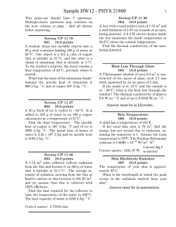


© Copyright 2026