
ABDOMINAL RADIOLOGY, FLUOROSCOPY PROCEDURE MANUAL & EDUCATIONAL GOALS
ABDOMINAL RADIOLOGY, FLUOROSCOPY PROCEDURE MANUAL & EDUCATIONAL GOALS TABLE OF CONTENTS Goals and Objectives for the GI/GU Rotations .................................................4 GI Resident Responsibilities .............................................................................8 Resident Curriculum ..........................................................................................15 Educational List .................................................................................................16 Procedures and Techniques ..............................................................................13 Preliminary Supine Abdominal Film Prior to GI Contrast Examination . .13 Plain Abdominal Films ............................................................................13 Contrast studies ......................................................................................15 Drugs and Contrast Media ......................................................................16 SITMARKS Colonic Transit Study...........................................................18 Esophagram ...........................................................................................20 Post-operative Esophagus……...............................................................21 Esophagram for Suspected Tracheal or Esophageal Injury (e.g. Boerhaave Syndrome)…............................................................23 Upper GI Series.......................................................................................23 Modified Barium Swallow .......................................................................27 Post-operative Stomach..........................................................................29 Bariatric Surgery......................................................................................30 Small Bowel Series (Follow Through).....................................................33 Gastrografin Small Bowel Challenge.......................................................36 Retrograde Small Bowel Series (through ileostomy)..............................36 Contrast Enema.......................................................................................37 Post-operative Colon...............................................................................38 Therapeutic (Cleansing) Enema..............................................................40 Defecography..........................................................................................41 Fistulography ..........................................................................................44 Endoscopic Retrograde Choledochopancreatography (ERCP) .............46 Post-Operative Cholangiography ...........................................................46 Hysterosalpingography ...........................................................................47 Cystography & VCUG.............................................................................48 Retrograde Urethrography......................................................................49 GI Curriculum & Reading Assignments.........................................................51 Additional Reference Textbooks.........................................................................52 Society of Abdominal Radiology Curriculum......................................................53 2 Welcome to Abdominal Radiology. The fluoroscopy service is an integral component of the Abdominal Imaging service. On this service, residents will learn to interpret plain radiographs of the abdomen and pelvis and to perform and interpret fluoroscopic studies of the gastrointestinal and genitourinary systems. Fluoroscopy is a learned skill and can only be mastered by performing as many procedures as possible on the several Stanford Hospital (and VA) rotations. 3 GOALS AND OBJECTIVES FOR THE GI/GU ROTATIONS Many of the goals and objectives apply to all GI/GU rotations and are listed immediately below. Those goals that are more specific to a particular rotation are listed separately. Goals and Objectives for ALL GI/GU Rotations 1. 2. Demonstrate learning of the knowledge-based objectives. Review the request, all applicable clinical history, pertinent laboratory tests and previous imaging studies to be certain that the proper test has been ordered, that the patient’s condition is such that the examination is safe and that any necessary preparation for the test has been completed before starting the examination. If the indication for the examination is unclear, contact the referring physician or another of the patient’s appropriate and knowledgeable health care providers. Perform all examinations in the appropriate way. If you have a question, ask before performing the examination. Accurately dictate all studies in a timely fashion. Communicate effectively and courteously with referring clinicians: a. Including obtaining relevant history for study interpretation. b. Regarding important findings on studies performed. Demonstrate learning of the clinical indications for ordering and using radiological examinations including advanced GI/GU imaging. Demonstrate responsible work ethic and professionalism. a. This includes being present at the GI/GU station by 8:30 am. b. Throughout the workday, this means completion of dictation of all reviewed studies in a timely manner, attendance at all departmental teaching conferences, and grand rounds presentations. Facilitate the learning of medical students, peers, and other professionals participating in the GI/GI service including speech pathologists, nurses, nurse practitioners, and speech therapy students. Build confidence in reading routine and STAT GI/GU studies. Review ACR Appropriateness Criteria and Standards regarding GI and GU tracts (including the Communications Standard). Follow up results of surgery or examinations performed by other clinical services to determine final diagnosis. 3. 4. 5. 6. 7. 8. 9. 10. 11. Core Competencies by Goal ! ! ! ! ! Medical Knowledge - Goals 1 through 13 Interpersonal and Communication Skills - Goals 2, 4, 5, 8, and 12 Practice Based Learning and Improvement - Goals 2, 3, 4 and 13 Professionalism Goals 5, 7, 8 and 9 Patient Care - Goals 2, 3, 4, 9 4 Assessment Tools Utilized ! ! ! ! Global ratings by faculty including rotation evaluation sheet Conference attendance logs In-service examination and through RADPrimer Plan — o Develop 360 degree evaluations o Individuals to be included - technologists on day shift in radiology core, technology supervisor for radiology Goals and Objectives for 1st and 2nd Fluoro Rotations Knowledge-Based Objectives At the end of these rotations, the resident should be able to… 1. Discuss the proper clinical and radiologic indications for the following studies: a. Esophagram (“Barium swallow”) b. Upper gastrointestinal series (UGI) c. Barium enema (BE) d. Small bowel follow-through (SBFT) e. Endoscopic retrograde cholangiopancreatogram (ERCP) f. Fistulograms g. Cystogram h. Voiding cystourethrogram i. Hysterosalpingogram (HSG) j. Defecography 2. State the physiologic properties, proper concentrations and proper indications for the use of the following contrast materials: a. Barium b. Water soluble contrast media (Gastrografin/Gastroview, Cystografin/CystoConray, Omnipaque) 3. Discuss the following information about Glucagon: a. Proper indications and dosages used in GI radiology b. Physiologic and side effects; contraindications 4. List the high risk factors, pretreatment and treatments for adverse reactions to intravenous contrast media. 5. Demonstrate ability to interpret plain films of the abdomen, including common conditions such as bowel obstruction, volvulus, pneumatosis, pneumoperitoneum, common support apparatus, devices and foreign bodies. 5 6. Achieve > 90% score on the Stanford Radiography and Fluoroscopy Inservice Exam. Technical Skills By end of the 2nd rotation, the resident should be able to… 1. Demonstrate basic knowledge of the equipment used during fluoroscopy, radiation safety features of the machines, and proper radiation safety techniques. 2. Demonstrate knowledge of patient positioning, and type of films that should be taken for the procedures listed in #2 above. 3. Competently and with limited supervision perform and interpret the following: a. b. c. d. e. f. g. h. i. j. Barium swallow (esophagrams; including post-operative) Upper GI series Contrast enema (including post-operative state) Small bowel follow through Fistulogram Cystogram Voiding cystourethrogram Hysterosalpingogram ERCP (interpretation only) Defecography Decision Making and Judgment Skills At the end of the rotation, the resident should be able to… 1. Review history of the patient for whom a procedure has been ordered and determine the appropriateness of the study requested. 2. Communicate with the referring physician about any recommendations for change in the type of procedure to be performed. 3. Communicate with the technologist about any special or additional views that should be obtained to demonstrate the pathology identified. 4. Read and dictate the studies performed, with the assistance of the faculty radiologist. 5. Communicate to the referring physician on the day of the exam any significant abnormalities identified on the examination. 6 Goals and Objectives for 3rd Rotation Knowledge Based Objectives At the end of the rotation, the resident should be able to… 1. Demonstrate continued increase in knowledge in the areas listed in the previous rotations. 2. Achieve a score > 80% on the RADPrimer Basic and Intermediate level Inservice Exam (GI & GU). Technical Skills At the end of the rotation, the resident should be able to independently perform and interpret most fluoroscopic and plain abdominal radiographic studies. Decision-Making and Judgment Skills At the end of the rotation, the resident should be able to demonstrate more advanced and independent decision-making and patient management skills. 7 GI RESIDENT RESPONSIBILITIES General Responsibilities The adult Fluoroscopy service handles adults and some children referred by the adult clinicians, surgeons or gastroenterologists for evaluation of abdominal disorders by plain radiography or fluoroscopy. The morning is dedicated to GI “fluoro” studies that the resident is expected to perform with supervision. The resident will review the abdominal plain films in the GI worklists on the PACS, as well as the fluoroscopy studies, and present her/his preliminary impression of the study to the faculty prior to dismissal of the patient. The afternoon is used for additional overflow studies, hysterosalpingograms, and GU studies such as cystograms or nephrostograms, as well as the afternoon plain films. Good, interested, well-trained technologists are critical to the performance of technically adequate GI studies. It is imperative that they be accorded the full respect and cooperation commensurate with their professional status. Scheduling Our general philosophy is to provide the best, most expeditious care of the patients as possible, with prompt reporting to the referring physician. There will be flux in the daily schedule, as we try to accommodate as many patients as possible. The technologist staffing of the fluoroscopic area consists of two technologists and, during most of the year, one or two students. One technologist arrives at 7:30 am and the other at 8:00 am. The technologists coordinate patient flow, film quality control and scheduling. Three fluoroscopic/radiographic rooms are available. No limit exists regarding the number of patients scheduled per day; all inpatients will be done the day of or after the physician requests the study. If possible, outpatients are scheduled according to the patients' wishes; however we limit the daily number in an effort to distribute the daily work volume. Diabetic and cardiac patients should be scheduled in the early morning, if possible, to allow medications to be regulated. Emergency cases will be added to the schedule automatically; a request for an emergency study usually means that, if the fluoro study demonstrates what the clinicians believe to be present, the patient will receive the appropriate interventional or surgical therapeutic procedure immediately following the radiologic examination. If the schedule is light, and a physician requests that an elective study be done on either an inpatient or outpatient that day for patient convenience, the study may be added on. 8 The technologists should stagger their one half hour lunch breaks if the cases will not all be completed by noon. One technologist should have lunch from 11:30 am -12:00 pm and the other from 12:00 - 12:30 pm. This allows one technologist to always be available in the area to finish any examinations not completed by noon and to obtain follow-up films on patients needing them (such as small bowel series, etc.). Technologists and students should also each have a fifteen-minute break in the morning and afternoon. If any delays are incurred in examining a patient at the time he/she is scheduled, the resident or staff is expected to greet the patient and assure her/him that we are aware they are available. Explain the delay and apologize. This demonstrates our respect and decreases their annoyance. A greeting and a smile go a long way; it also reduces the complaints the technologists and front desk personnel receive while the patient is waiting. The resident is expected to perform all studies. However, if the caseload is heavy and the resident falls behind, the faculty will help until the schedule is back on time. Every attempt should be made to get the resident to the noon conference, usually meaning an 11:45 AM departure. Patient Care and Priorities No patient should be coerced into a fluoroscopic exam. Patients may refuse to start or continue any examination. Be sure the patient understands both the nature and importance of the study. Many times patient lack of compliance is due to fear. Communicate with the patient's physician if a study cannot be completed. Patient triage is an important part of the resident’s job. The resident and technologist must continuously check to assess which patients are most medically unstable and make sure they are handled expeditiously. This includes getting them in and out of the fluoroscopy suite and expediting their exit from the department. Exam Preparation and Performance Each examination must be tailored to answer the question being asked. The protocols described in this manual refer to routine examinations. Although this is a reasonable basis from which to start, in many instances exams must be modified due to a patient’s history, clinical condition or information desired. Preparation for each study includes careful reading of the requisition to obtain history, special instructions or precautions. All patient records are now available online in EPIC. Check their records for history, indication for study and prior surgery, etc. In addition, note whether endoscopy or biopsy has recently been performed. BEs may be performed the day following the superficial biopsy that is obtained via a colonoscope, although large amounts of air often preclude an ideal study. NOTE: No study should be performed without a completed requisition. Prior studies, particularly plain films, GI, CT, US etc., must be reviewed. 9 Before beginning any study, the resident should introduce her/himself to the patient. An attempt should be made to ascertain the possibility of pregnancy in any female patient of childbearing age. Radiation to the abdomen is particularly likely to be harmful between the second and sixth week post conception but unnecessary radiation should be avoided at any stage of pregnancy. You should inquire of the patient, "Is there any possibility that you are pregnant?" If the answer is "yes" or if her menstrual period is overdue, the general rule is to postpone any abdominal radiographic procedures which are elective in nature. Patients should also be questioned about relevant symptoms, prior abdominal surgery, having been NPO after 9:00 pm, and whether they received the "Instruction Sheet" explaining the examination and whether they followed the routine preparation. If a patient has active ulcerative colitis or Crohn's disease, the only preparation is NPO after 9:00 pm of the night preceding the examination. If the patient has not received the "Instruction Sheet" detailing the examination, the procedure and length of time necessary to complete it should be explained to them. Explain breath holding during spot filming to all patients, i.e., "don't take a breath in, just stop breathing." Important concepts to keep in mind prior to proceeding with the study include: ! The use of rectal balloons should be limited if at all possible; rectal balloons must not be used with known or suspected proctitis or rectal carcinoma. ! A barium enema is contraindicated with fulminant colitis, toxic megacolon or suspected colonic perforation. ! If perforation of any portion of the GI tract is suspected, the study should be performed with iodinated water-soluble contrast material. (e.g., Gastrografin, Cystografin) ! In suspected aspiration, high osmolality iodinated water-soluble contrast material (e.g., Gastrografin) is contraindicated; when these contrast agents come into contact with the lung they cause a severe pneumonitis. Consult the attending radiologist regarding use of Omnipaque (a nonionic, isotonic iodinated agent) in complex situations. When the study has been completed, the resident will instruct the technologist if views other than routine are required. Any unusual anatomy should be mentioned or shown fluoroscopically to the technologist at this time. Immediately after completing each study, a short fluoroscopic impression written on the requisition is suggested. This should include comments pertaining to esophageal motility abnormalities, reflux, findings on palpation, etc., to refresh your memory when you view and report the study. After the initial set of radiographs has been obtained, it may be necessary to re-fluoroscope or obtain further views to clarify initial ambiguities. Patients may be more tolerant of this 10 "second look" or "extra films" if you explain this as a continuation of the examination rather than a re-examination or re-spot. Try to keep re-fluoroscopies to a minimum. Record Keeping and Educational Responsibilities All examinations are reviewed with the attending and dictated on the day of the examination except for SBFT examinations that may not yet be complete at the end of the workday. The resident is responsible for proofreading and initialing all dictated reports. 11 FLUOROSCOPIC TECHNIQUES Fluoroscopic techniques will vary somewhat among even experienced radiologists, and must be tailored to individual patients and conditions. For instance, a study performed 24 hours following bariatric surgery will be much more limited and focused than one performed electively on an ambulatory patient to evaluate vague abdominal pain. Before beginning a study, know what the specific goals are and make sure you answer the clinical question. “Good fluoroscopy is 95% based on anatomy and gravity.” (How must you position the patient in order to get the contrast material &/or air where you need it?) Example: when evaluating for a gastric outlet or duodenal disorder, the patient must be placed right side down with the head of the fluoro table elevated to have the contrast medium leave the stomach and pool within the duodenum. 12 PROCEDURES AND TECHNIQUES Preliminary Supine Abdominal Film Prior to GI Contrast Examination A preliminary (“scout”) film of the abdomen prior to a fluoroscopic exam is not routinely performed because of proven low-yield, radiation exposure and cost. However, it should be obtained in any patient who has had previous surgery or intervention (e.g. biopsy or balloon dilation) because surgical staple lines and other “artifacts” may simulate pathology, such as a leak of contrast medium. Note: If a scout film is obtained it must be interpreted as a separate, initial paragraph in the final report. Plain Abdominal Films (See McCort, Mindelzun, Filpi, Rennell-Abdominal Radiology and Baker S. The abdominal plain film) The information sought dictates the extent of examination performed. Techniques For adults, the lower edge of the film is centered on the superior margin of the symphysis pubis. For a large patient, two (14 x 17) films cross-wise. Lower edge of one on symphysis, lower edge of the other on iliac crest. With optimum exposure both lateral properitoneal fat lines should be visible on all films. Single Abdominal Film or KUB (kidneys, ureter and bladder) Indications ! ! Preliminary for many studies. Search for masses; calcifications, particularly gallbladder, appendix or kidney; position of tubes and catheters Three-Way Abdominal Study Necessary for evaluation for bowel perforation and obstruction. Left lateral decubitus, patient remains in this position 10 minutes prior to filming (ideally) ! Upright. ! Supine. ! ! For the upright and decubitus films, the X-ray beam must be horizontal. An upright PA chest is often obtained unless one has been done within a few hours. This enables the examiner to exclude lung or diaphragmatic disease as the cause of the abdominal symptoms. Lower lobe abnormalities are often better seen on an abdominal film than on chest x-rays. It will also confirm the presence of free intraperitoneal gas. 13 Evaluation of Plain Films of the Abdomen Analyze all films using a well-conceived search pattern. The following can serve as a model. Do not focus immediately on a perceived abnormality but evaluate all information available from the film. 1. Supporting Structures Start with “tubes, devices and altered anatomy.” Important clues are often available by noting evidence of prior surgery (e.g. skin staples, anastomotic bowel staple lines, “ostomies.”) Residents must be able to identify the most common types of catheters, drains, surgical devices and foreign bodies, including retained surgical “sponges” and needles. (See: “Abdominal Incision and Injection Sites,” STATdx) 2. Abdominal Calcifications Residents must be able to identify renal calculi, gallstones, pancreatic calcifications, vascular calcifications, injection granulomas, and calcified lymph nodes, among others. (See: Expert DDx: “Abdominal Calcifications” in STATdx) 3. Organomegaly and Masses Hepatomegaly will displace the hepatic flexure down and splenomegaly will displace the splenic flexure down and the stomach medially. The kidneys can be seen, at least partially, in most patients because of adjacent perirenal fat. Similarly, the bladder can usually be visualized because of surrounding perivesical fat. Masses may be evident by localized soft tissue density that displaces bowel and other structures. (Read: “RUQ masses”, “LUQ mass,” “Splenogegaly” in STATdx) 4. Bowel Gas and Fluid Normal stomach = gas + fluid. Normal small bowel (ambulatory patient) = minimal gas. Normal colon = no fluid; formed stool in left side of colon. Abnormal patterns: Ileus: Increased gas and fluid in small bowel (diameter > 3 cm) and colon (> 6 cm) without a transition point. Obstruction: Dilation upstream from a transition point; collapsed bowel downstream 14 Aerophagia: Increased gas within SB and colon, but no significantly dilated segments. Note: In patients with both ileus and ascites, the mesenteric bowel segments (SB and transverse and sigmoid colon) will be dilated while the ascending and descending colon and rectum will be collapsed, often resulting in a mistaken diagnosis of colonic obstruction. 5. Abnormal Gas Collections Read in STATdx: 1. “Pneumoperitoneum” 2. “Portal Venous Gas” 3. “Gas in Bile Ducts or Gallbladder” 4. “Pneumatosis of SB or Colon” 6. Abnormal Fluid Collection Residents must learn to recognize signs of ascites on plain films, such as the “flank stripe sign” and obscuration of the margin between the Liver and Kidney. (Read: “Ascites” in STATdx and in any standard abdominal radiology textbook.) CRITICAL RESULTS In the abdomen, critical test results that MUST to communicated with clinical teams and recorded are as follows: 1. 2. 3. 4. 5. 6. Non iatrogenic pneumoperitoneum Retained foreign body Bowel obstruction with strangulation Misplaced enteric tubes or instruments Complications of procedures or contrast reactions Leaks 15 CONTRAST MEDIA VARIBAR Thin Liquid Barium E-Z-M HD barium Liquid EZpaque barium VARIBAR Target Viscosity Honey E-Z-PASTE Barium Sulfate Esophogeal GASTROGRAFIN Diatrizoate Meglumine & Diatrizote Sodium Solution USP (identical to Gastroview) 40% (used by Speech Pathologists) 98% w/v (“thick” for double contrast UGI, esophaghram) 60%w/v 41%w/w (general purpose, esophagus, UGI, diluted ~ ½ for SBFT) 40% w/v 29% w/w (Speech Pathologist studies) 60% w/w (to coat marshmallow or cookie for esophagram) 37% Organically Bound Iodine (hypertonic, hyperosmolar for oral administration) CYSTOGRAFIN 300mL Diatrizoate Meglumine (similar to Cystoconray) Injection USP 30% (contrast enema in Large patients) CYSTOGRAFIN 300mL Diatrizoate Meglumine Injection USP 18% (nearly isosmolar; for rectal or bladder administration) OMNIPAQUE 10mL (iohexol)) 240mg/mL (isosmolar; for HSG, I.V. or oral administration) [very expensive; use sparingly] OMNIPAQUE 10mL (iohexol)) 300mgl/Ml (myelography, rarely for GI/GU) OMNIPAQUE 100mL (iohexol)) 350mgl/Ml (oral admin; risk of aspiration and leak) CONRAY 43 (iothalamate meglumine) 430 mg I/mL (hypertonic; used for nephrostograms and fistulograms) 16 Potential Contra-Indications Where GI or GU perforation is suspected use water-soluble contrast first. Oral barium not given in the presence of colonic obstruction. Don't give barium prior to ultrasound or CT examination. Drugs Glucagon Used to relax spasm of stomach, bowel or colon. Produces hypotonia throughout the GI tract almost immediately, but does not abolish peristalsis in the esophagus. To prevent nausea, give slowly over 1 minute I.V. Dosages Upper GI 0.1 - 0.5 mg I.V. Colon 1 mg I.M Post Op. Stomach 1 mg. I.M IV lasts 10 - 20 minutes. IM lasts 20 - 30 minutes. Contraindications Pheochromocytoma, insulinoma, diabetic on insulin. Metoclopramide (Reglan) To enhance peristalsis for small bowel enema or small bowel follow through. Stimulates gastric emptying. Facilitates duodenal intubation by feeding tube. Dose 20 mg PO 15 minutes before procedure. Contradictions GI hemorrhage, perforation or obstruction. 17 SITZMARKS Colonic Transit Study On day one the patient ingests a SITZMARKS capsule containing 24 bariumimpregnated radioopaque markers. On day 5, an abdominal supine radiograph is taken. How to interpret a SITZMARKS® test Reading the Results: If over 80% of the markers are passed by day 5, colonic transit is not grossly abnormal. If the remaining markers are scattered about the colon, the condition is most likely hypomotility or colonic inertia. If the remaining markers are accumulated in the rectum or rectosigmoid, the condition is most likely functional outlet delay, e.g., internal rectal prolapse, anismus. 1. If 5 or less markers remain, patient has grossly normal colonic transit 18 2. If most rings are scattered about the colon, patient most likely has hypomotility or colonic inertia. 3. If most rings are gathered in the rectosigmoid, patient has functional outlet obstruction. 19 FLUOROSCOPIC PROCEDURES 1. Esophagram Before any barium swallow is performed, a full history must be obtained. The examination can then be appropriately tailored. Ask specifically about prior surgical procedures and “food sticking in throat” (big pills, dry toast, meat). Preparation Although an urgent esophagram may be done without the patient fasting, the optimal preparation is NPO after midnight. Filming Spot (3/sec for 2 sec) films. On any esophagus where a morphologic abnormality is detected and further study not contraindicated, overhead esophagus films should be performed. The technologists should do this at the completion of your study, ideally with the patient positioned to show the lesion optimally, and while the patient is drinking barium. 1.A. Esophagram with a History of Dysphagia, Food Sticking, or Suspected Obstruction The patient is initially placed in the upright left posterior oblique position. The examination is begun with dilute (60%) barium. Teach the patient a slow consistent cadence "one, two three." The patient is asked to take one mouthful, hold it in his mouth and swallow when told. Follow the bolus from mouth to the gastroesophageal junction with fluoroscopy, observing the tail of the peristaltic wave. If no obstruction, aspiration or emesis is encountered, proceed as in the next section. If obstruction is encountered, spot views should be taken in at least two projections at the site, (e.g. LPO & RPO). Motility Study Following the upright swallow, place the patient in a prone 25° RAO projection with the table horizontal (“swimmer’s position”). Collimate to show from the thoracic inlet to the upper abdomen (16” field of view). Have the patient take one mouthful of barium and swallow only once. Fluoroscopically monitor the tail of barium column. Repeat at least twice. Record tertiary contractions and other abnormalities on spot films. Then have the patient drink barium continuously while obtaining distended films of the entire esophagus, including several of the EG junction. Have the patient bear down when the esophagus is distended at the EG junction to bring out a hiatal hernia or Schatzki ring. 20 An alternative to the prone position, especially for elderly or ill patients is filming in the supine LPO position, with the patient’s right side supported by an angle sponge. Hypopharynx If a morphologic lesion or motility disorder of the hypopharynx or upper esophagus (e.g. crico pharyngeal spasm, tumor, Zenker's) is suspected, spot films are necessary. Position the patient upright in a straight lateral or steep oblique position. Collimate to show from the hard palate to the C7-T1 interspace. Have the patient take one large mouthful of dilute barium (60%). Depress rapid filming button and then have the patient swallow. Watch fluoroscopy screen closely until the barium bolus passes. Turn the patient to AP position. Be sure the patient's head is elevated so that the mid point of the jaw is aligned with the posterior arches of the cervical spine (“chin up”). Repeat the sequence. At least two swallows in each position should be performed. Generally, only AP + Lat filming is helpful in diagnosis. Obliques may be helpful when C7 and thoracic inlet region is not well seen as in patients with large shoulders. The upper thoracic esophagus must be included for complete evaluation of a large Zenker diverticulum. Barium tablet or Food Bolus Administration A 13mm Barium tablet may be given to evaluate esophageal narrowing as in Schatzki ring, or any other cause of known or suspected mechanical narrowing of the esophagus. Occasionally the following is helpful. Bread or a marshmallow coated with a spread of heavy barium. The patient is placed in an upright LPO position, then instructed to chew as much as he does at home in the first bite and swallow it with a mouthful of light barium. The food bolus is observed as it passes through the esophagus, at least twice. 1.B Post-Operative Esophagus Studies These patients may be studied in the immediate post-operative period to rule out anastomotic leaks or later for suspected anastomatic stricture, severe reflux, etc. Gastrografin is the medium of choice. 21 The Ivor Lewis esophagectomy This and its many alternative procedures are the most common surgical procedures for resection of esophageal cancer. The distal esophagus and proximal stomach are resected and the EG anastomosis may be created at any level from the thoracic inlet to the subcarinal region. Mandatory reading: RG 2001; 21: 1119 and RG 2007; 27: 409 Post-Fundoplication Various forms of fundoplication are performed as anti-reflux surgical procedures, with the most common being the Nissen (360ºwrap) procedures. Esophagography is often requested in the early postoperative period to evaluate leak or high-grade (“too tight”) luminal narrowing through the wrap. Studies performed weeks or months later are usually to evaluate suspected complications such as dehiscence or slip of the fundoplication Early Post-operative Period. Following a scout film of the epigastric region, the patient is placed upright in the LPO position and given Gastrografin to drink while obtaining rapid sequence (2/sec) spot films of the E-G junction. Following several swallows and filming in both the LPO & RPO positions, the patient is placed supine in order to fill the gastric fundus. This allows evaluation for leak and the Gastrografin pool will outline the fundoplication as a “filling defect.” Later Post-operative Period. Barium is the contrast agent and a complete esophagram is performed (see above; “Esophagram”) 22 Mandatory reading: RadioGraphics 2005; 25:1485-1499 1.C. Suspected Aspiration, Tracheoesophageal Fistula or Tracheal Laceration A single swallow of Omnipaque should be performed, filming at 4 frames /sec. If no significant leak is detected, a single-contrast barium study should be completed with the patient examined in at least two positions that are at 90° to each other. (e.g., LPO and RPO) 1.D. Suspected Esophageal Laceration or Perforation (e.g., Boerhaave syndrome or trauma) Begin with a small swallow of water to see how the patient tolerates this. Follow with Gastrografin. Again, one swallow should be performed and monitored fluoroscopically with images. If a leak is demonstrated, the examination is completed. (Note: Lacerations of the esophagus in Boehaave syndrome are almost always in the distal esophagus and best demonstrated in the LPO position. If a leak is not demonstrated the patient MUST be given a swallow of 60% barium because barium will in 15-20 % of patients demonstrate a tear not visualized with water-soluble agents. 1.E. Esophagram in patients evaluated for lung transplantation End-stage lung disease patients, particularly those with idiopathic pulmonary fibrosis or cystic fibrosis often have clinically significant GERD. Reflux has been associated with bronchiolitis obliterans syndrome, which represents a leading cause of death after lung transplantation. Many surgical teams have suggested antireflux surgery to preserve the allografts. Patients at Stanford routinely undergo an esophagram in anticipation of surgery. We perform a complete esophagram emphasizing both anatomic and functional detail with special attention to the EG junction and reflux. All of these patients are deconditioned and many are on oxygen therapy. The examination can be quite trying for some of them. It is important to proceed slowly so as not to fatigue the patient and yet gather as much information as possible. Technique is the same as outlined above. J Amer Coll Surgeons 2013; 217: 586-597 1. F. Double contrast Upper Gastrointestinal Studies: Esophagram or Upper GI Series Double contrast examinations of the upper gastrointestinal system are generally reserved for evaluation of mucosal abnormalities of the esophagus, stomach, and duodenal bulb. A double contrast esophagram or double contrast upper GI 23 series may be ordered for the evaluation or detection of erosions, ulcers, polyps, malignancy, inflammation or infection. These exams are often a first-line evaluation for these indications, with subsequent confirmation usually obtained via upper endoscopy. Double contrast upper GI studies are contraindicated for patients in the immediate post-operative period status post gastrointestinal surgery, patients with recent trauma, and for patients with suspected aspiration, perforation, tracheoesophageal fistula, esophageal strictures or rings. The double contrast exam is a two part examination, with the first part of the exam utilizing a double contrast technique and the second part of the examination utilizing a single contrast technique. Whereas the double contrast technique is for evaluation of mucosal detail, the single contrast technique is for the evaluation of motility, masses, strictures, etc. The double contrast technique utilizes gas and high density barium as the two “contrast agents”. Specifically, effervescent granules (sodium bicarbonate crystals) are swallowed by the patient and these granules liberate gas (carbon dioxide) which distend the stomach and esophagus. This is then followed by the administration of thick barium which coats the surfaces of the mucosa. It is important to keep in mind that the distension may be uncomfortable for the patient and to work quickly to obtain the images necessary. Preparation Esophagram: The optimal preparation is NPO for at least 2 hours prior to the start of the exam. UGI Series: The optimal preparation is NPO after midnight. Medications are okay. Water is fine but no coffee or tea. Contrast agents Effervescent granules (sodium bicarbonate crystals) High density barium Filming Each organ should be seen in at least two views and during both components of the examination. Collimate as necessary, to include only the relevant organs of interest. Take single spot images during double contrast technique. 24 N.X. Double contrast esophagram You will need a packet of effervescent granules, a small amount of water (about 2 tablespoons), two small cups, prepared thick barium, and a large cup for this part of the examination. Place the thick barium in a large cup. Place the effervescent granules in one of the small cups and a small amount of water in the other small cup. Prior to starting this examination, describe to the patient how the granules taste (a very sour, strong lemon taste), explain that the granules will produce a lot of gas, and, importantly, to not let the gas escape. This can be suppressed by telling the patient to swallow when they feel the need to belch. Explain to the patient to take a large mouthful of barium to hold in their mouth when told and to swallow when told. Also, explain to the patient to move the cup of barium away from their center (away from their neck, chest, and upper abdomen). A. The patient is placed in the upright position, standing on the footboard, in the LPO position to offset the esophagus from the spine. B. Mix the small amount of water and the effervescent granules (it will start effervescing immediately) and give the mixture to the patient, having them drink it down as quickly and completely as possible. Alternatively, have the patient place the granules in his mouth and drink it down with a small amount of water. C. Take the small cup(s) away and give him the large cup of thick barium in their left hand. D. Center the image over as much of the esophagus as possible. E. The patient is then asked to take one mouthful, hold it in his/her mouth, tilt their head back, and swallow when told. F. Follow the bolus from the mouth to the gastroesophageal junction with fluoroscopy and look for gross abnormalities. G. Once optimal coating is achieved, seen as a thin sheet of barium lining the esophagus, take spot images to include the entire esophagus, gastroesophageal junction, and distended gastric fundus. H. Repeat E. through G. with the patient in the upright RPO positioning. I. If a double contrast Upper GI Series is requested, then follow the instructions below (section N.Y.). Otherwise, follow the guidelines for the single contrast esophagram as explained in the separate section. At the end of the single contrast esophagram, check for reflux using the described maneuvers. 25 N.Y. Double contrast Upper GI Series The double contrast Upper GI Series includes a double contrast examination of the esophagus, stomach, and duodenal bulb followed by a single contrast examination of the esophagus, stomach, and proximal small bowel. It is important to keep in mind the relative positions of the stomach and duodenal bulb to achieve an optimal double contrast evaluation of the structures of interest. For example, the gastric fundus is a relatively posterior structure. Therefore, to obtain a double contrast examination of the fundus, optimal positioning would be more prone, as prone imaging “traps” the gas in the more superiorly located structure which, in this case, is the gastric fundus. Anatomic variations are considerable and, ultimately, the goal is to obtain double contrast imaging of all portions of stomach (fundus, body, antrum) and the duodenal bulb. An exact prescription for the spot images to obtain may not always apply and it is important to be flexible. A. Follow the instructions for the double contrast esophagram as described above (N.X). It is important to complete the double contrast esophagram as quickly as possible. B. Once you have completed obtaining images of the esophagus in the LPO and RPO positions, quickly lower the table into the horizontal position with the patient lying on his/her back. C. Once the table is completely horizontal, perform maneuvers to coat the stomach with the thick barium as tolerated by the patient. 1. This can be achieved by rotating the patient to his/her left into the prone position, then back to his/her right in the supine position. 2. Alternatively, rock the patient from side-to-side in the AP and PA positions. D. Obtain single spot images of the stomach. LPO, PA, and RAO views should show the gastric body, fundus, and antrum adequately. E. Watch for a double contrast view of the duodenum during evaluation of the stomach and take spot images if it is adequately coated and distended. 1. The LPO position is usually effective to obtain double contrast imaging of the duodenum. 2. Sometimes the extreme RPO position may be useful. 3. Also, placing the table semi-upright may help trap gas in the duodenal bulb if other positions don’t work. 26 F. Assess the proximal small bowel for gross abnormalities, including diverticula and take additional spot images if necessary. G. Follow the instructions for the single contrast Upper GI series, including assessment for reflux using the described maneuvers. 1.G. Modified Barium Swallow (Video Fluoroscopic Swallowing Examination) (Performed With Speech Therapist) The video fluoroscopic swallowing examination (modified barium swallow) procedure is designed to study the anatomy and physiology of the oral preparatory, oral, pharyngeal and cervical esophageal stages of deglutition, especially in patients considered to be at risk for aspiration pneumonitis.. Small amounts of contrast material are used to minimize the risk while evaluating the physiology of the oral cavity and pharynx. Four consistencies of barium are used to investigate patient complaints of variable swallowing ability: thin and thick liquid barium, barium paste, and material requiring mastication. NOTE: This is not the study to evaluate post-operative anatomy (e.g., following ENT oncologic surgery), nor is this study to be used to evaluate patients with dysphagia; the standard esophagram is to be performed in this setting. Patients considered to be at risk for aspiration as well as having dysphagia or post-operative swallowing problems may require both a modified as well as a standard esophagram for complete evaluation. Preparation The examination is performed with a fluoroscopy unit equipped with a video recorder (usually room 1). The speech pathologist brings the video recorder for the examination as well as any type of "food" to be used in the examination. Examination The exam begins in the lateral position, with 5 cc of thin barium to assess the risk of aspiration and the patency of the pharynx. The patient is asked to hold the barium in his mouth while the image is centered and the video recorder is started. The fluoroscopic field includes the oral cavity and the cervical region. The image should be collimated to improve resolution. The patient is asked to swallow once and the fluoroscopy image is recorded until the bolus disappears from the field. After laryngeal protection and patency of the pharynx are demonstrated, the examination is continued in the same fashion with the following: ! ! ! ! 5 cc of thin barium 5 cc of thick barium. Repeat once. 5 cc of barium paste. Repeat once. 5-10 cc of barium mixed with pudding, applesauce or any other puree-like material. 27 ! 5 cc of barium paste mixed with a cookie or cracker. Repeat once. The following observations are noted in the lateral and A/P planes during the radiographic procedure: ! ! ! ! ! ! ! ! ! Lateral view Oral and pharyngeal transit Analysis of patterns of lingual movement Gross estimate of time elapsed before swallow reflex triggers Estimate of amount of vallecular residue after swallow Amount of material aspirated per bolus and reason for aspiration Timing of aspiration relative to triggering of swallow reflex, i.e. before, during or after the swallow Anterior/Posterior View Asymmetries in function, i.e. pharynx and vocal folds Viewing residues such as collection of material in the valleculae and residue in pyriform sinuses Examine the residue in pharynx after swallow, comparing the two sides Radiographs should be taken during the routine examination if significant findings are noted to create a PACS record for the referring clinicians. All video recorders will be kept by Rehabilitation Services where they will be filed as permanent records. 2. Single-Contrast Upper GI Study Indications for a single-contrast GI study: suspected gastritis, obstruction, tumor. The patient drinks 60% barium preparation. A. Begin with the patient in the upright LPO position. B. Under fluoroscopic monitoring, the patient should be instructed to drink four ounces of barium. Observe esophagus and spot any abnormality. Proceed rapidly to a study of the stomach. C. Place patient in prone RAO position. Spot antrum. Give patient large cup of barium with a straw. Observe esophageal peristalsis with single swallows. D. Then obtain spots of distended esophagus while the patient drinks barium continuously. E. Obtain barium-filled films of the body, antrum and duodenum. F. Turn patient to supine LPO position for air-contrast views of antrum and duodenum. 28 G. Turn patient RPO, take spot film of fundus. H. Return table to upright position, take compression shots of stomach and duodenum. Under fluoroscopy, attention should be paid to the greater and lesser curvatures. I. The patient should be instructed to finish the cup of barium (a second cup of barium may be administered). J. Technologist will obtain the following films: 1. 10" x 12" radiograph of stomach, LPO (table horizontal) 2. 10" x 12" radiograph of stomach, 45o RAO (table horizontal) 3. 14" x 17" radiograph of stomach to include all barium filled small bowel, prone PA (table horizontal) 3. The Acute Post-Operative Stomach The indications for an examination of the acute postoperative stomach are generally to evaluate for leak, perforation and obstruction. Common gastric surgical procedures include partial or complete gastrectomy (e.g. Bilroth 1 or 2), and various bariatric procedures. In the immediate post-operative period, a water-soluble single contrast (Gastrografin) examination should generally be utilized especially if there is a clinical concern about an anastomotic leak or perforation. An exception to this is when the patient has a known or strongly suspected tendency to aspirate into the tracheobronchial tree. Give water first then follow with Omnipaque. Aspiration of Gastrografin can produce a fatal chemical pneumonitis. Spot films: Various obliquities to demonstrate the altered anatomy and any anastomoses. Due to recent surgery, the patient may not be able to tolerate prone positioning. Steep oblique positioning with the head of the table elevated is often useful to facilitate gastric emptying. Overhead films AP, LPO and RPO References 1. Double contrast Gastrointestinal Radiology with Endoscopic Correlation. Igor Laufer, Chapter 8 - The Postoperative Stomach. 2. Radiology of the Postoperative Digestive Tract – Bruce Javors, Ellen Wolf, Springer , 2003 29 3.A. Evaluation of bariatric surgical procedures. Two categories of surgery are performed: restrictive and malabsorptive. Diagram of Surgical Options. Image credit: Walter Pories, M.D. FACS The most common (~85%) is the Roux-en-Y gastric bypass. This consists of formation of a 15–30 mL gastric pouch attached to a segment of jejunum with side-to-side gastrojejunostomy. At 100–150 cm a jejunojejunostomy is created. The jejunal loop from the stomach to the jejunum is referred to as the Roux limb. It is most often now placed antecolic. Varying the length of the Roux limb will alter the malabsorptive component of the procedure. Gastric banding. Purely restrictive procedure. More popular outside the USA. Adjustable silicone band with inflatable balloon creates adjustable stoma controlled by saline injected subcutaneous port. Look at “phi” angle, vertical line to line of plane of the gastric band (normal - 4-58 degrees). Laparoscopic sleeve gastrectomy consists of placing a staple line along the lesser curve of the stomach. Approximately 85% of the stomach volume is excluded and then removed. A gastric “tube” represents the remaining stomach. Jejunoileal bypass, the original bariatric operation, is no longer done, but we still see some patients who had this procedure years ago. Radiologic evaluation 30 The patient is often examined by fluoroscopy the day after surgery. Most of these examinations are performed with the remote controlled unit in room 1. The patient stands for the examination with a technologist in the room behind a barrier. It is critically important for the technologist to be with the patient at all times, as many of these patients are unstable and susceptible to vasovagal reactions. If the technologist needs to leave the room to process a film, then the patient needs to return to a wheelchair. The patient is a given a swallow of water to determine if water-soluble contrast materials can be used for the examination. Assuming that the patient does not aspirate, the 1st series of films is obtained in the AP position at 2 frames per second. Try to determine on this initial set of images (or simply by asking the patient) which of the operations has been performed. Generally, following the Roux-en-Y configuration, the ingested contrast material will flow inferiority and to the left whereas following the sleeve procedure, it will flow to the right. The patient is then studied in a slight LPO and a RPO projection, also at 2 frames per second. Once the fluoroscopic examination is completed, the technologist obtains an upright 14x17 in the AP projection. By that time enough contrast material has passed distally so that the jejunojejunal anastomosis can be optimally evaluated. Images need to be reviewed before the patient is allowed to leave the department. Mandatory Reading: [Bariatric Surgery in STATdx; RG 2006; 26: 1355] Imaging in Bariatric Surgery: A Guide to Postsurgical Anatomy and Common Complications Robert C. Chandler1, Gujjarrapa Srinivas1, Kedar N. Chintapalli1, Wayne H. Schwesinger2 and Srinivasa R. Prasad1, AJR, January 2008, Volume 190, Number 1 Imaging in bariatric surgery: service set-up, post-operative anatomy and complications, Shah et al, The British Journal of Radiology, 84 (2011), 101– 111,Abnormal findings on routine upper GI series following laparoscopic Roux-en-Y gastric bypass OBESITY SURGERY Raman, R., Raman, B., Raman, P., Rossiter, S., Curet, M. J., Mindelzun, R., Morton, J. M., 2007; 17 (3): 311-316 31 3.B. The Remote Post-Operative Stomach These patients can be studied with barium as the contrast medium, assuming there is no clinical concern for leak (e.g., signs of peritonitis). A preliminary film of the abdomen is taken. Spot films and overhead films are obtained in multiple oblique, supine and prone positions, similar to the standard procedure for a single contrast UGI. Use with Patient supine Use with Patient prone Compression devices. 4. Feeding Tube Placement Occasionally we will be asked to assist in the placement of a feeding tube under fluoroscopic guidance. We generally insist that the clinical team have at least attempted to place an enteric feeding tube (EFT) on the ward, as most of these will find their way into the duodenum without guidance. Fluoroscopy is not of assistance if the issue is that the patient refuses to cooperate with EFT placement or repeatedly pulls it out. Nevertheless, under some circumstances, such as surgically altered anatomy or a large hiatal hernia, fluoroscopy can be helpful. The patient should be sent to our department with the EFT in the stomach or at least with him as we do not stock these in the department. The initial placement of the EFT is with the patient in a sitting or supine position. Place several mL of anesthetic gel (Viscous lidocaine) in the nares for comfort and lubrication. With the neck flexed, slowly and gently advance the EFT to the 32 back of the throat. Tell the patient in advance that when he feels a gagging sensation, you will pause; then have him swallow as you advance the tube. If it has gone down the trachea, the patient will generally have spasms of coughing; if so, withdraw. Once the tube has entered the esophagus it is usually easy to advance it into the stomach. Largely by trial and error, the tube should be manipulated until the tip is near the pylorus. Hold the tip of the EFT against the pylorus until a peristaltic wave carries it through into the duodenum. Don’t jab at the pylorus! Try to advance the tip into at least the 2nd part of the duodenum. Troubleshooting: It can be helpful to administer about 2 syringes of air (via Luer lock 60 cc syringe attached to the EFT) into the stomach. This distends the stomach and stimulates peristalsis, both helpful in advancing the EFT. For difficult cases (HH, strictures, prior surgery) it may be helpful to administer a small amount of barium or Gastrografin through the EFT to outline the anatomy. The wire that comes with EFT is of little use in guiding it under fluoroscopy. Consider removing the existing wire and replacing it with a stiffer wire of the type used in Angiography, some of which we do keep in the Fluoroscopy suite. Reglan (metaclopromide, 10 mg) is a medication used to stimulate peristalsis. It is administered orally as a 10 mg tablet and is, therefore, of limited value if the patient is unable to swallow. It also takes some minutes to have effect. 5. Small Bowel Series (Routine Single Contrast Examination) Except when looking for acute small bowel obstruction, the patient should have refrained from eating and drinking since midnight. Most small bowel examinations, frequently termed small bowel follow-through (SBFT), are done in conjunction with UGI studies. If the patient has had a concomitant UGI, the patient drinks one more full cup of 20% barium after the routine UGI radiographs. If only a small bowel series (SBS) is ordered, the patient drinks two full glasses of 20% barium. Some radiologists suggest adding a capful of gastrografin in each glass of barium the patient drinks (mix well!); this may accelerate the small bowel transit time of the barium. During the small bowel examination, the patient should not lie on his back or left side, as the barium will remain in the gastric fundus. Except when routine radiographs or spot films are being taken, the patient should either be in upright (walking, standing or sitting) or right side down position to facilitate gastric emptying. It is important to have solid, continuous full column filling of the small bowel with barium to ensure adequate diagnostic visualization of all portions of the bowel. For this reason, the stomach must remain fairly full with barium during the 33 entire study; this may necessitate the patient having a 3rd, or even 4th, cup of barium during the course of the examination. NOTE: If the patient is intubated with either a nasogastric or small bowel tube, the barium is usually injected through the tube using a 60 ml catheter-tipped syringe. When instilling barium through a nasogastric tube, the patient should be placed in a head-elevated position (the X-ray table is raised) and the lower esophagus monitored fluoroscopically during installation of barium, as patients will invariably reflux barium around the tube. When esophageal reflux occurs, stop injecting and ask the patient to swallow their saliva. The resultant peristaltic wave will sweep the barium back into the stomach. Five minutes after the 2nd cup of barium has been ingested, a film is obtained. Initial - 14 X 17 radiograph of abdomen, prone, upper/mid abdomen (table horizontal). Subsequent - 14 x 17 radiographs of abdomen, prone, including the rectum are obtained at 15 minute intervals for the next hour and at 1/2 hour intervals for the next two hours until barium is in the right colon. All films should be viewed as soon as possible following their exposure. The patient should also be fluoroscoped and the bowel palpated using a compression device several times during the study. NOTE: If apparent compression defects are noted on any portions of the small bowel or if the loops appear abnormally separated, an AP supine film is obtained immediately. Prone films give better visualization of the small bowel loops because they tend to separate them slightly from one another due to the compression effect of the table on the patient's abdomen; at times, however, this compression is exaggerated (especially in slender patients) and "abnormal" compression defects or loop separation is observed. If the abnormalities are secondary to the patient's prone position, they will disappear on a supine film. If any questionable or definite abnormalities are identified on the 14 x 17 radiographs, the patient is returned to the fluoroscopic room and the bowel is manually palpated with graded compression (using the plastic “F Spoon”) Spot films are obtained of the questioned area and prone compression (using the pneumatic compression paddle) films are also obtained. If a definite abnormality is viewed fluoroscopically, an overhead routine radiograph (10 x 12 or 14 x 17) is obtained of the abnormal area with the patient in the position best demonstrating the lesion. 34 Most SBSs are completed within 1 to 1 1/2 hours post barium ingestion. Patients in whom the study takes longer than three hours usually have ileus (including secondary to drug therapy) or a partial or complete small bowel obstruction. These patients are usually inpatients and further films are obtained at times deemed clinically feasible by the radiologist; it may be necessary to follow the study for as long as 24 hours. In these instances, the patients are returned to the ward. They are then brought back to the fluoroscopic room for films at two to four hour intervals during the day. The last film is obtained around 10:00 pm; if barium still has not reached the colon, the patient is returned at 8:15 am the next morning and is again studied. In the above cases, by the time the patient is returned to the ward for the first time, or certainly the 2nd time, enough barium is usually distal to the ligament of Treitz so that the patient may be put back on NG suction if a nasogastric tube is in place. NOTE: If a small bowel tube is in place, suction may remove a large amount of barium from the intestinal loops. In no instance, should a patient with an indwelling nasogastric tube be permitted to become extremely uncomfortable, distended or nauseous; in all such instances, suction should begin immediately and independent of the state of the SBS examination. Giving ice water or food orally or Reglan 10 mg IV may accelerate SBSs, but only after filled films of the jejunum have been obtained. Because barium filled loops of small bowel are usually superimposed on each other in the pelvis, compression films are necessary to ensure adequate visualization of all portions of the small bowel. With the patient prone, fluoroscopically place pneumatic compression paddle under pelvis and then inflate balloon until bowel loops are separated. Spot pelvic small bowel, prone (table horizontal) When the head of the barium column is in the right colon, the patient is placed supine and the terminal ileum, cecum and ileo-cecal valve are visualized with several spot films taken. This will often require fluoroscopy in varying LPO and RPO positions with graded compression over the RLQ. 14 X 17 overhead radiograph of abdomen to include all barium filled small bowel, prone over inflated balloon (table horizontal) 5.A. Water soluble contrast challenge in patients with adhesive SBO (Gastrografin challenge). In the absence of signs of strangulation and following a CT scan, patients with persistent symptoms who are deemed to have an 35 adhesive small bowel obstruction can be given a 100cc Gastrografin challenge. This can be administered through an NG tube or per orally. The patient does not need to come down to the department as the contrast can be given on the ward. Nursing personnel should be instructed to place the patient into a right side dependent position to facilitate gastric emptying. Gastrografin is diagnostic and may be therapeutic because of its high osmolality, draws fluid into SB and stimulates peristalsis. Water-soluble contrast in the colon on a single abdominal film within 24 hours is a strong predictor of resolution. Fluoroscopy is not necessary. A Gastrografin challenge is deemed safe and appears to reduce surgical intervention and hospital stay, though conclusive evidence is lacking. 6. Retrograde or Antegrade Small Bowel Series- Diagnostic or therapeutic (Ileostomy Patient) This study may be requested to evaluate a patient with decreased ostomy output, suggesting SB obstruction. A Foley catheter is inserted into the “upstream” portion of the SB and the 20 mL Foley balloon is gently inflated within the bowel. Either barium or Cystografin may be used as contrast medium, and is administered by gravity pressure with an enema bag filled with the contrast medium. IV or IM glucagon may be given to stop peristalsis and to allow more complete filling. A similar technique can be utilized to evaluate the SB distal to the ostomy by directing the Foley catheter into the distal (”downstream”) SB segment. Films ! Spot films as necessary, during retrograde filling: ! 14 x 17 radiograph of the abdomen, supine, include pelvis (table horizontal) ! 14 x 17 radiograph of the abdomen, LPO, include pelvis (table horizontal) ! 14 x 17 radiograph of the abdomen, RPO, include pelvis (table horizontal) 36 ! Drain small bowel by placing enema bag on floor for 10-15 seconds. Patient experiences relief of distension with this passive drainage. After drainage, a quick fluoroscopic search is made for abnormalities. Small bowel loops are separated manually utilizing a compression paddle in a gloved hand. Spot film any definite abnormality. 7. Single-Contrast Barium or Water Soluble Enema Common Indications: suspected colonic obstruction, volvulus, diverticular disease (not acute). Method Explain examination to patient and how patient's cooperation will help. Patient supine LPO 20-30º. Have compression paddle on gloved hand as barium is instilled. Obtain spot films of the sigmoid colon early and in several obliquities as it is often redundant and may be obscured later in the study by overlapping loops of opacified colon or small bowel. Follow head of barium using paddle to separate loops of bowel and to compress the colon. Talk to the patient as the colon is filled - be reassuring, explain some discomfort is expected and tell the patient he/she is doing the examination well. Do not be misled by the appearance of undistended bowel. If contrast flow is obstructed or appears to pass through a perforation, stop the flow of contrast and take spot films of region. When contrast reaches splenic flexure, turn patient RPO; take spot films of the splenic flexure, and follow barium to cecum. Take spot films of the hepatic flexure in the LPO position (or whatever additional obliquities are necessary to “unwind” the twists and turns of the colon). After the colon is completely filled, stop the flow of contrast. Take filled and compression spot films of the cecum and ileo-cecal valve. Cecum is filled when the appendix fills, barium refluxes into the terminal ileum. Overhead films 1. 14x17 films should include both rectum and flexures unless the patient is too large when an additional film (11x14) of the pelvis should be taken. 2. With patient supine: Abdomen, AP; 14 x 17 3. Turn patient to right to RPO: Abdomen, 45º RPO must include splenic flexure; 14 x 17 37 4. Turn patient to left to LPO: Abdomen, 45º LPO must include hepatic flexure; 14 x 17 5. Keep patient LPO: Rectosigmoid, 45º LPO with tube angled 35º cephalad and centered on anterior superior spine; 11 x 14 6. Turn patient to left decubitus position: Lateral rectum; 10 x 12 7. After checking and clearing the filled colon films, the patient is instructed to completely evacuate the contrast medium (ideally, into the toilet), and a post-evacuation AP film of the abdomen is obtained. 8. Post-Operative Contrast Enema We evaluate many patients who have had all or a portion of their colon removed. Many of these patients will have a temporary ileostomy or colostomy and various anastomoses that must be evaluated prior to surgically restoring continuity of the bowel. It is important to understand the relevant altered anatomy and terminology in order to perform and interpret a meaningful exam. 8.A. S/P Sigmoid Resection with End-colostomy and Hartmann Pouch. This is the procedure usually performed for resection of sigmoid diverticular disease or carcinoma. The surgeon will often request contrast evaluation of the Hartmann pouch, as well as the remaining colon to detect residual disease and to assess the length of the remaining colon prior to planned re-anastomosis (usually several months following the initial surgery 38 Technique: 1. First, examine the Hartmann pouch by placing a Foley catheter into the rectum, inflating the balloon and instilling Cystografin to opacify the remaining recto-sigmoid colon. The surgical staple line should be identified in the “Scout” film in order to anticipate the length of colon that remains. Obtain spot films and an AP and LPO oblique of the pelvis. Then place the enema bag on the floor, open the valve, and allow partial decompression of the rectum for patient comfort, before proceeding to evaluation of the proximal colon. 2. Next, place a separate Foley catheter and balloon into the colon through the colostomy. By gravity, infuse Cystografin into the colon. As the colon fills, utilize the same patient positioning to obtain spot and overhead films as listed under Single Contrast…Enema. 8.B. S/P Low Anterior Proctectomy with Colo-Rectal Anastomosis The procedure is performed in patients with rectal carcinoma. Surgeons may anastomose the remaining rectum to the sigmoid colon in an end-to-end or endto-side fashion, with the latter being more common. Patients will usually have a diverting ileostomy for several months to allow complete healing of the anastomosis. Technique A Scout film of the abdomen and pelvis is obtained to include the symphysis pubis. Either a Foley catheter or a standard enema tip is inserted through the anus. Care should be taken to not inflate a balloon at the level of the 39 anastomosis as this may disrupt a fragile staple line or occlude a potential leak. Cystografin is infused by gravity with retrograde opacification of the entire colon. Particular attention is paid to the colo-rectal anastomosis, with several spot films taken during colonic filling. Other spot, overhead, and postevacuation films are taken as described under “Single Contrast…Enema.” 8.C. S/P Colectomy with Ilo-Anal Pouch and Anastomosis This procedure is performed in patients with severe colitis, usually ulcerative. An ileal “pouch” is created from distal SB segments to serve as a fecal reservoir, and the pouch is anastomosed to the rectum. All stapled rectal anastomoses are fragile and prone to leak and stricture. For this reason, the surgeon will usually perform a “Diverting” (double-barrel) ileostomy to divert the fecal stream for several months. We routinely perform a Cystografin enema to evaluate the pouch and anastomoses for leak or stricture prior to “take-down” of the ileostomy. Technique A Scout film of the lower abdomen should include the symphysis. Place a Foley catheter through the anus and inflate the 20 mL balloon within the ileal pouch. Gravity infuse Cystografin to fill the rectum, pouch, and distal ileum up to the ostomy site. Obtain multiple spot films in various obliquities during filling in order to detect leak. Do not over distend the pouch or fill the ostomy bag with contrast medium, as the latter will obscure visualization of the bowel. Make sure the rectum is filled with contrast; if the Foley balloon occludes the anastomosis, either partially deflate the Foley balloon or advance the catheter farther into the ileal pouch, as the ileo-anal anastomosis must be visualized. Obtain filled views of the lower abdomen and pelvis in the AP, LPO and lateral rectal positions. Then drain the contrast back into the enema bag, allow the patient to use the bathroom, and obtain an AP Abdominal-Pelvis film postevacuation. 9. Therapeutic Enema. Therapeutic enemas are used in varying clinical situations such as patients with cystic fibrosis and obstruction (DIOS) or in patients with intractable constipation. Although these are ordered sometimes as “Hypaque enemas or Gastrografin enemas,” these agents should not be used as they are hypertonic to blood.. Hypertonic solutions draw excessive amounts of fluid into the intestines and may lead to hypovolemia. For this reason, we use Cystografin 18% which is essentially isotonic, and does not need to be diluted. 40 Use a large bore rectal tube (not a Foley catheter) and try to reflux as much of the contrast into the terminal ileum as the patient can tolerate. Do not rush the examination. The patient is usually quite cooperative and will try to accommodate the treatment as their other option, surgery, is highly undesirable. Often laying the patient on his right side for a significant amount of time will help reflux. Images are generally not necessary except for a supine post evacuation film at the end of the examination. Do not hesitate to repeat the examination if the results are less than optimal. Remember that contact time by the contrast agent (in the right colon and in the ileum), when the patient has returned to their room, will often produce the desired results. In particularly recalcitrant clinical situations, you may add Mucomyst_- 90cc (6 gms) to the contrast enema. This would be done in consultation with a nurse from the CF Center, who would bring the Mucomyst to Fluoroscopy. 10. Defecography A. Patient sitting in lateral position in special chair. B. Rectum first filled with 3 syringes of 60% barium followed by 3 syringes of barium paste (E-Z-Paste, or equipment) C. Spot Film at rest D. “Squeeze” (Kegal) maneuver E. Bear down without allowing defecation F. Direct patient to defecate on command and film at 2/sec until patient feels empty. The Following Should Be Included On All Evaluations: 1. Anorectal angle. This is the angle between the rectum and the anus at the pubo-rectal sling. Mean value is 92º at rest and 135º on straining. 2. Position of the pelvic floor in relation to the pubococcygeal line. Draw a line from the lower edge of the pubis to the last coccygeal interspace. Measure the distance from this line to the anorectal junction. A measurement greater than 8.5 centimeters at rest is abnormal. Next, 41 measure this same during maximal strain. The difference between these two values should not be greater than 3.5 centimeters. 3. Diameter of the anal canal. What To Look For: 1. Increase in the anorectal angle with defecation. 2. Relaxation of the pubo-rectal muscle. 3. Opening of the anal canal. 4. Descent of the pelvic floor. 5. Significant evacuation of rectal contents. 6. Lack of bulging of the rectal vaginal septum (no rectocele). 7. Prior to evacuation, the patient should be asked to squeeze (Kegal maneuver), which should accentuate the pubo-rectal sling, decrease the anorectal angle, elevate the pelvic floor, and tighten the anal canal. Abnormalities: 1. Rectal intussusception (types) A. Rectal intussusception B. Intra-anal rectal intussusception C. External rectal prolapse D. Many intussusceptions originate from the anterior rectal wall. E. These appear to be progressive phases of the same abnormality. F. An enterocele is a loop of small bowel or sigmoid colon that is enclosed in the anterior wall of the rectal prolapse. 2. Rectocele, usually from the anterior wall. 3. Dyskinetic pubo-rectal muscle. This is characterized by paradoxical hypertonic muscle contractions and represents a lack of coordination of the pelvic floor musculature. 4. Descending perineum syndrome. 42 This is manifested by a descent of the pelvic floor greater then 8.5 centimeters and a greater than 3.5 centimeters change in the position of the pelvic floor on straining. 5. Incontinence. An enlarged anal canal and an abnormal increase in the anorectal angle. Usually greater than 130º. 6. Solitary rectal ulcer (usually anterior wall). Commonly associated with an intussusception. Virtually never seen on def. Study. A. Sigmoidocele Grades 1 ,2 or 3 References 1. Bartram, Mahieu: Radiology of the pelvic floor in Henry & SwashColoproctology and the pelvic floor. 1985. 2. Mahieu: Defecography in Margulis/Burhenne 4th Edition. Alimentary Tract Radiology. Defecography - Worksheet Anal/rectal angle (normal 90° at rest, 135° during straining) Puborectalis function (normal, decreased, dyskinetic) Anal canal measurement (less than 1.5 cm) Pelvic floor descent (less than 3.5 cm) 43 Supporting structures Abnormal findings: Rectocele Intrarectal Entrañal Externa rectal prolapse Enterocele Intussusception Descending perineum syndrome Dyskinetic puborectalis muscle (contraction of puborectalis muscle during defecation) Solitary rectal ulcer (anterior wall, 6-8 cm above the anus) Incontinence Fill out the above worksheet in order to help you dictate a complete report. 11. Fistulography Catheter injections, tube injections, wound injections and sinus tract injections all fall into this category. They are usually requested on postoperative patients with numerous complications including wound dehiscence, postoperative abscess, anastomotic breakdown and leak, ulceration or perforation. Prior to fistula injection the radiologist should review the surgical note and chart and discuss the procedure with the patient. Any drainage from tubes or bags should be checked for type of drainage (serous, urine, blood, bile, feces, etc.) and volume of drainage. Such basic observations can be quite useful in planning and interpreting a fistula injection. The toughest decision is often which tube to inject first in a patient with multiple wounds or catheters. Consultation with the patient's surgeon will often be useful in such cases. Preliminary films to include AP abdomen, lower chest (to include diaphragm) and lower abdomen, including the pelvis and entire symphysis pubis, are often necessary. Cross-table lateral views and obliques can be helpful to further determine the location in three dimensions of drainage catheters or foreign bodies. 44 Sterile, water-soluble contrast material, usually Conray 43, should be utilized. Gloves and as sterile a technique as possible are usually indicated. Usually most bandages and padding must be removed so that the radiologist is aware of the location and function of all catheters and tubing in place, as well as the location and reason for all surgical wounds and ostomies. Once this information is available, the examination is far easier to interpret. If a wound injection is being performed, usually gently inserting a small catheter into the wound as far as it will go easily is helpful to prevent leakage of contrast material out through the wound onto the skin. Generally, the largest catheter (e.g., Foley) that will fit into the tract should be used to minimize leakage around the catheter. Frequent checking under the fluoroscopic carriage is necessary to see leakage from a wound or to spot sites of communication to other skin openings. Contrast injection should be performed under fluoroscopic observation at all times. Gentle pressure should always be used. The injection should be discontinued if resistance is encountered or if the patient complains of pain thought to be due to the injection. Water-soluble contrast may cause a transient burning sensation, especially in recent wounds or abraded, inflamed tissues. Many fistulous tracts are poorly communicating, being obstructed by debris or edema. Passive filling is often the rule and, therefore, rotation of the patient through as much of a 360º arc as possible is desirable in an attempt to fill such fistula. Often it is not possible to roll the patient prone due to the presence of wounds or tubing. However, even the worst cases can often be rolled onto one side with assistance. Films After preliminary films are obtained and satisfactorily reviewed, spot films should be obtained during fluoroscopy. Key: Early films are often the most diagnostic. Spot films must have a sufficient field of view to recognize relevant anatomy and location (e.g., filling of proximal or distal SB, colon, vagina or bladder. If an enterocutaneous fistula is identified (by recognizing luminal filling and mucosal pattern) identify specifically which bowel segment is involved (proximal vs distal SB or colon) as this has important clinical implications. Subsequently, it may be impossible to determine where a fistula tract started without early films. Continue to inject until all diagnostic information necessary has been obtained. Usually spot films in the supine, LPO and RPO views are obtained. Overhead views, including AP and cross-table lateral should also be done. 45 12. Endoscopic Retrograde Choledochopancreatography (ERCP) This examination has been radically altered by the introduction of MRCP, 3D rendered CT and Ultrasound. Whereas previously most examinations were for diagnostic purposes, most today are done for intervention. Nonetheless it is important for the radiologist to understand how the examination is performed and optimized. Currently in our hospital ERCPs are performed away from the main department and we have less involvement with the procedures. You may practice in a center where the approach is different from that currently done at Stanford. Common indications are for identification and removal of common duct stones, as well as diagnosis, dilation and stenting of biliary strictures (malignant, inflammation, and post-surgical). ERCP studies to be dictated are found under “GI Fluoroscopy, unread.” Most can and should be interpreted soon after the study is completed. On rare occasions, it may be necessary to delay interpretation until the endoscopist’s procedure note is available for review in EPIC. Comparison with prior ERCPs (and other imaging studies) and dictated reports will often allow quick and accurate interpretation (e.g., follow-up dilation of an anastomotic stricture following liver transplant; or stent replacement in a patient with pancreatic carcinoma) without requiring extensive EPIC review, nor waiting for the transcribed report from the endoscopist. 13. Post-operative Cholangiography This procedure usually follows surgical intervention of the biliary tree (e.g., common duct stone extraction, liver transplantation with duct-duct anastomosis). A T-tube or some other indwelling biliary catheter will be in place. Technique Scout film of right upper quadrant. Transplant recipients and other immunesuppressed patients should have prophylactic antibiotic coverage (to be determined by the referring MD), as cholangiography may cause transient broad spectrum bacteremia. (Note: This can be minimized by using only low pressure injection of contrast material.) The external portion of the biliary catheter is cleaned with Betadine and/or alcohol. If the catheter has a Luer Lock end, that is used for connection to the injection tubing, which is a 30-50 cm in length of clear tubing. A 50 mL syringe is filled with Conray 43 and the connecting tubing is filled with 46 contrast medium (cm). Following connection to the biliary catheter, suction is applied to withdraw bile into the connecting tubing and to rid the tubing of air bubbles. (Note: Air bubbles must not be injected as these may simulate common duct stones or prevent filling of intrahepatic ducts). With low pressure injection of CM and fluoroscopy monitoring, the biliary tree is opacified. Contrast will normally flow through the CBD to the duodenum quite readily. To visualize the intrahepatic ducts optimally, the patient may be placed into a 10º Trendelenberg (head down) position. Take several AP and oblique spot films of the entire biliary tree. Raise the head of the table 10º, if necessary, to fill the CBD and demonstrate flow into the duodenum ( or Roux limb, in the case of a choledocho-enteric anastomosis. Get a post-procedure “overhead” film of the right upper quadrant in RPO position. Consider re-injecting CM to fill the biliary tree just before the overhead film is taken. 14. Hysterosalpingography. Get history, allergies, prior surgery. Most patients will have started on tetracyclines as the American College of Obstetricians and Gynecologists recommends empiric antibiotic treatment for women with a history of pelvic infection or when hydrosalpinx is diagnosed at the time of the study. Done 2-5 days after end of period- late follicular phase. Known contrast allergy, pregnancy, and active pelvic infection are absolute contraindications to the procedure. Encourage bladder emptying prior to procedure. Explain procedure. Be supportive and reassuring. Support the patient’s hips on a stack of towels to aid visualization of the cervix. Drape lower body as possible, female chaperone Clean vulva and cervix well with Betadine multiple times 6.8 F Shalkoff catheter with 1.4 ml balloon and 14 F 16cm stiffener. Occasionally may have to use tenaculum with Cohen acorn cannula. Place stiffener against cervical os, not in canal. Fill catheter with Omnipaque, eliminating air bubbles from tubing. Inflate balloon within the cervix or uterine lumen and inject CM slowly to decrease discomfort and tubal spasm. In presence of tubal spasm, wait, will often relax. Use Glucagon if needed. 47 Scout radiograph. At least 4 images. 1. Early for small filling defects 2.full filling 3. both obliques 4. Any other including prone to resolve any questions. Have a preliminary impression from the fluoroscopy so that you can get all the proper images. For example, prone may differentiate free spill versus large hydrosalpinx. At the end of the procedure, deflate balloon, inject additional contrast medium (if necessary) to visualize the uterine isthmus. After procedure, explain what to expect, sticky contrast material, slight bleeding, be aware of possible infection etc. Ref; Diagnostic imaging in infertility, Winfield, 1992 15. Cystography and Voiding cystourethrography (VCUG) Get history, indications for examination, infection history, allergies and prior contrast reactions (Weese DL, Greenberg HM, Zimmern PE. Contrast media reactions during voiding cystourethrography or retrograde pyelography. Urology. Jan 1993;41(1):81-4). Also be aware that during urodynamic evaluation, patients may have a vasovagal response and even syncope. Stop bladder filling, lay patient down, to correct situation. 1 cc sterile Neomycin sulfate ampule added to 300 mL Cystografin (or Cystoconray. (According to the American Urological Association’s Best Practice Policy Statement on Antimicrobial Prophylaxis, prophylactic antibiotics for cystourethrography or urodynamic studies are recommended only for patients with risk factors (Wolf JS Jr, et al Best practice policy statement on urologic surgery antimicrobial prophylaxis. J Urol. Apr 2008;179(4):1379-90.) Scout film of the abdomen and pelvis. Catheterize and drain the bladder using aseptic technique and materials in the Foley Catheterization kit. Instill the Cystografin through the catheter via gravity. For adults instill 300 to 400 mL of contrast material; false-negative examinations can occur with inadequate distention of the bladder. Some patients, especially those with neurogenic or post-operative bladders, may tolerate less volume. 48 Obtain early filling and fully distended AP, obliques and lateral fluoroscopic views. Check for reflux. Get supine 14x17 films covering kidneys and bladder. If VCUG is requested, ask the patient to try to tolerate maximal distention as this will help them urinate on demand. Many patients find it difficult to void on command and with an audience. It may help them to place their hand in warm water or to hear running water. For the VCUG, rotate the patient to the right 45 degrees.It is best to personally speak with a referring urologist as to whether the Foley catheter should remain in place or be removed. Some patients will be able to urinate around the catheter, while many cannot. Urethral strictures are often best visualized with the Foley catheter out.. Fluoro over bladder base and take multiple spots at 3 films/sec. Check for refluxGet 14x17 AP supine post void film of pelvis. Autonomic dysreflexia In patients with spinal cord injuries, bladder filling during cystography may be associated with vasovagal or even more severe neurologic and systemic reactions. Prior to undertaking such procedures, the radiologist should consult with the referring physician and read the relevant section of Uptodate on prophylaxis and treatment of autonomic dysreflexia. In spinal cord injury patients with lesions above the splanchnic sympathetic outflow tract (T5-T6), bladder filling during cystourethrography or urodynamics may trigger a life-threatening imbalance in reflexive sympathetic discharge. Signs of autonomic dysreflexia include piloerection, skin pallor, sudden and severe hypertension with compensatory bradycardia, and profuse sweating and flushing above the level of the injury. Autonomic dysreflexia is a potential life-threatening complication. The bladder should be drained and if the blood pressure is significantly elevated a short-acting antihypertensive medication (nifedipine or a nitrate) may be given. These medications may also be given prophylactically to patients prior to cystography. Blood pressure should be monitored throughout the cystogram. 16. Retrograde Urethrography (RUG). Urethrography is performed by the retrograde injection of Cystografin into the urethra. Common indications are trauma and stricture. Patients with blood at the urethral meatus after blunt or penetrating trauma and patients with a so called “floating prostate” on digital rectal examination should undergo a RUG. The examination may also be ordered in surgical patients who have undergone a urethroplasty for treatment of stricture. Get a scout film. Place the patient supine turned 45 degrees to the right. Clean the glans and urethral meatus with Betadine. Inject 2 % lidocaine jelly into the urethra (Lidocaine Hydrochloride Jelly USP, 2% 49 STERILE PAK URO-JET). Wait a couple of minutes. Place the tip of a pediatric catheter at the fossa navicularis (about 2 cm from the urethral meatus). Inject 1.5 cc of air into the balloon slowly. Do not over inject especially in patient with impeded sensorium. Pull the penis laterally to straighten the urethra. Inject contrast slowly. Take images as urethra fills. Contrast may or may not pass beyond the prostatic urethra. 50 GI CURRICULUM & READING ASSIGNMENTS During the 1st and 2nd year “Fluoro” rotations, the resident will access the Curriculum section of RADPrimer and will read the Basic Gastrointestinal and Genitourinary Lessons with specific attention to the following: Basic Diagnoses and Differential Diagnoses for the Esophagus, Stomach, Duodenum, Small Bowel, Colon, Biliary System, Urethra and Bladder. 1. During the 3rd Fluoro rotations, the resident will access the Intermediate GI & GU Lessons. 2. The resident will also access the Radiology Department Med Wiki and review the Resident Educational Resources. Among these are excellent review articles on common surgical procedures that are encountered frequently on the fluoroscopy service. These must be understood in order to recognize expected alterations in anatomy, as well as unexpected complications of these procedures. A. B. C. D. E. F. G. H. I. J. Cystectomy [RadioGraphics 2009; 29: 461] Esophagectomy [RG 2001; 21: 1119] [RG 2007; 27: 409] Gastrectomy [RG 2002; 22: 323] Bariatric Surgery [RG 2006; 26: 1355] [RG 2013; 32: 1161] Ileo-anal Anastomosis [RG 1997; 17: 81] [RG 2010; 30: 221] Liver Transplantation [RG 2007; 27: 1401] Anti-reflux Surgery [RG 2005; 25: 1485] Whipple Procedure [RG 2012; 32: 743] Hysterosalpingography [RG 2006;26:419-431] Ano-rectal Disease [RG 2006; 26: 1391-1407] 3. By the end of his/her 2nd fluoro rotation, the resident will also take and must achieve a passing (> 90%) score on the “Stanford Radiography and Fluoroscopy Inservice Exam.” By the end of his/her 3rd Fluoro rotation, the resident must achieve a passing score (>80%) on the RADPrimer Basic and Intermediate level GI & GU Exams.” 4. At the end of the 2nd week on service, the resident should seek feedback from the Abdominal Imaging faculty on service as to his/her progress. A composite written review based on the Inservice Exam results and input from all faculty who have had contact with the resident on service, will be entered into MedHub shortly after completion of the Fluoro rotation. * A large number of reference textbooks dealing with all aspects of abdominal imaging are available in the Fluoroscopy reading room, the resident on-call room, Lane Library and on loan from any of the Abdominal Imaging faculty. 51 ADDITIONAL REFERENCE LIST 1. Laufer, Igor. Double-contrast Gastrointestinal Radiology with Endoscopic Correlation, W.B. Saunders, Philadelphia, 1992. Book is available in Resident Library. Best images and our techniques. 2. Diagnostic Imaging: Abdomen (2nd Ed): Published by Amirsys® [Hardcover] Michael P. Federle, R. Brooke Jeffrey, Paula J. Woodward MD, Amir Borhani MD 3. Eisenberg, Ronald. GI Radiology. Book is in Resident Library. 4. Alimentary Tract Roentgenology edited by Margulis & Burhenne, Vol. 1 & 2. 4th ed., 1989 5. Practical Alimentary Tract Radiology. Same authors but much shorter. 6. McCort, Mindelzun, Filpi, Rennell. Abdominal Radiology, 1981. 7. McCort, Mindelzun, Trauma Radiology. Abdominal Chapter, 1990. 8. Diagnostic and Surgical Imaging Anatomy: Chest, Abdomen, Pelvis: Published by Amirsys® by Michael P. Federle, Melissa L. Rosado-deChristenson MD FACR, Paula J. Woodward MD and Gerald F. Abbott MD 9. Meyers, Morton. Dynamic Radiology of the Abdomen, 6th ed. SpringerVerlag. 10. Levine. Radiology of the esophagus, 1990. 11. 1990. 12. Stewart, Edward. Atlas of Endoscopic Retrograde CholangioPancreatography, 1977. On permanent reserve in Lane Medical Library and Reading Room. 13. Radiology of the Postoperative Digestive Tract – Bruce Javors, Ellen Wolf, Springer , 2003 14. Diagnostic imaging in infertility, Winfield, 1992 15. GI Radiology The requisites, Halper and Goodman, 1993 16. The abdominal plain film, S. Baker, 1992 52 The following developed by the Society of Gastrointestinal Radiology (Dennis Balfe, M.D., Spencer Gay, M.D., Duane Mezwa, M.D.) will serve to give you an outline of the many entities which you are likely to encounter during your rotations in the Abdominal section. All of these topics are covered in STATdx and in the RADPrimer GI curriculum. Similarly, a complete GU curriculum is covered in STATdx and RADPrimer, in addition to Diagnostic Imaging: Abdomen (Federle et al). CURRICULUM IN GASTROINTESTINAL RADIOLOGY I. II. Physics A. B. Composition of standard contrast agents KVp setting for standard studies Pharynx A. Technique of examination B. Normal anatomy C. Benign diseases 1. Congenital disorders a. Branchial cleft cysts b. Thyroglossal duct cysts 2. Webs 3. Diverticula 4. Foreign bodies 5. Trauma D. Malignant tumors 1. Primary squamous cancer 2. Salivary gland tumors 3. Lymphoma 4. Metastases E. Motility disorders 1. Normal pharyngeal motion 2. The modified barium swallow F. The postoperative pharynx 1. Total laryngectomy 2. Partial laryngectomies 53 III. Esophagus A. Technique of examination B. Normal anatomy C. Benign diseases 1. Congenital abnormalities 2. Diverticula 3. Trauma 4. Esophagitis a. Reflux b. Infectious c. Caustic d. Drug-induced 5. Barrett’s esophagus 6. Rings, webs, strictures 7. Varices 8. Benign tumors and tumor-like conditions 9. Extrinsic processes affecting the esophagus a. Vascular structures b. Mediastinal structures c. Pulmonary abnormalities d. Vertebral and paravertebral structures D. Malignant tumors 1. Squamous 2. Adenocarcinomas 3. Other malignant tumors E. Motility disorders 1. Normal esophageal motility 2. Primary motility disorders 3. Secondary motility disorders F. The postoperative esophagus IV. Stomach A. Normal anatomy and variations B. Technique of examination C. Benign diseases 1. Congenital 2. Diverticula 3. Gastritis a. Erosive b. Hemorrhagic c. Atrophic d. Granulomatous and gummatous i. Sarcoidosis ii. Crohn’s iii. Syphilis 54 D. E. V. e. Infectious f. Miscellaneous i. Eosinophilic ii. Amyloidosis 4. Peptic ulcer disease a. Epidemiology b. Etiology c. Healing of peptic ulcers d. Complications 5. Hypertrophic gastropathy 6. Varices 7. Motility disturbances Malignant diseases 1. Primary a. Adenocarcinoma b. Lymphoma c. GI stromal tumors d. Carcinoid 2. Metastatic a. Hematogenous b. Lymphatic c. Peritoneal The postoperative stomach 3. Technique of examination 4. Expected surgical appearance 5. Complications Duodenum A. Benign diseases 1. Congenital abnormalities 2. Diverticula 3. Hernia 4. Trauma 5. Inflammation a. Duodenitis b. Duodenal ulcers c. Crohn’s 6. Varices 7. Aortoduodenal fistula 8. Systemic diseases a. Sprue b. Whipple’s c. Scleroderma 55 Benign tumors B. Malignant diseases 1. Adenocarcinoma 2. Lymphoma 3. Metastatic disease VI. Small Intestine A. Technique of examination B. Normal anatomy and variants C. Benign diseases 1. Congenital disorders 2. Diverticula 3. Trauma 4. Vascular diseases a. Intestinal ischemia and infarction b. Radiation enteritis c. Scleroderma d. Vasculitides i. Henoch-Schonlein purpura ii. Polyarteritis nodosa iii. Systemic lupus erythematosis iv. NSAIDS enteritis e. Varices 5. Malabsorption a. Sprue b. Lymphangiectasia Inflammatory diseases c. Crohn’s d. Infectious and parasitic diseases Benign tumors e. Sporadic f. Associated with polyposis syndromes D. Malignant tumors 1. Adenocarcinoma 2. Lymphoma 3. Carcinoid 4. GI stromal tumors 5. Metastases a. Hematogenous b. Peritoneal VII. Colon and Appendix A. Technique of examination B. Normal anatomy C. Benign diseases 1. Congenital abnormalities 56 D. VIII. 2. Diverticular disease a. Demographics b. Pathogenesis c. Diverticulitis 3. Inflammatory diseases a. Crohn’s b. Ulcerative colitis c. Infectious colitis i. Pseudomembranous ii. Viral iii. Bacterial iv. Colitis in AIDS d. Appendicitis e. Ischemic colitis 4. Benign neoplasms a. Adenoma b. Mesenchymal tumors c. Polyposis syndromes 5. Motility disorders a. Demographics b. Etiology c. Dynamic proctography i. Technique ii. Findings iii. Surgical decision-making Malignant diseases 1. Adenocarcinoma a. Demography b. Etiology and risk factors c. Biologic behavior d. Staging e. Surgical decision-making 2. Other malignant tumors a. Lymphoma b. Metastases Pancreas A. Normal anatomy B. Congenital abnormalities and variants C. Imaging methods D. Pancreatitis 1. Etiology 2. Clinical staging E. Pancreatic neoplasms 1. Duct cell adenocarcinoma 2. Cystic pancreatic neoplasms 3. Islet cell tumors 57 IX. Liver A. Normal anatomy 1. Classical gross anatomy 2. Couinaud segmentation B. Congenital abnormalities C. Imaging methods D. Diffuse diseases of the liver 1. Cirrhosis 2. Diseases associated with infiltration a. Fatty infiltration b. Hemochromatosis c. Storage diseases 3. Vascular diseases a. Portal hypertension b. Portal vein occlusion c. Hepatic venous hypertension E. Focal diseases of the liver 1. Benign a. Focal fatty infiltration b. Cavernous hemangioma c. Liver cell adenoma d. Focal nodular hyperplasia e. Regenerating nodules in cirrhosis 2. Malignant a. Hepatocellular carcinoma i. Epidemiology ii. Etiology and risk factors iii. Surgical decision-making b. Metastases i. Variation in appearance ii. Pitfalls in diagnosis iii. Surgical metastasectomy c. Other malignant liver lesions F. Liver transplantation 1. Surgical candidates 2. Exected postoperative appearance 3. Complications X. Spleen A. Normal anatomy and variants B. Congenital abnormalities C. Splenomegaly 58 D. XI. XII. XIII. Focal lesions 1. Cysts 2. Hemangioma 3. Infarction 4. Abscess 5. Granulomatous disease Bile Ducts and Gallbladder A. Normal anatomy and variants B. Techniques of examination C. Congenital abnormalities 1. Choledochal cysts 2. Caroli’s disease D. Inflammatory diseases 1. Gallbladder a. Acute cholecystitis i. Calculous ii. Acalculous b. Chronic cholecystitis 2. Biliary ducts a. Primary sclerosing cholangitis b. Ascending cholangitis c. Recurrent pyogenic cholangitis d. AIDS cholangiopathy e. Ischemic injury f. Surgical injury i. Mechanisms ii. Bismuth classification iii. Surgical decision-making Peritoneal Spaces A. Normal anatomy and embryology B. Distribution of fluid collections C. Diseases of the peritoneum 1. Inflammatory diseases a. Bacterial peritonitis b. Sclerosing peritonitis 2. Primary tumors 3. Metastatic tumors Retroperitoneum A. Normal anatomy and embryology B. Retroperitoneal spaces C. Retroperitoneal planes D. Benign diseases 1. Fibrosis 2. Inflammatory diseases E. Malignant tumors 59 XIV. Mesenteries A. Normal anatomy and embryology B. Relationship to retroperitoneum C. Pathologic conditions 1. Primary 2. Arising in the bowel 3. Arising in the retroperitoneum 4. Arising in the peritoneum 5. Systemic diseases XV. Percutaneous Biopsy and Fluid Aspiration XVI. Multisystem Disorders A. Acute abdomen B. Trauma to the abdomen C. Collagen vascular diseases D. Syndromes involving the Gastrointestinal Tract 60

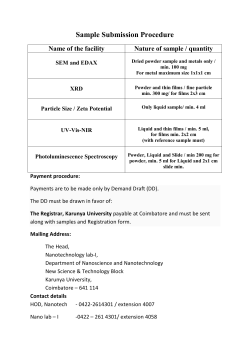


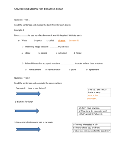
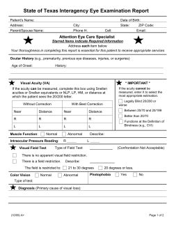
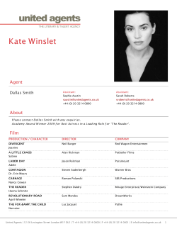

© Copyright 2026