
ADP-Glo™ Kinase Assay InstrucƟ ons for Use of Products
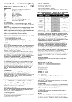
TECHNICAL MANUAL
ADP-Glo™ Kinase Assay
InstrucƟons for Use of Products
V6930, V9101, V9102, V9103 AND V9104
Revised 6/14
TM313
ADP-Glo™ Kinase Assay
All technical literature is available at: www.promega.com/protocols/
Visit the web site to verify that you are using the most current version of this Technical Manual.
E-mail Promega Technical Services if you have questions on use of this system: [email protected]
1. Description......................................................................................................................................... 1
2. Product Components and Storage Conditions ........................................................................................ 8
3. Preparing for the ADP-Glo™ Kinase Assay .......................................................................................... 10
3.A. Preparing the Kinase Detection Reagent ..................................................................................... 10
3.B. Generating a Standard Curve for Conversion of ATP to ADP ......................................................... 11
4. ADP-Glo™ Kinase Assay Protocols ..................................................................................................... 12
4.A. Kinase Reaction Buffer Considerations ....................................................................................... 13
4.B. ADP-Glo™ Kinase Assay Protocol .............................................................................................. 13
4.C. Optimizing Kinase Reaction Conditions...................................................................................... 14
4.D. Screening for Inhibitors Using the ADP-Glo™ Kinase Assay ......................................................... 15
4.E. Determining IC50 Values of Kinase Inhibitors .............................................................................. 16
5. General Considerations ..................................................................................................................... 17
6. References ........................................................................................................................................ 18
7. Composition of Buffers and Solutions ................................................................................................. 19
8. Related Products ............................................................................................................................... 19
9. Summary of Changes ......................................................................................................................... 20
1.
Description
The ADP-Glo™ Kinase Assay(a–g) is a luminescent ADP detection assay that provides a universal, homogeneous,
high-throughput screening method to measure kinase activity by quantifying the amount of ADP produced during a
kinase reaction. The ADP-Glo™ Kinase Assay can be used to monitor the activity of virtually any ADP-generating
enzyme (e.g., kinase or ATPase) using up to 1mM ATP. The ADP-Glo™ Kinase Assay is performed in a multiwell plate
and can detect kinase activity in a reaction volume as low as 5µl. The assay is performed in two steps; first, after the
kinase reaction, an equal volume of ADP-Glo™ Reagent is added to terminate the kinase reaction and deplete the
remaining ATP. Second, the Kinase Detection Reagent is added to simultaneously convert ADP to ATP and allow the
newly synthesized ATP to be measured using a luciferase/luciferin reaction (Figures 1 and 2). The light generated is
measured using a luminometer. Luminescence can be correlated to ADP concentrations by using an ATP-to-ADP
conversion curve (Figure 3). This assay is sensitive enough to detect very low amounts of ADP (20nM) and can
detect generated ADP in a reaction containing up to 1mM ATP in a linear fashion (Figure 3). The luminescent signal
generated is proportional to the ADP concentration produced and is correlated with kinase activity (Figure 4).
Promega CorporaƟon · 2800 Woods Hollow Road · Madison, WI 53711-5399 USA · Toll Free in USA 800-356-9526 · 608-274-4330 · Fax 608-277-2516
www.promega.com
TM313 · Revised 6/14
1
1.
Description (continued)
Furthermore, Figure 4, Panel B, shows the high assay performance for kinases with low ATP turnover. The ADP-Glo™
Kinase Assay can be performed with virtually any kinase and substrate combination and does not require radioactively
labeled components or antibodies. The kinase substrate can be a peptide, protein, lipid or sugar. Because of its high
sensitivity, the ADP-Glo™ Kinase Assay can be used with essentially all kinases and especially kinases with low enzyme
turnover, like growth factor receptor tyrosine kinases. Therefore, the assay is ideal for high-throughput screening
because a small amount of enzyme and low ATP-to-ADP conversion generate a high signal-to-background ratio.
(We define SB5, SB10 and SB20 as the amounts of enzyme needed to achieve signal-to-background ratios of 5, 10 and 20,
respectively.)
The ADP-Glo™ Kinase Assay relies on the properties of a proprietary thermostable luciferase (Ultra-Glo™ Recombinant
Luciferase) that is formulated to generate a stable “glow-type” luminescent signal and improve performance across a
wide range of assay conditions. The signal, which is produced by the luciferase reaction initiated by adding the Kinase
Detection Reagent, is stable for more than 3 hours (data not shown). This extended stability eliminates the need for a
luminometer equipped with injectors and allows batch-mode processing of multiple plates. In addition, the unique
combination of Ultra-Glo™ Recombinant Luciferase and proprietary formulations of the ADP-Glo™ Reagent and
Kinase Detection Buffer result in luminescence that is much less susceptible to interference from library compounds
than other luciferase- or fluorescence-based assays (1,2). In addition to providing biochemical values (e.g., Km of ATP
or Km of substrate) comparable to those reported in the literature, the ADP-Glo™ Kinase Assay can be used to
distinguish between ATP-competitive and noncompetitive kinase inhibitors (Figure 5).
Advantages of the ADP-Glo™ Kinase Assay
•
Broad linear range of ATP concentrations (micromolar to millimolar): Distinguish between ATPcompetitive and noncompetitive inhibitors.
•
Large dynamic range: High signal-to-background ratios at lower percent conversions of ATP to ADP allow use
of smaller amounts of enzyme.
•
High sensitivity: Detect 0.2pmol ADP with more than twofold difference over background.
•
Reliable, reproducible data: Routinely obtain Z´ factor values >0.7 even with low ATP consumption rates.
•
Universal assay: Use any kinase and kinase:substrate combination, including peptide, protein, lipid and sugar
substrates.
•
Positive correlation: Assay signal increases linearly with increasing product formation.
•
Luminescence-based ADP detection: Experience less overall assay interference from chemical compounds.
•
Batch plate processing: The luminescent signal is highly stable with <20% proportional increase in signal
after 3 hours.
•
Homogeneous, robust and non-radioactive assay.
2
Promega CorporaƟon · 2800 Woods Hollow Road · Madison, WI 53711-5399 USA · Toll Free in USA 800-356-9526 · 608-274-4330 · Fax 608-277-2516
TM313 · Revised 6/14
www.promega.com
Step 1
ADP-Glo™
Reagent
Add ADP-Glo™ Reagent to
completed ADP-generating
reactions and mix. Incubate
for 40 minutes.
Kinase
Detection
Substrate
Kinase
Detection
Buffer
Step 2
Record luminescence.
Kinase
Detection
Reagent
8050MA
Dispense Kinase Detection
Reagent to reaction and mix.
Incubate for 30–60 minutes.
Figure 1. Schematic representation of the ADP-Glo™ Kinase Assay protocol. Note: The 400-assay size of the
ADP-Glo™ Kinase Assay (Cat.# V6930) contains Kinase Detection Reagent that is already prepared and ready for use;
no reagent preparation is required.
Promega CorporaƟon · 2800 Woods Hollow Road · Madison, WI 53711-5399 USA · Toll Free in USA 800-356-9526 · 608-274-4330 · Fax 608-277-2516
www.promega.com
TM313 · Revised 6/14
3
1.
Description (continued)
Step 1: ATP Depletion
ADP-Glo™
Reagent
Step 2: ADP Detection
Kinase
Detection
Reagent
ATP
ADP
Luminescence
8051MA
Kinase or
ATPase Reaction
Figure 2. Principle of the ADP-Glo™ Kinase Assay. The assay is performed in two steps: 1) after the kinase or
ATPase reaction, ADP-Glo™ Reagent is added to terminate the kinase reaction and deplete the remaining ATP, and
2) the Kinase Detection Reagent is added to convert ADP to ATP and allow the newly synthesized ATP to be measured
using a luciferase/luciferin reaction. The light generated correlates to the amount of ADP generated in the kinase or
ATPase assay, which is indicative of kinase or ATPase activity.
4
Promega CorporaƟon · 2800 Woods Hollow Road · Madison, WI 53711-5399 USA · Toll Free in USA 800-356-9526 · 608-274-4330 · Fax 608-277-2516
TM313 · Revised 6/14
www.promega.com
1µM
3.0 × 10⁵
2.5 × 10⁵
2.0 × 10⁵
1.5 × 10⁵
r² = 0.99
1.0 × 10⁵
0.5 × 10
5
0
0
20
40
60
Luminescence (RLU)
Luminescence (RLU)
A.
2.0 × 10⁶
1.5 × 10⁶
1.0 × 10⁶
0
80 100 120
0.8 × 10⁷
r² = 0.99
0.4 × 10⁷
0
0
20
40
60
Luminescence (RLU)
Luminescence (RLU)
1.2 × 10⁷
0
20
40
60
80 100 120
Percent ATP-to-ADP Conversion
100µM
1.6 × 10⁷
r² = 0.99
0.5 × 10⁶
Percent ATP-to-ADP Conversion
2.0 × 10⁷
10µM
2.5 × 10⁶
1mM
1.8 × 10⁸
1.5 × 10
8
1.2 × 10⁸
0.9 × 10⁸
r² = 0.99
0.6 × 10⁸
8
0.3 × 10
0
80 100 120
Percent ATP-to-ADP Conversion
0
20
40
60
80 100 120
Percent ATP-to-ADP Conversion
B.
Signal-to-Background Ratios
100
80
60
40
20
10
5
4
3
2
1
0
1µM
80
54
41
28
15
8
4
4
3
2
2
1
10µM
135
110
84
57
31
16
8
7
6
4
2
1
100µM 125
97
77
56
31
17
9
8
6
5
3
1
117
91
74
51
28
14
8
7
6
4
2
1
1mM
8052MA
[ATP + ADP]
Percent ADP in an ATP + ADP Mixture
Figure 3. Sensitivity and linearity of the ADP-Glo™ Kinase Assay. Panel A. Four ATP-to-ADP conversion
curves were prepared at the indicated ATP+ADP concentrations in 25µl of 1X reaction buffer A (see Section 7 for
composition) in a solid white 96-well plate. (Conversion curves are described in Section 3.B.) ADP-Glo™ Kinase Assays
were performed using 25µl of ADP-Glo™ Reagent and 50µl of Kinase Detection Reagent at room temperature as
described in Section 4.B. Luminescence was recorded using a GloMax® 96 Microplate Luminometer (Cat.# E6501).
Values represent the mean of two replicates. There is a linear relationship between the luminescent signal and the
amount of ADP in the reaction buffer at all ATP+ADP concentration series tested. Panel B. The ADP-Glo™ Kinase
Assay is highly sensitive as shown by the high signal-to-background ratios. The ADP-Glo™ Kinase Assay luminescence
was measured 1 hour after adding the Kinase Detection Reagent. To determine signal stability, luminescence was
recorded again every hour. The signal is stable over a period of 5 hours generally with <20% increase after 3 hours,
while the signal-to-background ratio did not change (data not shown). RLU = relative light units.
Promega CorporaƟon · 2800 Woods Hollow Road · Madison, WI 53711-5399 USA · Toll Free in USA 800-356-9526 · 608-274-4330 · Fax 608-277-2516
www.promega.com
TM313 · Revised 6/14
5
1.
Description (continued)
Luminescence (RLU)
A.
8,000
7,000
6,000
5,000
4,000
3,000
2,000
1,000
0
EC50 = 28.4ng
0
2
1
3
Log10 IκKβ (ng)
B.
EC50
IκKβ (ng/reaction) 200 120
Average RLU
% ADP Produced
S:B Ratio
72 43.2 25.9 15.6 9.3 5.6 3.4 2.0 1.2
7,104 7,325 6,558 5,375 3,723 1,705 1,330 917
35.0 36.2 32.3 25.7 17.6
678
383
0
339
148
8.1
6.0
3.6
2.4
1.4
1.1
0
48.0 49.5 44.3 36.3 25.2 11.5
9.0
6.2
4.6
2.6
2.3
1
IκKβ (ng)
SB5
SB10
SB20
5.16
13.5
20.5
8053MA
C.
Figure 4. Demonstration of ADP-Glo™ Kinase Assay performance using Inhibitor of κB kinase β (IκKβ).
Panel A. The 10µl kinase reaction was performed in a 384-well plate in 1X reaction buffer A supplemented with 2mM
MnCl2, 2mM DTT and 100µM sodium vanadate using 200µM IκKtide peptide (Biomol) as a substrate and 1µM ATP at
room temperature for 1 hour. ADP was detected as described in Section 4.B using 10µl of ADP-Glo™ Reagent and 20µl
of Kinase Detection Reagent. Curve fitting was performed using GraphPad Prism® sigmoidal dose-response (variable
slope) software. Panel B. An ATP-to-ADP conversion curve in the 1µM series was performed at the same time to
correlate the %ADP produced to each RLU value. Panel C. Low amounts of enzyme (less than EC50) can be used in the
kinase reaction, while maintaining high signal-to-background ratios (S:B Ratio). Data points represent the average of
duplicate wells. SB5, SB10 and SB20 are defined as the amounts of enzyme needed to achieve signal-to-background ratios
of 5, 10 and 20, respectively.
6
Promega CorporaƟon · 2800 Woods Hollow Road · Madison, WI 53711-5399 USA · Toll Free in USA 800-356-9526 · 608-274-4330 · Fax 608-277-2516
TM313 · Revised 6/14
www.promega.com
A.
D.
10µM ATP, 10 minutes
Luminescence (RLU)
Luminescence (RLU)
5.0 × 10⁶
4.0 × 10⁶
3.0 × 10⁶
2.0 × 10⁶
1.0 × 10⁶
0
–1
IC50 = 31.22nM
0
1
2
3
4
4.0 × 10⁶
3.0 × 10⁶
2.0 × 10⁶
1.0 × 10⁶
0
5
10µM ATP, 10 minutes
5.0 × 10⁶
IC50 = 4.5nM
–2
–1
0
1
2
3
4
Log10[PKI], nM
Log10[H-89], nM
B.
E.
Luminescence (RLU)
1.0 × 10
100µM ATP, 30 minutes
0.8 × 10⁷
0.6 × 10⁷
0.4 × 10⁷
0.2 × 10⁷
0
–1
100µM ATP, 10 minutes
2.5 × 10⁷
Luminescence (RLU)
7
IC50 = 126nM
2.0 × 10⁷
1.5 × 10⁷
1.0 × 10⁷
0.5 × 10⁷
IC50 = 4.36nM
0
0
1
2
3
–2
5
4
–1
Log10[H-89], nM
0
1
2
3
4
Log10[PKI], nM
F.
1.6 × 10⁷
1.2 × 10
7
0.8 × 10⁷
0.4 × 10⁷
0
–1
IC50 = 737nM
0
1
2
3
Log10[H-89], nM
4
5
500µM ATP, 30 minutes
2.0 × 10⁷
1.6 × 10⁷
1.2 × 10⁷
0.8 × 10⁷
0.4 × 10⁷
IC50 = 2.7nM
0
–2
–1
0
1
2
Log10[PKI], nM
3
4
8054MA
500µM ATP, 60 minutes
2.0 × 10⁷
Luminescence (RLU)
Luminescence (RLU)
C.
Figure 5. Determining IC50 for ATP-competitive and noncompetitive inhibitors. Titrations of the protein
kinase A (PKA) noncompetitive inhibitor PKI and competitive inhibitor H-89 were performed in solid white, flatbottom 96-well plates in a total volume of 50µl using 0.1 unit/well PKA, 50µM Kemptide (PKA) Peptide Substrate
(Cat.# V5601) and the indicated concentration of inhibitor. Reactions were carried out at 10, 100 or 500µM ATP for
the time listed on each graph. Data points are the average of two determinations, and error bars are ± S.D. IC50 values
for H-89 determined using the ADP-Glo™ Kinase Assay increase with increasing ATP concentration (Panels A, B and C).
In contrast, the IC50 values for PKI show only a minimal change with increasing ATP concentrations (Panels D, E and F).
These compare favorably to the IC50 values reported for these compounds in the literature [3nM for PKI (3); 40nM for
H-89 at 10µM ATP (4)]. Curve fitting was performed using GraphPad Prism® sigmoidal dose-response (variable slope)
software.
Promega CorporaƟon · 2800 Woods Hollow Road · Madison, WI 53711-5399 USA · Toll Free in USA 800-356-9526 · 608-274-4330 · Fax 608-277-2516
www.promega.com
TM313 · Revised 6/14
7
2.
Product Components and Storage Conditions
PRODUCT
ADP-Glo™ Kinase Assay
SIZE
C A T. #
400 assays
V6930
The system is sufficient for 400 assays if performed in 384-well plates using 5µl, 5µl and 10µl of a kinase
reaction, ADP-Glo™ Reagent and Kinase Detection Reagent, respectively, per sample. The system also can
be used in 96-well plates using 25µl:25µl:50µl for a total of 80 assays. Note: This size of the system contains
Kinase Detection Reagent that is already prepared and ready for use; no reagent preparation is required.
Includes:
•
•
•
•
2ml ADP-Glo™ Reagent
4ml Kinase Detection Reagent
0.5ml Ultra Pure ATP, 10mM
0.5ml ADP, 10mM
PRODUCT
ADP-Glo™ Kinase Assay
SIZE
C A T. #
1,000 assays
V9101
The system is sufficient for 1,000 assays if performed in 384-well plates using 5µl, 5µl and 10µl of a kinase
reaction, ADP-Glo™ Reagent and Kinase Detection Reagent, respectively, per sample. The system also can be
used in 96-well plates using 25µl:25µl:50µl for a total of 200 assays. Includes:
•
•
•
•
•
5ml ADP-Glo™ Reagent
10ml Kinase Detection Buffer
1 vial Kinase Detection Substrate (Lyophilized)
500µl Ultra Pure ATP, 10mM
500µl ADP, 10mM
PRODUCT
ADP-Glo™ Kinase Assay
SIZE
C A T. #
10,000 assays
V9102
The system is sufficient for 10,000 assays if performed in 384-well plates using 5µl, 5µl and 10µl of a kinase
reaction, ADP-Glo™ Reagent and Kinase Detection Reagent, respectively, per sample. The system also can be
used in 96-well plates using 25µl:25µl:50µl for a total of 2,000 assays. Includes:
•
•
•
•
•
50ml
100ml
1 vial
5ml
5ml
ADP-Glo™ Reagent
Kinase Detection Buffer
Kinase Detection Substrate (Lyophilized)
Ultra Pure ATP, 10mM
ADP, 10mM
8
Promega CorporaƟon · 2800 Woods Hollow Road · Madison, WI 53711-5399 USA · Toll Free in USA 800-356-9526 · 608-274-4330 · Fax 608-277-2516
TM313 · Revised 6/14
www.promega.com
PRODUCT
ADP-Glo™ Kinase Assay
SIZE
C A T. #
100,000 assays
V9103
The system is sufficient for 100,000 assays if performed in 384-well plates using 5µl, 5µl and 10µl of a kinase
reaction, ADP-Glo™ Reagent and Kinase Detection Reagent, respectively, per sample. The system also can be
used in 96-well plates using 25µl:25µl:50µl for a total of 20,000 assays. Delivered as 10 × V9102. Includes:
• 10 × 50ml
• 10 × 100ml
•
10 vials
•
10 × 5ml
•
10 × 5ml
ADP-Glo™ Reagent
Kinase Detection Buffer
Kinase Detection Substrate (Lyophilized)
Ultra Pure ATP, 10mM
ADP, 10mM
PRODUCT
ADP-Glo™ Kinase Assay, Bulk Packaged*
SIZE
C A T. #
100,000 assays
V9104
The system is sufficient for 100,000 assays if performed in 384-well plates using 5µl, 5µl and 10µl of a kinase
reaction, ADP-Glo™ Reagent and Kinase Detection Reagent, respectively, per sample. The system also can be
used in 96-well plates using 25µl:25µl:50µl for a total of 20,000 assays. Includes:
• 10 × 50ml
• 10 × 100ml
•
10 vials
•
10 × 5ml
•
10 × 5ml
ADP-Glo™ Reagent
Kinase Detection Buffer
Kinase Detection Substrate (Lyophilized)
Ultra Pure ATP, 10mM
ADP, 10mM
*The V9104 components are packaged in a single box that is available through our Helix® On-Site Stocking Program.
Storage Conditions: Store the system at –20°C. Before use, thaw all components completely at room temperature.
Once thawed, mix all components thoroughly before use. Once prepared, dispense Kinase Detection Reagent (Kinase
Detection Buffer + Substrate) into aliquots and store at –20°C. Kinase Detection Buffer may form a precipitate when
thawed. See Section 3.A for a protocol to dissolve any precipitate. For convenience, ADP-Glo™ Reagent and Kinase
Detection Reagent may be used at room temperature (22°C) for 24 hours without loss of signal.
Promega CorporaƟon · 2800 Woods Hollow Road · Madison, WI 53711-5399 USA · Toll Free in USA 800-356-9526 · 608-274-4330 · Fax 608-277-2516
www.promega.com
TM313 · Revised 6/14
9
3.
Preparing for the ADP-Glo™ Kinase Assay
Materials to Be Supplied by the User
•
solid white, multiwell plate
•
multichannel pipette or automated pipetting station
•
kinase substrate
•
kinase
•
luminometer capable of reading multiwell plates
•
plate shaker
3.A. Preparing the Kinase Detection Reagent
!
Note: If you are using the 400-assay size of the ADP-Glo™ Kinase Assay (Cat.# V6930), no Kinase Detection Reagent
preparation is required; proceed to Section 3.B. If you are using another size of the ADP-Glo™ Kinase Assay (Cat.#
V9101, V9102, V9103 or V9104), prepare the Kinase Detection Reagent as described below.
Kinase Detection Buffer Preparation
The Kinase Detection Buffer may contain a precipitate depending on conditions used for storage and handling. There
is no observed change in performance of the ADP-Glo™ Kinase Assay if the buffer contains a precipitate. However, to
avoid clogging the pipette tips, the precipitate may be removed or solubilized according to the following steps.
1.
Thaw Kinase Detection Buffer at room temperature, and observe for the presence of precipitate.
2.
If a precipitate is present, incubate the Kinase Detection Buffer at 37°C with constant swirling for 15 minutes
to dissolve the precipitate. Alternatively, remove the precipitate from the Kinase Detection Buffer by carefully
pipetting the supernatant from the bottle.
Kinase Detection Reagent Preparation
1.
Equilibrate the Kinase Detection Buffer and Kinase Detection Substrate to room temperature before use.
2.
Transfer the entire volume of Kinase Detection Buffer into the amber bottle containing Kinase Detection
Substrate to reconstitute the lyophilized substrate. This forms the Kinase Detection Reagent.
3.
Mix by gently vortexing, swirling or inverting the contents to obtain a homogeneous solution. The Kinase
Detection Substrate should go into solution in less than one minute.
4.
The Kinase Detection Reagent should be used immediately or dispensed into aliquots and stored at –20°C. We
have shown that the reconstituted reagent remains stable with no loss of signal after several freeze-thaw cycles.
10
Promega CorporaƟon · 2800 Woods Hollow Road · Madison, WI 53711-5399 USA · Toll Free in USA 800-356-9526 · 608-274-4330 · Fax 608-277-2516
TM313 · Revised 6/14
www.promega.com
3.B. Generating a Standard Curve for Conversion of ATP to ADP
ADP-Glo™ Kinase Assay can detect small changes in ATP-to-ADP conversion. Whether assaying a low-activity kinase
whose ATP turnover rate is low or using a small amount of enzyme that produces a small amount of ADP, a high
signal-to-background ratio is obtained with the ADP-Glo™ Kinase Assay (Figure 3). The ADP-Glo™ Kinase Assay
produces excellent Z´-factor values (Table 1) as Z´ factors greater than 0.5 indicate excellent assay quality (5).
Table 1. Z´ Values Produced at Different Percent ADP in an ATP + ADP Mixture Using Two
Concentration Series.
ATP-to-ADP Conversion
10µM ATP + ADP
500µM ATP + ADP
1%
0.76
0.65
5%
0.82
0.84
10%
0.90
0.86
20%
0.88
0.92
To estimate the amount of ADP produced in the kinase reaction, we recommend creating a standard curve that
represents the luminescence corresponding to the conversion of ATP to ADP (the “ATP-to-ADP conversion curve”)
based on the ATP concentration used in the kinase reaction. These conversion curves represent the amounts of ATP
and ADP available in a reaction at the specified conversion percentage (Table 2). The standard samples used to
generate an ATP-to-ADP conversion curve are created by combining the appropriate volumes of ATP and ADP stock
solutions (Table 3). The sum of the ATP and ADP concentrations is denoted as “ATP+ADP” in this manual.
Table 2. Percent Conversion of ATP to ADP Represented by the Standard Curve.
% ADP
100
80
60
40
20
10
5
4
3
2
1
0
% ATP
0
20
40
60
80
90
95
96
97
98
99
100
1.
!
2.
Prepare 1ml of 1mM ATP and 500µl of 1mM ADP by diluting the supplied Ultra Pure ATP and ADP in preferred
1X kinase reaction buffer.
Use only the provided Ultra Pure ATP when performing the ADP-Glo™ Kinase Assay. Other sources of ATP may
contain ADP that could result in high background.
Add 90µl of 1X kinase reaction buffer to wells B1–B12, C1–C12 and D1–D12 of a 96-well plate.
Promega CorporaƟon · 2800 Woods Hollow Road · Madison, WI 53711-5399 USA · Toll Free in USA 800-356-9526 · 608-274-4330 · Fax 608-277-2516
www.promega.com
TM313 · Revised 6/14
11
3.B. Generating a Standard Curve for Conversion of ATP to ADP (continued)
3.
Combine the 1mM ATP and 1mM ADP solutions prepared in Step 1 in wells A1–A12 as indicated in Table 3 to
simulate the ATP and ADP concentrations at each percent conversion (see Table 2). Mix well. This is the 1mM
series.
Table 3. Preparation of the 1mM Series of ATP+ADP Standards.
Well Number
A1
A2
A3
A4
A5
A6
A7
A8
A9
A10
A11
A12
1mM ADP (µl)
100
80
60
40
20
10
5
4
3
2
1
0
1mM ATP (µl)
0
20
40
60
80
90
95
96
97
98
99
100
4.
Dilute the samples in wells A1–A12 by transferring 10µl of the sample in well A1 to well B1, 10µl from well A2
to well B2, etc. Mix well. This is the 100µM series.
5.
Dilute the samples in wells B1–B12 by transferring 10µl of the sample in well B1 to well C1, 10µl from well B2
to well C2, etc. Mix well. This is the 10µM series.
6.
Dilute the samples in wells C1–C12 by transferring 10µl of the sample in well C1 to well D1, 10µl from well C2
to well D2, etc. Mix well. This is the 1µM series.
7.
Transfer the amount needed (5 or 25µl) from the desired ATP+ADP series into separate wells of the new assay
plate or the assay plate where the kinase reactions are present.
8.
Follow the ADP-Glo™ Kinase Assay protocol described in Section 4.B, starting at Step 2.
4.
ADP-Glo™ Kinase Assay Protocols
Prior to performing the ADP-Glo™ Kinase Assay, prepare the reagents and ATP + ADP standards as described in
Section 3. Calculate the volume of ADP-Glo™ Kinase Assay Reagents required for your experiments, and allow that
volume to reach room temperature before use. Return the remaining ADP-Glo™ Reagent and Kinase Detection
Reagent to –20°C. For information about optimizing the kinase and kinase substrate concentrations, refer to Section
4.C. The ADP-Glo™ Reagent and the Kinase Detection Reagent may be used at room temperature (22°C) for 24 hours
without loss of signal.
12
Promega CorporaƟon · 2800 Woods Hollow Road · Madison, WI 53711-5399 USA · Toll Free in USA 800-356-9526 · 608-274-4330 · Fax 608-277-2516
TM313 · Revised 6/14
www.promega.com
4.A. Kinase Reaction Buffer Considerations
The ADP-Glo™ Kinase Assay is designed for use with 1X reaction buffer A (see Section 7 for composition) in the
presence of the kinase, ATP and substrate. If protein tyrosine kinase or IκKβ reactions are performed, we recommend
supplementing reaction buffer A with 2mM DTT, 2mM MnCl2 and 100µM sodium vanadate. Sphingosine kinase
reaction can be performed in 1X reaction buffer A supplemented with 0.5mM DTT.
!
If other reaction buffer compositions are preferred, make sure that the MgCl2 concentration is at least 0.5mM
after adding the ADP-Glo™ Reagent to the kinase reaction.
4.B. ADP-Glo™ Kinase Assay Protocol
The ADP-Glo™ Kinase Assay is performed in two steps once the kinase reaction is complete as outlined in Figure 1.
For 96-well plates, we recommend 25µl of kinase reaction, 25µl of ADP-Glo™ Reagent and 50µl of Kinase Detection
Reagent for a total volume of 100µl. For 384-well plates, volumes are reduced fivefold to 5µl of kinase reaction, 5µl
of ADP-Glo™ Reagent and 10µl of Kinase Detection Reagent. Other volumes may be used, provided the 1:1:2 ratio
of kinase reaction volume to ADP-Glo™ Reagent volume to Kinase Detection Reagent volume is maintained. The
ADP-Glo™ Kinase Assay Protocol for 384-well plates is outlined below.
!
1.
!
2.
!
Use the Ultra Pure ATP provided with the ADP-Glo™ Kinase Assay when setting up standard curves and running
kinase reactions.
Perform a 5µl kinase reaction using 1X kinase buffer (e.g., 1X reaction buffer A).
If the kinase reaction was not run at room temperature, equilibrate the plate to room temperature before adding
the ADP-Glo™ Reagent.
Add 5µl of ADP-Glo™ Reagent to stop the kinase reaction and deplete the unconsumed ATP, leaving only ADP
and a very low background of ATP.
The ADP-Glo™ Reagent is effective in terminating the kinase reaction; therefore, there is no need to add an
inhibitor to terminate the kinase reaction. We do not recommend modifying the protocol to add a kinasetermination reagent. However, if you do modify the protocol to add a kinase-termination reagent, do not use a
magnesium-chelating agent such as EDTA because the ADP-Glo™ Assay requires the presence of magnesium.
Note: The final Mg2+ concentration must be at least 0.5mM.
3.
Incubate at room temperature for 40 minutes.
4.
Add 10µl of Kinase Detection Reagent to convert ADP to ATP and introduce luciferase and luciferin to detect
ATP.
Promega CorporaƟon · 2800 Woods Hollow Road · Madison, WI 53711-5399 USA · Toll Free in USA 800-356-9526 · 608-274-4330 · Fax 608-277-2516
www.promega.com
TM313 · Revised 6/14
13
4.B. ADP-Glo™ Kinase Assay Protocol (continued)
5.
Incubate at room temperature for 30–60 minutes, depending on the ATP concentration used in the kinase
reaction (see Table 4).
Table 4. Incubation Time to Convert ADP to ATP.
ATP Concentration
10–100µM
100–500µM
500–1,000µM
Time
30 minutes
40 minutes
60 minutes
6.
Measure the luminescence with a plate-reading luminometer or charge-coupled device (CCD) camera.
Note: Instrument settings depend on the manufacturer. An integration time of 0.25–1 second per well should
serve as a guideline. The long half-life of the ADP-Glo™ Kinase Assay signal allows plates to be left longer at
room temperature before reading if desired.
4.C. Optimizing Kinase Reaction Conditions
For optimal performance when using the ADP-Glo™ Kinase Assay, optimize the amounts of kinase and kinase substrate
in the kinase reaction. If the amount of kinase or kinase substrate has been determined, proceed to Section 4.B.
When screening for kinase inhibitors, use an ATP concentration of 10µM or less to preferentially identify ATPcompetitive inhibitors. To preferentially identify ATP-noncompetitive inhibitors, use a higher ATP concentration
(up to 1mM). Optimize enzyme and substrate concentrations, reaction temperature and incubation time whenever the
ATP concentration is changed.
Notes:
1.
Use only the provided Ultra Pure ATP when performing the ADP-Glo™ Kinase Assay. Other sources of ATP may
contain ADP that could result in high background.
2.
We recommend optimizing the kinase reaction conditions at room temperature to ensure uniform temperature
across the plate during the ADP-Glo™ Kinase Assay.
Determining Optimal Substrate Concentration
1.
Make serial twofold dilutions of kinase substrate in 1X kinase reaction buffer across the plate using as much
kinase as is practical and the desired concentration of ATP (up to 1mM). As a control, perform the same titration
without kinase. Mix the plate, and incubate for the desired amount of time.
2.
Follow the ADP-Glo™ Kinase Assay protocol described in Section 4.B, starting at Step 2.
3.
Record luminescence.
Note: The optimal kinase substrate concentration will result in the largest difference in luminescence between
kinase reaction wells and wells that do not contain kinase.
14
Promega CorporaƟon · 2800 Woods Hollow Road · Madison, WI 53711-5399 USA · Toll Free in USA 800-356-9526 · 608-274-4330 · Fax 608-277-2516
TM313 · Revised 6/14
www.promega.com
Determining the Optimal Amount of Kinase
1.
!
Make serial twofold dilutions of kinase in 1X kinase reaction buffer across the plate using the desired amount of
ATP (up to 1mM) and kinase substrate (as determined previously). Mix the plate, and incubate for the desired
amount of time.
We recommend optimizing the kinase reaction conditions at room temperature to ensure uniform temperature
across the plate during the ADP-Glo™ Assay.
2.
Follow the ADP-Glo™ Kinase Assay protocol described in Section 4.B, starting at Step 2.
3.
Record luminescence.
Note: The optimal amount of kinase to use in subsequent compound screens and IC50 determinations is the
amount that produces luminescence within the linear range of the kinase titration curve and generates an
adequate signal-to-background ratio.
Because the ADP-Glo™ Kinase Assay is very sensitive, it can detect a very small amount of ADP with a high signal-tobackground ratio. A small amount of enzyme that produces low conversion of ATP to ADP is sufficient for use with the
ADP-Glo™ Kinase Assay due to the high signal-to-background ratio generated. Figure 4 shows that the amount of ADP
produced with a small amount of enzyme results in a signal-to-background ratio (SB) of 5, 10 or 20 (SB5, SB10 and SB20,
respectively).
4.D. Screening for Inhibitors Using the ADP-Glo™ Kinase Assay
The following protocol is an example; the actual volumes used can be adjusted as needed. The volume of the ADP-Glo™
Reagent used should equal the volume of the kinase reaction, and the volume of the Kinase Detection Reagent should
be double the amount of the kinase reaction. The volumes provided are intended for a 384-well plate (5µl:5µl:10µl
ratio); to perform the assay in a 96-well plate, increase the volumes fivefold.
1.
Add 1µl of test compound to each well except for 16 control wells, which receive 1µl of test compound vehicle.
2.
Add 2µl of 1X kinase reaction buffer containing 2.5X the optimal concentration of kinase, but no kinase substrate,
to eight of the control wells.
3.
Add 2µl of 1X kinase reaction buffer containing 2.5X the optimal concentrations of kinase and kinase substrate
to all remaining wells.
4.
Add 2µl of 1X kinase reaction buffer containing 2.5X the desired concentration of ATP (up to 1mM) to all wells.
Promega CorporaƟon · 2800 Woods Hollow Road · Madison, WI 53711-5399 USA · Toll Free in USA 800-356-9526 · 608-274-4330 · Fax 608-277-2516
www.promega.com
TM313 · Revised 6/14
15
4.D. Screening for Inhibitors Using the ADP-Glo™ Kinase Assay (continued)
5.
!
Mix the plate, and incubate for the desired amount of time.
We recommend optimizing the kinase reaction conditions at room temperature to ensure uniform temperature
across the plate during the ADP-Glo™ Assay.
6.
Follow the ADP-Glo™ Kinase Assay protocol described in Section 4.B, starting at Step 2.
7.
Record luminescence.
Note: Instrument settings depend on the manufacturer. An integration time of 0.25–1 second per well should
serve as a guideline.
4.E. Determining IC50 Values of Kinase Inhibitors
The following protocol is designed for a 96-well plate using a 25µl:25µl:50µl ratio. To perform the assay in a 384-well
plate, reduce volumes fivefold. Other volumes may be used, provided the 1:1:2 ratio of kinase reaction volume to
ADP-Glo™ Reagent volume to Kinase Detection Reagent volume is maintained.
1.
Add 10µl of 1X kinase reaction buffer to each well in columns 2 through 12.
2.
Add 20µl of the inhibitor diluted to the highest concentration desired in 1X kinase reaction buffer to the wells in
column 1.
3.
Transfer 10µl from the wells in column 1 to column 2. Mix well. Continue to make twofold serial dilutions across
the plate, mixing well before each transfer. Discard the 10µl removed from the last column.
4.
Add 10µl of 1X kinase reaction buffer containing 2.5X the optimal concentration of enzyme to all wells.
5.
Add 5µl of 1X kinase reaction buffer containing 5X the optimal concentration of ATP and 5X the optimal
concentration of substrate to all wells.
!
6.
!
Use only the provided Ultra Pure ATP when performing the ADP-Glo™ Kinase Assay. Other sources of ATP may
contain ADP that could result in high background.
Mix the plate, and incubate for the optimal amount of time.
We recommend optimizing the kinase reaction conditions so that the reaction can be performed at room
temperature to avoid formation of temperature gradients.
Note: If the kinase reaction was not run at room temperature, equilibrate the plate to room temperature before
adding the ADP-Glo™ Reagent.
7.
Follow the ADP-Glo™ Kinase Assay protocol described in Section 4.B, starting at Step 2.
8.
Record luminescence.
Note: Instrument settings depend on the manufacturer. An integration time of 0.25–1 second per well should
serve as a guideline.
16
Promega CorporaƟon · 2800 Woods Hollow Road · Madison, WI 53711-5399 USA · Toll Free in USA 800-356-9526 · 608-274-4330 · Fax 608-277-2516
TM313 · Revised 6/14
www.promega.com
5.
General Considerations
Temperature: The intensity and stability of the luminescent signal from the ADP-Glo™ Kinase Assay depends on the
rate of the luciferase reaction. Environmental factors that affect the rate of the luciferase reaction will result in a change
in the intensity of light output and stability of the luminescent signal. Temperature is one factor that affects the rate of
this enzymatic assay and thus the light output. For consistent results, equilibrate assay plates and reagent to room
temperature before adding the ADP-Glo™ Kinase Assay reagents. Insufficient equilibration may result in a temperature
gradient between the wells in the center and at the edge of the plate and therefore variability across the plate.
Solvents and Other Chemicals: The chemical environment in which the first or second step of the ADP-Glo™
Kinase Assay is performed will affect the enzymatic rates and thus luminescence intensity. We recommend a pH of 7–8
for the kinase buffer. Some vehicles used to resuspend the various test compounds or reagents used in the kinase
reaction buffer may interfere with the luciferase reaction and thus affect the light output of the assay. Various chemicals
were shown to be compatible with or tolerated by the ADP-Glo™ Kinase Assay (Table 5). Interference with the
luciferase reaction can be detected by assaying a parallel set of control wells without kinase or kinase substrate and
using ADP as a substrate.
Table 5. Solvents and Chemicals Compatible with the ADP-Glo™ Kinase Assay.
Chemical
Maximum Concentration Tolerated
NaCl
200mM
CaCl2
2.5mM
Calmodulin
5µM
DTT
10mM
MgCl2
50mM
MnCl2
5mM
DMSO
5%
Sodium orthovanadate
500µM
Tergitol-NP-9
0.1%
Tween®-20
0.2%
®
Triton X-100
0.2%
Plate Recommendations: We recommend using standard solid white, multiwell plates suitable for luminescence
measurements. Please contact your luminometer instrument manufacturer to find out which plates will work best for
your particular instrument.
Promega CorporaƟon · 2800 Woods Hollow Road · Madison, WI 53711-5399 USA · Toll Free in USA 800-356-9526 · 608-274-4330 · Fax 608-277-2516
www.promega.com
TM313 · Revised 6/14
17
5.
General Considerations (continued)
Testing for Compounds that Interfere with the ADP-Glo™ Kinase Assay: Test compounds that inhibit only
the kinase will result in lower luminescence compared to vehicle-only controls and are easily distinguishable from
compounds that inhibit other components of the assay. Test compounds that inhibit other components of the assay
either alone or together with the kinase might increase, decrease or have no effect on the luminescent signal, depending
on the level of inhibition of the kinase, luciferase or other enzyme components. To test hits from a kinase screen for the
possibility of chemical interference with enzymatic depletion of ATP or generation of the luminescent signal, set up
mock reactions without kinase but with all other assay components present, including a mixture of ADP and ATP to
mimic the kinase reaction results. Add the appropriate concentration of test compound (usually 10µM) or vehicle
control (e.g., 1% DMSO) to mock reactions without kinase present but containing final concentrations of 2µM ADP
and 8µM ATP (as described for generating a standard curve for conversion of ATP to ADP, Section 3.B). A test
compound that affects assay performance would alter luminescence by greater than 20% compared to vehicle control
reactions without test compounds. The results from testing 30 ATP-competitive inhibitors from different chemical
classes showed that none interfered with the ADP-Glo™ Kinase Assay (6). Test compounds that inhibit luciferase may
result in false hits. However, the unique combination of Ultra-Glo™ Recombinant Luciferase and proprietary buffer
compositions of the ADP-Glo™ Kinase Assay significantly reduces the number of false hits (1,7).
6.
References
1.
Auld, D.S. et al. (2009) A basis for reduced chemical library inhibition of firefly luciferase obtained from directed
evolution. J. Med. Chem. 52, 1450–8.
2.
Jia, Y. et al. (2008) Current in vitro kinase assay technologies: The quest for a universal format. Curr. Drug.
Discov. Technol. 5, 59–69.
3.
Walsh, D.A. and Glass, D.B. (1991) Utilization of the inhibitor protein of adenosine cyclic monophosphatedependent protein kinase, and peptides derived from it, as tools to study adenosine cyclic monophosphatemediated cellular processes. Methods Enzymol. 201, 304–16.
4.
Hidaka, H., Watanabe, M. and Kobayashi, K. (1991) Properties and use of H series compounds as protein kinase
inhibitors. Methods Enzymol. 201, 328–39.
5.
Zhang, J.H. et al. (1999) A simple statistical parameter for use in evaluation and validation of high throughput
screening assays. J. Biomol. Screen. 4, 67–73.
6.
Alli, C. et al. Nerviano Medical Sciences. ADP-Glo: The new luminescent ADP detection assay as a universal,
quantitative and robust alternative for kinase profiling. Poster #P3016. Society for Biomolecular Sciences, April
2009.
7.
Auld, D.S. (2009) Promega’s bioluminescent assays: Comparison of kinase assays using product depletion and
product formation. GTC Bio 4th Annual Protein Kinases in Drug Discovery Conference, Boston, MA, May 4–5,
2009.
18
Promega CorporaƟon · 2800 Woods Hollow Road · Madison, WI 53711-5399 USA · Toll Free in USA 800-356-9526 · 608-274-4330 · Fax 608-277-2516
TM313 · Revised 6/14
www.promega.com
7.
Composition of Buffers and Solutions
1X Kinase Reaction Buffer A
40mM Tris (pH 7.5)
20mM MgCl2
0.1mg/ml BSA
8.
Related Products
Protein Kinase Assay Systems
Product
ADP-Glo™ Max Assay
Size*
Cat.#
1,000 assays
V7001
Kinase-Glo® Luminescent Kinase Assay
10ml
V6711
Kinase-Glo® Plus Luminescent Kinase Assay
10ml
V3771
Kinase-Glo® Max Luminescent Kinase Assay
ProFluor® PKA Assay
ProFluor® Src-Family Kinase Assay
PepTag® Non-Radioactive PKC Assay
®
PepTag Non-Radioactive cAMP-Dependent Protein Kinase Assay
10ml
V6071
4 plate
V1240
4 plate
V1270
120 reactions
V5330
120 reactions
V5340
Size
Cat.#
*Products may be available in additional sizes.
SignaTECT® Protein Kinase Assay Systems
Product
®
SignaTECT cAMP-Dependent Protein Kinase (PKA) Assay System
96 reactions
V7480
SignaTECT® Protein Kinase C (PKC) Assay System
96 reactions
V7470
SignaTECT® Protein Tyrosine Kinase (PTK) Assay System
96 reactions
V6480
SignaTECT® Calcium/Calmodulin-Dependent Protein
Kinase (CaM KII) Assay System
96 reactions
V8161
®
SignaTECT DNA-Dependent Protein Kinase Assay System
96 reactions
V7870
SignaTECT® cdc2 Protein Kinase Assay System
96 reactions
V6430
Promega CorporaƟon · 2800 Woods Hollow Road · Madison, WI 53711-5399 USA · Toll Free in USA 800-356-9526 · 608-274-4330 · Fax 608-277-2516
www.promega.com
TM313 · Revised 6/14
19
8.
Related Products (continued)
SAM2® Biotin Capture Membranes
Product
SAM2® Biotin Capture Membrane
Size
Cat.#
96 samples
V2861
7.6 × 10.9cm
V7861
Purified Kinases
Product
cAMP-Dependent Protein Kinase, Catalytic Subunit
Size
Cat.#
2,500u
V5161
Protein Kinase C
1µg
V5261
EGF Receptor
10u
V5551
DNA-Dependent Protein Kinase
2,500u
V5811
Casein Kinase I
100u
V5631
Casein Kinase II
100u
V5621
More than 170 Kinase Enzyme Systems (KES) are available from Promega Corporation. Each system is optimized
for use with the ADP-Glo™ Kinase Assay. Each KES contains enzyme, substrate, reaction buffer and any supplemental
components needed to run the assay. See www.promega.com/kinase for more information.
9.
Summary of Changes
The following changes were made to the 6/14 revision of this document:
1.
Added Cat.# V6930 to Section 2.
2.
Added notes related to Cat.# V6930 to Figure 1, Section 2 and Section 3.A.
3.
Corrected the exponent of the y-axis values on the 10µM quadrant of Figure 3, Panel A.
4.
Updated legal information on next page.
5.
The document design was updated.
20
Promega CorporaƟon · 2800 Woods Hollow Road · Madison, WI 53711-5399 USA · Toll Free in USA 800-356-9526 · 608-274-4330 · Fax 608-277-2516
TM313 · Revised 6/14
www.promega.com
(a)
U.S. Pat. No. 8,183,007 and other patents pending.
(b)
(c)
U.S. Pat. Nos. 6,602,677, 7,241,584 and 8,030,017, European Pat. No. 1131441, Japanese Pat. Nos. 4537573 and 4520084 and other patents pending.
U.S. Pat. Nos. 7,083,911, 7,452,663 and 7,732,128, European Pat. No. 1383914 and Japanese Pat. Nos. 4125600 and 4275715.
(d)
U.S. Pat. No. 7,700,310, European Pat. No. 1546374 and other patents pending.
(e)
U.S. Pat. No. 7,741,067, Japanese Pat. No. 4485470 and other patents pending.
(f)
The method of recombinant expression of Coleoptera luciferase is covered by U.S. Pat. Nos. 5,583,024, 5,674,713 and 5,700,673.
(g)
Licensed from Lonza Noƫngham Ltd. under U.S. Pat. Nos. 6,599,711 and 6,911,319 and other pending and issued patents.
© 2009, 2012, 2014 Promega CorporaƟon. All Rights Reserved.
GloMax, Helix, Kinase-Glo, PepTag, ProFluor, SAM2 and SignaTECT are registered trademarks of Promega CorporaƟon. ADP-Glo, Ultra-Glo is a trademark
of Promega CorporaƟon.
GraphPad Prism is a registered trademark of GraphPad SoŌware, Inc. Tween is a registered trademark of ICI Americas, Inc. Triton is a registered
trademark of Union Carbide Chemicals & PlasƟcs Technology CorporaƟon.
Products may be covered by pending or issued patents or may have certain limitaƟons. Please visit our Web site for more informaƟon.
All prices and specificaƟons are subject to change without prior noƟce.
Product claims are subject to change. Please contact Promega Technical Services or access the Promega online catalog for the most up-to-date
informaƟon on Promega products.
Promega CorporaƟon · 2800 Woods Hollow Road · Madison, WI 53711-5399 USA · Toll Free in USA 800-356-9526 · 608-274-4330 · Fax 608-277-2516
www.promega.com
TM313 · Revised 6/14
21


© Copyright 2026