
Product Manual Auto-ubiquitinylation Kit Catalog #BML-UW0970-0001
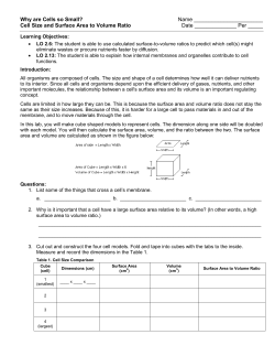
Product Manual
Auto-ubiquitinylation Kit
Catalog #BML-UW0970-0001
For assessment of protein Ub E3 ligase activity
Product Manual
USE FOR RESEARCH PURPOSES ONLY
Unless otherwise specified expressly on the packaging, all
products sold hereunder are intended for and may be used for
research purposes only and may not be used for food, drug,
cosmetic or household use or for the diagnosis or treatment of
human beings. Purchase does not include any right or license
to use, develop or otherwise exploit these products
commercially. Any commercial use, development or exploitation
of these products or development using these products without
the express written authorization of Enzo Life Sciences, Inc. is
strictly prohibited. Buyer assumes all risk and liability for the
use and/or results obtained by the use of the products covered
by this invoice whether used singularly or in combination with
other products.
LIMITED WARRANTY; DISCLAIMER OF WARRANTIES
These products are offered under a limited warranty. The
products are guaranteed to meet all appropriate specifications
described in the package insert at the time of shipment. Enzo
Life Sciences’ sole obligation is to replace the product to the
extent of the purchasing price. All claims must be made to Enzo
Life Sciences, Inc., within five (5) days of receipt of order. THIS
WARRANTY IS EXPRESSLY IN LIEU OF ANY OTHER
WARRANTIES OR LIABILITIES, EXPRESS OR IMPLIED,
INCLUDING
WARRANTIES
OF
MERCHANTABILITY,
FITNESS
FOR
A
PARTICULAR
PURPOSE,
AND
NONINFRINGEMENT OF THE PATENT OR OTHER
INTELLECTUAL PROPERTY RIGHTS OF OTHERS, AND ALL
SUCH WARRANTIES (AND ANY OTHER WARRANTIES
IMPLIED BY LAW) ARE EXPRESSLY DISCLAIMED.
TRADEMARKS AND PATENTS
Several Enzo Life Sciences products and product applications
are covered by US and foreign patents and patents pending.
FOR RESEARCH USE ONLY.
NOT FOR USE IN DIAGNOSTIC PROCEDURES.
Product Manual
Table of Contents
Please read
entire booklet
before
proceeding with
the assay.
Background ............................................................................. 2
Kit Description ......................................................................... 3
Suggested Application ............................................................ 3
Kit Components ....................................................................... 4
Storage .................................................................................... 4
Carefully note
the handling
and storage
conditions of
each kit
component.
Other Materials Needed .......................................................... 5
Auto-Ubiquinylation Assay ...................................................... 5
Analysis by Western Blotting ................................................... 6
Example Procedure for Western Blotting ................................ 7
Example Results for Western Blotting ..................................... 9
Please contact
Enzo Life
Sciences
Technical
Support if
necessary.
References .............................................................................. 9
Contact Information ............................................................... 10
1
Product Manual
BACKGROUND
The covalent attachment of ubiquitin to proteins
(ubiquitinylation) plays a fundamental role in the regulation of
cellular function through biological events involving cell cycle,
differentiation, immune responses, DNA repair, chromatin
structure, and apoptosis1,2,3,4.
Ubiquitinylation is achieved through three enzymatic steps. In
an ATP-dependent process, the ubiquitin activating enzyme
(E1) catalyzes the formation of a reactive thioester bond with
ubiquitin, followed by its subsequent transfer to the active site
cysteine of a ubiquitin carrier protein (E2). The selectivity of the
ubiquitin cascade for a particular substrate protein relies on the
interaction between the E2 conjugating enzyme (of which a cell
contains relatively few) and a ubiquitin-protein ligase (E3), of
which over 600 have been identified to date2,5.
The E3s are a large, diverse group of proteins, characterized by
one of several defining motifs. These include a HECT
(homologous to E6-associated protein C-terminus), RING
(really interesting new gene) or U-box (a modified RING motif
without the full complement of Zn2+-binding ligands) domain.
Whereas HECT E3s have a direct role in catalysis during
ubiquitinylation, RING and U-box E3s facilitate protein
ubiquitinylation. These latter two E3 types act as adaptor-like
molecules. They bring an E2 and a substrate into sufficiently
close proximity to promote the substrate's ubiquitinylation.
Although many RING-type E3s, such as MDM2 and c-Cbl, can
apparently act alone, others are found as components of much
larger multi-protein complexes, such as the anaphasepromoting complex.
Taken together, these multifaceted properties and interactions
enable E3s to provide a powerful, and specific, mechanism for
protein clearance within all cells of eukaryotic organisms
utilising the ubiquitin-proteasome system. The importance of
E3s is highlighted by the number of normal cellular processes
they regulate, and the number of diseases associated with their
loss of function or inappropriate targeting.
E3 ligases also undergo auto-ubiquitinylation, through
modification of specific lysine residues within an individual
ligase, providing a mechanism thought to be responsible for the
regulation of the E3 enzyme itself5,6.
2
Product Manual
KIT DESCRIPTION
This E3 ligase auto-ubiquitinylation kit enables proteins to be
tested for ubiquitin E3 ligase activity through assessment of
their ability to undergo auto-ubiquitinylation. Utilizing the first
three steps in the ubiquitin cascade the kit facilitates
ubiquitinylation of known or putative E3 ligase enzymes
followed by Western blot analysis using the highly sensitive
reagents provided or using antibodies to the specific protein of
interest (user supplied). A high integrity ubiquitin E3 ligase
enzyme is also provided for use as a positive control.
The Kit provides sufficient material for approximately 10 autoubiquitinylation assays.
SUGGESTED APPLICATION
1. Qualitative assessment of an Ub E3 ligase enzyme’s
activity through its ability to auto-ubiquitinylate.
2. Testing of proteins for auto-ubiquitinylation activity
allowing their identification as putative ubiquitin E3
ligases.
3. Ubiquitinylation of substrate proteins (user provided)
specific to a particular ubiquitin E3 ligase.
Note: Protocol provided covers applications 1-2. Assay set-up
can be readily modified for alternative applications by inclusion,
omission or substitution of specific components.
3
Product Manual
KIT COMPONENTS
1. Ubiquitin Activating Enzyme Solution, E1 (20X):
20X Ub E1
Ubiquitin E1 (BML-UW9410-0025).
Use 2.5μL per 50μL reaction.
25μL provided, sufficient for 10 x 50μL reactions.
2. UbcH5a (h)(rec.) (His) 20X:
20X E2
UbcH5a (BML-UW9050-0025).
Use 2.5μL per 50μL reaction.
25μL provided, sufficient for 10 x 50μL reactions.
3. Ubiquitin (10X):
10X Ubiquitin
Ubiquitin (BML-UW8795-0050).
Use 5μL per 50μL reaction.
50μL provided, sufficient for 10 × 50μL reactions.
4. Hdm2 (h) (rec.) (catalytic RING domain) (GST-tag):
20X E3 Control
Hdm2 RING domain (BML-UW0200-9090).
Use 2.5μL per 50μL reaction.
25μL provided, sufficient for 10 × 50μL reactions.
5. Mg2+-ATP Solution (20X):
20X Mg-ATP Solution
Mg-ATP (BML-EW9805-0025).
Use 2.5μL per 50μL reaction.
25μL provided, sufficient for 10 x 50μL reactions.
Note: Ensure Mg-ATP is fully dissolved by warming to
room temperature and mixing by vortex prior to use.
6. Ub E3 Ligase Buffer (10X):
10X Ub E3 Ligase Buffer
Use 5μL per 50μL reaction (BML-KW0965-0050)
50μL provided, sufficient for 10 x 50μL reactions
7. Ubiquitin-protein Conjugates, pAb:
Ubiquitin Antibody Solution
Ubiquitin, rabbit polyclonal antibody (BML-UG9511-0025)
25μL provided. Dilution of at least 1:500-1:1000
recommended for Western blotting.
STORAGE
All kit components should be stored at –80°C to ensure stability
and activity. Components with storage temperatures other than
4
Product Manual
-80°C can be stored at the temperature listed on the label OR
at -80°C. Avoid multiple freeze/thawing.
OTHER MATERIALS NEEDED
1. Eppendorf tubes (0.5mL)
2. 2x SDS-PAGE gel loading buffer (e.g. 0.25M Tris-Cl, pH
6.8, 4% SDS, 10% glycerol, 2% β-mercaptoethanol,
0.01% bromophenol blue).
3. DTT (Dithiothreitol) solution (50mM in dH20)
AUTO-UBIQUINYLATION ASSAY
The protocol set out in this section describes the running of
reactions to assess the auto-ubiquitinylation activity of the
control Ub E3 ligase enzyme provided and user supplied
proteins for subsequent analysis by Western blotting.
Hdm2 RING domain (UW0200) is provided as a control
ubiquitin E3 ligase for use in auto-ubiquitinylation assays.
Assay protocol
Note: recommended total reaction volume = 50 μL. *Adjust
dH2O volume in accordance with available Ub E3 ligase protein
concentration. A final assay concentration of 150-300nM is
recommended as a starting point for Ub E3 ligase autoubiquitinylation (e.g. use 2.5μL of 6μM Ub E3 ligase protein
solution).
Component
dH2O
10x Ub E3
ligase buffer
20x Ub E1
20x E2
10x Ubiquitin
20x E3
control (6μM)
*Sample E3
protein
50mM DTT
20x Mg-ATP
Sample
E3-Ub
Sample E3
(–ve control)
Hdm2-Ub
(+ve control)
Volume/μL
29.0
Hdm2
(–ve control)
31.5*
34.0*
5.0
5.0
5.0
5.0
2.5
2.5
5.0
2.5
2.5
5.0
2.5
2.5
5.0
2.5
2.5
5.0
-
-
2.5
2.5
X
X
-
-
1.0
2.5
1.0
-
1.0
2.5
1.0
-
31.5
Negative control reactions omitting Mg-ATP cofactors
demonstrate formation of auto-ubiquitinylated proteins is ATP
dependent (required for E1 activation) and hence derived from
the ubiquitin cascade.
5
Product Manual
Set-up assays/controls required as follows:
1. Add assay components to 0.5mL Eppendorf tube(s) in
order shown in table above. Keep all enzymes on ice
throughout.
2. Mix tube contents gently.
3. Incubate at 37°C for 60 minutes.
4. Quench assays by addition of 50μL 2x SDS-PAGE gel
loading buffer followed by heating to 95°C for 5 minutes.
Note: This step removes all Ub thioester linked species
(Ub-E1/Ub-E2) so only isopeptide linked Ub-E3 species
are detected using ubiquitin antibody/Western blotting.
5. Proceed directly to “Analysis by Western blotting” or store
at –20°C until ready.
ANALYSIS BY WESTERN BLOTTING
Summary of analysis steps
1. Separate proteins by SDS-PAGE.
2. Western transfer to PVDF membrane.
Note: Western blotting conditions appropriate for the transfer of
large proteins may be required to ensure good transfer of
Ubiquitinylated-E3 protein to PVDF membrane. For example,
use BSN transfer buffer 48mM Tris, pH9.2, 39mM glycine with
10% MeOH and 0.0375% SDS.
3. Block membrane with BSA/PBS-T solution.
4. Probe blot with either:
a) ubiquitin antibody supplied or
b) appropriate target protein specific primary antibody
in conjunction with suitable secondary antibodies.
5. Develop with western blotting detection reagents.
Note: Do NOT use milk in blocking/antibody binding solutions.
Please use 1% BSA in PBS or TBS Tween instead.
6
Product Manual
Materials Needed
1. SDS-PAGE gels - User prepared (10% standard / 4-15%
linear gradient)
2. Pre-stained SDS-PAGE molecular weight markers (e.g.
See Blue Plus 2)
3. PVDF membrane (e.g. Immobilon-P)
4. Anti-rabbit IgG secondary antibody (HRP linked) (e.g.
Goat Anti-Rabbit IgG-peroxidase antibody, Sigma,
A0545).
5. (If required) Target protein specific primary antibody (user
supplied) and appropriate secondary antibody-HRP
conjugate.
6. Western blotting detection reagents (e.g. ECL Reagent).
7. PBS solution 1X PBS.
8. PBS-T solution 1X PBS containing 0.2% Tween 20.
9. BSA/PBS-T blocking solution PBS-T containing 1%
bovine serum albumin (BSA).
Note: TBS-T can be used as an alternative to PBS-T if
required.
EXAMPLE PROCEDURE FOR WESTERN BLOTTING
1.
Apply 20μL of each quenched reaction to the SDSPAGE gel alongside selected molecular weight markers,
electrophorese, and transfer protein to PVDF membrane
according to standard procedures.
2.
Remove membrane from the transfer unit and block with
BSA/PBS-T blocking buffer for 1 hour at room
temperature on a rotor mixer.
Note: Drying PVDF membrane prior to blocking, as per
Manufacturers’ instructions, may considerably enhance
results.
3.
Wash membrane for 3 x 10mins with PBS-T on a
rocking platform at room temperature.
Ubiquitin-conjugate detection
4.
Dilute supplied ubiquitin antibody 1:500 or 1:1000 in
BSA/PBS-T.
5.
Incubate membrane with ubiquitin antibody solution
overnight at 4°C on a rotor mixer.
6.
Wash membrane for 3 x 10mins with PBS-T on a
rocking platform.
7
Product Manual
7.
Dilute appropriate anti-rabbit IgG secondary antibody
(HRP-linked)
according
to
the
manufacturer’s
instructions (e.g. 1:5000 in BSA/PBS-T).
8.
Incubate membrane with secondary antibody solution for
1 hour at room temperature on a rocking platform, or as
specified by the manufacturer.
9.
Wash membrane for 6 x 10mins with PBS-T on a
rocking platform.
10. Proceed to step 17.
Specific target protein detection (if required)
11. Dilute appropriate target protein specific primary
antibody according to manufacturer’s instructions.
12. Incubate membrane with target protein specific primary
antibody solution overnight at 4°C on a rotor mixer.
13. Wash membrane for 3 x 10mins with PBS-T on a
rocking platform.
14. Dilute appropriate secondary antibody according to the
manufacturer’s instructions (e.g. 1:5000 in BSA/PBS-T).
15. Incubate membrane with secondary antibody solution for
1 hour at room temperature on a rocking platform, or as
specified by the manufacturer.
16. Wash membrane for 6 x 10mins with PBS-T on a
rocking platform.
Analysis
17. Prepare western blotting detection reagent according to
the manufacturer’s instructions.
18. Incubate membrane
appropriate time.
with
detection
reagent
for
19. Detect emitted signal by luminography or CCD imaging
instrument.
8
Product Manual
EXAMPLE RESULTS FOR WESTERN BLOTTING
Figure: Western blot analysis of control Ub E3 ligase Hdm2 RING domain
auto-ubiquitinylation assays. Auto-ubiquitinylation assays set-up and run
as described in “Assay protocol”. Ubiquitinylated E3 ligase species were
detected by Western blotting as described in “Analysis by western
blotting”, using the provided ubiquitin antibody (UG9511) at a dilution of
1:1000 dilution.
Results demonstrate auto-ubiquitinylation of the control Hdm2 RING
domain ligase under the given assay conditions.
REFERENCES
1. Haas, A.L. and Siepmann, T. J. Pathways of ubiquitin
conjugation. FASEB J. 11, 1257-1268 (1997)
2. Hershko, A. and Ciechanover, A. The ubiquitin system.
Annu.Rev.Biochem. 67, 425-479 (1998)
3. Pickart, C.M. Mechanisms underlying ubiquitination.
Annu.Rev.Biochem. 70, 503-533 (2001)
4. Strous, G.J. and Govers, R. The ubiquitin-proteasome
system and endocytosis. J.Cell Sci. 112 ( Pt 10), 14171423 (1999)
5. Amemiya, Y., Azmi, P., and Seth, A. Autoubiquitination of
BCA2 RING E3 ligase regulates its own stability and
affects cell migration. Mol.Cancer Res. 6, 1385-1396
(2008)
6. Wu-Baer, F., Ludwig, T., and Baer, R. The UBXN1 protein
associates with autoubiquitinated forms of the BRCA1
tumor suppressor and inhibits its enzymatic function.
Mol.Cell Biol. (2010)
9
Product Manual
Global Headquarters
Enzo Life Sciences Inc.
10 Executive Blvd
Farmingdale, NY 11735
(p) 1-800-942-0430
(f) 1-631-694-7501
(e) [email protected]
Enzo Life Sciences (ELS) AG
Industriestrasse 17, Postfach
CH-4415 Lause / Switzerland
(p) +41/0 61 926 89 89
(f) +41/0 61 926 89 79
(e) [email protected]
For local distributors and detailed product information visit us online:
www.enzolifesciences.com
Catalog Number: BML-UW0970
Copyright 2012
Rev. June , 2014
10


© Copyright 2026