
TransAM Nrf2
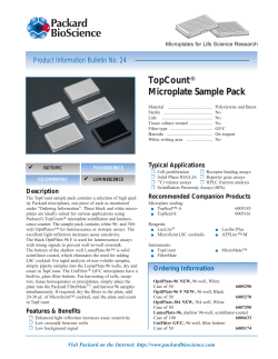
TransAM® Nrf2
(version A3)
Catalog Nos. 50296 & 50796
Active Motif North America
1914 Palomar Oaks Way, Suite 150
Carlsbad, California 92008, USA
Toll free:
877 222 9543
Telephone:
760 431 1263
Fax:
760 431 1351
Active Motif Europe
Avenue Reine Astrid, 92
B-1310 La Hulpe, Belgium
UK Free Phone:
France Free Phone:
Germany Free Phone:
Telephone:
Fax:
0800 169 31 47
0800 90 99 79
0800 181 99 10
+32 (0)2 653 0001
+32 (0)2 653 0050
Active Motif Japan
Azuma Bldg, 7th Floor
2-21 Ageba-Cho, Shinjuku-Ku
Tokyo, 162-0824, Japan
Telephone:
+81 3 5225 3638
Fax:
+81 3 5261 8733
Copyright 2014 Active Motif, Inc.
www.activemotif.com
Information in this manual is subject to change without notice and does not constitute a commitment on the part of Active Motif, Inc. It is supplied on an “as is” basis without any warranty of any
kind, either explicit or implied. Information may be changed or updated in this manual at any time.
This documentation may not be copied, transferred, reproduced, disclosed, or duplicated, in
whole or in part, without the prior written consent of Active Motif, Inc. This documentation is
proprietary information and protected by the copyright laws of the United States and international treaties.
The manufacturer of this documentation is Active Motif, Inc.
© 2014 Active Motif, Inc., 1914 Palomar Oaks Way, Suite 150; Carlsbad, CA 92008. All rights reserved.
All trademarks, trade names, service marks or logos referenced herein belong to their respective
companies.
www.activemotif.com
TABLE OF CONTENTS
Page
Overview . . . . . . . . . . . . . . . . . . . . . . . . . . . . . . . . . . . . . . . . . . . . . . . . . . . . . . . . . . . . . . . . . . . . . . . . . . . . 1
Flow Chart of Process . . . . . . . . . . . . . . . . . . . . . . . . . . . . . . . . . . . . . . . . . . . . . . . . . . . . . . . . . . . . . . . . 2
Introduction
Nrf2 Transcription Factor . . . . . . . . . . . . . . . . . . . . . . . . . . . . . . . . . . . . . . . . . . . . . . . . . . . . . . . . 3
Transcription Factor Assays . . . . . . . . . . . . . . . . . . . . . . . . . . . . . . . . . . . . . . . . . . . . . . . . . . . . . . . 4
TransAM Nrf2 . . . . . . . . . . . . . . . . . . . . . . . . . . . . . . . . . . . . . . . . . . . . . . . . . . . . . . . . . . . . . . . . . . . 4
Kit Performance and Benefits . . . . . . . . . . . . . . . . . . . . . . . . . . . . . . . . . . . . . . . . . . . . . . . . . . . . . . 5
Kit Components and Storage
Additional Materials Required . . . . . . . . . . . . . . . . . . . . . . . . . . . . . . . . . . . . . . . . . . . . . . . . . . . . 6
Protocols
Buffer Preparation and Recommendations . . . . . . . . . . . . . . . . . . . . . . . . . . . . . . . . . . . . . . . . . 7
Quick Chart for Preparing Buffers . . . . . . . . . . . . . . . . . . . . . . . . . . . . . . . . . . . . . . . . . . . . . . . . . 9
Nrf2 Transcription Factor Assay . . . . . . . . . . . . . . . . . . . . . . . . . . . . . . . . . . . . . . . . . . . . . . . . . . . 9
References . . . . . . . . . . . . . . . . . . . . . . . . . . . . . . . . . . . . . . . . . . . . . . . . . . . . . . . . . . . . . . . . . . . . . . . . . . 11
Appendix
Section A. Preparation of Nuclear Extract . . . . . . . . . . . . . . . . . . . . . . . . . . . . . . . . . . . . . . . . . 11
Section B. Troubleshooting Guide . . . . . . . . . . . . . . . . . . . . . . . . . . . . . . . . . . . . . . . . . . . . . . . . 13
Section C. Related Products . . . . . . . . . . . . . . . . . . . . . . . . . . . . . . . . . . . . . . . . . . . . . . . . . . . . . 14
Technical Services . . . . . . . . . . . . . . . . . . . . . . . . . . . . . . . . . . . . . . . . . . . . . . . . . . . . . . . . . . . . . . . . . . 16
www.activemotif.com
Overview
NF-E2-related factor, Nrf2, is a critical transcription factor in oxidative stress signaling. Nrf2 is a
basic leucine zipper transcription factor that binds to the antioxidant responsive element (ARE)
and may serve as a master regulator in cellular defense pathways in protecting a wide variety of
tissues from various toxic exposure.
TransAM® Nrf2 is a simple solution for studying Nrf2 activation in human, mouse and rat model
systems. The Kit is a 96-well plate based assay measuring DNA binding activity of Nrf2 using
colorimetric detection on a standard plate reader. It works with nuclear extracts from tissue or cell
samples and is able to detect activated Nrf2 in as little as 0.15 micrograms of nuclear extract.
With its patented TransAM® method*, Active Motif introduced the first ELISA-based kits to detect
and quantify transcription factor activation. TransAM Kits combine a fast, user-friendly format
with a sensitive, specific assay. TransAM Nrf2 Kits are designed specifically to detect and quantify
Nrf2 activation. Kits contain a 96-well plate to which oligonucleotide containing an ARE has been
immobilized. Nrf2 contained in nuclear extract then binds specifically to this oligonucleotide and
is detected through use of an antibody directed against Nrf2. Addition of a secondary antibody
conjugated to horseradish peroxidase (HRP) provides a sensitive colorimetric readout that is easily
quantified by spectrophotometry. The 96-well plate with individual strips of 8 wells is suitable for
manual use or for high-throughput screening applications.
product
format
catalog no.
TransAM® Nrf2
1 x 96 rxns
50296
TransAM® Nrf2
5 x 96 rxns
50796
* Technology covered by EAT-filed patents and licensed to Active Motif.
www.activemotif.com
1
Flow Chart of Process
2
1
7
6
5
4
3
8
9
10
11
12
A
B
C
D
E
F
Cell extract
containing activated
transcription factor
G
H
Oligonucleotide
coated plate
Add cell
extract
21
11
01
9
8
7
6
5
4
3
2
1
A
B
C
D
E
F
G
H
Add primary
antibody
1 hr .
1
9
8
7
6
5
4
3
2
11
10
12
A
B
C
D
E
F
G
H
Add anti-IgG
HRP conjugate
21
11
01
9
8
7
6
5
1 hr .
4
3
2
1
A
B
C
D
E
F
G
Add developing
and stop solution
H
1 hr .
1
5
4
3
2
7
6
A
B
C
D
E
F
G
H
www.activemotif.com
2
8
9
10
11
12
Introduction
Nrf2 Transcription Factor
Nrf2 (NF-E2 related factor, NFE2L2, from nuclear factor erythroid-derived 2-like 2) is a basic
leucine zipper (bZIP) transcription factor. Nrf2 binds to the antioxidant response element (ARE)
and positively regulates the expression of detoxifying enzyme genes (such as NAD(P)H:quinone
oxidoreductase1, NQO1) in response to antioxidants and xenobiotics. Higher levels of NQO1 gene
expression has been shown in liver, lung, colon, and breast tumors1.
A cytosolic inhibitor of Nrf2, Keap1/INrf2, retains Nrf2 in the cytoplasm under normal conditions
where the interaction of Nrf2 with INrf2 targets Nrf2 for ubiquitination and proteasomal degradation. However, after oxidative stress, Nrf2 is released from INrf2, translocates to the nucleus, and
results in the activation of ARE-mediated gene expression. Nrf2 is also synthesized de novo after
exposure to stress. In addition, Nrf2 controls its own degradation by regulating expression and
induction of INrf22. It has been shown that nuclear export and degradation pathways are activated
by around two hours after treatment with tert-butylhydroquinone (t-BHQ)3.
Nrf2 activation and degradation are important sensing mechanisms in the cellular response for
oxidative and electrophilic stressors4. The TransAM Nrf2 Kit provides a simple solution for studying
Nrf2 activation levels in tissue or cell extracts.
Transcription Factor Assays
To date, three methods are widely used to measure Nrf2 activation, either directly or indirectly:
1.
Nrf2 activation can be determined by Western blot by using antibodies specific for Nrf2 proteins. This method is time consuming (up to 2 days once the nuclear extracts are prepared),
and is not suitable for processing large numbers of samples.
2.
The DNA-binding capacity of Nrf2 can be assayed by gel retardation, also called electrophoretic mobility shift assay (EMSA). In this method, nuclear extracts are incubated with a radioactive double-stranded oligonucleotide probe containing the consensus sequence for Nrf2
binding. If Nrf2 is active in the nuclear extract, it will bind to the probe. Samples are then
resolved by electrophoresis on a native polyacrylamide gel, followed by autoradiography.
This method is sensitive, but like the previous procedure, it is time consuming (multiple days
of gel exposure may be required to achieve sufficient sensitivity) and it cannot be applied to
high-throughput screening. Gelshift assays also require special precautions and equipment
for handling radioactivity.
3.
Another method used to assay Nrf2 activation is based on reporter genes, typically luciferase
or β-galactosidase, placed under the control of a promoter containing the ARE consensus
sequence. However, the procedure is limited by the following issues: (i) reporter gene assays
have to be repeated several times to obtain statistically reliable data; and (ii) reporter gene
assays are sensitive to confounding factors that may influence the expression level of the
reporter gene. Therefore, assays have to be carefully standardized. This method is sensitive
and easy to perform with a large number of samples but requires efficient cell transfection
with the reporter plasmid.
www.activemotif.com
3
TransAM Nrf2
The TransAM Nrf2 Kit combines a fast and user-friendly ELISA format with a sensitive and specific
assay for transcription factors. TransAM Nrf2 Kits contain a 96-well plate on which has been
immobilized oligonucleotide containing the ARE consensus binding site (5´-GTCACAGTGACTCAGCAGAATCTG-3´). The active form of Nrf2 contained in nuclear extract specifically binds to this
oligonucleotide. The primary antibody used to detect Nrf2 recognizes an epitope on Nrf2 protein
upon DNA binding. Addition of an HRP-conjugated secondary antibody provides a sensitive colorimetric readout easily quantified by spectrophotometry. Once the nuclear extracts are prepared,
this assay is completed in less than 3.5 hours. As this assay is performed in 96-well plates, a large
number of samples can be handled simultaneously, enabling high-throughput automation. This
assay is specific for Nrf2 activation and has been shown to be 5-fold more sensitive and 20-fold
faster than the gel-retardation technique. With the 3.5-hour procedure of TransAM, we could
detect Nrf2 activation using as little as 0.6 µg of nuclear extract.
www.activemotif.com
4
Kit Performance and Benefits
Detection limit: > 0.6 µg nuclear extract/well.
Range of detection: TransAM provides quantitative results from 0.6 to 10 µg of nuclear extract per
well.
Cross-reactivity: TransAM Nrf2 specifically detects Nrf2 from human, mouse and rat origin.
Assay time: 3.5 hours.
Nrf2 activation (OD450 nm)
1
0.75
Unstimulated
D,L Sulforaphane Stimulated
0.5
0.25
0
0.63
1.25
2.5
5
10
Extract/well (µg)
Monitoring Nrf2 activation with the TransAM Nrf2 Kit:
0.625 to 10 µg of D,L Sulforaphane treated HepG2 nuclear extract (purple bars) and untreated HepG2 nuclear extract (copper bars) were assayed per well. Data shown are the results from wells assayed in duplicate.
www.activemotif.com
5
Kit Components and Storage
TransAM Nrf2 Kits are for research use only. Not for use in diagnostic procedures. Except for the
positive control extract that must be kept at -80ºC, kit components can be stored at -20ºC prior
to first use. Then, we recommend storing each component at the temperature indicated in the
table below. Store the Nrf2 antibody at 4°C after it has been thawed for use. All components are
guaranteed stable for 6 months from date of receipt when stored properly.
Reagents
Quantity
1 plate / 5 plates
Storage
Nrf2 antibody
10 µl / 25 µl
4°C
anti-rabbit HRP-conjugated antibody
10 µl / 50 µl
4°C
Wild-type oligonucleotide AM29
100 µl / 500 µl
(10 pmol/µl)
-20°C
Mutated oligonucleotide AM29
100 µl / 500 µl
(10 pmol/µl)
-20°C
Positive control extract
20 µl / 50 µl
(2.5 µg/µl)
-80°C
Dithiothreitol (DTT) (1 M)
100 µl / 500 µl
-20°C
Protease Inhibitor Cocktail 100 µl / 500 µl
-20°C
Herring Sperm DNA
100 µl / 500 µl
(1 µg/µl)
-20°C
Lysis Buffer AM1
10 ml / 50 ml
4°C
Binding Buffer AM1
10 ml / 50 ml
4°C
10X Wash Buffer AM2
22 ml / 110 ml
4°C
10X Antibody Binding Buffer AM3
2.2 ml / 11 ml
4°C
Developing Solution
11 ml / 55 ml
4°C
Stop Solution
11 ml / 55 ml
4°C
96-well Nrf2 assay plate
1 / 5
Plate sealer
1/5
Additional materials required
• Multi-channel pipettor
• Multi-channel pipettor reservoirs
• Rocking platform
• Microplate spectrophotometer capable of reading at 450 nm (655 nm as optional reference
wavelength)
www.activemotif.com
6
Protocols
Buffer Preparation and Recommendations
Preparation of Complete Lysis Buffer
We provide an excess of Lysis Buffer AM1 in order to perform the assay AND to prepare customized nuclear extracts. Please refer to the Appendix Section A for a protocol to prepare a nuclear
extract. Our Nuclear Extract Kit can also be purchased separately (Cat. Nos. 40010 & 40410). Lysis
Buffer AM1 contains phosphatase inhibitors to prevent dephosphorylation of transcription factors
during the extract preparation and the assay. The presence of these inhibitors gives a yellow
coloration to Lysis Buffer AM1. Prepare the amount of Complete Lysis Buffer required for the
assay by adding 1 µl of 1 M DTT and 10 µl Protease Inhibitor Cocktail per ml of Lysis Buffer AM1 (see
the Quick Chart for Preparing Buffers in this section). Some of the protease inhibitors lose their
activity after 24 hours once diluted. Therefore, we recommend using the Complete Lysis Buffer
immediately for cell lysis. The remaining amount should be discarded if not used in the same day.
Preparation of Complete Binding Buffer
Prepare the amount of Complete Binding Buffer required for the assay by adding 1 µl of 1 M DTT
and 10 µl of 1 µg/µl Herring Sperm DNA per ml of Binding Buffer AM1 (see the Quick Chart for
Preparing Buffers in this section). After use, discard remaining Complete Binding Buffer.
Preparation of 1X Wash Buffer
Prepare the amount of 1X Wash Buffer required for the assay as follows: For every 100 ml of 1X
Wash Buffer required, dilute 10 ml 10X Wash Buffer AM2 with 90 ml distilled water (see the Quick
Chart for Preparing Buffers in this section). Mix gently to avoid foaming. The 1X Wash Buffer may
be stored at 4°C for one week. The Tween 20 contained in the 10X Wash Buffer AM2 may form
clumps, therefore homogenize the buffer by incubating at 50ºC for 2 minutes and mixing prior to
use.
Preparation of 1X Antibody Binding Buffer
Prepare the amount of 1X Antibody Binding Buffer required for the assay as follows: For every
10 ml of 1X Antibody Binding Buffer required, dilute 1 ml 10X Antibody Binding Buffer AM3 with 9
ml distilled water (see the Quick Chart for Preparing Buffers in this section)*. Mix gently to avoid
foaming. Discard remaining 1X Antibody Binding Buffer after use. The BSA contained in the 10X
Antibody Binding Buffer AM3 may form clumps, therefore homogenize the buffer by warming to
room temperature and vortexing for 1 minute prior to use. Dilute the Nrf2 and HRP-conjugated
secondary antibodies with the 1X Antibody Binding Buffer to 1:1000. Depending on the particular
assay, the signal:noise ratio may be optimized by using higher dilutions of both antibodies. This
may decrease the sensitivity of the assay.
* Volumes listed refer to the preparation of buffer for diluting both the primary & secondary
antibodies.
www.activemotif.com
7
Developing Solution
The Developing Solution should be warmed to room temperature before use. The Developing
Solution is light sensitive, therefore, we recommend avoiding direct exposure to intense light
during storage. The Developing Solution may develop a yellow hue over time. This does not affect
product performance. A blue color present in the Developing Solution indicates that it has been
contaminated and must be discarded. Prior to use, place the Developing Solution at room temperature for at least 1 hour. Transfer the amount of Developing Solution required for the assay into
a secondary container before aliquoting into the wells (see the Quick Chart for Preparing Buffers
in this section). After use, discard remaining Developing Solution.
Stop Solution
Prior to use, transfer the amount of Stop Solution required for the assay into a secondary container (see the Quick Chart for Preparing Buffers in this section). After use, discard remaining Stop
Solution.
WARNING: The Stop Solution is corrosive. Wear personal protective equipment when
handling, i.e. safety glasses, gloves and labcoat.
Positive control extract (2.5 µg/µl)
The positive control nuclear extract is provided as a control for Nrf2 activation. Sufficient extract
is supplied for 20 reactions if using 5 µg per well. This extract is optimized to give a strong signal
when used at 0.6 to 10 µg/well. We recommend aliquoting the extract in 5 µl fractions and storing
at -80ºC. Avoid multiple freeze/thaw cycles of the extract.
Wild-type and mutated consensus oligonucleotides
The wild-type consensus oligonucleotide is provided as a competitor for Nrf2 binding in order to
monitor the specificity of the assay. Used at 20 pmol/well, the oligonucleotide will prevent Nrf2
binding to the probe immobilized on the plate. Conversely, the mutated consensus oligonucleotide should have no inhibitory effect on Nrf2 binding. Prepare the required amount of wild-type
and/or mutated consensus oligonucleotide by adding 2 µl of appropriate oligonucleotide to 43 µl
of Complete Binding Buffer per well being used (see the Quick Chart for Preparing Buffers in this
section). To allow for optimum competition, add the oligonucleotide to the well prior to addition
of the nuclear extract.
www.activemotif.com
8
Quick Chart for Preparing Buffers
Reagents to prepare
Components
1 well 1 strip 6 strips 12 strips
(8 wells) (48 wells) (96 wells)
Complete Lysis Buffer
DTT
Protease Inhibitor Cocktail
Lysis Buffer
Total Required
0.01 µl
0.12 µl
11.12 µl
11.25 µl
Complete Binding Buffer
DTT
Herring Sperm DNA
Binding Buffer
Total Required
0.04 µl 0.3 µl
0.45 µl 3.6 µl
44.5 µl 356.1 µl
45 µl 360 µl
Binding Buffer with
wt or mut oligont
Nrf2 wt or mut oligont Complete Binding Buffer
Total Required
0.1 µl 0.6 µl 1.2 µl
0.9 µl 5.4 µl 10.8 µl
89.0 µl 534.0 µl 1.07 ml
90.0 µl 540.0 µl 1.08 ml
2.16 µl
21.6 µl
2.14 ml
2.16 ml
4.3 µl
43.2 µl
4.27 ml
4.32 ml
2.0 µl 18.0 µl 108 µl
N/A
43.0 µl 342.0 µl 2.052 ml N/A
45.0 µl 360.0 µl 2.16 ml N/A
1X Washing Buffer
Distilled Water
10X Washing Buffer
Total Required
2.025 ml 16.2 ml 97.2 ml 194.4 ml
225.0 µl
1.8 ml 10.8 ml 21.6 ml
2.25 ml 18.0 ml 108.0 ml 216.0 ml
1X Antibody
Binding Buffer*
Distilled Water
202.5 µl
1.62 ml 9.72 ml 19.44 ml
10X Antibody Binding Buffer 22.5 µl 180.0 µl 1.08 ml 2.16 ml
Total Required
225.0 µl 1.8 ml 10.8 ml 21.6 ml
Developing Solution
Total Required
112.5 µl 900.0 µl
5.4 ml 10.8 ml
Stop Solution
Total Required
112.5 µl 900.0 µl
5.4 ml 10.8 ml
* Volumes listed refer to the preparation of buffer for diluting both the primary & secondary
antibodies.
Nrf2 Transcription Factor Assay
Determine the appropriate number of microwell strips required for testing samples, controls and
blanks in duplicate. If less than 8 wells in a strip need to be used, cover the unused wells with a
portion of the plate sealer while you perform the assay. The content of these wells is stable at
room temperature if kept dry and, therefore, can be used later for a separate assay. Store the
unused strips in the aluminum pouch at 4°C. Use the strip holder for the assay.
Prepare the Complete Lysis Buffer, Complete Binding Buffer, 1X Wash Buffer and 1X Antibody
Binding Buffer as described above in the section Buffer Preparation and Recommendations. Multichannel pipettor reservoirs may be used for dispensing the Complete Binding Buffer, Wash Buffer,
Antibody Binding Buffer, Developing Solution and Stop Solution into the wells being used.
Step 1: Binding of Nrf2 to its consensus sequence
1. Add 40 µl Complete Binding Buffer to each well to be used. If you wish to perform competitive binding experiments, add 40 µl Complete Binding Buffer that contains 20 pmol (2 µl) of
www.activemotif.com
9
the wild-type or mutated oligonucleotide (see the Buffer Preparation section above for a
description of competitive binding).
2.
Sample wells: Add 10 µl of sample diluted in Complete Lysis Buffer per well. We recommend using 5-20 µg of nuclear extract diluted in Complete Lysis Buffer per well. A protocol
for preparing nuclear extracts is provided on page 11.
Positive control wells: Add 5 µg of the provided positive control extract diluted in 10 µl of
Complete Lysis Buffer per well (2 µl of control extract in 8 µl of Complete Lysis Buffer per well).
Blank wells: Add 10 µl Complete Lysis Buffer only per well.
3.
Use the provided adhesive cover to seal the plate. Incubate for 1 hour at room temperature
with mild agitation (100 rpm on a rocking platform).
4.
Wash each well 3 times with 200 µl 1X Wash Buffer. For each wash, flick the plate over a sink
to empty the wells, then tap the inverted plate 3 times on absorbent paper towels.
Step 2: Binding of primary antibody
1.
Add 100 µl diluted Nrf2 antibody (1:1000 dilution in 1X Antibody Binding Buffer) to each well
being used.
2.
Cover the plate and incubate for 1 hour at room temperature without agitation.
3.
Wash the wells 3 times with 200 µl 1X Wash Buffer (as described in Step 1, No. 4).
Step 3: Binding of secondary antibody
1.
Add 100 µl of diluted HRP-conjugated antibody (1:1000 dilution in 1X Antibody Binding Buffer)
to all wells being used.
2.
Cover the plate and incubate for 1 hour at room temperature without agitation.
3.
During this incubation, place the Developing Solution at room temperature.
4.
Wash the wells 4 times with 200 µl 1X Wash Buffer (as described in Step 1, No. 4).
Step 4: Colorimetric reaction
1.
Add 100 µl Developing Solution to all wells being used.
2.
Incubate 2-15 minutes at room temperature protected from direct light. Monitor the blue
color development in the sample and positive control wells until it turns medium to dark
blue. Do not overdevelop.
3.
Add 100 µl Stop Solution. In presence of the acid, the blue color turns yellow.
4.
Read absorbance on a spectrophotometer within 5 minutes at 450 nm with a reference
wavelength of 655 nm. Blank the plate reader according to the manufacturer’s instructions
using the blank wells.
www.activemotif.com
10
References
1. 2. 3. 4.
Pi, J., et al. (2007) Free Radic Biol Med 42(12): 1797-1806.
Lee, O-H., et al. (2007) J Biol Chem 282(50): 36412-36420.
Theodore, M., et al. (2008) J Biol Chem 283(14): 8984-8994
Kobayashi, A., et al. (2006) Mol Cell Biol 26(1): 221-229.
Appendix
Section A. Preparation of Nuclear Extract
For your convenience, Active Motif offers a Nuclear Extract Kit (Cat. Nos. 40010 & 40410). This kit
contains buffers optimized for use in TransAM Kits, which serves to reduce inconsistencies in the
assay that may arise from using homemade or other buffers. If you prefer to make your own buffers, please refer to the following protocol.
This procedure can be used for a confluent cell layer of 75 cm2 (100-mm dish). The yield is approximately 0.15 mg of nuclear proteins for 9 x 10⁶ cells.
1. Wash cells with 10 ml ice-cold PBS/PIB. Discard PBS/PIB.
2. Add 10 ml ice-cold PBS/PIB and scrape the cells off the dish with a cell lifter. Transfer cells
into a pre-chilled 15 ml tube and spin at 300 x g for 5 minutes at 4°C.
3. Resuspend the pellet in 1 ml ice-cold HB buffer by gentle pipetting and transfer the cells into
a pre-chilled 1.5 ml tube.
4. Allow the cells to swell on ice for 15 minutes.
5. Add 50 µl 10% Nonidet P-40 (0.5 % final) and vortex the tube vigorously for 10 seconds.
6. Centrifuge the homogenate for 30 seconds at 4°C in a microcentrifuge. Remove the supernatant (cytoplasmic fraction) and, if you wish to save this for other uses, transfer it into a
pre-chilled microcentrifuge tube. (Store the cytoplasmic fraction at –80°C.)
7. Resuspend the nuclear pellet in 50 µl Complete Lysis Buffer and rock the tube gently on ice
for 30 minutes on a shaking platform.
8. Centrifuge for 10 minutes at 14,000 x g at 4°C and save the supernatant (nuclear extract).
Aliquot and store at –80°C. Avoid freeze/thaw cycles.
9. Determine the protein concentration of the extract by using a Bradford-based assay.
www.activemotif.com
11
Preparation of Buffers for Nuclear Extract
10X PBS
For 250 ml, mix:
0.1 M phosphate buffer, pH 7.5
3.55 g Na2HPO4 + 0.61 g KH2PO4
1.5 M NaCl
21.9 g
27 mM KCl
0.5 g
Adjust to 250 ml with distilled water. Prepare a 1X PBS solution by adding 10 ml 10X PBS to 90 ml
distilled water. Sterilize the 1X PBS by filtering through a 0.2 µm filter. The 1X PBS is at pH 7.5. Store
the filter-sterilized 1X PBS solution at 4°C.
PIB (Phosphatase Inhibitor Buffer)
125 mM NaF
For 10 ml, mix:
52 mg
250 mM β-glycerophosphate
0.55 g
250 mM p-nitrophenyl phosphate (PNPP) 1.15 g
25 mM NaVO3
31 mg
Adjust to 10 ml with distilled water. Mix the chemicals by vortexing. Incubate the solution at 50ºC
for 5 minutes. Mix again. Store at –20°C.
PBS/PIB
Prior to use, add 0.5 ml PIB to 10 ml 1X PBS.
HB (Hypotonic Buffer)
For 50 ml, mix:
20 mM Hepes, pH 7.5
0.24 g
5 mM NaF
12 mg
10 µM Na2MoO4
5 µl of a 0.1 M solution
0.1 mM EDTA
10 µl of a 0.5 M solution
Adjust pH to 7.5 with 1 N NaOH. Adjust volume to 50 ml with distilled water. Sterilize by filtering
through a 0.2 µm filter. Store the filter-sterilized solution at 4°C.
www.activemotif.com
12
Section B: Troubleshooting Guide
Problem/question
Possible cause
Recommendation
No signal or weak signal
Omission of key
reagent
Check that all reagents have been added in all wells in the
correct order
Substrate or conjugate
is no longer active
Test conjugate and substrate for activity
Enzyme inhibitor
present
Sodium azide will inhibit the peroxidase reaction, follow our
recommendations to prepare buffers
Plate reader settings
not optimal
Verify the wavelength and filter settings in the plate reader
Incorrect assay temperature
Bring substrate to room temperature
Inadequate volume of
Developing Solution
Check to make sure that correct volume is delivered by pipette
Developing time too
long
Stop enzymatic reaction as soon as the positive wells turn
medium-dark blue
Concentration of antibodies is too high
Increase antibody dilutions
Inadequate washing
Ensure all wells are filled with Wash Buffer and follow washing
recommendations
Incomplete washing
of wells
Ensure all wells are filled with Wash Buffer and follow washing
recommendations
Well cross-contamination
Follow washing recommendations
High background in all
wells
Uneven color development
High background in sample Too much nuclear
wells
extract per well
No signal or weak signal in
sample wells
Decrease amount of nuclear extract down to 1-2 µg/well
Concentration of antibodies is too high
Perform antibody titration to determine optimal working
concentration. Start using 1:1000 for primary antibody and
1:1000 for the secondary antibody. The sensitivity of the assay
will be decreased
Not enough nuclear
extract per well
Increase amount of nuclear extract to 50 µg/well
Nrf2 is poorly activated or inactivated in
nuclear fractions
Perform a time course for Nrf2 activation in the studied cell
line
Nuclear extracts are
not from correct
species
www.activemotif.com
13
Section C. Related Products
Transcription Factor ELISAs
Format
Catalog No.
TransAM® AML-1/Runx1
1 x 96-well plate
47396
TransAM® AML-3/Runx2
1 x 96-well plate
44496
TransAM® AP-1 Family
2 x 96-well plates
44296
TransAM® AP-1 c-Fos
1 x 96-well plate
44096
TransAM® AP-1 c-Jun
1 x 96-well plate
46096
TransAM® AP-1 FosB
1 x 96-well plate
45096
TransAM® AP-1 JunD
1 x 96-well plate
43496
TransAM® ATF-2
1 x 96-well plate
42396
TransAM® c-Myc
1 x 96-well plate
43396
TransAM® C/EBP a/b
1 x 96-well plate
44196
TransAM® CREB
1 x 96-well plate
42096
TransAM® pCREB
1 x 96-well plate
43096
TransAM® Elk-1
1 x 96-well plate
44396
TransAM® ER
1 x 96-well plate
41396
TransAM® FKHR (FOXO1/4)
1 x 96-well plate
46396
TransAM® GATA Family
2 x 96-well plates
48296
TransAM® GATA-4
1 x 96-well plate
46496
TransAM® GR
1 x 96-well plate
45496
TransAM® HIF-1
1 x 96-well plate
47096
TransAM® HNF Family
2 x 96-well plates
46296
TransAM® HNF-1
1 x 96-well plate
46196
TransAM® IRF-3 (Human)
1 x 96-well plate
48396
TransAM® IRF-3 (Mouse)
1 x 96-well plate
48496
TransAM® IRF-7
1 x 96-well plate
50196
TransAM® MAPK Family
2 x 96-well plates
47296
TransAM® MEF2
1 x 96-well plate
43196
TransAM® MyoD
1 x 96-well plate
47196
TransAM® NF-YA
1 x 96-well plate
40396
TransAM® NFATc1
1 x 96-well plate
40296
TransAM® NFkB Family
2 x 96-well plates
43296
TransAM® Flexi NFkB Family
2 x 96-well plates
43298
TransAM® NFkB p50
1 x 96-well plate
41096
TransAM® NFkB p50 Chemi
1 x 96-well plate
41097
TransAM® Flexi NFkB p50
1 x 96-well plate
41098
TransAM® NFkB p52
1 x 96-well plate
48196
TransAM® NFkB p52 Chemi
1 x 96-well plate
48197
TransAM® NFkB p65
1 x 96-well plate
40096
TransAM® NFkB p65 Chemi
1 x 96-well plate
40097
TransAM® Flexi NFkB p65
1 x 96-well plate
40098
TransAM® Nrf2
1 x 96-well plate
50296
TransAM® Oct-4
1 x 96-well plate
42496
TransAM® p53
1 x 96-well plate
41196
TransAM® PPARg
1 x 96-well plate
40196
TransAM® Sp1
1 x 96-well plate
41296
TransAM® Sp1/Sp3
1 x 96-well plate
40496
TransAM® STAT Family
2 x 96-well plates
42296
TransAM® STAT3
1 x 96-well plate
45196
TransAM® T-bet
1 x 96-well plate
51396
For a complete, up-to-date list of available TransAM® Kits, please visit www.activemotif.com/transam
www.activemotif.com
14
Nuclear, Cytoplasmic and Whole-cell Extraction
Format
Catalog No.
Nuclear Extract Kit
100 rxns
400 rxns
40010
40410
Histone Acetyltransferase and Deacetylase Activity
Format
Catalog No.
HAT Assay Kit (Fluorescent)
Recombinant p300 protein, catalytic domain
Recombinant GCN5 protein, active
HDAC Assay Kit (Fluorescent)
HDAC Assay Kit (Colorimetric)
1 x 96 rxns
5 µg
5 µg
1 x 96 rxns
1 x 96 rxns
56100
31205
31204
56200
56210
Histone Purification & Chromatin Assembly
Format
Catalog No.
Histone Purification Kit
Histone Purification Mini Kit
Chromatin Assembly Kit
HeLa Core Histones
10 rxns
10 rxns
10 rxns
36 µg
40025
40026
53500
53501
DNA Methylation
Format
Catalog No.
hMeDIP
MeDIP
MethylDetector™
MethylCollector™
MethylCollector™ Ultra
UnMethylCollector™
DNMT Activity / Inhibition Assay
Methylated DNA Standard Kit
Fully Methylated Jurkat DNA
Jurkat genomic DNA
10 rxns
10 rxns
50 rxns
25 rxns
30 rxns
30 rxns
96 rxns
3 x 2.5 µg
10 µg
10 µg
55010
55009
55001
55002
55005
55004
55006
55008
55003
55007
Co-Immunoprecipitation
Format
Catalog No.
Universal Magnetic Co-IP Kit
Nuclear Complex Co-IP Kit
25 rxns
50 rxns
54002
54001
SUMOylation
Format
Catalog No.
SUMOlink™ SUMO-1 Kit
SUMOlink™ SUMO-2/3 Kit
20 rxns
20 rxns
40120
40220
Recombinant Proteins
Format
Catalog No.
Recombinant c-Fos protein
5 µg
31115
Recombinant c-Jun protein
5 µg
31116
Recombinant c-Myc protein
5 µg
31117
Recombinant NFkB p50 protein
5 µg
31101
Recombinant NFkB p65 protein
5 µg
31102
Recombinant p53 protein
5 µg
31103
Recombinant p300 protein
4 µg
31124
Purified Sp1 protein
2 µg
31137
Recombinant STAT3 protein
10 µg
31140
For an up-to-date list of over 200 recombinant proteins, please visit www.activemotif.com/proteins.
www.activemotif.com
15
Technical Services
If you need assistance at any time, please call Active Motif Technical Service at one of the
numbers listed below.
Active Motif North America
1914 Palomar Oaks Way, Suite 150
Carlsbad, CA 92008
USA
Toll Free:
877 222 9543
Telephone: 760 431 1263
Fax:
760 431 1351
E-mail:
[email protected]
Active Motif Europe
Avenue Reine Astrid, 92
B-1310 La Hulpe, Belgium
UK Free Phone:
0800 169 31 47
France Free Phone: 0800 90 99 79
Germany Free Phone: 0800 181 99 10
Telephone: +32 (0)2 653 0001
Fax: +32 (0)2 653 0050
E-mail: [email protected]
Active Motif Japan
Azuma Bldg, 7th Floor
2-21 Ageba-Cho, Shinjuku-Ku
Tokyo, 162-0824, Japan
Telephone: +81 3 5225 3638
Fax:
+81 3 5261 8733
E-mail:
[email protected]
Visit Active Motif on the worldwide web at http://www.activemotif.com
At this site:
• Read about who we are, where we are, and what we do
• Review data supporting our products and the latest updates
• Enter your name into our mailing list to receive our catalog, MotifVations newsletter and
notification of our upcoming products
• Share your ideas and results with us
• View our job opportunities
Don’t forget to bookmark our site for easy reference!
www.activemotif.com
16


© Copyright 2026