
PDF hosted at the Radboud Repository of the Radboud University Nijmegen
PDF hosted at the Radboud Repository of the Radboud University
Nijmegen
This full text is a publisher's version.
For additional information about this publication click this link.
http://hdl.handle.net/2066/125153
Please be advised that this information was generated on 2014-10-15 and may be subject to
change.
NEED, QUEST & EVIDENCE
resting-state oscillations, neurofeedback,
and working memory training in ADHD
Martine van Dongen-Boomsma
NEED, QUEST & EVIDENCE
resting-state oscillations, neurofeedback, and working memory training in ADHD
The research in this thesis was largely supported by BrainGain, a Dutch research
consortium, funded by Smartmix, an initiative of the Netherlands Organization for
Scientific Research (NWO) to support applied research.
Publication of this thesis was financially supported by the Radboud University Nijmegen
and Karakter, The Netherlands.
ISBN
978-94-6259-075-5
Cover design and illustrations
Studio Lakmoes, The Netherlands
Lay-out
Promotie In Zicht, The Netherlands
Print
Ipskamp Drukkers, The Netherlands
© Martine van Dongen-Boomsma, 2014
All rights reserved. No parts of this publication may be reproduced or transmitted in any form or by any means without
prior written permission of the author.
NEED, QUEST & EVIDENCE
resting-state oscillations, neurofeedback,
and working memory training in ADHD
proefschrift
ter verkrijging van de graad van doctor
aan de Radboud Universiteit Nijmegen
op gezag van de rector magnificus prof. mr. S.C.J.J. Kortmann,
volgens besluit van het college van decanen
in het openbaar te verdedigen op donderdag 27 maart 2014
om 10:30 uur precies
door
Martine van Dongen-Boomsma
geboren op 29 juni 1978
te IJsselstein
Promotor
Prof. dr. J.K. Buitelaar
Copromotor
Dr. D.I.E. Slaats-Willemse
Manuscriptcommissie
Prof. dr. E.L.J.M. van Luijtelaar
Prof. dr. I. Tendolkar
Prof. dr. P.J.M. Prins (Universiteit van Amsterdam)
Contents
CHAPTER I
GENERAL INTRODUCTION, AIMS & OUTLINE OF THE THESIS
7
RESTING-STATE OSCILLATIONS IN ADHD
CHAPTER II
Relation between resting EEG to cognitive performance and clinical symptoms
in adults with attention-deficit/hyperactivity disorder
39
CHAPTER III
How the alpha peak frequency helps to unravel the neurophysiological
underpinnings of behavioral functioning in children with attention-deficit/
hyperactivity disorder
59
NON-PHARMACOLOGICAL INTERVENTIONS IN CHILDREN WITH ADHD
Frequency neurofeedback
CHAPTER IV
A randomized placebo-controlled trial of electroencephalographic
(EEG-) neurofeedback in children with attention-deficit/hyperactivity disorder
79
CHAPTER V
Does EEG-neurofeedback improve neurocognitive functioning in children with
attention-deficit/hyperactivity disorder? A systematic review and a double-blind
placebo-controlled study
97
Cogmed working memory training
CHAPTER VI
Working memory training in young children with ADHD: A randomized
placebo-controlled trial
133
CHAPTER VII SUMMARY
161
CHAPTER VIII GENERAL DISCUSSION, CONCLUSIONS, CLINICAL IMPLICATIONS
171
& DIRECTIONS FOR FUTURE RESEARCH
APPENDIX
SAMENVATTING
189
LIST OF PUBLICATIONS
195
DANKWOORD
197
CURRICULUM VITAE
199
I
General Introduction
8
Chapter I
General Introduction, Aims & Outline of the Thesis
9
General Introduction
Attention-deficit/hyperactivity disorder (ADHD) affects about 5% of the children worldwide
(Polanczyk, de Lima, Horta, Biederman & Rohde, 2007) and is associated with a high risk for
adverse psychiatric outcomes in adult life (Biederman et al., 2006), poorer educational and
vocational outcomes (Kuriyan et al., 2013), parental strain (Hinojosa, Hinojosa, FernandezBaca, Knapp & Thompson, 2012), and elevated financial costs by burden on health, social
care, and justice systems in society (Pelham, Foster & Robb, 2007).
The essential feature of children with ADHD, defined by the Diagnostic and Statistical
Manual of Mental Disorders (fifth ed.; DSM-5; American Psychiatric Association, 2013) is a
persistent pattern of inattention and/or hyperactivity-impulsivity that interferes with
adaptive functioning or development. Children presenting with these symptoms have been
described since almost three centuries. Due to variable opinions and increased knowledge,
the nomenclature of the clustered symptoms has been changed over time. Today, the
literature on ADHD consists of around twenty-three thousands papers worldwide with
more than half of these published the last decade. This amount demonstrates an
extraordinary effort by researchers to investigate the clinical concept, the etiological
factors, the underlying pathophysiology, and potential effective treatment modalities.
Despite this effort, there are still gaps in our knowledge about ADHD.
This general introduction comprises a historical overview of the clinical concept of ADHD,
the state of the art of resting-state oscillations in ADHD, and a preface of two non-pharmacological interventions in children with ADHD, i.e., neurofeedback (NF) and working memory
training (WMT). Then, the aims and outline of this thesis will be presented. In the subsequent
chapters (Chapter II-VI), five studies are presented, addressing the aims of the thesis.
These chapters will be followed by the summary (Chapter VII), the general discussion, the
main conclusions, clinical implications, and directions for future research (Chapter VIII).
Chapter I
10
“See the naughty, restless child
Growing still more rude and wild,
Till his chair falls over quite.
Philip screams with all his might,
Catches at the cloth, but then
That makes matters worse again.
Down upon the ground they fall,
Glasses, plates, knives, forks, and all.
How Mamma did fret and frown,
When she saw them tumbling down!
And Papa made such a face!
Philip is in sad disgrace.”
Figure 1 Fragment of the poem about Fidgety Philip by Hoffmann (1844).
General Introduction, Aims & Outline of the Thesis
11
The clinical concept of attention-deficit/hyperactivity disorder
from a historical perspective
Zappel-Philip (in English Fidgety Philip, Figure 1) was among the first references to a
hyperactive child, described in a German children’s book with poems about misbehaving
children (Hoffmann, 1844). However, the first scientific description of a condition in children
that most closely resembles the currently concept of ADHD, has recently been awarded to
Weikard (1742-1803) (Barkley & Peters, 2012). Weikard’s book in which lack of attention was
discussed, dated from around 1770-1775. It mainly comprised a description of the illness,
causes, and treatment. Especially the description of the inattentive symptoms has
remarkable similarities with the current conceptualization of inattentive symptoms in ADHD
(Barkley & Peters, 2012).
In 1798, Crichton published a chapter on disorders of attention in his medical textbook
dealing with an inquiry into the nature of mental disorders (Crichton, 1798; Lange, Reichl,
Lange, Tucha & Tucha, 2010).
A century later, three lectures to the Royal College of Physicians were published in which
Still described children with problems in sustained attention, hyperactivity, defiant and
aggressive behavior, and an excess of emotions; all in relation to a deficit in “inhibitory volition”
(Still, 1902). He assigned these problems to a major defect in “moral control” in their
behavior, and suggested the role for biological determinants (Still, 1902; Barkley, 2006b).
From 1917 to 1928, the focus was further shifted towards the underlying pathophysiology
of the earlier described problems, when an outbreak of encephalitis in the United States and
Europe caused major cognitive and behavioral problems among children, who survived this
disease. This ‘post-encephalitic hyperkinetic syndrome’ included -among others- the
cardinal features (i.e., inattention, hyperactivity, and impulsivity) of what is today called
ADHD. Since these features were observed in children who had experienced actual disease-related neurological impairments, it provided evidence that behavioral problems could
have biological causes (Ebaugh, 1923).
In 1932, the German physicians Kramer and Pollnow reported “Uber eine hyperkinetische
Erkrankung im Kindesalter” (Kramer & Pollnow, 1932). They made the point that symptoms
of this ‘hyperkinetic disease’ had previously been observed, but not distinguished from
other diseases with similar symptoms, such as the residual effects of the epidemic
encephalitis. Kramer and Pollnow established a concept of the hyperkinetic disease that
closely resembles the current concept of ADHD by recognizing the three core behavioral
symptoms of ADHD, together with an early onset and the persistence of the disorder into
adulthood (Rothenberger & Neumärker, 2005).
A rising number of reports on brain damage in children with hyperactivity hypothesized
that brain damage was the cause of this symptom. This idea was supported by the “Frontal
Lobe Ablation Studies of Monkeys” (1930-1940) in which frontal lobe lesions often caused
excessive restlessness, inability to sustain interest in activities, and behavioral disorganization
Chapter I
12
(Barkley, 2006a). This resulted in the development of the concept ‘minimal brain damage’
which stated that hyperactive behavior may be caused by minimal damage to the brain,
even when this could not be objectified (Ross & Ross, 1976; Barkley, 2006a).
In the 1960s, the idea that every child with hyperactivity had brain damage was challenged.
It was argued that children did exist with hyperactivity but without any classically traumatic
or infectious factor in their historical background and so a dysfunction of the diencephalon
was suggested (Denhoff, Laufer & Solomons, 1957). Therefore, the Oxford International
Study Group of Child Neurology advocated a shift in terminology by replacing the former
term by ‘minimal brain dysfunction’. Although this concept persisted until the 1980s, its
custom declined already in the 1960s when severe concerns arose (Rothenberger &
Neumärker, 2005). It was found that many cases of known brain damage or dysfunction did
not show hyperactivity or other symptoms postulated by the concept of minimal brain
damage or dysfunction. Furthermore, the concept was criticized to be too general and
heterogeneous.
In 1968, a definition of the concept of hyperactivity (at that time seen as the core feature
of the clinical concept) was incorporated in the DSM-II. It was labeled as ‘Hyperkinetic
Reaction of Childhood’, characterized by overactivity, restlessness, distractibility, and a
short attention span, especially in young children with the notification that the symptoms
usually diminished in adolescence. This definition was of great importance because it
replaced the etiological hypotheses of the past with a simple description of observable
behavior.
In 1971, the Canadian psychologist Douglas presented in a Presidential Address to the
Canadian Psychological Association her theory that deficits in sustained attention and
impulse control were more likely to account for the difficulties of these children than
hyperactivity. Her landmark paper shifted the focus of research within the field from
hyperactivity to attention deficit (Douglas, 1972).
With the release of the DSM-III in 1980, the disorder was renamed by ‘attention deficit disorder
(with or without hyperactivity)’, based on the recognition that both inattention and impulsivity
were significant symptoms in establishing a diagnosis (Barkley, 2006a). However, the two
subtypes were believed to correlate so highly that the disorder was regarded as a one-dimensional construct. This led to the abolition of these subtypes, and so the name was changed
into ‘attention- deficit/hyperactivity disorder (ADHD)’ in the DSM-III–R.
Next, however, critics argued that this one-dimensional construct could increase rates of
diagnostic errors (Atkins, Pelham & Licht, 1985, 1989). A multi-dimensional construct was
hypothesized and studies showed that two dimensions of ADHD consistently emerged in
exploratory factor analytic studies, i.e., an inattention factor and a combined impulsivity-hyperactivity factor (Pelham, Gnagy, Greenslade & Milich, 1992; Healey et al., 1993). This
led to the implementation of three subtypes of ADHD - predominantly inattentive,
predominantly hyperactive-impulsive, and a combined type - in the DSM-IV (Lahey et al.,
1994). The DSM-IV-TR did not modify the diagnostic classification of ADHD.
General Introduction, Aims & Outline of the Thesis
13
The International Statistical Classification of Diseases and Related Health Problems (10th
rev. ICD-10; World Health Organization, 1992), until recently the most common used
classification system of psychiatric disorders, had almost identical criteria for inattention,
hyperactivity, and impulsivity compared to the DSM-IV(-TR). However, there are differences;
the ICD-10 uses the term ‘hyperkinetic disorder’, and only recognizes a combined type.
Furthermore, the diagnosis demands the presence of criteria in each domain (inattention,
hyperactivity, and impulsivity), requires all necessary criteria to be present in two settings
and direct observation of the symptoms. Furthermore, the ICD-10 is stricter in the exclusion
criteria. For the differences between the DSM-IV and the ICD-10 in detail, see for example,
the paper by Tripp, Luk, Schaughency & Singh (1999).
In the DSM-5, special attention is given to guide clinicians in diagnosing adults with ADHD
with the ultimate goal that all people with ADHD can get care throughout their lives if
needed. In light of this purpose, the threshold criterion of at least five actual symptoms
after the age of 17 for the diagnosis was added. Furthermore, the subtypes were held but
renamed into presentation subtypes, rather than subtypes, the disorder was placed among
the neurodevelopmental disorders, the criterion for the age of onset was expanded from 7
to 12 years, and the possibility to specify the severity was included.
In sum, a behavioral pattern similar to what today is called ADHD, has been described for
centuries. After the first reference to a hyperactive child, more than 250 years ago, and
after more than 50 years of research, ADHD is still based on a description of symptoms,
established by classification systems.
Resting-state oscillations in ADHD
Resting-state oscillations
Electroencephalography (EEG), a method to examine brain cortical activity was introduced
by Berger (1929). An electroencephalogram (EEG) represents summed oscillations arising
from synchronous firing of large collections of neurons measured by electric field differences
on the scalp. The electrodes are often located far from the region from which the signals
originate, thereby causing a relatively poor source localization and spatial resolution.
Oscillations are characterized by their amplitude, phase, and frequency.
Oscillatory frequencies are clustered by temporal and functional similarities in frequencybands. Definitions of the boundaries of these bands vary, but are generally determined as;
delta (<4 Hertz [Hz]), theta (4-7 Hz), alpha (8-12 Hz), beta (13-30 Hz; often split into beta-1/
sensorimotor rhythm [SMR], 13-16 Hz, and beta-2, 17-30 Hz), and gamma (30-up to 200
Hz) (Onton & Makeig, 2009; Loo & Makeig, 2012; Saby & Marshall, 2012). In childhood,
boundaries of the corresponding bands appear to be lower (Saby & Marshall, 2012). Slow
oscillations (delta, theta, and alpha) have been supposed to be related to higher levels of the
hierarchy regarding brain processes (meaning they are linked to more basic and general
Chapter I
14
processes) than the fast oscillations (beta and gamma) (Engel et al., 2010). Slow oscillations
span relatively large cortical regions, hypothesized to serve the purpose of integration
across diverse cortical sites by synchronizing coherent activity and phase coupling across
widely spatially distributed neural assemblies (Nunez, 1995). Fast oscillations are distributed
over a smaller area, seen as elementary signals of the brain, and functionally related to
various brain processes (Schurmann, Demiralp, Basar-Eroglu & Basar, 1999). Although
clear-cut knowledge about the implications of frequency-bands is still topic of research,
they are supposed to represent certain neurocognitive processes and levels. In rest, delta
power has been related to a state of deep sleep. Together with theta power, they are
hypothesized to represent activity in brain systems that regulate behavior on the basis of
motivational drives and emotional appraisal, and involved in salience detection and
emotional learning (Knyazev, 2012). Alpha power has typically been associated with
inhibition of brain areas and may be involved in cognitive processes associated with
attention and memory (Jensen, Gelfand, Kounios & Lisman, 2002). According to the
inhibition hypothesis, alpha has been shown to play a role in the cortex by a selective
increase of power when that region is task irrelevant, while inhibition is released where
reduced alpha power is measured (Klimesch, Sauseng & Hanslmayr, 2007). Furthermore,
alpha power has been stated as a measure for resting-state arousal (Barry, Clarke, Johnstone,
Magee & Rushby, 2007). The three slow bands together (delta, theta, and alpha) are thought
to be reciprocally connected in a way that relative prevalence of alpha oscillations is
associated with inhibition of behavioral patterns peculiar to the delta and theta power
(Knyazev, 2007). Beta power has been hypothesized to play a role in motor networks
(Mackay, 1997; Jenkinson & Brown, 2011) but also in cognitive processes, like executive
functioning (Buschman, Denovellis, Diogo, Bullock & Miller, 2012; Groppe et al., 2013).
Gamma power has been associated with a wide variety of higher cognitive processes
including attention, perception, memory, and language (Benasich, Gou, Choudhury & Harris,
2008).
Resting-state oscillations in ADHD
Oscillations in ADHD during rest (i.e., awake but without directed cognitive and motor
activity) have been widely investigated.
First reports in children with behavioral problems (among them problems corresponding
with those seen in the currently concept of ADHD) indicated ‘EEG slowing’ (Jasper, Solomon
& Bradley, 1938), later labeled as ‘theta’ (Walter & Dovey, 1944). Nowadays, elevated theta
power is the most consistent finding in children and adults with ADHD (Mann, Lubar,
Zimmerman, Miller & Muenchen, 1992; Matsuura et al., 1993; Janzen, Graap, Stephanson,
Marshall & Fitzsimmons, 1995; Chabot & Serfontein, 1996; Clarke, Barry, McCarthy &
Selikowitz, 1998; Lazzaro et al., 1998; Bresnahan, Anderson & Barry, 1999; Lazzaro et al.,
1999; Clarke, Barry, McCarthy & Selikowitz, 2001d, 2001c; Bresnahan & Barry, 2002; Clarke,
Barry, McCarthy & Selikowitz, 2002, 2003a; Clarke et al., 2003b; Hermens et al., 2004;
General Introduction, Aims & Outline of the Thesis
15
Hermens, Kohn, Clarke, Gordon & Williams, 2005a; Hermens et al., 2005b; Bresnahan, Barry,
Clarke & Johnstone, 2006; Clarke et al., 2006; Clarke, Barry, McCarthy, Selikowitz &
Johnstone, 2007a; Hobbs, Clarke, Barry, McCarthy & Selikowitz, 2007; Clarke, Barry,
McCarthy, Selikowitz & Johnstone, 2008; Koehler et al., 2009; Barry et al., 2010; Woltering,
Jung, Liu & Tannock, 2012; Clarke et al., 2013). This is in line with the conclusion of a recent
meta-analysis in children with ADHD (Arns, Conners & Kraemer, 2012).
Diminished beta power has also been found in ADHD (Callaway, Halliday & Naylor, 1983;
Mann et al., 1992; Matsuura et al., 1993; Clarke et al., 2001c; Bresnahan & Barry, 2002;
Barry et al., 2010; Woltering et al., 2012), but far less consistently (i.e., not always a
difference was found between ADHD and controls) (Lazzaro et al., 1998; Lazzaro et al.,
1999; Clarke, Barry, McCarthy & Selikowitz, 2001a; Bresnahan et al., 2006; Hobbs et al.,
2007; Barry, Clarke, Johnstone & Brown, 2009; Koehler et al., 2009; Liechti et al., 2013).
Further, several studies have identified a subgroup (13-20%) of children with ADHD (Arns,
2012) with excess beta power (Chabot & Serfontein, 1996; Kuperman, Johnson, Arndt,
Lindgren & Wolraich, 1996; Clarke et al., 1998; Chabot, Orgill, Crawford, Harris & Serfontein,
1999; Clarke et al., 2001d; Clarke et al., 2013).
Research on the alpha power during rest also yielded inconsistent findings. This may
partially be explained by the great individual differences in alpha power and the choice of
analyzing and reporting data. The absolute power, the actual measured power in a
frequency-band, can be elevated, while the relative power, the percentage of power in a
frequency-band compared to for instance the overall power, can be diminished in the same
individual. This may partially clarify that some studies reported elevation for the absolute
alpha power (Bresnahan & Barry, 2002; Koehler et al., 2009) or for relative alpha power
(Chabot & Serfontein, 1996), one study reported an elevation for both absolute and relative
alpha (Lazzaro et al., 1999), another study reported elevation of absolute alpha and
diminished relative alpha in the same sample (El-Sayed, Larsson, Persson & Rydelius, 2002).
Further findings include absolute as well as diminished relative alpha power (Woltering et
al., 2012), diminished relative alpha power, without difference in absolute power (Clarke et
al., 2001a; Barry et al., 2010), or without a clear description whether relative or absolute
alpha was obtained (Clarke et al., 2001d), and diminished alpha power without reporting if
the results referred to absolute or relative power (Loo et al., 2009). Finally, some studies did
not find any difference regarding alpha power (absolute and relative) (Bresnahan et al.,
1999; Bresnahan et al., 2006).
In addition, elevated delta power has been found in ADHD (Matousek, Rasmussen & Gillberg,
1984; Clarke et al., 1998, 2001a, 2001c; Barry et al., 2010).
Recently, gamma power was shown to be reduced in ADHD children (Barry et al., 2010).
Two main underlying neurobiological hypotheses have been proposed about the findings
regarding resting-state oscillations in ADHD:
1) The hypo-arousal theory. This theory, first proposed by Satterfield & Dawson (1971),
was initially based on the reduced skin conductance in hyperactive children. Lubar
Chapter I
16
2)
(1991) further developed this theory, linking Satterfield and Dawson’s (1971) report of
hypo-arousal with Jasper and colleagues’ report (1938) that normal resting EEG power
is dominant in the theta and alpha bands, but shifts to the beta band during activity.
He hypothesized that the ability to produce beta power is diminished in subjects with
ADHD, and instead theta power is produced. Based on this hypothesis, a decrease of
beta power together with an increase of theta power (elevated theta/beta power ratio)
was supposed to represent a hypo-aroused state. However, an elevated theta/beta
power ratio was later found not to be directly related with arousal in ADHD (Barry,
Clarke, Johnstone, McCarthy & Selikowitz, 2009). The former hypothesis was expanded
with the suggestion that an excess of beta power reflects a hyper-arousal state (Clarke
et al., 2001d). A recent study further investigated this hypothesis by dividing children
with ADHD into an “excess beta group” and an “excess theta group” and measuring the
skin conductance level (SCL) as a marker of central nervous system arousal. Both ADHD
groups had a significant reduced SCL compared to the control group, but the two
groups did not differ from each other on SCL, indicating that ADHD children with
excess beta power are not hyper-aroused and that the theta/beta power ratio is not
associated with arousal (Clarke et al., 2013).
The maturational lag model (Gasser, Verleger, Bacher, & Sroka, 1988). This model refers
to the hypothesis that ADHD shows a lag in the normal timetable for development
(Burke & Edge, 2013). This model is based on findings that there is a decrease in the
relative contribution of low-frequency rhythms and an increase in higher-frequency
rhythms during the child’s development (Gibbs & Knott, 1949; Corbin & Bickford, 1955;
Matousek & Petersen, 1973; John et al., 1980). This model expects elevated amount of
slow waves (i.e., delta, theta, and alpha) and diminished fast waves (beta and gamma)
often found in ADHD.
The use of resting-state oscillations for diagnostic and prognostic purposes
The use of resting-state oscillations as diagnostic tool in ADHD has a long history. The idea
of the theta/beta power ratio for diagnostic purpose was introduced to discriminate healthy
children from children with ADHD and learning disorders (Lubar, 1991). Elevation of this
ratio in ADHD has been replicated frequently with a large mean effect size (ES) of around
0.7. Nevertheless, caution in its interpretation is recommended, because ES has shown a
decrease across the years, due to an increase of the theta/beta power ratio in controls (Arns
et al., 2012). In line, a recently published large study on the theta/beta power ratio, did not
find any difference at all between ADHD and controls (Loo et al., 2013). An analysis, taking
into account both the sensitivity and specificity, showed an accuracy of 58% in discrimination
on the theta/beta power ratio between children with and without ADHD (Ogrim et al.,
2012). So, the theta/beta power ratio cannot be regarded as a reliable diagnostic tool in
ADHD. Furthermore, the lack of deviation in the theta/beta power ratio when using
individual frequency-bands rather than fixed frequency-bands has been explained by
General Introduction, Aims & Outline of the Thesis
17
mediation of a low alpha peak frequency (APF) (Arns, Gunkelman, Breteler & Spronk, 2008;
Lansbergen, Arns, van Dongen-Boomsma, Spronk & Buitelaar, 2011a). This suggests the
existence of two subgroups in ADHD; one group with an actual excess of theta power
without any mediation of the APF, and a second group with alpha peaking at a lower
frequency, thereby ‘leaking’ into the theta band causing the fixed theta band estimate to be
falsely interpreted as inflated (Arns et al., 2012).
In addition, effort has been made to differentiate subgroups of ADHD based on resting-state
oscillations; greater abnormalities in the combined subtype compared to the inattentive
subtype have been found (Chabot & Serfontein, 1996; Clarke et al., 1998, 2001a; Clarke,
Barry, McCarthy & Selikowitz, 2001b; Barry & Clarke, 2009). Furthermore, several studies
have shown additional deviated oscillations in ADHD patients with comorbidity (e.g.,
learning disorders, see for example Barry, Clarke, McCarthy & Selikowitz (2009).
Resting-state oscillations have also been examined as potential predictive markers by
attempting to identify subgroups to predict treatment-response. Excess of beta, frontal
theta, and frontal alpha power have been supposed to predict a better stimulant treatment
response. In contrast, a slow individual APF has been proposed as a marker of treatment
resistance (Arns, 2012).
Resting-state oscillations and neurocognitive and behavioral correlates in ADHD
Research on the relationship between resting-state oscillations and neurocognitive and
behavioral correlates in ADHD is sparse. Apart from the study described in this thesis
(Chapter II), neurocognitive correlates of resting-state oscillations have only been studied
in children. An elevated theta power has been found to correlate with less accuracy on an
auditory oddball task (Hermens et al., 2005b), lower reaction time in an attention task (i.e.,
the Test of Variables of Attention [TOVA]) (Swartwood et al., 1998; Swartwood, Swartwood,
Lubar & Timmermann, 2003), and increased response variability on a continuous
performance task (CPT) (Loo & Smalley, 2008). Elevated alpha power was correlated with
less inhibition on a CPT (Loo & Smalley, 2008), slower reaction time, more variability on the
TOVA (Swartwood et al., 1998; Swartwood et al., 2003), and impaired attention in a Go/
No-Go task (Lansbergen et al., 2011a).
Furthermore, research on the relationship between resting-state oscillations and the core
behavioral symptoms of ADHD is also limited. Besides the study described in this thesis
(Chapter II), only one other study was performed in adults (Koehler et al., 2009), reporting
a positive relationship between theta power and inattention scores on the Adult ADHD
Self-Report Scale (Kessler et al., 2005). Furthermore, diminished gamma power was
correlated with higher inattention scores on the Conners’ Parent Rating Scale-Revised
(CPRS-R) (Conners, Sitarenios, Parker & Epstein, 1998; Barry et al., 2010). In another study,
beta power correlated positively with inattention and the total symptom-score on the
CPRS-R (Ogrim, Kropotov & Hestad, 2012). In yet two other studies, beta power was
positively correlated with impulsivity (Swartwood et al., 1998; Swartwood et al., 2003) and
Chapter I
negatively with inattention (Swartwood et al., 1998). Theta power correlated positively with
inattention and negatively with hyperactivity and impulsivity on the Conners’ Rating Scale
Revised (Ogrim et al., 2012). For the theta/beta power ratio, a weak correlation was found
with inattentive symptoms (Loo et al., 2013).
Inconsistency of these findings and the small amount of studies reporting correlations
between resting-state oscillations and neurocognitive and/or behavioral measures in ADHD
make firm conclusions not yet possible. The small amount of studies may be even smaller
than it seems at first sight; the studies by Swartwood and colleagues (1998; 2003) have
possibly used the same data set, suggested by exactly the same results.
18
So, despite the robust evidence for elevated theta power and - to a lesser degree - diminished
beta power, the findings on resting-state oscillations, and their correlations with neurocognitive and behavioral measures in ADHD are inconsistent. This may have different causes
and those will be further addressed in the discussion (Chapter VIII). This inconsistency has
led to - among other reasons - innovating technical aspects of the EEG, such as independent
component analysis (Loo & Makeig, 2012). Furthermore, research has been expanded to
other electrophysiological approaches. Slow cortical potentials (SCPs), for example, also
used as target for NF (see later in this introduction), are slow event-related direct-current
shifts of the EEG, originating from the upper cortical layer (Birbaumer, Elbert, Canavan &
Rockstroh, 1990). They are not oscillatory in nature but occur as a consequence of external
or internal events (Strehl et al., 2006). It has been suggested that SCP shifts in the negative
direction reflect increased excitability, while shifts in the positive direction reflect reduced
excitability (Birbaumer et al., 1990; Hinterberger et al., 2003). The contingent negative
variation, a SCP associated with cognitive preparation, has found to be reduced in children
with ADHD in several ERP studies suggesting dysfunctional regulation of energetically
resources in ADHD (Sergeant, 2005; Banaschewski & Brandeis, 2007). Other examples of
electrophysiological approaches are coherence analyses (Clarke et al., 2007b) and
event-related potentials (see for a review Johnstone, Barry & Clarke, 2013).
These approaches may have different purposes, like 1) to understand more about the neural
mechanism underlying ADHD and its relationship with the clinical concept of ADHD,
2) to detect a diagnostic or prognostic tool that sufficiently discriminates on individual
level, or 3) to detect an electrophysiological target suitable for treatment modalities.
Non-pharmacological interventions in children with ADHD;
neurofeedback and working memory training
National and international guidelines describe a multimodal, stepped-care treatment plan
with an individual approach for children with ADHD (Taylor et al., 2004; Landelijke Stuurgroep
Multidisciplinaire Richtlijnontwikkeling in de GGZ, 2005; National Institute of Mental Health
General Introduction, Aims & Outline of the Thesis
19
[NIMH], 2009). Psycho-education (for parents, older children, and their teachers) is seen as
the first step in treatment. It includes information about the etiology, clinical course, and
prognosis of ADHD, together with information about the various treatment options, possible
community supports, and school resources. Further treatment depends on the level of
disability, the context in which the problems exist and the age of the child. In sum, when the
child is below the age of 6, or has a mild expression of ADHD symptoms, first-line treatment
consists of behavioral interventions, parent training, and/or school liaison. If these
interventions have shown insufficient improvement, the use of medication has to be
considered. For children older than 6 years with severe and pervasive disabilities, first-choice
treatment is medication. Combined therapy (i.e., stimulants and behavioral therapy) has
shown additional benefits (e.g., improvement of symptoms other than the core behavioral
symptoms of ADHD, parent-child interaction, parental satisfaction, and social skills)
compared to stimulants alone (MTA Cooperative Group, 1999b, 1999a). However, the
general recommendation is to indicate combined therapy only if stimulant treatment alone
has shown insufficient improvement of the ADHD symptoms or in case of comorbidity with
other psychiatric disorders, such as oppositional defiant disorder (ODD) and conduct disorder.
Currently, the most effective treatment for severe ADHD is medication, with placebocontrolled large ESs on the core ADHD symptoms for amphetamine, methylphenidate
(Faraone & Buitelaar, 2010) and atomoxetine (Michelson et al., 2002; Banaschewski et al.,
2008). Although large ESs, interpretation of the implication for the individual patient, asks
for cautiousness; reanalysis of the NIMH-funded Multimodal Treatment of Attention Deficit
Hyperactivity Disorder (MTA) data showed that only 56% of the patients in the medication
group met the definition of success at the end of treatment (Swanson et al., 2001).
Furthermore, some concerns accompany the use of medication in children. First, side effects
have been reported and for some serious and life-threatening side effects, the risk is still
not fully known and will likely to stay uncertain due to their rarity (Graham et al., 2011).
The leading cause of negative attitudes in children with ADHD and their parents is based on
concerns regarding (negative) long-term effects (Berger, Dor, Nevo & Goldzweig, 2008).
Second, there is insufficient evidence of long-term efficacy of medication for ADHD (van de
Loo-Neus, Rommelse & Buitelaar, 2011). Third, the symptoms of ADHD reappear after
discontinuing drug treatment (Jensen et al., 2007; Murray et al., 2008). Based on these
concerns children with ADHD and their parents may be reserved about pharmacotherapy.
Therefore, non-pharmacological treatment modalities are needed. Currently, NF and WMT
are two of the most studied non-pharmacological treatment options in ADHD. In the
following part of this introduction these two interventions will be discussed regarding the
efficacy in children with ADHD.
Neurofeedback
After the first introduction of recording brain oscillations in humans by Berger in the 1920s,
it took decades until the first published experiments took place in modulating these
Chapter I
20
oscillations. These experiments started in the 1950s, leading to the first publication on
frequency NF (F-NF) in humans, showing control over their brain activity by entering the
‘alpha-state’, associated with a state of relaxation (Kamiya, 1968). In the same period, in
1965, Sterman accidentally discovered that cats could be trained to produce SMR and that
these trained cats had built resistance against a toxic substance known to provoke seizures
(Sterman, Wyrwicka & Howe, 1969). Sterman replicated this discovery successively in
monkeys, in humans, and finally in patients with epileptic symptoms (Sterman, Macdonald
& Stone, 1974). Eventually Lubar and Shouse were the first to report F-NF in a child with a
hyperkinetic disorder (Lubar & Shouse, 1976).
NF is a form of biofeedback targeting brain oscillations, including the conventional F-NF
and SCP-NF, and is defined as a process, in which sensors are placed on the scalp and
devices are used to monitor and provide moment-to-moment information about the
physiological brain activity, that is fed back to the individual to improve brain functioning
(Hammond et al., 2011). F-NF has been hypothesized to aim at tonic aspects of cortical
arousal, while SCP-NF has been hypothesized to aim at phasic excitability (Gevensleben et
al., 2009). The idea is that NF works via operant learning, in which simultaneous and
contingent feedback is given by positive reinforcement through visual and/or acoustic
signals when changes in the brain oscillations are made in the desired direction, leading to
voluntary modulation of these oscillations and controlling their underlying processes and
thereby enhancing the self-regulation (Gevensleben, Rothenberger, Moll & Heinrich, 2012).
In children with ADHD, F-NF protocols usually target the deviations of the resting-state
oscillations found in children with ADHD. For example, the child trains to increase the
production of beta activity while suppressing the production of theta activity, whereby the
feedback consists of rewarding the production of the desired frequency activity (Monastra
et al., 2005; Gevensleben et al., 2012). The SCP-NF protocol targets the hypothesized
impaired regulation of cortical excitation in ADHD (Sergeant, 2005; Banaschewski &
Brandeis, 2007) by practicing to produce positive and negative shifts (Moriyama et al.,
2012). Feedback consists of rewarding the changes in the polarity (i.e., positivity vs.
negativity) of the EEG over the sensorimotor cortex (Arns, de Ridder, Strehl, Breteler &
Coenen, 2009).
The first review addressing the efficacy of F-NF in children with ADHD concluded that F-NF
was ‘probably efficacious’ in children with ADHD (Monastra et al., 2005). This conclusion
was based on The Guidelines for Evaluation of Clinical Efficacy of Psychophysiological
Interventions (LaVaque et al., 2002), accepted by the Association for Applied Psychophysiology & Biofeedback as well as the International Society for Neurofeedback and Research,
and similar to those from the American Psychological Association. These guidelines contain
five levels of classification, ranging from ‘Not empirically supported’ to ‘Efficacious and
Specific’. The authors of this review further stated that although they reported beneficial
effects, studies with a more robust methodological design are needed (Monastra et al.,
2005), further underscored in the review by Loo & Barkley (2005). Since then, studies with
General Introduction, Aims & Outline of the Thesis
21
a better methodological design were performed and today’s literature on this topic counts
more than 20 randomized controlled trials. Using the same guidelines (LaVaque et al., 2002),
the meta-analysis by Arns and colleagues (2009) concluded that NF in children is ‘efficacious
and specific’. However, this conclusion was based on both randomized and non-randomized
studies, but not on placebo-controlled trials (not available at that moment). Most of the
more recently published reviews are more reserved about the efficacy of NF in children with
ADHD due to methodological shortcomings of the studies (e.g., lack of large samples and a
double-blind, placebo-controlled design) (Lofthouse, Arnold, Hersch, Hurt & DeBeus, 2012;
Lofthouse, Arnold & Hurt, 2012; Moriyama et al., 2012). Furthermore, conclusions about the
state of F-NF as monotherapy are regarded as presumptive, since the great majority of
studies investigated F-NF as an adjunctive treatment, and for ethical reasons other
treatments were usually not discontinued (Moriyama et al., 2012). The review by Gevensleben
and colleagues (2012) is characterized by a more positive line with the suggestion that
negative results may be ascribed to certain study shortcomings (such as the lack of transfer
strategies into daily life) and some directions for future research to optimize NF studies,
based on the use of common variables of earlier performed trials with positive results (e.g.,
age 7-14, standard protocols instead of personalized protocols, block wise treatment, two or
three times a week, including transfer trials, simple and clearly represented feedback, and
25-40 sessions) (Gevensleben et al., 2012).
Despite a rise of the number of randomized controlled trials (RCTs) by time of conducting
our F-NF study (in 2007), not any placebo-controlled trial was performed in children with
ADHD. By now, four published papers (Perreau-Linck, Lessard, Levesque & Beauregard,
2010; Lansbergen, van Dongen-Boomsma, Buitelaar & Slaats-Willemse, 2011b; Arnold et al.,
2012; van Dongen-Boomsma, Vollebregt, Slaats-Willemse & Buitelaar, 2013) describe a RCT
including a placebo-condition investigating the efficacy of F-NF on the core behavioral
symptoms of ADHD, among them two papers of our research group (Lansbergen et al.,
2011b; van Dongen-Boomsma et al., 2013); the latter is part of this thesis (Chapter IV) and
will be discussed later (Chapter VIII). The three other studies did not find a superior effect
of F-NF on the core behavioral symptoms of ADHD. This conclusion was in line with the
most recent systematic review/meta-analysis of randomized controlled trials of non-pharmacological interventions in children with ADHD; analyses of probably unblinded ratings
for NF in children with ADHD showed an ES of 0.29 (p = .07) (Sonuga-Barke et al., 2013).
Thus, so far, convincing evidence is lacking for beneficial effects of NF on the core behavioral
symptoms of ADHD in children. Even more, neurophysiological evidence that NF leads to
improvement of neural regulation is poor, and subsequently leads to significantly positive
effects on behavioral outcome measures, has sparsely been investigated. Although findings
suggest normalization of neurophysiological markers in ADHD after NF (Monastra, Monastra
& George, 2002; Heinrich, Gevensleben, Freisleder, Moll & Rothenberger, 2004) and even so
that improved behavioral outcome measures follow improved neural regulation (Kropotov
et al., 2005; Strehl et al., 2006; Doehnert, Brandeis, Straub, Steinhausen & Drechsler, 2008),
these findings should be interpreted with caution, especially due to inconsistent and weak
findings, methodological limitations and the small numbers of studies.
In spite of these concerns, F-NF is widely used in Europe and the United States of America
as a treatment modality for ADHD. In The Netherlands, F-NF is partly reimbursed by some
health insurance companies, despite the state of evidence. So, a methodological welldesigned study is needed to answer the question if daily practice F-NF merits its current
position as a widely used and evidence-based treatment by investigating its efficacy in
children with ADHD.
Chapter I
Working memory training
22
Decades of research on neurocognitive functioning in ADHD has yielded invaluable
knowledge about the neurocognitive deficits often found in ADHD. Deficits are found in
attention regulation (Losier, McGrath & Klein, 1996; Epstein et al., 2003), executive functions
(EFs) (Martinussen, Hayden, Hogg-Johnson & Tannock, 2005; Willcutt, Doyle, Nigg, Faraone
& Pennington, 2005), reward-related processes (Sonuga-Barke, Sergeant, Nigg & Willcutt,
2008), and timing (Noreika, Falter & Rubia, 2013). Problems in EFs are found in domains of
response inhibition, vigilance, planning, and working memory (WM), with the latter found
to be affected most strongly in children with ADHD (Willcutt et al., 2005). Neurocognitive
deficits in ADHD may have contributed to shift the focus of identifying treatment modalities
in ADHD from direct amelioration behavioral symptoms towards improvement of underlying
neurocognitive functioning in ADHD. Furthermore, this shift is even more understandable
by the finding that persistence of neurocognitive deficits in ADHD is strongly associated
with future occupational problems and morbidity (Barkley & Murphy, 2010; Biederman et
al., 2012).
An example of such a treatment modality is Cogmed WMT (CWMT). This training is based
on the idea that intensive training of WM may improve WM, other neurocognitive functions,
and ultimately diminish the core behavioral symptoms of ADHD. WM is defined as the ability
to temporarily hold information while simultaneously manipulating the information
(Baddeley, 1986), and is often regarded as a fundamental neurocognitive function underlying
other EFs (Klingberg et al., 2005).
Neurophysiological studies showed that maintenance of information in WM is associated
with elevated and sustained neural firing over a delay when information is kept in mind
(Funahashi, Bruce & Goldman-Rakic, 1989). Neuroimaging studies have mapped WM-related
activity to both sensory association cortices and the prefrontal cortex (Curtis & D’Esposito,
2003; Linden, 2007; Klingberg, 2010). Neurobiological studies indicated an important role
for dopamine in WM (Luciana, Depue, Arbisi & Leon, 1992; Muller, von Cramon & Pollmann,
1998; Diamond, Briand, Fossella & Gehlbach, 2004). The past decade, research has focused
on neural correlates of WMT. Increased activity of prefrontal and parietal regions (i.e., the
middle frontal gyrus and the superior and inferior parietal cortices) as well as thalamic and
caudate regions was found following WM training. Decreased brain activation in the right
General Introduction, Aims & Outline of the Thesis
23
cingulate sulcus was also found and hypothesized to reflect the decreased need for motor
planning (Olesen, Westerberg & Klingberg, 2004). Furthermore, changes in the density of
cortical dopamine D1 receptors, namely larger decreases in dopamine D1 binding potential,
were associated with larger improvements in WM in both prefrontal and parietal cortices
after WMT (McNab et al., 2009). Volumetric brain changes were also found following WM
training, represented by a reduced regional gray matter volume in the bilateral frontoparietal
regions and the left superior temporal regions (Takeuchi et al., 2011). These findings indicate
plasticity of the WM capacity (Klingberg, 2010), however, a specific neural mechanism
underlying WMT and transfer effects has not yet been indicated (Buschkuehl, Jaeggi &
Jonides, 2012) and research on this aspect in ADHD is lacking. See for a comprehensive
review on this topic, Buschkuehl and colleagues (2012).
So far, eight studies, among one study in this thesis (Chapter VI) examined the efficacy of
CWMT in children with ADHD (Klingberg, Forssberg & Westerberg, 2002; Klingberg et al.,
2005; Holmes, Gathercole & Dunning, 2009; Beck, Hanson, Puffenberger, Benninger &
Benninger, 2010; Gibson et al., 2011; Gray et al., 2012; Green et al., 2012); all but one used
an RCT design (Holmes et al., 2009). The study by Gibson and colleagues (2011) showed that
CWMT focuses on a component of WM that is less supposed to be affected in ADHD
(primary memory rather than secondary memory). Because, this study used a modified
training program, and was not designed to investigate the efficacy of CWMT in ADHD, this
study will not further be included in the discussion about the efficacy of CWMT in ADHD.
Our study will be presented in this thesis (Chapter VI), and reviewed in the discussion
(Chapter VIII).
Four out of the six studies investigated the efficacy of CWMT on the core behavioral
symptoms in ADHD. Three of them showed significant treatment effects (Klingberg et al.,
2005; Beck et al., 2010; Green et al., 2012). Five studies reported improvement on at least
one trained WM task (Klingberg et al., 2002; Klingberg et al., 2005; Holmes et al., 2009; Gray
et al., 2012; Green et al., 2012), four studies reporting non-trained neurocognitive
parameters, in which only two found significant improvement on some neurocognitive
parameters (i.e., in the domains of EFs [WM, response inhibition]), and attention (Klingberg
et al., 2002; Klingberg et al., 2005).
Findings of recently published reviews and meta-analyses on this topic are in line with the
mixed results of the individual studies. A review/meta-analysis on non-pharmacological
interventions in children with ADHD, reported no significant treatment effect (ES 0.24,
p = .34) when probably blinded assessments were analyzed for cognitive training, including
CWMT (Sonuga-Barke et al., 2013). This finding is not optimistic, but must be interpreted
with caution because it represents the total mean efficacy of all included cognitive training
modalities and only included one study on CWMT in children, namely the study by Klingberg
and colleagues (2005). Another meta-analytic review on non-pharmacological interventions
concerning children with ADHD concluded that WMT on the basis of average weighted
ES across the outcome measures (i.e., core behavioral symptoms and neurocognitive
Chapter I
24
performance), did not result in greater improvement in the treatment group in contrast to
the control group (Hodgson, Hutchinson & Denson, 2012). This conclusion was based solely
on the study by Klingberg and colleagues (2005). Furthermore, a meta-analytic review was
performed on different WMT programs, including 23 studies with clinical samples as well as
samples of typically developing children and adults (Melby-Lervag & Hulme, 2013). Results
indicated that WMT improved WM; however for verbal WM, these effects were not sustained
at follow-up, whereas for visuospatial WM, limited evidence suggested that such effects
might be maintained. In addition, there was no evidence of generalization of WMT to other
neurocognitive functions. However, because of mixed included samples (i.e., clinical and
non-clinical, children and adults), the inclusion of all types of WMT, and because the
findings are based on neurocognitive parameters, conclusions about the clinical implication
of CWMT for children with ADHD cannot be drawn. Shipstead and colleagues (2012) also
reviewed different WMT programs in clinical and non-clinical samples, including all ages,
and also expressed concerns about the claims for the efficacy of WMT. In sum, the authors
reported the problematic tendency among researchers to define change by the use of a
single measure, the use of invalid WM tasks that do not differ from the trained tasks, the
use of no-contact control groups, and inclusion of subjective non-blinded measurement of
change. In addition, they concluded that there is a need to demonstrate directly that WM
capacity increases in response to training, and that transfer effects take place to other
neurocognitive functions. Further, the authors recommended the use of a wider variety of
tasks might eliminate the possibility that results can be explained by task specific learning.
A recent meta-analysis on training-modalities targeting neurocognitive functioning in
children with ADHD, focusing explicitly on near vs. far transfer effects, concluded that
CWMT was associated with moderate near transfer effects only (d = 0.63); far transfer
effects were lacking (Rapport, Orban, Kofler & Friedman, 2013). Due to these findings,
further concerns about the potential transfer effects of these training-modalities were
expressed. A review including only CWMT studies in children with ADHD has been published
by Chacko and colleagues (2013). They included all studies, reported earlier in this
introduction, except for the study by Gibson and colleagues (2011), due to not meeting the
predetermined inclusion criteria and included one extra study, the study by Mezzacappa
and colleagues (2010). This latter study included children without a firm ADHD diagnosis,
but with elevated scores on an ADHD Rating Scale, rated by their teacher. The four trials
(Klingberg et al., 2005; Beck et al., 2010; Gray et al., 2012; Green et al., 2012) that were
determined as having an Evidence Based Treatment status, espoused by the Society for
Clinical Child and Adolescent Psychology (Silverman & Hinshaw, 2008), led to the conclusion
that CWMT can be regarded as a ‘Possibly Efficacious’ treatment for youth with ADHD.
Concerns like the inconsistent findings within and between studies, the weak evidence for
the hypothesized underlying working mechanism, the questionable quality of the placebo
condition and the differences between the sample characteristics were discussed. Future
directions were mainly based on these concerns, and consisted of the recommendations to
General Introduction, Aims & Outline of the Thesis
25
investigate CWMT in a more heterogeneous and so more clinical sample, with a smaller and
lower age range with broadening outcome measures referring functional impairment.
So far, current findings raise doubt about the efficacy of CWMT. Altogether, CWMT is (still)
not a well-established treatment modality in children with ADHD. Even more, a gap in the
literature exists for the young children with ADHD. Especially for the younger ADHD
population, in which even more barriers may rise in the decision to start medication (e.g.,
due to poor efficacy [Riddle et al, 2013] and far less knowledge about side effects compared
to older children), a non-pharmacological treatment option would be of great value. In
addition, since WM shows a fast development throughout preschool and early school-age
(Carlson, 2005), early training of WM may possibly prevent development of ADHD-related
neurocognitive deficits and behavioral problems. For these reasons, stepping up the effort
to investigate the efficacy of WMT in young children in ADHD is necessary.
Aims & Outline of the Thesis
For decades, resting-state oscillations in ADHD have been investigated with the most
consistent finding of elevated theta in adults as well as in children. However, also significant
inconsistent findings regarding resting-state oscillations in ADHD have been found. In
addition, there are few studies on the relationship between resting-state oscillations on one
hand and neurocognitive functioning and core behavioral symptoms on the other hand and
the findings are inconsistent. Thus, the role of resting-state oscillations in ADHD and their
relationship with neurocognitive and behavioral functioning is still unclear.
Furthermore, F-NF is a widely used treatment modality in children with ADHD. However,
whether F-NF is efficacious for this target population is still an open question. Besides,
neurophysiological evidence that NF improves neural regulation and that such an
improvement significantly affects behavioral outcome measures, is scarce.
The same is true for CWMT; widely used but with an unclear state of evidence regarding
efficacy in the treatment for children with ADHD. In addition, the efficacy of CWMT in
younger children with ADHD has not been studied.
Therefore, the aims of this thesis are:
a. To examine resting-state oscillations in ADHD and the relationship between
resting-state oscillations on one hand, and neurocognitive functioning and core
behavioral symptoms of ADHD on the other hand, in adults as well as in children with
ADHD. (Chapter II and III)
b. To investigate the efficacy of daily practice F-NF on core behavioral symptoms,
neurocognitive, and global clinical functioning as well as the safety of F-NF in children
with ADHD by using a stratified, semi-randomized, double-blind, placebo-controlled
treatment design. In addition, the neurophysiological background is addressed.
Chapter I
c.
26
(Chapter IV and V)
To investigate the efficacy of CWMT on core behavioral symptoms, neurocognitive,
daily executive, and global clinical functioning in young children with ADHD by using a
stratified, semi-randomized, triple-blind, placebo-controlled treatment design.
(Chapter VI)
To this end, data from three different samples were analyzed.
The first sample consisted of 24 adults with ADHD (combined subtype), based on the
DSM-IV, and 24 healthy controls (matched on age, gender, and IQ). Psychiatric
comorbidity was allowed, as well as regular use of psychotropic medication, provided
the use during the assessments. All adults performed a stop-signal task after recording
of a resting-state EEG. Resting-state oscillations were analyzed and further related to
the performance on the stop-signal task and the core behavioral symptoms of ADHD
(Chapter II).
The second sample consisted of 41 children (age between 8-15 years) with a primary
diagnosis of ADHD (all subtypes), based on the DSM-IV-TR, without any other
psychiatric disorder (except for ODD and anxiety disorders) or any other serious medical
condition. The use of a stable dosage of stimulants and/or atomoxetine was allowed,
provided the presence of room for improvement on behavioral level. The children in this
sample were semi-randomly assigned to F-NF or placebo-feedback for 30 sessions, twice
a week. Assignment was based on stratification on age, electrophysiological state of
arousal, and medication use. At baseline, resting-state oscillations were measured. Before
treatment and at study end, behavioral measurements were performed and a wide
selection of neurocognitive tasks was administered. Baseline resting-state oscillations
were explored and analyzed in relation to the performance on the neurocognitive tasks
and the core behavioral symptoms of ADHD (Chapter III). Efficacy of F-NF was
measured by analyzing the difference between groups on core behavioral symptoms
and global clinical functioning (Chapter IV) and neurocognitive functioning (Chapter V) at
study end compared to baseline measurements. Furthermore, electrophysiological
background was explored by analyzing the oscillations during the sessions (Chapter V).
The third sample consisted of 51, medication-free children (age between 5-7 years) with a
primary diagnosis of ADHD (all subtypes), based on the DSM-IV-TR, without any other
psychiatric disorder (except for ODD and Pervasive Developmental Disorder Not Otherwise
Specified) or any other serious medical condition. Children in this sample were semi-randomly
assigned to the active or the placebo condition of the Cogmed JM training program for 25
sessions, five times a week. Assignment was based on stratification on age and gender. At
baseline and at study end, behavioral measurements were performed and a comprehensive set
of neurocognitive tasks was administered. Efficacy was measured by analyzing the difference
between groups on core behavioral symptoms, neurocognitive, daily executive and global
clinical functioning at study end, compared to baseline measurements (Chapter VI).
General Introduction, Aims & Outline of the Thesis
27
References
American Psychiatric Association. (1968). Diagnostic and statistical manual of mental disorders. (2nd ed.).
Washington DC: Author.
American Psychiatric Association. (1980). Diagnostic and statistical manual of mental disorders. (3rd ed.).
Washington DC: Author.
American Psychiatric Association. (1987). Diagnostic and statistical manual of mental disorders. (3rd rev. ed.).
Washington DC: Author.
American Psychiatric Association. (1994). Diagnostic and statistical manual of mental disorders. (4th ed.).
Washington DC: Author.
American Psychiatric Association. (2000). Diagnostic and statistical manual of mental disorders. (4th text rev. ed.).
Washington DC: Author.
American Psychiatric Association. (2013). Diagnostic and statistical manual of mental disorders. (5th ed.).
Arlington, VA: American Psychiatric Publishing.
Arnold, L. E., Lofthouse, N., Hersch, S., Pan, X., Hurt, E., Bates, B. (...) Grantier, C. (2012). EEG Neurofeedback for
ADHD: Double-Blind Sham-Controlled Randomized Pilot Feasibility Trial. J Atten Disord. doi:
10.1177/1087054712446173
Arns, M., Gunkelman, J., Breteler, M., & Spronk, D. (2008). EEG phenotypes predict treatment outcome to stimulants
in children with ADHD. J Integr Neurosci, 7(3), 421-438.
Arns, M., de Ridder, S., Strehl, U., Breteler, M., & Coenen, A. (2009). Efficacy of neurofeedback treatment in ADHD:
the effects on inattention, impulsivity and hyperactivity: a meta-analysis. Clin EEG Neurosci, 40(3),
180-189.
Arns, M. (2012). EEG-Based Personalized Medicine in ADHD: Individual Alpha Peak Frequency as an Endophenotype
Associated with Nonresponse. Journal of Neurotherapy: Investigations in Neuromodulation, Neurofeedback
and Applied Neuroscience, 16(2), 123-141.
Arns, M., Conners, C. K., & Kraemer, H. C. (2012). A Decade of EEG Theta/Beta Ratio Research in ADHD: A
Meta-Analysis. J Atten Disord. doi: 10.1177/1087054712460087
Atkins, M. S., Pelham, W. E., & Licht, M. H. (1985). A comparison of objective classroom measures and teacher
ratings of Attention Deficit Disorder. J Abnorm Child Psychol, 13(1), 155-167.
Atkins, M. S., Pelham, W. E., & Licht, M. H. (1989). The differential validity of teacher ratings of inattention/
overactivity and aggression. J Abnorm Child Psychol, 17(4), 423-435.
Baddeley, A. (1986). Working Memory. Oxford, UK: Oxford University Press.
Banaschewski, T., & Brandeis, D. (2007). Annotation: what electrical brain activity tells us about brain function that
other techniques cannot tell us - a child psychiatric perspective. J Child Psychol Psychiatry, 48(5), 415-435.
doi: 10.1111/j.1469-7610.2006.01681.x
Banaschewski, T., Coghill, D., Santosh, P., Zuddas, A., Asherson, P., Buitelaar, J. (...) Taylor, E. (2008). [Long-acting
medications for the treatment of hyperkinetic disorders - a systematic review and European treatment
guidelines. Part 2: a quantitative evaluation of long-acting medications]. Z Kinder Jugendpsychiatr
Psychother, 36(2), 97-106; quiz 106-107. doi: 10.1024/1422-4917.36.2.97
Barkley, R. A. (2006a). Attention-deficit hyperactivity disorder. A Handbook for Diagnosis and Treatment. New York.
The Guilford Press.
Barkley, R. A. (2006b). The relevance of the still lectures to attention-deficit/hyperactivity disorder: a commentary.
J Atten Disord, 10(2), 137-140. doi: 10.1177/1087054706288111
Barkley, R. A., & Murphy, K. R. (2010). Impairment in occupational functioning and adult ADHD: the predictive
utility of executive function (EF) ratings versus EF tests. Arch Clin Neuropsychol, 25(3), 157-173. doi:
10.1093/arclin/acq014
Barkley, R. A., & Peters, H. (2012). The earliest reference to ADHD in the medical literature? Melchior Adam
Weikard’s description in 1775 of “attention deficit” (Mangel der Aufmerksamkeit, Attentio Volubilis). J Atten
Disord, 16(8), 623-630. doi: 10.1177/1087054711432309
Barry, R. J., Clarke, A. R., Johnstone, S. J., Magee, C. A., & Rushby, J. A. (2007). EEG differences between eyes-closed
and eyes-open resting conditions. Clin Neurophysiol, 118(12), 2765-2773.
Chapter I
28
Barry, R. J., & Clarke, A. R. (2009). Spontaneous EEG oscillations in children, adolescents, and adults: typical
development, and pathological aspects in relation to AD/HD. Journal of Psychophysiology 23, 157-173.
Barry, R. J., Clarke, A. R., Johnstone, S. J., & Brown, C. R. (2009). EEG differences in children between eyes-closed
and eyes-open resting conditions. Clin Neurophysiol, 120(10), 1806-1811. doi: 10.1016/j.clinph.2009.08.006
Barry, R. J., Clarke, A. R., Johnstone, S. J., McCarthy, R., & Selikowitz, M. (2009). Electroencephalogram theta/beta
ratio and arousal in attention-deficit/hyperactivity disorder: evidence of independent processes. Biol
Psychiatry, 66(4), 398-401. doi: 10.1016/j.biopsych.2009.04.027
Barry, R. J., Clarke, A. R., McCarthy, R., & Selikowitz, M. (2009). EEG coherence in children with attention-deficit/
hyperactivity disorder and comorbid reading disabilities. Int J Psychophysiol, 71(3), 205-210. doi: 10.1016/j.
ijpsycho.2008.09.003
Barry, R. J., Clarke, A. R., Hajos, M., McCarthy, R., Selikowitz, M., & Dupuy, F. E. (2010). Resting-state EEG gamma
activity in children with attention-deficit/hyperactivity disorder. Clin Neurophysiol, 121(11), 1871-1877. doi:
10.1016/j.clinph.2010.04.022
Beck, S. J., Hanson, C. A., Puffenberger, S. S., Benninger, K. L., & Benninger, W. B. (2010). A controlled trial of
working memory training for children and adolescents with ADHD. J Clin Child Adolesc Psychol, 39(6),
825-836. doi: 10.1080/15374416.2010.517162
Benasich, A. A., Gou, Z., Choudhury, N., & Harris, K. D. (2008). Early cognitive and language skills are linked to
resting frontal gamma power across the first 3 years. Behav Brain Res, 195(2), 215-222. doi: 10.1016/j.
bbr.2008.08.049
Berger, H. (1929). Über das elektrenkephalogramm des menschen. European Archives of Psychiatry and Clinical
Neuroscience 87(1), 527-570.
Berger, I., Dor, T., Nevo, Y., & Goldzweig, G. (2008). Attitudes toward attention-deficit hyperactivity disorder
(ADHD) treatment: parents’ and children’s perspectives. J Child Neurol, 23(9), 1036-1042. doi:
10.1177/0883073808317726
Biederman, J., Monuteaux, M. C., Mick, E., Spencer, T., Wilens, T. E., Silva, J. M. (...) Faraone, S. V. (2006). Young adult
outcome of attention deficit hyperactivity disorder: a controlled 10-year follow-up study. Psychol Med,
36(2), 167-179. doi: 10.1017/s0033291705006410
Biederman, J., Petty, C. R., Woodworth, K. Y., Lomedico, A., Hyder, L. L., & Faraone, S. V. (2012). Adult outcome of
attention-deficit/hyperactivity disorder: a controlled 16-year follow-up study. J Clin Psychiatry, 73(7),
941-950. doi: 10.4088/JCP.11m07529
Birbaumer, N., Elbert, T., Canavan, A. G., & Rockstroh, B. (1990). Slow potentials of the cerebral cortex and behavior.
Physiol Rev, 70(1), 1-41.
Bresnahan, S. M., Anderson, J. W., & Barry, R. J. (1999). Age-related changes in quantitative EEG in attentiondeficit/hyperactivity disorder. Biol Psychiatry, 46(12), 1690-1697.
Bresnahan, S. M., & Barry, R. J. (2002). Specificity of quantitative EEG analysis in adults with attention deficit
hyperactivity disorder. Psychiatry Res, 112(2), 133-144.
Bresnahan, S. M., Barry, R. J., Clarke, A. R., & Johnstone, S. J. (2006). Quantitative EEG analysis in dexamphetamine-responsive adults with attention-deficit/hyperactivity disorder. Psychiatry Res, 141(2), 151-159.
Burke, A., & Edge, A. (2013). Neurodevelopmental Pathways of Childhood ADHD into Adulthood: Maturational Lag,
Deviation, or Both? In S. Banerjee (Ed.), Attention Deficit Hyperactivity Disorder in Children and Adolescents.
Rijeka, Croatia: InTech.
Buschkuehl, M., Jaeggi, S. M., & Jonides, J. (2012). Neuronal effects following working memory training. Dev Cogn
Neurosci, 2 Suppl 1, S167-179. doi: 10.1016/j.dcn.2011.10.001
Buschman, T. J., Denovellis, E. L., Diogo, C., Bullock, D., & Miller, E. K. (2012). Synchronous oscillatory neural
ensembles for rules in the prefrontal cortex. Neuron, 76(4), 838-846. doi: 10.1016/j.neuron.2012.09.029
Callaway, E., Halliday, R., & Naylor, H. (1983). Hyperactive children’s event-related potentials fail to support
underarousal and maturational-lag theories. Arch Gen Psychiatry, 40(11), 1243-1248.
Carlson, S. M. (2005). Developmentally sensitive measures of executive function in preschool children. Dev
Neuropsychol, 28(2), 595-616. doi: 10.1207/s15326942dn2802_3
Chabot, R. J., & Serfontein, G. (1996). Quantitative electroencephalographic profiles of children with attention
deficit disorder. Biol Psychiatry, 40(10), 951-963. doi: 10.1016/0006-3223(95)00576-5
General Introduction, Aims & Outline of the Thesis
29
Chabot, R. J., Orgill, A. A., Crawford, G., Harris, M. J., & Serfontein, G. (1999). Behavioral and electrophysiologic
predictors of treatment response to stimulants in children with attention disorders. J Child Neurol, 14(6), 343-351.
Chacko, A., Feirsen, N., Bedard, A. C., Marks, D., Uderman, J. Z., & Chimiklis, A. (2013). Cogmed Working Memory
Training for Youth with ADHD: A Closer Examination of Efficacy Utilizing Evidence-Based Criteria. J Clin Child
Adolesc Psychol. doi: 10.1080/15374416.2013.787622
Clarke, A. R., Barry, R. J., McCarthy, R., & Selikowitz, M. (1998). EEG analysis in Attention-Deficit/Hyperactivity
Disorder: a comparative study of two subtypes. Psychiatry Res, 81(1), 19-29.
Clarke, A. R., Barry, R. J., McCarthy, R., & Selikowitz, M. (2001a). Age and sex effects in the EEG: differences in two
subtypes of attention-deficit/hyperactivity disorder. Clin Neurophysiol, 112(5), 815-826.
Clarke, A. R., Barry, R. J., McCarthy, R., & Selikowitz, M. (2001b). Age and sex effects in the EEG: development of
the normal child. Clin Neurophysiol, 112(5), 806-814.
Clarke, A. R., Barry, R. J., McCarthy, R., & Selikowitz, M. (2001c). Electroencephalogram differences in two subtypes
of attention-deficit/hyperactivity disorder. Psychophysiology, 38(2), 212-221.
Clarke, A. R., Barry, R. J., McCarthy, R., & Selikowitz, M. (2001d). EEG-defined subtypes of children with attention-deficit/hyperactivity disorder. Clin Neurophysiol, 112(11), 2098-2105.
Clarke, A. R., Barry, R. J., McCarthy, R., & Selikowitz, M. (2002). Children with attention-deficit/hyperactivity
disorder and comorbid oppositional defiant disorder: an EEG analysis. Psychiatry Res, 111(2-3), 181-190.
Clarke, A. R., Barry, R. J., McCarthy, R., & Selikowitz, M. (2003a). Hyperkinetic disorder in the ICD-10: EEG evidence
for a definitional widening? Eur Child Adolesc Psychiatry, 12(2), 92-99. doi: 10.1007/s00787-003-0315-5
Clarke, A. R., Barry, R. J., McCarthy, R., Selikowitz, M., Clarke, D. C., & Croft, R. J. (2003b). EEG activity in girls with
attention-deficit/hyperactivity disorder. Clin Neurophysiol, 114(2), 319-328.
Clarke, A. R., Barry, R. J., McCarthy, R., Selikowitz, M., Magee, C. A., Johnstone, S. J., & Croft, R. J. (2006). Quantitative
EEG in low-IQ children with attention-deficit/hyperactivity disorder. Clin Neurophysiol, 117(8), 1708-1714.
doi: 10.1016/j.clinph.2006.04.015
Clarke, A. R., Barry, R. J., McCarthy, R., Selikowitz, M., & Johnstone, S. J. (2007a). Effects of stimulant medications
on the EEG of girls with Attention-Deficit/Hyperactivity Disorder. Clin Neurophysiol, 118(12), 2700-2708.
doi: 10.1016/j.clinph.2007.08.020
Clarke, A. R., Barry, R. J., McCarthy, R., Selikowitz, M., Johnstone, S. J., Hsu, C. I. (...) Croft, R. J. (2007b). Coherence
in children with Attention-Deficit/Hyperactivity Disorder and excess beta activity in their EEG. Clin
Neurophysiol, 118(7), 1472-1479. doi: 10.1016/j.clinph.2007.04.006
Clarke, A. R., Barry, R. J., McCarthy, R., Selikowitz, M., & Johnstone, S. J. (2008). Effects of imipramine hydrochloride
on the EEG of children with Attention-Deficit/Hyperactivity Disorder who are non-responsive to stimulants.
Int J Psychophysiol, 68(3), 186-192. doi: 10.1016/j.ijpsycho.2008.01.007
Clarke, A. R., Barry, R. J., Dupuy, F. E., McCarthy, R., Selikowitz, M., & Johnstone, S. J. (2013). Excess beta activity in
the EEG of children with attention-deficit/hyperactivity disorder: A disorder of arousal? Int J Psychophysiol.
doi: 10.1016/j.ijpsycho.2013.04.009
Conners, C. K., Sitarenios, G., Parker, J. D., & Epstein, J. N. (1998). The revised Conners’ Parent Rating Scale
(CPRS-R): factor structure, reliability, and criterion validity. J Abnorm Child Psychol, 26(4), 257-268.
Corbin, H. P., & Bickford, R. G. (1955). Studies of the electroencephalogram of normal children: comparison of
viscal and automatic frequency analyses. Electroencephalogr Clin Neurophysiol, 7(1), 15-28.
Crichton, A. (1798). An inquiry into the nature and origin of mental derangement: Comprehending a concise system
of the physiology and pathology of the human mind and a history of the passions and their effects. London,
England: T. Cadell Hr. & W.Davies. Reprinted by AMS Press, New York, 1976.
Curtis, C. E., & D’Esposito, M. (2003). Persistent activity in the prefrontal cortex during working memory. Trends
Cogn Sci, 7(9), 415-423.
Denhoff, E., Laufer, M. W., & Solomons, G. (1957). Hyperkinetic impulse disorder in children’s behavior problems.
Psychosom Med, 19(1), 38-49.
Diamond, A., Briand, L., Fossella, J., & Gehlbach, L. (2004). Genetic and neurochemical modulation of prefrontal
cognitive functions in children. Am J Psychiatry, 161(1), 125-132.
Doehnert, M., Brandeis, D., Straub, M., Steinhausen, H. C., & Drechsler, R. (2008). Slow cortical potential
neurofeedback in attention deficit hyperactivity disorder: is there neurophysiological evidence for specific
effects? J Neural Transm, 115(10), 1445-1456. doi: 10.1007/s00702-008-0104-x
Chapter I
30
Douglas, V. (1972). Stop, look and listen: the problem of sustained attention and impulse control in hyperactive
and normal children. Can. J. Behav. Sci. , 4, 259-282.
Ebaugh, F. G. (1923). Neuropsychiatric sequelae of acute epidemic encephalitis in children. American Journal of
Diseases of Children, 25(89-97).
El-Sayed, E., Larsson, J. O., Persson, H. E., & Rydelius, P. A. (2002). Altered cortical activity in children with attention-deficit/hyperactivity disorder during attentional load task. J Am Acad Child Adolesc Psychiatry, 41(7),
811-819. doi: 10.1097/00004583-200207000-00013
Engel, A. K., Friston, K., Kelso, J. A. S., König, P., Kovács, I., MacDonald III, A. (...) Uhlhaas, P. (2010). Coordination in
behavior and cognition Dynamic Coordination in the Brain: From Neurons to Mind. Strüngmann Forum
Report (Vol. 5). Cambridge: The MIT Press.
Epstein, J. N., Erkanli, A., Conners, C. K., Klaric, J., Costello, J. E., & Angold, A. (2003). Relations between Continuous
Performance Test performance measures and ADHD behaviors. J Abnorm Child Psychol, 31(5), 543-554.
Faraone, S. V., & Buitelaar, J. (2010). Comparing the efficacy of stimulants for ADHD in children and adolescents
using meta-analysis. Eur Child Adolesc Psychiatry, 19(4), 353-364. doi: 10.1007/s00787-009-0054-3
Funahashi, S., Bruce, C. J., & Goldman-Rakic, P. S. (1989). Mnemonic coding of visual space in the monkey’s
dorsolateral prefrontal cortex. J Neurophysiol, 61(2), 331-349.
Gevensleben, H., Holl, B., Albrecht, B., Vogel, C., Schlamp, D., Kratz, O. (...) Heinrich, H. (2009). Is neurofeedback an
efficacious treatment for ADHD? A randomised controlled clinical trial. J Child Psychol Psychiatry, 50(7),
780-789.
Gevensleben, H., Rothenberger, A., Moll, G. H., & Heinrich, H. (2012). Neurofeedback in children with ADHD:
validation and challenges. Expert Rev Neurother, 12(4), 447-460. doi: 10.1586/ern.12.22
Gibbs, F. A., & Knott, J. R. (1949). Growth of the electrical activity of the cortex. Electroencephalogr Clin
Neurophysiol, 1(2), 223-229.
Gibson, B. S., Gondoli, D. M., Johnson, A. C., Steeger, C. M., Dobrzenski, B. A., & Morrissey, R. A. (2011). Component
analysis of verbal versus spatial working memory training in adolescents with ADHD: a randomized,
controlled trial. Child Neuropsychol, 17(6), 546-563. doi: 10.1080/09297049.2010.551186
Graham, J., Banaschewski, T., Buitelaar, J., Coghill, D., Danckaerts, M., Dittmann, R. W. (...) European Guidelines, G.
(2011). European guidelines on managing adverse effects of medication for ADHD. Eur Child Adolesc
Psychiatry, 20(1), 17-37. doi: 10.1007/s00787-010-0140-6
Gray, S. A., Chaban, P., Martinussen, R., Goldberg, R., Gotlieb, H., Kronitz, R. (...) Tannock, R. (2012). Effects of a
computerized working memory training program on working memory, attention, and academics in
adolescents with severe LD and comorbid ADHD: a randomized controlled trial. J Child Psychol Psychiatry,
53(12), 1277-1284. doi: 10.1111/j.1469-7610.2012.02592.x
Green, C. T., Long, D. L., Green, D., Iosif, A. M., Dixon, J. F., Miller, M. R. (...) Schweitzer, J. B. (2012). Will working
memory training generalize to improve off-task behavior in children with attention-deficit/hyperactivity
disorder? Neurotherapeutics, 9(3), 639-648. doi: 10.1007/s13311-012-0124-y
Groppe, D. M., Bickel, S., Keller, C. J., Jain, S. K., Hwang, S. T., Harden, C., & Mehta, A. D. (2013). Dominant frequencies
of resting human brain activity as measured by the electrocorticogram. Neuroimage, 79C, 223-233. doi:
10.1016/j.neuroimage.2013.04.044
Hammond, D. C., Bodenhamer-Davis, G., Gluck, G., Stokes, D., Harper, S. H., Trudeau, D. (...) Kirki, L. (2011). Standards
of Practice for Neurofeedback and Neurotherapy: A Position Paper of the International Society for
Neurofeedback & Research. Journal of Neurotherapy: Investigations in Neuromodulation, Neurofeedback
and Applied Neuroscience(1), 54-64. doi: 10.1080/10874208.2010.545760
Healey, J. M., Newcorn, J. H., Halperin, J. M., Wolf, L. E., Pascualvaca, D. M., Schmeidler, J., & O’Brien, J. D. (1993).
The factor structure of ADHD items in DSM-III-R: internal consistency and external validation. J Abnorm
Child Psychol, 21(4), 441-453.
Heinrich, H., Gevensleben, H., Freisleder, F. J., Moll, G. H., & Rothenberger, A. (2004). Training of slow cortical
potentials in attention-deficit/hyperactivity disorder: evidence for positive behavioral and neurophysiological effects. Biol Psychiatry, 55(7), 772-775. doi: 10.1016/j.biopsych.2003.11.013
Hermens, D. F., Williams, L. M., Lazzaro, I., Whitmont, S., Melkonian, D., & Gordon, E. (2004). Sex differences in adult
ADHD: a double dissociation in brain activity and autonomic arousal. Biol Psychol, 66(3), 221-233. doi:
10.1016/j.biopsycho.2003.10.006
General Introduction, Aims & Outline of the Thesis
31
Hermens, D. F., Kohn, M. R., Clarke, S. D., Gordon, E., & Williams, L. M. (2005a). Sex differences in adolescent ADHD:
findings from concurrent EEG and EDA. Clin Neurophysiol, 116(6), 1455-1463.
Hermens, D. F., Soei, E. X., Clarke, S. D., Kohn, M. R., Gordon, E., & Williams, L. M. (2005b). Resting EEG theta activity
predicts cognitive performance in attention-deficit hyperactivity disorder. Pediatr Neurol, 32(4), 248-256.
Hinojosa, M. S., Hinojosa, R., Fernandez-Baca, D., Knapp, C., & Thompson, L. A. (2012). Parental strain, parental
health, and community characteristics among children with attention deficit-hyperactivity disorder. Acad
Pediatr, 12(6), 502-508. doi: 10.1016/j.acap.2012.06.009
Hinterberger, T., Veit, R., Strehl, U., Trevorrow, T., Erb, M., Kotchoubey, B. (...) Birbaumer, N. (2003). Brain areas
activated in fMRI during self-regulation of slow cortical potentials (SCPs). Exp Brain Res, 152(1), 113-122.
doi: 10.1007/s00221-003-1515-4
Hobbs, M. J., Clarke, A. R., Barry, R. J., McCarthy, R., & Selikowitz, M. (2007). EEG abnormalities in adolescent males
with AD/HD. Clin Neurophysiol, 118(2), 363-371.
Hodgson, K., Hutchinson, A. D., & Denson, L. (2012). Nonpharmacological Treatments for ADHD: A Meta-Analytic
Review. J Atten Disord. doi: 10.1177/1087054712444732
Hoffmann, H. (1844). Lustige Geschichten und drollige Bilder mit 15 schön kolorierten Tafeln für kindern von 3-6
Jahren. Renamed Struwwelpeter for the third German edition (1958). Privately published.
Holmes, J., Gathercole, S. E., & Dunning, D. L. (2009). Adaptive training leads to sustained enhancement of poor
working memory in children. Dev Sci, 12(4), F9-15. doi: 10.1111/j.1467-7687.2009.00848.x
Janzen, T., Graap, K., Stephanson, S., Marshall, W., & Fitzsimmons, G. (1995). Differences in baseline EEG measures
for ADD and normally achieving preadolescent males. Biofeedback Self Regul, 20(1), 65-82.
Jasper, H., Solomon, P., & Bradley, C. (1938). Electroencephalographic analyses of behavior problem children. Am J
Psychiatry, 95(641).
Jenkinson, N., & Brown, P. (2011). New insights into the relationship between dopamine, beta oscillations and
motor function. Trends Neurosci, 34(12), 611-618. doi: 10.1016/j.tins.2011.09.003
Jensen, O., Gelfand, J., Kounios, J., & Lisman, J. E. (2002). Oscillations in the alpha band (9-12 Hz) increase with
memory load during retention in a short-term memory task. Cereb Cortex, 12(8), 877-882.
Jensen, P. S., Arnold, L. E., Swanson, J. M., Vitiello, B., Abikoff, H. B., Greenhill, L. L. (...) Hur, K. (2007). 3-year
follow-up of the NIMH MTA study. J Am Acad Child Adolesc Psychiatry, 46(8), 989-1002. doi: 10.1097/
CHI.0b013e3180686d48
John, E. R., Ahn, H., Prichep, L., Trepetin, M., Brown, D., & Kaye, H. (1980). Developmental equations for the electroencephalogram. Science, 210(4475), 1255-1258.
Johnstone, S. J., Barry, R. J., & Clarke, A. R. (2013). Ten years on: a follow-up review of ERP research in attention-deficit/hyperactivity disorder. Clin Neurophysiol, 124(4), 644-657. doi: 10.1016/j.clinph.2012.09.006
Kamiya, J. (1968). Conscious control of brain waves. Psychology Today, 1, 57-60.
Kessler, R. C., Adler, L., Ames, M., Demler, O., Faraone, S., Hiripi, E. (...) Walters, E. E. (2005). The World Health
Organization Adult ADHD Self-Report Scale (ASRS): a short screening scale for use in the general population.
Psychol Med, 35(2), 245-256.
Klimesch, W., Sauseng, P., & Hanslmayr, S. (2007). EEG alpha oscillations: the inhibition-timing hypothesis. Brain
Res Rev, 53(1), 63-88. doi: 10.1016/j.brainresrev.2006.06.003
Klingberg, T., Forssberg, H., & Westerberg, H. (2002). Training of working memory in children with ADHD. J Clin Exp
Neuropsychol, 24(6), 781-791.
Klingberg, T., Fernell, E., Olesen, P. J., Johnson, M., Gustafsson, P., Dahlstrom, K. (...) Westerberg, H. (2005).
Computerized training of working memory in children with ADHD--a randomized, controlled trial. J Am Acad
Child Adolesc Psychiatry, 44(2), 177-186.
Klingberg, T. (2010). Training and plasticity of working memory. Trends Cogn Sci, 14(7), 317-324. doi: 10.1016/j.
tics.2010.05.002
Knyazev, G. G. (2007). Motivation, emotion, and their inhibitory control mirrored in brain oscillations. Neurosci
Biobehav Rev, 31(3), 377-395. doi: 10.1016/j.neubiorev.2006.10.004
Knyazev, G. G. (2012). EEG delta oscillations as a correlate of basic homeostatic and motivational processes.
Neurosci Biobehav Rev, 36(1), 677-695. doi: 10.1016/j.neubiorev.2011.10.002
Koehler, S., Lauer, P., Schreppel, T., Jacob, C., Heine, M., Boreatti-Hummer, A. (...) Herrmann, M. J. (2009). Increased
EEG power density in alpha and theta bands in adult ADHD patients. J Neural Transm, 116(1), 97-104.
Chapter I
32
Kramer, F., & Pollnow, H. (1932). Uber eine hyperkinetische Erkrankung im Kindesalter. Aus der Psychiatrischen und
Nerven-Klinik der Charite in Berlin. Mschr Psychiat Neurol, 82, 21-40.
Kropotov, J. D., Grin-Yatsenko, V. A., Ponomarev, V. A., Chutko, L. S., Yakovenko, E. A., & Nikishena, I. S. (2005). ERPs
correlates of EEG relative beta training in ADHD children. Int J Psychophysiol, 55(1), 23-34. doi: 10.1016/j.
ijpsycho.2004.05.011
Kuperman, S., Johnson, B., Arndt, S., Lindgren, S., & Wolraich, M. (1996). Quantitative EEG differences in a
nonclinical sample of children with ADHD and undifferentiated ADD. J Am Acad Child Adolesc Psychiatry,
35(8), 1009-1017. doi: 10.1097/00004583-199608000-00011
Kuriyan, A. B., Pelham, W. E., Jr., Molina, B. S., Waschbusch, D. A., Gnagy, E. M., Sibley, M. H. (...) Kent, K. M. (2013).
Young adult educational and vocational outcomes of children diagnosed with ADHD. J Abnorm Child Psychol,
41(1), 27-41. doi: 10.1007/s10802-012-9658-z
Lahey, B. B., Applegate, B., McBurnett, K., Biederman, J., Greenhill, L., Hynd, G. W. (...) et al. (1994). DSM-IV field trials
for attention deficit hyperactivity disorder in children and adolescents. Am J Psychiatry, 151(11), 1673-1685.
Landelijke Stuurgroep Multidisciplinaire Richtlijnontwikkeling in de GGZ. (2005). Multidisciplinaire richtlijn ADHD
bij kinderen en jeugdigen. Utrecht: Trimbos-instituut.
Lange, K. W., Reichl, S., Lange, K. M., Tucha, L., & Tucha, O. (2010). The history of attention deficit hyperactivity
disorder. Atten Defic Hyperact Disord, 2(4), 241-255. doi: 10.1007/s12402-010-0045-8
Lansbergen, M. M., Arns, M., van Dongen-Boomsma, M., Spronk, D., & Buitelaar, J. K. (2011a). The increase in theta/
beta ratio on resting-state EEG in boys with attention-deficit/hyperactivity disorder is mediated by slow
alpha peak frequency. Prog Neuropsychopharmacol Biol Psychiatry, 35(1), 47-52. doi: 10.1016/j.
pnpbp.2010.08.004
Lansbergen, M. M., van Dongen-Boomsma, M., Buitelaar, J. K., & Slaats-Willemse, D. (2011b). ADHD and EEG-neurofeedback: a double-blind randomized placebo-controlled feasibility study. J Neural Transm, 118(2),
275-284. doi: 10.1007/s00702-010-0524-2
LaVaque, T. J., Hammond, D. C., Trudeau, D., Monastra, V. J., Perry, J., & Lehrer, P. (2002). Template for developing
guidelines for the evaluation of the clinical efficacy of psychophysiological interventions. Appl Psychophysiol
Biofeedback, 27(4), 273-281.
Lazzaro, I., Gordon, E., Whitmont, S., Plahn, M., Li, W., Clarke, S. (...) Meares, R. (1998). Quantified EEG activity in
adolescent attention deficit hyperactivity disorder. Clin Electroencephalogr, 29(1), 37-42.
Lazzaro, I., Gordon, E., Li, W., Lim, C. L., Plahn, M., Whitmont, S. (...) Meares, R. (1999). Simultaneous EEG and EDA
measures in adolescent attention deficit hyperactivity disorder. Int J Psychophysiol, 34(2), 123-134.
Liechti, M. D., Valko, L., Muller, U. C., Dohnert, M., Drechsler, R., Steinhausen, H. C., & Brandeis, D. (2013). Diagnostic
value of resting electroencephalogram in attention-deficit/hyperactivity disorder across the lifespan. Brain
Topogr, 26(1), 135-151. doi: 10.1007/s10548-012-0258-6
Linden, D. E. (2007). The working memory networks of the human brain. Neuroscientist, 13(3), 257-267. doi:
10.1177/1073858406298480
Lofthouse, N., Arnold, L. E., Hersch, S., Hurt, E., & DeBeus, R. (2012). A review of neurofeedback treatment for
pediatric ADHD. J Atten Disord, 16(5), 351-372. doi: 10.1177/1087054711427530
Lofthouse, N., Arnold, L. E., & Hurt, E. (2012). Current status of neurofeedback for attention-deficit/hyperactivity
disorder. Curr Psychiatry Rep, 14(5), 536-542. doi: 10.1007/s11920-012-0301-z
Loo, S. K., & Barkley, R. A. (2005). Clinical utility of EEG in attention deficit hyperactivity disorder. Appl Neuropsychol,
12(2), 64-76.
Loo, S. K., & Smalley, S. L. (2008). Preliminary Report of Familial Clustering of EEG Measures in ADHD. American
Journal of Medical Genetics Part B (Neuropsychiatric Genetics), 147(B), 107-109.
Loo, S. K., Hale, T. S., Macion, J., Hanada, G., McGough, J. J., McCracken, J. T., & Smalley, S. L. (2009). Cortical activity
patterns in ADHD during arousal, activation and sustained attention. Neuropsychologia, 47(10), 2114-2119.
doi: 10.1016/j.neuropsychologia.2009.04.013
Loo, S. K., & Makeig, S. (2012). Clinical utility of EEG in attention-deficit/hyperactivity disorder: a research update.
Neurotherapeutics, 9(3), 569-587. doi: 10.1007/s13311-012-0131-z
Loo, S. K., Cho, A., Hale, T. S., McGough, J., McCracken, J., & Smalley, S. L. (2013). Characterization of the theta to
beta ratio in ADHD: identifying potential sources of heterogeneity. J Atten Disord, 17(5), 384-392. doi:
10.1177/1087054712468050
General Introduction, Aims & Outline of the Thesis
33
Losier, B. J., McGrath, P. J., & Klein, R. M. (1996). Error patterns on the continuous performance test in non-medicated
and medicated samples of children with and without ADHD: a meta-analytic review. J Child Psychol
Psychiatry, 37(8), 971-987.
Lubar, J. F., & Shouse, M. N. (1976). EEG and behavioral changes in a hyperkinetic child concurrent with training of
the sensorimotor rhythm (SMR): a preliminary report. Biofeedback Self Regul, 1(3), 293-306.
Lubar, J. F. (1991). Discourse on the development of EEG diagnostics and biofeedback for attention-deficit/
hyperactivity disorders. Biofeedback Self Regul, 16(3), 201-225.
Luciana, M., Depue, R. A., Arbisi, P., & Leon, A. (1992). Facilitation of working memory in humans by a d2 dopamine
receptor agonist. J Cogn Neurosci, 4(1), 58-68. doi: 10.1162/jocn.1992.4.1.58
Mackay, W. A. (1997). Synchronized neuronal oscillations and their role in motor processes. Trends Cogn Sci, 1(5),
176-183. doi: 10.1016/s1364-6613(97)01059-0
Mann, C. A., Lubar, J. F., Zimmerman, A. W., Miller, C. A., & Muenchen, R. A. (1992). Quantitative analysis of EEG in
boys with attention-deficit-hyperactivity disorder: controlled study with clinical implications. Pediatr
Neurol, 8(1), 30-36.
Martinussen, R., Hayden, J., Hogg-Johnson, S., & Tannock, R. (2005). A meta-analysis of working memory
impairments in children with attention-deficit/hyperactivity disorder. J Am Acad Child Adolesc Psychiatry,
44(4), 377-384.
Matousek, M., & Petersen, I. (1973). Automatic evaluation of EEG background activity by means of age-dependent
EEG quotients. Electroencephalogr Clin Neurophysiol, 35(6), 603-612.
Matousek, M., Rasmussen, P., & Gillberg, C. (1984). EEG frequency analysis in children with so called minimal brain
dysfunction and related disorders. Advances Biological Psychiatry, 15, 102-108.
Matsuura, M., Okubo, Y., Toru, M., Kojima, T., He, Y., Hou, Y. (...) Lee, C. K. (1993). A cross-national EEG study of
children with emotional and behavioral problems: a WHO collaborative study in the Western Pacific Region.
Biol Psychiatry, 34(1-2), 59-65.
McNab, F., Varrone, A., Farde, L., Jucaite, A., Bystritsky, P., Forssberg, H., & Klingberg, T. (2009). Changes in cortical
dopamine D1 receptor binding associated with cognitive training. Science, 323(5915), 800-802. doi: 10.1126/
science.1166102
Melby-Lervag, M., & Hulme, C. (2013). Is working memory training effective? A meta-analytic review. Dev Psychol,
49(2), 270-291. doi: 10.1037/a0028228
Mezzacappa, E., & Buckner, J. C. (2010). Working memory training for children with attention problems or
hyperactivity: A schoolbased pilot study. School Mental Health, 2, 202-208. doi: 10.1007/s12310-010-9030-9
Michelson, D., Allen, A. J., Busner, J., Casat, C., Dunn, D., Kratochvil, C. (...) Harder, D. (2002). Once-daily atomoxetine
treatment for children and adolescents with attention deficit hyperactivity disorder: a randomized, placebo-controlled study. Am J Psychiatry, 159(11), 1896-1901.
Monastra, V. J., Monastra, D. M., & George, S. (2002). The effects of stimulant therapy, EEG biofeedback, and
parenting style on the primary symptoms of attention-deficit/hyperactivity disorder. Appl Psychophysiol
Biofeedback, 27(4), 231-249.
Monastra, V. J., Lynn, S., Linden, M., Lubar, J. F., Gruzelier, J., & LaVaque, T. J. (2005). Electroencephalographic
biofeedback in the treatment of attention-deficit/hyperactivity disorder. Appl Psychophysiol Biofeedback,
30(2), 95-114.
Moriyama, T. S., Polanczyk, G., Caye, A., Banaschewski, T., Brandeis, D., & Rohde, L. A. (2012). Evidence-based
information on the clinical use of neurofeedback for ADHD. Neurotherapeutics, 9(3), 588-598. doi: 10.1007/
s13311-012-0136-7
MTA Cooperative Group. (1999a). A 14-month randomized clinical trial of treatment strategies for attention-deficit/hyperactivity disorder. Arch Gen Psychiatry, 56(12), 1073-1086.
MTA Cooperative Group. (1999b). Moderators and mediators of treatment response for children with attention-deficit/hyperactivity disorderdisorder. Arch Gen Psychiatry, 56(12), 1088-1096.
Muller, U., von Cramon, D. Y., & Pollmann, S. (1998). D1- versus D2-receptor modulation of visuospatial working
memory in humans. J Neurosci, 18(7), 2720-2728.
Murray, D. W., Arnold, L. E., Swanson, J., Wells, K., Burns, K., Jensen, P. (...) Strauss, T. (2008). A clinical review of
outcomes of the multimodal treatment study of children with attention-deficit/hyperactivity disorder
(MTA). Curr Psychiatry Rep, 10(5), 424-431.
Chapter I
34
National Institute of Mental Health [NIMH]. (2009). Attention Deficit Hyperactivity Disorder: Diagnosis and
Management of ADHD in Children, Young People and Adults. Leicester UK: The British Psychological Society
& The Royal College of Psychiatrists.
Noreika, V., Falter, C. M., & Rubia, K. (2013). Timing deficits in attention-deficit/hyperactivity disorder (ADHD):
evidence from neurocognitive and neuroimaging studies. Neuropsychologia, 51(2), 235-266. doi: 10.1016/j.
neuropsychologia.2012.09.036
Nunez, P. L. (1995). Neocortical dynamics and human EEG rhythms. New York: Oxford University Press.
Ogrim, G., Kropotov, J., & Hestad, K. (2012). The quantitative EEG theta/beta ratio in attention deficit/hyperactivity
disorder and normal controls: sensitivity, specificity, and behavioral correlates. Psychiatry Res, 198(3),
482-488. doi: 10.1016/j.psychres.2011.12.041
Olesen, P. J., Westerberg, H., & Klingberg, T. (2004). Increased prefrontal and parietal activity after training of
working memory. Nat Neurosci, 7(1), 75-79.
Onton, J., & Makeig, S. (2009). High-frequency Broadband Modulations of Electroencephalographic Spectra. Front
Hum Neurosci, 3, 61. doi: 10.3389/neuro.09.061.2009
Pelham, W. E., Foster, E. M., & Robb, J. A. (2007). The economic impact of attention-deficit/hyperactivity disorder
in children and adolescents. J Pediatr Psychol, 32(6), 711-727. doi: 10.1093/jpepsy/jsm022
Pelham, W. E., Jr., Gnagy, E. M., Greenslade, K. E., & Milich, R. (1992). Teacher ratings of DSM-III-R symptoms for
the disruptive behavior disorders. J Am Acad Child Adolesc Psychiatry, 31(2), 210-218. doi: 10.1097/00004583199203000-00006
Perreau-Linck, E., Lessard, N., Levesque, J., & Beauregard, M. (2010). Effects of neurofeedback training on inhibitory
capacities in ADHD children: A single-blind, randomized, placebo-controlled study Journal of Neurotherapy.,
14, 229–242.
Polanczyk, G., de Lima, M. S., Horta, B. L., Biederman, J., & Rohde, L. A. (2007). The worldwide prevalence of ADHD:
a systematic review and metaregression analysis. Am J Psychiatry, 164(6), 942-948. doi: 10.1176/appi.
ajp.164.6.942
Rapport, M. D., Orban, S. A., Kofler, M. J., & Friedman, L. M. (2013). Do programs designed to train working memory,
other executive functions, and attention benefit children with ADHD? A meta-analytic review of cognitive,
academic, and behavioral outcomes. Clin Psychol Rev, 33(8), 1237-1252. doi: 10.1016/j.cpr.2013.08.005
Riddle, M. A., Yershova, K., Lazzaretto, D., Paykina, N., Yenokyan, G., Greenhill, L. (...) Posner, K. (2013). The Preschool
Attention-Deficit/Hyperactivity Disorder Treatment Study (PATS) 6-year follow-up. J Am Acad Child Adolesc
Psychiatry, 52(3), 264-278 e262. doi: 10.1016/j.jaac.2012.12.007
Ross, D., & Ross, S. (1976). Hyperactivity: research, theory and action. New York: Wiley.
Rothenberger, A., & Neumärker, K. (2005). Wissenschaftsgeschichte der ADHS. Kramer-Pollnow im Spiegel der Zeit.
Darmstadt: Steinkopff
Saby, J. N., & Marshall, P. J. (2012). The utility of EEG band power analysis in the study of infancy and early
childhood. Dev Neuropsychol, 37(3), 253-273. doi: 10.1080/87565641.2011.614663
Satterfield, J. H., & Dawson, M. E. (1971). Electrodermal correlates of hyperactivity in children. Psychophysiology,
8(2), 191-197.
Schurmann, M., Demiralp, T., Basar-Eroglu, C., & Basar, E. (1999). Selectively distributed gamma-band responses
studied in cortex, reticular formation, hippocampus, and cerebellum Brain Function and Oscillations. II.
Integrative Brain Function. Neurophysiology and Cognitive Processes (pp. 61-67). Berlin, Heidelberg: Springer.
Sergeant, J. A. (2005). Modeling attention-deficit/hyperactivity disorder: a critical appraisal of the cognitive-energetic model. Biol Psychiatry, 57(11), 1248-1255. doi: 10.1016/j.biopsych.2004.09.010
Shipstead, Z., Redick, T. S., & Engle, R. W. (2012). Is working memory training effective? Psychol Bull, 138(4),
628-654. doi: 10.1037/a0027473
Silverman, W., & Hinshaw, S. P. (2008). The second special issue on evidence-based psychosocial treatments for
children and adolescents: A 10-year update. Journal of Clinical Child and Adolescent Psychology Today, 37,
1-7. doi: 10.1080/15374410701817725
Sonuga-Barke, E. J., Sergeant, J. A., Nigg, J., & Willcutt, E. (2008). Executive dysfunction and delay aversion in
attention deficit hyperactivity disorder: nosologic and diagnostic implications. Child Adolesc Psychiatr Clin
N Am, 17(2), 367-384, ix. doi: 10.1016/j.chc.2007.11.008
Sonuga-Barke, E. J., Brandeis, D., Cortese, S., Daley, D., Ferrin, M., Holtmann, M. (...) Sergeant, J. (2013). Nonphar-
General Introduction, Aims & Outline of the Thesis
35
macological interventions for ADHD: systematic review and meta-analyses of randomized controlled trials
of dietary and psychological treatments. Am J Psychiatry, 170(3), 275-289. doi: 10.1176/appi.ajp.2012.
12070991
Sterman, M. B., Wyrwicka, W., & Howe, R. (1969). Behavioral and neurophysiological studies of the sensorimotor
rhythm in the cat. Electroencephalogr Clin Neurophysiol, 27(7), 678-679.
Sterman, M. B., Macdonald, L. R., & Stone, R. K. (1974). Biofeedback training of the sensorimotor electroencephalogram rhythm in man: effects on epilepsy. Epilepsia, 15(3), 395-416.
Still, G. (1902). Some abnormal psychical conditions in children. Lancet, 1, 1008-1012, 1077-1082, 1163-1168.
Strehl, U., Leins, U., Goth, G., Klinger, C., Hinterberger, T., & Birbaumer, N. (2006). Self-regulation of slow cortical
potentials: a new treatment for children with attention-deficit/hyperactivity disorder. Pediatrics, 118(5),
e1530-1540. doi: 10.1542/peds.2005-2478
Swanson, J. M., Kraemer, H. C., Hinshaw, S. P., Arnold, L. E., Conners, C. K., Abikoff, H. B. (...) Wu, M. (2001). Clinical
relevance of the primary findings of the MTA: success rates based on severity of ADHD and ODD symptoms
at the end of treatment. J Am Acad Child Adolesc Psychiatry, 40(2), 168-179. doi: 10.1097/00004583200102000-00011
Swartwood, J. N., Swartwood, M. O., Lubar, J. F., & Timmermann, D. L. (2003). EEG differences in ADHD-combined
type during baseline and cognitive tasks. Pediatr Neurol, 28(3), 199-204.
Swartwood, M. O., Swartwood, J. N., Lubar, J. F., Timmermann, D. L., Zimmerman, A. W., & Muenchen, R. A. (1998).
Methylphenidate effects on EEG, behavior, and performance in boys with ADHD. Pediatr Neurol, 18(3),
244-250.
Takeuchi, H., Taki, Y., Sassa, Y., Hashizume, H., Sekiguchi, A., Fukushima, A., & Kawashima, R. (2011). Working
memory training using mental calculation impacts regional gray matter of the frontal and parietal regions.
PLoS One, 6(8), e23175. doi: 10.1371/journal.pone.0023175
Taylor, E., Dopfner, M., Sergeant, J., Asherson, P., Banaschewski, T., Buitelaar, J. (...) Zuddas, A. (2004). European
clinical guidelines for hyperkinetic disorder -- first upgrade. Eur Child Adolesc Psychiatry, 13 Suppl 1, I7-30.
doi: 10.1007/s00787-004-1002-x
Tripp, G., Luk, S. L., Schaughency, E. A., & Singh, R. (1999). DSM-IV and ICD-10: a comparison of the correlates of
ADHD and hyperkinetic disorder. J Am Acad Child Adolesc Psychiatry, 38(2), 156-164. doi: 10.1097/00004583199902000-00014
van de Loo-Neus, G. H., Rommelse, N., & Buitelaar, J. K. (2011). To stop or not to stop? How long should medication
treatment of attention-deficit hyperactivity disorder be extended? Eur Neuropsychopharmacol, 21(8),
584-599. doi: 10.1016/j.euroneuro.2011.03.008
van Dongen-Boomsma, M., Vollebregt, M. A., Slaats-Willemse, D., & Buitelaar, J. K. (2013). A randomized placebo-controlled trial of electroencephalographic (EEG) neurofeedback in children with attention-deficit/
hyperactivity disorder. J Clin Psychiatry, 74(8), 821-827. doi: 10.4088/JCP.12m08321
Walter, W. G., & Dovey, V. J. (1944). Electro-encephalography in cases of sub-cortical tumour J Neurol Neurosurg
Psychiatry, 7(3-4), 57-65.
Willcutt, E. G., Doyle, A. E., Nigg, J. T., Faraone, S. V., & Pennington, B. F. (2005). Validity of the executive function
theory of attention-deficit/hyperactivity disorder: a meta-analytic review. Biol Psychiatry, 57(11),
1336-1346.
Woltering, S., Jung, J., Liu, Z., & Tannock, R. (2012). Resting state EEG oscillatory power differences in ADHD college
students and their peers. Behav Brain Funct, 8, 60. doi: 10.1186/1744-9081-8-60
World Health Organization. (1992). International statistical classification of diseases and related health problems.
(10th rev. ed.). Geneva: Author.
RESTING-STATE OSCILLATIONS
IN ADHD
II
Relation between resting EEG to
cognitive performance and
clinical symptoms in adults with attentiondeficit/hyperactivity disorder
Martine van Dongen-Boomsma, Marieke M. Lansbergen, Evelijne M. Bekker,
J.J. Sandra Kooij, Maurits van der Molen, J. Leon Kenemans & Jan K. Buitelaar
Neuroscience Letters, 2010; 469(1), 102-106
Chapter II
Abstract
40
Attention-deficit/hyperactivity disorder (ADHD) in children is characterized by elevated
levels of slow wave activity and reduced fast wave activity in resting-state electroencephalogram (EEG). In adults with ADHD, resting state EEG findings are scarce and
inconsistent.
The present study examined whether the disparate findings are due to EEG recording
conditions (i.e., eyes-open vs. eyes-closed). A second goal of the current study was to
assess relations between EEG spectral indices to performance measures obtained using a
stop-signal task, and to behavioral ADHD symptoms. The present study included 24 adults
with ADHD and 24 control adults.
The EEG results showed a greater reduction in alpha power from eyes-closed to eyes-open
(i.e., alpha attenuation) in ADHD compared to controls. In addition, the theta/beta power
ratio was negatively correlated to the speed of responding to choice stimuli.
These findings were interpreted vis-à-vis a biophysical model assuming that the hypoarousal in ADHD is due to an overdrive of the locus coeruleus resulting in inhibitory activity
of the thalamic reticular nucleus.
Relation between resting-state oscillations and behavioral functioning in adults with ADHD
41
Introduction
Attention-deficit/hyperactivity disorder (ADHD) affects 5 to 10% of all school-aged children
in European countries, and persists into adulthood in one third of the cases or more (Spencer,
Biederman & Mick, 2007). According to current views, multiple cognitive deficits contribute
to ADHD, such as deficient inhibitory control, impaired attention regulation, deficits in
working memory, and problems related to motivation and decision-making (Castellanos,
Sonuga-Barke, Milham & Tannock, 2006).
A considerable body of research focused on deficient inhibitory control in ADHD (e.g.,
Bekker et al., 2005; Lijffijt, Kenemans, Verbaten & van Engeland, 2005). The stop-signal
paradigm is a widely used task to assess response inhibition (Logan, 1994). Meta-analyses
reported significant deficits in the ability to inhibit responses in children and adults
diagnosed with ADHD (mean effect sizes [ESs] range from 0.58 to 0.79) (e.g., Lijffijt et al.,
2005). Moreover, it has been demonstrated that in adults with ADHD, the ability to inhibit
is specifically impaired over and beyond general deficits in response activation and execution
(Lijffijt et al., 2005).
Neurophysiological measures have been a major focus of research in ADHD. Brain electrical
activity can be recorded during rest or while individuals perform a cognitive task. In children
with ADHD, resting-state electroencephalographic (EEG) studies report elevated levels of
slow wave activity, especially theta activity, reduced amounts of alpha, and beta power, as
well as higher theta/beta and theta/alpha power ratios compared to control children (Barry,
Clarke & Johnstone, 2003). These findings attracted different interpretations. Some authors
suggested that these findings pointed to hypo-arousal in children with ADHD compared to
controls (Satterfield & Cantwell, 1974; Rowe et al., 2005). Alternatively, elevated theta/beta
power ratios in children with ADHD have been interpreted to suggest reduced cortical
control over increased subcortical drives (Schutter, Leitner, Kenemans & van Honk, 2006).
Few studies examined resting-state EEG in older individuals with ADHD. The most consistent
finding refers to elevated levels of theta activity in adolescents and adults with ADHD
(Lazzaro et al., 1999; Bresnahan & Barry, 2002; Hermens et al., 2004; Hermens, Kohn, Clarke,
Gordon & Williams, 2005a; Bresnahan, Barry, Clarke & Johnstone, 2006; Hobbs, Clarke,
Barry, McCarthy & Selikowitz, 2007; Koehler et al., 2009). Elevated levels of alpha activity
have also been reported in adolescents and adults with ADHD (Lazzaro et al., 1999;
Bresnahan & Barry, 2002; Koehler et al., 2009). The findings for beta oscillations are far
from consistent (Lazzaro et al., 1999; Bresnahan & Barry, 2002; Bresnahan et al., 2006;
Hobbs et al., 2007; Koehler et al., 2009). Finally, all studies, except one recent study (Koehler
et al., 2009), report an elevated theta/beta power ratio in adolescents and adults with
ADHD as compared to controls (Bresnahan & Barry, 2002; Hermens et al., 2005a; Bresnahan
et al., 2006; Hobbs et al., 2007). Summing up, the EEG literature relating to older individuals
with ADHD yielded disparate findings in contrast to the relatively consistent results reported
for children with ADHD.
Chapter II
42
The apparent discrepancies between children versus adolescent and adult ADHD studies of
resting state EEG might be due to various factors, including developmental change, study
design, EEG recording, and quantification procedures. In broad outline, it should be noted
that the large majority of studies in children refer to EEG recorded when eyes were closed,
whereas in three of seven adolescent and adults studies, recordings were done when eyes
were open (Lazzaro et al., 1999; Bresnahan & Barry, 2002; Bresnahan et al., 2006).
In order to evaluate this potentially critical factor, the current study will analyze EEG data
recorded in adults with ADHD both under eyes-open and eyes-closed conditions. In
addition, few studies - and only studies in children and adolescents - examined relations
between resting state EEG indices and attention/cognitive performance measures, and
those that did assess such relations yielded inconsistent results. One study examining the
relation between EEG theta power and performance accuracy on an auditory oddball task
in adolescents with ADHD reported a negative correlation for both ADHD individuals and
controls (Hermens et al., 2005b). Another study observed a positive relation between theta
power and the variability of responses when children with ADHD performed a Conners’
continuous performance task (CPT) and a negative relation between alpha and beta power
and the incidence of response omissions (Loo & Smalley, 2008). Finally, other investigators
failed to obtain significant relations between resting state EEG indices and performance
measures obtained when children with ADHD underwent a CPT (Swartwood, Swartwood,
Lubar & Timmermann, 2003). In view of the lack of studies examining relations between
resting- state EEG and neurocognitive task performance in older individuals with ADHD,
the present study will assess relations between resting-state EEG indices obtained under
conditions of eyes-open vs. eyes-closed and various performance measures that can be
derived from the frequently used stop-signal task (i.e., speed of responding and the
efficiency of response inhibition).
A final goal of the current study is to explore the relations between resting-state EEG and
behavioral symptoms observed in adults with ADHD. Recently, diminished frontal theta and
elevated frontal beta activity have been related to parent behavior ratings of improved
attention and hyperactivity/impulsivity in children with ADHD on methylphenidate (Loo,
Hopfer, Teale & Reite, 2004). However, another study failed to observe a significant relation
between theta activity and ratings of attention in children with ADHD (Swartwood et al.,
1998). In conclusion, the relation between resting-state EEG and ratings of behavioral
ADHD symptoms is still unclear.
In sum, based on previous studies examining stop-signal performance in individuals with
ADHD, we predicted that adults with ADHD would show a slower speed of responding and
less efficient response inhibition. In addition, we assumed that the apparent inconsistencies
between child versus adult studies of resting-state EEG in individuals with ADHD are related
to EEG recording conditions; eyes-open versus eyes-closed. Furthermore, we predicted a
negative relation between slow EEG frequencies and performance on the stop-signal task
based on the hypothesis that elevated slow resting-state oscillations signals hypo-arousal
Relation between resting-state oscillations and behavioral functioning in adults with ADHD
43
(Satterfield & Cantwell, 1974; Rowe et al., 2005) and on findings suggesting reduced
performance when attention is sub-optimal (Chee, Logan, Schachar, Lindsay & Wachsmuth,
1989). Finally, we will explore relations between resting-state EEG and ratings of behavioral
ADHD symptoms.
Methods
In the current study, we examined data available from a previous study (Bekker et al., 2005).
The methods, procedures, and materials have been described in detail elsewhere (Bekker et
al., 2005). Here, we will provide only the essentials for the present study.
Twenty-four outpatient adults diagnosed with ADHD combined subtype (34.3 ± 11.68 years;
range, 18-57 years; 12 men; 3 left-handed) were matched on age, IQ, and gender with 24
control participants (34.9 years; range, 18-57 years; 12 men; 1 left-handed). A semi-structured interview for adult ADHD (Kooij, 2002), four sections of the Diagnostic Interview
Schedule (Robins, Cottler, Bucholz & Compton, 1995), and the computerized Composite
International Diagnostic Interview (lifetime version 2.1) (Robins et al., 1988; ter Smitten,
Smeets & van den Brink, 1997) were used to evaluate current and childhood ADHD symptoms
and co-morbid disorders in all participants (see Bekker et al., 2005). Additionally, all
participants filled out translated versions of the Brown ADD Scale (BADDS; Brown, 1996),
the Conners’ Adult ADHD Rating Scales (CAARS; Conners Erhardt & Sparrow, 1999) and
the ADHD Rating Scale-IV (ADHD-RS) for current and childhood ADHD symptoms (DuPaul,
Power, Anastopoulos & Reid, 1998). An experienced physician and nurse made the Diagnostic
and Statistical Manual of Mental Disorders (4th ed.; DSM-IV; American Psychiatric Association,
1994) diagnosis of childhood-onset and current ADHD combined subtype. For the diagnosis
of ADHD, patients must have 1) met the criteria for the diagnosis of ADHD combined
subtype in childhood (i.e., 6 out of 9 DSM-IV criteria of inattention and 6 out of 9 DSM-IV
criteria of hyperactivity/impulsivity in childhood), 2) met the criteria for the diagnosis of
current ADHD combined subtype (i.e., at least 5 of 9 criteria DSM-IV criteria of inattention
and 5 out of 9 DSM-IV criteria of hyperactivity/impulsivity in adulthood (Kooij, 2002), and
3) experienced impairment in a moderate or severe level attributed to the ADHD symptoms.
Controls were excluded if currently suspected of ADHD (scores on the BADDS, CAARS, and
the DSM-IV Rating Scale had to be below the cut-off point for ADHD), diagnosed with a
developmental disorder in childhood (i.e., ADHD, oppositional defiant disorder, conduct
disorder, and/or autism), reporting an ADHD diagnosis among relatives, or treated by a
health-care professional. Participants gave written informed consent in accordance with
the guidelines of the Ethics Committee of the University Medical Center. All further
procedures were in compliance with the guidelines of the review board for Scientific
Research in Humans of the Faculty of Social Sciences.
Chapter II
44
Participants performed the stop-signal task after recording resting-state EEG during an
eyes-open and eyes-closed condition in a sound-attenuating cabin. The stop-signal task
consisted of the presentation of square-wave, black-on-white gratings with a high (4.8
cycles per degree [cpd]) or low (0.6 cpd) fundamental spatial frequency (750 milliseconds
[ms]). Participants were required to discriminate between the two gratings and press the
correct (left or right) button. Unpredictably, on 40% of the trials, a tone (1000 Hertz [Hz],
80 decibel, 400 ms) was presented binaurally through earplugs, which indicated that the
planned response to the grating should be withheld. The delay between go stimuli and stop
signals (stimulus onset asynchrony [SOA]) was adjusted using a tracking algorithm to yield
about 50% successful inhibitions, corrected for the estimated number of omissions. The
time-interval between successive trials varied from 1000 to 1250 ms. There were six trial
blocks that contained 76 trials without a stop-signal and 50 trials with a stop-signal. Mean
reaction time (MRT) was computed for correct responses to the choice stimulus, excluding
extremely fast or slow responses (i.e., < 150 ms and > 1250 ms post-stimulus). The time it
takes to inhibit the response to the choice stimulus was estimated using the formalizations
associated with the stop-signal task (Logan, 1994). The latency of response inhibition is
dubbed stop-signal reaction time (SSRT).
The EEG data were recorded using an elastic cap with 62 tin electrodes referenced to the left
mastoid. The ground electrode was placed within the cap between Fpz and Fz. Vertical electrooculogram (VEOG) was recorded from electrodes attached above and below the left eye
and the horizontal electrooculogram (HEOG) from the outer canthi of both eyes. Electrode
impedance was kept below 5 kOhm. EEG and EOG were amplified with a bandwidth of
0.05-50 Hz. The sampling rate was 1000 Hz. Participants were asked to sit quietly for four
minutes (min), two min with eyes-open and two min with eyes-closed, during which
resting-state EEG was recorded.
EEG and EOG data were analyzed using Brain Vision Analyzer software (Brainproducts,
Gilching, Germany). EEG signals were re-referenced off-line to the average of all electrodes
and the sampling-rate was changed to 256 Hz with a notch filter of 50 Hz. The 2-min
continuous EEG data were segmented into 2-s epochs, separately for the eyes-open and
eyes-closed condition. Trials with artifacts were rejected from further analysis (absolute
amplitude criterion of 120 μV; low activity criterion of 0.3 μV within a 50 ms time window)
and ocular artifact correction was conducted according to the Gratton et al. algorithm. The
average number of EEG epochs used for the Fast Fourier Transform analyses was 51 (SD =
10.6) for the eyes-open and 49 (SD = 12.6) for the eyes-closed condition in the ADHD group
and 56.2 (SD = 9.1) for the eyes-open and 57.2 (SD = 4.8) for the eyes-closed condition in
the control group. EEG data were Fourier transformed (Hanning window length of 20%) and
subsequently ln-transformed. Power estimates were derived from the average for the theta
(4-7.5 Hz), alpha (8-12 Hz), and beta (12.5-25 Hz) frequency-bands at frontal (Fz, F3, F4),
central (Cz, C3, C4), and parietal (Pz, P3, P4) sites. Theta/beta and theta/alpha power ratios
were calculated between the frequency-bands by dividing the power of the lower frequency-
Relation between resting-state oscillations and behavioral functioning in adults with ADHD
45
band by the power of the higher frequency-band. Additionally, mean alpha peak frequency
(APF) was defined as the frequency at which alpha power was maximum within 7.5-15 Hz
over the occipital electrodes (O1, Oz, O2) in the eyes-closed condition. Visual inspection was
conducted for peak frequencies occurring at the boundaries of the search window. One
adult with ADHD did not show a clear alpha peak and was excluded from the APF analysis.
Separate one-way ANOVAs were conducted for MRT, SSRT, and APF to examine group
differences. Separate repeated-measures analyses of variance (ANOVAs) were conducted
for absolute theta, alpha, and beta power and theta/alpha and theta/beta power ratios with
condition (eyes-open vs. eyes-closed), laterality (left, midline, right) and area (frontal,
central, parietal), as within-subjects factors, and group (ADHD vs. control participants) as
between-subjects factor. We were only interested in (interaction with) group effects. If the
omnibus ANOVAs revealed significant interaction effects with group, post-hoc analyses
were conducted to examine the group effect. Furthermore, parametric correlation analyses
were performed to investigate the relation between task performance (MRT and SSRT) and
EEG power values. Additionally, the relation between behavioral symptoms of ADHD and
EEG power was examined by computing non-parametric correlation coefficients between
EEG power values and the total score of the BADDS, the ADHD index of the CAARS, and the
attention-deficit and hyperactivity/impulsivity items in childhood as well as in the last 6
months for the ADHD-RS. The alpha level of significance was set at .01 two-tailed.
Results
As reported in a previous study (Bekker et al., 2005), the ADHD group had longer SSRTs than
the control group, F (1,46) = 7.12, p = .010 (mean ± SD, 185.2 ± 38.9 ms for the control and
237.3 ± 87.2 ms for the ADHD group, respectively). The ADHD group committed more
omission errors to the trials without stop-signals relative to the controls, F (1,46) = 4.24, p =
.045 (mean ± SD: 1.22% ± 1.18 for the control and 2.63% ± 3.05 for the ADHD group,
respectively)1, but the groups did not differ regarding MRTs, F (1,46) = 0.04, p = .842 (mean ±
SD, 463.3 ± 68.8 ms for the control and 467.9 ± 87.6 ms for the ADHD group, respectively).
Table 1 (see supplement) presents mean theta, alpha, and beta power, and theta/beta and
theta/alpha power ratios at midline electrodes, for the eyes-closed and eyes-open condition,
separately for the ADHD and control group. ANOVAs did not yield any significant main
group effect for theta, alpha, and beta power, for theta/alpha and theta/beta power ratios
or APF (all F -values < 1).
We found three significant interaction effects including group. First, a significant group
(ADHD vs. control) x area (frontal, central, parietal) x condition (eyes-closed vs. eyes-open)
interaction was found for alpha, F (2,92) = 4.90, p = .010. Post-hoc analyses per area revealed
a significant group x condition effect over parietal sites, F (1,46) = 4.69, p = .036, but not over
frontal or central sites. Although parietal alpha activity did not significantly differ between
Chapter II
46
groups in both conditions, the alpha attenuation from the eyes-closed to the eyes-open
condition was significantly greater for the ADHD than the control group (Figure 1). Second,
a trend for a group x laterality effect was found for theta/beta power ratio, F (2, 92) = 3.03,
p = .053, indicating that the theta/beta power ratio was larger for ADHD than for controls
at midline compared to lateral electrodes (Figure 2), but group differences did not reach
significance at either midline or lateral electrodes (F < 1). Finally, a significant group x
laterality x condition effect was found for the theta/alpha, F (2, 92) = 6.73, p = .002. Post-hoc
analyses per laterality revealed no significant group x condition effects.
Correlation analyses were conducted to assess relations between EEG and task performance
or behavioral symptoms (see supplement). Based on the significant interaction effects
including group for alpha activity, the theta/beta power ratio, and the theta/alpha ratio,
differences in power values were used in the correlation analyses. The reason for using
differences in power values in the analyses is to reduce the intra-individual variability in
power values. Negative correlations were observed between MRT and the theta/beta power
ratio (midline minus lateral; averaged across areas and eyes-closed/open conditions) for
both the ADHD and control group (r = -0.55, p = .005; and r = -0.62, p = .001, respectively).
The theta/alpha ratio (midline minus lateral sites; averaged across areas) also correlated
negatively with MRT; for the ADHD group in the eyes-open condition (r = -0.52, p = .010)
and for the control group in the eyes-closed condition (r = -0.52, p = .009). All other
correlations between EEG measures and performance measures or behavioral symptoms
failed to reach an acceptable significance level.
Figure 1 Mean alpha power over parietal sites in the eyes-open and eyes-closed condition
for ADHD adults and matched control participants. Error bars represent the standard error
of the mean.
Relation between resting-state oscillations and behavioral functioning in adults with ADHD
47
Figure 2 Mean theta/beta power ratio at left, midline and right electrode sites for adults
with ADHD and matched control controls. Error bars represent the standard error of the
mean.
Discussion
The current study set out to assess resting-state EEG in adults with ADHD and to relate EEG
indices to performance measures obtained using a stop-signal task, and to behavioral
symptoms characterizing ADHD. The data were derived from a previous report showing that
response inhibition was less efficient in the ADHD group compared to controls while the
speed of responding did not significantly differentiate between groups.
In contrast to previous studies examining EEG in adults with ADHD, the present study
assessed potential EEG differences between individuals with ADHD and controls under both
eyes-open and eyes-closed conditions. The current findings indicated that parietal alpha in
either condition failed to distinguish between the ADHD group and controls, but the
reduction in parietal alpha power between conditions (i.e., alpha attenuation) did
discriminate between the groups. This finding seems to underscore the importance of this
procedural detail (i.e., eyes-open vs. closed). Note the relatively large difference in parietal
alpha power between groups in the eyes-closed, but not in the eyes-open condition. Based
on the assumption that alpha activity reflects arousal state (Barry, Clarke, Johnstone, Magee
& Rushby, 2007) and the current findings showing that ADHD adults had a greater difference
in alpha power in the eyes-closed compared to the eyes-open condition than control adults,
we may speculate that adults with ADHD are hypo-aroused during an eyes-closed resting
condition (Satterfield & Cantwell, 1974). This interpretation is consistent with the hypoarousal model of ADHD assuming that individuals with ADHD are cortically hypo-aroused
and are seeking external stimulation in an attempt to normalize their arousal level.
Chapter II
48
The current study did not replicate previous findings of elevated theta power in adolescents
and adults ADHD relative to controls (Lazzaro et al., 1999; Bresnahan & Barry, 2002;
Hermens et al., 2004; Hermens et al., 2005a; Bresnahan et al., 2006; Hobbs et al., 2007;
Koehler et al., 2009). Although theta power was larger for the ADHD than the control group
at midline electrodes, these group differences did not reach an acceptable significance level.
A possible explanation points to the large variability in theta power across participants, as
indicated by high standard errors of the mean (Figure 2). The theta/beta power ratio
correlated negatively with the ADHD index of the CAARS. The latter finding was unexpected
given the recent observation reported by Koehler and colleagues (Koehler et al., 2009),
indicating a positive relation between theta and inattention scores on the Adult ADHD
Self-Report Scale. Before interpreting this apparent inconsistence, the robustness of the
current finding should be established in future research.
The theta/beta and theta/alpha power ratios, although not discriminating between groups,
were significantly related to performance in the ADHD and control group; larger ratios were
associated with faster responding to the choice stimuli in the stop-signal task. At first
glance, the observation of a positive relation between higher EEG ratios and faster
responding to the choice stimuli on the stop-signal-task seems at odds with previous
findings indicating that patients with ADHD are characterized by higher EEG ratios, most
notably the theta/beta power ratio, and with worse performance on the stop-signal task as
compared to control participants (Barry et al., 2003; Lijffijt et al., 2005). The current finding,
however, should be interpreted in relation to both speed and accuracy. Although groups did
not differ in the speed of responding, the ADHD group committed significantly more
omission errors relative to the controls (2.63% and 1.22%, respectively). This pattern
suggests a speed-accuracy trade-off in the ADHD group; that is, individuals with ADHD are
inclined to respond to the choice stimulus before it is completely analyzed. Thus, the current
pattern of higher EEG ratios associated with faster responding might index higher levels of
impulsivity rather than improved cognitive efficiency. The relation between slow EEG
frequencies and impulsivity is consistent with observations that elevated ratios of slow/fast
wave EEG activity are associated with risky decision-taking (Schutter & van Honk, 2005).
More specifically, it has been suggested that slow EEG frequencies are associated with
‘deeper’ motivational systems and fast EEG frequencies with ‘higher’ cognitive systems,
such that a balance towards slow frequencies biases the system to response activation
whereas a balance to fast frequencies would promote inhibition (Schutter et al., 2006).
To conclude, the current study showed that the alpha attenuation from the eyes-closed to
the eyes-open condition was larger in adults with ADHD compared to controls. This finding
was interpreted in terms of the hypo-arousal model of ADHD (Rowe et al., 2005). In addition,
the current study showed a positive correlation between theta/alpha and theta/beta power
ratios and the speed of responding in the stop-signal task. In adults with ADHD this pattern
was associated with a higher error rate relative to the control group. This finding was
interpreted in relation to previous findings suggesting a relation between an elevated theta/
Relation between resting-state oscillations and behavioral functioning in adults with ADHD
49
beta power ratio and impulsive behavior (Schutter et al., 2006). A unified account of both
findings is provided by a biophysical model presented by Rowe and colleagues (Rowe et al.,
2005). This model assumes that hypo-arousal of the cortex is due to a tonic overdrive of the
locus coeruleus resulting in an increase in inhibitory activity of the thalamic reticular
nucleus, which in turn results in elevated EEG slow waves. The cortical hypo-arousal affects
several mechanisms including the fronto-striatal circuitry implicated in the ability to inhibit
pre-potent and premature responses (Bradshaw, 2001). The biophysical model proposed by
Rowe and colleagues fitted data from an ADHD-medication study quite well and might
open up interesting avenues for future investigations of hypo-arousal and its behavioral
consequences in ADHD.
1
Note that the proportion of omission errors may violate the assumption of a normal distribution. To
obtain more normally distributed values, proportions of omission errors were transformed using the
arcsin function: arcsin(SQRT[p]).
Supplement
Table 1 Mean power (μV2) and mean power ratios (in parentheses standard error of
the mean) at midline electrodes for the ADHD and control group.
ADHD adults
theta
alpha
Chapter II
beta
theta/beta ratio
50
theta/alpha ratio
control adults
eyes-open
eyes-closed
eyes-open
eyes-closed
Fz
0.23 (0.10)
0.30 (0.13)
0.22 (0.13)
0.28 (0.16)
Cz
0.21 (0.07)
0.28 (0.13)
0.21 (0.13)
0.27 (0.17)
Pz
0.17 (0.07)
0.27 (0.17)
0.17 (0.08)
0.25 (0.14)
Fz
0.22 (0.15)
0.57 (0.35)
0.22 (0.13)
0.46 (0.25)
Cz
0.19 (0.13)
0.49 (0.32)
0.19 (0.10)
0.40 (0.21)
Pz
0.27 (0.17)
0.72 (0.43)
0.31 (0.20)
0.59 (0.30)
Fz
0.08 (0.05)
0.10 (0.07)
0.08 (0.04)
0.09 (0.05)
Cz
0.08 (0.08)
0.11 (0.08)
0.08 (0.07)
0.10 (0.08)
Pz
0.08 (0.05)
0.13 (0.08)
0.08 (0.05)
0.11 (0.08)
Fz
3.65 (1.94)
3.35 (1.37)
3.14 (1.47)
3.16 (1.31)
Cz
3.76 (1.82)
3.32 (1.74)
3.26 (2.11)
3.02 (1.57)
Pz
2.45 (1.07)
2.41 (1.25)
2.32 (1.09)
2.32 (1.04)
Fz
1.32 (0.58)
0.63 (0.26)
1.16 (0.60)
0.66 (0.30)
Cz
1.37 (0.60)
0.66 (0.25)
1.19 (0.58)
0.70 (0.35)
Pz
0.80 (0.43)
0.44 (0.24)
0.70 (0.41)
0.47 (0.25)
Note: Standard deviations of the mean are given in parentheses.
Abbreviations: ADHD, attention-deficit/hyperactivity disorder.
Correlation analyses
First, parametric correlation analyses were conducted to examine the relation between
brain oscillations at rest and task performance (SSRT, MRT). Second, non-parametric
correlation analyses were conducted to assess the relations between EEG and behavioral
symptoms of ADHD (i.e., total score of the BADDS, ADHD index of the CAARS, and attention
deficit and hyperactivity/impulsivity items in childhood and in the last 6 months for the
ADHD-RS).
Based on the significant interaction effects including group for alpha activity, the theta/
beta ratio, and the theta/alpha ratio, differences in power values were used in the correlation
Relation between resting-state oscillations and behavioral functioning in adults with ADHD
51
analyses for these variables to reduce the number of tests and to reduce the intra-individual variability in power values: 1) Parietal alpha power in the eyes-open condition was
subtracted from parietal alpha power in the eyes-closed condition, 2) the average theta/
beta ratio across conditions and lateral sites was subtracted from the average theta/beta
ratio across conditions at midline sites, and 3) the average theta/alpha ratio across lateral
sites was subtracted from the theta/alpha ratio at midline sites, separately for the eyes-open
and eyes-closed condition. For the correlation analyses with theta and beta power, mean
EEG power was calculated per area by averaging over lateral and midline electrodes. To
correct for the large number of statistical tests, the alpha level of significance was set at .01
two-tailed. Bonferroni corrections were not conducted, because the test statistics were
assumed to be highly dependent.
Tables 2 and 3 show Pearson’s correlation coefficients for the relation between EEG and
task performance on the stop-signal task. As presented in Table 2, a significant positive
correlation was obtained for the ADHD group between central beta power in the eyes-open
condition and SSRT (r = 0.54, p = .007). However, as illustrated in Figure 1, one outlier was
mainly responsible for these results. The correlation between SSRT and beta power was not
significant anymore in a post-hoc correlation analysis without this outlier (r = 0.32, p = .14).
Table 3 and Figure 2 present the significant negative relation between MRT and theta/beta
ratio for both the ADHD and control group (r = -0.55, p = .005; and r = -0.62, p = .001,
respectively). Furthermore, a significant negative correlation was observed between theta/
alpha ratio and MRT in the eyes-open condition for the ADHD group (r = -0.52, p = .010)
and in the eyes-closed condition for the control group (r = -0.52, p = .009) (see Table 3 and
Figures 3 and 4).
No significant correlations were observed between EEG measures and the behavioral
symptoms of ADHD.
Table 2 Pearson’s correlation coefficients between cognitive performance on the
stop-signal task and mean power in the theta and beta band across
electrodes, for each brain area (frontal, central and parietal) in the eyes-open
and eyes-closed condition, separately for the ADHD and control group.
ADHD
theta
eyes-closed
eyes-open
Chapter II
beta
eyes-closed
eyes-open
control
SSRT
MRT
SSRT
MRT
frontal
0.22
0.04
-0.06
-0.16
central
0.25
0.03
-0.02
-0.25
parietal
0.18
-0.03
0.11
-0.02
frontal
0.13
-0.09
-0.11
-0.28
central
0.22
0.00
-0.09
-0.40
parietal
0.27
-0.02
-0.00
-0.33
frontal
0.47*
0.39
0.26
0.13
central
0.47*
0.39
0.21
0.10
parietal
0.31
0.27
0.34
0.08
frontal
0.41*
0.23
-0.12
0.13
central
0.54**
0.34
0.14
0.09
parietal
0.42*
0.33
0.36
0.03
52
* p < .05 (two-tailed), ** p < .01 (two-tailed).
Abbreviations: ADHD, attention-deficit/hyperactivity disorder; SSRT, stop-signal reaction time;
MRT, mean reaction time.
Table 3 Pearson’s correlation coefficients between cognitive performance on the
stop-signal task and differences in power values, separately for the ADHD
and control group.
ADHD
parietal alpha (eyes-closed minus eyes-open)
control
SSRT
MRT
SSRT
MRT
0.15
0.17
0.11
0.21
TB (lateral sites minus midline sites)
-0.29
-0.55**
-0.22
TA eo (lateral sites minus midline sites)
-0.28
-0.52**
0.03
TA ec (lateral sites minus midline sites)
-0.15
-0.45*
-0.01
-0.62**
-0.39
-0.52**
* p < .05 (two-tailed), ** p < .01 (two-tailed).
Abbreviations: ADHD, attention-deficit/hyperactivity disorder; SSRT, stop-signal reaction time;
MRT, mean reaction time, TB, theta/beta power ratio; TA, theta/alpha power ratio; eo, eyes-open; ec, eyes-closed.
Relation between resting-state oscillations and behavioral functioning in adults with ADHD
53
Figure 1 The relation between mean beta power at central sites in the eyes-open condition
and stop-signal reaction time (SSRT) for both groups.
Figure 2 The relation between theta/beta ratio (averaged over conditions; midline minus
lateral sites) and mean reaction time (MRT) for both groups.
Figure 3 The relation between theta/alpha ratio (midline minus lateral sites) in the eyesChapter II
open (eo) condition and mean reaction time (MRT) for both groups.
54
Figure 4 The relation between theta/alpha ratio (midline minus lateral sites) in the eyesclosed (ec) condition and mean reaction time (MRT) for both groups.
Relation between resting-state oscillations and behavioral functioning in adults with ADHD
55
References
American Psychiatric Association. (1994). Diagnostic and statistical manual of mental disorders. (4th ed.).
Washington DC: Author.
Barry, R. J., Clarke, A. R., & Johnstone, S. J. (2003). A review of electrophysiology in attention-deficit/hyperactivity
disorder: I. Qualitative and quantitative electroencephalography. Clin Neurophysiol, 114(2), 171-183.
Barry, R. J., Clarke, A. R., Johnstone, S. J., Magee, C. A., & Rushby, J. A. (2007). EEG differences between eyes-closed
and eyes-open resting conditions. Clin Neurophysiol, 118(12), 2765-2773.
Bekker, E. M., Overtoom, C. C., Kenemans, J. L., Kooij, J. J., de, N. I., Buitelaar, J. K., & Verbaten, M. N. (2005). Stopping
and changing in adults with ADHD. Psychol Med, 35(6), 807-816.
Bradshaw, J. L. (2001). Developmental Disorders of the Frontostriatal System: Neuropsychological, Neuropsychiatric, and Evolutionary Perspectives. Philadelphia, PA: Psychology Press.
Bresnahan, S. M., & Barry, R. J. (2002). Specificity of quantitative EEG analysis in adults with attention deficit
hyperactivity disorder. Psychiatry Res, 112(2), 133-144.
Bresnahan, S. M., Barry, R. J., Clarke, A. R., & Johnstone, S. J. (2006). Quantitative EEG analysis in dexamphetamine-responsive adults with attention-deficit/hyperactivity disorder. Psychiatry Res, 141(2), 151-159.
Brown, T. E. (1996). Brown Attention-Deficit Disorder Scale. San Antonio, Texas: The Psychological Corporation.
Harcourt Brace & Company.
Castellanos, F. X., Sonuga-Barke, E. J., Milham, M. P., & Tannock, R. (2006). Characterizing cognition in ADHD:
beyond executive dysfunction. Trends Cogn Sci, 10(3), 117-123.
Chee, P., Logan, G., Schachar, R., Lindsay, P., & Wachsmuth, R. (1989). Effects of event rate and display time on
sustained attention in hyperactive, normal, and control children. J Abnorm Child Psychol, 17(4), 371-391.
Conners, C., Erhardt, D., & Sparrow, E. (1999). Conners’ Adult ADHD Rating Scales (CAARS). New York: Multihealth
Systems Inc.
DuPaul, G. J., Power, T. J., Anastopoulos, A. D., & Reid, R. (1998). ADHD Rating Scale IV. New York: Guilford
Publications.
Hermens, D. F., Williams, L. M., Lazzaro, I., Whitmont, S., Melkonian, D., & Gordon, E. (2004). Sex differences in adult
ADHD: a double dissociation in brain activity and autonomic arousal. Biol Psychol, 66(3), 221-233. doi:
10.1016/j.biopsycho.2003.10.006
Hermens, D. F., Kohn, M. R., Clarke, S. D., Gordon, E., & Williams, L. M. (2005a). Sex differences in adolescent ADHD:
findings from concurrent EEG and EDA. Clin Neurophysiol, 116(6), 1455-1463.
Hermens, D. F., Soei, E. X., Clarke, S. D., Kohn, M. R., Gordon, E., & Williams, L. M. (2005b). Resting EEG theta activity
predicts cognitive performance in attention-deficit hyperactivity disorder. Pediatr Neurol, 32(4), 248-256.
Hobbs, M. J., Clarke, A. R., Barry, R. J., McCarthy, R., & Selikowitz, M. (2007). EEG abnormalities in adolescent males
with AD/HD. Clin Neurophysiol, 118(2), 363-371.
Koehler, S., Lauer, P., Schreppel, T., Jacob, C., Heine, M., Boreatti-Hummer, A. (...) Herrmann, M. J. (2009). Increased
EEG power density in alpha and theta bands in adult ADHD patients. J Neural Transm, 116(1), 97-104.
Kooij, J. J. S. (2002). ADHD bij volwassenen. Inleiding in diagnostiek en behandeling. Lisse: Swets & Zeitlinger.
Lazzaro, I., Gordon, E., Li, W., Lim, C. L., Plahn, M., Whitmont, S. (...) Meares, R. (1999). Simultaneous EEG and EDA
measures in adolescent attention deficit hyperactivity disorder. Int J Psychophysiol, 34(2), 123-134.
Lijffijt, M., Kenemans, J. L., Verbaten, M. N., & Engeland, H. v. (2005). A meta-analytic review of stopping
performance in attention-deficit/hyperactivity disorder: deficient inhibitory motor control? J Abnorm
Psychol, 114(2), 216-222.
Logan, G. D. (1994). On the ability to inhibit thought and action. A user’s guide to the stop-signal paradigm.
Inhibitory Process in Attention, memory and Language, San diego, California, 189-239.
Loo, S. K., Hopfer, C., Teale, P. D., & Reite, M. L. (2004). EEG correlates of methylphenidate response in ADHD:
association with cognitive and behavioral measures. J Clin Neurophysiol, 21(6), 457-464.
Loo, S. K., & Smalley, S. L. (2008). Preliminary Report of Familial Clustering of EEG Measures in ADHD. American
Journal of Medical Genetics Part B (Neuropsychiatric Genetics), 147(B), 107-109.
Robins, L. N., Wing, J., Wittchen, H. U., Helzer, J. E., Babor, T. F., Burke, J. (...) Regier, D. A. (1988). The Composite
International Diagnostic Interview. An epidemiologic Instrument suitable for use in conjunction with
different diagnostic systems and in different cultures. Arch Gen Psychiatry, 45(12), 1069-1077.
Chapter II
56
Robins, L. N., Cottler, L. B., Bucholz, K. K., & Compton, W. M. (1995). Diagnostic Interview Schedule for DSM-IV
(DIS-IV). St Louis, Mo National Institute of Mental Health.
Rowe, D. L., Robinson, P. A., Lazzaro, I. L., Powles, R. C., Gordon, E., & Williams, L. M. (2005). Biophysical modeling
of tonic cortical electrical activity in attention deficit hyperactivity disorder. Int J Neurosci, 115(9),
1273-1305. doi: 10.1080/00207450590934499
Satterfield, J. H., & Cantwell, D. P. (1974). Proceedings: CNS function and response to methylphenidate in
hyperactive children. Psychopharmacol Bull, 10(4), 36-37.
Schutter, D. J., & van Honk, J. (2005). Electrophysiological ratio markers for the balance between reward and
punishment. Brain Res Cogn Brain Res, 24(3), 685-690. doi: 10.1016/j.cogbrainres.2005.04.002
Schutter, D. J., Leitner, C., Kenemans, J. L., & van Honk, J. (2006). Electrophysiological correlates of cortico-subcortical
interaction: a cross-frequency spectral EEG analysis. Clin Neurophysiol, 117(2), 381-387. doi: 10.1016/j.
clinph.2005.09.021
Spencer, T. J., Biederman, J., & Mick, E. (2007). Attention-deficit/hyperactivity disorder: diagnosis, lifespan,
comorbidities, and neurobiology. J Pediatr Psychol, 32(6), 631-642. doi: 10.1093/jpepsy/jsm005
Swartwood, J. N., Swartwood, M. O., Lubar, J. F., & Timmermann, D. L. (2003). EEG differences in ADHD-combined
type during baseline and cognitive tasks. Pediatr Neurol, 28(3), 199-204.
Swartwood, M. O., Swartwood, J. N., Lubar, J. F., Timmermann, D. L., Zimmerman, A. W., & Muenchen, R. A. (1998).
Methylphenidate effects on EEG, behavior, and performance in boys with ADHD. Pediatr Neurol, 18(3),
244-250.
ter Smitten, M. H., Smeets, R. M. W., & van den Brink, W. (1997). Composite International Diagnostic Interview,
version 2.1, 12-months (in Dutch). (1 ed.). Amsterdam: WHO-CIDI Training and Reference Center, Academic
Medical Center University of Amsterdam.
Relation between resting-state oscillations and behavioral functioning in adults with ADHD
57
III
How the individual alpha peak
frequency helps to unravel the
neurophysiological underpinnings of behavioral
functioning in children with
attention-deficit/hyperactivity disorder
Madelon A. Vollebregt, Martine van Dongen-Boomsma, Dorine Slaats-Willemse,
Jan K. Buitelaar* & Robert Oostenveld*
* joint last authors
Submitted for publication
Chapter III
Abstract
60
Attention-deficit/hyperactivity disorder (ADHD) has been associated with an elevated
resting-state theta/beta power ratio and elevated theta power. However, the potential
confounding effect of a low individual alpha peak frequency (IAPF) on the theta power
estimate has often been disregarded, when studying the relationship between ADHD and
the theta/beta power ratio or theta power alone. The current study assessed whether the
theta/beta power ratio and relative theta power correlated with behavioral functioning in
children with ADHD such as expected from previous work. Subsequently, the influence of
IAPF and the amount of supposed overlap between the individually determined alpha-band
and the fixed theta-band were studied.
For 38 children (age between 8-15 years) EEG data and investigator-scored ADHD IV Rating
Scales were available. Additional neurocognitive data were available for 32 children.
As expected, the theta/beta power ratio and theta were positively related to the ADHD
core-symptoms. This relationship strengthened when controlling for IAPF, although
correlations did not significantly differ from each other. Eight out of 38 (21%) children
showed a supposed overlap between their individually determined alpha-band and the
theta-band. Neurocognitive performance did not show any relationship with the theta/beta
power ratio or theta.
The results of this study confirmed that the theta/beta power ratio and theta indeed
correlated with behavioral symptoms in children with ADHD and underscore the relevance
of taking the IAPF into account.
Individual alpha peak frequency in children with ADHD
61
Introduction
Children with attention-deficit/hyperactivity disorder (ADHD) display impairments in
sustained attention and set-shifting (Weissman, Chu, Reddy & Mohlman, 2012), response
inhibition, vigilance, working memory, and planning (Martinussen, Hayden, Hogg-Johnson
& Tannock, 2005; Willcutt, Doyle, Nigg, Faraone & Pennington, 2005), reward processing
(Luman, Oosterlaan & Sergeant, 2005; Luman, Tripp & Scheres, 2010), and temporal
processing (Noreika, Falter & Rubia, 2013). The persistence of these neurocognitive
impairments is clinically relevant for ADHD by their strong association with impairment in
global functioning (Biederman et al., 2012).
The past decades, research has been conducted to understand the neurophysiological
underpinnings of behavioral (i.e., symptomatic and neurocognitive) functioning in children
with ADHD using quantitative electroencephalography (qEEG). A recent meta-analysis of
studies on oscillatory activity at the vertex during an eyes-open resting-state condition in
children with ADHD, showed that elevated absolute power in the theta-band is reported
most consistently in ADHD (Arns, Conners & Kraemer, 2012). Resting-state theta power has
been positively correlated with inattention on symptomatic (Loo, Hopfer, Teale & Reite,
2004; Ogrim, Kropotov & Hestad, 2012) and neurocognitive level (Swartwood et al., 1998;
Swartwood, Swartwood, Lubar & Timmermann, 2003; Hermens et al., 2005; Loo & Smalley,
2008) and negatively with hyperactive/impulsive symptoms (Ogrim et al., 2012). In addition,
a diminished resting-state beta power has been found, although 13-20% of patients
with ADHD showed excess beta power or beta spindles (Arns, 2012). Other inconsistent
findings suggest that beta power correlated positively with inattention and the overall
symptoms-score of ADHD (Ogrim et al., 2012), but correlated positively with impulsivity
(Swartwood et al., 1998; Swartwood et al., 2003) and negatively with inattention
(Swartwood et al., 1998).
Elevation of the ratio between power in the theta- and beta-band has been regarded as a
robust finding at the vertex in children with ADHD compared to controls (Arns et al., 2012).
The eyes-closed resting-state theta/beta power ratio showed a weak correlation with
inattention symptoms (Loo et al., 2013). Despite its robustness, caution in interpretation is
warranted for several reasons (Arns et al., 2012). Firstly, the accuracy of discrimination
between ADHD and controls based on this ratio is too low to serve as a diagnostic tool
(Monastra et al., 1999; Ogrim et al., 2012). Secondly, although fixed frequency-bands
showed a difference in theta/beta power ratio between boys with ADHD and healthy
controls, this difference was absent using individual alpha peak frequency (IAPF) to
determine individualized frequency-bands (Lansbergen, Arns, van Dongen-Boomsma,
Spronk & Buitelaar, 2011). These results suggested the existence of a group with an actual
excess of theta power without any IAPF mediation, and another group with a lower IAPF
which consequently ‘leaks’ into the theta-band, causing the theta-band power estimate to
falsely inflate when using a fixed frequency-band (Arns, 2012).
Chapter III
62
IAPF rises until the teenage years in healthy development (Chiang, Rennie, Robinson, van
Albada & Kerr, 2011). Developmental change seems an important observation since ADHD
is regarded as a neurodevelopmental disorder. Power in the alpha-band has been related to
functional inhibition of neuronal activity and processing (Klimesch, Sauseng & Hanslmayr,
2007). A failure to suppress incoming distracting information, hence to modulate alpha
power, is per definition a present core-feature of ADHD. Alpha power modulation during
task performance indeed has been shown aberrant in children and adults with ADHD
(Mazaheri et al., 2010; ter Huurne et al., 2013). Following the inhibition hypothesis, a low
IAPF has been hypothesized to slow the process of allowance and stopping of information
transfer (Grandy et al., 2013). In the literature, IAPF has been considered low if < 9 Hertz
(Hz) for 9-17 and < 8.5 Hz for 6-9 years old children (Arns, Gunkelman, Breteler & Spronk,
2008). Importantly, a low IAPF has been shown to be important by its relation with
non-response to stimulant medication in ADHD (Arns, 2012). Furthermore, the IAPF is
thought to be trait-like and considerably heritable (Posthuma, Neale, Boomsma & de Geus,
2001; van Beijsterveldt & van Baal, 2002; Smit et al., 2010; Grandy et al., 2013) suggesting
that despite changes during development, genetic factors play a lasting role.
Most research regarding these conventional electrophysiological measures focused on a
dichotomous difference between ADHD and controls rather than gradual changes. The few
studies that focused on gradual changes within ADHD have rather inconsistent methods
and results and were not always described in sufficient detail (e.g., concerning the use of
absolute or relative power and the use of correction for multiple statistical comparisons).
Differences between study designs (e.g., regarding age-range, neurocognitive tasks, and
medication use) further complicate comparisons.
The aim of this study was twofold. Rather than making a dichotomous distinction between
ADHD and controls, the theta/beta power ratio and theta power were first correlated with
behavioral functioning using a broad range of behavioral measures. Although Ogrim and
colleagues (2102) found a negative correlation between absolute theta power and
hyperactive/impulsive symptoms (rated by teacher), we expected a positive relationship,
driven by the elevated theta power found in children with ADHD with an inherent clinical
symptom-level of both inattention and hyperactivity/impulsivity. Likewise, a lower
performance on neurocognitive tests, with a higher theta/beta power ratio and relative
theta power was expected. Second, these relationships were studied while controlling for
IAPF, comparing zero-correlations with IAPF controlled correlations. Also, the amount of
children showing an IAPF for which overlap between individually determined alpha-band
and fixed theta-band can be expected was determined. Taken together, the current study
was designed to unravel the confounded interpretation of conventional electrophysiological
measurements due to low IAPFs.
Individual alpha peak frequency in children with ADHD
63
Methods
Participants
Data acquired from a clinical trial on EEG-neurofeedback in children with ADHD were
examined (http://www.clinicaltrials.gov; NCT00723684). The study was approved by the
Dutch Central Medical Ethics Committee (www.ccmo.nl) and conducted in accordance with
the declaration of Helsinki. All parents and all children * 12 years gave their written informed
consent before participation; children < 12 year gave oral assent. Findings related to
treatment efficacy and methodological procedures have been described in detail elsewhere
(van Dongen-Boomsma, Vollebregt, Slaats-Willemse & Buitelaar, 2013; Vollebregt, van
Dongen-Boomsma, Buitelaar & Slaats-Willemse, 2013). Here, we will provide the information
relevant for the present study only.
Children (8-15 years old) with a diagnosis of ADHD classified according to the Diagnostic
and Statistical Manual of Mental Disorders (4th ed., text rev.; DSM-IV-TR, American
Psychiatric Association, 2000) without any comorbid psychiatric diagnosis (except for
oppositional defiant disorder) or any serious other medical condition. Use of ADHDmedication was allowed albeit with clinically significant remaining ADHD symptoms, i.e., at
least six inattentive or hyperactive/impulsive symptoms above the clinical threshold.
Behavior
ADHD symptom rating. Total severity of inattentive and hyperactive/impulsive symptoms of
ADHD, according to the DSM-IV-TR based ADHD Rating Scale IV (ADHD-RS; Zhang, Faries,
Vowles & Michelson, 2005), was scored by the investigator in an interview with the parents,
using a 4-point Likert scale (0 = never occurs, 1 = occurs sometimes, 2 = occurs often, 3 =
occurs very often). The sum per subscale and the sum of all symptoms were used for further
analyses.
Neurocognitive performance. Participating children underwent a neurocognitive assessment
of approximately 90 minutes (min). Complete task descriptions can be found elsewhere
(Vollebregt et al., 2013). Sustained attention was measured with the Sustained Attention
Dots task (SA-DOTS), visuospatial memory with Visuospatial Sequencing (VSS), verbal
working memory with Digit Span from the Wechsler Intelligence Scale for Children-III,
verbal working and long term memory with The Rey Auditory-Verbal Learning Test (RAVLT),
instrumental/operant learning with the Instrumental Learning task, precision of time
perception with the Time Production task, and precision of time reproduction with the Time
Reproduction task.
Electrophysiology
Instruction. EEG was acquired during 10-min eyes-open and 10-min eyes-closed restingstate conditions. Children were instructed to sit quietly and fix their eyes on one spot during
the measurement. In between they had a small break.
Chapter III
64
Data processing. Data were processed and analyzed using MATLAB 2012a (The MathWorks,
Inc., Natrick, MA) and the FieldTrip EEG analysis toolbox (Oostenveld, Fries, Maris &
Schoffelen, 2011). Data segments showing artifacts such as vertical and horizontal EOG
exceeding 100 microvolt, muscle potentials, amplifier or electrode noise, were identified
using a semiautomatic routine and excluded from further analysis. When less than two
minutes data remained within a dataset, the EEG signal quality was regarded inadequate
and the subject was excluded from further analysis.
EEG system. EEG was recorded from 21 scalp electrodes placed according to the 10-20
system using the TruScan EEG system (DEYMED Diagnostic, Payette, ID). Electrode
impedance was kept below 10 kOhm. Electrode Fpz was used as ground and the common
reference was placed just anterior of electrode Fz. For all, but 8 children EEG data were
recorded with a bandwidth of 0.1-102 Hz and the sampling rate was 256 Hz. For 8 children
EEG data were recorded with a bandwidth of 0.1-64 Hz and a sampling rate of 128 Hz. Eye
movements were not separately recorded but were detectable in the frontal EEG channels.
Electrophysiological procedure. First, spectrally resolved power was calculated using a Fast
Fourier Transform. To make an informed choice of analysis parameters and to limit the
number of EEG variables, i.e., minimize multiple statistical comparisons for the subsequent
analysis, we used literature and a pilot analysis on independent data (see supplement for
details). These resulted in a selection of relative theta power at the vertex for further
analyses. The theta/beta power ratio was included based on literature findings. The
condition, electrode position, and bandwidth of theta and beta were chosen to be consistent
with (most) studies from the recent meta-analysis on the theta/beta power ratio (Arns et
al., 2012); power was estimated at electrode Cz at the vertex for theta (4-8 Hz) and beta
(13-21 Hz) frequency-bands in the eyes-open condition, using a time windows of 1 second
(sec) and a Hanning taper. The theta/beta power ratio was calculated by dividing the average
power over frequency bins within the theta-band by the average power over frequency bins
within the beta-band. Theta was derived by dividing the average power over frequencies
within the theta-band by the overall power of all frequencies measured at that electrode. As
from here, theta/beta power ratio (O/`) and theta mentioned together will be abbreviated
with O&O/`.
To investigate the possibility of alpha-band power leaking into the theta-band estimate, the
IAPF was determined. Figure 1 depicts how the IAPF may confound the estimate of theta
using illustrative data from two children. Obviously, higher alpha power influences the
theta-band estimate more. However, to limit the number of statistical comparisons, only
IAPF was a-priori selected for this study. To yield an accurate estimate of the IAPFs, we
constructed a power spectrum with higher resolution than used in the standard analysis.
Time windows of 3 sec were Hanning windowed and Fourier transformed, resulting in 1/3
Hz frequency resolution. The IAPF was determined by the maximum power attenuation
between the eyes-open and eyes-closed condition within 6-15 Hz at the occipital electrodes
(average O1 and O2). All outcomes were checked by visual inspection without prior
65
Individual alpha peak frequency in children with ADHD
a.
child ID 960
Ⱦ
Ʌ
eyes open
amplitude (arbitrary units)
amplitude (arbitrary units)
eyes closed
Ƚ peak frequency
at occipital electrodes:
8.7
0
5
10
15
20
25
frequency (Hz.)
0
5
10
15
20
25
30
frequency (Hz.)
b.
child ID 977
Ⱦ
Ʌ
eyes open
amplitude (arbitrary units)
amplitude (arbitrary units)
eyes closed
Ƚ peak frequency
at occipital electrodes:
7.7
0
5
10
15
20
25
frequency (Hz.)
0
5
10
15
20
25
30
frequency (Hz.)
Figure 1 This figure shows two data set examples how activity from the alpha-band may
lead to false interpretations of the theta-band power estimate. For illustrative purposes, the
1/f component has been removed from the power spectra in this figure. The striped area
under the alpha-curve indicates the area in which alpha power may falsely be estimated as
theta power. Figure a. depicts a case in which a – somewhat low – alpha peak frequency
results in ‘leaking into’ the theta power estimation. Since the amplitude, hence the power of
alpha is at least three times higher than of theta, this small overlap with the theta-band will
inflate the theta power estimate. Figure b. depicts a case in which the frequency of the alpha
peak is clearly low, resulting in even more ‘leakage’ into the theta power estimation. Note
that with a same peak frequency, a higher alpha amplitude (i.e., power) would have a bigger
influence on the theta power estimation.
knowledge on any of the other outcome variables. In case of multiple peaks, the peak
closest to 10 Hz. was chosen. Next, recognition of the determined peak was verified at the
vertex.
Chapter III
Statistics
Statistical analyses were performed with IBM SPSS Statistics, version 20.0 (Armonk, New
York; IBM Corp.). The significance level was set at p = .05, two-tailed. Imputation was used
to deal with random missing data to obtain the most accurate data set (Donders, van der
Heijden, Stijnen & Moons, 2006). We largely avoided the multiple comparison problem by
performing the partial correlations only on a few electrophysiological variables of interest,
predetermined in the independent pilot study.
First, the amount of children with an IAPF < 9 Hz, thereby supposedly displaying an overlap with
the fixed 4-8 Hz theta-band, and the correlation between IAPF and theta were determined.
O&O/` were then correlated with the variables of interest (i.e., variables derived from the
ADHD-RS filled out by investigator and the neurocognitive tasks) while controlling for IAPF.
Zero-order correlations were compared to correlations after keeping IAPF constant by
determining whether a significant relation was revealed or abolished after controlling for
IAPF, and by statistically comparing the correlation coefficients using a z-test. Pooled data
for imputed data sets do not yield p-values. Therefore, for imputed data sets, correlations were
reported on pooled data, while significance was determined based on the original data sets.
66
Results
Demographic and clinical characteristics
Forty-nine children were selected for EEG-measurement. For all children clinical information
and scores on the ADHD-RS were available. Six children were excluded due to inadequate
EEG-signal quality. Another five children were excluded from the main analyses because a
non-comparable EEG-system was used; these data were used for pilot analyses. Hence, for
38 children (10.5 ± 2.6 years, 84.2% boys) the relationship between ADHD-RS rated by
investigator and EEG data were analyzed. Six children were excluded from the treatment
study, consequently lacking administration of the neurocognitive test-battery in these
children. For the remaining 32 children (mean age 10.6 ± 2.2, 81.3% boys) neurocognitive
measurements were available. An overview of the selection procedure is depicted in figure
1 of the supplement. Descriptive characteristics can be found in table 1 of the supplement.
Imputation of missing data was used for the SA-DOTS and VSS (3.12%) and the Time
Reproduction task (6.25%).
Individual alpha peak frequency in children with ADHD
67
Overlap between individual alpha-band and relative theta power
In line with our hypothesis, eight children (21%) showed an IAPF < 9 Hz, supposedly creating
an overlap between the individual determined alpha-band and the conventionally defined
theta-band (4-8 Hz). Consequently, a significant negative correlation was found between
the IAPF and theta (r =-.412, p = .010) which disappeared after excluding these eight
children from analyses (r = -.280, p = .133). Eight additional children (another 21%) showed
an IAPF of 9 Hz, potentially creating a slight overlap (see Figure 1a).
Partial correlations
Significant relationships were found between theta and part of the ADHD symptoms
(inattentive symptoms: r = .112, p = .505; hyperactive/impulsive symptoms: r = .344, p = .034;
total symptoms: r = .315, p = .054). These became stronger after controlling for IAPF
(inattentive symptoms: r = .272, p = .104; hyperactive/impulsive symptoms: r = .396, p = .015;
total symptoms: r = .427, p = .008). Similarly, significant relationships between the theta/
beta power ratio and part of the ADHD symptoms (inattentive symptoms: r = .212, p = .202;
hyperactive/impulsive symptoms: r = .312, p = .057; total symptoms: r = .335, p = .040)
became stronger after controlling for IAPF (inattentive symptoms: r = .307, p = .065;
hyperactive/impulsive symptoms: r = .331, p = .045; total symptoms: r = .392, p = .017). The
differences between the zero-order and IAPF-controlled correlations were non-significant.
Also, no significant relationships were found between the O&O/` and any of the
neurocognitive measures with or without controlling for IAPF. Results can be found in table
2 of the supplement.
Discussion
In this study a gradual reference framework was used by correlating O&O/` with behavioral
functioning in children with ADHD. Furthermore, it was investigated whether IAPF influenced
this correlation by keeping IAPF variability constant. The hypotheses that led us to conduct
this study were twofold; 1) positive relationships between O&O/` and clinical symptoms
were expected based on the robust finding of elevated theta power in children with ADHD
with an inherent clinical symptom-level of inattention and hyperactivity/impulsivity; a
similar expectation was suggested regarding deviation of accompanying neurocognitive
ADHD-characteristics, 2) the IAPF was hypothesized to influence these relationships by
showing a supposed overlap between individually determined alpha-band and fixed
theta-band in part of the children, thereby potentially falsely overestimating theta.
In line with a dichotomous difference between ADHD and controls found in previous studies
(see the meta-analysis by Arns and colleagues, 2012) and as expected, a positive relationship
was found between theta/beta power ratio and the total and hyperactive/impulsive
symptom score on the ADHD-RS. In addition, also expected, a positive relationship was
Chapter III
68
found between theta and hyperactive/impulsive symptoms. Twenty-one percent of the
children in our study showed a supposed overlap between the individually based alpha-band
and fixed theta-band. Consequently, IAPF and theta correlated moderately. As hypothesized,
all relationships between the O&O/`, and core-symptoms of ADHD became stronger when
controlling for IAPF. However, the differences between zero-correlations and IAPFcontrolled correlations were non-significant. Still, the relationship between theta and total
symptom score after controlling for IAPF changed from non-significant to a strong
significant correlation. In contrast to what was expected, neurocognitive performance did
not show any relationship with O&O/`.
On symptomatic level, results confirmed our hypotheses; the O&O/` were related to
core-symptoms of ADHD and controlling for IAPF influenced these relationships. The
direction of the results however, was different from what would be expected based on
literature. Lansbergen and colleagues (2011) found that a dichotomous difference between
ADHD and controls was lacking when taking into account the IAPF. To come to this
conclusion, the theta frequency-band in that study was determined using IAPF as anchor
point (0.4*IAPF-0.6*IAPF). A shift of IAPF however, does not necessarily imply a proportional
shift of the other frequency-bands, among them the theta-band. Application of this method
on the current example data sets showed that the theta-band in Figure 1a. would become
3.5-5.2 Hz, resulting in an estimation of the theta-band based on the theta-band as well as
an additional lower peak. The theta-band in Figure 1b. would become 3.1-4.7 Hz, resulting
in an underestimation of theta since not the entire theta-band would be covered by this
new determined band. Although the results of Lansbergen and colleagues illustrated that
the IAPFs differ enough from 10 Hz to shift the bands away from the dichotomous
difference, the results do not necessarily imply an actual lack of the dichotomous difference
from the ‘normative’ theta- and beta-band. The current study aimed at unraveling the
influence of the IAPF-based alpha-band on the fixed theta-band of 4-8 Hz. By using a fixed
theta-band comparable to the majority of previous studies (Arns et al., 2012), and an
individual alpha-band comparable to Lansbergen and colleagues, we were able to show that
the relationship between the conventional O&O/`, and core-symptoms of ADHD became
stronger when controlling for IAPF rather than eliminated.
The hypothesis of a relationship between O&O/` and neurocognitive results could not be
confirmed by this study, which might be explained in different ways. Although neurocognitive
deficits have been recognized in ADHD, without partitioning the neurocognitive heterogeneity within the group of children with ADHD (Nigg, 2005), the measures might not be
sensitive enough to detect relationships such as with O&O/`. Furthermore, both
neurocognitive and neurophysiological measurements are more vulnerable for transient
state effects due to the short duration of measurement than a behavioral measurement,
which is based on a significantly longer time period (Kendler & Neale, 2010). Consequently,
transient state effects might be smallest in the confirmed behavioral hypotheses.
Individual alpha peak frequency in children with ADHD
69
The interpretation of our findings should take into account a number of limitations. First,
medication use has shown to have a large impact on the EEG activity in children with ADHD
(Swartwood et al., 1998; Loo et al., 2004). The majority of the children in our sample used
medication; yet, all children nevertheless displayed symptoms in the clinical range, meaning
that medication-use did not diminish ADHD-symptoms sufficiently. Also, our sample size
and hence the statistical power were relatively small. This prohibited analyses of
neurocognitive subtypes as suggested in the literature (Nigg, 2005). To control the false
alarm rate for the statistical inference, we a-priori chose a limited number of electrophysiological variables based on previous studies and our pilot-analysis. These choices restricted
the analyses to one electrode, enlarging the potential influence of noise and disallowing
topographical localization of the measures. The lack of a relationship between neurocognitive
results and other measures questions whether causal claims can be made about different
characteristics of ADHD; in particular the directionality and nature of relationship between
core-symptoms, neurocognitive characteristics, and neurophysiology (Kendler & Neale,
2010). Simultaneous measurement of neurocognition and neurophysiology might give
more insight as to whether these are part of a similar causal pathway; an important question
that needs to be addressed in the future (Kendler & Neale, 2010). As a last remark, this study
was performed under the assumption that findings can be captured within frequency-bands
with independent functions. A more integrated analysis will contribute to a more full
understanding of the underlying neurophysiology. In conclusion, this study confirmed the
influence of IAPF on the conventional EEG measures in ADHD. Until now, resting-state EEG
research in ADHD has been primarily focused on fixed theta- and beta-band; future research
should expand to studying individualized frequency band patterns.
Acknowledgments
The authors gratefully acknowledge the support of BrainGain, a Dutch research consortium,
funded by Smartmix, an initiative of the Netherlands Organization for Scientific Research
(NWO) to support applied research. We are thankful for the participation of children and
their parents and teachers.
Supplement
Chapter III
Independent pilot study
70
To make an informed choice of analysis parameters and to limit the number of EEG variables,
i.e., minimize multiple comparisons for the subsequent analysis, we used literature and a
pilot analysis on independent data. These data came from children participating in the
selection procedure from the same clinical trial, that were a priori excluded from analysis
due EEG-recording with a non-comparable EEG system (n = 5, all data available n = 4). In
these subjects EEG was recorded from 32 scalp electrodes placed according to the 10-20
system using the Acticap and BrainAmp system (Brain Products GmbH, Munich). The left
mastoid (earlobe) was used as online reference, offline the data were referenced to linked
mastoids (earlobes). Electrode Fpz was used as ground. Electrode impedance was kept
below 10 kOhm. The sampling rate was set to 256 Hz.
Regarding theta power, the pilot results showed – in line with the literature (Boutros,
Fraenkel & Feingold, 2005) – that relative rather than absolute theta power was more
predictive. Relative power is less sensitive to individual variation (accounting for the
variation in thickness and resistance of the skull). The analyses suggested no relationship
between the behavioral measures and absolute or relative beta power, consistent with the
ambiguous findings in the literature (Arns et al., 2012), although possibly also due to the
small pilot sample size. Alpha activity is generally observed strongest in the visual/occipital
regions when closing the eyes. To verify whether individualized alpha frequencies (IAPFs)
could also be derived from the vertex, the method to derive the IAPFs was applied to
occipital and vertex electrodes. The pilot data showed that it was possible to identify the
IAPF at the vertex.
Individual alpha peak frequency in children with ADHD
71
assessed for eligibility (n = 63)
excluded (n = 14)
• not meeting inclusion criteria (n = 14)
EEG-measurement (n = 49)
excluded (n = 11)
• EEG-measurement with a
different EEG-system (n = 5)
• poor EEG-signal quality (n = 6)
behavioral analyses (n = 38)
excluded from treatment study, hence no
neurocognitive data available (n = 6)
• deviation oscillatory activity < 1.5 SD from
normative database (n = 2)
• declined to follow the treatment (n = 4)
neurocognitive analyses (n = 32)
Figure 1 CONSORT flow diagram of study participants.
Abbreviations: CONSORT, Consolidated Standards of Reporting Trials, n, number; EEG, electroencephalographic;
SD, standard deviation.
Table 1 Descriptive characteristics
descriptive characteristics
neurocognitive analyses
(n = 32)
age, m (SD), y
10.5 ± 2.6
10.6 ± 2.2
gender, n (%)
male
32 (84.2)
26 (81.3)
race, n (%)
Caucasian
Black
36 (94.7)
2 (5.3)
30 (93.8)
2 (6.3)
handedness, n (%)
right
left
34 (89.5)
4 (10.5)
29 (90.6)
3 (9.4)
104.5 ± 17.1x
103.8 ± 15.9
medication for ADHD, n (%)
psychostimulants
atomoxetine
no medication
21 (55.3)
1 (2.6)
16 (42.1)
18 (56.3)
1 (3.1)
13 (40.6)
melatonin, n (%)
8 (21.1)
7 (21.9)
ADHD subtype, n (%)
combined
inattentive
hyperactive/impulsive
27 (71.1)
10 (26.3)
1 (2.6)
23 (71.9)
8 (25.0)
1 (3.1)
comorbidity, n (%)
oppositional defiant disorder
anxiety disorders
dyslexia
5 (13.2)
4 (10.5)
5 (13.2)
5 (15.6)
3 (9.4)
2 (6.3)
32.2 ± 9.2
18.3 ± 4.1
13.8 ± 7.0
32.2 ± 8.8
18.3 ± 4.2
13.9 ± 6.9
Chapter III
full scale IQ, m (SD)
72
EEG analyses
core symptoms analyses
(n = 38)
ADHD-RS, m (SD)
total score
inattentive symptom score
hyperactive/impulsive symptom score
Note: x indicates that the n is two points lower than the rest.
Abbreviations: EEG, electroencephalographic; n, number; m, mean; SD, standard deviation; y, years,
IQ, Intelligent Quotient; ADHD-RS, ADHD Rating Scale IV.
73
Individual alpha peak frequency in children with ADHD
Table 2 Partial correlations
test variables
ADHD-RS
total symptoms
inattentive symptoms
hyperactive/impulsive symptoms
no control IAPF
control IAPF
O`
O
O`
O
.335*
.212
.312
.315
.112
.344*
.392*
.307
.331*
.427**
.272
.396*
SA-DOTSx
mean response time
standard deviation of response time
number of hits
number of correct rejections
.046
-.105
.121
.015
-.007
-.060
-.010
-.126
.034
-.126
.186
.052
-.057
-.133
.165
-.009
VSSx
number of correct trials
number of identified targets
number of identified targets in correct order
number of false alarms
-.169
-.227
-.121
.236
-.111
-.196
-.051
.210
-.147
-.210
-.095
.220
.003
-.111
.073
.125
Digit Span-WISC-III
forward repetition of digits
backward repetition of digits
-.085
-.027
-.075
.110
-.052
-.004
.071
.212
RAVLT
direct recalled words
delayed recalled words
.177
-.065
-.021
-.162
.171
-.057
-.052
-.141
Instrumental Learning task
high reward % targets chosen in reward condition
high reward % target chosen in neutral condition
reach learning criterion in reward condition
reach learning criterion in neutral condition
.098
.075
-.158
-.096
-.029
-.107
.105
-.110
.116
.088
-.154
-.110
.030
-.067
.131
-.163
Time Production task
mean deviation
standard deviation from mean deviation
.082
-.002
.019
-.075
.082
.019
.020
-.004
Time Reproduction taskx
mean deviation
standard deviation from mean deviation
.055
.025
.111
.110
.044
.014
.039
.049
* p ) .05, ** p ) .01.
Note: x indicates that the r is displayed for pooled results after imputation.
Abbreviations; IAPF, individual alpha peak frequency; O, relative theta power; O`, theta/beta ratio; ADHD-RS, ADHD
Rating Scale IV; SA-DOTS, Sustained Attention Dots task; VSS, Visuospatial Sequencing; Digit Span-WISC-III,
Digit Span from the Wechsler Intelligence Scale for Children-III; RAVLT, Rey Auditory-Verbal Learning Test.
Chapter III
References
74
American Psychiatric Association. (2000). Diagnostic and statistical manual of mental disorders. (4th text rev. ed.).
Washington DC: Author.
Arns, M., Gunkelman, J., Breteler, M., & Spronk, D. (2008). EEG phenotypes predict treatment outcome to stimulants
in children with ADHD. J Integr Neurosci, 7(3), 421-438.
Arns, M. (2012). EEG-Based Personalized Medicine in ADHD: Individual Alpha Peak Frequency as an Endophenotype
Associated with Nonresponse. Journal of Neurotherapy: Investigations in Neuromodulation, Neurofeedback
and Applied Neuroscience, 16(2), 123-141.
Arns, M., Conners, C. K., & Kraemer, H. C. (2012). A Decade of EEG Theta/Beta Ratio Research in ADHD: A
Meta-Analysis. J Atten Disord. doi: 10.1177/1087054712460087
Biederman, J., Petty, C. R., Woodworth, K. Y., Lomedico, A., Hyder, L. L., & Faraone, S. V. (2012). Adult outcome of
attention-deficit/hyperactivity disorder: a controlled 16-year follow-up study. J Clin Psychiatry, 73(7),
941-950. doi: 10.4088/JCP.11m07529
Boutros, N., Fraenkel, L., & Feingold, A. (2005). A four-step approach for developing diagnostic tests in psychiatry:
EEG in ADHD as a test case. J Neuropsychiatry Clin Neurosci, 17(4), 455-464. doi: 10.1176/appi.
neuropsych.17.4.455
Chiang, A. K., Rennie, C. J., Robinson, P. A., van Albada, S. J., & Kerr, C. C. (2011). Age trends and sex differences of alpha
rhythms including split alpha peaks. Clin Neurophysiol, 122(8), 1505-1517. doi: 10.1016/j.clinph.2011.01.040
Donders, A. R., van der Heijden, G. J., Stijnen, T., & Moons, K. G. (2006). Review: a gentle introduction to imputation
of missing values. J Clin Epidemiol, 59(10), 1087-1091. doi: 10.1016/j.jclinepi.2006.01.014
Grandy, T. H., Werkle-Bergner, M., Chicherio, C., Lovden, M., Schmiedek, F., & Lindenberger, U. (2013). Individual
alpha peak frequency is related to latent factors of general cognitive abilities. Neuroimage, 79, 10-18. doi:
10.1016/j.neuroimage.2013.04.059
Hermens, D. F., Soei, E. X., Clarke, S. D., Kohn, M. R., Gordon, E., & Williams, L. M. (2005). Resting EEG theta activity
predicts cognitive performance in attention-deficit hyperactivity disorder. Pediatr Neurol, 32(4), 248-256.
Kendler, K. S., & Neale, M. C. (2010). Endophenotype: a conceptual analysis. Mol Psychiatry, 15(8), 789-797. doi:
10.1038/mp.2010.8
Klimesch, W., Sauseng, P., & Hanslmayr, S. (2007). EEG alpha oscillations: the inhibition-timing hypothesis. Brain
Res Rev, 53(1), 63-88. doi: 10.1016/j.brainresrev.2006.06.003
Lansbergen, M. M., Arns, M., van Dongen-Boomsma, M., Spronk, D., & Buitelaar, J. K. (2011). The increase in theta/
beta ratio on resting-state EEG in boys with attention-deficit/hyperactivity disorder is mediated by slow
alpha peak frequency. Prog Neuropsychopharmacol Biol Psychiatry, 35(1), 47-52. doi: 10.1016/j.
pnpbp.2010.08.004
Loo, S. K., Hopfer, C., Teale, P. D., & Reite, M. L. (2004). EEG correlates of methylphenidate response in ADHD:
association with cognitive and behavioral measures. J Clin Neurophysiol, 21(6), 457-464.
Loo, S. K., & Smalley, S. L. (2008). Preliminary Report of Familial Clustering of EEG Measures in ADHD. American
Journal of Medical Genetics Part B (Neuropsychiatric Genetics), 147(B), 107-109.
Loo, S. K., Cho, A., Hale, T. S., McGough, J., McCracken, J., & Smalley, S. L. (2013). Characterization of the theta to
beta ratio in ADHD: identifying potential sources of heterogeneity. J Atten Disord, 17(5), 384-392. doi:
10.1177/1087054712468050
Luman, M., Oosterlaan, J., & Sergeant, J. A. (2005). The impact of reinforcement contingencies on AD/HD: a review
and theoretical appraisal. Clin Psychol Rev, 25(2), 183-213. doi: 10.1016/j.cpr.2004.11.001
Luman, M., Tripp, G., & Scheres, A. (2010). Identifying the neurobiology of altered reinforcement sensitivity in
ADHD: a review and research agenda. Neurosci Biobehav Rev, 34(5), 744-754. doi: 10.1016/j.
neubiorev.2009.11.021
Martinussen, R., Hayden, J., Hogg-Johnson, S., & Tannock, R. (2005). A meta-analysis of working memory
impairments in children with attention-deficit/hyperactivity disorder. J Am Acad Child Adolesc Psychiatry,
44(4), 377-384.
Mazaheri, A., Coffey-Corina, S., Mangun, G. R., Bekker, E. M., Berry, A. S., & Corbett, B. A. (2010). Functional
disconnection of frontal cortex and visual cortex in attention-deficit/hyperactivity disorder. Biol Psychiatry,
67(7), 617-623. doi: 10.1016/j.biopsych.2009.11.022
Individual alpha peak frequency in children with ADHD
75
Monastra, V. J., Lubar, J. F., Linden, M., VanDeusen, P., Green, G., Wing, W. (...) Fenger, T. N. (1999). Assessing
attention deficit hyperactivity disorder via quantitative electroencephalography: an initial validation study.
Neuropsychology, 13(3), 424-433.
Nigg, J. T. (2005). Neuropsychologic theory and findings in attention-deficit/hyperactivity disorder: the state of
the field and salient challenges for the coming decade. Biol Psychiatry, 57(11), 1424-1435. doi: 10.1016/j.
biopsych.2004.11.011
Noreika, V., Falter, C. M., & Rubia, K. (2013). Timing deficits in attention-deficit/hyperactivity disorder (ADHD):
evidence from neurocognitive and neuroimaging studies. Neuropsychologia, 51(2), 235-266. doi: 10.1016/j.
neuropsychologia.2012.09.036
Ogrim, G., Kropotov, J., & Hestad, K. (2012). The quantitative EEG theta/beta ratio in attention deficit/hyperactivity
disorder and normal controls: sensitivity, specificity, and behavioral correlates. Psychiatry Res, 198(3),
482-488. doi: 10.1016/j.psychres.2011.12.041
Oostenveld, R., Fries, P., Maris, E., & Schoffelen, J. M. (2011). FieldTrip: Open source software for advanced analysis
of MEG, EEG, and invasive electrophysiological data. Comput Intell Neurosci, 2011. doi: 10.1155/2011/156869
Posthuma, D., Neale, M. C., Boomsma, D. I., & de Geus, E. J. (2001). Are smarter brains running faster? Heritability
of alpha peak frequency, IQ, and their interrelation. Behav Genet, 31(6), 567-579.
Smit, D. J., Boersma, M., van Beijsterveldt, C. E., Posthuma, D., Boomsma, D. I., Stam, C. J., & de Geus, E. J. (2010).
Endophenotypes in a dynamically connected brain. Behav Genet, 40(2), 167-177. doi: 10.1007/s10519-0099330-8
Swartwood, J. N., Swartwood, M. O., Lubar, J. F., & Timmermann, D. L. (2003). EEG differences in ADHD-combined
type during baseline and cognitive tasks. Pediatr Neurol, 28(3), 199-204.
Swartwood, M. O., Swartwood, J. N., Lubar, J. F., Timmermann, D. L., Zimmerman, A. W., & Muenchen, R. A. (1998).
Methylphenidate effects on EEG, behavior, and performance in boys with ADHD. Pediatr Neurol, 18(3),
244-250.
ter Huurne, N., Onnink, M., Kan, C., Franke, B., Buitelaar, J., & Jensen, O. (2013). Behavioral Consequences of
Aberrant Alpha Lateralization in Attention-Deficit/Hyperactivity Disorder. Biol Psychiatry. doi: 10.1016/j.
biopsych.2013.02.001
van Beijsterveldt, C. E., & van Baal, G. C. (2002). Twin and family studies of the human electroencephalogram: a
review and a meta-analysis. Biol Psychol, 61(1-2), 111-138.
van Dongen-Boomsma, M., Vollebregt, M. A., Slaats-Willemse, D., & Buitelaar, J. K. (2013). A randomized placebo-controlled trial of electroencephalographic (EEG) neurofeedback in children with attention-deficit/
hyperactivity disorder. J Clin Psychiatry, 74(8), 821-827. doi: 10.4088/JCP.12m08321
Vollebregt, M. A., van Dongen-Boomsma, M., Buitelaar, J. K., & Slaats-Willemse, D. (2013). Does EEG-neurofeedback improve neurocognitive functioning in children with attention-deficit/hyperactivity disorder? A
systematic review and a double-blind placebo-controlled study. J Child Psychol Psychiatry. doi: 10.1111/
jcpp.12143
Weissman, A. S., Chu, B. C., Reddy, L. A., & Mohlman, J. (2012). Attention mechanisms in children with anxiety
disorders and in children with attention deficit hyperactivity disorder: implications for research and practice.
J Clin Child Adolesc Psychol, 41(2), 117-126. doi: 10.1080/15374416.2012.651993
Willcutt, E. G., Doyle, A. E., Nigg, J. T., Faraone, S. V., & Pennington, B. F. (2005). Validity of the executive function
theory of attention-deficit/hyperactivity disorder: a meta-analytic review. Biol Psychiatry, 57(11),
1336-1346.
Zhang, S., Faries, D. E., Vowles, M., & Michelson, D. (2005). ADHD Rating Scale IV: psychometric properties from a
multinational study as a clinician-administered instrument. Int J Methods Psychiatr Res, 14(4), 186-201.
NON-PHARMACOLOGICAL
INTERVENTIONS
IN CHILDREN WITH ADHD
Frequency neurofeedback
IV
A randomized placebo-controlled trial
of electroencephalographic (EEG)
neurofeedback in children with
attention-deficit/hyperactivity disorder
Martine van Dongen-Boomsma, Madelon A. Vollebregt, Dorine Slaats-Willemse*
& Jan K. Buitelaar*
* joint last authors
Journal of Clinical Psychiatry, 2013; 74(8), 821-827
Chapter IV
Abstract
80
A double-blind, randomized, placebo-controlled study was designed to assess the efficacy
and safety of electroencephalographic (EEG) neurofeedback in children with attention-deficit/hyperactivity disorder (ADHD). The study started in August 2008 and ended in July 2012
and was conducted at Karakter Child and Adolescent Psychiatry University Centre in
Nijmegen, The Netherlands.
Forty-one children (8-15 years) with a DSM-IV-TR diagnosis of ADHD were randomly
assigned to EEG-neurofeedback or placebo-neurofeedback treatment for 30 sessions, given
as 2 sessions per week. The children were stratified by age, electrophysiological state of
arousal, and medication use. Everyone involved in the study, except the neurofeedback
therapist and the principal investigator, was blinded to treatment assignment. The primary
outcome was the severity of ADHD symptoms on the ADHD Rating Scale IV, scored at
baseline, during treatment, and at study end. Clinical improvement as measured by the
Clinical Global Impressions-Improvement (CGI-I) scale was a secondary outcome.
While total ADHD symptoms improved over time in both groups (p < .001), there was no
significant treatment effect, i.e., group x time interaction (F (1,39) = 0.36, p = .554); the same
was true for clinical improvement as measured by the CGI-I scale (p = .092). No clinically
relevant side effects were observed. Among the children and their parents, guessing
treatment assignment was not better than chance level (p = .224 for children, p = .643 for
parents).
EEG-neurofeedback was not superior to placebo-neurofeedback in improving ADHD
symptoms in children with ADHD.
Trial Registration: ClinicalTrials.gov identifier: NCT00723684.
EEG-neurofeedback in children with ADHD
81
Introduction
A substantial proportion of children with attention-deficit/hyperactivity disorder (ADHD) fails
to respond favorably to the first-line treatment medication (Brown et al., 2005). Indications
that long-term use of medication affects growth, neural functioning and the cardiovascular
system (Graham et al., 2011) and the absence of evidence for long-term efficacy of medication
for ADHD (Spencer, Biederman, Wilens & Faraone, 2002; van de Loo-Neus, Rommelse &
Buitelaar, 2011) points to the need for non-pharmacological treatment options.
Electroencephalographic (EEG) neurofeedback is such an option. With EEG-neurofeedback,
the hypothesis is that voluntary modulation of specific brain activity patterns can be learned
by operant learning strategies via provision of continuous real-time feedback, i.e., positive
reinforcement when changes are made in the desired direction, through visual and/or acoustic
signals representing the brain activity (Gevensleben, Rothenberger, Moll & Heinrich, 2012).
Most often, the aim of EEG-neurofeedback is to increase beta activity (or sensorimotor rhythm
(SMR), 12-15 Hz over the motor cortex), while suppressing theta activity (Monastra et al.,
2005). This goal is based on the observation that slow-wave activity (primarily theta [4-7 Hz])
is increased and fast-wave activity (beta [12-30 Hz]) is decreased in most patients with ADHD
(see the review by Barry et al, 2003). Different EEG-neurofeedback treatment protocols are in
use. For example, a predetermined protocol (mostly a theta/beta protocol) can be used that
does not necessary require pre-treatment EEG analysis to assess the individual resting-state
EEG. Alternatively, a pre-treatment quantitative electroencephalogram (qEEG) analysis is
performed, and, after comparison of findings with those from a normative database, a
personalized treatment protocol focusing on the resting-state EEG features of that individual
is drawn up. The first method has the advantage that a standardized treatment protocol is
used, and the second has the advantage that treatment is personalized and targeted to the
specific EEG deviations of that individual.
Recent reviews are reserved about the efficacy of EEG-neurofeedback in children with ADHD,
despite the finding of medium to large effect sizes (ESs), mainly because of methodological
shortcomings of the studies (Gevensleben et al., 2012; Lofthouse, Arnold, Hersch, Hurt &
DeBeus, 2012; Lofthouse, Arnold & Hurt, 2012; Moriyama et al., 2012). Although the most
recent published studies have more robust methodological designs, only 3 of more than 20
published randomized controlled trials (RCTs) included a placebo condition (Perreau-Linck,
Lessard, Levesque & Beauregard, 2010; Lansbergen, van Dongen-Boomsma, Buitelaar &
Slaats-Willemse, 2011; Arnold et al., 2012). A systematic review and meta-analysis of RCTs of
non-pharmacological interventions in children with ADHD reported non-significant results
for the blinded rating of symptoms (p = .07) (Sonuga-Barke et al., 2013). Moreover, none of
the three published placebo-controlled trials showed EEG-neurofeedback to be superior to
placebo-neurofeedback. The question whether EEG-neurofeedback is a safe treatment has still
to be addressed. As far as we know, our pilot study was the first to systematically monitor
safety (Lansbergen et al., 2011).
At the time our study was designed and begun, EEG-neurofeedback was thought to be a
promising treatment for ADHD. So, we expected significant improvement of ADHD symptoms
after EEG-neurofeedback compared to placebo-neurofeedback.
This current study is a valuable addition to the existing literature because of a larger study
sample, the use of qualified neurofeedback therapists, the double-blind design and the
inclusion of only participants with a deviant pre-treatment EEG. The latter made it possible
to apply personalized EEG-neurofeedback.
In sum, the present, double-blind, randomized, placebo-controlled trial was designed to
critically evaluate the efficacy in reducing ADHD symptoms and the safety of EEG-neurofeedback in children with ADHD. The study was registered on ClinicalTrials.gov (identifier:
NCT00723684).
Methods
Chapter IV
Trial design
82
This study started as a triple-blind, placebo-controlled treatment trial, with stratified
randomization for age (younger vs. older than 12 years), electrophysiological state of arousal
(hyper-arousal vs. hypo-arousal), and use of medication (with vs. without medication).
After our pilot study (Lansbergen et al., 2011), we made 2 changes: (1) Automatically adjusted
reward thresholds in the EEG-neurofeedback condition were changed into manually
adjusted reward thresholds, with the consequence that the neurofeedback therapist was no
longer blinded to treatment assignment; note that the children, their parents and teachers,
and the raters were still blinded to treatment assignment. (2) Active learning strategies were
introduced, so that children could integrate the learned strategies into daily life.
Children with ADHD were stratified and then randomly assigned in a double-blind manner
(1:1 assignment using random block sizes of two) to either EEG-neurofeedback or placebo-neurofeedback (treatments to be given twice per week for a total of 30 sessions). The
assignment was done by the principal investigator, who was not involved in data collection.
All people involved in the study were blinded to treatment assignment, with the exception
of the neurofeedback therapist and the principal investigator, who were not involved in data
collection, data entry, and data analysis. Since both participants and raters were still blinded
to treatment assignment, this study was labeled as double-blind.
Participants
Children (aged 8-15 years) were included if 1) they had been clinically diagnosed with ADHD
according to the Diagnostic and Statistical Manual of Mental Disorders (4th ed., text rev.;
DSM-IV-TR, American Psychiatric Association, 2000), 2) they had an (estimated) full-scale
intelligence quotient (IQ) of at least 80, 3) their qEEG, a technique to produce a visual map
of different frequencies and locations of a signal measured from the brain using EEG,
EEG-neurofeedback in children with ADHD
83
deviated at least 1.5 standard deviations (SDs) from normative data, 4) they did not use
psychoactive drugs, or they used a stable dose of psychostimulants or atomoxetine, and 5)
there was room for improvement, defined as a minimum score of 2 on a 4-point Likert scale
for at least 6 items of the ADHD Rating Scale IV (ADHD-RS; Zhang, Faries, Vowles &
Michelson, 2005). Children were excluded if they 1) were involved in individual or group
psychotherapy, 2) used medication other than psychostimulants or atomoxetine, 3) had a
comorbid disorder other than oppositional defiant disorder or any anxiety disorder, 4) had
a neurological disorder and/or a cardiovascular disease, 5) participated in another clinical
trial at the same time, 6) had received EEG-neurofeedback in the past, or 7) used alcohol or
drugs.
Psychostimulants or atomoxetine were permitted because the majority of severely affected
children with ADHD in The Netherlands uses medication. The discontinuation of medication
would have been ethically questionable due to the consequence of withholding an
evidence-based treatment; moreover, the exclusion of children on medication would have
limited the generalizability of findings.
A psychologist or doctor screened potential participants for eligibility by asking their
parents a number of questions over the telephone. Current ADHD symptoms and other
psychiatric symptoms were checked. The Dutch version of the Autism Screening
Questionnaire (ASQ; Berument, Rutter, Lord, Pickles & Bailey, 1999) was used to screen for
autism spectrum disorders. Children who screened positive for ADHD symptoms underwent
an extensive diagnostic procedure, including the ADHD-RS and a developmental and
psychiatric interview with a child and adolescent psychiatrist, who confirmed the diagnosis
on the basis of the findings. The presence of comorbid disorders was assessed with the
Diagnostic Interview Schedule for Children (Shaffer, Fisher, Lucas, Dulcan & Schwab-Stone,
2000; Steenhuis, Serra, Minderaa & Hartman, 2009). General functioning was measured
using the Children’s Global Assessment Scale (CGAS; Shaffer et al., 1983), and the severity
of ADHD was assessed with the Clinical Global Impressions-Severity Scale (Guy, 1976). If
intelligence had not been assessed in the past 1.5 years, two subtests of the Wechsler
Intelligence Scale for Children 3rd Edition (WISC-III) were administered (i.e., Vocabulary and
Block Design) to estimate intelligence (Wechsler, 1949, 1989, 1991). Finally, a 20-minutes
(min) EEG was recorded to assess whether the child’s qEEG deviated from the NeuroGuide
normative database (Thatcher, 1998).
As predetermined, recruitment started in August 2008 and ended in May 2012. Children
were recruited from among referrals to Karakter Child and Adolescent Psychiatry University
Centre in Nijmegen, The Netherlands, and from responders to advertisements in the
magazine Balans (the Dutch association of parents with children with learning or behavioral
disorders). The study was approved by the Dutch Central Medical Ethics Committee (www.
ccmo.nl) and conducted in accordance with the Declaration of Helsinki. All parents and all
children older than 12 years gave their written informed consent before participation;
children younger than 12 year gave oral assent. Travel expenses were partially reimbursed.
All children received a gift certificate worth 10 euro and a small present during evaluation.
Sample size was calculated for the primary outcome, on the basis of the following
considerations. Double-blind, placebo-controlled trials have shown an ES of 0.6 or more for
the first-line treatment of ADHD with medication (Michelson et al., 2002; Faraone &
Buitelaar, 2010). Pilot open-label studies with EEG-neurofeedback also report an ES of
about 0.6 (Fuchs, Birbaumer, Lutzenberger, Gruzelier & Kaiser, 2003). With an alpha error of
.05, we calculated that a sample of 60 children in the EEG-neurofeedback arm and 60 in the
placebo-neurofeedback arm would enable us to detect treatment effects with an ES of 0.5
and a power of 80.0%.
Chapter IV
Interventions
84
The Neurofeedback Instituut Nederland provided the EEG-neurofeedback and placeboneurofeedback training. Individualized EEG-neurofeedback protocols based on visual inspection
of the raw EEG and qEEG were used for EEG-neurofeedback training.
To determine whether EEG data deviated from the NeuroGuide database, a minimum of 10
minutes of de-artifacted raw EEG per condition (i.e., eyes-open and eyes-closed) was
acquired. The aim of the EEG-neurofeedback training was to normalize power within
individually determined frequency bands and electrode sites by receiving feedback on their
real-time EEG signal. During the 45-min sessions, after preparation, the children watched a
film for 20 min while sitting quietly on a chair in an ‘active focusing state’ with eyes open.
They were instructed to try to self-regulate their brain activity by receiving positive feedback.
Positive feedback was provided by brightening the computer screen and by presenting
auditory tones. Most children in the EEG-neurofeedback group were trained to increase the
presence of SMR or low-beta activity while simultaneously suppressing the presence of
theta activity, meaning that when the production of SMR remained above threshold and/or
the theta/beta remained below threshold positive feedback was given. Reward threshold
levels were manually adjusted so that the child was rewarded about 80% of the time (i.e.,
received positive feedback). Consequently, the amount of reward remained at about the
same level across sessions and across groups. An identical procedure was provided in the
placebo-neurofeedback group, except that children in the placebo-neurofeedback group
received feedback on a simulated EEG signal, consisting of a random signal similar to real
EEG. BrainMaster Atlantis hardware and software were used to provide both training
modalities (BrainMaster Technologies; Bedford, Ohio). Feedback on real EEG and simulated
EEG signals seemed similar, in experiences in an earlier study (Logemann, Lansbergen, van Os,
Bocker & Kenemans, 2010) and in our pilot study (Lansbergen et al., 2011), such that
participants did not know whether they had received real or placebo-neurofeedback.
At each session the child was given a sticker, and 30 stickers were rewarded, with a small
present given at the last appointment.
Recruitment and assessments were performed at Karakter Child and Adolescent Psychiatry
University Centre in Nijmegen, The Netherlands.
EEG-neurofeedback in children with ADHD
85
Outcomes
Efficacy measures
The primary endpoint was efficacy, measured as the difference before and after training of
the total severity of inattentive and hyperactive/impulsive symptoms of ADHD according to
the ADHD-RS, scored by the investigator in an interview with the parents at baseline; after
6, 10, and 20 sessions, and at study end, using a 4-point Likert scale (0 = never occurs, 1 =
occurs sometimes, 2 = occurs often, 3 = occurs very often). Additional analyses were
performed for teacher-reported symptoms conducted on the ADHD-RS at baseline, after 10
and 20 sessions, and at study end. The Clinical Global Impressions-Improvement scale
(CGI-I) (Guy, 1976), a widely used scale to evaluate clinical effects in intervention studies,
was administered in a final interview by the investigator and was used as an additional
outcome measure. The CGI-I consists of a single item 7-point scale (1 = very much improved,
2 = much improved, 3 = minimally improved, 4 = no change, 5 = minimally worse, 6 = much
worse, 7 = very much worse). Responders were defined as children who were rated as very
much improved or much improved. Another outcome measurement was the global
improvement in functioning, which was assessed as the difference between baseline and
end-of-study scores on the CGAS (scale 0–100, with 0 = most affected global functioning
and 100 = best global functioning).
Safety measures
Potential adverse effects of the intervention were measured with the Pittsburgh Side
Effects Rating Scale (PSERS), a scale often used in drug treatment studies (Pelham et al.,
1993; Sandler & Bodfish, 2008), using the total score for all items (4-point scale: 0 = not
present, 1 = mild, 2 = moderate, 3 = severe) at baseline, in between, and at study end
(Pelham et al., 1993; Sandler & Bodfish, 2008). For this study, three items were added to the
original scale, i.e., epileptic seizures, nausea, and feeling agitated. Side effects on sleep
quality were assessed by summing the scores of 14 insomnia items on the Dutch version of
the Sleep Disorders Questionnaire (SDQ) (Sweere, 1998) (5-point scale: 0 = never, 1 = rarely,
2 = sometimes, 3 = usually, 4 = always) at baseline and at study end.
Feasibility outcome
Parents and children were asked about their experience with the training and whether they
thought the child had received EEG-neurofeedback or placebo-neurofeedback training.
Statistical methods
Statistical analyses were performed with the IBM SPSS Statistics, version 20.0 (Armonk,
New York; IBM Corp.). For each parameter, mean and SD were computed. The significance
level was set at p = .05 (two-tailed). Repeated-measures analyses of variance, with time
as within-subjects factor and group (EEG-neurofeedback vs. placebo-neurofeedback) as
between-subjects factor were performed separately for the sum of inattentive symptoms,
Chapter IV
the sum of hyperactive/impulsive symptoms, the sum of all symptoms on the ADHD-RS, the
total sum of adverse events (PSERS), the total sum of sleep problems as rated by the SDQ,
and the CGAS. For the analysis of the ADHD-RS scores, as rated by the investigator, the
within-subjects factor time had five levels (i.e., baseline, after 6, 10, and 20 sessions, and at
study end). For the analysis of the teacher-rated ADHD-RS, the PSERS, the SDQ, and the
CGAS, the within-subjects factor time had two levels (i.e., baseline and study end).
Differences between the groups on the CGI-I at study end were tested by a t-test. Post-hoc
analysis of covariance was performed with the covariates gender, age, medication, and
electrophysiological state of arousal.
In preliminary analyses, the efficacy and safety of the EEG-neurofeedback treatment of the
first 8 patients (automatic thresholding, no implementation of active learning strategies)
and of another 14 patients (manual thresholding and implementation of active learning
strategies) were assessed. As there were no differences in efficacy and safety between
these two groups, the data of the two series of EEG-neurofeedback were summed, and
results for the whole sample are reported.
Results
Demographic and clinical characteristics
86
In total, 63 children and their parents were eligible for the study and were examined clinically
(Figure 1). Twenty-two subjects were excluded. One child withdrew during selection. Four
children were included but not enrolled; just before training started, the parents and/or
child decided to withdraw because it was difficult fitting the sessions into their daily
schedule. Seventeen children either did not meet the inclusion criteria or did meet exclusion
criteria and were excluded for the following reasons: no room for improvement (n = 6), no
deviant EEG (n = 3), epileptic activity on EEG (n = 1), comorbid Gilles de la Tourette (n = 1),
no ADHD (but dysthymic disorder) (n = 1), too great a burden to participate (n = 1), unstable
use of ADHD-medication (n = 1) and a combination of criteria (n = 3) (above the cut-off
score on the SCQ, unstable use of ADHD-medication and too great a burden to participate
[n = 2], above cut-off score on the SCQ and no room for improvement [n = 1]).
Thus, 41 children participated in the study. The mean (SD) age of the sample was 10.62
(2.25) and there were 34 boys; 22 children were allocated to the EEG-neurofeedback group
(8 in the pilot study, 14 post-pilot study), and 19 were allocated to the placebo-neurofeedback group. As expected as a result of randomization, no significant differences were found
between the two groups on baseline characteristics (Table 1). All 41 children completed
training. Two children unintentionally changed the dosage of their medication during the
treatment phase (one increased the dosage of the psychostimulant, and the other
incidentally introduced drug-free weekends and holidays).
EEG-neurofeedback in children with ADHD
enrollment
87
assessed for eligibility (n = 63)
excluded (n = 22)
• not meeting inclusion criteria (n = 17)
• declined to participate (n = 5)
• other reasons (n = 0)
randomized (n = 41)
allocation
allocated to EEG-neurofeedback (n = 22)
allocated to placebo-neurofeedback (n = 19)
follow-up
lost to follow-up (n = 0)
discontinued intervention (n = 0)
lost to follow-up (n = 0)
discontinued intervention (n = 0)
analysis
analyzed (n = 22)
• excluded from analysis (n = 0)
analyzed (n = 19)
• excluded from analysis (n = 0)
Figure 1 CONSORT flow diagram of study participants.
Abbreviations: CONSORT, Consolidated Standards of Reporting Trials, n, number; EEG, electroencephalographic.
Efficacy outcomes
Table 2 presents detailed statistical results for all study measures by treatment group.
ADHD-RS as rated by the investigator. ADHD symptoms decreased over time (F (1,39) = 26.56,
p < .001) to a similar extent in both groups and there was no group x time interaction effect
(F (1,39) = 0.36, p = .554) (Figure 2). Similar results were observed when the inattentive and
hyperactive/impulsive scores were analyzed separately.
ADHD-RS as rated by the teacher. As nine teacher questionnaires were missing for the
end-of-study assessment, last observation carrier forward (LOCF) data were used, except
for two end-of-study measurements for which a baseline measurement was the only data
Table 1 Descriptive baseline demographic and clinical characteristics by treatment
group (n = 41)
age, m (SD), y
EEGneurofeedback
(n = 22)
10.5 (2.2)
placeboneurofeedback
(n = 19)
10.7 (2.3)
gender, n (%)
male
female
19 (86.4)
3 (13.6)
15 (78.9)
4 (21.1)
race, n (%)
Caucasian
Black
20 (91)
2 (9)
18 (95)
1 (5)
108.8 (19.4)
102.1 (12.2)
characteristics
Chapter IV
full-scale IQ, m (SD)
88
analysis
T,r2
p-value
p = .734
p = 1.000
p = 1.000
P = .205
p = .726
medication for ADHD, n (%)
psychostimulants
atomoxetine
no medication
11 (50)
1 (4.5)
10 (45.5)
14 (73.7)
0 (0)
5 (26.3)
EEG arousal, n (%)
hypo-aroused
hyper-aroused
19 (86.4)
3 (13.6)
14 (73.7)
5 (26.3)
ADHD subtype, n (%)
combined
inattentive
hyperactive/impulsive
17 (77.3)
4 (18.2)
1 (4.5)
13 (68.4)
5 (26.3)
1 ( 5.3)
comorbidity, n (%)
oppositional defiant disorder
anxiety disorders
dyslexia
5 (22.7)
3 (13.6)
2 (9)
1 (5.3)
2 (10.5)
3 (15.8)
p = .191
p = 1.000
p = .649
ADHD-RS-INV, m (SD)
total symptoms
inattentive symptoms
hyperactive/impulsive symptoms
30.6 (7.5)
17.0 (5.1)
13.6 (5.5)
32.0 (9.6)
18.2 (3.4)
13.8 (7.9)
p = .601
p = .369
p = .942
ADHD-RS-Teacher, m (SD)
total symptoms
inattentive symptoms
hyperactive/impulsive symptoms
23.6 (14.8)
13.1 (7.5)
10.6 (8.4)
25.7 (12.8)
13.9 (6.2)
11.8 (8.2)
p = .639
p = .712
p = .632
CGI-S, n (%)
3- mildly ill
4- moderately ill
5- markedly ill
3 (13.6)
12 (54.5)
7 (31.8)
0 (0)
11 (57.9)
8 (42.1)
CGAS, m (SD)
51.3 (6.6)
51.6 (5.6)
p =.513
p =.543
p = .405
p = .703
2
Abbreviations: EEG, electroencephalographic; n, number; T, independent sample t-test; r , chi-square test;
p-value, probability value; m, mean; SD, standard deviation; y, years; IQ, Intelligence Quotient; ADHD, attentiondeficit/hyperactivity disorder; ADHD-RS, ADHD Rating Scale IV; INV, Investigator; CGI-S scale, Clinical GlobalImpressions Severity scale; CGAS, Children’s Global Assessment Scale.
24.0 (7.0)
4.1 (4.3)
25.3 (8.3)
5.5 (5.5)
SDQ
PSERS
5.6 (4.9)
26.3 (6.3)
51.6 (5.6)
25.2 (12.5)
13.4 (5.9)
11.8 (8.3)
32.0 (9.6)
18.2 (3.4)
13.8 (7.9)
baseline
m (SD)
3.9 (4.2)
24.9 (9.2)
54.8 (4.5)
3.6 (0.5)
18.9 (10.2)
11.0 (4.8)
8.0 (6.6)
26.3 (7.2)
13.8 (3.1)
12.5 (6.3)
study end
m (SD)
placeboneurofeedbacka
F(1,37)
0.45, p = .509
0.25, p = .624
0.53, p = .473
F(1,37)
13.54, p = .001
7.63, p = .009
15.74, p < .001
F(1,39)
6.30, p = .016
F(1,37)
5.42, p = .025
F(1,39)
0.10, p = .754
F(1,37)
0.05, p = .818
F(1,38)
1,96, p = .169
p = .092
F(1,39)
0.36, p = .554
0.17, p = .682
2.26, p = .141
F(1,39)
26.56, p < .001
27.17, p < .001
10.80, p = .002
F(1,38)
15.47, p < .001
group x time-effect
F, p
time-effect
F, p
analyses
Note: a Mean and standard deviation (in parentheses) are given for each parameter at baseline and study end. b Reduced scores reflect improvement, except for the CGI and CGAS.
Abbreviations: m, mean; SD, standard deviation; p, probability value; ADHD-RS, ADHD Rating Scale IV; -INV, rated by the investigator; -T, rated by the teacher; CGI-I, Clinical
Global Impressions-Improvement scale; CGAS, Children’s Global Assessment Scale; SDQ, Sleep Disorders Questionnaire; PSERS, Pittsburgh Side Effects Rating Scale.
3.2 ( 0.8)
58.1 (9.1)
51.3( 6.6)
19.3 (11.4)
11.3 (5.7)
8.0 (7.0)
CGASb
23.6 (14.8)
13.1 (7.5)
10.6 (8.4)
ADHD-RS-T
total symptoms
inattentive symptoms
hyperactive/impulsive
symptoms
23.4 (9.5)
13.2 (6.0)
10.2 (5.3)
study end
m (SD)
CGI-Ib
30.6 (7.5)
17.0 (5.1)
13.6 (5.5)
ADHD-RS-INV
total symptoms
inattentive symptoms
hyperactive/impulsive
symptoms
baseline
m (SD)
EEGneurofeedbacka
Table 2 Results for all study outcomes by treatment group (n = 41)
EEG-neurofeedback in children with ADHD
89
present. Teacher-rated ADHD symptoms decreased significantly over time (F (1,37) = 13.54,
p = .001), without a difference between groups (F (1,37) = 0.45, p = .509). Similar results were
obtained for the inattentive and hyperactive/impulsive scores.
CGI-I. On the CGI-I scale, 4 of 22 children (18%) in the EEG-neurofeedback group were rated
as ‘much improved’, 9 of 22 (41%) in the EEG-neurofeedback group and 8 of 19 (42%) in the
placebo-neurofeedback group were rated as ‘minimally improved’, and 9 of 22 (41%) in the
EEG-neurofeedback group and 11 of 19 (58%) in the placebo-neurofeedback group were
rated as unchanged at the end of the study. The differences between the groups were not
significant (p = .092). None of the children deteriorated.
CGAS. One end-of-study value was missing in the EEG-neurofeedback group. The CGAS
score increased significantly over time (F (1,38) = 15.47, p < .001), but increased similarly in the
two groups (F (1,38) = 1.96, p = .169).
Chapter IV
Safety outcomes
90
Adapted SDQ. Two end-of-study scores were missing in the placebo-neurofeedback group.
Total sleep problems decreased significantly over time (F (1,37) = 5.42, p = .025), but similarly
in the two groups (F (1,37) = 0.05, p = .818).
Adapted PSERS. Two values were missing in the EEG-neurofeedback group; the LOCF method
was used for the missing data. The total number of adverse events decreased significantly
over time (F (1,39) = 6.30, p = .016) and decreased similarly in the two groups (F (1,39) = 0.10,
p = .754).
Post-hoc analyses
Post-hoc analyses with the covariates age, gender, medication use, and state of electrophysiological arousal did not reveal any significant treatment effect (i.e., group by time
interaction) for any outcome. After correction, almost all significant results became nonsignificant, except for the effect of time on the CGAS, which changed to a marginally
significant level ( F (1,34) =3.48, p = .071).
Feasibility examination
Among the children, 10 of 41 (24%) correctly guessed which treatment they had received,
13 of 41 (32%) guessed incorrectly, and 10 of 41 (24%) did not know; data were missing for
8 of 41 (20%) children. Among the parents, 14 of 41 (34%) guessed the treatment
assignment correctly, 19 of 41 guessed incorrectly (46%), and 6 of 41 did not know (15%);
data were missing for 2 of 41 (5%) parents. Fisher exact tests showed that the children and
their parents did not guess treatment assignment significantly better than chance level
(p = .224 for children, p = .643 for parents).
EEG-neurofeedback in children with ADHD
91
EEG-neurofeedback
placebo-neurofeedback
36
total sum ADHD Rating Scale IV
34
32
30
28
26
24
22
20
baseline
after 6
sessions
after 10
sessions
after 20
sessions
study end
Figure 2 Mean total summed score and 95% confidence intervals for the ADHD Rating
Scale IV over time as rated by the investigator and shown by treatment group.
Discussion
This study examined the safety and efficacy of EEG-neurofeedback treatment for core
symptoms in children with ADHD, using a double-blind, randomized, placebo-controlled
design with blinded participants and raters. Treatment assignment was not guessed better
than chance level. EEG-neurofeedback was not superior to placebo-neurofeedback in
affecting ADHD symptoms or other secondary efficacy outcomes. The intervention was
safe as no adverse effects were reported. Post-hoc analyses with the covariates age, gender,
medication, and electrophysiological state of arousal did not lead to any significant results
compared to the main analyses. These findings are in line with those of our previous
feasibility pilot study (Lansbergen et al., 2011) and two recently published placebo-controlled EEG-neurofeedback studies (Perreau-Linck et al., 2010; Arnold et al., 2012). Moreover,
a recent systematic review and meta-analysis of randomized controlled trials of non-pharmacological interventions in children with ADHD concluded that the significant effect size
of unblinded ratings could not have been replicated if blinded ratings were used
(meta-analysis of seven open-label and one triple-blind EEG-neurofeedback studies)
(Sonuga-Barke et al., 2013). Thus, it seems that methodologically sound studies do not
confirm the efficacy of EEG-neurofeedback in children with ADHD.
Chapter IV
92
Changing from automatic to manual thresholds did not result in larger effects for EEGneurofeedback, nor did the addition of active learning strategies. Making passive learning
active by adopting learning strategies is hypothesized to be an important aspect of the
working mechanism of EEG-neurofeedback (Gevensleben et al., 2012). Our findings did not
support this hypothesis.
Unfortunately, we were unable to recruit a sufficient number of participants to meet our
planned sample size. Post-hoc, our sample had 80% power to detect a treatment effect of
0.90. However, since there was virtually no difference between the effect of EEG-neurofeedback and placebo-neurofeedback in the smaller sample, it is unlikely that our negative
results were due to limited statistical power.
The study was carefully designed to tackle the methodological shortcomings of previous
studies, resulting in a randomized placebo-controlled trial with blinded participants and
raters, an extended selection procedure, and several behavior and safety evaluations of
both interventions. Conducting such study has drawbacks. First, the 50% chance of
receiving placebo-neurofeedback treatment probably adversely influenced recruitment.
During our entire clinical trial, patients with ADHD had access to EEG-neurofeedback in the
general clinical practice without the risk of being assigned to placebo-neurofeedback and
treatment costs were fully reimbursed by health insurance companies. Another potential
limitation is the change from a triple-blind to a double-blind design (which means that the
neurofeedback therapist was no longer blinded); however, participants and raters were still
blinded to treatment assignment. The use of medication by most participants may have
influenced the ability to detect a significant effect of EEG-neurofeedback. At this time,
follow-up data are not available yet; we plan to re-assess all participants after six months
and will describe these findings in a separate report. Last, because most children were
white, the generalizability of findings to other races cannot readily be assumed.
In conclusion, our results seriously question claims that EEG-neurofeedback is an effective
treatment for children with ADHD. Further research with more participants is needed to
determine whether this traditional form of neurofeedback is effective in particular patient
subgroups.
Acknowledgments
This study was supported by BrainGain, a Dutch research consortium, funded by Smartmix,
an initiative of the Netherlands Organization for Scientific Research (NWO) to support applied
research. We are grateful for the participation of the children and their parents and teachers,
and we appreciate the invaluable support of Ms. Nadine Schalk, our research assistant
(Karakter Child and Adolescent Psychiatry University Centre, Nijmegen, The Netherlands.
Ms. Nadine Schalk has no potential conflict of interest.
EEG-neurofeedback in children with ADHD
93
Clinical Points
% The findings are in line with a recent meta-analysis that concluded that EEG-neurofeedback does not have proven efficacy as a treatment for children with ADHD.
% Guidance regarding EEG-neurofeedback as a treatment for children with ADHD must be
in line with these current findings.
% Further research on this topic is needed to determine whether EEG-neurofeedback is of
clinical relevance in subgroups of children with ADHD.
Chapter IV
References
94
American Psychiatric Association. (2000). Diagnostic and statistical manual of mental disorders. (4th text rev. ed.).
Washington DC: Author.
Arnold, L. E., Lofthouse, N., Hersch, S., Pan, X., Hurt, E., Bates, B. (...) Grantier, C. (2012). EEG Neurofeedback for
ADHD: Double-Blind Sham-Controlled Randomized Pilot Feasibility Trial. J Atten Disord. doi: 10.1177/
1087054712446173
Berument, S. K., Rutter, M., Lord, C., Pickles, A., & Bailey, A. (1999). Autism screening questionnaire: diagnostic
validity. Br J Psychiatry, 175, 444-451.
Brown, R. T., Amler, R. W., Freeman, W. S., Perrin, J. M., Stein, M. T., Feldman, H. M. (...) American Academy of
Pediatrics Subcommittee on Attention-Deficit/Hyperactivity, D. (2005). Treatment of attention-deficit/
hyperactivity disorder: overview of the evidence. Pediatrics, 115(6), e749-757. doi: 10.1542/peds.2004-2560
Faraone, S. V., & Buitelaar, J. (2010). Comparing the efficacy of stimulants for ADHD in children and adolescents
using meta-analysis. Eur Child Adolesc Psychiatry, 19(4), 353-364. doi: 10.1007/s00787-009-0054-3
Fuchs, T., Birbaumer, N., Lutzenberger, W., Gruzelier, J. H., & Kaiser, J. (2003). Neurofeedback treatment for
attention- deficit/hyperactivity disorder in children: a comparison with methylphenidate. Appl Psychophysiol
Biofeedback, 28(1), 1-12.
Gevensleben, H., Rothenberger, A., Moll, G. H., & Heinrich, H. (2012). Neurofeedback in children with ADHD:
validation and challenges. Expert Rev Neurother, 12(4), 447-460. doi: 10.1586/ern.12.22
Graham, J., Banaschewski, T., Buitelaar, J., Coghill, D., Danckaerts, M., Dittmann, R. W. (...) European Guidelines, G.
(2011). European guidelines on managing adverse effects of medication for ADHD. Eur Child Adolesc
Psychiatry, 20(1), 17-37. doi: 10.1007/s00787-010-0140-6
Guy, W. (1976). ECDEU Assessment Manual for Psychopharmacology-Revised. Rockville, US: Dept. of Health,
Education and Welfare, ADAMHA, MIMH Psychopharmacology Research Branch.
Lansbergen, M. M., van Dongen-Boomsma, M., Buitelaar, J. K., & Slaats-Willemse, D. (2011). ADHD and EEG-neurofeedback: a double-blind randomized placebo-controlled feasibility study. J Neural Transm, 118(2),
275-284. doi: 10.1007/s00702-010-0524-2
Lofthouse, N., Arnold, L. E., Hersch, S., Hurt, E., & DeBeus, R. (2012). A review of neurofeedback treatment for
pediatric ADHD. J Atten Disord, 16(5), 351-372. doi: 10.1177/1087054711427530
Lofthouse, N., Arnold, L. E., & Hurt, E. (2012). Current status of neurofeedback for attention-deficit/hyperactivity
disorder. Curr Psychiatry Rep, 14(5), 536-542. doi: 10.1007/s11920-012-0301-z
Logemann, H. N., Lansbergen, M. M., van Os, T. W., Bocker, K. B., & Kenemans, J. L. (2010). The effectiveness of
EEG-feedback on attention, impulsivity and EEG: a sham feedback controlled study. Neurosci Lett, 479(1),
49-53. doi: 10.1016/j.neulet.2010.05.026
Michelson, D., Allen, A. J., Busner, J., Casat, C., Dunn, D., Kratochvil, C. (...) Harder, D. (2002). Once-daily atomoxetine
treatment for children and adolescents with attention deficit hyperactivity disorder: a randomized, placebo-controlled study. Am J Psychiatry, 159(11), 1896-1901.
Monastra, V. J., Lynn, S., Linden, M., Lubar, J. F., Gruzelier, J., & LaVaque, T. J. (2005). Electroencephalographic
biofeedback in the treatment of attention-deficit/hyperactivity disorder. Appl Psychophysiol Biofeedback,
30(2), 95-114.
Moriyama, T. S., Polanczyk, G., Caye, A., Banaschewski, T., Brandeis, D., & Rohde, L. A. (2012). Evidence-based
information on the clinical use of neurofeedback for ADHD. Neurotherapeutics, 9(3), 588-598. doi: 10.1007/
s13311-012-0136-7
Pelham, W. E., Jr., Carlson, C., Sams, S. E., Vallano, G., Dixon, M. J., & Hoza, B. (1993). Separate and combined effects
of methylphenidate and behavior modification on boys with attention deficit-hyperactivity disorder in the
classroom. J Consult Clin Psychol, 61(3), 506-515.
Perreau-Linck, E., Lessard, N., Levesque, J., & Beauregard, M. (2010). Effects of neurofeedback training on inhibitory
capacities in ADHD children: A single-blind, randomized, placebo-controlled study Journal of Neurotherapy.,
14, 229–242.
Sandler, A. D., & Bodfish, J. W. (2008). Open-label use of placebos in the treatment of ADHD: a pilot study. Child
Care Health Dev, 34(1), 104-110. doi: 10.1111/j.1365-2214.2007.00797.x
EEG-neurofeedback in children with ADHD
95
Shaffer, D., Gould, M. S., Brasic, J., Ambrosini, P., Fisher, P., Bird, H., & Aluwahlia, S. (1983). A children’s global
assessment scale (CGAS). Arch Gen Psychiatry, 40(11), 1228-1231.
Shaffer, D., Fisher, P., Lucas, C. P., Dulcan, M. K., & Schwab-Stone, M. E. (2000). NIMH Diagnostic Interview Schedule
for Children Version IV (NIMH DISC-IV): description, differences from previous versions, and reliability of
some common diagnoses. J Am Acad Child Adolesc Psychiatry, 39(1), 28-38. doi: 10.1097/00004583200001000-00014
Sonuga-Barke, E. J., Brandeis, D., Cortese, S., Daley, D., Ferrin, M., Holtmann, M. (...) Sergeant, J. (2013). Nonpharmacological interventions for ADHD: systematic review and meta-analyses of randomized controlled trials of
dietary and psychological treatments. Am J Psychiatry, 170(3), 275-289. doi: 10.1176/appi.ajp.2012.12070991
Spencer, T. J., Biederman, J., Wilens, T. E., & Faraone, S. V. (2002). Novel treatments for attention-deficit/
hyperactivity disorder in children. J Clin Psychiatry, 63 Suppl 12, 16-22.
Steenhuis, M. P., Serra, M., Minderaa, R. B., & Hartman, C. A. (2009). An Internet version of the Diagnostic Interview
Schedule for Children (DISC-IV): correspondence of the ADHD section with the paper-and-pencil version.
Psychol Assess, 21(2), 231-234. doi: 10.1037/a0015925
Sweere, Y., Kerkhof, G. A., De Weerd, A. W., Kamphuisen, H. A., Kemp, B., & Schimsheimer, R. J. . (1998). The validity
of the Dutch Sleep Disorders Questionnaire (SDQ). J.Psychosom.Res., 45, 549-555.
Thatcher, R. (1998). EEG normative databases and EEG biofeedback. J Neurotherapy, 2, 8-39.
van de Loo-Neus, G. H., Rommelse, N., & Buitelaar, J. K. (2011). To stop or not to stop? How long should medication
treatment of attention-deficit hyperactivity disorder be extended? Eur Neuropsychopharmacol, 21(8),
584-599. doi: 10.1016/j.euroneuro.2011.03.008
Wechsler, D. (1949). Manual for the Wechsler Intelligence Scale for children. New York: The Psychological
Corporation.
Wechsler, D. (1989). Manual for the Wechsler Preschool and Primary Scale of Intelligence Revised (WPPSI-R) New
York: The Psychological Corporation.
Wechsler, D. (1991). Manual for the Wechsler Intelligence Scale for Children. (3rd ed.). San Antonio: The
Psychological Corporation.
Zhang, S., Faries, D. E., Vowles, M., & Michelson, D. (2005). ADHD Rating Scale IV: psychometric properties from a
multinational study as a clinician-administered instrument. Int J Methods Psychiatr Res, 14(4), 186-201.
V
Does EEG-neurofeedback improve
neurocognitive functioning in children with
attention-deficit/hyperactivity disorder?
A systematic review and a double-blind
placebo-controlled study
Madelon A. Vollebregt*, Martine van Dongen-Boomsma*, Jan K. Buitelaar
& Dorine Slaats-Willemse
* joint first authors
Journal of Child Psychology and Psychiatry, epub ahead of print
Chapter V
Abstract
98
The number of placebo-controlled randomized studies relating to EEG-neurofeedback and
its effect on neurocognition in ADHD is limited. For this reason, a double-blind, randomized,
placebo-controlled study was designed to assess the effects of EEG-neurofeedback on
neurocognitive functioning in children with attention-deficit/hyperactivity disorder
(ADHD), and a systematic review on this topic was performed.
Forty-one children (8-15 years) with a DSM-IV-TR diagnosis of ADHD were randomly
allocated to EEG-neurofeedback or placebo-neurofeedback treatment for 30 sessions, twice
a week. Children were stratified by age, electrophysiological state of arousal, and medication
use. Neurocognitive tests measuring executive functioning, attention, reward-related
processes, and timing were administered before and after treatment. Researchers, teachers,
children, and their parents, with the exception of the neurofeedback-therapist, were all
blinded to treatment assignment. Outcome measures were the changes in neurocognitive
performance before and after treatment.
No significant treatment effect on any of the neurocognitive variables was found. A
systematic review of the current literature also did not find any systematic beneficial effect
of EEG-neurofeedback on neurocognitive functioning.
Overall, the existing literature and this study fail to support any benefit of neurofeedback
on neurocognitive functioning in ADHD, possibly due to small sample sizes and other study
limitations.
Trial Registration: ClinicalTrials.gov identifier: NCT00723684.
Neurocognition after EEG-neurofeedback in children with ADHD
99
Introduction
Attention-deficit/hyperactivity disorder (ADHD) is the most common childhood mental
disorder, affecting about 5% of all children worldwide (Polanczyk, de Lima, Horta, Biederman
& Rohde, 2007) with an increasing prevalence over the last decade (Getahun et al., 2013).
ADHD affects children’s personal development substantially and is associated with
impairments in social and emotional development, and poor academic and vocational
outcomes (Wehmeier, Schacht & Barkley, 2010). Consequently, the substantial burden on
families and society in general is notable (Biederman, 2005; Biederman et al., 2012). Because
of the severity and long-term nature of the impairments associated with ADHD, efforts
have been made to understand the underlying deficits and identify effective treatments for
ADHD.
ADHD & neurocognitive dysfunctions
Neurocognitive models of ADHD have attempted to explain the behavioral symptoms in
underlying impairments in executive functions (EFs), attention regulation, reward-related
processes, and timing. Associations between ADHD and EFs are found in domains of
response inhibition, vigilance, working memory, and planning (Martinussen, Hayden,
Hogg-Johnson & Tannock, 2005; Willcutt, Doyle, Nigg, Faraone & Pennington, 2005).
ADHD-related attention problems are described as weak performances in selective and
sustained attention, and attention shifting tasks (Weissman, Chu, Reddy & Mohlman, 2012).
Studies on reward-related processes in ADHD indicate a preference for small immediate
rewards over later larger rewards (for review see Sonuga-Barke, Sergeant, Nigg & Willcutt,
2008). Finally, timing deficits have consistently been found in subjects with ADHD in three
major domains, i.e., motor timing, perceptual timing, and temporal foresight (for review see
Noreika, Falter & Rubia, 2013). Differentiation could be made between timing deficits and
delay deficits (Sonuga-Barke, Bitsakou & Thompson, 2010; de Zeeuw, Weusten, van Dijk,
van Belle & Durston, 2012).
ADHD & EEG-neurofeedback
Concerns about the safety and long term efficacy of first-line treatment medication in
ADHD have led to interest in developing alternative non-pharmacological treatment
approaches. Electroencephalographic neurofeedback (EEG-NF) is based on the rationale
that voluntary modulation of specific brain activity patterns can be learned by operant
learning strategies. In other words, by providing continuous real time feedback, i.e., positive
reinforcement when changes are made in the desired direction, the self-regulation of
ongoing neuronal oscillations in one or more frequency-bands can be enhanced
(Gevensleben, Rothenberger, Moll & Heinrich, 2012). Resting-state electroencephalogram
(EEG) in the majority of children with ADHD is characterized by increased slow-wave activity
and decreased fast-wave activity, primarily theta and beta activity respectively, and higher
Chapter V
100
theta/beta and theta/alpha ratios compared to controls (for review see Barry, Clarke &
Johnstone, 2003), often referred to as an hypo-aroused physiological state. Therefore most
neurofeedback protocols focus on these frequency-bands (Monastra et al., 2005). A
minority of children with ADHD has shown increased power of beta activity (Clarke, Barry,
McCarthy, Selikowitz & Brown, 2002), creating a subgroup with a hyper-aroused
physiological state.
The placebo-controlled randomized trials published to date, have not found superior effects
of EEG-NF compared to placebo-neurofeedback (PL-NF) (Perreau-Linck, Lessard, Levesque
& Beauregard, 2010; Lansbergen, van Dongen-Boomsma, Buitelaar & Slaats-Willemse,
2011; Arnold et al., 2012; van Dongen-Boomsma, Vollebregt, Slaats-Willemse & Buitelaar,
2013). In addition, a systematic review and meta-analysis of randomized controlled trials
(RCTs) of non-pharmacological interventions in children with ADHD including EEG-NF
studies, reported non-significant results for the blinded rating of symptoms (p = .07)
(Sonuga-Barke et al., 2013).
Most studies have focused on behavioral outcome measures. However, it is worthwhile to
examine whether EEG-NF is able to improve neurocognitive functioning in ADHD because
the persistence of neurocognitive deficits is strongly associated with occupational problems
and morbidity (Barkley & Murphy, 2010; Biederman et al., 2012).
The objectives of this paper were two-fold: (1) to systematically review the existing literature
on the effects of two modalities of EEG-NF, namely frequency NF (F-NF) and Slow Cortical
Potential (SCP)-NF on neurocognitive functioning and (2) to assess the effect of F-NF on
neurocognitive functioning in a double-blind, placebo-controlled trial in children with
ADHD.
Review on neurocognitive outcome measures after EEG-NF in children with ADHD
A literature research was carried out in PubMed for the period between 1994 and May 2012
by combining the following MeSH terms; (‘Attention Deficit Disorder with Hyperactivity’[MeSH]) AND (‘Biofeedback, Psychology’[MeSH] OR ‘Neurofeedback’[MeSH]). A final
search was conducted to check for the most recent published trials (February 2013). The
database search outlined above was supplemented by manual searches. The inclusion
criteria that were applied to the publications retrieved were a) study was peer reviewed, b)
diagnosis of ADHD was classified by the Diagnostic and Statistical Manual of Mental
Disorders (3rd ed.; DSM-III; American Psychiatric Association, 1980), DSM-III-R (American
Psychiatric Association, 1987), DSM-IV (American Psychiatric Association, 1994), DSM-IV-TR
(American Psychiatric Association, 2000), or the ICD-10 (World Health Organization, 1992),
c) age was in the range from 0-18 years, d) the study was an RCT, e) F-NF and/or SCP-NF
was used as treatment modality, f) neurocognitive data were reported in the publication. In
total, 10 randomized controlled trials met these inclusion criteria (Linden, Habib & Radojevic,
1996; Heinrich, Gevensleben, Freisleder, Moll & Rothenberger, 2004; Levesque, Beauregard
& Mensour, 2006; Leins et al., 2007; Holtmann et al., 2009; Perreau-Linck et al., 2010;
Neurocognition after EEG-neurofeedback in children with ADHD
101
Bakhshayesh, Hansch, Wyschkon, Rezai & Esser, 2011; Steiner, Sheldrick, Gotthelf & Perrin,
2011; Wangler et al., 2011; Arnold et al., 2012). See Table 1 of the supplement.
The 10 selected EEG-NF studies were quite heterogeneous in their design and methodology.
The studies included a range of different sample sizes and control conditions (e.g., passive
control conditions vs. active control conditions). Investigators were blinded to treatment
assignment in some studies while in others they were not. The NF-protocol as well as the
duration, frequency, and number of sessions varied between studies. Significant differences
between the studies were also noted in terms of the participants’ characteristics (especially
the use of medication), the statistical methods used, and the choice of neurocognitive
tasks. The differences in neurocognitive tasks employed also render a meta-analysis
impossible. However, areas in which the studies overlapped included the use of predominantly
male participants in the same age range (mean round 10 years) as well as a common
inclusion criterion that subjects must have a full-scale intelligence quotient (FSIQ) of more
than 80 points.
Three of the 10 studies reported significant improvement on at least one neurocognitive
variable for the NF condition superior to the control condition (Heinrich et al., 2004;
Holtmann et al., 2009; Bakhshayesh et al., 2011). More specifically, treatment (i.e., time x
group interaction) effects were seen for the variable representing impulsivity on the
stop-signal task (Holtmann et al., 2009) and for all variables representing attention on the
paper-and-pencil attention test (Bakhshayesh et al., 2011). However, in the paper-and-pencil attention test, the number of errors also increased significantly more in the F-NF
condition than in the control condition, suggesting that improved speed came at the
expense of accuracy. Note that speed-accuracy trade-off calculation was not reported. One
study appeared to show a time x group effect on the composite variable of the Kaufman-BRIEF
Intelligence Test, a German intelligence scale. However, this was not explicitly reported
(Linden et al., 1996). The study investigating the efficacy of SCP-NF showed a time x group
effect for the variable representing impulsivity on a Continuous Performance Task (Heinrich
et al., 2004).
Overall, these studies had many methodological limitations (including small sample sizes,
increasing the chance for type II errors), and the majority failed to show positive
neurocognitive effects of F-NF or SCP-NF. Taken together, these studies suggest that there
is no systematic beneficial effect of these two types of NF on neurocognitive functioning.
Methods
Trial design
This study was designed as a triple-blind, placebo-controlled treatment trial, with stratified
randomization for age (younger vs. older than 12 years), electrophysiological state of
arousal (hyper-arousal vs. hypo-arousal), and use of medication (with vs. without
medication). After a pilot study (Lansbergen et al., 2011), two adaptations were made: 1) In
the F-NF condition, reward thresholds were changed from automatic into manual
adjustment, resulting in the unblinding of the neurofeedback-therapist (NF-therapist).
Participants and raters remained blinded, creating a double-blind study. 2) Active learning
strategies were implemented, so that children could apply the learned strategies into daily life.
Chapter V
Participants
102
Children (8-15 years) were included if 1) they had been clinically diagnosed with ADHD
according to the criteria of the DSM-IV-TR (American Psychiatric Association, 2000), 2)
they had an FSIQ of at least 80, 3) their quantitative EEG (qEEG) deviated at least 1.5
standard deviations (SD) from normative data, 4) they did not use psychopharmaca or used
a stable dose of ADHD medication, and 5) there was room for improvement, defined as a
minimum score of 2 on a 4-point Likert scale (0-3) for at least 6 items of the ADHD Rating
Scale IV (ADHD-RS; Zhang, Faries, Vowles & Michelson, 2005). Children were excluded if
they 1) were involved in psychotherapy, 2) used medication other than ADHD medication,
3) had a comorbid disorder other than oppositional defiant disorder or any anxiety disorder,
4) had a neurological disorder and/or a cardiovascular disease, 5) participated in another
clinical trial simultaneously, 6) had received NF in the past, or 7) used alcohol or drugs.
A doctor or psychologist screened potential children via a telephone interview with their
parents in which ADHD symptoms and other psychiatric symptoms were checked. The
Dutch version of the Social Communication Questionnaire (SCQ; Berument, Rutter, Lord,
Pickles & Bailey, 1999) was used to screen for autism spectrum disorders. The presence of
other comorbid disorders was assessed with the Diagnostic Interview Schedule for Children
(Shaffer, Fisher, Lucas, Dulcan & Schwab-Stone, 2000; Steenhuis, Serra, Minderaa &
Hartman, 2009). A positive screening-outcome was followed by a diagnostic procedure,
including the ADHD-RS and a developmental and psychiatric interview with a child and
adolescent psychiatrist. General functioning was measured by the Children’s Global
Assessment Scale (CGAS; Shaffer et al., 1983) and the severity of ADHD was assessed with
the Clinical Global Impressions-Severity scale (CGI-S; Guy, 1976). If an intelligence test had
not taken place over the past 1.5 years, two subtests of the Wechsler Intelligence Scale for
Children (WISC-III) were administered (i.e., Vocabulary and Block Design) to estimate
intelligence (Wechsler, 1991). Finally, 20 minutes (min) of de-artifacted raw EEG in an
eyes-open and eyes-closed condition was acquired to determine deviation from the
NeuroGuide database (Thatcher, Walker, Biver, North & Curtin, 2003).
Recruitment started in August 2008 and ended in May 2012. Children were recruited from
referrals to Karakter Child and Adolescent Psychiatry University Centre in Nijmegen, and
from responders to advertisements in the journal of the Dutch Parents Association for
Children with Developmental Disorders. The study was approved by the Dutch Central
Medical Ethics Committee (www.ccmo.nl) and conducted in accordance with the declaration
of Helsinki. All parents and children older than 12 years gave their written informed consent
Neurocognition after EEG-neurofeedback in children with ADHD
103
before participation; children younger than 12 year gave oral assent. Travel costs were
partially reimbursed. All children received a 10-euro gift certificate and a small present after
collecting 30 stickers, given after each session.
The study was registered in the Clinical trial register under ‘Project ADHD and EEG-Neurofeedback THERapy’; www.clinicaltrials.gov; NCT00723684.
Interventions
The Neurofeedback Instituut Nederland B.V. provided both the F-NF and the PL-NF training.
Individualized F-NF protocols based on visual inspection of the raw EEG and qEEG were
used for F-NF training.
The F-NF training was intended to normalize power within individually determined frequency-bands and electrode sites by receiving feedback on their real-time EEG-signal. In this
study, personalized protocols were used to address different EEG abnormalities, i.e.,
hypo-arousal vs. hyper-arousal, in children with ADHD, consisting of a protocol focusing on
the EEG abnormality in that child.
Children watched a film for 20 min in an ‘active focusing state’ with eyes open. They were
instructed to attempt to self-regulate their brain activity. Positive feedback was provided by
brightening the computer screen and presentation of auditory tones. Most children in the
F-NF group were trained to increase the presence of the sensory motor rhythm (SMR) or
low-beta activity while simultaneously suppressing the presence of theta activity, meaning
that when the production of SMR remained above threshold, and/or the theta/beta
remained below threshold, positive feedback was given. Reward threshold levels were
manually adjusted to 80% for each training target (i.e., frequency- band and/or location).
Therefore, the actual percentage reinforcement depended on the amount of co-occurrence
of desirable activity towards training targets (e.g., theta power going downwards at P3
while simultaneously going downwards at P4). Reinforcement was 80% when all training
targets were achieved simultaneously only. When assuming no correlation in activity
between the different training targets, the reinforcement was 0.8 to the power of the
number of training targets (e.g., training theta power downwards and beta power upwards
resulted in a rewarding percentage of 64%). In practice, the reinforcement lay between 0.8
and 0.8 to the power of the number of training targets. Thresholds were manually adjusted
according to the expertise of the NF-therapist. No specific guideline or protocol was
followed. This method was in line with the objective of this study to investigate the efficacy
of NF as delivered in ‘care as usual’, in which decisions about adjustments of the threshold
are determined by the involved clinical NF-therapist. All of the NF-therapists were BCIA
certified (Biofeedback Certification International Alliance, 10200 W 44th Ave, Suite 310,
Wheat Ridge CO 80033-2840). An identical procedure was provided in the PL-NF group,
except that children in the PL-NF group received feedback on a simulated EEG signal,
consisting of a random signal similar to real EEG, in accordance with the procedure of an
earlier study (Logemann, Lansbergen, van Os, Bocker & Kenemans, 2010). BrainMaster
Atlantis hardware and software provided both training modalities (BrainMaster Technologies;
Bedford, Ohio). Feedback on real EEG and simulated EEG signals seemed similar in experience
in an earlier study and in our pilot study (Logemann et al., 2010; Lansbergen et al., 2011).
The behavioral effects of this study have been published elsewhere (van Dongen-Boomsma
et al., 2013).
Chapter V
Neurocognitive outcomes
104
All children included in the study underwent a neurocognitive assessment of 90 min before
and after treatment. Two versions of the neurocognitive battery controlled for a possible
task-order effect. If available, different versions of the tasks were administered before and
after the treatment to control for a potential learning effect. Complete task descriptions can
be found in the supplement (subsection; ‘neurocognitive task descriptions’). Below, the
neurocognitive tasks are briefly described.
Sustained Attention Dots task (SA-DOTS). The Continuous Performance Task from the
computerized neurocognitive test-battery of the Amsterdamse Neuropsychologische Taken
(ANT; de Sonneville, Schmidt, Michel & Batzler, 1990; de Sonneville, 1999) was used to
measure sustained attention. Variables of interest were the number of correct responses,
the mean reaction time (RT) in milliseconds (ms) on correct responses and its standard
deviation, and the number of premature responses (RT < 150 ms). Note there was a trade-off
to be made between RT and accuracy. Therefore, the expected (negative) relationship
between RT and number of correct trials was addressed by performing similar analyses
while controlling for each other.
Visuospatial Sequencing (VSS). To measure visuospatial memory, the VSS subtest from the
ANT (de Sonneville, 1999) was used. The number of correct trials and the number of targets
identified in the correct order were determined and used for analyses.
Digit Span from the Wechsler Intelligence Scale for Children-III (DS-WISC-III). To measure
verbal working memory, the DS (forward and backward) from the WISC-III (Wechsler, 1991;
de Kort et al., 2002) was used. The total number of correctly recalled forward digit-sequences and backward digit-sequences compared to an age-norm was the variable of interest.
The Rey Auditory-Verbal Learning Test (RAVLT). Verbal working memory and long term verbal
memory was assessed using the Dutch adaptation of the RAVLT. In the Dutch version Rey’s
procedure (Rey, 1964) is applied without an interference trial (van den Burg & Kingma,
1999). In this form, the AVLT was administered in the present study. The total number of
immediately recalled words over all five presentations and the amount of words recalled 20
min after the last presentation were chosen as the variables of interest.
Instrumental Learning task. Instrumental learning tasks are widely used instruments that
have their origin in the instrumental/operant learning principle (Thorndike, 1898). A version
of this task appropriate for children was created derived from two example instrumental
learning paradigms (O’Doherty et al., 2004; Pessiglione, Seymour, Flandin, Dolan & Frith,
2006). The variables of interest were the total number of choices of high versus low
Neurocognition after EEG-neurofeedback in children with ADHD
105
probability actions in reward trials and the trial at which the learning criterion was reached.
The learning criterion was defined as 8 consecutive high probability actions.
Time Production task. To measure precision of time perception, a time production task was
constructed based on the task description of van Meel, Oosterlaan, Heslenfeld & Sergeant
(2005). The mean absolute discrepancy and its standard deviation between stimulus length
and response length were measured.
Time Reproduction task. To measure precision of time reproduction, a task was constructed
based on the task description of Rommelse, Oosterlaan, Buitelaar, Faraone & Sergeant
(2007). The mean absolute discrepancy and its standard deviation between stimulus length
and response length were measured.
Sample size
Sample size was calculated for the primary outcome and was based on the following
considerations. Double-blind, placebo-controlled trials have shown an effect size (ES) of 0.6
or more for the first-line treatment of ADHD with medication (Michelson et al., 2002;
Faraone & Buitelaar, 2010). Pilot open-label studies with EEG-neurofeedback also report an
ES around 0.6 (Fuchs, Birbaumer, Lutzenberger, Gruzelier & Kaiser, 2003). With an alpha
error of .05, a sample of 60 children in the EEG- neurofeedback arm and 60 in the placebo-neurofeedback group and a power of 80.0% would enable treatment effects to be
detected with an ES of 0.5.
Randomization
Participating children were stratified and subsequently randomly assigned (1:1 assignment
using random block sizes of two), double-blindly, to either F-NF or PL-NF. The principal
investigator who was not involved in data collection performed this. Randomization by
means of minimization was applied, including EEG profile, age, and medication use as
factors (Han, Enas & McEntegart, 2009). The treatment group that most strongly would
minimize the imbalance was chosen to allocate the participant.
Blinding
All people involved in the study were blinded to treatment assignment, except the
NF-therapist and the principal investigator, who were not involved in data collection, data
entry, and data analysis.
Statistical methods
As first step, we analyzed all neurocognitive variables at group level. Variables of the
Instrumental Learning task, Time Reproduction task, and the Time Production task were
created using MATLAB R2009a (The Math-Works, Inc., Natrick, MA). All statistical analyses
were conducted employing the IBM SPSS Statistics, version 20.0 (Armonk, New York; IBM
Corp.). The significance level was set at p = .05. Imputation of missing data was used to
Chapter V
106
obtain the most accurate data set (Donders, van der Heijden, Stijnen & Moons, 2006).
To optimize control for the variance at baseline, baseline was used as a covariate in analyses
of the covariance (ANCOVA). For each neurocognitive parameter the endpoint measurement
was the dependent variable while the baseline measurement was a covariate, and group
(F-NF vs. PL-NF) was the fixed factor.
To reduce within-group error variance and to eliminate confounding, additional ANCOVAs
were performed (Field, 2009). For all main analyses, we also conducted ANCOVAs with age,
gender, FSIQ, medication use, and electrophysiological arousal as covariates to control for
their possible influence.
To confirm the reliable use of ANCOVA, all required assumptions were tested per variable,
except the assumption for independence of the sample, which was not expected to be
present in this experimental design and the independence of the covariate and treatment
effect, which was covered, by randomization and stratification. B-weights, the unstandardized
regression coefficients, represent the relationship between the groups and the outcome
variable included in the analysis. In this study, a positive value indicates an effect for the
F-NF group, a negative value an effect for the PL-NF group. The significance on the t-tests
tells whether this relationship is significant.
The next step was to examine whether participants might show significant and reliable
individual changes on neurocognitive variables that might be overlooked at group level. The
Reliable Change Index (RCI) was used to address this issue. The RCI-method, described by
Jacobson & Truax (1991) was subsequently applied to a placebo-controlled medication
study in children with ADHD (Buitelaar, van der Gaag, Swaab-Barneveld & Kuiper, 1995).
This method was also used by another NF study (Perreau-Linck et al., 2010). The RCI was
calculated for each individual, i, using the following formula:
RCI =
Di – P i
SE
In which Di is the observed change between pre- and post- measurement, Pi the mean
change score of the placebo group, and SE the corresponding standard error. If a child
exceeded the critical value of (-)1.96 (equaling our significance value set at p = .05), it was
said to reliably change on this measure.
Results
The demographic and clinical characteristics are summarized in Table 1. In sum, 41 children
(mean age 10.6 ± 2.3 83.0 % boys, and estimated FSIQ of 105.7 ± 16.7) were included. Of
these, 22 children were assigned to the F-NF group (8 of the pilot-study, 14 post pilot-study)
and 19 children to the PL-NF group (no differences made after pilot). Analyses with respect
to the neurocognitive results performed on the sample without the pilot NF sample (n = 33)
Neurocognition after EEG-neurofeedback in children with ADHD
107
Table 1 Demographic characteristics
characteristics
frequency
neurofeedback
(n = 22)
placeboneurofeedback
(n = 19)
analysis
T, r2
p-value
age, m (SD), y
10.5 (2.2)
10.7 (2.3)
p = .734
gender (%)
male
female
86.4
13.6
78.9
21.1
race (%)
Caucasian
Black
91
9
95
5
108.8 (19.4)
102.1 (12.2)
full scale IQ, m (SD)
p = 1.000
p = 1.000
p = .205
p = .726
medication for ADHD (%)
psychostimulants
atomoxetine
no medication
50
4.5
45.5
73.7
0
26.3
EEG arousal (%)
hypo-aroused
hyper-aroused
86.4
13.6
73.7
26.3
ADHD subtype (%)
combined
inattentive
hyperactive/impulsive
77.3
18.2
4.5
68.4
26.3
5.3
comorbidity (%)
oppositional defiant disorder
anxiety disorders
dyslexia
22.7
13.6
9
5.3
10.5
15.8
p = .513
p = .543
p = .191
p = 1.000
p = .649
Abbreviations: n, number; T, independent sample t-test; r2, chi-square test; p, probability; m, mean; SD, standard
deviation; y, years; IQ, Intelligent Quotient; ADHD, attention-deficit/hyperactivity disorder; EEG, electroencephalographic.
and the total sample (n = 41), did not yield different results, and therefore data of the total
sample will be presented. Further, we did test for blinding of the participants. This test
revealed that guessing treatment assignment was not better than at chance level (p = .224
for children, p = .643 for parents). For a complete overview of the clinically examination
procedure and the administered NF, see Figure 1 and Table 2, both in the supplement.
Neurocognitive characteristics
All variables were distributed normally within groups, unless specifically stated and dealt
with accordingly (e.g., by removing outliers, defined as 25% of the size of the largest leaf
entry in the clustering feature tree, based on the default definition used by SPSS 20.0). All
Table 2 Baseline and endpoint scores on all neurocognitive parameters. Also depicted
are the change scores between baseline and endpoint and the mean individual
interaction effect size
neurocognitive parameter
(m and SD)
F-NF (n ) 22)
PL-NF (n ) 19)
551.9 (31.5)
3417.2 (873.6)
1533.8 (736.8)
556.6 (29.6)
3434.9 (824.5)
1649.6 (674.5)
Visuospatial Sequencing
correct trials
identified in correct order
19.6 (2.5)
90.9 (9.7)
18.3 (2.9)
86.7 (10.8)
Digit Span-WISC-III
z-score forward and backward
10.6 (3.2)
10.0 (2.7)
RAVLT
direct recall
delayed recall
46.6 (8.4)
9.7 (2.6)
44.2 (6.7)
9.6 (2.5)
Instrumental Learning task
high probability action
reach learning criterion
44.8 (3.9)
17.3 (2.7)
44.9 (4.1)
16.8 (2.0)
Time Production task
MAD from 1 sec (ms)
SD from MAD (ms)
205.5 (80.4)
182.5 (76.2)
224.9 (74.0)
208.2 (84.4)
Time Reproduction task
MAD from trial (ms)
SD from MAD (ms)
2043.2 (1254.0)
2178.1 (1584.8)
2694.4 (1740.2)
2947.5 (1644.2)
Chapter V
Sustained Attention Dots
correct trials
response time correct trials
SD response time correct trials
108
baselinea
* p < .01.
Note: Data were used without using imputed data or outliers. a Independent sample t-tests between groups for
which p > .20. b Time x group interactions of repeated measures ANOVAs for which p > .10.
Abbreviations: m, mean; SD, standard deviation; F-NF, frequency neurofeedback; n, number; PL-NF, placeboneurofeedback; Cohen's d, the difference between groups (PL-NF subtracted from F-NF) for the change scores
between endpoint and baseline divided by the pooled standard deviation of the change scores taking into
account the sample size; ?, direction of the change score in opposite direction than direction hypothesized to
be improvement; Digit Span-WISC-III, Digit Span from the Wechsler Intelligence Scale for Children-III; RAVLT,
Rey Auditory Verbal Learning Test; MAD, mean absolute deviation.
Neurocognition after EEG-neurofeedback in children with ADHD
endpoint
change score
109
effect sizeb
Cohen’s d
F-NF (n ) 22)
PL-NF (n ) 19)
F-NF (n ) 22)
PL-NF (n ) 19)
549.2 (29.7)
3146.7 (1058.1)
1464.9 (891.8)
555.5 (32.2)
2981.5 (653.5)
1504.3 (777.2)
-0.9 (14.9) ?
-354.5 (571.9)
-155.5 (551.9)
-0.8 (25.1) ?
-511.7 (342.4)
-212.5 (336.8)
-0.01
0.34
0.13
19.1 (3.8)
87.6 (16.0)
19.4 (3.3)
90.3 (12.0)
0.0 (3.7)
-1.5 (15.3) ?
1.1 (3.0)
3.8 (11.2)
-0.34
-0.42
11.5 (3.4)
11.5 (2.4)
0.9 (2.8)
1.5 (2.2)
-0.24
45.5 (9.0)
9.7 (2.5)
45.1 (7.0)
9.8 (2.5)
-1.1 (7.5) ?
0.0 (2.5)
0.9 (6.0)
0.2 (2.2)
-0.30
0.15
42.5 (7.3)
17.9 (4.6)
40.2 (7.2)
18.0 (4.5)
-2.3 (8.2) ?
0.6 (5.8) ?
4.8 (7.9)
1.2 (4.4) ?
-0.90
-0.12
228.2 (113.7)
338.1 (473.0)
230.4 (83.8)
209.7 (102.8)
22.7 (83.8) ?
155.5 (429.5) ?
5.5 (65.0) ?
1.5 (97.2) ?
0.23
0.50
2672.4 (1604.7)
3089.6 (1597.1)
2388.5 (1556.2)
2317.6 (1281.6)
473.9 (949.3) ?
734.1 (1453.1) ?
310.3 (1258.5) ?
619.6 (1412.3) ?
0.15
0.08*
assumptions confirmed permission for using ANCOVA as statistical test method. However,
for some variables removing outliers was needed to meet these assumptions. A maximum
of 4.9% was removed in favor of creating a normal distribution. Imputation of missing data
was used for the SA-DOTS, the VSS, the Instrumental Learning task, and the Time
Reproduction task with an average of 4.9% and a maximal imputation of 14.6%. Table 2,
among others, gives an overview of baseline values and shows that there was no difference
between groups at baseline.
Chapter V
Neurocognitive outcomes
110
All main outcomes are depicted in Table 2 and Figure 1.
SA-DOTS. Two outliers were detected in the F-NF group and three in the PL-NF group. No
treatment effect was found on any of the variables (correct: t(33) = -0.090, p = .928; RT:
t(33) = 0.868, p = .385; SD of RT: t(33) = 0.109, p = .913). When additionally controlling for the
number of correct trials, RT still did not reveal a treatment effect (t(32) = 0.864, p = .387).
Likewise, the number of correct trials did not reveal a treatment effect after additionally controlling
for RT (t(32) = -0.075, p = .940). Making more than eight premature responses was defined as
an outlier. After treatment no difference in premature responses between groups was found
(Fisher’s Exact Test: p = 1.000).
VSS. Two outliers were detected in the F-NF group and excluded from the dataset. The
number of correct trials after controlling for baseline score did not show a treatment effect
(t(36) = -0.672, p = .502), neither did the number of targets identified in correct order (t(36)
= -0.810, p = .418).
DS-WISC-III. No significant treatment effect was found on the norm-score of forward and
backward DS after controlling for the baseline-score (t(38) = 0.586, p = .561).
RAVLT. No significant treatment effect was found on direct- and delayed recall after controlling
for the baseline-score (direct: t(38) = 0.591, p = .558; delayed: t(38) = -0.290, p = .773).
Instrumental Learning task. Two outliers were detected in the PL-NF group when exploring
the data and were excluded from the dataset. No treatment effect was observed on the
number of high probability actions (t(36) = 1.003, p = .316) or on the moment at which the
learning criterion of 8 consecutive high probability actions was reached (t(36) = -0.028,
p = .978).
Time Production task. Two outliers were detected in the F-NF group and excluded from the
dataset. No significant treatment effect was found on the mean absolute deviation (MAD;
in ms) from 1 second after controlling for baseline-score (t(36) = 0.599, p = .553). A trend
towards more variance in response length (standard deviation of MAD) in the F-NF than
PL-NF group was observed (t(36) = 1.833, p = .075).
Time Reproduction task. No significant treatment effect was found on the MAD from the
trial (t(38) = 1.771, p = .077). A significant treatment effect, the F-NF fluctuating more in
response length than the PL-NF, was observed on the SD of MAD (t(38)= 2.674, p = .008).
Neurocognition after EEG-neurofeedback in children with ADHD
PL-NF better
-7 -6 -5 -4 -3 -2 -1
0
1
F-NF better
2
3 4
5
6 7
111
p-value
Sustained Attention - dots
n/10 of correct responses (-1.416 - 1.292)
response time 100*ms (1.82 - 4.72)
SD response time 100*ms (-3.07 - 3.43)
.928
.385
.913
Visual Spatial Sequencing
n of correct trials (-2.88 - 1.41)
n/10 of correct order identified (-1.217 - 0.506)
.502
.418
Rey Auditory-Verbal Learning Test
direct recall (-5.33 - 2.92)
delayed recall (-1.48 - 1.11)
.558
.773
Digit Span WISC-III
total z-value (FW & BW) (-1.91 – 1.05)
.561
Instrumental Learning task
n of high probability actions (-2.28 - 7.05)
reach learning criterion (-1.53 - 1.45)
.568
.978
Time Production task
MAD from 1s 100*ms (-3.5 - 6.4)
SD of MAD 100*ms (1.9 - 38.4)
.553
.075
Time Reproduction task
MAD from trial 100*ms (-0.7 - 13.8)
SD of MAD 100*ms (2.8 - 18.5)
.077
.008
-7 -6 -5 -4 -3 -2 -1
PL-NF better
0
1
2
3
4
5
6 7
F-NF better
Figure 1 95% confidence intervals for each neurocognitive parameter
Based on B-weights, this figure shows that despite a relatively small sample size and thus
limited statistical power, there is no reason to reject the null-hypothesis. A positive value
indicates an effect for the EEG-neurofeedback group; a negative value indicates an effect
for the placebo-neurofeedback group.
Note: Values of which lowering is hypothesized to be an improvement are indicated with a preceding arrow ( ) and
scales are inverted.
Abbreviations: PL-NF, placebo-neurofeedback group; F-NF, frequency neurofeedback group; p-value, probabilityvalue; n, number; ms, milliseconds; SD, standard deviation; Digit Span-WISC-III, Digit Span from the Wechsler
Intelligence Scale for Children-III; FW, forward; BW, backward; MAD, mean absolute deviation; s, seconds.
Covariates analyses
To control for the influence of age, gender, FSIQ, medication, and electrophysiological
arousal, these variables were added as covariates to the main ANCOVA with the
neurocognitive parameter as dependent variable, and group as fixed variable. These analyses
SA-DOTS
SA-DOT
0.6
Digit SpanWISC-III
VSS
0.5
F-NF group
PL-NF group
improvement
0.4
0.3
0.2
0.1
deterioration
0
-0.1
-0.2
-0.3
-0.4
-0.5
r
ard ecall
& b ed d
ack igit
wa s
rd)
rw
(fo
tria
ls
en
in tified
co
rre targ
ct
ord ets
er
ect
no
f id
orr
fc
RT
of
no
Instrumental
Learning task
Time Production
task
Time Reproduction
task
0.4
improvement
0.3
0.2
0.1
0
-0.1
-0.2
-0.3
-0.4
-0.5
nd
eco
nd
1s
rom
MA
Df
Df
rom
SD
of
MA
Df
MA
of
SD
1s
rom
eco
tria
l
l
tria
rom
MA
Df
pro
ba
n
rea bilit of h
y
ch
i
lea actio gh
rni
ns
ng
cri
tie
rio
n
-0.6
n
rec of
all dire
ed
c
wo tly
rds
no
rec f d
all ela
ed
y
wo ed
rds
112
RAVLT
RAV
0.5
deterioration
Chapter V
0.6
SD
no
fc
orr
ect
res
po
ns
es
on
res corr
po ect
ns
es
-0.6
Figure 2 Proportion of children that shows improvement or deterioration on the Reliable
Change Index per group for each variable.
Note: The preceding arrow ( ) indicates an inverted scale; values of which lowering is hypothesized to be an
improvement.
Abbreviations: SA-DOTS, Sustained Attention Dots; VSS, Visuospatial Sequencing; Digit Span-WISC-III, Digit Span
from the Wechsler Intelligence Scale for Children-III; F-NF, frequency neurofeedback; PL-NF, placebo-neurofeedback; n, number; SD, standard deviation; RT, reaction time; RAVLT, Rey Auditory Verbal Learning Test; MAD, mean
absolute deviation.
Neurocognition after EEG-neurofeedback in children with ADHD
113
did not reveal any significant treatment effects and also abolished the previous ANCOVAresults that suggested a potential treatment effect.
Reliable Change Index
Similar to results of Perreau-Linck and colleagues (2010), each participant improved on at
least one measure, however, each participant also deteriorated on at least one measure.
Figure 2 displays the percentage children that showed improvement and deterioration per
group for each variable. These results did not yield a different conclusion than group
analyses did; i.e., F-NF was not superior to PL-NF in improvement on the neurocognitive
measures. When focusing on the few children that showed a significant behavioral
improvement (i.e., a clinical response), see van Dongen-Boomsma and colleagues (2013),
each of these children showed improvement on some neurocognitive measures but
deterioration on others.
Post-hoc analyses of the F-NF training
EEG-data during the sessions were available for 10 children (14 children were part of the
pilot group in which EEG recordings were not saved and for 4 additional children, data were
missing). Mean power was calculated per trained frequency-band and electrode for the
first, 10th, 20th, and last session. Seven children showed a change in power towards one of
the training targets. However, the variability between sessions was great and no children
showed such a desired change in more than one frequency-band. Moreover, all children
additionally showed a change in power away from a training target. Clinical responders
showed an EEG change in the desired as well as non-desired direction also.
Discussion
This study evaluated whether or not F-NF had beneficial effects on neurocognitive functioning
in children with ADHD, based on the results of our placebo-controlled double-blind design,
and on a review of the existing literature.
No significant improvement of neurocognitive functioning after F-NF compared to PL-NF
was found, which is in line with previous analyses of behavioral effects on the same dataset
(van Dongen-Boomsma et al., 2013). Participants who showed positive behavioral responses
to F-NF did not show any sign of neurocognitive improvement. In addition, RCIs assessing
individual changes in neurocognitive measures for each participant yielded essentially the
same results. Furthermore, the only significant interaction effect found was in favor of the
PL-NF.
The systematic review suggests that neurocognitive improvements occur over time, but
when compared with the control conditions, only two out of the nine RCTs reported a
treatment effect of F-NF on some neurocognitive variables (i.e., impulsivity and attention).
Chapter V
114
The findings of these two RCTs are likely based on chance since the family-wise error rate is
large when conducting such a high number of statistical tests. Also, the few papers that
report neurocognitive improvements had significant methodological limitations.
The most likely explanation why we did not find improvement of neurocognitive
functioning after F-NF is that F-NF is not an effective treatment in ADHD. This conclusion
is in line with three recently published placebo-controlled F-NF studies reporting no
superior effect on the core behavior symptoms of ADHD (Perreau-Linck et al., 2010;
Lansbergen et al., 2011; Arnold et al., 2012; van Dongen-Boomsma et al., 2013). Yet another
explanation is that neurocognitive improvement takes longer to manifest and may only be
detectable at later time periods after end of the study. Furthermore, the results are based
on a selected battery of neurocognitive tests, reflecting neurocognitive functions
hypothesized to be impaired in ADHD (Nigg, 2005). The battery focussed more on attentional
processes, since these have been shown to be most sensitive to EEG-NF on behavioral level
(Arns, de Ridder, Strehl, Breteler & Coenen, 2009). However, not all hypothesized impaired
neurocognitive functions were (fully) represented by the chosen test-battery, as is the case
for conflict resolution and inhibition. The current study was conducted with care, especially
with respect to study design and implementation of a comprehensive neurocognitive
test-battery. Due to the requirement of a deviant pre-treatment EEG, this study enabled the
child to train specific EEG deviations, in line with the hypothesis that EEG-NF improves or
even normalizes deviant pre-treatment brain activity. This requirement did not lead to generalizability problems, because 95% of the participating children did have a deviant
pre-treatment EEG.
Nevertheless, this study has some limitations. First, children with all subtypes of ADHD and
with an FSIQ of at least 80 points were included in this study. Thus, clear findings of
improvements in subgroups of ADHD or children with a significant lower IQ cannot be made.
Second, the current cohort is smaller in size than planned, due to recruitment difficulties.
Especially the F-NF pilot group, which could have shown improvement driven by implemented
learning strategies, was small (n = 14). However, all 95%-CIs of the B-weights, the
unstandardized regression coefficients (Figure 1) are centered around zero, which suggests
that the significant and marginal effects that were found for three parameters were possibly
based on chance. Type II errors due to a lack of power are therefore less likely than if the
95%-CIs had not been centered around zero. The RCI analyses also suggest that power was
not the most likely explanation of the failure to find an effect. Third, the NF-therapist was
not blinded, allowing for the possibility of a different attitude or bias towards the child,
depending on group assignment. Fourth, to arrive at a normal distribution, up to 4.9% of
the data was removed and to deal with missing data, imputation was used for an average
of 4.9%, up to 14.6% of the data. Although these procedures were necessary to perform
analyses in the most valid way, these procedures are still regarded as limitations of the
study. It should also be noted that the current findings are based on a Caucasian sample and
thus should not be presumed to be applicable to other races. Finally, this study aimed to
Neurocognition after EEG-neurofeedback in children with ADHD
115
investigate neurofeedback training delivered in ‘care as usual’. Applying 80% positive
feedback per condition leaded to a relatively low amount of reward in the more complex
protocols. This decision was made in congruency with ‘care as usual’, but adds a limitation
to this study-design. Furthermore, EEG-data from children in the F-NF group (after the two
protocol adaptations) recorded during the sessions, showed that not all desired training
directions were met. Significant improvement on group level can only solidly be interpreted
if all training conditions hypothesized to improve ADHD (on either behavioral or
neurocognitive level) are actually improved in the desired direction. In ‘care as usual’,
decisions about adjustments of the threshold were determined by the involved clinical
NF-therapist. Future research should focus on different ways to deliver neurofeedback. In
addition, the influence of F-NF on neurocognitive domains not covered by the current study
therapy should be investigated.
This study was unable to establish positive treatment effects on neurocognitive functioning
after F-NF compared to PL-NF. This finding is in line with a systematic review of the current
literature, but maybe influenced by the existing study limitations.
Key Points
% This double-blind, randomized, placebo-controlled study could not demonstrate superior
effects of F-NF on neurocognitive functioning.
% A systematic review of the existing literature on this topic was also unable to find a firm
indication of superior neurocognitive improvement after EEG-NF compared to control
conditions.
% The systematic review as well as this small study does not support significant benefits of
EEG-NF in its current form on neurocognitive functioning of children with ADHD, however
this finding is probably influenced by methodological limitations.
Acknowledgements
This study was supported by BrainGain, a Dutch research consortium, funded by Smartmix,
an initiative of the Netherlands Organization for Scientific Research (NWO) to support
applied research.
The authors would like to thank the participating children and their parents and teachers,
and appreciate the invaluable support of Mrs. Nadine Schalk, research management
assistant (Karakter Child and Adolescent Psychiatry University Centre, Nijmegen, the
Netherlands) and dr. M. Lansbergen (former senior researcher at the Radboud University
Nijmegen Medical Centre, Donders Institute for Brain, Cognition and Behaviour, Department
of Psychiatry, Nijmegen, the Netherlands).
116
Chapter V
Neurocognition after EEG-neurofeedback in children with ADHD
117
Supplement
enrollment
assessed for eligibility (n = 63)
excluded (n = 22)
• not meeting inclusion criteria (n = 17)
• declined to participate (n = 5)
• other reasons (n = 0)
randomized (n = 41)
allocation
allocated to EEG-neurofeedback (n = 22)
allocated to placebo-neurofeedback (n = 19)
follow-up
lost to follow-up (n = 0)
discontinued intervention (n = 0)
lost to follow-up (n = 0)
discontinued intervention (n = 0)
analyses
analyzed (n = 22)
• excluded from analysis (n = 0)
analyzed (n = 19)
• excluded from analysis (n = 0)
Figure 1 CONSORT flow diagram of study participants.
Abbreviations: CONSORT, Consolidated Standards of Reporting Trials; n, number; EEG, electroencephalographic.
Chapter V
Table 1 Overview studies
Study
age (years)1
(m, SD)
n (total)
n (F-NF)
n (controls)
duration
frequency
n sessions
blinding
P
R
NFT
Linden et al., 1996
5-15
18
9 (F-NF)
9 (WL)
45 minutes
2/week
40 sessions
P: no
R: yes
NFT: NAP
Heinrich et al., 2004
10.8 (1.8)
22
13 (SCP-NF)
9 (WL)
50 minutes
5/week
25 sessions
P: no
R: no
NFT: no
Lévesque et al., 2006
10.2 (0.9)
20
15 (F-NF)
5 (no F-NF)
60 minutes
3/week
40 sessions
No blinding
Leins et al., 2007
9.2 (1.5)
38
19 (F-NF)
19 (SCP-NF)
60 minutes
5/week3
3x10 sessions
P: yes
R: NA
NFT: no
Holtmann et al., 2009
10.3 (1.2)
34
20 (F-NF)
14 (CAST)
30 minutes
2/week
20 sessions
P: no
R: NA
NFT: no
9
5 (F-NF)
4 (PL)
60 minutes
2/day5
40 sessions
P: yes
R: no
NFT: yes
118
Perreau-Linck et al., 2010 10.4 (1.7)
Wangler et al., 2011
9.6 (1.2)
94
59 (F-NF/SCP-NF)
35 (AST)
50 minutes
2/day6
36 sessions
P: no
R: yes
NFT: no
Bakhshayesh et al., 2011
9.3 (1.9)
35
18 (F-NF)
17 (EMG-F)
30 minutes
2-3/week
30 sessions
P: no
R: NA
NFT: no
Steiner et al., 2011
12.4 (0.9)
36
10 (F-NF)
11 (SCF-AT)
15 (WL)
30 minutes
2/week
32 sessions
No blinding
Neurocognition after EEG-neurofeedback in children with ADHD
NC- tests
results2
results2
Cohen’s d
K-BIT
K-BIT*
1.26
CPT
CPT Impulsivity errors*
-0.39
IVA-CPT
Digit Span
c- Stroop
IVA-CPT ***
Digit Span*
c- Stroop*
0.34
0.17
0.94-1.18
TAP-7
HAWIK-II
TAP-7 for theta/beta*4
HAWIK-II for theta/beta****
0.66, -4
0.82
TAP-7 for SCP***-****
HAWIK-II for SCP***
0.92-1.09, 0.77
0.54
Stop signal-t
Stop signal-t impulsivity****
-1.03
CPT
Digit Span
Spatial span
VF/CWI-t
Key search
Zoo map
Six part test
Beels/Mesula’s-ct
Child CAT
TEA-ch
D2
All participants showed significant change in
the desired direction on at least one measure with
equivalent change occurring in both groups.
-
ANT
CPT7
ANT** - ****
-0.345-0.171
p-p attention-t
CPT
p-p attention-t *-**** 8
CPT**
0.68-0.99
-0.70
IVA-CPT
No significant effects at all
-
119
Chapter V
Table 1 Overview studies
120
Study
age (years)1
(m, SD)
n (total)
n (F-NF)
n (controls)
duration
frequency
n sessions
blinding
P
R
NFT
Arnold et al., 2012
8.9 (1.7)
39
26 (F-NF)
13 (PL)
45 minutes
2-3/week
Max. 40 sessions
P: yes
R: NA
NFT: NAP
Note: 1 If mean and standard deviation were not available, the inclusion criterion was given. 2 Results are
represented by Cohen’s d interaction when there were significant time effects. Time effects are depicted in
standard black and time x group effects in italic and bold in favor of the frequency neurofeedback group. If
there are no time effects and/or time x group effects in favor of the frequency neurofeedback group, these are
not mentioned. 3 3 phases of 10 sessions in two weeks, breaks of 4-6 weeks in between, transfer trials between
phases. 4 Since this study contained active treatment groups only, time effect rather than treatment effects are
given. Time effects consisted of either baseline-endpoint or baseline-follow-up measurements. Analyses were
performed for ‘below and above average achievers’ and are reported as such respectively, separated by a
comma. 5 spread over 7-9 weeks. 6 2-3 times a week with a break of 2-3 weeks between the two blocks. 7 Due
to adaptation of the CPT during the study, the authors decided not to publish the results (personal
communication with one of the authors). 8 On the paper-and-pencil attention test, there are time-effects for
the variables speed and total concentration score, but also for the variable error. So the effects on variable
error are in the opposite direction. Time x group effects are seen for these variables too, and also for the
variable reaction time. For the CPT there was a time effect for commissions only.
* p < .05, ** p <.01, *** p < .005, **** p ) .001. When different p-values are within one test, the minimum and
maximum p-values are presented. When there is also a time x group interaction, only this p-value is reported.
Abbreviations: m, mean; SD, standard deviation; n, number; F-NF, frequency neurofeedback; P, participants;
R, rater; NFT, neurofeedback-therapist; NC-tests, neurocognitive tests; Cohen’s d, effect size of interaction;
et al., et alii, meaning ‘and others’; WL, waiting list; NAP, not applicable; K-BIT, Kaufman-BRIEF Intelligence Test;
SCP-NF, Slow Cortical Potential-neurofeedback; CPT, continuous performance task; IVA-CPT, Integrated Visual
and Auditory Continuous Performance Task; c-Stroop, counting Stroop; NA, not available; TAP-7, Testbatterie
zur Aufmerksamkeits-prufung; HAWIK-II, Hamburger-Wechsler-Intelligentenztest fur Kinder; CAST,
Computerized Attention Skills Training; -t, test; PL, placebo; VF/CWI-t, Verbal Fluency Color Word Interference
test; Beels/Mesula’s-ct, Beels and Mesula’s cancellation task; Child CAT, Children’s Apperception Test; TEA-ch,
Test of Everyd; d2, d2 Test of Attention; AST, Attention Skills Training; ANT, Attentional Network Test; EMG-F,
electromyograph-feedback; p-p attention-t, paper-and-pencil attention test; SCF-AT, Standard Computer
Format-Attention Training; tx, tests; W-abbr.-IQ, Wechsler Abbreviated Scale of Intelligence; BRC-tx, 7 Brain
Resource Center computer-based normed neuropsychological tests.
Neurocognition after EEG-neurofeedback in children with ADHD
NC- tests
results2
results2
Cohen’s d
Achievement-tx
Timed math-t
W-abbr-IQ
BRC-tx
“No apparent advantage of active treatment over
placebo” (no quantitative data reported)
-
121
Table 2 Characteristics of the administered EEG-neurofeedback for the EEG-neurofeedback group
Chapter V
participant
122
electrode
position
low frequencies (Hz) beta, low range (Hz) beta, high range (Hz)
train downwards
train upwards
train downwards
1
F3, F4
4-7
12-15
20-30
2
F3, F4
4-7
12-15
20-30
3
C3, C4
4-7
12-15
4
P3, C4
4-6
12-15
5
P3, P4
4-7
12-15
6
Fz
4-7
12-15
7
C3, C4
4-7
12-15
8
C3, C4
12-15
15-20 + 20-25
9
P3, P4
4-5
12-15
24-27
4-7
12-14
20-30
11-13
18-23 +24-28
10
F3, F4
11
F3, F4
12
CZ
4-7
15-20
13
CZ
4-7
12-16
14
P3, P4
11-13 +12-15
20-25
15
Fz
3-5
12-15
15-18
16
F3, F4
3-5
12-15
17
C3, C4
5-7
12-15
18
CZ
3-4
15-18
19
F3, F4
20
P3, P4
3-6
12-15
21
CZ
3-7
12-15
22
CZ
4-7
15-18
15-18 +18-20
Note: Electrode positions are according to the international 10-20 system. The trained frequencies deviated 1.5
SD or more from the normative NeuroGuide database and were trained in the opposite direction.
Abbreviations: SD, standard deviation; EEG-neurofeedback, electroencephalographic neurofeedback; Hz, Hertz.
Neurocognitive task descriptions
Sustained Attention Dots task. Dot patterns of random asymmetric 10 x 10 centimeter
configurations were randomly presented. Children covered the buttons of a mouse with
both hands using their index fingers and had to response ‘yes‘ with their dominant hand if
a four-dots pattern was presented, and ‘no’ with their non-dominant hand if a three- or
five-dots pattern was presented. In case of an opposite response an audible error-signal
was presented. The interval between a response and the next stimulus was fixed at 250
Neurocognition after EEG-neurofeedback in children with ADHD
123
milliseconds (ms). Reaction times were allowed to vary between 150 and 1000 ms.
Responses outside these bounds were labeled as non-valid trials and therefore automatically
replaced by a new trial. After 12 practice trials, a total of 600 valid trials were presented in
20-30 minutes (min).
Visuospatial Sequencing. In a 3 x 3 matrix of nine circles, several circles were pointed at with
a computer-driven hand. Children had to point at the same circles in correct order with a
self-driven hand without having any time constraints. Difficulty level increased after every
correct trial by an increase in number of circles or by an increase in complication of the
spatial pattern, hence the distance between circles pointed at. One practice trial and a fixed
amount of 24 experimental trials were presented in 5-10 min.
Digit Span from the Wechsler Intelligence Scale for Children-III. In the first block, sequences
of digits were verbally presented, which the child had to repeat in forward order in the first
block and in backward order in the second block. The maximum sequence-length depended
on the number of correctly repeated sequences. For each block, 2 trials of each sequence-length (two-eight digits) were presented until two repeatedly incorrect sequences
of the same length occurred, at the most 14 presented trials.
The Rey Auditory-Verbal Learning Test. Fifteen unrelated concrete nouns were read aloud in
five learning trials with an interval of 2 seconds (sec) between words. This series of words
was presented five times. After each presentation immediate recall was tested. Twenty min
after the fifth presentation, the number of words correctly recalled indexed long-term
memory.
Instrumental Learning Task. In this modified version, each trial involves the simultaneous
presentation of a pair of cartoon-pictures. Children were required to choose between one
of two stimuli: one associated with a high probability of obtaining feedback (70%) and the
other with a low probability of obtaining feedback (30%). The task consists of two different
pairs of pictures, each alone signifies the onset of one of 2 distinct trial types: reward or
neutral. In the reward trials, positive feedback involved the presentation of a smiley and a
point, whereas neutral feedback involved the presentation of a smiley alone. After 14
practice trials, two blocks, each containing 40 trials and two different sets of cartoon-pairs,
were performed in a total duration of about 12 min. Children were encouraged to ‘break the
record’ which was fictively set at 25 points (62.5% rewarded responses).
Time Reproduction task. In this task, two light bulbs were presented on the lateral sides of a
computer screen. A trial started with the word ‘kijk’ (‘look’ in Dutch) displayed above the left
light bulb for 3000 ms. Then the left light bulb turned yellow for different durations (4, 8,
12, 16, or 20 sec) in random order. As soon as the light bulb turned back to white, ‘jouw
beurt’ (‘your turn’ in Dutch) were displayed above the right light bulb. This indicated that the
duration of the stimulus had to be reproduced as accurately as possible by pressing a button
for the same amount of time as the stimulus presentation, turning the right light bulb
yellow. After release of the button, both light bulbs remained white for 1500 ms. After three
practice trials, 20 experimental stimuli were presented in around 20 min. Children were not
Chapter V
informed about the length of the intervals and did not receive feedback concerning their
performance.
Time Production task. In this task, one-sec intervals had to be produced. After presentation
of a fixation cross for 200 ms, an auditory tone of 800 Hz was presented for 50 ms. The end
of the tone announced the start of the interval. Children were instructed to produce as
accurately as possible the one-sec interval by pressing a button at the end of the one-sec
interval. 1500 ms after the subject’s response, visual feedback (1000 ms) was given,
indicating whether the response was correct, too short, or too long. A response was correct,
if it fell between the lower and upper boundary set by a dynamic tracking algorithm.
Boundaries were set at 500 to 1500 ms at the beginning of the task. If the response fell
within or outside these boundaries, the boundaries of the subsequent trial were respectively
narrowed or widened by 100 ms. The task consisted of 10 practice trials and 80 experimental
trials are administered. Before the task started a picture of a cartoon-cow was presented for
one sec 10 times. Children were instructed to develop a strategy to ‘get a feeling’ of how
long a sec is. These variables were created using MATLAB 7.5.0.
124
Neurocognition after EEG-neurofeedback in children with ADHD
125
References
American Psychiatric Association. (1980). Diagnostic and statistical manual of mental disorders. (3rd ed.).
Washington DC: Author.
American Psychiatric Association. (1987). Diagnostic and statistical manual of mental disorders. (3rd rev. ed.).
Washington DC: Author.
American Psychiatric Association. (1994). Diagnostic and statistical manual of mental disorders. (4th ed.).
Washington DC: Author.
American Psychiatric Association. (2000). Diagnostic and statistical manual of mental disorders. (4th text rev. ed.).
Washington DC: Author.
Arnold, L. E., Lofthouse, N., Hersch, S., Pan, X., Hurt, E., Bates, B. (...) Grantier, C. (2012). EEG Neurofeedback for
ADHD: Double-Blind Sham-Controlled Randomized Pilot Feasibility Trial. J Atten Disord. doi:
10.1177/1087054712446173
Arns, M., de Ridder, S., Strehl, U., Breteler, M., & Coenen, A. (2009). Efficacy of neurofeedback treatment in ADHD: the
effects on inattention, impulsivity and hyperactivity: a meta-analysis. Clin EEG Neurosci, 40(3), 180-189.
Bakhshayesh, A. R., Hansch, S., Wyschkon, A., Rezai, M. J., & Esser, G. (2011). Neurofeedback in ADHD: a single-blind
randomized controlled trial. Eur Child Adolesc Psychiatry, 20(9), 481-491. doi: 10.1007/s00787-011-0208-y
Barkley, R. A., & Murphy, K. R. (2010). Impairment in occupational functioning and adult ADHD: the predictive
utility of executive function (EF) ratings versus EF tests. Arch Clin Neuropsychol, 25(3), 157-173. doi:
10.1093/arclin/acq014
Barry, R. J., Clarke, A. R., & Johnstone, S. J. (2003). A review of electrophysiology in attention-deficit/hyperactivity
disorder: I. Qualitative and quantitative electroencephalography. Clin Neurophysiol, 114(2), 171-183.
Berument, S. K., Rutter, M., Lord, C., Pickles, A., & Bailey, A. (1999). Autism screening questionnaire: diagnostic
validity. Br J Psychiatry, 175, 444-451.
Biederman, J. (2005). Attention-deficit/hyperactivity disorder: a selective overview. Biol Psychiatry, 57(11),
1215-1220. doi: 10.1016/j.biopsych.2004.10.020
Biederman, J., Petty, C. R., Woodworth, K. Y., Lomedico, A., Hyder, L. L., & Faraone, S. V. (2012). Adult outcome of
attention-deficit/hyperactivity disorder: a controlled 16-year follow-up study. J Clin Psychiatry, 73(7),
941-950. doi: 10.4088/JCP.11m07529
Buitelaar, J. K., van der Gaag, R. J., Swaab-Barneveld, H., & Kuiper, M. (1995). Prediction of clinical response to
methylphenidate in children with attention-deficit hyperactivity disorder. J Am Acad Child Adolesc
Psychiatry, 34(8), 1025-1032.
Clarke, A. R., Barry, R. J., McCarthy, R., Selikowitz, M., & Brown, C. R. (2002). EEG evidence for a new conceptualisation of attention deficit hyperactivity disorder. Clin Neurophysiol, 113(7), 1036-1044.
de Kort, W., Compaan, E., Bleichrodt, N., Resing, W., Schittekatte, M., Bosmans, M., & Verhaeghe, P. (2002). WISC-III
NL. Handleiding. London, UK: The Psychological Corporation.
de Sonneville, L. M., Schmidt, E., Michel, U., & Batzler, U. (1990). Preliminary neuropsychological test results. Eur J
Pediatr, 149 Suppl 1, 39-44.
de Sonneville, L. M. (1999). Amsterdam neuropsychological tasks: A computer-aided assessment program.
Computers in Psychology, 6, 187-203.
de Zeeuw, P., Weusten, J., van Dijk, S., van Belle, J., & Durston, S. (2012). Deficits in cognitive control, timing and
reward sensitivity appear to be dissociable in ADHD. PLoS One, 7(12), e51416. doi: 10.1371/journal.
pone.0051416
Donders, A. R., van der Heijden, G. J., Stijnen, T., & Moons, K. G. (2006). Review: a gentle introduction to imputation
of missing values. J Clin Epidemiol, 59(10), 1087-1091. doi: 10.1016/j.jclinepi.2006.01.014
Faraone, S. V., & Buitelaar, J. (2010). Comparing the efficacy of stimulants for ADHD in children and adolescents
using meta-analysis. Eur Child Adolesc Psychiatry, 19(4), 353-364. doi: 10.1007/s00787-009-0054-3
Field, A. (2009). Discovering Statistics Using SPSS. (third edition ed.). London, United Kingdom: Sage Publications
Limited.
Fuchs, T., Birbaumer, N., Lutzenberger, W., Gruzelier, J. H., & Kaiser, J. (2003). Neurofeedback treatment for attention-deficit/hyperactivity disorder in children: a comparison with methylphenidate. Appl Psychophysiol
Biofeedback, 28(1), 1-12.
Chapter V
126
Getahun, D., Jacobsen, S. J., Fassett, M. J., Chen, W., Demissie, K., & Rhoads, G. G. (2013). Recent trends in childhood
attention-deficit/hyperactivity disorder. JAMA Pediatr, 167(3), 282-288. doi: 10.1001/2013.jamapediatrics.401
Gevensleben, H., Rothenberger, A., Moll, G. H., & Heinrich, H. (2012). Neurofeedback in children with ADHD:
validation and challenges. Expert Rev Neurother, 12(4), 447-460. doi: 10.1586/ern.12.22
Guy, W. (1976). ECDEU Assessment Manual for Psychopharmacology-Revised. Rockville, US: Dept. of Health,
Education and Welfare, ADAMHA, MIMH Psychopharmacology Research Branch.
Han, B., Enas, N. H., & McEntegart, D. (2009). Randomization by minimization for unbalanced treatment allocation.
Stat Med, 28(27), 3329-3346. doi: 10.1002/sim.3710
Heinrich, H., Gevensleben, H., Freisleder, F. J., Moll, G. H., & Rothenberger, A. (2004). Training of slow cortical
potentials in attention-deficit/hyperactivity disorder: evidence for positive behavioral and neurophysiological effects. Biol Psychiatry, 55(7), 772-775. doi: 10.1016/j.biopsych.2003.11.013
Holtmann, M., Grasmann, D., Cionek-Szpak, E., Hager, V., Panzner, N., Beyer, A., & Stadler, C. (2009). Spezifische
wirksamkeit von neurofeedback auf die impulsivität bei ADHS. Kindheit und Entwicklung, 18(2), 95-104.
Jacobson, N. S., & Truax, P. (1991). Clinical significance: a statistical approach to defining meaningful change in
psychotherapy research. J Consult Clin Psychol, 59(1), 12-19.
Lansbergen, M. M., van Dongen-Boomsma, M., Buitelaar, J. K., & Slaats-Willemse, D. (2011). ADHD and EEG-neurofeedback: a double-blind randomized placebo-controlled feasibility study. J Neural Transm, 118(2),
275-284. doi: 10.1007/s00702-010-0524-2
Leins, U., Goth, G., Hinterberger, T., Klinger, C., Rumpf, N., & Strehl, U. (2007). Neurofeedback for children with
ADHD: a comparison of SCP and Theta/Beta protocols. Appl Psychophysiol Biofeedback, 32(2), 73-88. doi:
10.1007/s10484-007-9031-0
Levesque, J., Beauregard, M., & Mensour, B. (2006). Effect of neurofeedback training on the neural substrates of
selective attention in children with attention-deficit/hyperactivity disorder: a functional magnetic
resonance imaging study. Neurosci Lett, 394(3), 216-221. doi: 10.1016/j.neulet.2005.10.100
Linden, M., Habib, T., & Radojevic, V. (1996). A controlled study of the effects of EEG biofeedback on cognition and
behavior of children with attention deficit disorder and learning disabilities. Biofeedback Self Regul, 21(1),
35-49.
Logemann, H. N., Lansbergen, M. M., van Os, T. W., Bocker, K. B., & Kenemans, J. L. (2010). The effectiveness of
EEG-feedback on attention, impulsivity and EEG: a sham feedback controlled study. Neurosci Lett, 479(1),
49-53. doi: 10.1016/j.neulet.2010.05.026
Martinussen, R., Hayden, J., Hogg-Johnson, S., & Tannock, R. (2005). A meta-analysis of working memory
impairments in children with attention-deficit/hyperactivity disorder. J Am Acad Child Adolesc Psychiatry,
44(4), 377-384.
Michelson, D., Allen, A. J., Busner, J., Casat, C., Dunn, D., Kratochvil, C. (...) Harder, D. (2002). Once-daily atomoxetine
treatment for children and adolescents with attention deficit hyperactivity disorder: a randomized, placebo-controlled study. Am J Psychiatry, 159(11), 1896-1901.
Monastra, V. J., Lynn, S., Linden, M., Lubar, J. F., Gruzelier, J., & LaVaque, T. J. (2005). Electroencephalographic
biofeedback in the treatment of attention-deficit/hyperactivity disorder. Appl Psychophysiol Biofeedback,
30(2), 95-114.
Nigg, J. T. (2005). Neuropsychologic theory and findings in attention-deficit/hyperactivity disorder: the state of
the field and salient challenges for the coming decade. Biol Psychiatry, 57(11), 1424-1435. doi: 10.1016/j.
biopsych.2004.11.011
Noreika, V., Falter, C. M., & Rubia, K. (2013). Timing deficits in attention-deficit/hyperactivity disorder (ADHD):
evidence from neurocognitive and neuroimaging studies. Neuropsychologia, 51(2), 235-266. doi: 10.1016/j.
neuropsychologia.2012.09.036
O’Doherty, J., Dayan, P., Schultz, J., Deichmann, R., Friston, K., & Dolan, R. J. (2004). Dissociable roles of ventral and
dorsal striatum in instrumental conditioning. Science, 304(5669), 452-454. doi: 10.1126/science.1094285
Perreau-Linck, E., Lessard, N., Levesque, J., & Beauregard, M. (2010). Effects of neurofeedback training on inhibitory
capacities in ADHD children: A single-blind, randomized, placebo-controlled study Journal of Neurotherapy.,
14, 229–242.
Pessiglione, M., Seymour, B., Flandin, G., Dolan, R. J., & Frith, C. D. (2006). Dopamine-dependent prediction errors
underpin reward-seeking behaviour in humans. Nature, 442(7106), 1042-1045. doi: 10.1038/nature05051
Neurocognition after EEG-neurofeedback in children with ADHD
127
Polanczyk, G., de Lima, M. S., Horta, B. L., Biederman, J., & Rohde, L. A. (2007). The worldwide prevalence of ADHD:
a systematic review and metaregression analysis. Am J Psychiatry, 164(6), 942-948. doi: 10.1176/appi.
ajp.164.6.942
Rey, A. (1964). L’examen clinique en psychologie [clinical psychological examination. Paris: Presses Universitaires
de France.
Rommelse, N. N., Oosterlaan, J., Buitelaar, J., Faraone, S. V., & Sergeant, J. A. (2007). Time reproduction in children
with ADHD and their nonaffected siblings. J Am Acad Child Adolesc Psychiatry, 46(5), 582-590. doi: 10.1097/
CHI.0b013e3180335af7
Shaffer, D., Gould, M. S., Brasic, J., Ambrosini, P., Fisher, P., Bird, H., & Aluwahlia, S. (1983). A children’s global
assessment scale (CGAS). Arch Gen Psychiatry, 40(11), 1228-1231.
Shaffer, D., Fisher, P., Lucas, C. P., Dulcan, M. K., & Schwab-Stone, M. E. (2000). NIMH Diagnostic Interview Schedule
for Children Version IV (NIMH DISC-IV): description, differences from previous versions, and reliability of
some common diagnoses. J Am Acad Child Adolesc Psychiatry, 39(1), 28-38. doi: 10.1097/00004583200001000-00014
Sonuga-Barke, E., Bitsakou, P., & Thompson, M. (2010). Beyond the dual pathway model: evidence for the
dissociation of timing, inhibitory, and delay-related impairments in attention-deficit/hyperactivity disorder.
J Am Acad Child Adolesc Psychiatry, 49(4), 345-355.
Sonuga-Barke, E. J., Sergeant, J. A., Nigg, J., & Willcutt, E. (2008). Executive dysfunction and delay aversion in
attention deficit hyperactivity disorder: nosologic and diagnostic implications. Child Adolesc Psychiatr Clin
N Am, 17(2), 367-384, ix. doi: 10.1016/j.chc.2007.11.008
Sonuga-Barke, E. J., Brandeis, D., Cortese, S., Daley, D., Ferrin, M., Holtmann, M. (...) Sergeant, J. (2013). Nonpharmacological interventions for ADHD: systematic review and meta-analyses of randomized controlled trials of
dietary and psychological treatments. Am J Psychiatry, 170(3), 275-289. doi: 10.1176/appi.ajp.2012.12070991
Steenhuis, M. P., Serra, M., Minderaa, R. B., & Hartman, C. A. (2009). An Internet version of the Diagnostic Interview
Schedule for Children (DISC-IV): correspondence of the ADHD section with the paper-and-pencil version.
Psychol Assess, 21(2), 231-234. doi: 10.1037/a0015925
Steiner, N. J., Sheldrick, R. C., Gotthelf, D., & Perrin, E. C. (2011). Computer-based attention training in the schools
for children with attention deficit/hyperactivity disorder: a preliminary trial. Clin Pediatr (Phila), 50(7),
615-622. doi: 10.1177/0009922810397887
Thatcher, R. W., Walker, R. A., Biver, C. J., North, D. N., & Curtin, R. (2003). Quantitative EEG normative databases:
Validation and clinical correlation. Journal of Neurotherapy, 7(3/4), 87-121.
Thorndike, E. (1898). Animal Intelligence: An Experimental Study of the Associative Processes in Animals.
Psychological Monographs: General and Applied, 2(4).
van den Burg, W., & Kingma, A. (1999). Performance of 225 Dutch school children on Rey’s Auditory Verbal
Learning Test (AVLT): parallel test-retest reliabilities with an interval of 3 months and normative data. Arch
Clin Neuropsychol, 14(6), 545-559.
van Dongen-Boomsma, M., Vollebregt, M. A., Slaats-Willemse, D., & Buitelaar, J. K. (2013). A randomized placebo-controlled trial of electroencephalographic (EEG) neurofeedback in children with attention-deficit/
hyperactivity disorder. J Clin Psychiatry, 74(8), 821-827. doi: 10.4088/JCP.12m08321
van Meel, C. S., Oosterlaan, J., Heslenfeld, D. J., & Sergeant, J. A. (2005). Motivational effects on motor timing in
attention-deficit/hyperactivity disorder. J Am Acad Child Adolesc Psychiatry, 44(5), 451-460. doi: 10.1097/01.
chi.0000155326.22394.e6
Wangler, S., Gevensleben, H., Albrecht, B., Studer, P., Rothenberger, A., Moll, G. H., & Heinrich, H. (2011).
Neurofeedback in children with ADHD: specific event-related potential findings of a randomized controlled
trial. Clin Neurophysiol, 122(5), 942-950. doi: 10.1016/j.clinph.2010.06.036
Wechsler, D. (1991). Manual for the Wechsler Intelligence Scale for Children. (3rd ed.). San Antonio: The
Psychological Corporation.
Wehmeier, P. M., Schacht, A., & Barkley, R. A. (2010). Social and emotional impairment in children and adolescents with
ADHD and the impact on quality of life. J Adolesc Health, 46(3), 209-217. doi: 10.1016/j.jadohealth.2009.09.009
Weissman, A. S., Chu, B. C., Reddy, L. A., & Mohlman, J. (2012). Attention mechanisms in children with anxiety
disorders and in children with attention deficit hyperactivity disorder: implications for research and practice.
J Clin Child Adolesc Psychol, 41(2), 117-126. doi: 10.1080/15374416.2012.651993
Chapter V
Willcutt, E. G., Doyle, A. E., Nigg, J. T., Faraone, S. V., & Pennington, B. F. (2005). Validity of the executive function
theory of attention-deficit/hyperactivity disorder: a meta-analytic review. Biol Psychiatry, 57(11),
1336-1346.
World Health Organization. (1992). International statistical classification of diseases and related health problems.
(10th rev. ed.). Geneva: Author.
Zhang, S., Faries, D. E., Vowles, M., & Michelson, D. (2005). ADHD Rating Scale IV: psychometric properties from a
multinational study as a clinician-administered instrument. Int J Methods Psychiatr Res, 14(4), 186-201.
128
Neurocognition after EEG-neurofeedback in children with ADHD
129
NON-PHARMACOLOGICAL
INTERVENTIONS
IN CHILDREN WITH ADHD
Cogmed working memory training
VI
Working memory training in
young children with ADHD: A randomized
placebo-controlled trial
Martine van Dongen-Boomsma*, Madelon A. Vollebregt*, Jan K. Buitelaar**
& Dorine Slaats-Willemse**
* joint first authors, ** joint last authors
Accepted for publication in the Journal of Child Psychology and Psychiatry
Chapter VI
Abstract
134
Until now, working memory training (WMT) has not reached sufficient evidence as effective
treatment for attention-deficit/hyperactivity disorder (ADHD) core behavioral symptoms in
children with ADHD; for young children with ADHD, no studies are available. To this end, a
triple-blind, randomized, placebo-controlled study was designed to assess the efficacy of
Cogmed WMT (CWMT) in young children with ADHD.
Fifty-one children (5-7 years) with a DSM-IV-TR diagnosis of ADHD (without current
psychotropic medication) were randomly assigned to the active (adaptive) or placebo
(non-adaptive) training condition for 25 sessions during five weeks. The compliance
criterion (* 20 sessions) was met for 47 children. The primary outcome measure concerned
the core behavioral symptoms of ADHD, measured with the ADHD Rating Scale IV
(ADHD-RS). Secondary outcome measures were neurocognitive functioning, daily executive
functioning, and global clinical functioning. The influence of the increase in difficulty level
(index-improvement) for the treatment group was also analyzed.
A significant improvement in favor of the active condition was found on a verbal working
memory task (p = .041; adapted Digit Span of the Wechsler Intelligence Scale for Children-III,
backward condition). However, it did not survive correction for multiple testing. No significant
treatment effect on any of the primary or other secondary outcome measurements was
found. The index-improvement significantly contributed to ADHD-RS and the Behavior
Rating Inventory of Executive Function, both rated by the teacher, but revealed no significant
group difference.
This study failed to find robust evidence for benefits of CWMT over the placebo training on
behavioral symptoms, neurocognitive, daily executive, and global clinical functioning in
young children with ADHD.
Trial Registration: ClinicalTrials.gov identifier: NCT00819611.
Working memory training in children with ADHD
135
Introduction
Persistence of neurocognitive deficits in attention-deficit/hyperactivity disorder (ADHD) is
strongly associated with occupational problems and morbidity (Barkley & Murphy, 2010;
Biederman et al., 2012). Medication is currently the most effective treatment for ADHD
(Faraone & Buitelaar, 2010). However, insufficient knowledge about long-term safety
(Berger, Dor, Nevo & Goldzweig, 2008) and efficacy (van de Loo-Neus, Rommelse &
Buitelaar, 2011), as well as the continuous need for treatment (Jensen et al., 2007; Murray
et al., 2008) call for non-pharmacological alternatives. Treatment alternatives have been
developed, focusing on improvement of behavioral symptoms by training of ADHD-related
neurocognitive functions. One of these cognitive training possibilities is Cogmed working
memory training (CWMT). CWMT is based on the rationale that working memory (WM) (i.e.,
the ability to temporarily hold information while simultaneously manipulating the
information [Baddeley, 1986]) is regarded as a fundamental higher-order function, underlying
other executive functions (EFs) (Klingberg et al., 2005), and essential for goal-directed
behavior (Molfese & Molfese, 2002). Training of WM might improve WM capacity, other
neurocognitive functions, and core behavioral symptoms of ADHD.
So far, six studies examined the efficacy of the original version of CWMT in children with
ADHD (Klingberg, Forssberg & Westerberg, 2002; Klingberg et al., 2005; Holmes, Gathercole
& Dunning, 2009; Beck, Hanson, Puffenberger, Benninger & Benninger, 2010; Gray et al.,
2012; Green et al., 2012); all but one (Holmes et al., 2009) used a randomized controlled trial
design. Three out of four studies that investigated the efficacy on the core behavioral
symptoms of ADHD showed significant treatment effects (Klingberg et al., 2005; Beck et al.,
2010; Green et al., 2012). Five studies reported on neurocognitive data and found
improvement on at least one trained WM task (Klingberg et al., 2002; Klingberg et al., 2005;
Holmes et al., 2009; Gray et al., 2012; Green et al., 2012). Four of these studies reported
non-trained neurocognitive outcome measures, with two showing significant improvement
(i.e., on WM, response inhibition, and attention) (Klingberg et al., 2002; Klingberg et al.,
2005).
Recently, two meta-analyses on non-pharmacological treatment studies in children with
ADHD were unable to derive an overall significant ES for the core symptoms of ADHD for
cognitive training (Hodgson, Hutchinson & Denson, 2012; Sonuga-Barke et al., 2013) as well
as for several neurocognitive functions (Hodgson et al., 2012). In two meta-analytic reviews
(Melby-Lervag & Hulme, 2013; Rapport, Orban, Kofler & Friedman, 2013) and two reviews
(Shipstead, Redick & Engle, 2012; Chacko et al., 2013) further concerns were expressed
about the efficacy of WM training (WMT). Inconsistent findings within and between studies,
yielded doubt about the generalization of the trained task effect in this training (Chacko et
al., 2013).
In sum, WMT is not yet a well-established treatment in school-aged children with ADHD.
Data on the efficacy of WMT in a younger target group are not available yet. A relationship
between ADHD symptoms and WM deficits has already been observed at preschool age
(Kalff et al., 2002; Thorell & Wåhlstedt, 2006; Skogan et al., 2013). Whereas medication has
been found to be less effective in younger children with ADHD (Riddle et al., 2013), WM
shows a rapid development throughout preschool and early school-age (Carlson, 2005).
Training children at this young age, before larger demands from school exist, could be
beneficial by increasing WM capacity and thereby preventing development of cognitive
and/or behavioral problems (Rueda, Posner & Rothbart, 2005; Thorell, Lindqvist, Bergman
Nutley, Bohlin & Klingberg, 2009). Therefore, investigating the efficacy of WMT in younger
children in ADHD is worthwhile.
The present study investigates the efficacy of CWMT in young children (age 5-7 years) with
ADHD on behavioral, neurocognitive, and global clinical functioning. To this end, a tripleblind, randomized, placebo-controlled design was used, including an extensive diagnostic
procedure and neurocognitive battery assessing different EFs and attention in ADHD.
Chapter VI
Methods
136
Trial design
The Cogmed JM training program designed for the age 4-7 years (developed by Cogmed
Cognitive Medical Systems AB, Stockholm, Sweden) was used for the active condition and
its (non-adaptive) placebo version. Children with ADHD were randomly and triple-blindly
assigned to the active condition (the working memory group [WMG]) or the placebo
condition (placebo group, [PLG]) with stratification for age and gender. A research team
member, not involved in data collection, assigned the children (in predetermined random
order and 1:1 allocation). The study was triple-blind since the participants (the children,
their parents, and teachers), the training coaches, and the investigators were blinded to
treatment assignment. Endpoint of the study was defined as the date of the assessment
after the training, within two weeks after the last training session.
Participants
To be included in the study, children had to meet the following criteria 1) age between
5.5-7.3 years, 2) a diagnosis of ADHD classified according to the criteria of the Diagnostic
and Statistical Manual of Mental Disorders (4th ed., text rev.; DSM-IV-TR; American
Psychiatric Association, 2000), 3) a full scale intelligence quotient (FSIQ) of at least 80, 4)
without current psychotropic medication, and 5) access to a computer with Windows Vista/
XP/7 and speakers, and access to internet. Children were excluded in case of 1) current
intensive (i.e., weekly) psychotherapy, 2) another comorbid psychiatric diagnosis except for
pervasive developmental disorder not otherwise specified or oppositional defiant disorder,
3) any neurological and/or cardiovascular disease, currently or in the past, 4) a serious
motor or perceptual handicap, and 5) participation in another clinical trial.
Working memory training in children with ADHD
137
A trained psychologist screened potential participants in a telephone interview with the
parents. For children with a positive screening for ADHD symptoms, eligibility was assessed
and a diagnostic procedure scheduled that included a developmental and psychiatric
assessment with a board certified child and adolescent psychiatrist or a certified child mental
health psychologist. This assessment also included the ADHD Rating Scale IV (ADHD-RS;
Zhang, Faries, Vowles & Michelson, 2005). Autism spectrum disorders were screened with the
Social Communication Questionnaire (Berument, Rutter, Lord, Pickles & Bailey, 1999) and the
presence of other psychiatric disorders was screened with the Diagnostic Schedule for Children
(Shaffer, Fisher, Lucas, Dulcan & Schwab-Stone, 2000; Steenhuis, Serra, Minderaa & Hartman,
2009). All were subsequently examined in a clinical assessment. If intelligence had not been
assessed in the past 1.5 years, the subtests picture completion, vocabulary, block design, and
similarities from the Wechsler Preschool and Primary Scale of Intelligence Revised (Wechsler,
1989), translated in Dutch (WPPSI-R NL; van der Steene, 1997) were administered.
Recruitment (May 2009-March 2013, predetermined) and assessments were performed at
Karakter Child and Adolescent Psychiatry University Centre in Nijmegen, The Netherlands.
Children were recruited among referrals to the centre and from responders to advertisements
on the website and magazine of the Dutch Parents Association of Children with ADHD, and
advertisements in two Dutch local newspapers. The training took place at home, except for
one child who trained at school due to the lack of computer-facilities at home.
The study was approved by the local Dutch Medical Ethics Committee and conducted in
accordance with the declaration of Helsinki. All parents gave their written informed consent
before participation; their children gave their oral assent. Travel expenses were partially
reimbursed. All children received a gift certificate of 10 euro.
At the start of the study, the only published randomized controlled trial on CWMT reported
an effect size (ES) of 0.6 for their primary outcome (Klingberg et al., 2005). Given an alpha
error of .05 and an estimated dropout of 10%, a sample of 50 children per group gives a
power of 81.2% to detect treatment effects of 0.6 ES.
Interventions
Both conditions were translated in Dutch by Cogmed qualified practice BeterBrein
(Groningen, The Netherlands). The training consisted of 25 sessions of 15 minutes, five days
a week. Both conditions included seven visuospatial WM tasks. For all tasks, a number of
visual stimuli were presented sequentially on the computer screen and the child had to
remember both their location and order to subsequently respond by mouse clicking the
targets in correct order. In the active condition, the software adjusted task difficulty based
on the child’s performance. The placebo condition was identical to the active condition,
except that the items to be remembered did not exceed the starting level of two items.
Training data were uploaded to a server after each training session. The parents were
instructed to encourage the child during the training course, and gave small rewards every
five sessions and after training completion. A certified Cogmed coach contacted the parents
every week to evaluate the performance and motivation of the child with a standardized
questionnaire. A complier was defined by fulfilment of at least 20 sessions as defined in the
study by Klingberg (2005).
Behavioral outcomes
Chapter VI
The primary outcome measure was the difference between baseline and endpoint on the
total symptom-score of the ADHD-RS rated by the investigator (ADHD-RS-INV), using a
4-point Likert scale (0 = never occurs, 1 = occurs sometimes, 2 = occurs often, 3 = occurs
very often). The teacher also rated the ADHD-RS at baseline and endpoint (ADHD-RS-T).
Another outcome measurement was the change between baseline and endpoint on the
Dutch version of the Behavior Rating Inventory of Executive Function (BRIEF), filled out by
parents (BRIEF-P) and teacher (BRIEF-T) (see appendix for detailed information) (Gioia,
Isquith, Guy & Kenworthy, 2000; Smidts & Huizinga, 2009). The BRIEF-P was additionally
used to determine WM deviance at baseline by using the test-scores of the WM subscale.
The amount of children that scored above clinical threshold (t * 6.5) was determined.
138
Neurocognitive outcomes
A neurocognitive assessment of approximately 60 minutes included the adapted Digit Span
from the Wechsler Intelligence Scale for Children-III (DS-WISC-III) to measure verbal WM
(Wechsler, 1949; Smidts, 2003; Raaijmakers et al., 2008), the Knox Cubes Leidse Diagnostische
test (LDT) to measure short term memory for spatial sequences (Schroots & van Alphen de
Veer, 1976), the Sentences of the WPPSI-R NL (Sentences) to assess memory for sentences
(Wechsler, 1991), the Shortened Raven Coloured Progressive Matrices (RAVEN) for
non-verbal reasoning ability (Raven, 1998), the Day-Night Stroop Task (DNST) to measure
motor inhibition (Gerstadt, Hong & Diamond, 1994), the Sustained Attention Dots task,
version 02K (SA-DOTS-02K) to measure visual sustained attention (de Sonneville, Schmidt,
Michel & Batzler, 1990; de Sonneville, 1999) and the Shape School to assess inhibition and
switching processes (Espy, 1997; Smidts & Groot, 2005). The appendix contains the
neurocognitive task descriptions.
Global clinical functioning outcomes
Global clinical functioning served as a secondary outcome measure. The Clinical Global
Impressions-Improvement scale (CGI-I) was administered after the training to measure
training effect. The CGI-I consists of a single item 7-point scale (1 = very much improved, 2
= much improved, 3 = minimally improved, 4 = no change, 5 = minimally worse, 6 = much
worse, 7 = very much worse) (Guy, 1976). Responders were defined as children that were
rated ‘much improved’ or ‘very much improved’.
Global improvement was assessed as the difference between baseline and endpoint on the
Children’s Global Assessment Scale (CGAS) (scale 0–100, with 0 = most affected global
functioning and 100 = best global functioning) (Shaffer et al., 1983).
Working memory training in children with ADHD
139
Index-improvement
Trained task effects were assessed through a task improvement index (index-improvement)
provided by Cogmed, calculated by subtracting the ‘start index’ (average performance on
day 2 and 3 from four of the tasks in the program) from the ‘max index’ (average of the best
two trials over the course of the training).
Statistical methods
Statistical analyses were performed with IBM SPSS Statistics, version 20.0 (Armonk, New
York; IBM Corp.). For each parameter, mean (M) and standard deviation (SD) were computed.
The significance level was set at p = .05 (two-tailed). To evaluate the impact of multiple
comparisons, a Bonferroni correction was performed, in which the amount of dependent
variables corrected for was defined per level (the initial p-value of the matching level was
divided by 6 behavioral, 18 neurocognitive, and 2 global clinical variables, resulting in a
significance level of p = .008, p = .002, and p = .025 respectively). Missing data were
imputed, using multiple imputation with 20 iterations (Donders, van der Heijden, Stijnen &
Moons, 2006). Outliers (defined as 25% of the size of the largest leaf entry in the clustering
feature tree, based on the default definition used by SPSS 20.0) were removed. When all
assumptions were valid, analysis of the covariance (ANCOVA) was applied to optimize
control for the variance at baseline. For each parameter the endpoint measurement was the
dependent variable, the baseline measurement a covariate, and group the independent
variable. For the tasks containing a RT-accuracy trade-off (DNST, SA-DOTS-02K, and the
Shape School), accuracy was an additional covariate. To inspect the overall pattern of the
results, the 95% confidence intervals (95%-CIs) of the b-values were calculated. When the
parametric assumptions were not valid, non-parametric testing on the change score
(baseline subtracted from endpoint score) was performed on non-imputed data, or the
Reliable Change Index (RCI) (Jacobson & Truax, 1991) was measured in case of a very small
remaining sample. Difference between the groups on the CGI-I was tested by a chi-square
test. ESs (Cohen’s d; the difference between the change scores [endpoint-baseline] of each
group divided by the pooled standard deviations of both groups at baseline) were calculated
on the non-imputed data.
Post-hoc, the difference between the start index and max index in the WMG was analyzed
using a paired samples t-test. The difference between groups regarding the start index was
calculated using a Mann-Whitney-Wilcoxon test, since it was a priory known that per
definition the variance of distributions would be different between groups. The influence of
the index-improvement in the WMG was evaluated for the DS-WISC-III, the total scores on
the BRIEF-P, BRIEF-T, ADHD-RS-INV, and ADHD-RS-T, and the CGI-I. These scales were
thought to reflect near to far transfer, respectively. If ANCOVAs were performed in the main
analyses, they were repeated post-hoc with the index-improvement as additional covariate.
If non-parametric tests were performed in the main analyses, a Pearson correlation between
the change scores and index-improvement was determined. The CGI-I was tested using an
analysis of the variance (ANOVA) with the CGI-I as fixed factor and the index-improvement
as dependent variable. We calculated the correlation between the WM deviance measured
with the BRIEF-P at baseline and the index-improvement to determine the importance of
baseline deficits to the ability to improve on the training. Finally, in addition to the analysis
of all compliers describe above, an intention-to-treat (ITT) analysis was performed on all
randomized children (independent of treatment completion) using the ADHD-RS-INV.
Results
Chapter VI
All variables were distributed normally within groups and variances were homogeneous
among groups, unless explicitly stated and dealt with accordingly. See Table 2 for all
statistical results. See the appendix for detailed results and an overview of the 95%-CIs of
the group difference b-values. Note that time differences, i.e., improvement independent of
the groups, were often present.
140
Demographic and clinical characteristics
In total, 53 children were eligible for the study and were clinically examined (Figure 1). Two
children were excluded; one due to technical problems and one due to a FSIQ<80.
Twenty-seven children were allocated to the WMG and 24 to the PLG. Four children finished
less than 20 sessions and were defined as decliners. Forty-three children completed all
training-sessions; two children completed 21 and two 22 training-sessions. Complier
analyses thus were conducted for 47 children. Expected as a result of randomization, no
differences were found on baseline characteristics (see Table 1). All children were
psychotropic medication-naïve, except for one child that quit medication (pipamperon and
methylphenidate) more than two months before the start of the study.
WM deficits at baseline
For six children, the WM subscale of the BRIEF-P was missing. A clinical threshold (t * 6.5)
was reached by 29.3% of the children (12/41). Another 31.7% (13/41) scored subthreshold
(under clinical threshold but t * 6).
Behavioral functioning outcomes
There were no treatment effects of the primary outcome measurement, i.e., any of the
ADHD-RS-INV subscales (total: t(41) = -0.237, p = .813; inattentive: p = .380; hyperactive/
impulsive: t(41) = -0.655, p = .512). Neither were treatment effects observed on the
ADHD-RS-T or the BRIEF-P and BRIEF-T composite scores.
Working memory training in children with ADHD
enrollment
141
assessed for eligibility (n = 53)
not meeting inclusion criteria (n = 2)
randomized (n = 51)
allocation
allocation to active conditon (n = 27)
allocation to placebo condition (n = 24)
ITT-analyses
follow-up
lost to follow-up (n = 0)
discontinued intervention (n = 1), due to:
• severe stress
lost to follow-up (n = 0)
discontinued intervention (n = 3), due to:
• insufficient motivation
• stressfull life event
• technical problems
analyses
analyzed (n = 26)
analyzed (n = 21)
Figure 1 CONSORT flow diagram of study participants.
Abbreviations: CONSORT, Consolidated Standards of Reporting Trials, n, number; ITT, intention-to-treat.
Neurocognitive functioning outcomes
A significant difference in favor of the WMG was found on the backward (BW) condition
of the DS-WISC-III (p = .041), which did not survive the Bonferroni corrected significance
level of p = .002. No treatment effect was found on the forward condition (p = .980), or any
of the other neurocognitive functioning outcomes.
Global clinical functioning outcomes
There were no significant differences on the global clinical functioning outcomes (CGI-I:
r2-test, p = .124; CGAS: t(44) = -0.658, p = .514).
Table 1 Descriptive characteristics
characteristics
WMG
(n = 26)
PLG
(n = 21)
analysis
(T, r2)
age, m (SD), y
6.5 ± 0.6
6.6 ± 0.7
p = .536
gender, n (%)
male
18 (69.2)
16 (76.2)
p = .596
race, n (%)
Caucasian
full-scale IQ, m (SD)
Chapter VI
BRIEF WM t-score (m ± SD)
142
x
26 (100)
21 (100)
p = 1.00
99.9 ± 10.2
96.8 ± 10.7
p = .325
60.3 ± 7.4
61.1 ± 7.2
p = .749
p = .168
ADHD subtype, n (%)
combined
inattentive
hyperactive/impulsive
21 (80.8)
2 (7.7)
3 (11.5)
12 (57.1)
2 (9.5)
7 (33.3)
comorbidity, n (%)
pervasive developmental disorder NOS
oppositional defiant disorder
developmental coordination disorder
disruptive behavior NOS
nocturnal enuresis
parent-child relational problem
no comorbidity
0 (0)
1 (3.8)
0 (0)
0 (0)
1 (3.8)
0 (0)
24 (92.3)
1 (4.8)
2 (9.5)
2 (9.5)
1 (4.8)
0 (0)
1 (4.8)
14 (66.7)
p = 1.00
x
WMG n = 22, PLG n = 19.
Abbreviations: WMG, working memory group; n, number; PLG, placebo group; T, independent sample t-test; r2,
chi-square test; m, mean; SD, standard deviation; y, years; p, probability; IQ, Intelligence Quotient; BRIEF,
Behavior Rating Inventory of Executive Function; WM, working memory; ADHD, attention-deficit/hyperactivity
disorder; NOS, not otherwise specified.
The relation between index-improvement and outcome measures
There was a significant difference between groups (p < .001) on the start index. Nevertheless,
the WMG significantly improved on the task, as illustrated by a significant difference
between the start index and the max index (t(25) = 15.59, p < .001). The mean indeximprovement of all Cogmed JM users in 2012 (18.44 ± 6.17; S. Söderqvist, Cogmed employee,
personal communication, October, 17, 2013) did not significantly differ from the mean
index-improvement of the WMG in the current sample (16.60 ± 5.170; t(26) = -1.818,
p = .081). A correlation analysis (chosen because of violation of normality) showed no
relationship between index-improvement and the DS-WISC-III-BW (r = 0.129, p = .568).
Also, the BRIEF-P and the ADHD-RS-INV were not influenced by the index-improvement as
covariate. The outcome of the BRIEF-T and ADHD-RS-T however, were significantly
influenced by the index-improvement, even though no group difference was revealed
Working memory training in children with ADHD
143
(Global Executive Composition BRIEF-T: index-improvement: t(42) = 2.401, p = .016;
treatment-effect: t(42) = 1.623, p = .105; ADHD-RS-T: index-improvement: t(42) = 2.073,
p = .038; treatment-effect: t(42) = 1.267, p = .205). Lastly, an ANOVA between the
improvement CGI-I categories as fixed factor and the index-improvement from the WMG
as dependent variable, did not show an effect (F(20) = 0.145, p = .866). We were also
unable to establish a significant correlation between the baseline WM t-score on the
BRIEF-P and the index-improvement (r = -.268, p = .228).
Compliance to the training
Data on the ADHD-RS-INV were available for two non-compliers that finished 16 and 17
sessions, but not for those that finished 6 and 13 sessions. In addition, data were available
for four children (three for the primary outcome) that were defined as compliers, but did
not finish all 25 sessions. Since an ITT-analysis would only expand the data with two
children, children who completed all sessions were compared to children who completed
fewer sessions instead. Of eight children that did not complete all sessions of training, only
one was from the WMG. Assumptions to perform ANCOVA were valid after removal of
outliers. An ANCOVA controlling for baseline showed significant group differences between
children that did and did not complete the entire training, independent of treatment
assignment (total: t(40) = -3.172, p = .003; inattentive: t(40) = -2.858, p = .007; hyperactive/
impulsive: t(4) = -2.901, p = .006). Note however, that group sizes were very unequal
(completers n = 38, non-completers n = 5).
Discussion
This study was designed to investigate the efficacy of CWMT in young children with ADHD.
A triple-blind, randomized, placebo-controlled design was used with outcome measures
concerning behavioral, neurocognitive, and global clinical functioning.
The results showed a significant improvement in favor of the active condition on a verbal
WM task, but this effect did not survive correction for multiple testing. Further, both the
active and the placebo condition improved on many outcome measures over time. However,
no additional effect in favor of the active condition was found on any of the primary or
other secondary outcome measurements. The task improvement index influenced the BRIEF
and ADHD-RS outcome, both evaluated by teacher, but correction for this variable did not
yield significant group differences.
The hypothesis underlying WMT is that WM performance principally reflects the effects of
a general-purpose attentional system and that effective WM training should 1) lead to a
growth in a domain-general attentional capacity, and 2) show transfer effects to untrained
tasks (Barnett & Ceci, 2002; Shipstead et al., 2012). Near-transfer effects are effects on
tasks closely related to trained tasks, whereas far-transfer effects are effects on tasks not
Chapter VI
Table 1 Results on behavioral, neurocognitive, and global clinical functioning
144
WMG-baseline
WMG-endpoint
PLG-baseline
ADHD-RS–INV, m (SD)
total Sx
inattentive Sx
hyperact./imp. Sx
35.9 ± 6.1
16.8 ± 3.8
19.0 ± 4.1
32.4 ± 5.7
15.0 ± 4.7
17.4 ± 3.2
32.3 ± 4.1
15.4 ± 2.4
16.9 ± 3.6
ADHD-RS-T, m (SD)
total Sx
inattentive Sx
hyperact./imp. Sx
31.2 ± 7.2
13.5 ± 5.7
17.7 ± 4.7
27.5 ± 10.1
12.6 ± 6.5
15.0 ± 5.3
29.3 ± 6.7
14.9 ± 4.9
14.3 ± 5.3
60.4 ± 12.8
91.4 ± 11.8
151.8 ± 20.7
58.4 ± 11.4
94.4 ± 10.2
152.8 ± 13.9
56.8 ± 10.0
92.6 ± 11.2
149.4 ± 17.1
50.3 ± 14.6
88.8 ± 21.4
149.2 ± 31.7
57.4 ± 12.9
93.1 ± 20.2
150.4 ± 28.2
54.7 ± 13.4
92.7 ± 14.5
147.4 ± 22.5
DS-WISC-III, m (SD)
FW
BW
5.7 ± 0.9
2.6 ± 0.7
5.9 ± 1.3
2.9 ± 0.9
5.4 ± 1.0
2.8 ± 0.8
LDT, m (SD)
FW
BW
7.4 ± 2.9
4.0 ± 1.6
8.7 ± 2.1
4.9 ± 2.2
5.8 ± 2.4
3.4 ± 1.6
BRIEF-P, m (SD)
BRI
MI
GEC
BRIEF-T, m (SD)
BRI
MI
GEC
Sentences, m (SD)
21.0 ± 5.9
21.4 ± 6.0
20.1 ± 4.5
RAVEN, m (SD)
22.0 ± 5.9
24.7 ± 4.8
20.5 ± 5.7
DNST, m (SD)
control time
switch time
difference
35.0 ± 11.0
35.6 ± 7.5
0.6 ± 11.3
28.4 ± 4.2
33.9 ± 7.1
5.5 ± 5.7
35.7 ± 11.4
39.4 ± 8.7
4.9 ± 9.7
1315.0 ± 247.2
231.8 ± 79.2
1255.5 ± 249.6
278.0 ± 97.4
1370.6 ± 317.5
289.2 ± 83.0
1780.1 ± 449.8
587.2 ± 454.1
1635.7 ± 426.0
511.9 ± 536.7
1630.9 ± 632.7
455.0 ± 434.5
1603.7 ± 478.6
503.7 ± 354.1
1778.4 ± 790.8
560.1 ± 326.5
1626.8 ± 422.7
302.3 ± 251.0
1992.1 ± 481.2
502.2 ± 351.1
2133.4 ± 554.5
516.4 ± 333.2
1945.4 ± 712.7
302.9 ± 345.0
2079.8 ± 949.9
552.9 ± 1035.4
2102.2 ± 552.8
353.1 ± 278.0
2221.0 ± 791.0
545.8 ± 348.4
SA-DOTS-02K, m (SD)
RT
SD-RT
Shape School, m (SD)
Control
red RT
red SD-RT
yellow RT
yellow SD-RT
Switch
red RT
red SD-RT
yellow RT
yellow SD-RT
Working memory training in children with ADHD
PLG-endpoint
n
145
time effect
group effect
test
Cohen’s d
t(41) = 2.719**
NAP
t(41) = 3.789***
t(41) = -0.237
t(41) = -0.655
ANCOVA
MWW
ANCOVA
0.28
0.40
0.03
t(42) = 2.103*
t(42) = 2.732**
t(42) = 2.320*
t(42) = -0.398
t(42) = 0.318
t(42) = -1.175
t(43) = 6.740***
t(43) = 2.654**
t(43) = 4.110***
t(43) = 0.553
t(43) = -0.617
t(43) = -0.307
t(43) = 4.017***
NAP
t(43) = 2.593**
t(43) = -0.095
t(43) = -0.109
22-19
30.3 ± 7.4
14.9 ± 4.1
15.4 ± 5.0
ANCOVA
18-14
25.5 ± 7.7
14.0 ± 4.7
11.5 ± 4.9
ANCOVA
15-16
57.2 ± 10.0
92.7 ± 13.3
149.9 ± 20.7
-0.01
0.00
-0.02
0.21
-0.25
-0.03
14-14
53.1 ± 10.8
96.3 ± 15.5
149.4 ± 22.6
22-21
ANCOVA
MWW
ANCOVA
MWW
NAP
0.21
0.93
*
5.4 ± 0.9
2.4 ± 1.0
26-19
MWW
NAP
0.04
0.13
-
7.0 ± 2.7
4.1 ± 2.3
-0.62
-0.04
0.03
21.2 ± 5.1
25-21
t(44) = 8.973***
t(44) = 0.800
ANCOVA
-0.13
23.7 ± 5.0
26-21
t(44) = 6.174***
t(44) = -0.106
ANCOVA
-0.09
NA
NA
t(39) = 1.782
NA
NA
t(39) = 1.399
NA
NA
ANCOVA+
NA
NA
0.63
t(40) = 2.104*
t(40) = 2.863**
t(40) = -0.288
t(40) = 0.136
20-20
34.6 ± 10.0
37.9 ± 6.9
3.2 ± 8.2
ANCOVA+
23-20
1231.0 ± 410.7
311.6 ± 93.0
0.28
0.29
ANCOVA+
1474.7 ± 714.9
387.0 ± 416.5
1566.3 ± 669.1
255.2 ± 275.6
24-15
23-12
24-13
18-10
t(35) = 1.428
t(31) = -0.113
t(33) = 0.966
t(24) = 0.395
t(35) = 0.018
t(31) = -0.976
t(33) = 0.203
t(24) = -0.738
-0.26
-0.10
-0.07
-0.08
1743.3 ± 590.6
325.3 ± 301.2
1728.3 ± 718.0
185.6 ± 168.0
22-11
17-8
21-13
18-10
t(29) = 2.924**
t(21) = 0.346
t(30) = 1.032
t(24) = 1.995
t(29) = -0.679
t(21) = 1.581
t(30) = -1.179
t(24) = -1.803
-0.62
0.52
-0.67
-1.17
Table 1 Continued
WMG-baseline
CGI-S, n (%)
3-mildly ill
4-moderately ill
5-markedly ill
CGI-I, n (%)
2-much improved
3-minimally improved
4-no change
5-minimally worse
Chapter VI
CGAS, m (SD)
146
WMG-endpoint
PLG-baseline
NAP
1 (5)
17 (85)
2 (10)
1 (4.2)
17 (70.8)
6 (25)
NAP
NAP
1 (4)
11 (44)
12 (48)
1 (4)
51.2 ± 6.8
53.8 ± 6.7
52.3 ± 7.0
* p < .05, ** p < .01, *** p < .001.
Note: ‘-‘ indicates a non-significant (p <.05) result after non-parametric testing.
Abbreviations: WMG, working memory group; PLG, placebo group; n, number of children for which both a
baseline and endpoint measurement were available; Cohen’s d of interaction, the difference in change scores
(endpoint-baseline) between groups (WMG-PLG) divided by the pooled baseline standard deviation based on
the standard deviation while taking into account the overlapping non-imputed n per group; ADHD-RS-IV, attention-deficit/hyperactivity disorder Rating Scale IV; -INV, rated by the investigator; SD, standard deviation;
Sx, symptoms; hyperact./imp., hyperactive/impulsive; NAP, not applicable; ANCOVA, analysis of covariance with
the measurement at baseline as covariate; MWW, Mann–Whitney–Wilcoxon; -T, rated by the teacher; m, mean;
BRIEF, Behavior Rating Inventory of Executive Function; -P, rated by parent; BRI, Behavioral Regulation Index;
MI, Metacognition Index; GEC, Global Executive Composition; DS-WISC-III, adapted Digit Span of the Wechsler
Intelligence Scale for Children-III; FW, forward condition; BW condition, backward; LDT, Knox Cubes Leidse
Diagnostische Test; Sentences, Sentences of the Wechsler Preschool and Primary Scale of Intelligence revised,
translated in Dutch; RAVEN, Shortened Raven Coloured Progressive Matrices; DNST, Day-Night Stroop Task;
NA, not analysed; +, additionally controlling for correct responses; SA-DOTS-02K, Sustained Attention Dots
task, version 02K; RT, response time; SD-RT, standard deviation response time; CGI-S, Clinical Global-Impressions
Severity scale; CGI-I; Clinical Global Impressions-Improvement scale;r2; chi-square test; CGAS, Children’s Global
Assessment Scale.
closely related to trained tasks (Melby-Lervag & Hulme, 2013). This study suggested
improvement on the trained visuospatial WM represented by a significant improvement on
the training in the active condition. A near-transfer effect was not maintained after
correcting for multiple testing. Far-transfer effects were absent. These results were in line
with recent meta-analyses and reviews (Shipstead et al., 2012; Chacko et al., 2013;
Melby-Lervag & Hulme, 2013; Rapport et al., 2013), which questioned if WMT could enable
transfer effects. Rapport and colleagues explicitly targeted near vs. far transfer effects in
their meta-analysis and concluded that CWMT was associated with moderate near transfer
effects only (d = 0.63). Cognitive and behavioral far transfer effects were lacking. Moreover,
higher ESs were found for unblinded raters than for blinded raters (Rapport et al., 2013;
147
Working memory training in children with ADHD
PLG-endpoint
n
time effect
group effect
test
Cohen’s d
NAP
24-20
NAP
NAP
NAP
NAP
25 -21
NAP
-
r2 test
NAP
26-21
t(44) = 4.593***
t(44) = -0.658
ANCOVA
0.38
2 (9.5)
3(14.3)
15 (71.4)
1 (4.8)
53.2 ± 9.3
Sonuga-Barke et al., 2013). Our study showed an ES of 0.28 for the total score on the
ADHD-RS-investigator, which is in accordance with the mean ES of 0.24 for cognitive
training as reported by probably blinded raters in a meta-analysis on non-pharmacological
treatment studies in children with ADHD (Sonuga-Barke et al., 2013).
Despite the robust trial design, this study had some limitations. First, the CWMT placebo
condition has been criticized in that it does not match with the intensity of parents’ support
of the active condition. Although the placebo condition is designed to be as long and
intense as the active condition, due to a stable difficulty level along with improvement in
training skills, training is less challenging and training time per session is shorter in the
placebo condition than in the active condition. This possibly influenced the quality of
Chapter VI
148
interaction with the parent and involvement of the coach during training negatively (Chacko
et al., 2013). In line with the concern by Chacko and colleagues, compliance in the current
study was found to be different between groups, since 87.5% of the children who did not
complete the training were in the PLG. Comparison between children who did and did not
complete the training showed a group difference in favor of the completers’ group. The
impact of these findings would be more relevant if treatment effects had been found in this
study. Second, although the coach was able to encourage parents and child, motivation and
support of parents and child in this study - in contrast to CWMT in clinical practice – could
not be based on individual training results because the coach was blinded to group
assignment (due to the triple-blind design). Furthermore, treatment outcome studies in
youth with ADHD have shown that support and motivation may improve parent reports of
ADHD symptoms (Sonuga-Barke, Daley, Thompson, Laver-Bradbury & Weeks, 2001),
suggesting a need to work in partnership with the family. Third, since our study showed
time effects independent of group assignment, inclusion of a passive control condition (i.e.,
a waiting list group) might have differentiated between non-specific training effects and
learning effects. Fourth, the sample was smaller in size than planned, due to recruitment
difficulties, leading to an elevated chance for making a type-II error. The fact that the
95%-CIs of the b-values were centred around zero (see Figure 1, appendix), supported an
actual lack of difference between groups rather than a type-II error. Furthermore, imputation
was necessary to deal with missing data. Due to the sample characteristics, current findings
should not be presumed to be applicable to races other than Caucasian, other age-ranges
than 5-7 years, or children with IQs lower than 80.
There are several possible explanations for the lack of transfer effects of CWMT to ADHD
symptoms and global functioning. First, weekly call-contact between therapist and parents
in CWMT may be insufficient to keep parents active and involved. Second, coaching based
on training results -equal to coaching in clinical practice- might already enlarge the possible
efficacy. Third, CWMT training can be seen as an intensive but rather short intervention. A
much longer training period may be more effective. This may however have other trade-off
effects, such as lower feasibility, less compliance, and larger drop-out percentage, and may
induce side-effects as well, such as fatigue. Fourth, an active transfer component might be
further created if WM and EF exercises are implemented in daily life. Practising in real life
situations during meal times, leisure activities, etc. might be a decisive factor regarding
efficacy. Another explanation is that an intervention that improves a single EF component
such as WM may be too limited to lead to meaningful reductions of behavioral symptoms.
In this way, CWMT is possibly too much a stand-alone intervention. Probably a broader set
of EFs, such as inhibition, set shifting, and WM, should be addressed. Yet another possibility
is that WM deficits are not part of the causal pathway to ADHD symptoms. Rather, WM
deficits may be associated with ADHD as a form of ‘cognitive comorbidity’. Moreover, it has
been suggested that CWMT would be more accurately classified as a short-term memory
training (Gibson et al., 2011; Shipstead et al., 2012; Rapport et al., 2013) than a WMT. The
Working memory training in children with ADHD
149
claim of CMWT is that improving the targeted memory functioning would lead to
improvement regarding the academic, behavioral, and cognitive benefits. However, the
specific targeted memory is not clear-cut the most impaired executive function in ADHD
(Rapport et al., 2013).
This study provides clinical implications by casting doubts on claims that CWMT is an
effective treatment in young children with ADHD. We were able to provide support for the
hypothesis that changes can be brought in aspects of WM in children as young as 5-7 year
old, but unable to support the hypothesis that changes in trained measures can affect
untrained measures. Future studies might consider a more specific (i.e., coaching based on
training results) and active role for the coach (e.g. weekly face-to-face contacts), with the
consequence that the coach should be unblinded and should focus on better understanding
of affected components and the neural bases of WM to optimize the possible effects of this
training. This recommendation is based on the general concerns about enabling transfer
effects in cognitive training programs (Shipstead et al., 2012; Chacko et al., 2013;
Melby-Lervag & Hulme, 2013), and although some studies showed neural indices for WMT
(Olesen, Westerberg & Klingberg, 2004; McNab et al., 2009; Takeuchi et al., 2011), a specific
neural mechanism underlying WMT and transfer effects has not yet been indicated
(Buschkuehl, Jaeggi & Jonides, 2012).
Key Points
% So far, reviews and meta-analyses did not prove WMT to be a well-established treatment
for children with ADHD.
% The present study failed to support robust benefit of CWMT on behavioral, neurocognitive,
and global clinical functioning in young children with ADHD, i.e., no transfer effect was
found.
% Guidance regarding CWMT as a treatment for children with ADHD must be in line with
these findings.
% Future research might focus on discovering a specific neural mechanism underlying WMT
and transfer effects.
Acknowledgments
This study was supported by BrainGain, a Dutch research consortium, funded by Smartmix,
an initiative of the Netherlands Organization for Scientific Research (NWO) to support
applied research.
We are grateful for the participation of the children and their parents and teachers. And we
appreciate the support of dr. Leo de Sonneville (Department of Clinical Child and Adolescent
Psychology, Leiden University, The Netherlands) in the selection of the neurocognitive
outcome measures.
Appendix
Methods
Chapter VI
Behavior Rating Inventory of Executive Function (BRIEF)
150
The Dutch BRIEF consists of 75 items. Each item pertains to specific everyday behavior,
relevant to executive functioning of which 72 comprise the eight clinical scales. The
remaining items comprise (among others) two validity scales ‘Negativity’ (the extent to
which the respondent answers selected BRIEF items in an unusually negative manner
relative to clinical samples) and ‘Inconsistency’ (the extent to which the respondent answers
similar BRIEF items in an inconsistent manner relative to the clinical samples, a measure of
validity). The items of the BRIEF are categorized into eight clinical scales: ‘Inhibit’, ‘Shift’,
‘Emotional Control’, ‘Initiate’, ‘Working Memory’, ‘Plan/Organize’, ‘Organization of Materials’,
and ‘Monitor’. Two composite scores can be obtained based on these eight scales: Behavior
Regulation Index (BRI) based on the first three and the Metacognition Index (MI) based on
the latter four scales. BRI and MI form the summary score Global Executive Composite
(GEC). In case the inconsistency score was acceptable, the BRI, MI, and GEC scales were the
variables of interest.
Adapted Digit Span from the Wechsler Intelligence Scale for Children-III
(DS-WISC-III)
This task required the child to repeat strings of words, i.e., concrete unrelated nouns, which
were read aloud. In the forward condition (FW), this repetition should be in similar order, in
the backward condition (BW) in reversed order. The maximum presented sequence-length
depended on the number of correctly repeated sequences. For each block, 2 trials of each
sequence-length (two-six words) were presented until two repeatedly incorrect sequences
of the same length occurred, with a maximum of 10 presented trials per condition. The total
number of correctly recalled words the child repeated in FW and BW order were the variables
of interest.
Knox Cubes Leidse Diagnostische Test (LDT)
In this task the child had to repeat a series of ticking blocks in FW and BW order. First, after
having watched the instructor ticking series of blocks, series should be repeated in FW
order. The second part of the task required BW repeating. Preceding the task, two 2-series
practice trials were offered on which the child was not scored. The actual task started with
one 2-series trial, followed by four 3-series, four 4-series, and three 5-series. The task
finished when the child repeated three sequences erroneously or after the last presented
trial. The total number of correctly FW and BW repeated taps were the variables of interest.
Working memory training in children with ADHD
151
Sentences from the WPPSI-R NL (Sentences)
Sentences with variable length of 2-19 words were read aloud by the instructor and had to
be repeated by the child. Maximum possible score varied with length from one to five points
per sentence. Individual score was acquired by subtracting the number of errors (i.e.,
omission of a word, interchange of words, substitution by a new word, addition of a new
word) from the maximum score. The sum score of all sentences was the variable of interest.
Shortened Raven Coloured Progressive Matrices (RAVEN)
Thirty-six items of the RAVEN were presented in three sets (A, Ab, B); 12 items per set. Each
of the set of items started with a problem and ascended in order of the difficulty by building
on the argument of what was done before. This ascending order of the difficulty approach
provided the respondent with five opportunities to become familiar with the frame and
method of thought required to solve the problems. The variable of interest was the total
number of problems solved.
Day-Night Stroop task (DNST)
This task was programmed and presented using Presentation software (Neurobehavioral
Systems, Inc). DNST was administered in two blocks of trials; a control and conflict block.
Each block contained 16 randomly ordered and even pictures of a sun and a moon. In the
first block, congruent responses were required; hence the child had to say day when a sun
was depicted and night when a moon was depicted. In the second block, incongruent
responses were required, saying night to the sun-picture and day to the moon-picture.
Instructions were given while the appropriate pictures were presented on the screen.
Understanding of the instructions was monitored by asking the child to correctly respond
to the two possible pictures. It was explicitly mentioned that the word should be pronounced
correctly to avoid a mixture of the answers. If such a mixture did occur, it was counted
incorrect. If an immediate self-correction was made after an error, the picture response was
counted correct, but also as a self-correction. The variable of interest was completion time
of the congruent condition minus that of the incongruent condition, while controlling for
the amount of correct responses on the incongruent condition.
Sustained Attention Dots task (SA-DOTS, version 02K)
This task consists of a series of 240 valid trials. On the computer screen a house with three
windows was continuously presented. On each trial a yellow bee, blue bird, or purple
butterfly was presented in one of windows in a random order. The child had to respond yes
if a bee picture was presented, and no if a bird or butterfly picture was presented. Because
of balanced presentations of the animal presentations, a ‘no’-response (bird or butterfly
picture) was required twice as often as a ‘yes’-response (bee picture). The child was told to
press the ‘yes’-button when a bee was displayed in one of the windows and the ‘no’-button
when he or she saw another animal. In case of an error, the computer generated a beep
signal. Because of the variable response latencies, the task was self-paced. The variables of
interest were the mean and fluctuation of reaction time (RT and SD-RT, respectively) on hits
while controlling for the amount of hits.
Chapter VI
Shape School
The Shape School was modified into a computerized version. This task was programmed and
presented using Presentation software (Neurobehavioral Systems, Inc). It consists of four
blocks; the first block is a control condition in which the child had to push the button (a red
right button or yellow left button) of the color of the figure (red or yellow) that appeared
on the screen. Second, in the inhibition condition; the child had to respond by pushing the
button of the correct color only when the figure looked happy, and had to suppress this
response when the figure looked sad. Third, in the switch condition; the child had to respond
to the color of the figure, but when the figure wears a hat, the child had to push the button
of the contrasting color. In the last condition, both inhibiting and switching had to be
applied. Only this ‘both’ condition was used for analyses. Variables were derived from the
output log-files by using Matlab 2012a (The MathWorks, Inc., Natrick, MA). The variables of
interest were the mean RT and SD-RT on correct trials while controlling for the number of
correct responses, the total number of correct inhibitions, and the total amount of omissions
in both the control and switch condition.
152
Results
Behavioral functioning outcomes
Despite randomization, the WMG was significantly more affected on two variables of
interest at baseline (i.e., ADHD-RS-INV total score [p = .037] and ADHD-RS-T subscale
hyperactive/impulsive [p = .049]).
ADHD-RS-INV. Three outliers were removed to reach a normal distribution. Homogeneity of
variance was violated in the inattention subscale (p = .026). Therefore, this subscale was
tested non-parametrically. For the other subscales, ANCOVAs were performed. 4.6% of the
data were imputed at baseline and at endpoint. There were no treatment effects (i.e.,
differences between groups) for any of the ADHD-RS-INV subscales (total: t(41) = -0.237,
p = .813; inattentive: p = .380; hyperactive/impulsive: t(41) = -0.655, p = .512)
ADHD-RS-T. Two outliers were removed to reach normality. ANCOVAs were performed on all
subscales after imputation of 15.6% of the data at baseline and 20.0% at endpoint. No
treatment effects on any of the ADHD-RS-T subscales were found (total: t(45) = -0.398,
p = .691; inattentive: t(42) = 0.318, p = .750; hyperactive/impulsive: t(42) = -1.175, p = .240).
BRIEF-P. No outliers were removed. Data from one child were removed due to an
unacceptable inconsistency scale. Data from four children at baseline and five at endpoint
contained high negativity scores. At endpoint, ANCOVAs were performed on all subscales
Working memory training in children with ADHD
153
after imputation of 13.0% of the data at baseline and 26.1% at endpoint. No differences
between groups for any of the BRIEF-P composite scores or total score were found (BRI:
t(43) = 0.553, p = .581; MI: t(43) = -0.617, p = .537; GEC: t(43) = -0.307, p = .759).
BRIEF-T. No outliers were removed. Data from 3 children were removed due to an
unacceptable inconsistency scale. Data from 1 child at baseline contained high negativity
scores. At endpoint, ANCOVAs were performed on all subscales after imputation of 17.4%
of the data at baseline and 32.6% at endpoint. No treatment effects on any of the BRIEF-T
composite scores or total score were found (BRI: t(43) = -0.095, p = .924; MI: p = .613; GEC:
t(43) = 0.256, p = .798).
Neurocognitive functioning outcomes
Despite randomization, two significant differences between groups were found on the
variables of interest at baseline (i.e., LDT-FW (p = .027), SA-DOTS-O2K SD-RT (p = .024).
DS-WISC-III. After removal of four outliers, data remained skewed. No data were missing.
Differences between groups were analyzed non-parametrically. A significant difference in
favor of the WMG was found on the BW condition (p = .041), which did not remain
significant after multiple comparisons correction. No significant differences were found
between the groups on the FW condition (p = .980).
LDT. Normality was still violated after removal of two outliers. Therefore, non-parametric
tests were performed, without the two outliers. There were no data missing. For both
conditions, no differences between groups were detected in the amount of correctly
repeated tapping of the blocks (FW: p = .898; BW: p = 1.00).
Sentences. There were no outliers and 2.1% of the data were imputed at baseline. ANCOVAs
were performed. The groups did not differ from each other on the correctly repeated
sentences score (t(44) = 0.800, p = .424)
RAVEN. No outliers were removed and no data were missing. An ANCOVA showed no
difference between the two groups on the total score (t(44) = -0.106, p = .916)
DNST. Four outliers were removed to reach normality. 4.7% of the data at baseline and 2.3%
at endpoint were imputed. Afterwards, an ANCOVA was performed. The difference in RT
between the switch and control condition while controlling for the amount of correct
answers did not differ between groups (t(39) = 1.399, p = .162)
SA-DOTS-O2K. Three outliers were removed. Data were imputed for 2.3% at endpoint.
ANCOVAs were performed while controlling for hits and showed no differences between
groups (RT: t(40) = -0.288, p = .773; SD-RT: t(40) = 0.136, p = .892)
Shape School. One outlier was removed to reach normality. Data from four children at
baseline and five children at endpoint were missing. Imputation was not possible due to
non-random missing values of children with no correct responses (RT’s and SD-RT’s
missing) or one correct response (SD-RT’s missing) to all 3 stimuli of a certain trial-type (24
stimuli, 8 trial-types). ANCOVAs were performed despite the small variable sample size.
There were no differences between groups (Control condition –red– RT: t(35) = 0.018,
p = .986; SD-RT: t(31)= -0.976, p = .337; –yellow– RT: t(33) = 0.203, p = .840; SD-RT: t(24)
= -0.738, p = .468. Switch condition –red– RT: t(29) = -0.679, p = .502; –yellow– RT: t(21)
= 1.581, p = .129) barring two marginal significant differences in the switch condition; one
in favor of WMG (red SD-RT: t(30) = -1.179, p = .248) and one in favor of PLG (yellow SD-RT:
t(24) = -1.803, p = .084).
Due to a leftward skewed distribution and the small remaining sample, the Reliable Change
Index was calculated (including the outliers) for the number of omissions and correct
inhibitions. The change in amount of control and switch stimuli that was omitted and
correctly inhibited before and after training was not clearly different between groups. See,
table 1 of this appendix for statistical details.
Table 1 Amount and percentage reliable improvement and deterioration per group on
the Shape School task, according to the Reliable Change Index
Chapter VI
improvement, n (%)
inhibition
control red
control yellow
switch red
switch yellow
154
omission
control red
control yellow
switch red
switch yellow
deterioration, n (%)
WMG
PLG
WMG
PLG
3/23 (13.0)
5/23 (21.7)
1/24 (4.2)
1/24 (4.2)
1/17 (5.9)
3/17 (17.6)
2/17 (11.8)
3/17 (17.6)
4/23 (17.4)
3/23 (13.0)
5/24 (20.8)
4/24 (16.7)
0/17 (0)
3/17 (17.6)
2/17 (11.8)
4/17 (23.5)
1/24 (4.2)
3/24 (12.5)
6/24 (25)
2/24 (8.3)
2/17 (11.8)
3/17 (17.6)
4/17 (23.5)
3/17 (17.6)
0 (0)
0 (0)
1/24 (4.2)
1/24 (4.2)
2/17 (11.8)
1/17 (5.9)
2/17 (11.8)
1/17 (5.9)
Abbreviations: n, number; WMG, working memory group; PLG, placebo group.
Global clinical functioning outcomes
As expected due to randomization, no significant differences between groups were found
on the variables of interest at baseline.
CGI-I. Four percent from the WMG and 9.5% from the PLG were rated as ‘much improved’;
44.0% in the WMG and 14.3% in the PLG were rated as ‘minimally improved’, and 48.0% in
the WMG and 71.4% in the PLG were rated as ‘unchanged’ at endpoint. 4.0% from the WMG
and 4.8% from the PLG was rated as ‘minimally worse’. The difference between the groups
was not significant (r2-test, p = .124). None of the children showed clear deterioration.
CGAS. No outliers were removed and no data were missing. An ANCOVA was performed.
The score on the CGAS did not significantly differ between the two groups (t(44) = -0.658,
p = .514).
Working memory training in children with ADHD
PLG better
-8 -7 -6 -5 -4 -3 -2 -1
0
1
WMG better
2 3 4
5
6
7
8
155
p-value
ADHD-RS-INV
total Sx (-4.205-3.298)
hyperact./imp. Sx (-2.925-1.459)
.813
.512
ADHD-RS-T
total Sx (-5.720-3.789)
inattentive Sx (-2.643-3.665)
hyperact./imp. Sx (-4.497-1.126)
.691
.750
.240
CGAS (-5.217-2.650)
.514
BRIEF-P
BRI (-3.168-5.646)
MI (-7.524-3.922)
GEC (-8.966-6.537)
.581
.537
.759
BRIEF-T
BRI (-5.710-5.183)
GEC (-10.514-13.667)
.924
.798
Sentences (-1.124-2.675)
.424
RAVEN (-2.267-2.040)
.916
DNST (-1.156-6.925)
.162
SA-DOTS-O2K*
RT (-223.268-168.522)
SD-RT (-52.819-60.961)
.784
.888
Shape School*
control
red RT (-390.819-397.643)
red SD-RT (-395.736-139.579)
yellow RT (-358.843-438.352)
yellow SD-RT (-448.346-212.203)
switch
red RT (-619.625-310.691)
red SD-RT (-51.422-377.647)
yellow RT (-828.443-222.004)
yellow SD-RT (-141.042-95.081)
.98
.337
.840
.468
.502
.129
.248
.084
-8 -7 -6 -5 -4 -3 -2 -1
PLG better
0
1
2 3 4
5
WMG better
6
7
8
Figure 1 95% confidence intervals of the b-values for each parameter for which an
ANCOVA could be performed; i.e., parametric assumptions were valid. A positive value
indicates an effect for the working memory group, a negative value an effect for the
placebo group. Note that values of which lowering is hypothesized to be an improvement
are indicated with an arrow ( ).
* additionally controlling for correct responses.
Abbreviations: ANCOVA, analysis of the covariance; PLG, placebo group; WMG, working memory group; ADHD-RS,
attention-deficit/hyperactivity disorder Rating Scale IV; -INV, rated by the investigator; -T, rated by the teacher;
Sx, symptoms; hyperact./imp., hyperactive/impulsive; CGAS, Children’s Global Assessment Scale; BRIEF, Behavior
Rating Inventory of Executive Function; -P, rated by parents; BRI, Behavioral Regulation Index; MI, Metacognition
Index; GEC, Global Executive Composition; Sentences, Sentences of the Wechsler Preschool and Primary Scale of
Intelligence revised, translated in Dutch; RAVEN, Shortened Raven Coloured Progressive Matrices; DNST,
Day-Night Stroop Task, SA-DOTS-02K, Sustained Attention Dots task, version 02K; RT, response time; SD-RT,
standard deviation response time.
Chapter VI
References
156
American Psychiatric Association. (2000). Diagnostic and statistical manual of mental disorders. (4th text rev. ed.).
Washington DC: Author.
Baddeley, A. (1986). Working Memory. Oxford, UK: Oxford University Press.
Barkley, R. A., & Murphy, K. R. (2010). Impairment in occupational functioning and adult ADHD: the predictive
utility of executive function (EF) ratings versus EF tests. Arch Clin Neuropsychol, 25(3), 157-173. doi:
10.1093/arclin/acq014
Barnett, S. M., & Ceci, S. J. (2002). When and where do we apply what we learn? A taxonomy for far transfer.
Psychol Bull, 128(4), 612-637.
Beck, S. J., Hanson, C. A., Puffenberger, S. S., Benninger, K. L., & Benninger, W. B. (2010). A controlled trial of
working memory training for children and adolescents with ADHD. J Clin Child Adolesc Psychol, 39(6),
825-836. doi: 10.1080/15374416.2010.517162
Berger, I., Dor, T., Nevo, Y., & Goldzweig, G. (2008). Attitudes toward attention-deficit hyperactivity disorder
(ADHD) treatment: parents’ and children’s perspectives. J Child Neurol, 23(9), 1036-1042. doi:
10.1177/0883073808317726
Berument, S. K., Rutter, M., Lord, C., Pickles, A., & Bailey, A. (1999). Autism screening questionnaire: diagnostic
validity. Br J Psychiatry, 175, 444-451.
Biederman, J., Petty, C. R., Woodworth, K. Y., Lomedico, A., Hyder, L. L., & Faraone, S. V. (2012). Adult outcome of
attention-deficit/hyperactivity disorder: a controlled 16-year follow-up study. J Clin Psychiatry, 73(7),
941-950. doi: 10.4088/JCP.11m07529
Buschkuehl, M., Jaeggi, S. M., & Jonides, J. (2012). Neuronal effects following working memory training. Dev Cogn
Neurosci, 2 Suppl 1, S167-179. doi: 10.1016/j.dcn.2011.10.001
Carlson, S. M. (2005). Developmentally sensitive measures of executive function in preschool children. Dev
Neuropsychol, 28(2), 595-616. doi: 10.1207/s15326942dn2802_3
Chacko, A., Feirsen, N., Bedard, A. C., Marks, D., Uderman, J. Z., & Chimiklis, A. (2013). Cogmed Working Memory
Training for Youth with ADHD: A Closer Examination of Efficacy Utilizing Evidence-Based Criteria. J Clin Child
Adolesc Psychol. doi: 10.1080/15374416.2013.787622
de Sonneville, L. M., Schmidt, E., Michel, U., & Batzler, U. (1990). Preliminary neuropsychological test results. Eur J
Pediatr, 149 Suppl 1, 39-44.
de Sonneville, L. M. (1999). Amsterdam neuropsychological tasks: A computer-aided assessment program.
Computers in Psychology, 6, 187-203.
Donders, A. R., van der Heijden, G. J., Stijnen, T., & Moons, K. G. (2006). Review: a gentle introduction to imputation
of missing values. J Clin Epidemiol, 59(10), 1087-1091. doi: 10.1016/j.jclinepi.2006.01.014
Espy, K. A. (1997). The Shape School: Assessing executive function in preschool children. Developmental Neuropsychology, 13, 495-499.
Faraone, S. V., & Buitelaar, J. (2010). Comparing the efficacy of stimulants for ADHD in children and adolescents
using meta-analysis. Eur Child Adolesc Psychiatry, 19(4), 353-364. doi: 10.1007/s00787-009-0054-3
Gerstadt, C. L., Hong, Y. J., & Diamond, A. (1994). The relationship between cognition and action: performance of
children 3 1/2-7 years old on a Stroop-like day-night test. Cognition, 53(2), 129-153.
Gioia, G. A., Isquith, P. K., Guy, S. C., & Kenworthy, L. (2000). Behavior rating inventory of executive function. Child
Neuropsychol, 6(3), 235-238. doi: 10.1076/chin.6.3.235.3152
Gray, S. A., Chaban, P., Martinussen, R., Goldberg, R., Gotlieb, H., Kronitz, R. (...) Tannock, R. (2012). Effects of a
computerized working memory training program on working memory, attention, and academics in
adolescents with severe LD and comorbid ADHD: a randomized controlled trial. J Child Psychol Psychiatry,
53(12), 1277-1284. doi: 10.1111/j.1469-7610.2012.02592.x
Green, C. T., Long, D. L., Green, D., Iosif, A. M., Dixon, J. F., Miller, M. R. (...) Schweitzer, J. B. (2012). Will working
memory training generalize to improve off-task behavior in children with attention-deficit/hyperactivity
disorder? Neurotherapeutics, 9(3), 639-648. doi: 10.1007/s13311-012-0124-y
Guy, W. (1976). ECDEU Assessment Manual for Psychopharmacology-Revised. Rockville, US: Dept. of Health,
Education and Welfare, ADAMHA, MIMH Psychopharmacology Research Branch.
Working memory training in children with ADHD
157
Hodgson, K., Hutchinson, A. D., & Denson, L. (2012). Nonpharmacological Treatments for ADHD: A Meta-Analytic
Review. J Atten Disord. doi: 10.1177/1087054712444732
Holmes, J., Gathercole, S. E., & Dunning, D. L. (2009). Adaptive training leads to sustained enhancement of poor
working memory in children. Dev Sci, 12(4), F9-15. doi: 10.1111/j.1467-7687.2009.00848.x
Jacobson, N. S., & Truax, P. (1991). Clinical significance: a statistical approach to defining meaningful change in
psychotherapy research. J Consult Clin Psychol, 59(1), 12-19.
Jensen, P. S., Arnold, L. E., Swanson, J. M., Vitiello, B., Abikoff, H. B., Greenhill, L. L. (...) Hur, K. (2007). 3-year
follow-up of the NIMH MTA study. J Am Acad Child Adolesc Psychiatry, 46(8), 989-1002. doi: 10.1097/
CHI.0b013e3180686d48
Kalff, A. C., Hendriksen, J. G., Kroes, M., Vles, J. S., Steyaert, J., Feron, F. J. (...) Jolles, J. (2002). Neurocognitive
performance of 5- and 6-year-old children who met criteria for attention deficit/hyperactivity disorder at 18
months follow-up: results from a prospective population study. J Abnorm Child Psychol, 30(6), 589-598.
Klingberg, T., Forssberg, H., & Westerberg, H. (2002). Training of working memory in children with ADHD. J Clin Exp
Neuropsychol, 24(6), 781-791.
Klingberg, T., Fernell, E., Olesen, P. J., Johnson, M., Gustafsson, P., Dahlstrom, K. (...) Westerberg, H. (2005).
Computerized training of working memory in children with ADHD--a randomized, controlled trial. J Am Acad
Child Adolesc Psychiatry, 44(2), 177-186.
McNab, F., Varrone, A., Farde, L., Jucaite, A., Bystritsky, P., Forssberg, H., & Klingberg, T. (2009). Changes in cortical
dopamine D1 receptor binding associated with cognitive training. Science, 323(5915), 800-802. doi: 10.1126/
science.1166102
Melby-Lervag, M., & Hulme, C. (2013). Is working memory training effective? A meta-analytic review. Dev Psychol,
49(2), 270-291. doi: 10.1037/a0028228
Molfese, D., & Molfese, V. (2002). Developmental and clinical variations in executive functions Developmental
variations in learning: Applications to social, executive function, language, and reading skills. Maywah, New
Jersey: Lawrence Erlbaum Associates.
Murray, D. W., Arnold, L. E., Swanson, J., Wells, K., Burns, K., Jensen, P. (...) Strauss, T. (2008). A clinical review of
outcomes of the multimodal treatment study of children with attention-deficit/hyperactivity disorder
(MTA). Curr Psychiatry Rep, 10(5), 424-431.
Olesen, P. J., Westerberg, H., & Klingberg, T. (2004). Increased prefrontal and parietal activity after training of
working memory. Nat Neurosci, 7(1), 75-79.
Raaijmakers, M. A., Smidts, D. P., Sergeant, J. A., Maassen, G. H., Posthumus, J. A., van Engeland, H., & Matthys, W.
(2008). Executive functions in preschool children with aggressive behavior: impairments in inhibitory
control. J Abnorm Child Psychol, 36(7), 1097-1107. doi: 10.1007/s10802-008-9235-7
Rapport, M. D., Orban, S. A., Kofler, M. J., & Friedman, L. M. (2013). Do programs designed to train working memory,
other executive functions, and attention benefit children with ADHD? A meta-analytic review of cognitive,
academic, and behavioral outcomes. Clin Psychol Rev, 33(8), 1237-1252. doi: 10.1016/j.cpr.2013.08.005
Raven, J. R. J. C. C. J. H. (1998). Manual for Raven’s Progressive Matrices and Vocabulary Scales.
Riddle, M. A., Yershova, K., Lazzaretto, D., Paykina, N., Yenokyan, G., Greenhill, L. (...) Posner, K. (2013). The Preschool
Attention-Deficit/Hyperactivity Disorder Treatment Study (PATS) 6-year follow-up. J Am Acad Child Adolesc
Psychiatry, 52(3), 264-278 e262. doi: 10.1016/j.jaac.2012.12.007
Rueda, M. R., Posner, M. I., & Rothbart, M. K. (2005). The development of executive attention: contributions to the
emergence of self-regulation. Dev Neuropsychol, 28(2), 573-594. doi: 10.1207/s15326942dn2802_2
Schroots, J. J. F., & van Alphen de Veer, R. J. (1976). Leidse Diagnostische Test: Handleiding [Leiden Diagnostic Test:
Manual] Lisse: Swets & Zitlinger.
Shaffer, D., Gould, M. S., Brasic, J., Ambrosini, P., Fisher, P., Bird, H., & Aluwahlia, S. (1983). A children’s global
assessment scale (CGAS). Arch Gen Psychiatry, 40(11), 1228-1231.
Shaffer, D., Fisher, P., Lucas, C. P., Dulcan, M. K., & Schwab-Stone, M. E. (2000). NIMH Diagnostic Interview Schedule
for Children Version IV (NIMH DISC-IV): description, differences from previous versions, and reliability of
some common diagnoses. J Am Acad Child Adolesc Psychiatry, 39(1), 28-38. doi: 10.1097/00004583200001000-00014
Shipstead, Z., Redick, T. S., & Engle, R. W. (2012). Is working memory training effective? Psychol Bull, 138(4),
628-654. doi: 10.1037/a0027473
Chapter VI
158
Skogan, A. H., Zeiner, P., Egeland, J., Rohrer-Baumgartner, N., Urnes, A. G., Reichborn-Kjennerud, T., & Aase, H.
(2013). Inhibition and working memory in young preschool children with symptoms of ADHD and/or
oppositional-defiant disorder. Child Neuropsychol. doi: 10.1080/09297049.2013.838213
Smidts, D., & Huizinga, M. (2009). BRIEF Executieve Functies Gedragsvragenlijst. Handleiding Amsterdam: Hogrefe
Uitgevers.
Smidts, D. P. (2003). Development of executive processes in early childhood. Melbourne, Victoria, Australia:
University of Melbourne.
Smidts, D. P., & Groot, P. (2005). The shape school (Computer program) [E-Prime computer Software]. Amsterdam
Vrije Universiteit Amsterdam.
Sonuga-Barke, E. J., Daley, D., Thompson, M., Laver-Bradbury, C., & Weeks, A. (2001). Parent-based therapies for
preschool attention-deficit/hyperactivity disorder: a randomized, controlled trial with a community sample.
J Am Acad Child Adolesc Psychiatry, 40(4), 402-408. doi: 10.1097/00004583-200104000-00008
Sonuga-Barke, E. J., Brandeis, D., Cortese, S., Daley, D., Ferrin, M., Holtmann, M. (...) Sergeant, J. (2013). Nonpharmacological interventions for ADHD: systematic review and meta-analyses of randomized controlled trials
of dietary and psychological treatments. Am J Psychiatry, 170(3), 275-289. doi: 10.1176/appi.
ajp.2012.12070991
Steenhuis, M. P., Serra, M., Minderaa, R. B., & Hartman, C. A. (2009). An Internet version of the Diagnostic Interview
Schedule for Children (DISC-IV): correspondence of the ADHD section with the paper-and-pencil version.
Psychol Assess, 21(2), 231-234. doi: 10.1037/a0015925
Takeuchi, H., Taki, Y., Sassa, Y., Hashizume, H., Sekiguchi, A., Fukushima, A., & Kawashima, R. (2011). Working
memory training using mental calculation impacts regional gray matter of the frontal and parietal regions.
PLoS One, 6(8), e23175. doi: 10.1371/journal.pone.0023175
Thorell, L. B., & Wåhlstedt, C. (2006). Executive functioning deficits in relation to symptoms of ADHD and/or ODD
in preschool children. Infant and Child Development, 15, 503-518.
Thorell, L. B., Lindqvist, S., Bergman Nutley, S., Bohlin, G., & Klingberg, T. (2009). Training and transfer effects of
executive functions in preschool children. Dev Sci, 12(1), 106-113. doi: 10.1111/j.1467-7687.2008.00745.x
van de Loo-Neus, G. H., Rommelse, N., & Buitelaar, J. K. (2011). To stop or not to stop? How long should medication
treatment of attention-deficit hyperactivity disorder be extended? Eur Neuropsychopharmacol, 21(8),
584-599. doi: 10.1016/j.euroneuro.2011.03.008
van der Steene, G. B. A. (1997). WPPSI-R. Wechsler Preschool and Primary Scale of Intelligence. Vlaams-Nederlandse
Aanpassing. . Lisse: Swets & Zeitlinger.
Wechsler, D. (1949). Manual for the Wechsler Intelligence Scale for children. New York: The Psychological
Corporation.
Wechsler, D. (1989). Manual for the Wechsler Preschool and Primary Scale of Intelligence Revised (WPPSI-R) New
York: The Psychological Corporation.
Wechsler, D. (1991). Manual for the Wechsler Intelligence Scale for Children. (3rd ed.). San Antonio: The
Psychological Corporation.
Zhang, S., Faries, D. E., Vowles, M., & Michelson, D. (2005). ADHD Rating Scale IV: psychometric properties from a
multinational study as a clinician-administered instrument. Int J Methods Psychiatr Res, 14(4), 186-201.
Working memory training in children with ADHD
159
VII
SUMMARY
162
Chapter VII
Summary
163
Attention-deficit/hyperactivity disorder (ADHD) affects about 5% of the children worldwide
(Polanczyk, de Lima, Horta, Biederman & Rohde, 2007) and is associated with a high risk for
adverse psychiatric outcomes in adult life (Biederman et al., 2006), poorer educational and
vocational outcomes (Kuriyan et al., 2013), parental strain (Hinojosa, Hinojosa, FernandezBaca, Knapp & Thompson, 2012), and elevated financial costs by burden on health, social
care, and justice systems in society (Pelham, Foster & Robb, 2007).
The essential feature of children with ADHD, defined by the Diagnostic and Statistical
Manual of Mental Disorders (fifth ed.; DSM5; American Psychiatric Association, 2013) is a
persistent pattern of inattention and/or hyperactivity-impulsivity that interferes with
adaptive functioning or development. Children presenting with these symptoms have been
described since almost three centuries, but the nomenclature of the clustered symptoms
has been changed over time, due to variable opinions and increased knowledge.
Despite a remarkable effort to investigate the clinical concept and to discover etiological
factors, the underlying pathophysiology, and potential effective treatment modalities, there
are still gaps in our knowledge about ADHD.
To contribute to the knowledge about ADHD, this thesis addressed two important topics,
i.e., resting-state oscillations and non-pharmacological interventions. The first part
of this thesis (Chapter II and III) explored resting-state oscillations in ADHD and the
relationship between resting-state oscillations on one side and neurocognitive and
behavioral functioning on the other side in adults and children with ADHD. The second part of
this thesis (Chapter IV, V, and VI) comprised two RCTs in children with ADHD, investigating
the efficacy of frequency neurofeedback (F-NF) and Cogmed working memory training
(CWMT).
NF is a form of biofeedback targeting brain oscillations, including conventional F-NF and
slow cortical potential-NF, and is defined as a process, in which sensors are placed on the
scalp and devices are used to monitor and provide moment-to-moment information about
the physiological brain activity, that is fed back to the individual to improve brain functioning
(Hammond et al., 2011). NF is hypothesized to work via operant learning, in which
simultaneous and contingent feedback is given by positive reinforcement through visual
and/or acoustic signals when changes in the brain oscillations are made in the desired
direction, leading to voluntary modulation of these oscillations and controlling their
underlying processes, thereby enhancing the self-regulation (Gevensleben, Rothenberger,
Moll & Heinrich, 2012).
CWMT is based on the idea that intensive training of working memory (WM) may improve
WM, other neurocognitive functions, and ultimately diminish the core behavioral symptoms
of ADHD. WM is the ability to temporarily hold information while simultaneously
manipulating the information (Baddeley, 1986) and is often regarded as a fundamental
neurocognitive function underlying other executive functions (Klingberg et al., 2005).
Resting-state oscillations in ADHD
Chapter VII
Chapter II
164
The case-control study described in this chapter was conducted 1) to investigate
resting-state oscillations in adults with ADHD compared to healthy controls in an
eyes-closed and eyes-open condition, and 2) to investigate the correlations between
resting-state oscillations and performance on a neurocognitive task (a stop-signal task,
measuring response inhibition) in both groups and the correlations between resting-state
oscillations and the core behavioral symptoms in ADHD. The latter goal was of particularly
interest because it was the first study investigating neurocognitive correlations in adults. To
address both goals, for 24 adults with the combined subtype of ADHD and 24 healthy adults
the resting-state electroencephalogram (EEG) in both eyes-open and eyes-closed condition
was analyzed. Subsequently, the correlation analyses were performed. Adults with ADHD
showed a greater reduction in alpha power from eyes-closed to eyes-open (i.e., alpha
attenuation) compared to healthy controls. Regarding the correlation with neurocognitive
functioning, theta/beta power ratio was negatively correlated to the speed of responding to
choice stimuli; for the ADHD group this was probably at the expense of accuracy. No
significant relation was found between resting-state oscillations and the core behavioral
symptoms in adults with ADHD.
Chapter III
The study in this chapter focused on exploring whether the relationship between the theta/
beta power ratio and/or relative theta power on one hand and neurocognitive functioning
and behavioral symptoms on the other hand is confounded by a low alpha peak frequency
(APF). This focus was based on earlier work in which a lower APF was actually responsible
for the finding of an elevated theta/beta power ratio in a subgroup of children with ADHD.
This led to the suggestion that the most robust finding of elevated theta power in ADHD
(incorporated in the elevated ratio) is sometimes misinterpreted because of alpha peaking
at a lower frequency thereby leaking into the theta-band power estimation. To investigate
the influence of the APF on correlations between theta/beta power ratio and relative theta
power on one hand and neurocognitive and behavioral functioning on the other hand,
resting-state EEG, neurocognitive data, and ADHD symptom scores were analyzed. For 38
children (age between 8-15 years), resting-state oscillations data and ADHD symptom
scores were available. Additional neurocognitive data were available for 32 children. The
individual APF was measured by using both the eyes-open and eyes-closed resting-state
recordings. The frequency-bands were analyzed using the eyes-open condition. A significant
positive relationship was found between the theta/beta power ratio and the overall ADHD
symptom score and the hyperactive/impulsive symptom sub-score. Further, a significant
positive relationship between relative theta power and the hyperactive/impulsive symptom
sub-score was found. Both relationships became stronger when controlling for the
Summary
165
individual APF. Eight out of 38 (21%) children showed a low APF (i.e., an APF of 9 Hz or
lower), creating a supposed overlap between their individually determined alpha-band and
the theta-band. Neurocognitive functioning did not show any relationship with the theta/
beta power ratio or relative theta power. The results of this study confirmed that the theta/
beta power ratio and relative theta power indeed correlated with behavioral symptoms in
children with ADHD and stressed the important role for individual APF in studies concerning
electrophysiological underpinnings in ADHD.
Non-pharmacological interventions in children with ADHD
Frequency neurofeedback
Chapter IV described a study investigating the efficacy and safety of daily practice F-NF in
children with ADHD by a double-blind, semi-randomized, and placebo-controlled study. For
this purpose, 41 children (8-15 years) with ADHD were semi-randomly allocated to F-NF or
placebo-NF treatment for 30 sessions, twice a week. Children were stratified by age, electrophysiological state of arousal, and medication use. Everyone involved in the study, except
the NF-therapist, was blinded to treatment assignment. Although both groups improved
over time, this study did not find any benefit of F-NF compared to placebo-NF on the core
behavioral symptoms in ADHD. In addition, no superior effect of F-NF was found on global
clinical functioning. In both groups no relevant side effects were observed. The feasibility of
this study-design by implementing a placebo-NF, in which the feedback signal was based on
a simulated EEG signal, was satisfied by the finding that guessing treatment assignment
was not better than chance level. Chapter V contained a second study in the same
population, with the aim to detect possible efficacy of F-NF on neurocognitive functioning
and included a systematic review on this topic. In this study, a wide range of neurocognitive
tests was administered before and after treatment, chosen to reflect hypothesized impaired
neurocognitive functioning in ADHD (i.e., attention, executive functioning, reward-related
processes and timing). Nor significant effects in favor of F-NF on any of the neurocognitive
variables was found by analyzing differences on group level, neither by analyzing individual
changes. The included systematic review of the current literature also did not indicate any
systematic beneficial effect of F-NF on neurocognitive functioning. In addition, based on
analyzing neurophysiological measures (i.e., trained EEG targets), no support for improved
core behavioral symptoms based on improved neural regulation was found.
In sum, F-NF was not found to be superior to placebo-NF in improving the core behavioral
symptoms of ADHD, global clinical functioning, and neurocognitive functioning in children
with ADHD. No support was found for the neurophysiological hypothesis.
Chapter VII
Cogmed working memory training
166
Chapter VI consisted of a study investigating the efficacy of CWMT on the core behavioral
symptoms, neurocognitive, daily executive, and global clinical functioning in children with
ADHD. A triple-blind, semi-randomized, and placebo-controlled study was conducted. For
this purpose, 51 children (5-7 years) with ADHD were semi-randomly assigned to the active
or placebo condition of the Cogmed JM training program for 25 sessions, five times a week.
Children were stratified by age and gender. Everyone involved in the study was blinded to
treatment assignment. Although children improved over time (independent on groups
assignment), this study did not find any benefit of CWMT compared to the placebo condition
on the core behavioral symptoms of ADHD. In addition, no superior effect was found on
neurocognitive functioning (based on a wide battery of neurocognitive tasks), daily
executive, and global clinical functioning. Although children in the active condition
improved on the trained WM task, no superior effects were found on the non-trained
measures (the so-called transfer effects). In sum, this study failed to find evidence for
benefits of CWMT training compared to the placebo condition on the core behavioral
symptoms of ADHD, neurocognitive, daily executive and global clinical functioning in young
children with ADHD. In other words, no evidence for transfer effects was found.
In the discussion (Chapter VIII) the findings of the five studies are discussed and clinical
implications are given. These three points are regarded as the most relevant lessons from
this thesis;
1)
2)
3)
Resting-state oscillations may offer a great value to the knowledge about ADHD.
However, currently, unraveling their role has not yet been finished. Alpha power and
alpha peak frequency do merit a more prominent role in detecting the electrophysiological underpinnings of ADHD. Furthermore, correlations between resting-state
oscillations on one hand and core behavioral symptoms of ADHD and neurocognitive
functioning on the other hand have been found; however the inconsistency of these
findings makes firm conclusions not yet possible.
The studies described in this thesis regarding F-NF and CWMT in children with ADHD
could not prove superior benefit compared to the placebo condition. It is possible that
methodological and other limitations of these studies have prevented us from finding
specific treatment effects with large ESs comparable to those of ADHD-medication.
However, since specific treatment effects were consistently lacking at all levels rather
than found to be inconsistent, it is less likely that the negative findings are only due to
limitations of the studies. Of course, future research should address these limitations
to justify more firm conclusions about the efficacy of F-NF and CWMT.
Regarding F-NF and CWMT, guidance to parents and their children with ADHD must be
in line with the actual findings of current research.
Summary
167
Further, directions for future research are given. Suggestions for improvement of the
present studies are discussed. The most relevant recommendation for future research is to
optimize earlier study designs to enlarge the chance to detect potential specific and unique
effects of F-NF and CWMT to justify more definitive conclusions about the true impact of
these interventions in the treatment of children with ADHD.
Chapter VII
References
168
American Psychiatric Association. (2013). Diagnostic and statistical manual of mental disorders. (5th ed.).
Arlington, VA: American Psychiatric Publishing.
Baddeley, A. (1986). Working Memory. Oxford, UK: Oxford University Press.
Biederman, J., Monuteaux, M. C., Mick, E., Spencer, T., Wilens, T. E., Silva, J. M. (...) Faraone, S. V. (2006). Young adult
outcome of attention deficit hyperactivity disorder: a controlled 10-year follow-up study. Psychol Med,
36(2), 167-179. doi: 10.1017/s0033291705006410
Gevensleben, H., Rothenberger, A., Moll, G. H., & Heinrich, H. (2012). Neurofeedback in children with ADHD:
validation and challenges. Expert Rev Neurother, 12(4), 447-460. doi: 10.1586/ern.12.22
Hammond, D. C., Bodenhamer-Davis, G., Gluck, G., Stokes, D., Harper, S. H., Trudeau, D. (...) Kirki, L. (2011). Standards
of Practice for Neurofeedback and Neurotherapy: A Position Paper of the International Society for
Neurofeedback & Research. Journal of Neurotherapy: Investigations in Neuromodulation, Neurofeedback
and Applied Neuroscience(1), 54-64. doi: 10.1080/10874208.2010.545760
Hinojosa, M. S., Hinojosa, R., Fernandez-Baca, D., Knapp, C., & Thompson, L. A. (2012). Parental strain, parental
health, and community characteristics among children with attention deficit-hyperactivity disorder. Acad
Pediatr, 12(6), 502-508. doi: 10.1016/j.acap.2012.06.009
Klingberg, T., Fernell, E., Olesen, P. J., Johnson, M., Gustafsson, P., Dahlstrom, K. (...) Westerberg, H. (2005).
Computerized training of working memory in children with ADHD--a randomized, controlled trial. J Am Acad
Child Adolesc Psychiatry, 44(2), 177-186.
Kuriyan, A. B., Pelham, W. E., Jr., Molina, B. S., Waschbusch, D. A., Gnagy, E. M., Sibley, M. H. (...) Kent, K. M. (2013).
Young adult educational and vocational outcomes of children diagnosed with ADHD. J Abnorm Child Psychol,
41(1), 27-41. doi: 10.1007/s10802-012-9658-z
Pelham, W. E., Foster, E. M., & Robb, J. A. (2007). The economic impact of attention-deficit/hyperactivity disorder
in children and adolescents. J Pediatr Psychol, 32(6), 711-727. doi: 10.1093/jpepsy/jsm022
Polanczyk, G., de Lima, M. S., Horta, B. L., Biederman, J., & Rohde, L. A. (2007). The worldwide prevalence of ADHD:
a systematic review and metaregression analysis. Am J Psychiatry, 164(6), 942-948. doi: 10.1176/appi.
ajp.164.6.942
Summary
169
VIII
GENERAL DISCUSSION, CONCLUSIONS,
CLINICAL IMPLICATIONS & DIRECTIONS
FOR FUTURE RESEARCH
172
Chapter VIII
General Discussion, Conclusions, Clinical Implications & Directions for Future Research
173
The aim of the present thesis was two-fold. The first aim was to investigate resting-state
oscillations and to examine the correlations between these oscillations on one hand, and
neurocognitive functioning and the core behavioral symptoms in attention-deficit/
hyperactivity disorder (ADHD) on the other hand. The second aim was to investigate the
efficacy of two non-pharmacological interventions in children with ADHD, i.e., frequency
neurofeedback (F-NF) and Cogmed working memory training (CWMT). Here, the findings of
the studies will be discussed, followed by the clinical implications and directions for future
research.
Resting-state oscillations in ADHD
Despite the bulk of research on resting-state oscillations in ADHD, the inconsistency of the
findings complicates their interpretation. This inconsistency of resting-state findings may
have different causes; differences between studies regarding methodological aspects, such
as different sample characteristics (e.g., age, gender, and subtypes of ADHD) and technical
aspects of the EEG recordings (e.g., differences in EEG registration and analysis and
measurement, like relative and absolute power, and difference in the use of an eyes-closed
and/or eyes-open condition). Furthermore, in studies on the correlation of resting-state
oscillations on one hand and neurocognitive functioning and core behavioral symptoms in
ADHD on the other hand, also the different ways of dealing with multiple testing can explain
inconsistency. In addition, differences in neurocognitive tasks and behavioral rating scales
may contribute to the inconsistency of the findings. Furthermore, inconsistency can also be
based on the heterogeneity of ADHD itself. Besides, measurement over a short time interval,
like recording resting-state oscillations, has been supposed to be more vulnerable to
transient state effects, compared to behavioral measurements, which are based on a
significantly longer time period (Kendler & Neale, 2010). Another possible explanation is
based on the idea that these oscillations may reflect too complicated, too basic and/or too
far away processes, to give a clear-cut electrophysiological fingerprint of the clinical and
neurocognitive aspects of ADHD. Finally, of course a combination of these hypothesized
causes may play a role in clarifying the inconsistency.
The first study in this thesis (Chapter II) was designed 1) to investigate resting-state
oscillations in adults with ADHD, compared to healthy controls, and 2) to investigate
correlations between resting-state oscillations on one hand and performance on a
stop-signal task (measuring response inhibition) in both groups and core behavioral
symptoms in the ADHD group on the other hand. Given the idea that the inconsistency of
the resting-state oscillations findings may be partially based on the different use regarding
the eyes-open and/or an eyes-closed condition, this study analyzed the results under both
conditions. To address the possibly influence on findings based on difference in dealing with
Chapter VIII
174
multiple testing, the alpha level of significance was set at p = .01, two-tailed. Analyses of
the mean absolute theta, alpha, beta power, the theta/beta power, and theta/alpha power
ratios did not yield any significant difference between adults with ADHD and healthy
controls for both conditions. So, in contrast to previous findings, this study did not replicate
the most consistent finding of elevated theta power. The finding of a large variability in the
theta power across participants may have prevented to find a significant result. Further
analyses showed a greater reduction in alpha power from eyes-closed to eyes-open (i.e.,
alpha attenuation) in ADHD compared to the healthy controls. This finding is in line with a
recent study in children with ADHD (Fonseca, Tedrus, Bianchini & Silva, 2013) and may
reflect a hypo-arousal state (in the eyes-closed condition), based on the hypothesis that
alpha activity is associated with arousal (Barry, Clarke, Johnstone, Magee & Rushby, 2007).
Further exploration revealed no significant correlations between resting-state oscillations
and the core behavioral symptoms of ADHD. However, positive significant correlations were
found between the theta/beta power and theta/alpha ratios on one hand and the speed of
responding on the stop-signal task on the other hand for both groups. These correlations
were attributed to increased impulsivity, because not only the speed, but also the error rate
was increased in the ADHD group; a so-called speed-accuracy trade-off effect was
hypothesized in this group. However, for the control group, a speed-accuracy trade-off was
probably not the case (no higher error rate was found), and so for this group these findings
could not reflect impulsivity. Neglecting the latter remark, the findings for the ADHD group
together were seen as supportive to the biophysical model presented by Rowe and
colleagues (2005), in which hypo-arousal of the cortex is caused by a tonic overdrive of the
locus coeruleus. This leads to an increase in inhibitory activity of the thalamic reticular
nucleus, which in turn results in increased EEG slow waves.
Altogether, although the intention to diminish noise by analyzing both conditions and to
deal with multiple testing, these results still indicate inconsistency of resting-state
oscillations findings in ADHD, especially regarding the resting-state theta activity. Of course
these findings may have been influenced by study limitations, such as a small sample, only
reporting the absolute power, and the use of only one neurocognitive task. Correlations,
however, between resting-state oscillations on one hand and neurocognitive functioning
and core behavioral symptoms in ADHD on the other hand can be regarded as weak.
The design of the second study (Chapter III) was driven by the hypothesis that a low alpha
peak frequency (APF) may influence the estimation of the most robust finding of an elevated
theta power (incorporated in the theta/beta power ratio) in ADHD (Arns, Conners & Kraemer,
2012), thereby influencing the relationship between this finding and neurocognitive
functioning and the core behavioral symptoms. The hypothesis that a low APF can lead to
misinterpretation of theta power was proposed in earlier work; the study by Lansbergen and
colleagues (2011a) showed a difference in the theta/beta power ratio between children with
ADHD and healthy controls when using fixed frequency-bands, while this difference was
General Discussion, Conclusions, Clinical Implications & Directions for Future Research
175
absent when the APF was used to determine individual frequency-bands, due to a lower
individual APF in a subgroup of the sample. Our hypotheses were 1) theta and the theta/
beta power ratio are positively related to impaired neurocognitive functioning and the core
behavioral symptoms of ADHD, and 2) a low APF influences these relationships by showing
an overlap between alpha-band based on APF and the fixed theta-band, thereby falsely
overestimating theta. As expected, a positive relationship was found between the theta/
beta power ratio and the total symptom score and hyperactive/impulsive symptom
sub-score. In addition, also expected, a positive relationship was found between relative
theta and hyperactive/impulsive symptoms. Twenty-one percent of the children in our
study showed an overlap between the alpha-band based on APF and the fixed theta-band.
All relationships between theta and the theta/beta power ratio, and core symptoms of
ADHD became stronger when controlling for APF. In addition, the relationship between
theta and total symptom score after controlling for APF changed from non-significant to a
strong significant correlation. Based on the study by Lansbergen and colleagues (2011a),
the direction of the influence of the individual APF in this study was not expected.
Lansbergen and colleagues (2011a) found that a dichotomous difference between ADHD
and controls was absent regarding the theta/beta power ratio when taking into account the
individual APF. To come to this conclusion, the theta frequency-band in that study was
determined using APF as anchor point. A shift of individual APF, however, does not
necessarily imply a proportional shift of the other frequency-bands, among them the
theta-band. Application of this method may have resulted in an underestimation of theta
power since not the entire theta-band would be covered by this new determined band.
Although those results illustrated that the individual APFs differ enough from 10 Hertz (Hz)
to shift the bands away from the dichotomous difference, the results did not necessarily
imply an actual lack of the dichotomous difference from the ‘true’ theta- and beta-band.
The current study aimed at unraveling the influence of the APF-based alpha-band on the
fixed theta-band of 4-8 Hz. By using a fixed theta-band comparable to the majority of
previous studies (Arns et al., 2012), and an alpha-band based on APF comparable to
Lansbergen and colleagues (2011a), we were able to show that the relationship between the
conventional theta and the theta/beta power ratio, and core symptoms of ADHD became
stronger when controlling for APF rather than eliminated. In contrast to our hypothesis,
neurocognitive performance did not show any relationship with theta and the theta/beta
power ratio. The fact that this study could not confirm the hypothesis for neurocognitive
functioning may have different causes. One cause may be the neurocognitive heterogeneity
found in ADHD (Nigg, 2005). Another is the vulnerability of both the neurocognitive as well
as the EEG measurements for transient state effects due to the short duration of the
measurements, compared to the behavioral measurement, which is based on a significantly
longer time period (Kendler & Neale, 2010). Finally, interpretation of the findings should
take into account the study limitations. First, the sample size and so the statistical power
was small, and also made analyses of neurocognitive subtypes as suggested in the literature
not feasible. Second, the majority of the children in our sample used medication also during
the assessment; yet, all children displayed symptoms in the clinical range, meaning that
medication-use did not diminish ADHD-symptoms sufficiently. Third, the a-priori and
conscious choice (based on previous studies as well as on our pilot-analysis) to include a
limited number of electrophysiological variables, restricted the analyses to one electrode,
enlarging the potential influence of noise and not allowing for topographical localization of
the measures. Despite these limitations, this study supported the hypothesized influence of
APF in ADHD.
Chapter VIII
Non-pharmacological interventions in children with ADHD
176
Most child and adolescent mental health specialists agree that new evidence-based
non-pharmacological treatment modalities are needed for children with ADHD. This
agreement may be based on different arguments, related to limitations of the first-line
treatment medication, and/or to the need to provide an alternative treatment. For parents
and the older child, the same arguments may be true. However, until now, not one non-pharmacological treatment modality has proven to be sufficient efficacious to claim an
important role in the treatment of ADHD. Even, the benefit of behavioral therapy as an
add-on modality above medication is only proven (MTA Cooperative Group, 1999b, 1999a)
and advised in a minority of children with ADHD (Taylor et al., 2004; Landelijke Stuurgroep
Multidisciplinaire Richtlijnontwikkeling in de GGZ, 2005; National Institute of Mental Health
[NIMH], 2009). Nevertheless, the rise of non-pharmacological interventions is significant
and often so the claim for their efficacy (distributed by social media, newspapers and
magazines), causing parents, children, and often even health insurance companies to
believe in these treatments, although they have not yet been proven to be effective and
safe. Based on the grounded general agreement about the need for new evidence-based
interventions and the potential harmfulness and unnecessary costs of widely administered
non-evidence based treatments for children with ADHD, we felt encouraged to investigate
the efficacy and the safety of two non-pharmacological treatments options, i.e., F-NF and
CWMT.
Frequency neurofeedback
By now, quite many randomized controlled trials, reviews, and two meta-analyses
concerning the efficacy of NF in children, have been published. However, the question about
its efficacy has still been unanswered; the three randomized placebo controlled trials did
not find a superior effect of F-NF over placebo-NF on the core behavioral symptoms of
ADHD (Perreau-Linck, Lessard, Levesque & Beauregard, 2010; Lansbergen, van DongenBoomsma, Buitelaar & Slaats-Willemse, 2011b; Arnold et al., 2012). This conclusion was in
line with the most recent systematic review/meta-analysis on randomised controlled trials
General Discussion, Conclusions, Clinical Implications & Directions for Future Research
177
of non-pharmacological interventions in children with ADHD. Analyses of probably
unblinded ratings for NF in children with ADHD showed an effect size (ES) of 0.29 (p = .07)
(Sonuga-Barke et al., 2013). Furthermore, neurophysiological evidence that NF improves
neural regulation and subsequently significantly affects behavioral outcome, has sparsely
been investigated. The study in this thesis (described in Chapter IV and V) could not prove
superior efficacy compared to the placebo condition on core behavioral symptoms,
neurocognitive, and global clinical functioning in children with ADHD by using a double-blind,
semi-randomized, placebo-controlled treatment design investigating daily practice
personalized F-NF. In addition, analyses on the trained EEG-targets for children in the NF
group across the sessions, did not find support for increased neural regulation after F-NF.
Furthermore, analyzing these targets for the clinical responders only, no support for
improved core behavioral symptoms due to improved neural regulation was found. No
adverse side effects were found, indicating F-NF to be at least a safe intervention. A major
limitation of this study was the small sample size. Furthermore, the implementation of
mental learning strategies and the change from automatically into manually thresholds
after the pilot period could only be of benefit for an even smaller sample. Taking into
account these study limitations, a firm conclusion that daily clinical practice F-NF as
performed widely in The Netherlands does not bring any benefit to children with ADHD is
not appropriate. Nevertheless, this study adds serious doubts about the efficacy of F-NF,
strengthened by similar negative findings (meaning no specific treatment effects were
found) of the three other published placebo-controlled studies and the most recent
systematic review/meta-analysis (including F-NF as well as SCP-neurofeedback)
(Perreau-Linck et al., 2010; Lansbergen et al., 2011b; Arnold et al., 2012; Sonuga-Barke et al.,
2013). Interestingly, although no superior effects of F-NF were found compared to the
placebo condition, all placebo-controlled studies (including our studies) did find
improvement over time on the core behavioral symptoms of ADHD in the active as well as
the placebo condition.
There are two possible explanations for the negative findings on efficacy of F-NF;
1) There is in reality no effect, so the null-hypothesis is correctly conserved. This would
implicate that the originally idea that regulating, whether it is improving, normalizing
or rather controlling resting-state oscillations can lead to improvement on behavioral
and/or neurocognitive level in children with ADHD has proven to be incorrectly. The
time effects found are then purely based on non-specific treatment and/or placebo
effects.
2) Actually there is an effect, so the null-hypothesis is incorrectly conserved. In this case,
methodological (e.g., too small sample) and/or other shortcomings create a type-II
error. This suggests that characteristics of the performed studies have prevented to
find an effect that in reality did exist.
In my opinion, to come to more definitive conclusions about whether or not F-NF is an
efficacious treatment modality in children with ADHD, future studies should address all the
Chapter VIII
178
following aspects. First, a larger sample is necessary to overcome the power-discussion.
Second, the operant learning principles may be expanded by the development of a paradigm
in which instructions are clearly goal-directed and in which the child is encouraged to actively
attempt to reach a certain ‘brain-state’. Third, more and better attempts to facilitate potential
transfer effects are necessary. Furthermore, future studies should answer the key question
whether there is supporting evidence, i.e., neurophysiological background, for a specific effect
of F-NF. This can be reached by a similar method used in our study on a larger sample (i.e.,
analyzing EEG-targets during the sessions and correlations between these neurophysiological
measures and behavioral outcome measures). Knowledge derived by neurofeedback studies
with a more experimental set-up, such as tomographic NF (tNF) (Liechti et al., 2012),
magnetoencephalographic NF (MEG-NF) (Foldes, Vinjamuri, Wang, Weber & Collinger, 2011),
and real-time functional magnetic resonance imaging NF (fMRI-NF) (Weiskopf, 2012; Sulzer
et al., 2013) may help to further unravel the neurophysiological background.
If these improved future studies don’t bear specific treatment effects on a proven neurophysiological background, other effects must be ascribed to non-specific treatment and/or
placebo effects. It would then be of valuable interest to know the background of these
potential non-specific treatment effects. Implementing a third (next to an active and
placebo condition), passive control condition would enable to further unravel the potential
non-specific treatment effects like learning and expectancy effects. If a learning effect, like
enhanced self-control clarifies (partially) the found time effects, it would offer great
opportunities to develop new treatment modalities without the intermediate role of an EEG.
Cogmed working memory training
In recent years the amount of studies investigating the efficacy of cognitive training
programs with among them WMT has been rising. In addition, a number of valuable reviews
and meta-analyses have now been published. Focusing on cognitive training literature in
ADHD children, including WMT, one systematic review/meta-analysis reported an ES of 0.24
(p = .34) for studies using probably blinded assessments, leading to the conclusion that
efficacy of cognitive training cannot be stated until blinded studies will demonstrate
efficacy (Sonuga-Barke et al., 2013). In addition, another meta-analytic review on non-pharmacological interventions concerning children with ADHD concluded that WMT did not
result in greater improvement compared to the control condition (Hodgson, Hutchinson &
Denson, 2012). Furthermore, a meta-analytic review and a review on WM programs
seriously question the efficacy of WMT mainly due to insufficient evidence about
generalization to non-trained skills, i.e., evidence for transfer effects (Shipstead, Redick &
Engle, 2012; Melby-Lervag & Hulme, 2013). Transfer effects refer to improvements on
trained WM-tasks leading to improvement on non-trained WM tasks (near-transfer effects),
other neurocognitive functioning, and behavioral functioning (far transfer effects).
Concerns about transfer effects were further expressed by a recent meta-analysis on
training-modalities targeting neurocognitive functioning in children with ADHD because
General Discussion, Conclusions, Clinical Implications & Directions for Future Research
179
CWMT was associated with moderate near transfer effects only (d = 0.63), so no far transfer
effects were found (Rapport, Orban, Kofler & Friedman, 2013). The review by Chacko and
colleagues (2013) focused exclusively on CWMT in children with ADHD and children with an
elevated ADHD symptom-score. In this review, reasonable critical concerns are outlined, like
the inconsistent findings within and between studies, the weak evidence for the hypothesized
underlying working mechanism, the questionable quality of the placebo condition and the
differences between the sample characteristics. Future directions were mainly based on
these concerns, and consisted of the recommendations to investigate CWMT in a more
heterogeneous and so more clinical sample, with a smaller and lower age range with
broadening outcome measures referring functional impairment.
The study in this thesis (Chapter VI) addressed all the directions, discussed by Chacko and
colleagues (2013). This study found improvement on the trained visuospatial WM
represented by a significant improvement on the training in the active condition, however a
near-transfer effect was not maintained after correcting for multiple testing and far-transfer
effects were absent at all. No side effects were found (measured, but not reported). So, the
hypothesis that changes in the adaptability of (some aspects of) WM may already be able in
the pre-school developmental period (Carlson, 2005) was supported by improvement on
the trained tasks, however, it was not supported by transfer to non-trained WM measures.
Although addressing the earlier mentioned directions, this study also had some limitations.
First of all, the sample size was smaller than planned. However, taking into account that the
95%-CIs of the b-values were all centered around zero, an actual lack of efficacy in favor
of the active condition is more likely than a type-II error. The second limitation is the
absence of feedback on the individual training performance of the child. In order to maintain
triple-blindness, but in contrast to training applied in clinical setting, the therapist was
blinded to the training progress of the child. Therefore, coaching based on personal training
results was lacking. Personal feedback may be a prerequisite to challenge past performances
and keep the child motivated to improve WM capacity. Therefore, this study might leave out
an important aspect of the training efficacy.
Several factors may contribute to the lack of transfer effects in CWMT. First, CWMT training
can be seen as an intensive but rather short intervention. A longer training period may be
more effective. This may however have other undesirable effects, such as lower feasibility,
less compliance, and a larger drop-out percentage and may induce side effects as well, such
as fatigue (in case the frequency is kept the same) or loss of motivation. Second, the weekly
call between therapist and parents may be insufficient to keep parents and child motivated.
Third, implementation of WM exercises in daily life might be crucial to facilitate potential
transfer effects. Fourth, it might be insufficient to train a single executive function (EF)
component, i.e., WM, to diminish behavioral symptoms. Probably a broader set of executive
functions EFs should be addressed. Yet another possibility is that WM deficits are not part
of the causal pathway to ADHD symptoms. Rather, WM deficits may be associated with
ADHD as a form of ‘cognitive comorbidity’.
Chapter VIII
So, the conclusion, based on this study, is that the results suggest no benefit of CWMT on
behavioral, neurocognitive, and global clinical functioning in young children with ADHD.
Furthermore, this study supports earlier concerns about the efficacy of CWMT by not
finding any transfer effects.
Future research needs a larger study sample. Another issue that needs to be addressed in
future research is investigating the efficacy of CWMT with a more prominent, guiding role
for the therapist. In addition, like for EEG-NF, studies should optimize the facilities to create
potential transfer effects. Furthermore, similar to my recommendations regarding F-NF,
further unraveling non-specific effects is necessary. This can be investigated by expanding
our study design by implementing a third (next to an active and placebo condition) passive
control condition. In case of a significant learning effect, aspects of the offered training do
improve core behavioral symptoms, neurocognitive, daily executive, and/or global clinical
functioning. This can guide future research to develop new treatment modalities.
Another concern is the actual lack of a specific neural mechanism underlying WMT and
transfer effects that would fit within one single framework (Buschkuehl, Jaeggi & Jonides,
2012). Further research should focus on the potential neural correlates of WMT.
180
Conclusions
1)
2)
Resting-state oscillations may offer a great value to the knowledge about ADHD.
However, currently, unraveling their role in ADHD has not yet been finished. Alpha
power and alpha peak frequency do merit a more prominent role in detecting the
electrophysiological underpinnings of ADHD. Correlations between resting-state oscillations
on one hand and core behavioral symptoms of ADHD and neurocognitive functioning
on the other hand have been found, however the inconsistency of the findings makes
firm conclusions prematurely.
The studies described in this thesis regarding F-NF and CWMT in children with ADHD
could not prove superior benefit compared to the placebo condition. It is possible that
methodological and other limitations of these studies have prevented us from finding
specific treatment effects with large ESs comparable to those of ADHD-medication.
However, since specific treatment effects were consistently lacking at all levels rather
than found to be inconsistent, it is less likely that the negative findings are only due to
limitations of the studies. Of course, future research should address these limitations
to justify more firm conclusions about the efficacy of F-NF and CWMT.
General Discussion, Conclusions, Clinical Implications & Directions for Future Research
181
Clinical Implications
Guidance to parents and their children with ADHD regarding F-NF and CWMT must be in
line with the actual findings; there is not yet a proven benefit of any of both treatment
options. Further, in my opinion, the currently insufficiently scientific backing for the efficacy
of both treatment options in children with ADHD, questions the (partially) reimbursement
of these interventions by health insurance companies.
Directions for Future Research
Resting-state oscillations in ADHD
1)
2)
3)
4)
To further diminish inconsistent findings based on noise regarding resting-state oscillations,
future studies investigating resting-state oscillations and their neurocognitive and
behavioral correlates should use as far as possible similar methodological (e.g., same
sample characteristics and the same way of dealing with multiple testing) and technical
aspects of the EEG recordings (e.g., EEG registration and analysis, reporting relative
rather than absolute power, and measurement of the same condition; eyes-closed and/
or eyes-open).
To better understand the electrophysiological underpinnings of ADHD, more research
focusing on correlation between resting-state oscillations and the core behavioral
symptoms of ADHD and neurocognitive functioning is needed. Especially, the role of
alpha activity must be further investigated in ADHD. Recent literature further
suggesting an inhibitory role for alpha activity in ADHD. This suggestion should be
used as a launching pad for future research (Mazaheri et al., 2010; ter Huurne et al.,
2013).
Technical innovations of EEG, such as independent component analysis may be valuable
in strengthen the role of the EEG in ADHD (Loo & Makeig, 2012).
Furthermore, other electrophysiological approaches should be applied more in ADHD
research, like coherence analyses (Clarke et al., 2007) and event-related potentials
(Johnstone, Barry & Clarke, 2013).
Non-pharmacological interventions in children with ADHD
Frequency neurofeedback
1) Future studies should include a larger sample to overcome the power-discussion.
2) In future research projects, the operant learning paradigm may be expanded with a
paradigm in which instructions are clearly goal-directed and in which the child is
encouraged to actively attempt to reach a certain ‘brain-state’.
3) Studies should optimize the facilities to create potential transfer effects.
4)
5)
Chapter VIII
6)
182
Future research should focus on the question whether there is a neurophysiological
background for F-NF that makes F-NF a valuable treatment for children with ADHD.
This can be reached by analyzing EEG targets across the sessions and correlations
between these neurophysiological measures and behavioral outcome measures.
Knowledge from studies with a more experimental set-up may help to further unravel
the neurophysiological background, like tNF (Congedo, Lubar & Joffe, 2004; Liechti et
al., 2012), MEG-NF (Foldes et al., 2011), and fMRI-NF (Weiskopf, 2012; Sulzer et al.,
2013).
Non-specific treatment effects must be further investigated. Implementing a third
(next to an active and placebo condition), passive control condition would enable to
further disentangle the non-specific treatment effects like learning and expectancy
effects. If enhanced self-control clarifies (partially) time effects, it would offer great
opportunities to develop new treatment modalities without the intermediate role of an
EEG.
At the same time, research on more innovative methods of NF in children with ADHD
can be of great value in developing new treatment modalities and/or enlarging the
knowledge about ADHD.
Cogmed working memory training
1) Future studies should include a larger sample to overcome the power-discussion.
2) Future studies should address the potential contributing role of the therapist. A study
should be conducted with a comparable design to our study, but than with a more
active role for the therapist, providing a context in which it is possible to optimally
motivate parents and children throughout the whole training. Unblinding of the
therapist should be considered, enabling the therapist to reinforce the child depending
on individual training results.
3) Studies should optimize the facilities to create potential transfer effects.
4) As for F-NF and in a similar way, non-specific treatment effects should be further
investigated. This can guide future research to develop new treatment modalities.
5) Future research should focus on potential neural correlates of WMT in ADHD by
performing methodological sound studies with a wide variety of neurocognitive,
behavioral measures, as well as neuroimaging measures.
6) The concern that currently available cognitive training modalities, with among them
WMT, may not be able to create sufficient (transfer) effects, may encourage future
research to further develop alternative cognitive based training programs for children
with ADHD, for example expanding the training focus with other EFs rather than WM
alone, or focusing on self-control. This latter is hypothesized to balance between
reward-driven and cognitive control systems proposed by Rutledge, van den Bos,
McClure & Schweitzer (2012).
General Discussion, Conclusions, Clinical Implications & Directions for Future Research
183
For all above mentioned directions, a prerequisite to justify more definitive conclusions is
the use of a sufficient large sample. In my opinion, to fulfill this need, at least all academic
Mental Health Institutions should have the priority to put effort in including every referred
patient in a research project, unless the patient doesn’t fit or doesn’t want to participate.
Lastly, I want to encourage future research to develop efficacious and safe non-pharmacological treatment options, taking into account the expected costs and the cost-effectiveness
of such treatments and also the accessibility and availability for the affected child and his/
her parents. Especially in this relatively economical hard time, affordable but effective
treatments are urgently needed.
Chapter VIII
References
184
Arnold, L. E., Lofthouse, N., Hersch, S., Pan, X., Hurt, E., Bates, B. (...) Grantier, C. (2012). EEG Neurofeedback for
ADHD: Double-Blind Sham-Controlled Randomized Pilot Feasibility Trial. J Atten Disord. doi:
10.1177/1087054712446173
Arns, M., Conners, C. K., & Kraemer, H. C. (2012). A Decade of EEG Theta/Beta Ratio Research in ADHD: A
Meta-Analysis. J Atten Disord. doi: 10.1177/1087054712460087
Barry, R. J., Clarke, A. R., Johnstone, S. J., Magee, C. A., & Rushby, J. A. (2007). EEG differences between eyes-closed
and eyes-open resting conditions. Clin Neurophysiol, 118(12), 2765-2773.
Buschkuehl, M., Jaeggi, S. M., & Jonides, J. (2012). Neuronal effects following working memory training. Dev Cogn
Neurosci, 2 Suppl 1, S167-179. doi: 10.1016/j.dcn.2011.10.001
Carlson, S. M. (2005). Developmentally sensitive measures of executive function in preschool children. Dev
Neuropsychol, 28(2), 595-616. doi: 10.1207/s15326942dn2802_3
Chacko, A., Feirsen, N., Bedard, A. C., Marks, D., Uderman, J. Z., & Chimiklis, A. (2013). Cogmed Working Memory
Training for Youth with ADHD: A Closer Examination of Efficacy Utilizing Evidence-Based Criteria. J Clin Child
Adolesc Psychol. doi: 10.1080/15374416.2013.787622
Clarke, A. R., Barry, R. J., McCarthy, R., Selikowitz, M., Johnstone, S. J., Hsu, C. I. (...) Croft, R. J. (2007). Coherence
in children with Attention-Deficit/Hyperactivity Disorder and excess beta activity in their EEG. Clin
Neurophysiol, 118(7), 1472-1479. doi: 10.1016/j.clinph.2007.04.006
Congedo, M., Lubar, J. F., & Joffe, D. (2004). Low-resolution electromagnetic tomography neurofeedback. IEEE
Trans Neural Syst Rehabil Eng, 12(4), 387-397. doi: 10.1109/tnsre.2004.840492
Foldes, S. T., Vinjamuri, R. K., Wang, W., Weber, D. J., & Collinger, J. L. (2011). Stability of MEG for real-time
neurofeedback. Conf Proc IEEE Eng Med Biol Soc, 2011, 5778-5781. doi: 10.1109/iembs.2011.6091430
Fonseca, L. C., Tedrus, G. M., Bianchini, M. C., & Silva, T. F. (2013). Electroencephalographic alpha reactivity on
opening the eyes in children with attention-deficit hyperactivity disorder. Clin EEG Neurosci, 44(1), 53-57.
doi: 10.1177/1550059412445659
Hodgson, K., Hutchinson, A. D., & Denson, L. (2012). Nonpharmacological Treatments for ADHD: A Meta-Analytic
Review. J Atten Disord. doi: 10.1177/1087054712444732
Johnstone, S. J., Barry, R. J., & Clarke, A. R. (2013). Ten years on: a follow-up review of ERP research in attention-deficit/hyperactivity disorder. Clin Neurophysiol, 124(4), 644-657. doi: 10.1016/j.clinph.2012.09.006
Kendler, K. S., & Neale, M. C. (2010). Endophenotype: a conceptual analysis. Mol Psychiatry, 15(8), 789-797. doi:
10.1038/mp.2010.8
Landelijke Stuurgroep Multidisciplinaire Richtlijnontwikkeling in de GGZ. (2005). Multidisciplinaire richtlijn ADHD
bij kinderen en jeugdigen. Utrecht: Trimbos-instituut.
Lansbergen, M. M., Arns, M., van Dongen-Boomsma, M., Spronk, D., & Buitelaar, J. K. (2011a). The increase in theta/
beta ratio on resting-state EEG in boys with attention-deficit/hyperactivity disorder is mediated by slow
alpha peak frequency. Prog Neuropsychopharmacol Biol Psychiatry, 35(1), 47-52. doi: 10.1016/j.
pnpbp.2010.08.004
Lansbergen, M. M., van Dongen-Boomsma, M., Buitelaar, J. K., & Slaats-Willemse, D. (2011b). ADHD and EEG-neurofeedback: a double-blind randomized placebo-controlled feasibility study. J Neural Transm, 118(2),
275-284. doi: 10.1007/s00702-010-0524-2
Liechti, M. D., Maurizio, S., Heinrich, H., Jancke, L., Meier, L., Steinhausen, H. C. (...) Brandeis, D. (2012). First clinical
trial of tomographic neurofeedback in attention-deficit/hyperactivity disorder: evaluation of voluntary
cortical control. Clin Neurophysiol, 123(10), 1989-2005. doi: 10.1016/j.clinph.2012.03.016
Loo, S. K., & Makeig, S. (2012). Clinical utility of EEG in attention-deficit/hyperactivity disorder: a research update.
Neurotherapeutics, 9(3), 569-587. doi: 10.1007/s13311-012-0131-z
Mazaheri, A., Coffey-Corina, S., Mangun, G. R., Bekker, E. M., Berry, A. S., & Corbett, B. A. (2010). Functional
disconnection of frontal cortex and visual cortex in attention-deficit/hyperactivity disorder. Biol Psychiatry,
67(7), 617-623. doi: 10.1016/j.biopsych.2009.11.022
Melby-Lervag, M., & Hulme, C. (2013). Is working memory training effective? A meta-analytic review. Dev Psychol,
49(2), 270-291. doi: 10.1037/a0028228
General Discussion, Conclusions, Clinical Implications & Directions for Future Research
185
MTA Cooperative Group. (1999a). A 14-month randomized clinical trial of treatment strategies for attention-deficit/hyperactivity disorder. Arch Gen Psychiatry, 56(12), 1073-1086.
MTA Cooperative Group. (1999b). Moderators and mediators of treatment response for children with attention-deficit/hyperactivity disorderdisorder. Arch Gen Psychiatry, 56(12), 1088-1096.
National Institute of Mental Health [NIMH]. (2009). Attention Deficit Hyperactivity Disorder: Diagnosis and
Management of ADHD in Children, Young People and Adults. Leicester UK: The British Psychological Society
& The Royal College of Psychiatrists.
Nigg, J. T. (2005). Neuropsychologic theory and findings in attention-deficit/hyperactivity disorder: the state of
the field and salient challenges for the coming decade. Biol Psychiatry, 57(11), 1424-1435. doi: 10.1016/j.
biopsych.2004.11.011
Perreau-Linck, E., Lessard, N., Levesque, J., & Beauregard, M. (2010). Effects of neurofeedback training on inhibitory
capacities in ADHD children: A single-blind, randomized, placebo-controlled study Journal of Neurotherapy.,
14, 229–242.
Rapport, M. D., Orban, S. A., Kofler, M. J., & Friedman, L. M. (2013). Do programs designed to train working memory,
other executive functions, and attention benefit children with ADHD? A meta-analytic review of cognitive,
academic, and behavioral outcomes. Clin Psychol Rev, 33(8), 1237-1252. doi: 10.1016/j.cpr.2013.08.005
Rowe, D. L., Robinson, P. A., Lazzaro, I. L., Powles, R. C., Gordon, E., & Williams, L. M. (2005). Biophysical modeling
of tonic cortical electrical activity in attention deficit hyperactivity disorder. Int J Neurosci, 115(9),
1273-1305. doi: 10.1080/00207450590934499
Rutledge, K. J., van den Bos, W., McClure, S. M., & Schweitzer, J. B. (2012). Training cognition in ADHD: current
findings, borrowed concepts, and future directions. Neurotherapeutics, 9(3), 542-558. doi: 10.1007/s13311012-0134-9
Shipstead, Z., Redick, T. S., & Engle, R. W. (2012). Is working memory training effective? Psychol Bull, 138(4),
628-654. doi: 10.1037/a0027473
Sonuga-Barke, E. J., Brandeis, D., Cortese, S., Daley, D., Ferrin, M., Holtmann, M. (...) Sergeant, J. (2013). Nonpharmacological interventions for ADHD: systematic review and meta-analyses of randomized controlled trials
of dietary and psychological treatments. Am J Psychiatry, 170(3), 275-289. doi: 10.1176/appi.
ajp.2012.12070991
Sulzer, J., Haller, S., Scharnowski, F., Weiskopf, N., Birbaumer, N., Blefari, M. L. (...) Sitaram, R. (2013). Real-time fMRI
neurofeedback: progress and challenges. Neuroimage, 76, 386-399. doi: 10.1016/j.neuroimage.2013.03.033
Taylor, E., Dopfner, M., Sergeant, J., Asherson, P., Banaschewski, T., Buitelaar, J. (...) Zuddas, A. (2004). European
clinical guidelines for hyperkinetic disorder -- first upgrade. Eur Child Adolesc Psychiatry, 13 Suppl 1, I7-30.
doi: 10.1007/s00787-004-1002-x
ter Huurne, N., Onnink, M., Kan, C., Franke, B., Buitelaar, J., & Jensen, O. (2013). Behavioral Consequences of
Aberrant Alpha Lateralization in Attention-Deficit/Hyperactivity Disorder. Biol Psychiatry. doi: 10.1016/j.
biopsych.2013.02.001
Weiskopf, N. (2012). Real-time fMRI and its application to neurofeedback. Neuroimage, 62(2), 682-692. doi:
10.1016/j.neuroimage.2011.10.009
APPENDIX
SAMENVATTING
LIST OF PUBLICATIONS
DANKWOORD
CURRICULUM VITAE
188
Appendix
Samenvatting
189
Samenvatting
Aandachtstekortstoornis met hyperactiviteit (ADHD) komt wereldwijd bij ongeveer 5% van
de kinderen voor en is daarmee één van de meest voorkomende neuropsychiatrische
aandoeningen die in de kindertijd ontstaat (Polanczyk, de Lima, Horta, Biederman & Rohde,
2007). ADHD is geassocieerd met een verhoogd risico op andere psychiatrische stoornissen
op volwassen leeftijd (Biederman et al., 2006), lagere prestaties ten aanzien van onderwijs
en opleiding (Kuriyan et al., 2013), een grote belasting voor ouders/verzorgers (Hinojosa,
Hinojosa, Fernandez-Baca, Knapp & Thompson, 2012) en hoge financiële kosten op sociaalmaatschappelijk niveau (Pelham, Foster & Robb, 2007).
ADHD kenmerkt zich door een persisterend patroon van inattentie en/of hyperactiviteit/
impulsiviteit, dat interfereert met leeftijdsadequaat functioneren, gedefinieerd door de
“Diagnostic and Statistical Manual of Mental Disorders” (fifth ed.; DSM5; American
Psychiatric Association, 2013). Kinderen met deze symptomen worden al drie eeuwen
beschreven, maar de naamgeving van deze symptomen samen is door de eeuwen heen
gewijzigd door veranderde visies en toegenomen kennis.
Ondanks een opmerkelijke hoeveelheid wetenschappelijk onderzoek, zijn er nog steeds
relevante hiaten in de kennis over ADHD. Om bij te dragen aan de kennis over ADHD,
behandelde dit proefschrift twee belangrijke onderwerpen, namelijk hersengolven in rust
en niet-farmacologische interventies. Het eerste deel van dit proefschrift (Hoofdstuk II
en III) onderzocht hersengolven in rust bij ADHD en de relatie tussen hersengolven in rust
aan de ene kant en neurocognitief en gedragsmatig functioneren aan de andere kant bij
volwassenen en kinderen met ADHD. Het tweede deel van dit proefschrift (Hoofdstuk IV, V
en VI) beschreef twee gerandomiseerde klinische onderzoeken bij kinderen met ADHD met
als doel de effectiviteit van frequentie neurofeedback (F-NF) en Cogmed werkgeheugen
training (CWMT) te onderzoeken.
NF is een vorm van biofeedback gericht op het elektro-encefalogram (EEG) en bestaat uit
zowel conventionele F-NF als ‘slow cortical potential’-NF. NF is een methode waarbij met
behulp van electroden op de hoofdhuid registratie plaatsvindt van de hersenactiviteit, die
vervolgens wordt teruggekoppeld aan degene die de NF ondergaat (Hammond et al., 2011).
De hypothese is dat NF werkt via het operante leerprincipe, waarbij terugkoppeling
plaatsvindt door positieve bekrachtiging via visuele en/of akoestische signalen, op het
moment dat verandering in de hersenactiviteit plaatsvindt in de gewenste richting.
Zodoende kan vrijwillige modulatie van deze hersenactiviteit optreden, gepaard gaande met
modulatie van onderliggende processen met als uiteindelijk doel het vergroten van de
zelfregulatie (Gevensleben, Rothenberger, Moll & Heinrich, 2012).
CWMT is gebaseerd op het idee dat intensieve training van het werkgeheugen mogelijk
andere neurocognitieve functies verbetert en zo - in het geval van ADHD - de ADHD-kernsymptomen vermindert. Het werkgeheugen is een onderdeel van het executief functioneren.
Het omvat het vermogen informatie tijdelijk vast te houden en tegelijkertijd te bewerken
(Baddeley, 1986) en wordt gezien als een fundamentele neurocognitieve functie, onderliggend
aan andere executieve functies (Klingberg et al., 2005).
Hersengolven in rust bij ADHD
Appendix
Hoofdstuk II
190
De ‘case-control’ studie in Hoofdstuk II richtte zich 1) op het onderzoeken van de
hersengolven in rust bij volwassenen met ADHD vergeleken met gezonde volwassenen in
een ogen-dicht en ogen-open conditie, en 2) op het onderzoeken van correlaties tussen
hersengolven in rust en het presteren op een neurocognitieve taak (een ‘stop-signal task’,
die respons inhibitie meet) in volwassenen met ADHD en gezonde volwassenen en correlaties
tussen deze hersengolven in rust en de ADHD-kernsymptomen in volwassenen met ADHD.
Dit tweede punt was vooral interessant omdat dit de eerste studie was die dergelijke neurocognitieve correlaties bij volwassenen met ADHD onderzocht. Om deze studie uit te voeren,
werd het EEG in rust voor 24 volwassenen met ADHD (gecombineerde subtype) en 24
gezonde volwassenen in zowel de ogen-dicht als de ogen-open conditie geanalyseerd.
Volwassenen met ADHD lieten een grotere afname zien van de alpha ‘power’ van de
ogen-dicht naar de ogen-open conditie. Er werd geen significante relatie gevonden tussen
de hersengolven in rust en de ADHD-kernsymptomen bij volwassenen met ADHD. Voor de
correlatie met de neurocognitieve taak bleek de theta/beta ‘power’ ratio negatief
gecorreleerd met de responssnelheid; voor de volwassenen met ADHD ging dit waarschijnlijk
ten koste van de nauwkeurigheid.
Hoofdstuk III
De studie in Hoofdstuk III richtte zich op het onderzoeken van de invloed van een lage
alpha piek frequentie (APF) op de relatie tussen de theta/beta power ratio en de relatieve
theta power in rust enerzijds en neurocognitief en gedragsmatig functioneren anderzijds.
Deze onderzoeks-focus ontstond naar aanleiding van eerder onderzoek waaruit bleek dat
een lagere APF verantwoordelijk was voor de verhoogde theta/beta power ratio in een
subgroep bij kinderen met ADHD. Dit leidde tot de suggestie dat verhoogde theta power (als
onderdeel van de verhoogde theta/beta power ratio) als meest robuuste bevinding bij ADHD
- als het gaat over hersengolven in rust - soms verkeerd wordt geïnterpreteerd. Dit wordt
dan veroorzaakt door het pieken van alpha op een lagere frequentie, waardoor deze mee
wordt genomen in de schatting van theta power. Om de invloed van de APF te onderzoeken
op de correlaties tussen de theta/beta power ratio en de relatieve theta power enerzijds en
neurocognitief en gedragsmatig functioneren anderzijds, werden data betreffende
hersengolven in rust, neurocognitief functioneren en ADHD-kernsymptomen geanalyseerd.
Voor 38 kinderen (8-15 jaar) waren data van hersengolven in rust en scores van ADHDkernsymptomen beschikbaar. Voor 32 kinderen waren ook neurocognitieve data voorhanden.
Samenvatting
191
De individuele APF werd gemeten door het gebruik van zowel de ogen-open als de
ogen-dicht conditie. De frequentiebanden werden geanalyseerd op basis van de ogen-open
conditie. Een significant positieve relatie werd gevonden tussen de theta/beta power ratio
en de totale score op de ADHD-kernsymptomen en de score op de sub-schaal hyperactiviteit. Tevens werd een significante relatie gevonden tussen relatieve theta power en de
sub-schaal hyperactiviteit/impulsiviteit. Beide relaties werden sterker wanneer gecontroleerd
werd voor de individuele APF. Acht van de 38 kinderen (21%) lieten een verlaagde APF zien
(een APF van 9 Hz of lager), waardoor een overlap ontstond tussen hun individuele
alpha-band en de theta-band. Een relatie tussen de theta/beta power ratio en/of relatieve
theta power in rust en het neurocognitief functioneren werd niet gevonden. De resultaten
van deze studie bevestigden dat de theta/beta power ratio en relatieve theta power
inderdaad correleren met de ADHD-kernsymptomen. De bevindingen suggereren verder een
belangrijke rol voor de individuele APF in de onderliggende elektrofysiologie bij ADHD.
Niet-farmacologische interventies bij kinderen met ADHD
Frequentie neurofeedback
Hoofdstuk IV beschreef een studie bij kinderen met ADHD naar de effectiviteit van F-NF op
de ADHD-kernsymptomen en het globaal klinisch functioneren en de veiligheid, zoals deze
gegeven wordt in de dagelijkse klinische praktijk. Dit onderzoek betrof een dubbel-blinde,
semi-gerandomiseerde, placebo-gecontroleerde studie. Eenenveertig kinderen (8-15 jaar)
met ADHD werden semi-willekeurig toegewezen aan F-NF of placebo-NF voor 30 sessies
met een frequentie van tweemaal per week. Stratificatie werd toegepast voor leeftijd, elektrofysiologische staat van ‘arousal’ en medicatiegebruik. Alle betrokkenen in deze studie,
behalve de NF-therapeut, waren blind ten aanzien van groepstoewijzing. Hoewel beide
groepen op het niveau van ADHD-kernsymptomen verbetering lieten zien, kon de studie
geen superieur effect aantonen voor F-NF groep ten opzichte van de placebo-NF groep.
Bovendien werd er ook geen superieur effect gevonden op globaal klinisch functioneren. Er
werden geen relevante bijwerkingen gevonden. Haalbaarheid van deze studie-opzet ten
aanzien van de geïmplementeerde placebo-NF, waarbij het feedback signaal gebaseerd was
op een gesimuleerd EEG signaal, bleek uit de bevinding dat het raden van de groepstoewijzing niet beter was dan op basis van kans. Hoofdstuk V omvatte verdere analyses van
dezelfde studie met als doel de effectiviteit van F-NF op neurocognitief functioneren te
onderzoeken en bevatte tevens een systematische ‘review‘ over dit onderwerp. Daarnaast
werd het EEG gedurende de sessies geanalyseerd met als doel te onderzoeken of er toename
van neurale regulatie plaatsvond gedurende de F-NF. Een breed palet aan neurocognitieve
taken werden voor en na de behandeling afgenomen. Deze neurocognitieve taken waren
uitgekozen op grond van de veronderstelde neurocognitieve disfuncties bij kinderen met
ADHD (aandacht, executief functioneren, belonings-gerelateerde processen en ‘timing’).
Zowel op groepsniveau als op individueel niveau werden geen significante effecten
gevonden op neurocognitief functioneren ten gunste van F-NF. De systematische review
over dit onderwerp liet tevens geen superieur effect zien van F-NF op neurocognitief
functioneren. Bovendien werd geen steun gevonden voor toegenomen neurale regulatie na
F-NF.
Samenvattend toonden deze studies geen superieur effect aan van F-NF ten opzichte van
placebo-NF op de ADHD-kernsymptomen, globaal klinisch en neurocognitief functioneren.
Er werd bovendien geen steun gevonden voor de neurofysiologische hypothese van F-NF.
Appendix
Cogmed werkgeheugen training
192
Hoofdstuk VI betrof een studie bij jonge kinderen met ADHD naar de effectiviteit van
CWMT op de ADHD-kernsymptomen, het neurocognitief, dagelijks executief en globaal
klinisch functioneren. Dit onderzoek betrof een triple-blinde, semi-gerandomiseerde, placebo-gecontroleerde studie. Eenenvijftig kinderen (5-7 jaar) met ADHD werden semi-willekeurig toegewezen aan de actieve of placebo conditie van het Cogmed JM training programma
voor 25 sessies met een frequentie van vijf keer per week. Stratificatie werd toegepast voor
leeftijd en geslacht. Alle betrokkenen in deze studie waren blind ten aanzien van groepstoewijzing. Hoewel kinderen vooruitgang lieten zien op diverse maten, toonde deze studie geen
superieur effect aan van CWMT ten opzichte van de placebo conditie op de ADHD-kernsymptomen, neurocognitief, dagelijks executief en globaal klinisch functioneren. Kinderen
in de actieve conditie lieten wel verbetering zien op de getrainde werkgeheugentaak, maar
geen superieure verbetering op de niet-getrainde taken. Samenvattend kon deze studie
geen bewijs vinden voor de effectiviteit van CWMT bij jonge kinderen met ADHD. Met
andere woorden, deze studie liet geen transfer-effecten zien.
In de discussie (Hoofdstuk VIII) werden de bevindingen van de vijf studies besproken en
werd ingegaan op de betekenis van de bevindingen voor de klinische praktijk. De drie
belangrijkste lessen van dit proefschrift waren:
1)
2)
Hersengolven in rust kunnen mogelijk een belangrijke bijdrage leveren aan onze kennis
over ADHD. Echter, de puzzel over de betekenis van deze hersengolven in ADHD is nog
helemaal niet opgelost. Alpha power en APF verdienen in elk geval een meer prominente
rol in de zoektocht naar de elektrofysiologische achtergrond van ADHD. Correlaties
tussen hersengolven in rust enerzijds en ADHD-kernsymptomen en neurocognitief
functioneren anderzijds zijn aangetoond; echter inconsistentie van deze bevindingen
maken duidelijke conclusies nu (nog) niet mogelijk.
De studies naar de effectiviteit van F-NF en CWMT bij kinderen met ADHD in dit
proefschrift toonden geen superieur effect aan van de behandelconditie ten opzichte
van de placebo conditie. Het is mogelijk dat methodologische en andere tekortkomingen
van deze studies oorzaak zijn van het niet vinden van grote specifieke behandeleffec-
Samenvatting
3)
193
ten zoals die gevonden worden bij ADHD-medicatie; hoewel de resultaten niet
inconsistent waren, maar significant afwezig waren op alle niveaus. Echter, de
beperkingen van beide onderzoeken mogen niet genegeerd worden en moeten worden
aangepakt in toekomstig wetenschappelijk onderzoek.
Voorlichting aan ouders en hun kinderen met ADHD over F-NF en CWMT dient in lijn te
zijn met de actuele onderzoeksresultaten.
Aanbevelingen voor toekomstig wetenschappelijk onderzoek werden vervolgens besproken.
Er werd stilgestaan bij de beperkingen van de huidige studies. Het belangrijkste advies voor
toekomstig wetenschappelijk onderzoek was het optimaliseren van eerdere studies om de
mogelijk specifieke en unieke effecten van F-NF en CWMT verder te onderzoeken, met als
doel een meer definitieve uitspraak te kunnen doen over de werkelijke betekenis van beide
potentiele behandelmethoden bij kinderen met ADHD.
Appendix
Referenties
194
Baddeley, A. (1986). Working Memory. Oxford, UK: Oxford University Press.
Biederman, J., Monuteaux, M. C., Mick, E., Spencer, T., Wilens, T. E., Silva, J. M. (...) Faraone, S. V. (2006). Young adult
outcome of attention deficit hyperactivity disorder: a controlled 10-year follow-up study. Psychol Med,
36(2), 167-179. doi: 10.1017/s0033291705006410
Gevensleben, H., Rothenberger, A., Moll, G. H., & Heinrich, H. (2012). Neurofeedback in children with ADHD:
validation and challenges. Expert Rev Neurother, 12(4), 447-460. doi: 10.1586/ern.12.22
Hammond, D. C., Bodenhamer-Davis, G., Gluck, G., Stokes, D., Harper, S. H., Trudeau, D. (...) Kirki, L. (2011). Standards
of Practice for Neurofeedback and Neurotherapy: A Position Paper of the International Society for
Neurofeedback & Research. Journal of Neurotherapy: Investigations in Neuromodulation, Neurofeedback
and Applied Neuroscience(1), 54-64. doi: 10.1080/10874208.2010.545760
Hinojosa, M. S., Hinojosa, R., Fernandez-Baca, D., Knapp, C., & Thompson, L. A. (2012). Parental strain, parental
health, and community characteristics among children with attention deficit-hyperactivity disorder. Acad
Pediatr, 12(6), 502-508. doi: 10.1016/j.acap.2012.06.009
Klingberg, T., Fernell, E., Olesen, P. J., Johnson, M., Gustafsson, P., Dahlstrom, K. (...) Westerberg, H. (2005).
Computerized training of working memory in children with ADHD--a randomized, controlled trial. J Am Acad
Child Adolesc Psychiatry, 44(2), 177-186.
Kuriyan, A. B., Pelham, W. E., Jr., Molina, B. S., Waschbusch, D. A., Gnagy, E. M., Sibley, M. H. (...) Kent, K. M. (2013).
Young adult educational and vocational outcomes of children diagnosed with ADHD. J Abnorm Child Psychol,
41(1), 27-41. doi: 10.1007/s10802-012-9658-z
Pelham, W. E., Foster, E. M., & Robb, J. A. (2007). The economic impact of attention-deficit/hyperactivity disorder
in children and adolescents. J Pediatr Psychol, 32(6), 711-727. doi: 10.1093/jpepsy/jsm022
Polanczyk, G., de Lima, M. S., Horta, B. L., Biederman, J., & Rohde, L. A. (2007). The worldwide prevalence of ADHD:
a systematic review and metaregression analysis. Am J Psychiatry, 164(6), 942-948. doi: 10.1176/appi.
ajp.164.6.942
List of Publications
195
List of Publications
Published
Martine Boomsma & Maarten O. Hoekstra. (2004). Allergie en stress: Iets om ons druk over
te maken? Nederlands Tijdschrift voor Allergie, 4(6), 217-222.
Marieke M. Lansbergen, Martijn Arns, Martine van Dongen-Boomsma, Desirée Spronk &
Jan K. Buitelaar. (2011). The increase in theta/beta ratio on resting-state EEG in boys with
attention-deficit/hyperactivity disorder is mediated by slow alpha peak frequency. Progress
in Neuro-Psychopharmacology & Biological Psychiatry, 35(1), 47-52.
Marieke M. Lansbergen, Martine van Dongen-Boomsma, Jan K. Buitelaar & Dorine SlaatsWillemse. (2011). ADHD and EEG-neurofeedback: a double-blind randomized placebocontrolled feasibility study. Journal of Neural Transmission, 118(2), 275-284.
Martine van Dongen-Boomsma, Marieke M. Lansbergen, Evelijne M. Bekker, Sandra J.J.
Kooij, Maurits van der Molen, J. Leon Kenemans & Jan K. Buitelaar. (2010). Relation between
resting EEG to cognitive performance and clinical symptoms in adults with attentiondeficit/hyperactivity disorder. Neuroscience Letters, 469(1), 102-106.
Martine van Dongen-Boomsma, Madelon A. Vollebregt, Dorine Slaats-Willemse* & Jan K.
Buitelaar*. (2013). A randomized placebo-controlled trial of electroencephalographic (EEG)
neurofeedback in children with attention-deficit/hyperactivity disorder. Journal of Clinical
Psychiatry, 74(8), 821-827.
Madelon A. Vollebregt*, Martine van Dongen-Boomsma*, Jan K. Buitelaar & Dorine SlaatsWillemse. (2013). Does EEG-neurofeedback improve neurocognitive functioning in children
with attention-deficit/hyperactivity disorder? A systematic review and a double-blind
placebo-controlled study. Journal of Child Psychology and Psychiatry, epub ahead of print.
Accepted for publication
Martine van Dongen-Boomsma*, Madelon A. Vollebregt*, Jan K. Buitelaar** & Dorine
Slaats-Willemse**. Working memory training in young children with ADHD: A randomized
placebo-controlled trial. Journal of Child Psychology and Psychiatry.
Submitted for publication
Madelon A. Vollebregt, Martine van Dongen-Boomsma, Dorine Slaats-Willemse, Jan K.
Buitelaar** & Robert Oostenveld**. How the alpha peak frequency helps to unravel the
neurophysiologic underpinnings of behavioral functioning in children with attentiondeficit/hyperactivity disorder.
Martine van Dongen-Boomsma*, Madelon Vollebregt*, Dorine Slaats-Willemse & Jan K.
Buitelaar. EEG-neurofeedback in children with ADHD; How to resolve the debate about its
efficacy?
Madelon A. Vollebregt*, Martine van Dongen-Boomsma*, Jan K. Buitelaar & Dorine SlaatsWillemse. EEG-neurofeedback bij kinderen met ADHD; gebrek aan overtuigend bewijs?
Appendix
* joint first authors, ** joint last authors
196
Dankwoord
197
Dankwoord
Grote dankbaarheid gaat uit naar alle volwassenen en kinderen en diens ouders en
leerkrachten die deelnamen aan de studies beschreven in dit proefschrift!
Professor dr. Jan Buitelaar. Beste Jan, heel dankbaar ben ik je voor de mogelijkheid die je
creëerde onderzoek te combineren met de opleiding tot (kinder- en jeugd)psychiater.
Daarnaast dank ik je voor de jarenlange begeleiding en de schat aan kennis en ervaring die
je deelde. Bovenal dank ik je voor het gestelde vertrouwen in mijn professionele capaciteiten.
Dr. D. Slaats-Willemse. Beste Dorine, je betrouwbare, kundige, positieve en down-to-earth
stijl van begeleiden heeft bijgedragen aan het plezier van onderzoek doen. Veel dank!
Madelon, je bent van goud! Dank voor alles wat je hebt betekend in dit onderzoekstraject!
Cecile, dank je wel voor de leuke collega die je bent en de vriendschap de afgelopen jaren!
Nadine, je was de spil in het PANther en WORM-web! Je inzet en bijdrage aan beide projecten
zijn heel waardevol en onmisbaar geweest!
Marieke, dank je wel voor de eerste fijne periode van samenwerken!
Dorith, Kina, Sascha, Gabriëlle en alle andere Karakter-medewerkers die hebben bijgedragen
aan één of meerdere projecten beschreven in dit proefschrift, dank jullie wel!
Dank aan het opleidingsteam van de afdeling Psychiatrie van het UMC St. Radboud en van
Karakter, die mij hebben gesteund in het doen van onderzoek gedurende mijn opleidingstraject.
Jullie hebben altijd met me meegedacht en dat heb ik zeer gewaardeerd!
Leidinggevenden en de Raad van Bestuur van Karakter, dank jullie wel voor de gegeven
ruimte om dit promotietraject af te ronden.
Leden van de manuscriptcommissie en andere leden van de corona, hartelijk dank voor de
tijd die jullie hebben genomen voor het lezen van mijn proefschrift en de komst naar de
openbare verdediging.
Mama, Papa, Karin en Paulien. Dank voor het warme thuis, voor wie jullie zijn en dank voor
jullie geloof in mij!
Mijn dankbaarheid die verder gaat dan in woorden te vatten, gaat uit naar jou, Ernst.
Gedurende dit promotietraject, heb je me altijd gesteund; “Tine, je bent er bijna!”. Ernst, je
geeft me de vrijheid om te zijn wie ik ben en je gelooft onvoorwaardelijk in me. Leven met
jou is heel fijn! Daarnaast ben je de vader van onze kinderen, dat een heel héél groot
goed is.
Appendix
Allerliefste grote grootste knapperd, Nuna, en allerliefste kleine grootste knapperd, Laya!
Jullie ‘zijn’ maakt mama jullie mama! Elke dag is het een cadeau om jullie mama te mogen
zijn! Mama houdt van jullie voor altijd (tot de hemel en weer terug…) en onvoorwaardelijk!
198
Curriculum Vitae
199
Curriculum Vitae
Martine Boomsma werd geboren op 29 juni 1978 te IJsselstein. Na het behalen van haar
Gymnasium diploma (1996) aan het St. Bonifacius College te Utrecht, behaalde zij haar
propedeuse HBO-Verpleegkunde (1997) en startte in datzelfde jaar met de studie Geneeskunde
aan de Universiteit van Utrecht. Na haar studie (2004), werkte ze als arts-assistent op
de afdeling Verloskunde van het Wilhelmina Kinderziekenhuis te Utrecht. Na een klein,
inspirerend jaar, besloot zij dat naast de vroegste ontwikkeling van het kind, de gehele
ontwikkeling van het kind haar intrigeerde en een centrale plek mocht krijgen in haar
uiteindelijke specialisme. Zij besloot kinder- en jeugdpsychiater te willen worden. Zij werkte
vervolgens een aantal maanden als art-assistent Psychiatrie bij Altrecht. Vervolgens werd
zij per 1 september 2005 aangenomen voor de opleiding tot psychiater aan het UMC St.
Radboud en de opleiding tot kinder- en jeugdpsychiater bij Karakter. Beide opleidingen
werden gecombineerd met promotieonderzoek onder leiding van professor dr. Buitelaar en
dr. Slaats-Willemse, resulterend in dit proefschrift. In oktober 2011 rondde zij haar opleiding af
tot psychiater, in 2012 de opleiding tot kinder- en jeugdpsychiater. Sindsdien werkt zij bij
Karakter Universitair Centrum als kinder- en jeugdpsychiater en als arts-onderzoeker.
Martine is in 2008 getrouwd met Ernst van Dongen en samen hebben zij twee dochters;
Nuna (2009) en Laya (2011).


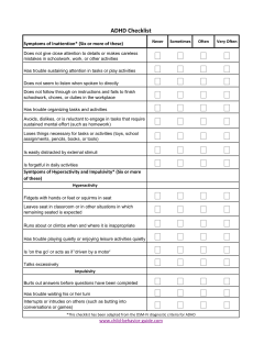


© Copyright 2026