
| Research Environmental Medicine
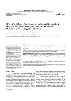
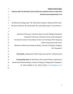
Research | Environmental Medicine
Serum Cadmium Levels in Pancreatic Cancer Patients from the East Nile
Delta Region of Egypt
Alison M. Kriegel,1 Amr S. Soliman,2 Qing Zhang,3 Nabih El-Ghawalby,4 Farouk Ezzat,4 Ahmed Soultan,4
Mohamed Abdel-Wahab,4 Omar Fathy,4 Gamal Ebidi,4 Nadia Bassiouni,4 Stanley R. Hamilton,5
James L. Abbruzzese,6 Michelle R. Lacey,7 and Diane A. Blake 1
1Department of Biochemistry, Tulane University Health Sciences Center, New Orleans, Louisiana, USA; 2Department of Epidemiology,
University of Michigan School of Public Health, Ann Arbor, Michigan, USA; 3Department of Epidemiology, University of Texas M.D.
Anderson Cancer Center, Houston, Texas, USA; 4Gastrointestinal Surgery Center, Mansoura University, Mansoura, Egypt; 5Division of
Pathology and Laboratory Medicine, and 6Department of Gastrointestinal Medical Oncology, University of Texas M.D. Anderson Cancer
Center, Houston, Texas, USA; 7Department of Mathematics, Tulane University, New Orleans, Louisiana, USA
The northeast Nile Delta region exhibits a high incidence of early-onset pancreatic cancer. It is well
documented that this region has one of the highest levels of pollution in Egypt. Epidemiologic
studies have suggested that cadmium, a prevalent pollutant in the northeast Nile Delta region, plays
a role in the development of pancreatic cancer.
OBJECTIVE: We aimed to assess serum cadmium levels as markers of exposure in pancreatic cancer
patients and noncancer comparison subjects from the same region in Egypt.
DESIGN AND PARTICIPANTS: We assessed serum cadmium levels of 31 newly diagnosed pancreatic
cancer patients and 52 hospital comparison subjects from Mansoura, Egypt.
EVALUATION/MEASUREMENTS: Serum cadmium levels were measured using a novel immunoassay
procedure.
RESULTS: We found a significant difference between the mean serum cadmium levels in patients
versus comparison subjects (mean ± SD, 11.1 ± 7.7 ng/mL vs. 7.1 ± 5.0 ng/mL, respectively; p =
0.012) but not in age, sex, residence, occupation, or smoking status. The odds ratio (OR) for pancreatic cancer risk was significant for serum cadmium level [OR = 1.12; 95% confidence interval
(CI), 1.04–1.23; p = 0.0089] and farming (OR = 3.25; 95% CI, 1.03–11.64; p = 0.0475) but not
for age, sex, residence, or smoking status.
CONCLUSIONS: The results from this pilot study suggest that pancreatic cancer in the East Nile
Delta region is significantly associated with high levels of serum cadmium and farming.
RELEVANCE TO CLINICAL PRACTICE/PUBLIC HEALTH: Future studies should further investigate the
etiologic relationship between cadmium exposure and pancreatic carcinogenesis in cadmium-exposed
populations.
KEY WORDS: cadmium, East Nile Delta region, environmental exposure, immunoassays, occupational exposure, pancreatic cancer, pollution. Environ Health Perspect 113:113–119 (2005).
doi:10.1289/ehp.8035 available via http://dx.doi.org/ [Online 25 August 2005]
Pancreatic cancer is one of the most deadly
forms of cancer. Although it is the fourth leading cause of cancer deaths in the United States,
it accounts for only 2% of all newly diagnosed
cancers each year. Five-year survival rates for
patients diagnosed with pancreatic cancer in
the United States average only 4.4%
(American Cancer Society 2005). In developing countries, pancreatic cancer appears to be
extremely rare. Studies in Egypt, Algeria, and
Iraq suggest a low incidence of the disease in
the Middle East (Al-Bahrani et al. 1982;
Mokhtar 1991; Parkin 1986; Parkin et al.
1997; Sherif and Ibrahim 1987). Risk factors
for this disease have generally been grouped
into four categories: a) cigarette smoking (Doll
et al. 1994; Howe et al. 1991); b) chronic pancreatitis (largely from alcohol consumption)
(Lowenfels et al. 1993) and genetic predisposition (Brand and Lynch 2000; Flanders and
Foulkes 1996; Goggins et al. 1996; Lowenfels
et al. 1997); c) diabetes mellitus and macro- or
micronutrients (Bueno de Mesquita et al.
1992; Everhart and Wright 1995); and
d) occupational and environmental contamiEnvironmental Health Perspectives
nation from exposure to pesticides and fertilizers (Anderson et al. 1996; Fontham and
Correa 1989; Hoppin et al. 2000), manufacturing paints and pigments (Norell et al. 1986;
Raymond and Bouchardy 1990; Sheffet et al.
1982), metalworking (Mallin et al. 1986;
Maruchi et al. 1979; Park and Mirer 1996;
Rotimi et al. 1993; Silverstein et al. 1988;
Sparks and Wegman 1980), and soldering (Ji
et al. 1999).
The incidence of pancreatic cancer in
Egypt has been previously studied with
particular attention to Dakahlia Province
(Soliman et al. 2002). This primarily rural
province is the largest of all the provinces in
the East Nile Delta region. The population in
this region exhibits an unusually high rate of
early-onset pancreatic cancer. Although most
cases in the United States occur in patients
older than 65 years of age (Lowenfels and
Maisonneuve 1999), the incidence rates for
patients younger than 65 years in the East
Nile Delta region were more than twice as
high as those observed in Americans in the
same age group and significantly higher than
• VOLUME 114 | NUMBER 1 | January 2006
those seen in other parts of Egypt (Soliman
et al. 2002). The reason for the incidence of
early-onset pancreatic cancer in the East Nile
Delta region is unclear. There is currently no
good evidence that the first three risk factors
named above would preferentially affect residents of the East Nile Delta region relative to
the rest of Egypt. It is, however, well documented that this region has one of the highest
levels of pollution in Egypt.
Nile River water is seriously contaminated
with heavy metals, pesticides, and hydrocarbons as a result of increasing discharge of
untreated industrial wastes and agricultural
irrigation wastewater (Badawy et al. 1995).
Several reports on Dakahlia Province show
high levels of heavy metal and organocholorine
pesticides in the soil and water in this region
(Abdel-Haleem et al. 2001; Dekov et al. 1997;
Reinhardt et al. 2001; Siegel et al. 1994). High
concentrations of heavy metals, including cadmium, are among the pollutants in the water.
Plants and fish grown in this water are also
contaminated with heavy metals (AbdelSabour 2001; Abou-Arab and Abou Donia
2000), which can in turn accumulate in
humans and animals that feed on these contaminated foods (Osfor et al. 1998). The serum
cadmium levels of residents of Dakahlia
Province are almost 10-fold higher than those
of residents from cadmium-polluted areas in
Cairo and 32 times higher than reference levels
Address correspondence to A.S. Soliman, Department
of Epidemiology, School of Public Health, University
of Michigan, 109 Observatory Rd., Rm. 2550, Ann
Arbor, MI 48109 USA. Telephone: (734) 764-5469.
Fax: (734) 764-3192. E-mail: [email protected]
We thank D. Garabrant (University of Michigan
School of Public Health) for his comments during
the data analysis.
This work was supported by Eli Lilly Research; the
Topfer Research fund from the M.D. Anderson
Cancer Center; National Cancer Institute grants CA
K07 090241 and R03 CA099513-01; and University
of Michigan Cancer Center Support grant 5 P30
CA46592 to A.S.S. Stipend support for A.M.K. was
provided by the Office of Science (Biological and
Environmental Research), U.S. Department of
Energy (grant DE-FG02-98ER62704 to D.A.B.).
The authors declare they have no competing
financial interests.
Received 21 February 2005; accepted 25 August
2005.
113
Kriegel et al.
for healthy populations in the United States
(Hossny et al. 2001; Osfor et al. 1998;
Soliman et al. 2002). Cadmium is a known
human carcinogen (Boffetta 1993) and has
recently been implicated as a cause of pancreatic cancer (Schwartz and Reis 2000). Two
main risk factors for pancreatic cancer, age and
cigarette smoking, are also associated with cadmium exposure. Cadmium accumulates in the
body over time because there are no specific
mechanisms for its removal. The half-life of
this metal in the body ranges from 10 to
30 years, with an average of 15 years (Jin et al.
1998). In addition, cigarette smoking is a significant source of cadmium. One cigarette contains 1–2 µg cadmium (Goyer 1996), and
inhaled cadmium is absorbed much more efficiently than is ingested cadmium (Friberg
1984). Measurement of cadmium in the pancreas of autopsy patients showed significantly
higher levels in smokers than in nonsmokers
(Elinder et al. 1976). Urinary cadmium levels,
commonly used as an indication of life-long
exposure, are also significantly higher in smokers versus nonsmokers (Schwartz et al. 2003).
In this study, we assessed serum cadmium
levels of newly diagnosed histologically confirmed pancreatic cancer patients with hospital comparison subjects from the same region
in Egypt, using a novel immunoassay procedure. We have also examined the contributions of age, residence, smoking status, and
profession to overall risk for pancreatic cancer. Although the sample size in this study is
small, it is our hope that these initial data will
act as a springboard for larger, more in-depth
studies that will analyze the relationship
between cadmium and pancreatic cancer in a
more detailed fashion.
study. Pancreatic cancer was confirmed by
reviewing the histopathologic slides of all
patients both at Mansoura University and at
M.D. Anderson Cancer Center. No patients
with chronic pancreatitis were included in this
study. Histopathologic examination of the tissues of the pancreatic cancer patients included
in the study revealed no signs of chronic pancreatitis. All patients were Egyptian citizens,
permanent residents of Dakahlia Province, and
recruited before receiving chemotherapy or
radiotherapy. There were no restrictions based
on age, sex, or tumor stage. In addition,
52 comparison subjects were recruited from
the ear-nose-throat and ophthalmology departments at Mansoura University General
Hospital on the same medical campus of
Mansoura University. The comparison subjects
were chosen by systematic random sampling
from inpatients admitted for surgeries related
to nonchronic illnesses. After the nature of the
study had been fully explained, informed oral
and/or written consent was obtained from each
person in the study.We used an intervieweradministered questionnaire, which included
questions about lifetime occupational, residential, and smoking histories. Information was
also collected about family history of pancreatic cancer.
Sample collection. For each patient and
comparison subject, 10 mL of blood was collected into a sterile nonheparinized Vacutainer
tube. The sample was allowed to clot for
5–10 min and then centrifuged for 10 min at
13,000 rpm. At least 2 mL of clear serum from
each sample was transferred to glass tubes,
which were labeled with patient name and
identification number, clinic name, and collection time and date. Specimens were frozen at
–20°C until they were transferred (within
3 weeks) to M.D. Anderson Cancer Center.
Samples were hand-carried in dry ice during
transport from Egypt to Houston to New
Orleans, Louisiana, where they were stored at
A
H
Cd(II) + protein
H
H
HNO3
Cd(II)
Apoprotein
B
Cd(II)
EDTA
C
Cd(II)–EDTA
EDTA
E
E
E
Washing
Materials and Methods
114
D
P
S
E
Reading
Absorbance
Materials. The 2A81G5 monoclonal antibody,
which recognizes Cd(II)–EDTA complexes,
was prepared from a hybridoma generated in
the Blake laboratory (Blake et al. 1996). A
Cd(II)–EDTA–horseradish peroxidase (HRP)
conjugate was prepared as described by
Darwish and Blake (2002, 2001). We purchased a pooled human serum sample from
Intergen (Milford, MA, USA); this serum sample was analyzed by graphite-furnace atomic
absorption spectroscopy (AAS) and shown to
be free of endogenous cadmium (data not
shown). ELISA high-binding 96-well plates
were a product of Corning-Costar (Cambridge,
MA, USA). We obtained AAS standard cadmium (1,000 mg/L in 2% nitric acid) from
Perkin-Elmer (Norwalk, CT, USA).
Patient selection. Between September 2001
and February 2002, 31 newly diagnosed
patients with adenocarcinoma of the pancreas
from the Gastrointestinal Surgery Center of
Mansoura University, Dakahlia Province in
Egypt were recruited to participate in this
[Cd(II)]
Anti-mouse antibody
2A81G5 antibody
E
Cd(II)–EDTA–HRP
S
Substrate
P
Product
Figure 1. Modified one-step assay for Cd(II) in human serum. (A) Cadmium was first displaced from metallothionein and other proteins under acidic conditions. (B) An EDTA-containing buffer was added to the serum
to form Cd(II)–EDTA complexes, which are recognized by the 2A81G5 antibody. (C) Microwell plates were
coated and blocked as described in “Materials and Methods.” A serum sample was mixed 1:1 with enzymelabeled Cd(II)–EDTA and subsequently added to the coated wells. The enzyme-labeled Cd(II)–EDTA competes with the Cd(II)–EDTA complexes from the serum sample for immobilized antibody binding sites. (D)
Peroxidase substrate was added to the wells and absorbance was read at 450–650 nm.
VOLUME
114 | NUMBER 1 | January 2006 • Environmental Health Perspectives
Serum cadmium and pancreatic cancer
–80°C. The study was approved by the human
subject committees of the Universities of
Texas, Tulane, and Michigan in the United
States and Mansoura University in Egypt.
Modification of the one-step immunoassay
for cadmium in serum. The method described
by Soliman et al. (2002) required relatively high
quantities of the anti-cadmium monoclonal
antibody, 2A81G5. Therefore, we modified the
original method by using a goat anti-mouse
antibody to capture and concentrate the
2A81G5 antibody on the microwell plate, as
shown in Figure 1. Preliminary experiments
demonstrated that when goat anti-mouse IgG,
coated at a concentration of 2.0 µg/mL, was
used to capture mouse monoclonal antibody,
5-fold less 2A81G5 was required for the subsequent cadmium assay (data not shown).
Pooled human serum versus pooled
Egyptian comparison serum. We calculated
standard curves in two separate sample matrices
using the modified one-step immunoassay for
cadmium. We compared a pooled human
serum sample from Intergen Co. (Norcross,
GA, USA) with a mixture of all the comparison
samples from Egypt in a competition assay.
Twenty-five microliters of each of the
52 Egyptian comparison samples were combined together and mixed well. Various concentrations of atomic absorption grade
cadmium were spiked into the Egyptian pooled
serum as well as Intergen’s pooled serum
(Norcross, GA, USA). We then performed an
immunoassay and compared inhibition curves.
Determination of cadmium in human
serum samples. We diluted goat anti-mouse
IgG1 (heavy chain specific; Jackson ImmunoResearch Laboratories, West Grove, PA, USA)
into HEPES-buffered saline (HBS; 137 mM
NaCl, 3 mM KCl, and 10 mM HEPES,
pH 7.4) to a concentration of 2 µg/mL. The
diluted antibody (50 µL) was introduced
to each well of a 96-well high-binding microwell plate and incubated for 2 hr at 37°C.
The plates were washed three times with
Absorbance (450–650 nm)
2.00
Mean
LOD
1.50
1.00
0.50
0.00
0.01
0.1
1
10
100
1,000
Spiked Cd(II) (ng/mL)
Figure 2. Typical standard curve using the one-step
assay for Cd(II) in pooled human serum. See
“Materials and Methods” for details. Data shown
were the means of four replicates with a best-fit line
(solid). Dashed lines on either side of the main curve
represent the best-fit line of the mean values ± 2 SD.
Environmental Health Perspectives
phosphate-buffered saline containing 0.05%
Tween-20. The wells were blocked with 3%
bovine serum albumin in HBS at 37°C for
1 hr, followed by a wash step. The antiCd(II)–EDTA antibody, 2A81G5, was diluted
to 0.5 µg/mL in HBS. A 50 µL aliquot of the
diluted antibody was added to each well and
incubated at room temperature for 1 hr, followed by a wash step.
Egyptian serum samples were thawed at
room temperature and mixed well. A 75 µL
aliquot of serum was added to 75 µL of 5%
HNO3 and mixed well. The mixture was incubated at room temperature for 5 min and then
spun at 15,000 rpm for 5 min. Acidification of
the sample allows the release of cadmium from
metallothionein and other protein ligands. A
90 µL aliquot of the supernatant was removed
and conditioned with 10 µL of a 10X concentrated buffer (1.37 M NaCl, 30 mM KCl,
50 mM EDTA, 100 mM HEPES). The presence of EDTA allows the formation of
Cd(II)–EDTA complexes. The sample was
neutralized with 10 M KOH. Neutralized
serum was added to an equal volume of
0.1 µg/mL Cd(II)–EDTA–HRP and mixed
well. The mixture (50 µL/well) was added to
three separate wells. After 1 hr incubation at
room temperature, the plate was washed.
3,3´,5,5´-Tetramethylbenzidine peroxidase
substrate (50 µL/well; Kirkegaard-Perry
Laboratories, Gaithersburg, MD, USA) was
added to each well, and color formation was
stopped after 10 min with an equal volume of
1 N HCl. The absorbance of each well was
measured in a dual-wavelength mode
(450–650 nm) using a Vmax Kinetic Microplate
Reader (Molecular Devices Corporation,
Sunnyvale, CA, USA). For preparation of a
standard curve, pooled human serum from
Intergen was spiked with various concentrations
of atomic-absorption–grade cadmium standard
and treated in a manner identical to that used
for the serum samples noted above.
Data analysis for the immunoassay. We
calculated standard curves on each immunoassay plate in replicates of four. We determined
the mean value and SD of the absorbance provided by each cadmium concentration. These
mean absorbance values (y) were plotted on a
curve versus the cadmium concentrations (x)
and used to fit the equation y = a0 – (a1 × x)/(a2
+ x), where y is the experimental absorbance, x
is cadmium concentration in the standard or
sample, a0 is the absorbance in the absence of
cadmium, a 1 is the difference between the
absorbance in the absence of cadmium and at a
saturating concentration of cadmium, and a2 is
the Cd(II) concentration that produces a 50%
inhibition of signal (IC50). To account for the
variability in the data, confidence bounds for
each standard Cd(II) concentration were computed. The upper and lower curves were fit
from the mean values ± 2 SD, respectively.
• VOLUME 114 | NUMBER 1 | January 2006
The limit of detection (LOD) was determined
with the use of the standard curves. The
absorbance value for zero cadmium (y-intercept) for the lower curve (mean – 2 SD) was
determined and then applied to the upper
curve (mean + 2 SD) to find the cadmium
concentration designated as the LOD. A visual
depiction of how the LOD was determined is
provided in Figure 2.
Each Egyptian serum sample was analyzed
in triplicate in a particular assay. The mean
absorbance value and SD were determined for
each sample. We used SDs to help eliminate
nonprecise values. Mean absorbance values for
each serum sample were applied to the mean
standard curve performed on the same plate to
determine the cadmium concentration for that
sample. When the sample was analyzed multiple times, the cadmium concentrations were
averaged. Each original serum sample was
diluted by a factor of 4.57 during the
immunoassay procedure. To obtain the actual
cadmium concentration of the serum samples,
the assay cadmium concentration was multiplied by this number.
Statistical methods. For those samples that
fell below the LOD (18 of 52), we used the
method described by Helsel (1990) to assign a
value for the cadmium concentration. In this
method, one assumes that the actual cadmium
concentration in these samples is described by
a Gaussian distribution whose lower bound is
zero and whose upper bound is the LOD of
the immunoassay. The median cadmium concentration in these samples would be 50% of
the LOD, and this value was therefore
assigned to each nondetect sample. This is a
conservative method of handling immunoassay data; the actual differences between the
cases and the comparisons might be even
greater than reported herein. The logarithm of
serum cadmium level was used as the continuous exposure because the distribution of serum
cadmium level was skewed. We used the
Student’s t-test to test the differences among
means of the logarithms of serum cadmium
level. Because both transformed and untransformed analytical methods yielded comparable
conclusions, we present the analysis based on
the untransformed data for better comparison
with other studies.
We also aimed to find a subset of predictors for pancreatic cancer disease status. The
main response variable was disease status and
potential predictors included age, sex, serum
cadmium level, smoking, and farming. We
analyzed the longest occupation reported by
study subjects as farming and nonfarming.
Farming jobs were defined as being a farmer
or a housewife who lived in a rural area.
Nonfarming occupations included administrators, barbers, businessmen, teachers, students, and women who lived in urban areas
but reported their job title as “housewife.”
115
Kriegel et al.
Nonfarming also included industrial workers,
such as painters and welders. We did not
include residence in the final model because
of its high correlation with occupation.
Residence alone was not significant in a logistic regression model that did not include
occupation. In this analysis, a study subject
was considered a smoker if he or she reported
ever smoking 100 cigarettes.
Multiple logistic regression models were fitted based on the variables chosen from the univariate analysis. The logistic regression models
were designed to estimate the probability of
having pancreatic cancer, whereas incorporating
the covariates of age, sex, serum cadmium levels, occupation, and smoking. p-Values < 0.05
are reported as significant. There is no collinearity among the predictors. The Hosmer and
Lemeshow goodness-of-fit test was used to test
the model fitting. All the statistical tests were
two sided. Data were analyzed using the
Statistical Analysis System package (version 8.2;
SAS Institute Inc., Cary, NC, USA).
Results
One-step immunoassay for cadmium. In this
study we used a previously published
immunoassay procedure to quantify cadmium
in serum samples. The assay was selective for
cadmium; other metal ions, including manganese, cobalt, copper, zinc, magnesium, mercury, calcium, nickel, iron, and lead, did not
significantly interfere with the assay even when
tested at concentrations considerably higher
than those present in human serum (Darwish
and Blake 2002). The original one-step
immunoassay for cadmium designed by
Darwish and Blake (2002) required 125 ng of
2A81G5 antibody to be coated in each microwell. With the large number of Egyptian
serum samples to be assayed, an unacceptable
quantity of antibody would have been
required for these experiments. We therefore
developed a modification of the original
Percent total absorbance
120
Intergen
Egyptian
100
80
60
40
20
0
0.01
0.1
1
10
100
1,000
Spiked Cd(II) (ng/mL)
Figure 3. Pooled human serum versus pooled
Egyptian comparison serum. Binding curves were
calculated with cadmium spiked into the pooled
human serum from Intergen or into a sample of
pooled Egyptian comparison serum. IC 50 values
were very similar, 16.74 ng/mL and 17.44 ng/mL for
Intergen and Egyptian samples, respectively.
Values reported are mean ± SD (n ≥ 6).
116
method that reduced the amount of 2A81G5
necessary for the capture of the Cd(II)–EDTA
complexes by 5-fold. An overall schematic of
the modified one-step assay is shown in
Figure 1. In human serum, cadmium usually
exists as a complex with metallothionein and
other proteins. In order to determine the total
level of cadmium in a serum sample, cadmium
must be extracted from these proteins, a
process most effectively achieved by acidification of the sample. Addition of a conditioning
buffer containing EDTA allowed the formation of Cd(II)–EDTA complexes. This unlabeled Cd(II)–EDTA competed with a known
concentration of enzyme-labeled Cd(II)–
EDTA to bind to the 2A81G5 antibody
immobilized on the microwell plate. After a
wash step to remove unbound Cd(II)–EDTA
and Cd(II)–EDTA–HRP, a chromogenic peroxidase substrate was added and HRP activity
was detected. A standard curve was prepared
by adding varying concentrations of atomic
absorption grade cadmium to a pooled human
serum sample that was treated in an identical
manner as the patient samples. This standard
curve was then used to determine the concentration of cadmium in each sample based on
the observed spectrophotometric absorbance.
Pooled human serum used preparation of
the standard curve. In previous studies
(Darwish and Blake 2002), a pooled human
serum sample obtained from Intergen was used
as the sample medium for preparing the
standard curves on each of the immunoassay
plates. In the present study, however, we were
concerned that this pooled serum sample, from
U.S. donors, might not be an appropriate
medium for analysis of Egyptian donors. A
large pooled serum sample from Egyptian
donors was not available, so we prepared a
small pooled sample by combining an equal
volume (25 µL) of all the Egyptian noncancer
comparison samples available from this study
(a total of 52). The two pooled serum samples
(from Intergen and the Egyptian comparisons)
were spiked with various concentrations of
cadmium and the subsequent inhibition curves
were compared. As shown in Figure 3, the two
curves were nearly identical. IC50 values were
very similar, 16.74 ng/mL and 17.44 ng/mL
for Intergen and Egyptian samples, respectively. These data indicate that the Egyptian
samples did not contain any endogenous components that might affect immunoassay performance and that the pooled human serum from
Intergen was an appropriate medium for all
subsequent standard curves.
Standard curve and determination of LODs
in the immunoassay. A typical standard curve,
obtained by adding known concentrations of
cadmium and EDTA to a pooled human serum
sample, is shown in Figure 2. As the concentration of unlabeled cadmium increased, less of the
enzyme-labeled Cd(II)–EDTA was able to bind
to the 2A81G5 immobilized in the microwell;
absorbance in the assay decreased as the unlabeled cadmium concentration increased. Four
replicates of each standard concentration were
Table 1. Characteristics of the study population [n (%)].
Case (n = 31)
Characteristic
Age (years)
< 60
≥ 60
Sex
Male
Female
Residence
Rural
Urban
Smoking
Yes
No
Occupation
Farmer
Housewife
Industrial
Professional
Farming-related occupation
Yes
No
Serum cadmium level (ng/mL)
Total
Mean ± SD
Median (range)
Smokers
Mean ± SD
Median (range)
Nonsmokers
Mean ± SD
Median (range)
Comparison (n = 52)
21 (67.7)
10 (32.3)
41 (78.8)
11 (21.2)
19 (61.3)
12 (38.7)
30 (57.7)
22 (42.3)
15 (48.4)
16 (51.6)
15 (28.8)
37 (71.2)
11 (35.5)
20 (64.5)
21 (40.4)
31 (59.6)
7 (22.6)
10 (32.3)
8 (25.8)
6 (19.4)
3 (5.8)
19 (36.5)
11 (21.2)
19 (36.5)
11 (35.5)
20 (64.5)
8 (15.4)
44 (84.6)
p-Value
0.3023
0.8198
0.0989
0.8161
0.0805
0.0570
11.1 ± 7.7
11.0 (27.9)
7.1 ± 5.0
5.7 (24.2)
0.0120
11.8 ± 9.3
13.7 (27.0)
7.1 ± 4.7
6.4 (16.5)
0.1386
10.7 ± 6.9
11.0 (27.0)
7.1 ± 5.3
5.5 (24.2)
0.0380
p-Values for quantitative variables were calculated using Student’s t-test; p-values for proportions and qualitative variables were calculated using the chi-square test.
VOLUME
114 | NUMBER 1 | January 2006 • Environmental Health Perspectives
Serum cadmium and pancreatic cancer
analyzed. The average values of each four standard replicates were plotted on the graph and
fitted using the equation described in “Materials
and Methods” (Figure 2). Variability (2 × SD)
was determined and graphed as described in
“Materials and Methods” (Figure 2). These
upper and lower limit curves were used to determine the LOD of the immunoassay, as shown
in Figure 2. The LODs were determined for
each standard curve and ranged between 0.4
and 1.0 ng/mL cadmium for the pancreatic cancer patients and between 0.3 and 1.0 ng/mL
cadmium for the comparison subjects. These
ranges show the consistency of standard curves
performed from day to day and the high sensitivity of the assay. Each sample was assayed at
least one time in triplicate. Many of the samples
were assayed on multiple days, also in triplicate.
The average absorbance value was detected and
then applied to the mean standard curve like the
one shown in Figure 2 to obtain a serum cadmium concentration.
Characteristics of the study population.
There were no significant differences between
the pancreatic cancer patients and the comparison subjects in terms of age, sex, residence,
smoking status, or occupation, as shown in
Table 1. Across the study population, age
ranged from 21 to 75 years, with a median age
of 52 in the cases and 49 in the comparison
subjects. There were slightly more males than
females in each group. Most study subjects
(52% and 71% in cases and comparison subjects, respectively) were urban dwellers, and
almost two-thirds were nonsmokers. Less than
half of the cases had professional backgrounds,
compared with two-thirds of the comparison
subjects. There was, however, a significant difference in mean serum cadmium levels between
the cases and comparisons (p = 0.012). In
patients with pancreatic cancer, the mean cadmium level was 11.1 ± 7.7 ng/mL, whereas in
the comparisons the mean ± SD was 7.1 ±
5.0 ng/mL. Although pancreatic cancer patients
who reported histories of smoking showed
higher but not significant differences in serum
cadmium levels than those of comparison subjects who reported histories of smoking (11.8 ±
9.3 ng/mL and 7.1 ± 4.7 ng/mL, p = 0.14, in
patients and comparison subjects, respectively),
there were statistically higher serum cadmium
levels in nonsmoker patients than in nonsmoker comparison subjects (10.7 ± 6.9 ng/mL
and 7.1 ± 5.3 ng/mL, p = 0.038, in patients and
comparisons, respectively.
A comparison of the different ranges of
cadmium levels determined in the serum samples of the case and comparison populations is
shown in Figure 4. Most comparison subjects
exhibited serum cadmium levels between the
LOD and < 10 ng/mL, with only one
> 20 ng/mL Cd(II). The pancreatic cancer
patients showed an overall trend toward
higher serum cadmium concentrations. Most
cases demonstrated serum cadmium levels
between 10 and 20 ng/mL, and 4 of 31 exhibited cadmium levels > 20 ng/mL.
Variation in cadmium levels in the study
population. Within the study population as a
whole, there were no significant differences in
serum cadmium levels as a function of sex,
residence (rural or urban), or smoking status,
as shown in Table 2. There was a trend
toward significance on the basis of occupation
(p = 0.0827) with farmers showing the highest levels (mean ± SD, 13.34 ± 8.29 ng/mL),
followed by industrial workers (8.68 ±
Sex
Male
Female
Residence
Rural
Urban
Smoking
Yes
No
Occupation
Farmer
Housewife
Industrial
Professional
Percent total samples
30
20
10
0
< LOD
LOD– < 10 ng/mL 10–20 ng/mL
> 20 ng/mL
Figure 4. Distribution of samples and serum Cd(II)
concentrations in comparison subjects versus pancreatic cancer patients. The values reported are the
percentage of the total samples for that population.
Environmental Health Perspectives
In this pilot study, we tested the hypothesis that
pancreatic cancer patients in this Egyptian population were exposed to higher levels of cadmium than were noncancer subjects. Serum
cadmium levels were used as a marker for cadmium exposure. It is difficult to find a perfect
dose estimator for cadmium. Urinary cadmium
levels are often used; however, studies with animals and humans have shown that renal damage may lead to higher than normal cadmium
excretion (Friberg et al. 1985; Nordberg and
Piscator 1972). Jarup et al. (1997) have shown
No.
Mean ± SD
Median (range)
p-Value
49
34
9.21 ± 7.09
7.72 ± 5.21
7.77 (27.88)
6.40 (19.19)
0.2966
30
53
8.52 ± 6.31
8.65 ± 6.51
7.31 (20.11)
6.86 (27.42)
32
51
8.75 ± 6.86
8.5 ± 6.16
7.08 (27.42)
6.86 (26.96)
10
29
19
25
13.34 ± 8.29
7.56 ± 6.05
8.68 ± 5.46
7.84 ± 6.14
0.9281
0.8634
11.88 (1.83–28.79)
5.94 (0.91–27.88)
7.77 (0.91–16.45)
5.03 (1.37–25.14)
0.0827
p-Values for quantitative variables were calculated using Student’s t-test; p-values for proportions and qualitative variables were calculated using the chi-square test.
Table 3. ORs of risk factors for pancreatic cancer.
Comparison
Case
40
Discussion
Table 2. Serum cadmium concentration (ng/mL) in the combined patients and comparison sample.
60
50
5.46 ng/mL) and professionals (7.84 ±
6.14 ng/mL).
Risk factors for pancreatic cancer. The
association between pancreatic cancer risk and
subject age, residence (urban or rural), smoking status, and occupation (farming or nonfarming) is indicated by the odds ratios (ORs)
shown in Table 3. Both serum cadmium levels
and farming were independently associated
with increased risk of pancreatic cancer. The
OR for serum cadmium levels was 1.12 [95%
confidence interval (CI), 1.04–1.23; p =
0.0089]. Farming was associated with
increased risk for pancreatic cancer OR = 3.25
(95% CI, 1.03–11.64) (Table 3).
Serum cadmium level (ng/mL, mean ± SD)
Age [years, n (%)]
< 60
≥ 60
Sex [n (%)]
Male
Female
Smoking [n (%)]
Yes
No
Farming-related occupation [n (%)]
Yes
No
Case
(n = 31)
Comparison
(n = 52)
OR
(95% CI)
p-Value
11.1 ± 7.7
7.1 ± 5.0
1.12 (1.04–1.23)
0.0089
21 (67.7)
10 (32.3)
41 (78.8)
11 (21.2)
1
2.25 (0.71–7.26)
0.1677
19 (61.3)
12 (38.7)
30 (57.7)
22 (42.3)
2.06 (0.51–8.61)
1
0.311
11 (35.5)
20 (64.5)
21 (40.4)
31 (59.6)
0.54 (0.13–2.19)
1
0.3859
11 (35.5)
20 (64.5)
8 (15.4)
44 (84.6)
3.25 (1.03–11.64)
1
0.0475
ORs were calculated using logistic regression analysis. p-Values for quantitative variables were calculated using
Student’s t-test; p-values for proportions and qualitative variables were calculated using the chi-square test.
• VOLUME 114 | NUMBER 1 | January 2006
117
Kriegel et al.
that blood cadmium can provide a better dose
estimate than urinary cadmium concentrations, especially when tubular proteinuria is
present. During high cadmium exposures, the
cadmium in the blood increases relatively
rapidly until, after some months, it reaches a
concentration that corresponds to the intensity
of exposure. If the exposure stops, the blood
cadmium decreases with an initial half-time of
2–3 months (Elinder et al 1994; Jarup et al.
1983). Cadmium accumulated in the body,
however, will continue to influence blood levels. Even after exposure ceases, the concentration in the blood never returns to preexposure
levels. Thus blood cadmium has been proposed
as one of the more accurate estimators of accumulated body burden (Alfven et al. 2002).
Only about 10% of whole-blood cadmium is
circulating in serum, but serum levels appear to
correlate with blood levels (Lauwerys et al.
1994). The use of serum rather than whole
blood in this study allowed us much greater
flexibility in collecting, storing, and transporting patient samples.
In the patient population studied here,
serum cadmium levels and farming were strong
independent risk factors for pancreatic cancer,
whereas age, sex, smoking, and residence were
not. The highly significant association of pancreatic cancer with serum cadmium level is also
consistent with other epidemiologic evidence.
The strongest suspicion of an association
between cadmium exposure and pancreatic
cancer has been reported in Louisiana (Blot
et al. 1978; Lemus et al. 1996; Tchounwou
et al. 1996). Industrial activity along the
Mississippi River has led to an accumulation of
contaminants in southern Louisiana (Mielke
et al. 2000). Seafood and rice are popular food
items in the local diet. Both rice and fish harvested from cadmium-polluted areas may contain high levels of cadmium (Ikeda 1992;
Modigosky et al. 1991). A case–control study
in Louisiana showed a significantly increased
risk for pancreatic cancer associated with rice
consumption among Cajuns, with a dose–
response relationship (Falk et al. 1988).
Louisiana residents are also exposed to cadmium by inhalation. Lemus et al. (1996) have
shown that 64 of 315 samples (20.3%) of
indoor and outdoor air from 53 households in
Louisiana exceeded the U.S. Environmental
Protection Agency’s permissible levels for cadmium (Lemus et al. 1996). It is interesting to
note the association between pancreatic cancer
risk and farming in this study (OR = 3.25, p =
0.0475).
Although smoking has been identified as a
strong risk factor for pancreatic cancer in previous studies (Doll et al. 1994; Howe et al.
1991), it was not significant in this study.
There are at least two possible explanations for
this finding. First, most subjects (approximately
two-thirds) in both the case and comparison
118
groups were nonsmokers. Second, cadmium is
a by-product of cigarette smoke, and adjusting
for serum cadmium levels in this study may
have controlled for the effect of smoking. There
was also no significant association between risk
of pancreatic cancer and residence. Although
rural residents might be expected to show
increased risk because of exposure to pesticides
and fertilizers (Anderson et al. 1996; Fontham
and Correa 1989), exposure of urban industrial
workers employed in manufacturing (Norell
et al. 1986; Sheffet et al. 1982), metalworking
(Maruchi et al. 1979; Rotimi et al. 1993;
Silverstein et al. 1988), and soldering (Ji et al.
1999) may be equally significant. There may
also be other important differences in cadmium
exposure (e.g., diet and air quality) that distinguish urban from rural populations. Reports
from urban areas in Egypt have shown high levels of cadmium in organ meats that are frequently consumed by the local population
(Abou-Arab and Abou Donia 2000). A recent
study on markers of environmental pollution in
Egypt has also shown significantly higher levels
of oxidative damage in urban populations,
compared with rural populations (Soliman
et al. 2004). The higher risk of pancreatic cancer associated with farming in this study may
reflect the intense exposure to cadmium and
other farming-related occupations in this
population. Thus, two major risk factors may
have overshadowed the relationship between
smoking and cadmium levels in this study.
The first was the exceptionally high levels of
environmental pollution in our study region.
The second included the high dietary intake
of rice and fish grown in polluted soils and
water of the study region. The National Food
Consumption Study in Egypt has shown that
residents of our study region of Dakahlia
Province consume more fish and rice than does
any other group in Egypt (Galal and Khorshid
1995). Unfortunately, neither detailed environmental exposures nor dietary intake was
measured in the present study.
Studies with rodents showed the ability of
the pancreas to accumulate high concentrations of cadmium. Mice injected with 4 mg/kg
CdCl2 expressed a 2.5-fold increase in DNA
synthesis in the pancreas, which Andrews et al.
(1990) and Hellman (1986) interpreted as a
sign of increased metallothionein synthesis.
Rats injected with 4 and 8 mg/kg cadmium
demonstrated 9.8- and 17.9-fold increases,
respectively, in pancreatic metallothionein levels (Wormser and Calp 1988). Cadmium
interferes with the use of essential metals such
as calcium, zinc, selenium, and iron.
Deficiencies of these essential metals, in conjunction with protein and vitamin deficiencies,
exaggerate cadmium toxicity by increasing
absorption through the gut and enhancing
retention in different organs (Waalkes 1986).
Studies on adult Wistar rats showed that zinc
VOLUME
deficiency markedly increases cadmium accumulation in various organs (Waalkes 1986).
Studies from our research region (Galal and
Khorshid 1995) showed that 46% of women
have a calcium intake < 50% of the required
daily allowances (RDA); the consumption of
B12, a marker of protein intake, is also < 50%
of the RDA in 33% of women in the region.
Nutritional deficiencies in our study region
may be a major factor in cadmium accumulation in body organs of the local population.
Cadmium is a potent carcinogen in
rodents. Injecting rats with CdCl2 resulted in
a significant increase (from 2.2% to 8.5%) in
the incidence of pancreatic islet cell tumors
(Poirier et al. 1983). In experiments designed
to determine the carcinogenic effects of
repeated exposure to CdCl 2 , Fischer and
Wistar rats showed a very high incidence of
dose-related pancreatic metaplasia, as reflected
by pancreatic hepatocyte formation (Konishi
et al. 1990). These pancreatic hepatocytes are
thought to arise from the ductal and interstitial cells of the pancreas, which resemble oval
cells of the liver (Rao et al. 1986). Cadmium
may activate oncogenes such as c-myc, mdm2,
and cellular tumor antigen p53; inhibit tumor
suppressor genes such as wild-type p53 and
p27; and consequently promote the proliferation of initiated cells (Fang et al. 2002).
The present study has several important
strengths. The Dakahlia region has documented high levels of pollution and is therefore
a unique setting for studying environmental
risk factors and cancer occurrence. The stability of population without migration in
Dakahliam maximizes the chances of life-long
environmental exposure (Aly and Shields
1996). The high incidence of young pancreatic
cancer patients (younger than 50 years of age)
provides a unique model system for studying
the effect of early environmental exposures on
pancreatic carcinogenesis (Soliman et al.
2002). Finally, the wide range of occupational
and lifestyle factors in this region provide help
to investigate the occupational and epidemiologic risk factors associated with pancreatic
cancer. The relatively small sample size of this
rare cancer may limit the generalization of
results.
In summary, this pilot study has shown a
statistically significant association between
pancreatic cancer and serum cadmium levels
and farming. Future studies should expand on
this pilot investigation by studying a larger
number of pancreatic cancer patients and by
collecting extensive information on the lifetime occupational, residential, and environmental exposures and dietary influences in
order to clarify the role of cadmium in pancreatic cancer etiology in this population. Several
molecular mechanisms have been identified by
which cadmium may influence pancreatic cells
(Chai et al. 1999; Merali and Singhal 1976;
114 | NUMBER 1 | January 2006 • Environmental Health Perspectives
Serum cadmium and pancreatic cancer
Shimoda et al 2001; Waalkes et al. 1992), and
a better understanding of these processes is
under study in our laboratories. It will also be
important to examine genetic susceptibility
and markers of genetic damage caused by
environmental exposures to clarify the role of
such exposures in pancreatic carcinogenesis.
REFERENCES
Abdel-Haleem AS, Sroor A, El-Bahi SM, Zohny E. 2001. Heavy
metals and rare earth elements in phosphate fertilizer components using instrumental neutron activation analysis.
Appl Radiat Isot 55:569–573.
Abdel-Sabour MF. 2001. Cadmium status in Egypt. J Environ Sci
(China) 13:351–360.
Abou-Arab AA, Abou Donia MA. 2000. Heavy metals in Egyptian
spices and medicinal plants and the effect of processing
on their levels. J Agric Food Chem 48:2300–2304.
Al-Bahrani ZR, Al-Mondhiry HA, Al-Saleem T. 1982. Carcinoma
of the pancreas in Iraq. Oncology 39:353–357.
Alfven T, Jarup L, Elinder C-G. 2002. Cadmium and lead in blood in
relation to low bone mineral density and tubular proteinuria.
Environ Health Perspect 110:699–702.
Aly HY, Shields M. 1996. A model of temporary migration: the
Egyptian case. Int Migr 34:431–447.
American Cancer Society. 2005. Cancer Facts and Figures
2005. Atlanta, GA:American Cancer Society.
Anderson KE, Potter JD, Mack TM. 1996. Pancreatic cancer. In:
Cancer Epidemiology and Prevention (Schottenfeld D,
Fraumeni JF Jr, eds). 2nd ed. New York:Oxford University
Press, 725–771.
Andrews GK, Kage K, Palmiter-Thomas P, Sarras MP Jr. 1990.
Metal ions induce expression of metallothionein in pancreatic exocrine and endocrine cells. Pancreas 5:548–554.
Badawy MI, Wahaab RA, Abou Waly HF. 1995. Petroleum and
chlorinated hydrocarbons in water from Lake Manzala and
associated canals. Bull Environ Contam Toxicol 55:258–263.
Blake DA, Chakrabarti P, Khosraviani M, Hatcher FM, Westhoff
CM, Goebel P, et al. 1996. Metal binding properties of a
monoclonal antibody directed towards metal-chelate
complexes. J Biol Chem 271:27677–27685.
Blot WJ, Fraumeni JF Jr, Stone BJ. 1978. Geographic correlates
of pancreas cancer in the United States. Cancer 42:373–380.
Boffetta P. 1993. Carcinogenicity of trace elements with reference to evaluations made by the International Agency for
Research on Cancer. Scand J Work Environ Health 19(suppl
1):67–70.
Brand RE, Lynch HT. 2000. Hereditary pancreatic adenocarcinoma. A clinical perspective. Med Clin North Am 84:665–675.
Bueno de Mesquita HB, Maisonneuve P, Moerman CJ, Walker
AM. 1992. Aspects of medical history and exocrine carcinoma of the pancreas: a population-based case-control
study in the Netherlands. Int J Cancer 52:17–23.
Chai F, Truong-Tran AQ, Ho LH, Zalewski PD. 1999. Regulation of
caspase activation and apoptosis by cellular zinc fluxes and
zinc deprivation: a review. Immunol Cell Biol 77:272–278.
Darwish IA, Blake DA. 2001. One-step competitive immunoassay for cadmium ions: development and validation for
environmental water samples. Anal Chem 73:1889–1895.
Darwish IA, Blake DA. 2002. Development and validation of a
one-step immunoassay for determination of cadmium in
human serum. Anal Chem 74:52–58.
Dekov VM, Komy Z, Araujo F, Van Put A, Van Grieken R. 1997.
Chemical composition of sediments, suspended matter,
river water and ground water of the Nile (Aswan-Sohag
traverse). Sci Total Environ 201:195–210.
Doll R, Peto R, Wheatley K, Gray R, Sutherland I. 1994. Mortality
in relation to smoking: 40 years’ observations on male
British doctors. BMJ 309:901–911.
Elinder C-G, Friberg L, Nordberg GF, Kjellstrom T, Oberdoerster
G. 1994. Biological Monitoring of Metals. Chemical Safety
Monographs. WHO/EHG/94.2. Geneva:World Health
Organization, International Programme on Chemical
Safety.
Elinder C-G, Lind B, Kjellstrom T, Linnman L, Friberg L. 1976.
Cadmium in kidney cortex, liver, and pancreas from
Swedish autopsies. Estimation of biological half time in
kidney cortex, considering calorie intake and smoking
habits. Arch Environ Health 31:292–302.
Everhart J, Wright D. 1995. Diabetes mellitus as a risk factor for
pancreatic cancer. A meta-analysis. JAMA 273:1605–1609.
Falk RT, Pickle LW, Fontham ET, Correa P, Fraumeni JF Jr. 1988.
Environmental Health Perspectives
Life-style risk factors for pancreatic cancer in Louisiana: a
case-control study. Am J Epidemiol 128:324–336.
Fang MZ, Mar W, Cho MH. 2002. Cadmium affects genes
involved in growth regulation during two-stage transformation of Balb/3T3 cells. Toxicology 177:253–265.
Flanders TY, Foulkes WD. 1996. Pancreatic adenocarcinoma:
epidemiology and genetics. J Med Genet 33:889–898.
Fontham ET, Correa P. 1989. Epidemiology of pancreatic cancer.
Surg Clin North Am 69:551–567.
Friberg L. 1984. Cadmium and the kidney. Environ Health Perspect
54:1–11.
Friberg L, Elinder C-G, Kjellstro” m T, Nordberg GF. 1985.
Cadmium and Health: A Toxicological and Epidemiological
Appraisal. Boca Raton, FL:CRC Press.
Galal O, Khorshid A. 1995. Development of Food Consumption
Monitoring System. National Agricultural Research
Project. Cairo:Ministry of Agriculture.
Goggins M, Schutte M, Lu J, Moskaluk CA, Weinstein CL,
Petersen GM, et al. 1996. Germline BRCA2 gene mutations in
patients with apparently sporadic pancreatic carcinomas.
Cancer Res 56:5360–5364.
Goyer RA. 1996. Toxic effects of metals. In: Casarett and Doull’s
Toxicology: The Basic Science of Poisons (Klaassen CD,
ed). New York:McGraw-Hill, 691–736.
Hellman B. 1986. Evidence for stimulatory and inhibitory effects
of cadmium on the [3H]thymidine incorporation into various
organs of the mouse. Toxicology 40:13–23.
Helsel DR. 1990. Less than obvious—statistical treatment of data
below the detection limit. Environ Sci Technol 24:1766–1774.
Hoppin JA, Tolbert PE, Holly EA, Brock JW, Korrick SA, Altshul
LM, et al. 2000. Pancreatic cancer and serum organochlorine levels. Cancer Epidemiol Biomarkers Prev 9:199–205.
Hossny E, Mokhtar G, El-Awady M, Ali I, Morsy M, Dawood A
2001. Environmental exposure of the pediatric age groups
in Cairo City and its suburbs to cadmium pollution. Sci
Total Environ 273:135–146.
Howe GR, Jain M, Burch JD, Miller AB. 1991. Cigarette smoking
and cancer of the pancreas: evidence from a populationbased case-control study in Toronto, Canada. Int J Cancer
47:323–328.
Ikeda M. 1992. Biological monitoring of the general population
for cadmium. IARC Sci Publ 118:65–72.
Jarup L, Persson B, Elinder C-G. 1997. Blood cadmium as an indicator of dose in a long-term follow-up of workers exposed
to cadmium. Scand J Work Environ Health 23:31–36.
Jarup L, Rogenfelt A, Elinder C-G, Nogawa K, Kjellstrom T. 1983.
Biological half-time of cadmium in the blood of workers
after cessation of exposure. Scand J Work Environ Health
9:327–331.
Ji BT, Silverman DT, Dosemeci M, Dai Q, Gao YT, Blair A. 1999.
Occupation and pancreatic cancer risk in Shanghai, China.
Am J Ind Med 35:76–81.
Jin T, Lu J, Nordberg M. 1998. Toxicokinetics and biochemistry
of cadmium with special emphasis on the role of metallothionein. Neurotoxicology 19:529–535.
Konishi N, Ward JM, Waalkes MP. 1990. Pancreatic hepatocytes
in Fischer and Wistar rats induced by repeated injection of
cadmium chloride. Toxicol Appl Pharmacol 104:149–156.
Lauwerys RR, Bernard AM, Roels HA, Buchet JP. 1994.
Cadmium: exposure markers as predictors of nephrotoxic
effects. Clin Chem 40:1391–1394.
Lemus R, Abdelghani AA, Akers TG, Horner WE. 1996. Health
risks from exposure to metals in household dusts. Rev
Environ Health 11:179–189.
Lowenfels AB, Maisonneuve P. 1999. Pancreatic cancer:
development of a unifying etiologic concept. Ann NY Acad
Sci 880:191–200.
Lowenfels AB, Maisonneuve P, Cavallini G, Ammann RW,
Lankisch PG, Andersen JR, et al. 1993. Pancreatitis and
the risk of pancreatic cancer. International Pancreatitis
Study Group. N Engl J Med 328:1433–1437.
Lowenfels AB, Maisonneuve P, DiMagno EP, Elitsur Y, Gates
LK Jr, Perrault J, et al. 1997. Hereditary pancreatitis and
the risk of pancreatic cancer. International Hereditary
Pancreatitis Study Group. J Natl Cancer Inst 89:442–446.
Mallin K, Berkeley L, Young Q. 1986. A proportional mortality ratio
study of workers in a construction equipment and diesel
engine manufacturing plant. Am J Ind Med 10:127–141.
Maruchi N, Brian D, Ludwig J, Elveback LR, Kurland LT. 1979.
Cancer of the pancreas in Olmsted County, Minnesota,
1935–1974. Mayo Clin Proc 54:245–249.
Merali Z, Singhal RL. 1976. Prevention by zinc of cadmiuminduced alterations in pancreatic and hepatic functions.
Br J Pharmacol 57:573–579.
Mielke HW, Gonzales CR, Smith MK, Mielke PW. 2000.
Quantities and associations of lead, zinc, cadmium, manganese, chromium, nickel, vanadium, and copper in fresh
• VOLUME 114 | NUMBER 1 | January 2006
Mississippi delta alluvium and New Orleans alluvial soils.
Sci Total Environ 246:249–259.
Modigosky SR, Alvarez-Hernandez X, Glass J. 1991. Lead, cadmium and aluminum accumulation in the red swamp crayfish Procambarus clarkii G. collected from roadside
drainage ditches in Louisiana. Arch Environ Contam
Toxicol 20:253–258.
Mokhtar N, ed. 1991. Cancer Pathology Registry (1985–1989).
Cairo:National Cancer Institute of Cairo University.
Nordberg G, Piscator M. 1972. Influence of long-term cadmium
exposure to cadmium on urinary excretion of protein and
cadmium in mice. Environ Physiol Biochem 2:37–49.
Norell S, Ahlbom A, Olin R, Erwald R, Jacobson G, LindbergNavier I, et al. 1986. Occupational factors and pancreatic
cancer. Br J Ind Med 43:775–778.
Osfor MM, el-Dessouky SA, el-Sayed A, Higazy RA. 1998.
Relationship between environmental pollution in Manzala
Lake and health profile of fishermen. Nahrung 42:42–45.
Park RM, Mirer FE. 1996. A survey of mortality at two automotive
engine manufacturing plants. Am J Ind Med 30:664–673.
Parkin DM, ed. 1986. Cancer Occurrence in Developing
Countries. IARC Sci Publ 75.
Parkin DM, Muir CS, Whelan SL, Gao YT, Ferlay J, Powell J.
1997. Cancer incidence in five continents. IARC Sci Publ
143:986–987.
Poirier LA, Kasprzak KS, Hoover KL, Weak ML. 1983. Effects of
calcium and magnesium acetates on the carcinogenicity of
cadmium chloride in Wistar rats. Cancer Res 43:4575–4581.
Rao MS, Scarpelli DS, Reddy JK. 1986. Transdifferentiated
hepatocytes in rat pancreas. Curr Top Dev Biol 20:63–78.
Raymond L, Bouchardy C. 1990. Risk factors of cancer of the
pancreas from analytic epidemiologic studies. Bull Cancer
77:47–68.
Reinhardt EG, Stanley DJ, Schwarcz HP. 2001. Human-induced
desalinization of Manzala Lagoon, Nile Delta, Egypt: evidence from isotopic analysis of benthic invertebrates.
J Coastal Res17:431–442.
Rotimi C, Austin H, Delzell E, Day C, Macaluso M, Honda Y.
1993. Retrospective follow-up study of foundry and engine
plant workers. Am J Ind Med 24:485–498.
Schwartz GG, Il’yasova D, Ivanova A. 2003. Urinary cadmium,
impaired fasting glucose, and diabetes in the NHANES III.
Diabetes Care 26:468–470.
Schwartz GG, Reis IM. 2000. Is cadmium a cause of human
pancreatic cancer? Cancer Epidemiol Biomarkers Prev
9:139–145.
Sheffet A, Thind I, Miller AM, Louria DB. 1982. Cancer mortality
in a pigment plant utilizing lead and zinc chromates. Arch
Environ Health 37:44–52.
Sherif M, Ibrahim AS, eds. 1987. Cancer Registry of Metropolitan
Cairo: The Profile of Cancer in Egypt (1972–1987).
Cairo:National Cancer Institute of Cairo University.
Shimoda R, Nagamine T, Takagi H, Mori M, Waalkes MP. 2001.
Induction of apoptosis in cells by cadmium: quantitative
negative correlation between basal or induced metallothionein concentration and apoptotic rate. Toxicol Sci
64:208–215.
Siegel FR, Sladboda ML, Stanley DJ. 1994. Metal pollution loading, Manazlah Lagoon, Nile Delta, Egypt; implications for
aquaculture. Environ Geol 23:89–98.
Silverstein M, Park R, Marmor M, Maizlish N, Mirer F. 1988.
Mortality among bearing plant workers exposed to metalworking fluids and abrasives. J Occup Med 30:706–714.
Soliman AS, El-Ghawalby N, Ezzat F, Bondy ML, Soultan A,
Abdel-Wahab M, et al. 2002. Unusually high rate of youngonset pancreatic cancer in the East Nile Delta region of
Egypt. Int J Gastrointes Can 32:143–151.
Soliman AS, Vulimiri SV, Kleiner HE, Shen J, Eissa S, Seifeldin I,
et al. 2004. High levels of oxidative DNA damage in lymphocyte DNA of premenopausal breast cancer patients from
Egypt. Int J Environ Health Sci 14:121–134.
Sparks PJ, Wegman DH. 1980. Cause of death among jewelry
workers. J Occup Med 22:733–736.
Tchounwou PB, Abdelghani AA, Pramar YV, Heyer LR,
Steward CM. 1996. Assessment of potential health risks
associated with ingesting heavy metals in fish collected
from a hazardous-waste contaminated wetland in
Louisiana. Rev Environ Health 11:191–203.
Waalkes MP. 1986. Effect of zinc deficiency on the accumulation of cadmium and metalothionein in selected tissues of
the rat. J Toxicol Environ Health 18:301–313.
Waalkes MP, Coogan TP, Barter RA. 1992. Toxicological principles of metal carcinogenesis with special emphasis on
cadmium. Crit Rev Toxicol 22:175–201.
Wormser U, Calp D. 1988. Metallothionein induction by cadmium
and zinc in rat secretory organs. Experientia 44:754–755.
119


© Copyright 2026