
Hajdu-Cheney Syndrome: Case Study in Diagnostic Imaging
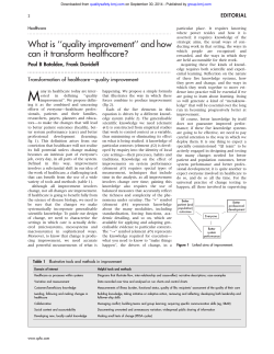
Downloaded from ard.bmj.com on October 15, 2014 - Published by group.bmj.com
Annals of the Rheumatic Diseases 1994; 53: 276-279
276
CASE STUDIES IN DIAGNOSTIC IMAGING SERIES EDITOR: V N CASSAR-PULLICINO*
Hajdu-Cheney syndrome
M A R O'Reilly, D G Shaw
Department of
Radiology,
Hospital for Sick
Children,
Great Ormond Street,
London WCl,
United Kingdom
M A R O'Reilly
D G Shaw
*Department of Diagnostic
Imaging,
The Robert Jones
and Agnes Hunt
Orthopaedic Hospital,
Oswestry,
Shropshire SY10 7AG,
United Kingdom.
Correspondence to:
Dr Marian O'Reilly,
Department of Radiology,
St George's Hospital,
Blackshaw Road,
London SW17,
United Kingdom.
4\
t,-
Clinical history
The patient, a female child, was born at 37
week gestation by a spontaneous vaginal
delivery following an uneventful pregnancy.
The parents were not consanguinous. Two
older siblings were alive and well. Respiratory
difficulties with cyanotic episodes occurred
during the first few months of life.
Multiple congenital deformities were noted.
The face was dysmorphic with a prominent
premaxilla. There was hypertelorism and the
eyes sloped downwards with narrow palpebral
fissures. The jaw was small with a thin upper
lip. The palate was high and arched and the
uvula appeared rotated forwards and fixed to
the palate. There was excess skin around the
neck and back. The arms were short, with
flexion deformity at the wrist and radial
4
deviation. The hands appeared short and
stubby and the fingers were clubbed.
As the child grew older, generalised joint
laxity and hyperextensibility developed. Height
and weight remained on the third percentile for
age but head circumference was above the 98th
percentile, with an enlarged pituitary fossa on
skull radiographs. Motor development was
delayed but normal intelligence was apparent.
Kyphoscoliosis was treated with a brace and
eventually spinal fusion. The chest deformity
led to a restrictive ventilatory defect and
recurrent chest infections.
The permanent teeth were all lost soon after
eruption. At the age of 15 years, delayed
puberty was apparent with prepubertal serum
levels of LH, FSH and oestradiol and a bone
age of only 12 years.
Radiological findings
Skull radiographs showed basilar impression,
multiple wormian bones and an enlarged
elongated pituitary fossa (fig 1). The calvarium
was thickened and the cranial sutures were
widely separated.
There was discrepancy in the length of
the paired long bones, resulting in valgus at the
knees and bowing of the fibulae (fig 2). The
ulnae were bowed, resulting in a non-united
fracture of the proximal shaft of the ulna on the
right (fig 3). Both radial heads were dislocated.
There was generalised osteopenia.
Radiographs of the spine revealed a severe
kyphoscoliosis (fig 4). The lumbar vertebrae
were tall with narrow disc spaces and there
was slight anterior wedging of the vertebral
bodies at T9 to T 1I (fig 5). The neural
foramina in the lumbar region were enlarged
v
A
4
(fig 5).
The most characteristic features, however,
were seen in the hands (fig 6). Osteolysis
of the terminal phalanges with overlying
soft tissue swelling developed in late childhood. The toes were only partially affected
(fig 7).
Figure 1 Lateral view of the neck and skull base shows basilar invagination
pituitaryfossa (open arrowheads) is enlarged and elongated, and the clivus is
(large arrowhead). Multiple wormian bones (closed arrowheads) are present.
.steep
Differential diagnosis
Acro-osteolysis refers to the dissolution of bone
at the phalanges. It is commonly associated
with other diseases such as scleroderma, sarcoidosis, neuropathic disorders, and rheumatoid
or psoriatic syndromes. It can be asssociated
with nephropathy in childhood. The pathogenesis may be vascular, neurovascular,
traumatic (due to burns and frostbite), toxic
(due to PVC or ergot poisoning), metabolic or
infective. It may also be idiopathic.
Downloaded from ard.bmj.com on October 15, 2014 - Published by group.bmj.com
277
Hajdu-Cheney syndrome
-
mid-portion of the phalanges or at the periarticular surfaces (table 1). Most of the
rheumatological diseases cause erosions at the
distal tufts and surrounding juxta-articular
areas. Neuropathic disorders, such as diabetes
mellitus and leprosy, may cause sclerosis
and pencilling or pointing of the phalanges.
The primary osteolysis syndromes of HajduCheney and Rothmund may cause a band-like
area of lucency in the distal phalangeal shaft
which is more sharply defined than that seen
in occupational or traumatically induced
acro-osteolysis.
The Hajdu-Cheney syndrome (familial idiopathic acro-osteolysis) was first described in
1948 by Hajdu and Kauntze,' and more
extensively reported by Cheney.2
Most cases are sporadic but inheritance is
likely to be autosomal dominant. Although this
is a congenital disorder, the correct diagnosis
is rarely made until later childhood, when the
characteristic hand changes occur.
Figure 2 Both tibiae and fibulae. There is valgus at the knees. Both fibulae are
bowed distally (arrow 5) and the appearances suggest early pseudarthroses.
The additional radiographic findings in this
patient make a congenital syndrome more
likely. The major ones to consider are progeria,
pyknodysostosis and the Hajdu-Cheney
syndrome. This patient does not display the
characteristic facies, skin changes and
premature ageing seen in progeria. Pyknodysostosis includes many of the features
described here but there are two major differences between this and the Hajdu-Cheney
syndrome: in pyknodysostosis the bones are
dense and the mandibular angle is almost
t
180O degrees.
_
The most likely diagnosis is that of the
Hajdu-Cheney syndrome.
Figure 3 Right forearm. Mesomelic shortening of the
forearm bones. The radial head is dislocated (arrow).
Discussion
In acro-osteolysis, resorption of bone may
occur at the phalangeal tufts, in the
The ulna is bowed and there is a non-united fracture of the
proximal shaft (open arrow). There is acro-osteolysis of the
terminal phalanx of the thumb (curved arrow). Diffuse
osteopenic change.
Downloaded from ard.bmj.com on October 15, 2014 - Published by group.bmj.com
O'Reilly, Shaw
278
The affected child has a distinctive facial
appearance, short stature and retarded puberty.
Intelligence is normal. The eyes slope down-
..........l......
wards and there is telecanthus and synophrys.
Optic atrophy can develop. The philtrum is
long and the nostrils are anteverted. The
mandible is small, the palate highly arched and
the occiput prominent. The neck is short and
the ears are low-set.
Multiple wormian bones occur in the skull
(fig 1). These are intrasutural bones occurring
most frequently along the lambdoidal sutures.
The presence of a few wormian bones is
normal up to the age of six months and only
when multiple are they considered significant
(table 2).
Table 2 Causes of multiple wormian bones
Cleidocranial dysostosis
.
...
Figure 4 Thoraco-lumbar spi1nze-frontal view. Severe kyphoscoliosis. The lumbar
vertebral bodies are enlarged wirth narrowed disc spaces.
Osteogenesis imperfecta
Congenital hypothyroidism
Hypophosphatasia
Pyknodysostosis
Progeria
~~~~~~~~~~~~Pachydermoperiostosis
Hajdu-Cheney syndrome
Down's syndrome
Idiopathic
'.i
Table 1 Causes of acro-osteolysis related to the radiological
appearances of the phalanges
Resorption of the phalangeal tuft
Scleroderma
Raynaud's disease
Sarcoidosis
Psoriatic arthropathy
Neuropathic disease
Thromboangitis obliterans
Thermal injuries and trauma
Hyperparathyroidism
Epidermolysis bullosa
Porphyria
Progeria
Pachydermoperiostosis
Resorption of the mid-portion of the phalanges
Polyvinyl chloride tank cleaners
Hyperparathyroidism
Haidu-Cheney syndrome
Rothmund's syndrome
Periarticular resorption
Psoriatic arthropathy
Hyperparathyroidism
Scleroderma
Erosive osteoarthritis
Thermal injuries
Figure 5 Thoraco-lumbar spine-lateral view, taken with
the patient wearing a brace. Kyphoscoliosis. The lumbar
vertebrae are tall with reduced disc heights. Posterior
scalloping of the vertebral bodies (arrows) and enlarged
neuralforamina. DWuse osteopenia.
Downloaded from ard.bmj.com on October 15, 2014 - Published by group.bmj.com
279
Hajdu-Cheney syndrome
Other characteristic radiological features in
the skull in the Hajdu-Cheney syndrome are
the elongated pituitary fossa, thickened skull
vault, and prominant occiput. Progressive
basilar invagination develops and a serious
complication of the disease can be foramen
magnum impaction. The spine is kyphotic.
The lumbar vertebral bodies are tall and the
disc spaces are narrowed. Schmorl's nodes and
vertebral collapse may develop. Other features
are the absence of the frontal sinuses and early
loss of teeth.
The upper limbs are mesomelic. There is
discrepancy in the length of the paired long
bones, resulting in valgus at the knees and
dislocation of the radial heads. The fibulae are
long and bowed and they project proximally.
The bones may be osteopenic and there is a
high incidence of fractures. joint laxity is also
a feature.
The classic acro-osteolysis develops in late
childhood. The terminal phalanges tend to be
small and trangular in appearance with an
increase in the amount of
surrounding soft
tissue. Sometimes the acro-osteolysis takes the
form of transverse lytic defects across the shafts
of the phalanges. The feet are less severely
affected. Pain is a frequent manifestation,
especially in the hands.
Treatment of the disorder can only be
symptomatic. The prognosis can be serious,
depending on the degree of ventilatory
restriction caused by the chest deformity and
the severity of neurological impairment caused
by basilar invagination.
Figure 6 Left hand. Soft tissue swelling about the terminal phalanges and distal
interphalangeal joints, with severe osteolysis of the terminal phalanges (arrows).
The fourth andfifth metacarpals are hypoplastic.
N, Kauntze R. Cranio-skeletal dysplasia. Br Radiol
1 Hajdu
1948: 21: 42.
2 Cheney W D. Acro-osteolysis. AmerJ7 Roentgenol 1965; 94:
ad _
Figure 7 Both feet.
Acro-osteolysis of the
terminal phalanges
(arrows) is less severe than
in the hands. There is
bilateral hallux valgus and
thefourth andfifth
metatarsals on the left are
hypoplastic.
£
595.
S~~~~~
Downloaded from ard.bmj.com on October 15, 2014 - Published by group.bmj.com
Hajdu-Cheney syndrome.
M A O'Reilly and D G Shaw
Ann Rheum Dis 1994 53: 276-279
doi: 10.1136/ard.53.4.276
Updated information and services can be found at:
http://ard.bmj.com/content/53/4/276.citation
These include:
References
Article cited in:
http://ard.bmj.com/content/53/4/276.citation#related-urls
Email alerting
service
Receive free email alerts when new articles cite this article. Sign up in
the box at the top right corner of the online article.
Notes
To request permissions go to:
http://group.bmj.com/group/rights-licensing/permissions
To order reprints go to:
http://journals.bmj.com/cgi/reprintform
To subscribe to BMJ go to:
http://group.bmj.com/subscribe/


© Copyright 2026