
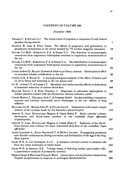
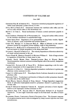
Inhibition of CD26 enzyme activity with pro-boropro stimulates rat
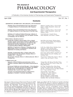
From www.bloodjournal.org by guest on October 15, 2014. For personal use only.
1995 85: 3602-3609
Inhibition of CD26 enzyme activity with pro-boropro stimulates rat
granulocyte/macrophage colony formation and thymocyte
proliferation in vitro
LA Bristol, W Bachovchin and L Takacs
Updated information and services can be found at:
http://www.bloodjournal.org/content/85/12/3602.full.html
Articles on similar topics can be found in the following Blood collections
Information about reproducing this article in parts or in its entirety may be found online at:
http://www.bloodjournal.org/site/misc/rights.xhtml#repub_requests
Information about ordering reprints may be found online at:
http://www.bloodjournal.org/site/misc/rights.xhtml#reprints
Information about subscriptions and ASH membership may be found online at:
http://www.bloodjournal.org/site/subscriptions/index.xhtml
Blood (print ISSN 0006-4971, online ISSN 1528-0020), is published weekly by the American
Society of Hematology, 2021 L St, NW, Suite 900, Washington DC 20036.
Copyright 2011 by The American Society of Hematology; all rights reserved.
From www.bloodjournal.org by guest on October 15, 2014. For personal use only.
Inhibition of CD26 Enzyme Activity with Pro-boropro Stimulates Rat
Granulocyte/Macrophage Colony Formation and Thymocyte
Proliferation In Vitro
By Lynn A. Bristol, William Bachovchin, and Laszlo Takacs
CD26 dipeptidyl peptidase (DPPIV) is involved
in thereguletion of proliferation of some hematopoietic and T-lineage
cells. Here, we show that Pro-boropro apotent inhibitor of
DPP activity has a costimulating effect
in hematopoieticcolony assays for macrophage and,
to a lesser extent, for granu-
locyte colonies and hasstimulating
a
effect in organ cultures
of immature thymocytes. Basedon theseandotherevidences, we propose that the mechanism by whichCD26
regulates proiiferation is associated with its DPP activity.
0 1995 by The American Society of Hematology.
B
of CD26 via cross-linking and internalization is also paralleled by increased cell growth. These results support the
concept that CD26 ectoenzymatic activity has an important
regulatory role in hematopoietic and early T-cell proliferation.
ONE MARROW (BM)-derived stem cells migrate into
the thymus where theprecursor of the T cells, the triple
negative (CD4-/CDK/TCR-) thymocyte, proliferates and
differentiates into major histocompatibility complex-restricted, self-tolerant mature, single positive (CD4+/CD8- or
CD4-/CD8+), T-cell receptor (TCR)-positive T cells.'-s A
small subpopulation of BM cells can reconstitute both the
hematopoietic and lymphoid elements of lethally irradiated
The mechanisms involved in the commitment of stem
cells to the T-lineage, T-stem cell thymus-immigrating activity and to the intrathymic stages of precursor T-cell development and proliferation require a more detailed analysis. To
analyze the mechanism of early T-cell proliferation and its
relation to hematopoietic proliferation in the rat, we generated monoclonal antibodies (MoAbs) against CD4-ICDS(double negative [DN]) blasts. One of these antibodies (1.3)
recognizes the ectoenzyme, dipeptidyl dipeptidase ZV (DPP
IV; E.C.N.3.4.14.5.) or CD26, and has costimulatory activity
in BM colony and DN cell proliferation assays.8-'
The mechanism of CD26-mediated signal modulation in
human T cells has been ascribed to its association with
CD45R0, a pathway independent of DPP IV enzyme activity, that increases tyrosine phosphorylation of key proteins
involved in signal transduction via the TCR.'' In the present
study, we have examined the requirements for CD26 enzyme
function on hematolymphoid cells for their ability to proliferate. Inhibition of CD26 enzymatic activity with Pro-boropro,
a transition-state-like competitive inhibitor," increased the
number of hematolymphoid cells several-fold in vitro. In
addition, we found that CD26 is not associated with CD45
on rat BM cells or thymocytes or on a rat-mouse DN hybridoma cell line and that a reduction in cell surface expression
From the Unit of Special Projects, Laboratory of Physiological
and Pharmacological Studies, National Institute on Alcohol and
Alcohol Abuse, National institutes of Health, Rockville, MD; and the
Department of Biochemistry, Tufrs University, School of Medicine,
Boston, MA.
Submitted September 22, 1994; accepted February 7, 1995.
During his stay in Hungary, L.T. was supported by OTKA Grant
No. 457.
Address reprint requests to Lciszld Takrics,Amgen, Inc, Amgen
Center Bldg 8-l-A-236, 1840 Dehavilland Dr, Thousand Oaks, CA,
91320-1 789.
The publication costs of this article were defrayed in part by page
charge payment. This article must therefore be hereby marked
"advertisement" in accordance with 18 U.S.C. section 1734 solely to
indicate this fact.
0 1995 by The American Society of Hematology.
0006-4971/95/8512-0036$3.00/0
3602
MATERIALS AND METHODS
Animals. The 6- toIO-week-old pregnant and nonpregnant female Buffalo rats were purchased from the National Cancer Institute
(Frederick Cancer Research Facility, Frederick, MD) and housed in
the animal facility at the National Institute on Alcoholism and Alcohol Abuse (Rockville, MD).
Antibodies and hybridomas. The 1.3 MoAb (anti-CD26 or DPP
IV, IgGl isotype) was described in detail
The antirat
CD26-specific MoAb 8.6A3 was received from Dr B.U. Pauli (Cancer Biology Laboratory, Department of Pathology, Cornel1 University, College of Veterinary Medicine, Ithaca, NY). The antimouse
CD45 cytoplasmic peptide antiserum was obtained from Dr LS.
Trowbridge (The Salk Institute, San Diego, CA). The mousepan
CD45 MoAb was received from Dr R. Hodes (National Institute on
Aging, National Institutes of Health, Bethesda, MD). The 9.1 l
MoAb (mouse antihuman IgM, IgGl isotype) was obtained from Dr
E. Rajnavolgyi (Department of Immunology, Eotvos Lorind University, God, Hungary). The OX-7 (antirat Thy-1.1, phycoerythrin-labeled) and OX-l (antirat pan CD45 leukocyte common antigen,
fluorescein isothiocyanate [FITCJ-labeled and unlabeled) MoAbs
were purchased from Accurate Biochemicals, Inc (Westbury, NY).
MOPC 21 (IgG1 isotype) was purchased from The Binding Site,
Ltd (San Diego, CA); purified normal mouse IgG was obtained from
Sigma (St Louis, MO); goat antimouse IgG was from ICN (Lisle,
IL); biotinylated antimouse IgG and biotinylated antirabbit IgG were
from Vector Laboratories, Inc (Burlingame, CA); and FITC-conjugated streptavidin was from Becton Dickinson (Mountain View,
CA).
Interspecies hybridomas were generated from rat CD4-/CD8cells and mouse BW5147.G.1.4 cells as previously described* and
were maintained in complete RPM1 medium (Advanced Biotechnologies Inc [ABI], Columbia, MD) containing 10% fetal calfserum
(FCS; Hyclone, Logan, UT), 1% penlstrep (Biofluids, Rockville,
MD), and 1% L-glutamine (Biofluids). The rat DNlBW hybridoma,
RDNH 22.108, was shown by cytofluorographic analysis to express
CD26.
Isolation of BM cells and splenic T cells. BM cells were isolated
by standard procedures. T cells from spleens were enriched by a
panning procedure as previously described.'
BM colony assays. Unfractionated BM cells were plated at a
density of 1 to 2 X IO" cells/Z mL QBSF-56 medium(Quality
Biologicals, Inc, Gaithersburg, MD) containing 0.8% methylcellulose (Fluka, Rokonkoma, NY), 3% conditioned spleen cell supernatant (spleen cells stimulated with concanavalin A [ 5 ,ug/mL; Sigma]
for 3 days followed by removal of concanavalin A from supernates
by repeated passage over a thyroglobulin-agarose column [Sigma]),
or a mixture of recombinant cytokines (100 pg/mL human recombiBlood, Vol 85, No 12 (June 15), 1995:pp 3602-3609
From www.bloodjournal.org by guest on October 15, 2014. For personal use only.
3603
STIMULATION OF CFU VIA CD26 INHIBITION
nant interleukin-la [riL-la; from Dr K. Matshushima, National Cancer Institute, National Institutes of Health, Frederick, MD], 100 “g/
mL human granulocyte colony-stimulating factor [Amgen Inc. Thousand Oaks, CA], 200 ng/mL rat stem cell factor [Amgen], 20 ng/
mL human IL-6 [Immunex Corp. Seattle, WA], 250 mU/mL human
erythropoietin [Amgen], and 50 U/mL mouse IL-3 [Immunex COrpl)
and 20% FCS, 1% antibiotics, 1% L-glutamine, and purified MoAbs
(1.3 or MOPC 21) with or without Pro-boropro in 3.5-cm plastic
petri dishes. Results are expressed as the mean number of colonies
% SD of triplicate plates from day-5 or day-7 time points and are
representative of three experiments.
Thymic lobe organ culture assay. Thymic lobes from 17-dayold fetuses or I- to2-day-old neonates were cultured on sterile
polycarbonate membranes in Click’s medium supplemented with
10% FCS and antibiotics containing the DPP IV inhibitor. The total
number of cells from each culture was determined by counting single
cell suspensions of mashed lobes. Data are presented as the mean
number of cells 2 SD and are representative of three experiments.
Chemical cross-linking of surface-iodinated cells, immunoprecipitation, and sodium dodecyl sulfate-polyacrylamide gel electrophoresis (SDS-PAGE) analysis of immunoprecipitates. Cells were surface-iodinated by the lactoperoxidase method.” ’251-labeledcells
were chemically cross-linked with 2 mmoVL disuccinimidyl suberate
(DSS; Pierce Chemical CO, Rockford, IL) or dimethyl sulfoxide
(DMSO) as a control at 4°C for 30 minutes according to Volarevic
et all3 and were lysed in 0.5% Triton X-100 or 0.5% NP 40 lysis
buffer containing 50 mmollL Tris (pH 7.5) and 150 mmol/L NaCl
(TBS), 10 pg/mL leupeptin (Fluka), 10 pg/mL aprotinin (Fluka),
1 m o V L phenylmethylsulfonyl fluoride (Fluka), and 1.8 mg/mL
iodoacetamide (Fluka) for 30 minutes at 4°C. In some cases, immunoprecipitation was performed from digitonin ( O S % , 1%) lysates as
described by Torimoto et al.”’ The detergent lysates were incubated
with Sepharose-coupled protein-A (Pharmacia, Piscataway, NJ) that
was preincubated with MoAbs or polyclonal antibodies. The precipitate was eluted in nonreducing or reducing SDSPAGE loading
buffer for 7.5% one-dimensional slab gel analysis. Gels were dried
and exposed to Kodak XAR-5 film (Eastman-Kodak, Rochester,
NY). In preclearing experiments, immunoprecipitates were precleared first with Sepharose-coupled protein-A as described above;
1.3 antibody was added later, captured with Sepharose protein-A,
and analyzed as described for regular immunoprecipitates. Preclearing efficiency was studied in preliminary experiments and showed
that the standard conditions used here remove over 90% of immunoprecipitable proteins in the preclearing step (data not shown).
Immunoblotting of chemically cross-linked cells. The 0.5% Triton X 100 or 0.5% NP 40 detergent lysates from chemically crosslinked cells were immunoprecipitated with various MoAbs, run on
7.5% SDS-PAGE gels, and transferred to Nitrocellulose (Schleicher
and Schuell, Keene, NH). Membranes were blocked overnight at
4°C in TBS (50 mmol/L Tris [pH 7.51and 150 mmoVL NaCI)
containing 0.1% Tween 20 and 1% bovine serum albumin (BSA)
and were probed with an antirat CD26 MoAb (8.6A3; 5 pg/mL for
1 hour at room temperature). Identical blots that were exposed to
normal mouse IgG were run in parallel and served as a negative
control. Membranes were reacted with biotin-conjugated goat antimouse Ig (1:2OOO; Vector Laboratories) followed by horseradish
peroxidase (HRP-conjugated avidin ( I :loOO, Arnersham Corp, Arlington Heights, IL). Immunoreactive proteins were visualized by
enhanced chemiluminescence using reagents obtained from Amersham.
Modulation of suqace expression of DPP W . Unfractionated
BM cells, thymocytes, or RDNH 22.108 cells (5 X 10⁶/mL) were
incubated at 37°C with 5 pg/mL of 1.3 or control 9.1 1 MoAb; and,
at 0, 0.5, 1, 3, 6, 12, and 24 hours, cells were analyzed for DPP IV
expression by cytofluorographic analysis. Cells expressing high levels of CD26’ were gated and analyzed.
CytojZuorography. For detection ofDPP W expression on 1.3
MoAb-treated BM, thymocytes, and the RDNH 22.108 hybridoma,
cells were reacted with biotinylated antimouse Ig (10 pg) followed
by FITC-streptavidin (1: 100 dilution). 9.1 1 MoAb-treated cells were
reacted with 1.3 MoAb (2 pg) followed by biotinylated antimouse
Ig and FITC-streptavidin. Surface expression of pan CD45 and Thy
1.1 on 1.3 and 9.11 MoAb-treated cells was measured using FITCOX-l (1:30) and phycoerythrin-OX-7 (1:30) MoAbs, respectively.
Cell surface staining was analyzed on a linear scale using a Coulter
Epics cytofluorometer (Coulter Electronics, Miami, FL).
RESULTS
Inhibition of CD26 enzyme activity on hematopoietic cells
and thymocytes with Pro-boropro stimulates proliferation in
vitro. Using the rat CD26-reactive 1.3MoAb,we established a role for CD26 in modulating growth-regulatory signals of hematopoietic and early T-lineage cell^.^.^ Here, we
analyze the effect of a transition-state-like competitive inhibitor of DPP IV, Pro-boropro, in biologic models of hematopoiesis and T-cell development in vitro. Treatment of BM
cells in semisolid colony assays containing 0.8% methylcellulose with Pro-boropro and 3% conditioned splenic supernatants stimulated an approximately twofold increase in
macrophage colony-forming units (CFU; see Fig IA). Granulocyte colonies were stimulated in numbers in all experiments, aithough to a lesser extent than were macrophage
colonies. Mixed colonies showed no change or were reduced
in number. Macrophage-lineage cells showed a greater sensitivity to Pro-boropro than granulocytic cells, with maximum
stimulation occurring at
m o m concentration versus
m o m , respectively. This concentration range of Proboropro was shown to inhibit the enzyme activity of porcine
kidney CD26l’ and rat CD26 purified from lamina propria
by
The stimulatory effect of Pro-boropro on the number of
colonies was similar in the presence of a defined recombinant
cytokine mixture (Fig 1B). In the presence of the same recombinant cytokines, the 1.3 MoAb stimulated colony formation close to twofold as cornpard withan
isotypematched control MoAb (Fig 1C); different types of colonies
were not scored independently in these experiments.
Given the possible involvement of CD26 in early T-cell
pr~liferation,~.~.’~.’~
the effect of Pro-boropro was assayed
on thymic lobe organ cultures, a system that more closely
approximates the thymic microenvironment than do thymocyte suspension cultures. At an optimum concentration of 2
X
m o m , addition of Pro-boropro alone to 17-day-old
fetal thymic lobes resulted in a twofold increase in total cell
number (Fig 1D).
Downmodulation of CD24 on the cell sugace does not
comodulate CD45 (OX-l)expression on hematopoietic and
early T cells. To examine whether the CD26/ CD45 association is detectable in the rat hematopoietic and T lineage,
CD45 downmodulation by CD26-specific MoAb was tested
first, as described by Torimoto et al.” BM cells, thymocytes,
and a rat DN thymocyte hybridoma (RDNH 22.108) were
cultured in the presence of 1.3 and 9.1 1 isotype-matched
control MoAb and were analyzed by fluorescence-activated
From www.bloodjournal.org by guest on October 15, 2014. For personal use only.
3604
BRISTOL, BACHOVCHIN, AND TAKACS
A.
B.
l
o
10-8
107
10-6
l*
10-5
10-4
0
Pro-boro-Pro M
C.
10-61 0 - 8 1 0 7
Pro-boro-Pro M
D.
Fig 1. Pro-boropro stimulates BM macrophage and early T-lineage cell proliferation in vitro. (A, B, and C) Unfradionated BM cells were
cultured in semisolid medium containing increasing concentrations of Pro-boropro (A and B) or 5 pglmL soluble 1.3 MoAb or an isotypematched control MoAb (MOPC; C) in 3% conditioned splenic supernatant (A) or a mixture of recombinant cytokines (B and C). Results are
expressed as the mean number of colonies k SD of triplicate plates on day 5 of culture. Different types of colonies were scored by their
morphology (A), or all the colonies were counted (B and C). Statistically significant changes are marked with asterisks P, P < .m;
**, P <
.W).(D) Thymic lobes from 17-day-old fetuses werecultured on polycarbonate membraneswith or without daily administration of 2 x
mol/L Pro-boropro. Results are presemted astotal call number (*, P < .051.
cell sorter analysis for CD26, OX-l (rat pan-CD45), as well
as OX-7 (Thy 1.1) surface expression.
On BM cells, 1.3 MoAb downmodulated the surface level
of CD26 within 3 hours, with a sustained decrease at the
12-hour (Fig 2) and 24-hour (not shown) time points. Comparing mean fluorescence of control MoAb and 1.3-treated
and stained BM cells, the degree of 1.3 downmodulation is
estimated to be 75%. Culturing of cells with an irrelevant
MoAb 9.1 1 did not affect CD26 expression. Similar experiments have been performed with radmouse hybridoma cells
and with thymiclobe organ cultures and have provided results
comparable with those obtained with BM cells (not shown).
Titration of 1.3 MoAb concentration has been performed and
has shown that 1.3 downmodulates CD26 levels in the same
range that it shows effect in the colony assays. Downmodulation of CD26 with the 1.3 MoAb did not co-downmodulate
OX-l (CD45); rather, the level of OX-l expression on BM
cells continued to increase even after CD26 expression had
reachedaminimum.Elevatedexpression
of OX-l was an
apparent effect of culturing, as evidenced by 9.1
1 MoAb treatment. A coassociation of CD45 with Thy-l has also been
reported for T cells,I3 but we did not observe a change in Thy
1.l expression after 1.3 or 9.1 1 MoAb treatment.
The effect of 1.3 MoAb cross-linking of CD26 resulted
in the internalization of the protein rather than in the cleavage
and subsequent solubilization, because we could not detect
From www.bloodjournal.org by guest on October 15, 2014. For personal use only.
3605
STIMULATION OF CFU VIA CD26 INHIBITION
800
0
z
0
\\
500'
0
1.3 circles
OX-l squares
Thy. 1 triangles
.
3
6
1.3treatment
open svmbols
9
1
2
Time/hours
Fig2.
Downmodulation of CD26 cell surfaceexpressionby
1.3
MoAb on BM cells doesnot influencethe level of CD45(OX-l) expression. Unfractionated BM cells incubated in the presence of 1.3 (open
symbols) or 9.11 control isotype-matched (closed symbols). MoAbs
of CD26 (circles),pan CD45 (OXwere analyzedfor surface expression
1; squares), and Thy 1.1 (OX-7; triangles) by fluorescence-activated
cell sorter. Results are presented as median channel fluorescence.
Gates were set to include bright 1.3-positive cells that express low
levels of CD45 and Thy.l.1. Irrelevant MoAb staining was detectable
at 520 median channel fluorescence.
increased DPP IV enzyme activity in supernatants from cultured cells.
CD26 is not associated with CD45 (OX-l) on the cell
surface of hematolymphoid cells in the rat. 1.3 MoAb immunoprecipitates from surface-iodinated cells analyzed by
reducing SDS-PAGE contained the 125-kD and 110-kD
forms of DPP IV for BM cells and thymocytes, respectively
(data not shown), as previously described for rat DPP IV,8*9
but contained no proteins in the range of the molecular
weight corresponding to OX-l (rat pan CD45, 180 to 220
kD).
The presence of CD26 in cross-linked complexes was
confirmed by immunoblotting (Fig 3) of the reduced 1.3
MoAb precipitates from nonradiolabeled, DSS cross-linked
BM cells (Fig 3A and B) and thymocytes (Fig 3C and D)
with another rat CD26-specific MoAb, 8.6A3. Immunoblots
of BM cells showed the presence of multiple immunoreactive proteins, the 1 2 5 4 3 form of DPP IV and a prominent
250-kD protein complex, which most likely represents two
cross-linked DPP IV molecules. DSS cross-linked DPP IV
on thymocytes to a complex of approximately 230 kD in
size, again most likely representing cross-linked DPPIV dimers. In addition, some very faint higher molecular weight
cross-linked proteins (approximately 270, 330, and 340 kD)
were detected with the 8.6A3 MoAb in 1.3 MoAb immunoprecipitates from DSS cross-linked thymocytes as well as
from BM cells, presumably representing multimeric forms
of the major complexes.
Coassociation of CD26 with CD45 has been shown by
immunoprecipitation from digitonin lysates." To test the
possibility of whether association between these two membrane antigens is detectable in mild detergent, experiments
have been performed as described by Torimoto et al.'' As
shown on Figure 4, 1.3 does not coprecipitate any protein
in addition to CD26 in the presence of 0.5% or 1% digitonin
from BM or from the thymus, respectively. Although, it is
generally accepted that the OX-l MoAb recognizes all of
the isoforms of CD45,16 wetested a polyclonal antibody that
recognizes the common intracellular domain of mouse CD45
molecules and another mouse CD45-specific reagent that
recognizes all of the mouse CD45 isoforms. Because none
of these reagents react with rat CD45, the ratlmouse 1.3/
CD26 hybridoma line was used inthis experiment. Figure 4B
shows that OX-l and the anticytoplasmic peptide polyclonal
anti-CD45 antibody precipitate proteins in the range of 200
kD from cells that have been treated with DMSO as a control
for the chemical cross-linking agent dithio-bis-succinimidylpropionate (DSP). Cross-linking with DSP results in the
appearance of broad, high molecular weight cross-linked
complexes (Fig 4C), but no band is visible in the CD45
immunoprecipitates in which CD26 is seen with 1.3 MoAb.
Preclearing of the complexes with OX-l or the anti-CD45
cytoplasmic segment-specificreagent does not remove CD26
precipitable by 1.3 MoAb.
DISCUSSION
CD26 membrane-associated serine protease has been implicated as a cosignaling molecule in human T ~ e l l s , ~ ~ - ~ ~
mouse t h y m o ~ y t e s , ' ~ ,rat
' ~ . thymocytes,
~~
and BM cells.839
Torimoto et all0 offered an explanation for the mechanisms
of signal modulation mediated by a human CD26-specific
MoAb by showing that one isoform of leukocyte common
antigen CD45RO is associated with CD26 on the cell surface.
Internalization of human CD26 by a MoAb cointernalizes
CD45RO; therefore, signals mediated by tyrosine phosphoproteins, such as the c-chain of the TCR or lck-kinase, will
last longer because of the lower rate of dephosphorylation
by CD45 phosphatase." A protein kinase C-dependent pathway for tyrosyl phosphorylation in T lymphocytes has also
been implicated in signaling through human CD26.24
In the rat, phosphorylation of tyrosyl residues imparted
by l .3 MoAb cross-linking of CD26 on BMcells and thymocytes was insignificant or only slightly above that observed
for cells cultured with an isotype-matched irrelevant control
MoAb (not shown), and the 1.3 MoAb did not trigger the
mobilization of Ca2+ in BM cells or thymocytes,* which
raises the important question as to whether CD26 uses other
signaling pathways for different hematopoietic lineages or
during different stages of development for a lineage-committed hematolymphoid cell type.
As shown here, decreased cell surface levels of CD26
enzyme or decreased enzyme activity resulting from internalization of the protein either by 1.3 MoAb cross-linking or
by direct inhibition with Pro-boropro can promote BM granulocyte-CFU and macrophage-CFU formation. To answer
the question whether intact CD26, DPP IV activity on the
cell surface is the sole requirement or whether additional
CD45 association is necessary for CD26-mediated signal
modulation in our models, we tested whether common leukocyte antigen CD45 is associated with CD26 on rat BM cells
and thymocytes.
In contrast to Torimoto et a1," we did not analyze specific
isoforms of CD45 because of the lack of rat isoform-specific
From www.bloodjournal.org by guest on October 15, 2014. For personal use only.
3606
AND
Bone
Marrow
DMSO
A. anti-CD26
B. control Ig
DSS
DSS
TAKACS
DMSO
7
D
340 kD 250 kD -
1251kD -
Thymus C. anti-CD26
DSS
DMSO
330 kD 270 kD 230 kD -
110 kD -
reagents. Nevertheless. by several independent criteria, eg,
cointernalization studies and chemical cross-linking. we
were not able to show that CD26 is associated with CD45
molecules. OX-l MoAb'" and the pan CD45 polyclonal reagent react with all isoforms: these data show that no detectable association exists between CD26 and CD45 in the rat.
and the majority of cell surface CD45 molecules are not
associated with CD26. Furthermore. the data obtained with
the rat-mouse hybridcelllineand
the mouse cytoplasmic
domain and pan CD45-specific reagents suggest that the rat
CD26 lacks the ability to associate with the mouse CD45 in
a detectable form. However, based on these negative results.
we cannot exclude the possibility that the failure to detect
association of CD45 with CD26 in our experiments was
because of the different sensitivity of the methods and reagents. We conclude that the major regulatory effect of
CD26 is mediated by its DPPactivity in the models presented
D. control lg
DSS
DMSO
Fig 3. CD26-associated protein complexes on the cellsurfaceofchemicallycross-linked
rat BM cells and thymocytes do
not contain CD45 (OX-l). 1.3 and
OX-l (CD451 MoAbs and normal
mouseIgG immunoprecipitates
from DMSO or DSS cross-linked
BM cells (A and B1 and thymocytes IC and D) were separated
by reducing SDS-PAGE, and the
Western-transferred proteins
were immunoblotted with an
antirat CD26 MoAb, 8.6A3 (A and
C) or normal mouse IgGas a control (Band D). The reaction was
revealed by a sensitive chemiluminescent method. Background
levels were set to maximum to
visualize low levels of
crosslinked complexes in the high
molecular weight range.
here. Additional CD45-mediated regulation might exist, but
we were not able to detect it.
Preliminary experiments performed to analyze the target
cells and target molecules of 1.3 MoAb or Pro-boropro in
the in vitro colony assays suggest that FCS andor accessory
cell-associated as well as precursor cell surface-associated
DPP IV enzymes could be involved in peptidase regulation.
The CD26 serine protease shows proteolytic activity for
cleaving Xaa-proline dipeptides from the N-terminus of
polypeptide^.'^"' This suggests that CD26 has the potential
for playing a role in the breakdown or modulation of cytokines and other factors that are involved in regulating different stages of hematopoietic and T-lymphoid cell growth.
Many cytokines (IL-Io, erythropoietin, IL-2, stem cell inhibitor [macrophage-inhibitory protein-la]. granulocyte colony-stimulating factor, IL-3, and basic fibroblastgrowth factor), neurotransmitters, and hormones (mouse prolactin,
From www.bloodjournal.org by guest on October 15, 2014. For personal use only.
STIMULATION OFCFU
VIA CD26INHIBITION
3607
- 200
- 110
- 96
. 64
. 45
c;
KD
""_
Fig 4. Association between CD26 and CD45 cannot be detected in mild detergent (digitonin) or on
an interspecies hybrid cell line. (A) Thymocytes were
surface-iodinated with lZ5l
and lysed with 1% or 0.5%
digitonin solution, and immunoprecipitates were
run
on nonreducing or reducing
gels as labeled. Surfaceiodinated 22.108 hybridoma cells have been crosslinked withDSP (C) or DMSO (B) as control, lysed in
0.570 NP40 lysis buffer, and immunoprecipitated
with antiratCD26 (1.31, antirat panCD45 MoAb (OXl ) , antimouse CD45 cytoplasmic tail polyclonal antibody (nCyt.t), anti CD45 polyclonal
antiserum
(PCD451, or rat IgG as control. Some DSP crosslinked samples have been precleared with an antiCD45 reagent and immunoprecipitated in the next
step with 1.3, as labeled on thefigure. Samples were
run on nonreducinggels.
-
- 110
- 96
-64
""_
-
I
KD
-
- 110
- 96
- 64
-
45
-
45
-
31
-
31
~
DMSO
mouse growth hormone, neuropeptide Y, substance-P, and
neurotensin fragment 9-1 3) contain DPP IV-susceptible
bonds. Prolin residues tend to accumulate at the N-terminal
of proteins and have been shown to have a critical role in
determining tertiary structure because of cis-trans isomerism." Therefore, it is possible that cleavage of N-terminal Xaapro residues by DPP IV can effectively change the
activity or other biologic properties of cytokines involved in
the regulation of hematopoietic cell growth.
The widerangeofpotentialsubstratesmightexplainthe
stimulatory effect ofPro-boroproon rat hematopoieticand early
T-cell proliferation in vitro that is in contrast to the reported
inhibitory effect of CD26 inhibitors on proliferation and IL-2
production of antigen-stimulated peripheral T cells.". ""
Peptidase regulation of cytokine activity has been shown
or suggested to be the function of other surface peptidases
that are related to CD26. These structurally and functionally
similar membrane enzymes include CDIO, CDI3, CD26,
.~".
DSP X LINKED
and aminopeptidase
Inhibitors of these ectoenzymes,
in vivo or in vitro, or their association with other important
membrane antigens showed the involvement of peptidase
regulation in
inflammatory
responses?'
tumorigene~is,'""~~
humanimmunodeficiencyvirus
infection?' and neuropeptide a~tion~'-~'
(see detailed review by
Shipp and Look)."
Based on the effect of a specific CD26 inhibitor, Proboropro, we suggest that CD26 plays an important role in
peptidase regulation of hematopoietic cytokines.
ACKNOWLEDGMENT
We thank Dr B. Pauli for 8.6A3 MoAb, Dr I. Trowbridge for the
CD45 anticytoplasmic peptide reagent, and E. Romm and E. Mclntosh for technical help.
REFERENCES
1. MacDonald HR. Howe RC, Pedrazzini T, Lees RK, Budd RC,
Schneider R, Liao NS, ZinkernagelRM,Louis JA, RauletDH: T-
From www.bloodjournal.org by guest on October 15, 2014. For personal use only.
3608
cell lineages, repertoire selection and tolerance induction. Immunol
Rev104:157, 1988
2. Blackman M, Kappler J, Marrack P: The role of the T cell
receptor in positive and negative selection of developing T cells,
Science 248:1335, 1990
3. Fowlkes BJ, Edison L, Mathieson BJ, Chused TM: Early T
lymphocytes differentiation in vivo of adult intrathymic precursor
cells. J Exp Med 162:802, 1985
4. Patterson DJ, Green JR, Jefferies WA, Puklavec M, Williams
AF: The MRC OX 44 antigen marks functionally relevant subsets
among rat thymocytes. J Exp Med 165:1, 1987
5. Kingston R, Jenkinson EJ, OwenJJ: A single stem cell can
recolonize an embryonic thymus, producing phenotypically distinct
T-cell populations. Nature 317:811, 1987
6. McCarthy KF, Hale ML, Fehnel PL: Purification and analysis
of rat hematopoietic stem cells by flow cytometry. Cytometry 8:296,
1987
7. Hale ML, Greiner DL, McCarthy KF: Characterization of rat
prothymocyte with monoclonal antibodies recognizing rat lymphocyte membrane antigenic determinants. Cell Immunol 107:188, 1987
8. Bristol LA, Finch L, Romm EV, Takacs L: Characterization
of a novel rat thymocyte costimulating antigen by the monoclonal
antibody 1.3. J Immunol 148:332, 1992
9. Bristol LA, Sakaguchi K, Appella E, Doyle D, Takacs L: Thymocyte co-stimulating antigen is CD26 (dipeptidyl-peptidase IV):
CO-stimulationof granulocyte, macrophage and T-lineage cell proliferation via CD26. J Immunol 149:367, 1992
10. Torimoto Y, Dang NH, Vivier E, Tanaka T, Schlossman SF,
Morimoto C: Coassociation of CD26 (dipeptidyl peptidase IV) with
CD45 on the surface of human T lymphocytes. J Immunol 147:2514,
1991
11. Flentke GR, Munoz E, Huber BT. Plaut AG, Kettner CA,
Bachovchin WW: Inhibition of dipeptidyl aminopeptidase IV (DPIV) by Xaa-boropro dipeptides and use of these inhibitors to examine
the role of DP-IVin T-cell function. Proc Natl Acad Sci USA
88:1556, 1991
12. Weissman AM, Baniyash M, Hou D, Samuelson LE, Burgess
WH, Klausner RD: Molecular cloning of the zeta chain of the T cell
antigen receptor. Science 239:1018, 1988
13. Volarevic S, Bums CM, Sussman JJ, Ashwell JD: Intimate
association of Thy-l and the T-cell antigen receptor with the CD45
tyrosine phosphatase. Proc Natl Acad Sci USA 87:7085, 1990
14. Naquet P, Macdonald HR. Brekelmans P, Barbet J, Marchetto
S, Van Ewijk W, Pierres M: A novel T cell-activating molecule
(THAM) highly expressed on CD4-CD8- murine thymocytes. J
Immunol 141:4101, 1988
15. Vivier I, Marguet D, Naquet P, Bonicel J, Black D, Li
CX-Y, Bernard A-M, Gorvel J-P, Pierres M: Evidence that thymocyte-activating molecule is mouse CD26 (dipeptidyl peptidase IV).
J Immunol 147:447, 1991
16. Sunderland CA, McMaster WR, Williams A F : Purification
with monoclonal antibody of a predominant leukocyte common antigen and glycoprotein from rat thymocytes. Eur J Immunol 9:155,
1979
17. Dang NH, Torimoto Y, Shimamura K, Tanaka T, Daley JF,
Schlossman SF, Morimoto C: 1F7 (CD26): A marker of thymic
maturation involved in the differential regulation of the CD3 and
CD2 pathways of human thymocyte activation. J Immunol 147:2825,
1991
18. Dang NH, Torimoto Y, Deusch K, Schlossman SF, Morimoto
C: CO-mitogenic effect of solid phase immobilized anti-IF7 on human CD4 T cell activation via CD2 and CD3 pathways. J Immunol
144:4092, 1990
19. Dang NH, Torimoto Y , Sugita K, Daley IF, Schow P, Prado
C, Schlossman SF, Morimoto C: Cell surface modulation of CD26
BRISTOL, BACHOVCHIN, AND TAKACS
by anti-IF7 monoclonal antibody, analysis of cell surface expression
and human T cell activation. J Immunol 145:3963, 1990
20 Dang NH, Torimoto Y, Sugita K, Daley JF, Schow P, Prado
C, Schlossman SF, Morimoto C: Cell surface modulation of CD26
by anti-1F7 monoclonal antibody. Analysis of surface expression
and human T cell activation. J Immunol 145:3963, 1990
21. Fleischer B, Sturm E, de Vries JE, Spits H: Triggering of
cytotoxic T lymphocytes and NK cells viathe Tp103 pathway is
dependent on the expression of the T cell receptor CD3 complex. J
Immunol 141:1103, 1988
22. Hegen M,Niedobitek G, Eberhardt-Klein C, Stein H, Fleischer
B: The T cell triggering molecule Tp103 is associated with dipeptidyl
aminopeptidase IV activity. J Immunol 144:2908, 1990
23. Bavuois B: Murine thymocytes possess specific cell surface
associated exoaminopeptidase activities: Preferential expression by
immature CD4-lCD8- subpopulation. Eur J Immunol 20:459, 1990
24. Munoz E, Blazquez MV, Madueno JA, Rubio G, Pena J:
CD26 induces T-cell proliferation by tyrosine protein phosphorylation. Immunology 77:43, 1992
25. Yoshimoto T, Fischl M, Orlowski RO, Walter R: Post-proline
cleaving enzyme and post-proline dipeptidyl aminopeptidase. Comparison of two peptidases with high specificity for proline residues.
J Biol Chem 253:3708, 1978
26. Heins J, Welker P, Schonlein C, Born I, Hartmdt B, Neubert
K, Tsuru D, Barth A: Mechanism ofproline-specific proteinases:
Substrate specificity of dipeptidyl peptidase IV from pig kidney and
proline-specific endopeptidase from Flavobacterium meningosepticum. Biochem Biophys Acta 954:161, 1988
27. Naider F, YaronA: Proline-dependent structural and biological
properties of peptides and proteins. Crit Rev Biochem Mol Biol
28:31, 1993
28. Schon E, Demuth H-U, Eichmann E, Horst H-J, Komer I-J,
Kopp J, Mattern T, Neubert K, No11 F, Ulmer AJ, Barth A, Ansorge
S: Dipeptidyl peptidase IV in human T lymphocytes. Impaired induction of interleukin 2 and gamma interferon due to specific inhibition
of dipeptidyl peptidase IV. Scand J Immunol 29:127, 1989
29. Schon E, Jahn S, Kiessig ST, Demuth H-U, Neubert K, Barth
A, Von Baehr R, Ansorge S: The role of dipeptidyl peptidase IV in
human T lymphocyte activation. Inhibitors and antibodies against
dipeptidyl peptidase IV suppress lymphocyte proliferation and immunoglobulin synthesis in vitro. Eur J Immunol 17:1821, 1987
30. Subramanyam M, Gutheil WG, Bachochin WW, Huber B:
Mechanism of HIV-Tat induced inhibition of antigen specific T cell
responsiveness. J Immunol 150:2544, 1993
31. Letarte M, Vera S, Tran R, Addis JBL, Onizuka W,Quackenbush EJ, Jongeneel CV, McInnes RR: Common acute lymphocytic
leukemia antigen is identical to neutral endopeptidase. J Exp Med
168:1247, 1988
32. Look AT, Ashum RA, Shapiro LH, Peiper SC: Human myeloid plasma membrane glycoprotein CD 13 is identical to aminopeptidase N. J Clin Invest 83:1299, 1989
33. Wu Q , Lahti JM, Air GM, Burrows PD, Cooper MD: Molecular cloning of the murine BP1/6C3 antigen: A member of zinc dependent metallopeptidase family. Proc Natl Acad Sci USA 87:993,1990
34. Wu Q, Li L, Cooper MD, Pierres M, Gorvel J P Aminopeptidase A activity of the murine B-lymphocyte differentiation antigen
BP-I/6C3. Proc Natl Acad Sci USA 88:676, 1991
35. Wang J, Cooper MD: Histidine residue in the zinc-binding
motif of aminopeptidase A is critical for enzymatic activity. Proc
Natl Acad Sci USA 901222, 1993
36. Umezawa H, Aouyagi T, Suda H, Hamada M, Takeuchi T:
Bestatin, an inhibitor of aminopeptidase B, produced by actinomycetes. J Antibiot (Tokyo) 29:97, 1976
37. Nemoto K, Abe F, Karakawa K, Horinishi H, Umezawa H:
From www.bloodjournal.org by guest on October 15, 2014. For personal use only.
STIMULATION OF CFU VIA CD26 INHIBITION
3809
44. Shipp MA, Tan GE, Chen CY, Switzer SN, Hersh LB. Stein
Enhancement of colony formation of mouse bone marrow cells by
H,Sunday M E , Reinherz E L CDlO/neutralendopeptidase24.11
Ubenimex. J Antibiot (Tokyo) 15894, 1987
38. Shibuya K, Chiba S,Hmo M, Miyagawa K,Takaku F, Miyahydrolyzes bornbesin-like peptides and regulates the growth
of small
zano K Enhancing effect of ubenimex (Bestatin) on proliferation
cell carcinomas of the lung. Roc NatlAcad Sci USA88:10662,
1991
and differentiationof hemopoietic progenitor cells, and suppressive
effect on proliferationof leukemiccell lines via peptidase regulation. 45.Morrison M E , Vijayasamdhi S , EngelsteinD,Albino A P ,
Biomed Pharmacother 4571, 1991
Houghton A N : A marker of neoplastic progression of human mela39. TsukagoshiS:A new antitumor drug with immunomodulating nocytes is a cell surface ectopeptidase. J ExpMed 177:1135, 1993
activity, ubenimex (bestatin). Gan To Kagaku Ryoho 147, 1987
46. Kameoka J, Tanaka T, Nojima Y,Schlossman SF, Morimoto
U, Brienza S , Canon C, Musset C: Direct association of adenosine deaminase with
40. Mathe G, Umezawa H, Misset
a T cell activation
antigen, CD26. Science 261:466, 1993
M, Reizenstein P Immunomodulatingpropererties of bestatinin
cancer patients. A phase
I1 trial. Biomed Pharmacother 40:379,1986
47.CallebautC,KurstB,Jacotot
E, HovanessianAG:Tcell
JL
41. Mathe G, Blazsek I, Canon C, Gil-Delgado M, Misset
activation antigen, CD26, as a cofactor for entry of HIV in CD4+
From experimental to clinical attempts in immunorestoration with
cells. Science 262:2045, 1993
bestatin and zinc. Comp Immunol Microbiol Infect Dis 9:241, 1986
48. Ahmad S, Ward P E Role of aminopeptidase. activity in the
42. Abe F, Schneider M, Black PL, Talmadge J Chemoimmuregulation of the pressor activityof criculating angiotensins. J Pharnotherapy with cyclophosphamide and bestatin in experimental memacol Exp Ther 252543, 1990
tastasis in mice. Cancer Immunol Immunother 29:231, 1989
49. Batt CM, Jensen LL, Hanesworth JM, Harding J W , Wright
43. Shipp MA, Stefan0 GB, Switzer SN,
Griffin JD, Reinherz EL: JW: Intracerebroventricularlyapplied peptidase inhibitors increase
CD 10 (CALLA)/neutral endopeptidase 24.1 1 modulates inflamma- endogeneous angiotensin levels. Brain Res 529:126, 1990
tory peptide-induced changes in neutrophil morphology, migration,
50. Shipp M, Look T Hematopoietic differentiation antigens that
and adhesion proteins and is itself regulated by neutrophil activation.aremembrane-associatedenzymes:Cutting
is thekey.Blood
Blood 78:1834, 1991
82:1052,1993


© Copyright 2026