
Arthroscopic Sternoclavicular Joint Resection Arthroplasty: A
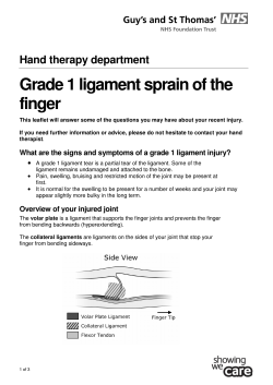
Technical Note
Arthroscopic Sternoclavicular Joint Resection Arthroplasty: A
Technical Note and Illustrated Case Report
Ryan J. Warth, M.D., Jared T. Lee, M.D., Kevin J. Campbell, B.S., and
Peter J. Millett, M.D., M.Sc.
Abstract: Open resection arthroplasty of the sternoclavicular (SC) joint has historically provided good long-term results
in patients with symptomatic osteoarthritis of the SC joint. However, the procedure is rarely performed because of the risk
of injury to vital mediastinal structures and concern regarding postoperative joint instability. Arthroscopic decompression
of the SC joint has therefore emerged as a potential treatment option because of many recognized advantages including
minimal tissue dissection, maintenance of joint stability, avoidance of posterior SC joint dissection, expeditious recovery,
and improved cosmesis. There are, however, safety concerns given the proximity of neurovascular structures. In this
article we demonstrate a technique for arthroscopic SC joint resection arthroplasty in a 26-year-old active man with
bilateral, painful, idiopathic degenerative SC joint osteoarthritis. This case also highlights the pearls and pitfalls of
arthroscopic resection arthroplasty for the SC joint. There were no perioperative complications. Four months postoperatively, the patient had returned to full activities, including weightlifting, without pain or evidence of SC joint
instability. One year postoperatively, the patient showed substantial improvements in the American Shoulder and Elbow
Surgeons score; Single Assessment Numeric Evaluation score; Quick Disabilities of the Arm, Shoulder and Hand score; and
Short Form 12 Physical Component Summary score over preoperative baseline values.
S
ymptomatic osteoarthritis of the sternoclavicular
(SC) joint is an uncommon pathology that can
produce significant pain and disability in some patients.
Although conservative management typically results in
symptomatic improvement, a small subset of patients
will have recalcitrant pain and continued shoulder
disability. Surgical management is therefore indicated
after a course of nonoperative therapy fails to provide
symptomatic relief.
Traditionally, open resection arthroplasty of the SC
joint has been the treatment of choice in patients with
From the Steadman Philippon Research Institute (R.J.W., J.T.L., K.J.C.,
P.J.M.), Vail, Colorado; and The Steadman Clinic (J.T.L., P.J.M.), Vail,
Colorado, U.S.A.
The authors report the following potential conflict of interest or source of
funding in relation to this article: Steadman Philippon Research Institute,
501(c) nonprofit. Ossur, Smith & Nephew Endoscopy, Siemens Medical Solutions, and Opedix. Corporate sponsors for the Steadman Philippon Research
Institute. P.J.M. receives support from Arthrex, GameReady, and Vumedi.
Received August 2, 2013; accepted September 23, 2013.
Address correspondence to Peter J. Millett, M.D., M.Sc., Center for
Outcomes-based Orthopaedic Research, Steadman Philippon Research Institute, 181 W Meadow Dr, Ste 1000, Vail, CO 81657, U.S.A. E-mail: drmillett@
thesteadmanclinic.com
! 2014 by the Arthroscopy Association of North America
2212-6287/13545/$36.00
http://dx.doi.org/10.1016/j.eats.2013.09.019
Arthroscopy Techniques, Vol
-,
intractable pain and dysfunction as a result of SC joint
osteoarthritis. Good to excellent outcomes have been
reported after open resection arthroplasty in several
series1-3; however, an open approach to the SC joint
requires some posterior dissection, potentially placing
vital mediastinal structures at risk. In addition, overresection of the medial clavicle with a large capsulotomy may lead to postoperative instability because of
the need for intraoperative ligament detachment.
As reported by Tavakkolizadeh et al.4 in 2009,
arthroscopy of the SC joint was originally thought to
enhance patient outcomes by improving visualization,
minimizing surgical trauma, limiting the risk to vital
retrosternal structures, avoiding postoperative instability, and hastening recovery times while also
improving cosmesis. Because of these advantages and
with the addition of new encouraging outcomes data,5
SC joint arthroscopy has become a viable and, potentially, favorable modality for the treatment of symptomatic SC joint osteoarthritis in carefully selected
patients. However, very few orthopaedic surgeons or
arthroscopists have experience with the technique.
Therefore the purposes of this article are (1) to
describe pertinent arthroscopic anatomy related to the
SC joint, (2) to present and demonstrate a technique for
arthroscopic SC joint resection arthroplasty, and (3) to
No
-
(Month), 2013: pp e1-e9
e1
e2
R. J. WARTH ET AL.
illustrate a case in which bilateral SC joint decompressions were necessary in a young, active patient
with symptomatic degenerative osteoarthritis of the SC
joint.
Surgical Anatomy
Ligamentous Stabilizers
Relevant periarticular ligaments involved with joint
stability include the anterior and posterior capsular
ligaments, the intra-articular disk ligament, the interclavicular ligament, and the costoclavicular ligament.
The anterior capsular ligament is a discrete thickening
of the anterior capsule that overlies the costoclavicular
ligament. The ligament runs from the posterior aspect
of the manubrium to the posterior and superior aspect
of the medial clavicle, just superior to the articular
surface. Capsular tissue that is inferior and lateral to the
anterior capsular ligament is relatively patulous and has
been described as a preferred location for intra-articular
injections and arthroscopic portal placement.6
The posterior capsular ligament corresponds to a
thickening of the entire posterior capsule. The ligament
arises on the posterior aspect of the manubrium and
attaches to the posterosuperior aspect of the medial
clavicle near the insertion of the anterior capsular ligament. Several studies have found the posterior
capsular ligament to be the most important ligament for
anteroposterior stabilitydrestoring its function has
been the primary focus of newer reconstruction techniques.7,8
The interclavicular ligament is a thick band that runs
along the superior bony surfaces between both the left
and right SC joints and inserts superomedially onto
each SC joint capsule. Contrary to previous reports,8,9
we have consistently found the interclavicular ligament
in all specimens. In a recent cadaveric study performed
at our institution, we found that the central portion of
the ligament has limited connectivity to the superior
aspect of the manubrium and likely plays a minimal
role in resisting superior migration of the medial clavicle as a result of a downward force on the distal clavicle
(unpublished data, Steadman Philippon Research
Institute, 2013).
The intra-articular disk ligament is a very thick and
dense ligament that arises from the chondral junction of
the first rib and passes through the SC joint to create 2
separate joint spaces. The intra-articular portion of the
ligament, commonly referred to as the intra-articular
disk, covers the cartilaginous portion of the medial
clavicle and attaches posteriorly and superiorly to the
articular cartilage. At its periphery, the intra-articular
disk also forms various attachments with the SC joint
capsule, thus reinforcing the anterior and posterior
capsular ligaments. Several studies have documented
variable morphologies of the intra-articular disk.
DePalma10 found the intra-articular disk to be complete
in 97% of specimens. However, other authors have
found that up to 52% of patients may have an
incomplete disk.6,9,11,12 van Tongel et al.6 found an
association between degenerative articular disks and
increased articular surface degeneration, both of which
increased with patient age. Histologic studies have
found the intra-articular disk to be a fibrocartilaginous
structure similar to the menisci in the knee and the
intra-articular disk of the acromioclavicular joint.12 The
disk is more closely associated with the clavicle than the
sternum, and most of the motion across the SC joint
occurs between the articular cartilage of the manubrium and the medial surface of the disk.13 The primary
function of the intra-articular disk has been debated in
the past; however, researchers have suggested that the
disk functions to decrease perceived bony incongruity
between the articular surfaces10 and to diminish the
force transmission between the medial clavicle and the
manubrium.14
The costoclavicular ligament, also known as the
rhomboid ligament, is a stout ligament found between
the costochondral junction of the first rib and the
costoclavicular tubercle on the inferior aspect of the
medial clavicle. Cave15 described the ligament has being
composed of 2 separate anterior and posterior fascicles
with an intervening bursa. The anterior fascicle ran
from medial to lateral and the posterior fascicle ran
from lateral to medial, resulting in a “twisted” appearance. However, our recent dissections have indicated
that the costoclavicular ligament sometimes exists as a
single, robust ligament since we were unable to reliably
distinguish between anterior and posterior fasciculi
(unpublished data, Steadman Philippon Research
Institute, 2013). Several authors have proposed that the
costoclavicular ligament is the predominant stabilizer of
the medial clavicle.1,15,16 It has been shown that preservation of this ligament is essential to maintain stability after resection arthroplasty. Echoing the results of
other authors,17 we recommend resection of no more
than 8 to 10 mm of bone from the medial clavicle to
preserve the many functions of the intact costoclavicular ligament.
Chondral Surfaces
The articular surfaces of the medial clavicle have been
described as being composed of hyaline cartilage;
however, histologic studies have suggested that
fibrocartilage may become predominant as a person
ages.10,18 In our dissection we found that only 67% of
the medial clavicle was covered with articular cartilage
(unpublished data, Steadman Philippon Research
Institute, 2013)dmost of this cartilage was found
anteriorly and inferiorly, confirming the results reported by other authors.6 The remaining portions of the
medial clavicle that were not covered with articular
ARTHROSCOPIC SC JOINT RESECTION ARTHROPLASTY
cartilage served as attachment sites for the interclavicular ligament, the anterior and posterior SC ligaments, and the intra-articular disk. In our dissection of
the medial clavicle, we also identified a tubercle located
anteriorly at approximately the 9:30 clock-face position
(end-on view of the right clavicle from medial to
lateral) that extended longitudinally along the insertion
of the clavicular head of the pectoralis major muscle
(pectoralis ridge). We believe this to be an important
landmark to assess rotational alignment of the medial
clavicle during ligament reconstruction and orientation
during SC joint arthroscopy (Fig 1).
Case Description
A 26-year-old right handedominant office worker
sustained an anterior labral tear to his left shoulder
while weightlifting approximately 2 years before presentation. He underwent arthroscopic labral repair by a
surgeon in his hometown and, during rehabilitation,
began to have pain in both the right and left SC joints.
He went back to his surgeon, who then found an
anterior labral tear in the patient’s right shoulder for
which he underwent arthroscopic repair. However, the
SC joint pain was never alleviated despite extensive
physical therapy and multiple corticosteroid injections.
Because of this pain, he was very limited in his recreational activities and sought treatment by the senior
surgeon.
Fig 1. (A) Cadaveric dissection photograph of a right medial clavicle from
anterior to posterior showing the
insertional area of the anterior capsular
ligament, posterior capsular ligament,
and intra-articular disk ligament
(asterisk). The intra-articular disk can
also been seen as it covers the articular
cartilage of the medial clavicle (arrow).
(B) End-on view of right medial clavicle
with remaining intra-articular disk. (C)
The intra-articular disk is flipped upward, showing the articular cartilage of
the medial clavicle. Small perforations
can be seen through the intra-articular
disk (solid arrow). The pectoralis ridge
is present at the anterior 9:30 clock-face
position (dashed arrow) and may be an
important landmark to aid in joint
orientation during arthroscopy of the
SC joint. (D) With the intra-articular
disk removed, it can be seen that the
articular cartilage covers approximately
67% of the medial clavicle. The pectoralis ridge can also be identified
anteriorly at the 9:30 clock-face position (dashed arrow).
e3
On physical examination, glenohumeral range of
motion was full and symmetric bilaterally. The patient
had full rotator cuff strength with negative apprehension, Speed, and O’Brien tests. He was tender to
palpation over both SC joints with mild swelling and
with no evidence of instability. He had moderate
crepitation over his SC joints with a simulated throwing
motion; however, no subluxation or dislocation was
observed. Outside magnetic resonance images showed
bilateral SC joint degeneration with possible degeneration of the intra-articular disks. A computed tomography scan showed cystic and sclerotic changes of both
medial clavicles with manubrial irregularity consistent
with SC joint osteoarthritis. A small subchondral cyst
was also noted on the anterior aspect of the left medial
clavicle (Fig 2). A diagnosis of bilateral SC joint
degenerative osteoarthritis without instability was
made. Because of recalcitrant pain and failed nonoperative therapy, the patient elected to undergo surgical
treatment with arthroscopic SC joint decompression
after an extensive discussion of the risks and benefits of
surgery. We recommend a staged approach. Thus
arthroscopic decompression of the right SC joint was
undertaken first, and the left SC joint was treated 17
days later.
Surgical Technique
In this case, the surgical techniques for SC joint
decompression were identical for both the left and right
e4
R. J. WARTH ET AL.
Fig 2. Computed tomography scans
obtained at initial clinical presentation.
(A) A coronal slice shows irregularity
and sclerotic changes of both medial
clavicles. (B) A subsequent coronal slice
shows irregularities of the manubrial
articular facets; however, this finding is
not uncommon in asymptomatic individuals.21 (C) An axial slice shows a
subchondral cyst on the left medial
clavicle (arrow), which was removed
with an arthroscopic curette (Fig 6).
Sclerotic changes can also be seen over
the articular surfaces of both medial
clavicles. (D) A subsequent axial slice
shows an intra-articular loose body
located within the right SC joint
(arrow).
SC joints. The intraoperative findings and therapeutic
interventions for both joints are summarized in Table 1.
A detailed description of the procedure for decompression of the patient’s left SC joint is provided later
and in Video 1.
Equipment and Personnel
In addition to standard arthroscopic equipment, a
radiofrequency tissue ablator (ArthroCare, Austin, TX)
and 30! and 70! 2.7-mm-diameter arthroscopes
(Smith & Nephew Endoscopy, Andover, MA) were used
to achieve complete visualization of the articular
surfaces and the intra-articular disk. The on-call
thoracic surgeon was made aware of the procedure, and
a thoracotomy tray was also immediately available in
the event of inadvertent thoracic penetration of
arthroscopic instruments. The infusion pump was set at
a pressure of 30 mm Hg to ensure capsular distention, to
maintain adequate visualization, and to achieve hemostasis throughout the procedure. Because of technical complexity, a skilled first assistant was necessary
to aid in patient safety. The role of the assistant was to
help with patient positioning, arthroscopic visualization,
and intraoperative dynamic examination such that the
Table 1. Summary of Intraoperative Findings, Therapeutic Interventions, and Instrumentation Required for Therapeutic
Interventions
Joint
Intraoperative Findings
Therapeutic Intervention
Right SC joint
Synovitis
Loose bodies
Intra-articular disk tear
Synovectomy
Morcellation
Total discectomy
Chondral degeneration of
clavicular facet
Chondral degeneration of
manubrial facet
Synovitis
Complete intra-articular disk
<8 mm of bone resected
Left SC joint
Debridement of entire chondral surface
Synovectomy
Total discectomy
Chondral degeneration of
clavicular facet
<8 mm of bone resected
Subchondral cyst
Chondral degeneration of inferior
manubrial facet
Complete excision
Debridement of inferior chondral surface
Instrumentation
Required
ArthroCare radiofrequency probe
Arthrex 4.0-mm high-speed shaver
ArthroCare arthroscopic biter
ArthroCare hook-tip radiofrequency probe
Arthrex 4.0-mm high-speed burr
Arthrex 4.0-mm high-speed shaver
Arthrex 4.0-mm high-speed shaver
ArthroCare radiofrequency probe
ArthroCare arthroscopic biter
ArthroCare hook-tip radiofrequency probe
Arthrex 4.0-mm full-radius resector
Arthrex 4.0-mm high-speed burr
Arthrex 4.0-mm high-speed shaver
ArthroCare manual rasp
ArthroCare angled curette
Arthrex 4.0-mm high-speed shaver
ARTHROSCOPIC SC JOINT RESECTION ARTHROPLASTY
e5
operation could be completed in a safe and efficient
manner.
Fig 3. A sterile marking pen was used to outline the surface
contours of the medial clavicle, sternal facet, and jugular
notch. Portal positions (circles) were planned to minimize
damage to the anterior SC ligament.
Patient Positioning
The patient was taken to the operating room and was
placed supine in approximately 30! of reverse Trendelenburg inclination. After the induction of general
anesthesia, an endotracheal tube was placed to ensure a
secure airway given the proximity of the trachea to the
operative field. A bolster was placed beneath the
thoracic spine and between the scapulae to retract the
scapulae, thus increasing the operating space between
the medial clavicle and the manubrium. At this point,
examination under anesthesia was undertaken and
showed no evidence of either SC joint instability or
glenohumeral instability. The left upper extremity was
Fig 4. (A) After the spinal needle was inserted with 30! of caudal angulation from the horizontal plane, the joint was insufflated
with normal saline solution to distend the joint capsule and to aid in arthroscope angulation. (B) After the spinal needle was
removed, a small stab incision was made in the same location with a No. 11 blade. (C) The 2.7-mm arthroscope was gradually and
bluntly inserted into the clavicular compartment of the joint with approximately 30! of caudal angulation from the horizontal plane
to maximize visualization while also minimizing damage to the anterior SC ligament.19,22 A distinct “pop” was heard as the
arthroscope penetrated the joint capsule and therefore entered the SC joint. (D) Under direct visualization through the inferior
viewing portal, a spinal needle was inserted into the joint through the previously marked site for superior portal placement with
approximately 30! of caudal angulation from the vertical plane (outside-in technique). (E) After removal of the spinal needle, a
second small stab incision was made in the same location to prepare for insertion of arthroscopic working instruments using the
same angulation as the previously removed spinal needle. Fraying of the intra-articular disk was also seen in the accompanying
arthroscopic view from the inferior portal (asterisk). (F) Arthroscopic instruments were inserted with approximately 30! of caudal
angulation from the vertical plane to minimize damage to the anterior SC ligament. Diagnostic arthroscopy and debridement were
then begun.
e6
R. J. WARTH ET AL.
Fig 5. Arthroscopic view from the inferior portal showing
significant chondral wear of the left medial clavicle before
joint decompression. A large chondral flap can be identified
(arrow) in addition to significant fraying of the medial
clavicular articular cartilage (asterisk).
then widely prepared and draped free to the contralateral midclavicular line such that intraoperative dynamic examination could be performed without
compromising sterility. The floor controls for the
arthroscopic instruments were situated such that the
operating surgeon could stand on the side of the
affected shoulder while also facing the high-definition
arthroscope output monitors, which were situated
above the head of the operating table. The assistant was
positioned near the left shoulder of the operating surgeon throughout the case.
Fig 6. Arthroscopic view from the superior portal after
resection of approximately 7 mm of bone from left medial
clavicle articular facet. Resection of 5 to 10 mm of bone is
necessary to expose the underlying cancellous bone without
damaging the supporting ligaments, such as the costoclavicular and superior SC ligaments.17,19 In this case approximately 7 mm of bone was resected.
Portal Placement
The SC joint was palpated to identify important bony
landmarks. A marking pen was then used to outline the
coronal contour of the medial clavicle, the manubrial
articular facet, and the jugular notch to aid in arthroscopic portal placement (Fig 3). Approximately 1 cm
inferior to the inferior joint line, an 18-gauge spinal
needle was introduced into the joint with 30! of
cephalad angulation from the horizontal plane. The
joint was then insufflated with normal saline solution
to distend the joint capsule and to determine the
appropriate angle for portal placement. After removal
of the spinal needle, a small stab incision was made
through the skin and subcutaneous tissues with a No.
11 blade. The 2.7-mm arthroscope was gently inserted
into the clavicular (lateral) compartment of the SC
joint, again with 30! of cephalad angulation, with care
taken not to penetrate or otherwise damage the posterior capsule because this has been shown to be the
most important ligament for maintaining anteroposterior SC joint stability.7,8 A distinct “pop” was
heard as the arthroscope bluntly penetrated the anterior joint capsule and thus entered the joint. Once this
inferior viewing portal was established, an accessory
superior portal was created under direct arthroscopic
visualization by an outside-in technique. A second
small stab incision was made 1 cm superior to the superior joint line to avoid damaging the anterior SC
ligament.19,20 Arthroscopic instruments were then
inserted using a technique similar to that described
earlier; however, the devices were initially angulated
inferiorly toward the inferior portal with approximately
30! of caudal angulation from the vertical plane.
Figure 4 shows the step-by-step procedure for establishing arthroscopic portals.
Diagnostic Arthroscopy
After creation of both arthroscopic portals, diagnostic
arthroscopy was performed using a combination of 30!
and 70! arthroscopes to fully evaluate the joint. Diagnostic arthroscopy involves inspection of the intraarticular disk and a complete evaluation of the clavicular
articular surface. Because the disk is typically complete,
the sternal articular surface can only be seen by repositioning the arthroscope more medially after the disk
has been resected. A switching stick may be used to
rotate the working and viewing portals, which can aid in
complete visualization. In this case diagnostic arthroscopy showed significant synovitis, a complete intraarticular disk, and chondral damage involving both the
sternal and clavicular facets of the SC joint (Fig 5).
Joint Decompression
Arthroscopic synovectomy was first performed with a
radiofrequency probe (ArthroCare) to aid in visualization while also removing a potential source of joint
e7
ARTHROSCOPIC SC JOINT RESECTION ARTHROPLASTY
Fig 7. (A) As viewed through the
inferior portal, a subchondral cyst,
which was identified on a preoperative
computed tomography scan (Fig 1C),
was successfully located on the anterior
aspect of the medial clavicle during
arthroscopic decompression of the left
SC joint. An angled curette was introduced into the joint in preparation for
cyst removal. (B) By use of an angled
curette, the subchondral cyst was
completely removed and its margins
were resected to a stable border.
pain. The clavicular facet was visualized, and 6 to 7 mm
of bone was resected with a 4.0-mm high-speed burr
and shaver (Arthrex, Naples, FL), creating approximately 12 mm of space between the clavicular and
manubrial articular facets (Fig 6). A small subchondral
cyst, which was identified on a preoperative computed
tomography scan, was also removed from the medial
clavicle with an angled curette (Fig 7). The intraarticular disk, which was found to be complete without
central perforation, was resected to a stable margin
using an arthroscopic biter and a hook-tip radiofrequency probe (ArthroCare). Remnants of the intraarticular disk were removed with a 4.0-mm high-speed
full-radius resector (Arthrex). The sternal facet was
then visualized and debrided inferiorly where chondral
wear was clearly evident. A remnant of the intraarticular disk was also removed from this area
(Fig 8). Bone was not resected from the sternal facet.
Throughout the decompression procedure, care was
taken to preserve the anterior, posterior, and superior
SC joint capsule and associated ligaments. Under direct
visualization through both arthroscopic portals, dynamic SC joint examination was undertaken by moving the arm through a full range of motion to ensure
joint stability and relief of bony impingement. The
portal sites were then closed with buried No. 3-0
absorbable sutures (Monocryl; Johnson & Johnson,
Fig 8. (A) Arthroscopic view from the
superior portal showing articular
degeneration of the inferior aspect of the
sternal facet. A remnant of the previously resected intra-articular disk can be
seen (asterisk). (B) The tip of the probe
points to the inferior aspect of the sternal
facet after debridement of chondral wear
and resection of the intra-articular disk
remnant. The clavicular facet can also be
seen (asterisk), highlighting the relative
preservation of the sternal facet relative
to the clavicular facet, which has been
recommended by other authors.5,19
New Brunswick, NJ). The patient was placed in a sling
and a cryotherapy device (GameReady [CoolSystems],
Concord, CA) for comfort before transfer to the postanesthesia care unit. The shoulder was not immobilized postoperatively.
Postoperative Rehabilitation
Early passive motion was initiated and encouraged in
the immediate postoperative period. Active and activeassisted motion began at approximately 1 week postoperatively, and the sling was used for the first 2 weeks
for comfort, followed by a gradual return to normal
activities.
Results
The patient returned to his high-demand recreational
activities, including weightlifting, without modification
at approximately 4 months postoperatively. One year
after surgery, the American Shoulder and Elbow Surgeons score improved by 52 points; the Single Assessment Numeric Evaluation score improved by 31 points;
the Quick Disabilities of the Arm, Shoulder and Hand
score improved by 38.8 points; and the Short Form 12
Physical Component Summary score improved by 17.9
points (Table 2). Results may be optimized after
consideration of the pearls and pitfalls presented in
Table 3.
e8
R. J. WARTH ET AL.
Table 2. Summary of Outcome Scores Obtained 12 Months
After Bilateral Arthroscopic SC Joint Decompression
Outcomes Measure
Preoperative Postoperative
ASES score (scale from 0 to 100,
where 100 indicates best score)
SANE score (scale from 0 to 100,
where 100 indicates best score)
QuickDASH score (scale from 100 to 0,
where 0 indicates best score)
SF-12 PCS score (scale from 0 to 100,
where 100 indicates best score)
43
95
60
91
41.1
2.3
33.9
51.4
ASES, American Shoulder and Elbow Surgeons; QuickDASH, Quick
Disabilities of the Arm, Shoulder and Hand questionnaire; SANE,
single assessment numeric evaluation; SF-12 PCS, Short Form 12
Physical Component Summary.
Discussion
Several authors have reported satisfactory outcomes
after resection arthroplasty of the degenerative SC
joint.1-3 Rockwood et al.1 reported good to excellent
outcomes in 8 patients with SC joint arthritis after open
resection with a mean follow-up of 5.7 yearsdall patients were completely satisfied with their results.
Pingsmann et al.2 evaluated outcomes in 8 women who
underwent open isolated resection arthroplasty of the
SC joint for painful arthritis. Again, all 8 patients were
completely satisfied with the result at a mean follow-up
of 31 months. In patients with anterior instability,
Panzica et al.3 also reported good to excellent outcomes
in 6 patients treated with open resection arthroplasty
with a mean follow-up of 9.9 years.
Although good to excellent outcomes have been reported after open resection arthroplasty of the SC joint,
this operation is rarely performed because of concern
regarding vital mediastinal structures located deep to
the operative field and a general unfamiliarity with
local surgical anatomy. In addition to a prolonged recovery period, open capsulotomy can compromise the
patency of supporting ligaments that confer joint stability. Arthroscopy of the SC joint, on the other hand,
has several advantages that are typically viewed as
improvements over the open approach. Arthroscopy
allows for a 360! view from within the joint, thus
allowing for a complete evaluation of all the joint surfaces and the intra-articular disk. Surgical risks to the
mediastinal vessels, trachea, and supporting SC ligaments are mitigated when proper instrument insertion
technique is used and when bony resection of the
medial clavicle does not exceed 10 mm.17 When
compared with the open approach, arthroscopy of the
SC joint may also hasten recovery and provide excellent
cosmesis.
In this case, bilateral arthroscopic SC joint resections
were performed in a young, active male patient without
Table 3. Valuable Pearls and Pitfalls for Arthroscopic Decompression of SC Joint
Preoperative imaging
Equipment and personnel
Patient positioning
Portal placement
Diagnostic arthroscopy
SC joint decompression
Postoperative rehabilitation
Pearls
Pitfalls
MRI is used for evaluation of the intra-articular disk.
CT scan is used for evaluation of joint congruity, joint
angulation, cyst formation, and loose bodies.
Endotracheal intubation should always be performed
because the operative field is nearly immediately superficial
to the trachea and mediastinal vessels.
Always work with a skilled first assistant.
At a minimum, always notify an on-call general surgeon of
the procedure and always have a thoracotomy tray
immediately available.
Use an interscapular bolster of sufficient size to open up
both SC joints anteriorly.
Evaluate the patient for SC joint instability (EUA) to
eliminate the possibility that instability may have been
present before the surgical procedure.
Initial joint insufflation may be incomplete in patients with
an intact intra-articular disk.
The inferior portal should be created first with 30! cephalad
angulation from the horizontal plane.
The superior portal should be created using an outside-in
technique under direct visualization with a spinal needle.
Always first identify the posterior capsule to ensure that the
arthroscope is situated within the joint.22
Bone should not be resected from the sternal facet.19,22
Irregularity of the manubrium is a common
imaging finding and should not elevate suspicion
for joint degeneration.21
Lack of experience of the primary surgeon and/or
supporting personnel may place the patient at risk
of severe complications.
Passive motion should be encouraged immediately
after surgery.
A sling should only be used for comfort.
An interscapular bolster that is too small will not
create the space needed to operate effectively.
Portals initially created either too perpendicular or
too parallel to the coronal plane minimize
visualization and working space.
Instruments inserted hastily or with high force and
speed place the posterior SC ligament and vital
retrosternal structures at risk of perforation.
Lack of a systematic approach may result in
missed diagnoses.
Removing >10 mm of the medial clavicle increases
the risk of postoperative SC joint instability.17
Immobilization of the affected arm is unnecessary
and increases the risk of shoulder stiffness.
CT, computed tomography; EUA, examination under anesthesia; MRI, magnetic resonance imaging.
ARTHROSCOPIC SC JOINT RESECTION ARTHROPLASTY
perioperative complications or postoperative SC joint
instability. The patient returned to his preinjury level of
function 4 months postoperatively and showed substantial improvements in outcomes scores over his
preoperative baseline values at 1 year of follow-up
(Table 2). Recently, Tytherleigh-Strong and Griffith5
also reported no complications and no cases of postoperative instability in a series of 10 patients with SC
joint osteoarthritis treated with arthroscopic medial
clavicle excision. After a mean follow-up period of 28
months, these 10 patients showed considerable improvements in outcomes scores compared with preoperative baseline values.
Despite its numerous advantages, arthroscopy of the
SC joint is an uncommon procedure and should not be
performed by inexperienced surgeons. Generally, this
procedure should only be performed in high-volume
SC joint referral centers with cardiothoracic backup in
which surgeon experience and anatomic familiarity are
sufficient to safely and correctly perform the operation.
When performed in experienced hands, however,
arthroscopic SC joint decompression can be a safe and
effective treatment method for symptomatic SC joint
osteoarthritis.
References
1. Rockwood CA Jr, Groh GI, Wirth MA, Grassi FA. Resection arthroplasty of the sternoclavicular joint. J Bone Joint
Surg Am 1997;79:387-393.
2. Pingsmann A, Patsalis T, Michiels I. Resection arthroplasty
of the sternoclavicular joint for the treatment of primary
degenerative sternoclavicular arthritis. J Bone Joint Surg Br
2002;84:513-517.
3. Panzica M, Zeichen J, Hankemeier S, Gaulke R, Krettek C,
Jagodzinski M. Long-term outcome after joint reconstruction or medical resection arthroplasty for anterior SCJ
instability. Arch Orthop Trauma Surg 2010;130:657-665.
4. Tavakkolizadeh A, Hales PF, Janes GC. Arthroscopic
excision of sternoclavicular joint. Knee Surg Sports Traumatol Arthrosc 2009;17:405-408.
5. Tytherleigh-Strong G, Griffith D. Arthroscopic excision of
the sternoclavicular joint for the treatment of sternoclavicular osteoarthritis. Arthroscopy 2013;29:1487-1491.
6. van Tongel A, MacDonald P, Leiter J, Pouliart N, Peeler J.
A cadaveric study of the structural anatomy of the sternoclavicular joint. Clin Anat 2012;25:903-910.
e9
7. Spencer EE, Kuhn JE, Huston LJ, Carpenter JE, Hughes RE.
Ligamentous restraints to anterior and posterior translation
of the sternoclavicular joint. J Shoulder Elbow Surg 2002;11:
43-47.
8. Bearn JG. Direct observations on the function of the
capsule of the sternoclavicular joint in the clavicular
support. J Anat 1967;101:159-170 (pt 1).
9. Renfree KJ, Wright KW. Anatomy and biomechanics of
the acromioclavicular and sternoclavicular joints. Clin
Sports Med 2003;22:219-237.
10. DePalma AF. Surgical anatomy of the acromioclavicular
and sternoclavicular joints. Surg Clin North Am 1963;43:
1541-1550.
11. Shimada K, Takeshige N, Moriyama H, Miyauchi Y,
Shimada S, Fujimaki E. Immunohistochemical study of
extracellular matrices and elastic fibers in a human
sternoclavicular joint. Okajimas Folia Anat Jpn 1997;74:
171-179.
12. Emura K, Arakawa T, Terashima T, Miki A. Macroscopic
and histological observations on the human sternoclavicular joint disc. Anat Sci Int 2009;84:182-188.
13. Barbaix E, Lapierre M, Van Roy P, Clarijs JP. The sternoclavicular joint: Variants of the discus articularis. Clin
Biomech (Bristol, Avon) 2000;15(suppl 1):S3-S7.
14. Brossman J, Stäbler A, Preidler K, Trudell D, Resnick D.
Sternoclavicular joint: MR imagingdAnatomic correlation. Radiology 1996;198:193-198.
15. Cave AJ. The nature and morphology of the costoclavicular ligament. J Anat 1961;95:170-179.
16. Tubbs RS, Shah NA, Sullivan BP, et al. The costoclavicular
ligament revisited: A functional and anatomic study. Rom
J Morphol Embryol 2009;50:475-479.
17. Bisson LJ, Dauphin N, Marzo JM. A safe zone for resection of the medial end of the clavicle. J Shoulder Elbow Surg
2003;12:592-594.
18. Jurik AG. Imaging of the sternocostoclavicular region. New
York: Springer; 2007;29-35.
19. Tytherleigh-Strong G, Getgood AM, Griffiths DE.
Arthroscopic intra-articular disk excision of the sternoclavicular joint. Am J Sports Med 2012;40:1172-1175.
20. Wijeratna MD, Turmezei TD, Tytherleigh-Strong G.
The relevant anatomy of the sternoclavicular joint in
relation to arthroscopic surgical approaches. Skeletal Radiol
2013;42:473-478.
21. Tuscano D, Banerjee S, Terk MR. Variations in normal
sternoclavicular joints; a retrospective study to quantify
SCJ asymmetry. Skeletal Radiol 2009;38:997-1001.
22. Tytherleigh-Strong G. Arthroscopy of the sternoclavicular
joint. Arthrosc Tech 2013;2:e141-e145.


© Copyright 2026