
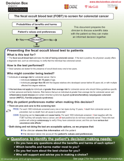
Customizing chemotherapy for colon cancer: the potential of gene expression profiling
Drug Resistance Updates 7 (2004) 209–218 Customizing chemotherapy for colon cancer: the potential of gene expression profiling John M. Mariadason a,∗ , Diego Arango b , Leonard H. Augenlicht a a Department of Oncology, Montefiore Medical Center, Albert Einstein Cancer Center, 111, East 210th Street, Bronx, NY 10467, USA b Department of Medical Genetics, Biomedicum Helsinki, Haartmaninkatu 8, FIN-00014 University of Helsinki, Finland Received 7 May 2004; accepted 19 May 2004 Abstract The value of gene expression profiling, or microarray analysis, for the classification and prognosis of multiple forms of cancer is now clearly established. For colon cancer, expression profiling can readily discriminate between normal and tumor tissue, and to some extent between tumors of different histopathological stage and prognosis. While a definitive in vivo study demonstrating the potential of this methodology for predicting response to chemotherapy is presently lacking, the ability of microarrays to distinguish other subtleties of colon cancer phenotype, as well as recent in vitro proof-of-principle experiments utilizing colon cancer cell lines, illustrate the potential of this methodology for predicting the probability of response to specific chemotherapeutic agents. This review discusses some of the recent advances in the use of microarray analysis for understanding and distinguishing colon cancer subtypes, and attempts to identify challenges that need to be overcome in order to achieve the goal of using gene expression profiling for customizing chemotherapy in colon cancer. © 2004 Elsevier Ltd. All rights reserved. Keywords: Microarray; Gene expression profiling; Colon cancer; 5-FU 1. Introduction Colorectal cancer is a leading cause of cancer related death in the western world. However, current treatment strategies for this disease are far from optimal, due in part to the inability to accurately distinguish subgroups of patients that differ in their prognosis and the probability of response to treatment. Based upon their histopathology, colorectal cancers are classified utilizing either the traditional Dukes staging system or the tumor node mucosa (TNM) staging system (Compton, 2002). While use of the TNM staging system is now becoming standard practice, we will use the Dukes staging system throughout this review as it is the predominant staging system used in the studies discussed herein. Colorectal cancers are classified as Dukes A when the tumor is confined to the mucosa, Dukes B when locally advanced, Dukes C if positive for lymph node metastasis, and as Dukes D when distant metastases are detected at the time of diagnosis (Compton, 2002). While the Dukes (and TNM) staging system identifies broad patient groups that vary in their long-term prognosis, considerable heterogene∗ Corresponding author. Tel.: +1 718 920 2025; fax: +1 718 882 4464. E-mail address: [email protected] (J.M. Mariadason). 1368-7646/$ – see front matter © 2004 Elsevier Ltd. All rights reserved. doi:10.1016/j.drup.2004.05.001 ity exists within each of these stages with regards to both prognosis and response to chemotherapy. For treatment of colorectal cancer, 5-fluorouracil (5-FU) remains the agent with the highest clinical efficacy (Moertel et al., 1995a,b), and (often in combination with other agents), has become the standard treatment for patients with locally advanced and metastatic disease. However, approximately 80% of these patients do not benefit from this treatment, either because they have been surgically cured and need no further treatment, or because their tumor is refractory to 5-FU-based chemotherapy. Therefore, there is clear need to develop biomarkers capable of distinguishing between these patient sub-groups. First, patients unlikely to respond to 5-FU can be spared the toxicity, time, and expense associated with this treatment regimen, and more important, can be placed on alternate therapies. To this end, irinotecan and oxaliplatin are now effective alternative treatment options (Conti et al., 1996; Cvitkovic and Bekradda, 1999; de Gramont et al., 1997; Machover et al., 1996), while the anti-VEGF antibody and inhibitor of tumor angiogenesis, bevacizumab, was recently approved by the US FDA as a first-line therapy for metastatic colon cancer (Ferrara et al., 2004). Many chemotherapeutic agents also may promote the acquisition of multidrug resistance (Morin, 2003; Vasilevskaya and O’Dwyer, 2003). 210 J.M. Mariadason et al. / Drug Resistance Updates 7 (2004) 209–218 Administration of the agent likely to induce maximal response in the first course of treatment is therefore critical to enhance overall treatment success. Second, identification of markers that predict response may provide important biological insights into the mechanisms that determine response to therapy. Particularly, such studies may identify avenues by which tumor cells can be manipulated in order to maximize response to the primary agent. A good example was the identification of thymidylate synthase (TS) as the enzyme targeted by 5-FU, and the subsequent discovery that an excess of intracellular reduced folates are necessary for maximal inhibition of TS activity (Kinsella et al., 1997). In turn, clinical trials demonstrated that administration of 5-FU + folinic acid (leucovorin) resulted in improved response rates compared to administration of 5-FU alone (Dannenberg, 2002; Kinsella et al., 1997). For these reasons, the identification of biomarkers capable of predicting 5-FU response in colon cancer has been a topic of intense investigation (Longley et al., 2003). A number of studies have examined the predictive value of the 5-FU target enzyme, TS, and related enzymes that affect 5-FU metabolism including thymidine phosphorylase (TP) and dipyrimidine dehydrogenase (DPD) (Metzger et al., 1998; Salonga et al., 2000). However, while several reports have linked low TS expression with improved response to 5-FU in vivo, others have shown no relationship between these parameters (Allegra et al., 2003; Berglund et al., 2002; Findlay et al., 1997; Johnston et al., 2003). Likewise, in vitro, while TS is often overexpressed in cell lines selected for resistance to 5-FU by continuous exposure (Johnston et al., 1992), studies of unselected panels of cell lines have failed to consistently show a correlation between intrinsic cellular TS levels and 5-FU response (Grem et al., 2001; Mirjolet et al., 1998), suggesting alterations in TS levels may be more closely linked to acquired rather than intrinsic resistance to 5-FU. The predictive efficacy of TP is also unclear, with both high and low levels of TP linked to 5-FU response depending on whether the studies were performed in vitro or in vivo, respectively (Metzger et al., 1998; Saito et al., 1999; Schwartz et al., 1995). Several studies also suggest that factors involved in regulating cell growth and apoptosis, including p53, myc, and the ratio of anti-apoptotic to pro-apoptotic bcl-2 family members (Violette et al., 2002), can predict 5-FU response. For example, improved response to 5-FU and prolonged survival has been observed in patients with tumors wild type for p53 (Ahnen et al., 1998; Benhattar et al., 1996; Goh et al., 1995), although contrasting findings have also been observed (Allegra et al., 2003). In our own investigations, we have established that low level amplification of c-myc was associated with longer overall survival in response to 5-FU-based adjuvant therapy (Augenlicht et al., 1997). Furthermore, these findings were extended to demonstrate that tumors with amplification of c-myc, that also retained wild-type p53 function, had significantly improved response to 5-FU both in vitro and in vivo (Arango et al., 2001). The mechanistic explanation for this interaction was the demonstration that high levels of c-myc represses p53 induction of p21WAF1 in response to drug treatment, promoting the induction of apoptosis over cell cycle arrest (Arango et al., 2003; Seoane et al., 2002). It has also been reported that various allelic deletions, and mismatch repair status, may identify tumor subsets with differential 5-FU sensitivity. For example, tumors that retain heterozygosity at either 17p or 18q show improved response to 5-FU-based adjuvant therapy (Barratt et al., 2002). Likewise, tumors that are mismatch repair deficient have been reported to show improved response to 5-FU (Elsaleh et al., 2000, 2001; Elsaleh and Iacopetta, 2001), although studies reporting no difference, and the converse, have also been published (Barratt et al., 2002; Ribic et al., 2003). While plausible explanations for many of these discrepancies have been offered, including, for example, differences in the median age (which in turn may effect DNA methylation status) of the patient cohorts in the studies linking MMR status with response to 5-FU (Iacopetta et al., 2003; Ribic et al., 2003), the predictive efficacy of these markers remains insufficient to permit their use in routine clinical practice. A further limitation of the use of these predictors is that they are often designed to predict response to a specific agent (5-FU), and thus generally fail to identify alternative treatment options. A robust assay, capable of predicting the probability of response of a given tumor to the multiple chemotherapeutic regimens that are increasingly becoming available, would therefore, have significant clinical utility. 2. Gene expression profiling for the classification and prognosis of colorectal cancer The development of colorectal tumorigenesis requires mutation of multiple key genes (Fodde et al., 2001). In turn, these mutations, through both direct and indirect mechanisms and clonal selection, result in alterations in expression of hundreds if not thousands of genes (Notterman et al., 2001; Zou et al., 2002). Therefore, it is not surprising that colorectal tumors vary significantly in terms of prognosis and response to therapy. The complexity of the genetic abnormalities that define a given colorectal tumor argue that an assay capable of collectively considering all of this variability may be more informative for the classification and determination of prognosis and response to therapy. The sequencing of the human genome, combined with the development of high throughput screening technologies such as microarray analysis, now makes such an approach possible. The utility of gene expression profiling for the classification and prognosis of most cancer types, including leukemia (Bullinger et al., 2004; Golub et al., 1999), lung (Beer et al., 2002; Bhattacharjee et al., 2001), breast (Sorlie et al., 2001; Van de Vijver et al., 2002; Van ’t Veer et al., 2002), brain (Pomeroy et al., 2002), gastric (Boussioutas et al., 2003; Tay et al., 2003), prostate (Dhanasekaran et al., 2001) and ovarian J.M. Mariadason et al. / Drug Resistance Updates 7 (2004) 209–218 cancer (Schaner et al., 2003) is now clearly established. With regards to colorectal cancer, almost two decades ago Augenlicht et al. demonstrated that gene expression profiling could successfully distinguish between normal colonic mucosa, benign adenoma and malignant carcinoma (Augenlicht et al., 1987), and furthermore, that profiling the normal-appearing colonic mucosa could distinguish patients who were at elevated risk for tumor development (FAP and HNPCC family members), from low risk individuals (Augenlicht et al., 1991). More recently, and since the advent of modern microarray technologies, several studies have demonstrated the ability of gene expression profiling to distinguish among other clinically important colorectal cancer subgroups. Multiple studies have demonstrated the ability of microarrays to successfully distinguish between colonic tumor tissue and adjacent normal mucosa. Importantly, this can be done in an unsupervised manner, with no prior selection of differentially expressed genes (Alon et al., 1999; Bertucci et al., 2004; Notterman et al., 2001; Zou et al., 2002). Illustrating an additional layer of sensitivity of this methodology, Notterman et al. demonstrated that gene expression profiling could separate normal colonic tissue from adenoma, as well as adenoma from adenocarcinoma in an unsupervised manner (Notterman et al., 2001). Extending this further still, Bertucci et al. recently demonstrated that metastatic (Dukes D) versus non-metastatic colon tumors (Dukes A–C) could be distinguished using microarrays in an unsupervised manner (Bertucci et al., 2004). Finally, two studies from Torben Orntoft’s group demonstrated the capability of gene expression profiling to distinguish normal tissue from colorectal tumors of different Dukes staging, in an unsupervised manner (Birkenkamp-Demtroder et al., 2002; Frederiksen et al., 2003). However, while these results are encouraging, not all tumor stages were classified correctly, indicating the need for further methodological improvements, and possibly the use of supervised data analysis strategies, to improve classification. While unsupervised analyses have been successful at discriminating colon tumor from normal tissue, and to a lesser extent among colon tumors of different histopathological stage, the identification of gene expression profiles or signatures associated with more subtle subgroups of colorectal cancer have required the use of more complex, supervised, analytical approaches. Selaru et al. used a training set of samples to develop and train an artificial neural network (ANN) capable of discriminating sporadic colorectal adenomas and cancers, from inflammatory bowel disease-associated dysplasias and cancers. Importantly, when applied to a validation set of samples, this ANN was able to accurately discriminate between these two tumor types (Selaru et al., 2002). Gene subsets differentially expressed between tumors from the left and right colon, or from tumors with or without microsatellite instability, have also been described (Mori et al., 2003, 2004). However, while the gene lists generated in these studies are certainly interesting biologically, for the most part their predictive 211 powers have not been tested either internally (using a cross validation strategy such as a “leave one out” analysis), or externally, using an independent sample set. Collectively, these studies have significant clinical implications for improving the management of colorectal cancer. The ability of expression profiling to predict prognosis or the likelihood of metastasis (Bertucci et al., 2004), identifies patients that require more rigorous follow up and/or more aggressive treatment. Gene expression profiling has also shed considerable light on the molecular mechanisms and pathways that characterize transformation of the colonic epithelium, and have identified a number of potential targets for treatment. Examples of the latter include the secreted integrin-binding protein osteopontin, which was identified as progressively upregulated in parallel with the progression of colorectal carcinoma from adenoma, to B1 through D stage cancers, and finally liver metastases (Agrawal et al., 2002, 2003). Likewise, using the SAGE methodology, Saha et al. identified the protein tyrosine phosphatase, PRL-3, as a gene consistently upregulated in metastatic colon cancers (Saha et al., 2001). Similarly, and consistent with previous reports (Sarris and Lee, 2001), nucleoside diphosphate kinase (NM23) was identified using a combination of gene expression profiling and tissue microarrays, to be upregulated in colon tumors compared to the normal colonic mucosa, and further, to have higher expression levels in tumors with more favorable outcome (Bertucci et al., 2004). 3. Gene expression profiling for the prediction of response of colon cancer cells to chemotherapy While the ability of gene expression profiling to distinguish between tumor tissue and normal colonic mucosa, as well as among colon tumors of different histopathological grade has been demonstrated, a definitive study demonstrating the utility of this approach for prediction of response of colon cancer to chemotherapy is presently lacking. Nevertheless, a number of encouraging studies, both in other cancer types, and in colon cancer cell lines in vitro, highlight the potential of gene expression profiling for this purpose. For example, Kihara et al. demonstrated the feasibility of microarray-based expression profiling to predict survival in esophageal cancer patients receiving 5-FU based adjuvant chemotherapy (Kihara et al., 2001). The authors developed a “drug resistance score” based upon 52 genes each of whose level of expression was correlated with survival, and thus possibly response to 5-FU. This drug resistance score was shown to accurately predict survival in six independent patient samples (Kihara et al., 2001). Focusing specifically on colon cancer, we recently utilized a panel of 30 colon carcinoma cell lines to identify genes correlated with response to 5-FU in vitro (Mariadason et al., 2003) (Fig. 1). Furthermore, using a “leave one out” cross validation strategy, we formally demonstrated the ability of these genes to predict response to 5-FU-induced apoptosis. 212 J.M. Mariadason et al. / Drug Resistance Updates 7 (2004) 209–218 agents, or specific combinations of therapies, would be most appropriate for treatment of a specific tumor. However, while this study illustrates the potential of this methodology for predicting response of colon cancer to specific chemotherapeutic agents, it is limited by the fact that it was performed in vitro. Therefore, the immediate challenge remains the demonstration of the utility of gene expression profiling for the prediction of response in patients. Collection of such gene expression and supporting clinical data is ongoing at our and other institutions. 4. Gene expression profiling for understanding mechanisms of 5-FU action An additional utility of gene expression profiling studies is that they have provided a number of interesting biological insights into both the factors that determine 5-FU response, and the pathways induced upon 5-FU exposure. 4.1. Determinants of intrinsic 5-FU resistance and likelihood of response Fig. 1. Genes correlated with 5-FU-induced apoptosis. Response of panel of 30 colon cancer cell lines to 5 M 5-FU-induced apoptosis was determined by propidium iodide staining and FACS analysis following 72 h. Basal gene expression ratios of the same 30 cell lines were correlated with 5-FU-induced apoptosis, and 420 significantly correlated genes identified. Genes more highly expressed in 5-FU-sensitive cells are shown in cluster A, and those more highly expressed in 5-FU resistant cells are shown in cluster B. The predictive efficacy of this gene subset was subsequently validated using a leave one out cross validation analysis. Importantly, this study demonstrated that measurement of multiple, rather than single markers, results in more accurate prediction of drug response when compared to four previously reported determinants of 5-FU response—TS and TP activity, and p53 and MMR status. The study also illustrated the potential of gene expression profiling to predict response to each of many chemotherapeutic agents. This was shown by the fact that reanalysis of the same microarray data was able to identify a signature capable of predicting response to camptothecin (CPT). We have subsequently demonstrated similar efficacy for prediction of response to oxaliplatin, the non-steroidal anti-inflammatory drug sulindac, and the histone deacetylase-inhibitor butyrate, using the same gene expression data (unpublished findings). The ability to predict probability of response to different agents from a single assay has the added potential of determining whether single To identify genes that determine the probability of response to 5-FU, several studies have correlated basal levels of gene expression with the magnitude of 5-FU-induced apoptosis or growth arrest in panels of cell lines or human tumor xenografts. Consistent with published reports, some of these studies identified previously established determinants of 5-FU response. In the landmark study of Scherf et al, which generated a large matrix of data linking the basal gene expression profiles of the NCI panel of 60 cell lines with response to the multitude of drugs tested in the NCI screen, dipyrimidine dehydrogenase (DPD) was identified as highly negatively correlated with 5-FU response. This finding is consistent with DPD being the rate limiting enzyme in 5-FU metabolism, and supports the idea that high DPD levels may confer resistance to 5-FU by reducing exposure of cells to the active forms of 5-FU (Scherf et al., 2000). Similarly, Zembutsu et al. determined the basal gene expression profile of 85 human xenografts from a variety of cancers, and identified TS as negatively correlated with 5-FU response (Zembutsu et al., 2002). These studies also identified a number of novel links between basal levels of gene expression and 5-FU response. Using a panel of 39 cell lines from various tumor types, Dan et al. identified the anti-apoptotic gene survivin as more highly expressed in 5-FU resistant cells, while members of the aldo-keto reductase and aldehyde dehydrogenase families, and galectin 4, were more highly expressed in 5-FU responsive cells (Dan et al., 2002). Establishing a previously unrecognized link, Zembutsu et al. reported a significant correlation between expression levels of mdr3 and mdr4, and 5-FU response. In our own study using a panel of 30 colon cancer cell lines, we identified a positive correlation between basal expression levels of the pro-apoptotic Bak J.M. Mariadason et al. / Drug Resistance Updates 7 (2004) 209–218 protein and sensitivity to 5-FU. We validated this finding by demonstrating that Bak was localized to the mitochondria following 5-FU treatment, which in turn was linked to release of cytochrome c (Mariadason et al., 2003). Consistent with this finding, induction of Bak protein in response to 5-FU in colon cancer cell lines has previously been demonstrated (Nita et al., 1998). In addition to Bak, 5-FU response has also been linked to the basal levels of expression or mutation status of other bcl-2 family members including Bcl-2, Bcl-xL , Bax and Bid (Sax et al., 2002; Violette et al., 2002; Zhang et al., 2000). Our study also identified a subset of genes with a role in protein processing and folding, including a number of chaperones, as more highly expressed in 5-FU resistant cell lines. Chaperones can play a role in protecting cells from environmental stress by binding denatured proteins and dissociating protein aggregates (Leppa and Sistonen, 1997), raising the possibility that higher basal expression of such proteins may enhance the ability of these cells to survive following 5-FU-induced genotoxic stress. Consistent with this, an upregulation of stress response genes, including chaperonin 10 (Maxwell et al., 2003) and vitamin D3-upregulated protein 1 (Takahashi et al., 2002) have recently been reported in microarray studies examining genes induced in response to 5-FU treatment. A limitation of these studies, however, is that in many cases, it remains to be determined whether the genes identified confer sensitivity or resistance by directly modulating 5-FU action, or whether they represent surrogate markers of a broader response phenotype. 213 nism of 5-FU-induced cell killing (Hwang et al., 2001). Finally, using cDNA microarrays, Maxwell et al. identified a number of genes altered in expression by 5-FU in vitro (Maxwell et al., 2003). Examples of the genes upregulated by 5-FU included those with a role in signal transduction (K-ras), apoptosis (COP9 homolog, FLIP protein), cell cycle (cdk2, cdc2, cyclin G) and cell structure (gelsolin), as well as genes such as MAT-8, annexin II and IV and FGF receptor 2 which are expressed at the cell surface (Maxwell et al., 2003). 4.3. Determinants of acquired 5-FU resistance A few investigators have also applied expression profiling to gain insight into the mechanisms of acquired 5-FU resistance. Using microarrays in which a panel of cell lines selected for resistance to 5-FU were compared to parental cells, Wang et al. identified an upregulation of the 5-FU target gene, TS (and the closely linked Yes-1 gene), in 5-FU resistant cells (Wang et al., 2001). Importantly, using Southern blotting and fluorescence in situ hybridization, the increase in TS mRNA levels was linked to amplification of the TS gene, which is consistent with in vivo findings (Wang et al., 2004). Also upregulated in expression in 5-FU resistant cells were CAK1 antigen and alpha catenin, while checkpoint suppressor 1 and c-rel were downregulated in expression (Wang et al., 2001). 5. Challenges and recent technological advances 4.2. Genes induced in response to 5-FU Gene expression profiling studies have also been used to characterize genes induced in response to 5-FU treatment, and have identified a number of novel pathways modulated by this agent. Clarke et al. determined the effect of treatment of late stage colon tumors with 5-FU and mitomycin on gene expression in vivo, by comparing RNA extracted from biopsies isolated pre-treatment or following 6 weeks of continuous drug treatment (Clarke et al., 2003). They demonstrated a coordinate downregulation in expression of genes required for RNA and protein synthesis, including a number of HnRNP’s and snRNP’s, ribosomal proteins, translation initiation factors, tRNA synthases, and genes involved in protein folding, following 5-FU/mitomycin treatment. The authors interpret these changes as likely reflecting the inhibition of cell proliferation induced by 5-FU/mitomycin. A number of these genes have been shown to be regulated by c-myc (Boon et al., 2001; Coller et al., 2000), which was also downregulated by drug treatment, suggesting a possible mechanism for their coordinate regulation. Using SAGE analysis, Hwang et al. demonstrated that ferredoxin reductase (FR) was induced by 5-FU in vitro in a p53-dependent manner. Follow up studies demonstrated that FR may contribute to apoptosis via the excessive generation of reactive oxygen species, a previously unknown mecha- A major factor limiting the advancement of gene expression profiling as a tool for the prediction of response to chemotherapy, is access to fresh frozen patient tumor samples with documented clinical follow up. A strong emphasis needs to be placed in Surgical and Pathology departments on the importance of developing tumor banks of fresh frozen tissue. Without these samples, and follow-up information, the potential of the currently available technologies cannot be fully realized. Once samples are acquired, a further challenge is dealing with the heterogeneity of tumor tissue. Maximizing the percentage of tumor cells in a given sample is certainly critical for gene expression analyses that involve, for example, discrimination of tumor cells from normal tissue, or classification of tumor grade (Gillespie et al., 2001). To maximize the percentage of tumor cells in a heterogeneous sample most investigators presently use “macrodissection” and apply criteria such as the requirement that samples comprise at least 50% tumor cells prior to inclusion for further analysis (Bertucci et al., 2004). An alternative approach has been to mathematically subtract non-tumor-cell derived signatures from the gene expression data “in silico”, including stromal and muscle-cell specific genes, and genes related to immune function expressed in lymphocytes (Stuart et al., 2004). The util- 214 J.M. Mariadason et al. / Drug Resistance Updates 7 (2004) 209–218 ity of this approach was demonstrated by the improved clustering of colorectal samples of different Dukes staging subsequent to removal of the non-tumor cell profiles (Frederiksen et al., 2003). While these approaches have produced encouraging results, improvements in tissue processing may enhance these distinctions even further. To this end, laser capture-based microdissection of tumor cells currently represents the gold standard (Crnogorac-Jurcevic et al., 2002; Ma et al., 2003). However, for analyses that aim to determine prognosis or response to chemotherapy, it is unclear whether analysis of pure populations of tumor cells or whether consideration of the tumor cells in the context of its host environment, may be more informative (Perou et al., 1999). For example, consideration of the gene expression signatures of non-tumor cells may be essential for prediction of response to specific therapies. Illustrating this, St. Croix et al. carefully isolated endothelial cells from colorectal tumors or normal mucosa and compared gene expression profiles using SAGE analysis. This study identified a number of genes differentially expressed between tumor and normal endothelium, which the authors conclude may have significant implications for the development of anti-angiogenic therapies (St Croix et al., 2000). Different approaches to sample preparation are therefore necessary depending upon the question being addressed. With regards to which approach may be more informative in terms of prognosis and prediction of response to chemotherapy, a definitive study that compares the use of whole versus microdissected tumor is needed. An exciting novel methodology termed transcriptional imaging, which has the potential to circumvent the issue of tissue heterogeneity, is being developed by Singer and colleagues at the Albert Einstein College of Medicine (Femino et al., 1998; Wilson et al., 2002). Here, probes directed against different target genes are generated by labeling with different fluorochromes. Addition of this cocktail of probes to a heterogeneous tissue sample results in hybridization of the individual probes to their respective target mRNA sequence. This hybridization is most evident at the specific site at which the target gene is transcribed (transcription site), where transcript levels are most concentrated. Using this approach, transcription sites for specific loci can be identified and characterized in situ in individual cells (Femino et al., 1998; Levsky et al., 2002). Levsky et al. demonstrated the potential of this technology for identifying the profiles of activation of 11 different loci in individual cells, while Wilson et al. used a variation of this methodology to demonstrate that the HDAC-inhibitor, butyrate, induced repression of c-myc in colon cancer cells by inducing a pause in c-myc transcription (Levsky et al., 2002; Wilson et al., 2002). Importantly, and because spectral analysis of each transcription site is possible, this methodology further enables the identification and quantification of multiple fluorescent wavelengths at a single transcription site. Therefore, by designing probes with combinations of multiple fluorochromes, it is possible to simultaneously detect the expression of a large number of loci using a small number of spectrally distinct probes. Thus, transcriptional profiling on a single cell basis can be achieved for each of a large number of cells in a manner that preserves the architecture of the tissue. Theoretically, this approach may be able to identify sub-populations of cells with important phenotypes (e.g. metastatic potential, drug resistance) that may represent only a minor percentage of the overall cell population. Such sensitivity may not be achievable through microdissection of individual cells coupled with gene expression profiling, because such cells cannot be identified a priori, and therefore the number of individual cells to be microdissected and analyzed would need to be very large. While the amounts of RNA required for microarray analysis was previously a limiting factor, significant progress has been made in RNA amplification protocols that has significantly reduced the amount of starting RNA required (1–5 g) for expression profiling. There is also considerable interest in protocols that enable extraction of good quality RNA from archived formalin fixed paraffin embedded (FFPE) sections (Coudry et al., 2004; Ding et al., 2004; Erlander et al., 2003). Should this develop into a truly viable and routine methodology, and the RNA extracted be of sufficient quality for use in microarray analyses, the opportunities for performing retrospective studies using archived samples would be significant. Given that tissue availability remains a major limitation in establishing the potential of gene expression profiling for predicting probability of response to chemotherapy, the establishment of systems that improve the ability to integrate microarray datasets from multiple investigators would be of significant benefit. Currently, microarrays are performed on many different platforms, and a variety of strategies for experimental design and data analysis are utilized to process and interpret the data, which makes integration of different datasets a major challenge. To deal with this issue, a number of consortia have been formed and several positive systems implemented. Examples include the recommended use of a universal reference RNA pool for platforms that utilize a two-color hybridization system such as cDNA microarrays (Novoradovskaya et al., 2004), and compliance with the MIAME standards, which are designed to enhance interpretation of gene expression data through standardizing the documentation of each step of the microarray experiment including methods of array design, tissue preparation, hybridization, image acquisition and analysis, and data normalization (Brazma et al., 2001). Furthermore, the constantly improving annotation of the human genome, development of more advanced and user-friendly bioinformatics tools and resources, and the ability to incorporate progressively more comprehensive gene sets on array platforms, will undoubtedly contribute towards improving data integration. Nevertheless there is a continued need for individual investigators to increase compliance with the aforementioned recommendations of experimental design and documentation, to utilize J.M. Mariadason et al. / Drug Resistance Updates 7 (2004) 209–218 the most widely available microarray platforms when possible, and to deposit databases in public repositories, in order to maximize the potential of this methodology. In addition to advances in gene expression profiling methods, such as the gradual movement towards profiling using whole genome arrays, advances have also been made in other high throughput profiling technologies. For example, array-based platforms such as BAC arrays are now available for screening genomic DNA for genomic imbalances such as insertions and deletions (comparative genome hybridization or CGH arrays), a characteristic event in the development and progression of a number of human cancers (Cai et al., 2002; Ishkanian et al., 2004). It is also possible to hybridize DNA to the same cDNA or oligonucleotide microarrays used for mRNA profiling, and thus obtain information on DNA copy number. Using such a strategy, Pollack et al. conducted an elegant study in which changes in DNA copy number in a cohort of breast cancers were correlated with parallel microarray measurements of mRNA levels (Pollack et al., 2002). This study concluded that approximately 12% of the variation in gene expression could be attributed to underlying variation in gene copy number (Pollack et al., 2002). It is also becoming increasingly possible to use array-based approaches to screen DNA for epigenetic modification such as methylation, using CpG island arrays (Gitan et al., 2002; Yan et al., 2000), or for single nucleotide polymorphisms (SNPs), using high density oligonucleotide arrays (Dong et al., 2001; Lindblad-Toh et al., 2000). Finally, high throughput proteomic technologies are also becoming increasing accessible to investigators (Hanash, 2003). These platforms may provide important information not afforded by gene expression profiling. For example, expression profiling may fail to provide information regarding the functionality or likelihood of induction of critical drug response-determining genes. For example, the pro-apoptotic genes p53 and Bax may be inactivated by mutation (Hollstein et al., 1991; Rampino et al., 1997), while inducibility of p16 or mdr1 can vary according to the extent of promoter methylation (Herman et al., 1995; Shannon and Iacopetta, 2001), neither of which may be evident by gene expression profiling. It is conceivable therefore that “systems biology” approaches involving the integration of gene expression profiling data with complementary genomic, epigenetic, and proteomic data, could ultimately provide the most accurate method for determining both prognosis and response to chemotherapy of colon cancer. In conclusion, the power of gene expression profiling as a tool for class discovery and prediction of prognosis has now been firmly established for a number of cancer types. A number of in vivo and in vitro experiments have also demonstrated the potential of this methodology for distinction among more subtle cancer phenotypes, suggesting its utility as a tool for customizing chemotherapy in colon cancer is an attainable goal in the foreseeable future. 215 References Agrawal, D., Chen, T., Irby, R., Quackenbush, J., Chambers, A.F., Szabo, M., Cantor, A., Coppola, D., Yeatman, T.J., 2002. Osteopontin identified as lead marker of colon cancer progression, using pooled sample expression profiling. J. Natl. Cancer Inst. 94, 513–521. Agrawal, D., Chen, T., Irby, R., Quackenbush, J., Chambers, A.F., Szabo, M., Cantor, A., Coppola, D., Yeatman, T.J., 2003. Osteopontin identified as colon cancer tumor progression marker. C. R. Biol. 326, 1041–1043. Ahnen, D.J., Feigl, P., Quan, G., Fenoglio-Preiser, C., Lovato, L.C., Bunn, Jr., P.A., et al., 1998. Ki-ras mutation and p53 overexpression predict the clinical behavior of colorectal cancer: a Southwest Oncology Group study. Cancer Res. 58, 1149–1158. Allegra, C.J., Paik, S., Colangelo, L.H., Parr, A.L., Kirsch, I., Kim, G., Klein, P., Johnston, P.G., Wolmark, N., Wieand, H.S., 2003. Prognostic value of thymidylate synthase, Ki-67, and p53 in patients with Dukes’ B and C colon cancer: a National Cancer Institute-National Surgical Adjuvant Breast and Bowel Project collaborative study. J. Clin. Oncol. 21, 241–250. Alon, U., Barkai, N., Notterman, D.A., Gish, K., Ybarra, S., Mack, D., Levine, A.J., 1999. Broad patterns of gene expression revealed by clustering analysis of tumor and normal colon tissues probed by oligonucleotide arrays. Proc. Natl. Acad. Sci. USA 96, 6745–6750. Arango, D., Corner, G.A., Wadler, S., Catalano, P.J., Augenlicht, L.H., 2001. c-myc/p53 interaction determines sensitivity of human colon carcinoma cells to 5-fluorouracil in vitro and in vivo. Cancer Res. 61, 4910–4915. Arango, D., Mariadason, J.M., Wilson, A.J., Yang, W., Corner, G.A., Nicholas, C., Aranes, M.J., Augenlicht, L.H., 2003. c-Myc overexpression sensitises colon cancer cells to camptothecin-induced apoptosis. Br. J. Cancer. 89, 1757–1765. Augenlicht, L.H., Taylor, J., Anderson, L., Lipkin, M., 1991. Patterns of gene expression that characterize the colonic mucosa in patients at genetic risk for colonic cancer. Proc. Natl. Acad. Sci. USA 88, 3286–3289. Augenlicht, L.H., Wadler, S., Corner, G., Richards, C., Ryan, L., Multani, A.S., Pathak, S., Benson, A., Haller, D., Heerdt, B.G., 1997. Lowlevel c-myc amplification in human colonic carcinoma cell lines and tumors: a frequent, p53-independent mutation associated with improved outcome in a randomized multi-institutional trial. Cancer Res. 57, 1769–1775. Augenlicht, L.H., Wahrman, M.Z., Halsey, H., Anderson, L., Taylor, J., Lipkin, M., 1987. Expression of cloned sequences in biopsies of human colonic tissue and in colonic carcinoma cells induced to differentiate in vitro. Cancer Res. 47, 6017–6021. Barratt, P.L., Seymour, M.T., Stenning, S.P., Georgiades, I., Walker, C., Birbeck, K., Quirke, P., 2002. DNA markers predicting benefit from adjuvant fluorouracil in patients with colon cancer: a molecular study. Lancet 360, 1381–1391. Beer, D.G., Kardia, S.L., Huang, C.C., Giordano, T.J., Levin, A.M., Misek, D.E., et al., 2002. Gene-expression profiles predict survival of patients with lung adenocarcinoma. Nat. Med. 8, 816–824. Benhattar, J., Cerottini, J.P., Saraga, E., Metthez, G., Givel, J.C., 1996. p53 mutations as a possible predictor of response to chemotherapy in metastatic colorectal carcinomas. Int. J. Cancer. 69, 190–192. Berglund, A., Edler, D., Molin, D., Nordlinder, H., Graf, W., Glimelius, B., 2002. Thymidylate synthase and p53 expression in primary tumor do not predict chemotherapy outcome in metastatic colorectal carcinoma. Anticancer Res. 22, 3653–3659. Bertucci, F., Salas, S., Eysteries, S., Nasser, V., Finetti, P., Ginestier, C., et al., 2004. Gene expression profiling of colon cancer by DNA microarrays and correlation with histoclinical parameters. Oncogene 23, 1377–1391. Bhattacharjee, A., Richards, W.G., Staunton, J., Li, C., Monti, S., Vasa, P., et al., 2001. Classification of human lung carcinomas by mRNA 216 J.M. Mariadason et al. / Drug Resistance Updates 7 (2004) 209–218 expression profiling reveals distinct adenocarcinoma subclasses. Proc. Natl. Acad. Sci. USA 98, 13790–13795. Birkenkamp-Demtroder, K., Christensen, L.L., Olesen, S.H., Frederiksen, C.M., Laiho, P., Aaltonen, L.A., et al., 2002. Gene expression in colorectal cancer. Cancer Res. 62, 4352–4363. Boon, K., Caron, H.N., van Asperen, R., Valentijn, L., Hermus, M.C., van Sluis, P., et al., 2001. N-myc enhances the expression of a large set of genes functioning in ribosome biogenesis and protein synthesis. EMBO J. 20, 1383–1393. Boussioutas, A., Li, H., Liu, J., Waring, P., Lade, S., Holloway, A.J., Taupin, D., Gorringe, K., Haviv, I., Desmond, P.V., Bowtell, D.D., 2003. Distinctive patterns of gene expression in premalignant gastric mucosa and gastric cancer. Cancer Res. 63, 2569–2577. Brazma, A., Hingamp, P., Quackenbush, J., Sherlock, G., Spellman, P., Stoeckert, C., et al., 2001. Minimum information about a microarray experiment (MIAME)-toward standards for microarray data. Nat. Genet. 29, 365–371. Bullinger, L., Dohner, K., Bair, E., Frohling, S., Schlenk, R.F., Tibshirani, R., Dohner, H., Pollack, J.R., 2004. Use of gene-expression profiling to identify prognostic subclasses in adult acute myeloid leukemia. N. Engl. J. Med. 350, 1605–1616. Cai, W.W., Mao, J.H., Chow, C.W., Damani, S., Balmain, A., Bradley, A., 2002. Genome-wide detection of chromosomal imbalances in tumors using BAC microarrays. Nat. Biotechnol. 20, 393–396. Clarke, P.A., George, M.L., Easdale, S., Cunningham, D., Swift, R.I., Hill, M.E., Tait, D.M., Workman, P., 2003. Molecular pharmacology of cancer therapy in human colorectal cancer by gene expression profiling. Cancer Res. 63, 6855–6863. Coller, H.A., Grandori, C., Tamayo, P., Colbert, T., Lander, E.S., Eisenman, R.N., Golub, T.R., 2000. Expression analysis with oligonucleotide microarrays reveals that MYC regulates genes involved in growth, cell cycle, signaling, and adhesion. Proc. Natl. Acad. Sci. USA 97, 3260–3265. Compton, C.C., 2002. Surgical pathology of colorectal cancer. Humana Press, Totowa, New Jersey. Conti, J.A., Kemeny, N.E., Saltz, L.B., Huang, Y., Tong, W.P., Chou, T.C., Sun, M., Pulliam, S., Gonzalez, C., 1996. Irinotecan is an active agent in untreated patients with metastatic colorectal cancer. J. Clin. Oncol. 14, 709–715. Coudry, R., Meireles, S., Stoyanova, R., Carpino, A., Cooper, H.A., Engstrom, P.F., Clapper, M.L., 2004. Gene expression profiling of formalin-fixed, paraffin-embedded archival tissue following laser capture microdissection. Proc. Am. Assoc. Cancer Res. 45, LB-79. Abstract. Crnogorac-Jurcevic, T., Efthimiou, E., Nielsen, T., Loader, J., Terris, B., Stamp, G., et al., 2002. Expression profiling of microdissected pancreatic adenocarcinomas. Oncogene 21, 4587–4594. Cvitkovic, E., Bekradda, M., 1999. Oxaliplatin: a new therapeutic option in colorectal cancer. Semin. Oncol. 26, 647–662. Dan, S., Tsunoda, T., Kitahara, O., Yanagawa, R., Zembutsu, H., Katagiri, T., Yamazaki, K., Nakamura, Y., Yamori, T., 2002. An integrated database of chemosensitivity to 55 anticancer drugs and gene expression profiles of 39 human cancer cell lines. Cancer Res. 62, 1139–1147. Dannenberg, P.V., 2002. Molecular markers of chemotherapy resistance in colorectal cancer. Humana Press, Totowa, New Jersey. de Gramont, A., Vignoud, J., Tournigand, C., Louvet, C., Andre, T., Varette, C., et al., 1997. Oxaliplatin with high-dose leucovorin and 5fluorouracil 48-h continuous infusion in pretreated metastatic colorectal cancer. Eur. J. Cancer 33, 214–219. Dhanasekaran, S.M., Barrette, T.R., Ghosh, D., Shah, R., Varambally, S., Kurachi, K., Pienta, K.J., Rubin, M.A., Chinnaiyan, A.M., 2001. Delineation of prognostic biomarkers in prostate cancer. Nature 412, 822–826. Ding, L., Chu, S., Kunitake, S., MacNair, J., Cheung, E., Ghosh, M., et al., 2004. Gene expression profiling of microdissected human breast cancer cells from formalin-fixed paraffin-embedded sections using microarrays. Proc. Am. Assoc. Cancer Res. 45, LB-142. Abstract. Dong, S., Wang, E., Hsie, L., Cao, Y., Chen, X., Gingeras, T.R., 2001. Flexible use of high-density oligonucleotide arrays for single-nucleotide polymorphism discovery and validation. Genome Res. 11, 1418–1424. Elsaleh, H., Iacopetta, B., 2001a. Microsatellite instability is a predictive marker for survival benefit from adjuvant chemotherapy in a population-based series of stage III colorectal carcinoma. Clin. Colorectal Cancer 1, 104–109. Elsaleh, H., Joseph, D., Grieu, F., Zeps, N., Spry, N., Iacopetta, B., 2000. Association of tumour site and sex with survival benefit from adjuvant chemotherapy in colorectal cancer. Lancet 355, 1745–1750. Elsaleh, H., Powell, B., McCaul, K., Grieu, F., Grant, R., Joseph, D., Iacopetta, B., 2001b. P53 alteration and microsatellite instability have predictive value for survival benefit from chemotherapy in stage III colorectal carcinoma. Clin. Cancer Res. 7, 1343–1349. Erlander, M.G., Ellis, M.J., Olson, J., Wittliff, J.L., Smith, B., Younger, J., Sgroi, D.C., 2003. Global gene expression profiling from formalinfixed paraffin-embedded (FFPE) tissues Implications for retrospective and prospective clinical studies. Proc. Am. Soc. Clin. Oncol. 22, 125. Abstract 498. Femino, A.M., Fay, F.S., Fogarty, K., Singer, R.H., 1998. Visualization of single RNA transcripts in situ. Science 280, 585–590. Ferrara, N., Hillan, K.J., Gerber, H.P., Novotny, W., 2004. Case history: Discovery and development of bevacizumab, an anti-VEGF antibody for treating cancer. Nat. Rev. Drug Discov. 3, 391–400. Findlay, M.P., Cunningham, D., Morgan, G., Clinton, S., Hardcastle, A., Aherne, G.W., 1997. Lack of correlation between thymidylate synthase levels in primary colorectal tumours and subsequent response to chemotherapy. Br. J. Cancer 75, 903–909. Fodde, R., Smits, R., Clevers, H., 2001. APC, signal transduction and genetic instability in colorectal cancer. Nat. Rev. Cancer 1, 55–67. Frederiksen, C.M., Knudsen, S., Laurberg, S., Orntoft, T.F., 2003. Classification of Dukes’ B and C colorectal cancers using expression arrays. J. Cancer Res. Clin. Oncol. 129, 263–271. Gillespie, J.W., Ahram, M., Best, C.J., Swalwell, J.I., Krizman, D.B., Petricoin, E.F., Liotta, L.A., Emmert-Buck, M.R., 2001. The role of tissue microdissection in cancer research. Cancer J. 7, 32–39. Gitan, R.S., Shi, H., Chen, C.M., Yan, P.S., Huang, T.H., 2002. Methylation-specific oligonucleotide microarray: a new potential for high-throughput methylation analysis. Genome Res. 12, 158–164. Goh, H.S., Yao, J., Smith, D.R., 1995. p53 point mutation and survival in colorectal cancer patients. Cancer Res. 55, 5217–5221. Golub, T.R., Slonim, D.K., Tamayo, P., Huard, C., Gaasenbeek, M., Mesirov, J.P., et al., 1999. Molecular classification of cancer: class discovery and class prediction by gene expression monitoring. Science 286, 531–537. Grem, J.L., Danenberg, K.D., Behan, K., Parr, A., Young, L., Danenberg, P.V., Nguyen, D., Drake, J., Monks, A., Allegra, C.J., 2001. Thymidine kinase, thymidylate synthase, and dihydropyrimidine dehydrogenase profiles of cell lines of the National Cancer Institute’s Anticancer Drug Screen. Clin. Cancer Res. 7, 999–1009. Hanash, S., 2003. Disease proteomics. Nature 422, 226–232. Herman, J.G., Merlo, A., Mao, L., Lapidus, R.G., Issa, J.P., Davidson, N.E., Sidransky, D., Baylin, S.B., 1995. Inactivation of the CDKN2/p16/MTS1 gene is frequently associated with aberrant DNA methylation in all common human cancers. Cancer Res. 55, 4525– 4530. Hollstein, M., Sidransky, D., Vogelstein, B., Harris, C.C., 1991. p53 mutations in human cancers. Science 253, 49–53. Hwang, P.M., Bunz, F., Yu, J., Rago, C., Chan, T.A., Murphy, M.P., et al., 2001. Ferredoxin reductase affects p53-dependent, 5-fluorouracilinduced apoptosis in colorectal cancer cells. Nat. Med. 7, 1111–1117. Iacopetta, B., Elsaleh, H., Zeps, N., 2003. Microsatellite instability in colon cancer. N. Engl. J. Med. 349, 1774–1776, author reply 1774– 1776. Ishkanian, A.S., Malloff, C.A., Watson, S.K., DeLeeuw, R.J., Chi, B., Coe, B.P., et al., 2004. A tiling resolution DNA microarray with complete coverage of the human genome. Nat. Genet. 36, 299–303. J.M. Mariadason et al. / Drug Resistance Updates 7 (2004) 209–218 Johnston, P.G., Benson, 3rd, A.B., Catalano, P., Rao, M.S., O’Dwyer, P.J., Allegra, C.J., 2003. Thymidylate synthase protein expression in primary colorectal cancer: lack of correlation with outcome and response to fluorouracil in metastatic disease sites. J. Clin. Oncol. 21, 815–819. Johnston, P.G., Drake, J.C., Trepel, J., Allegra, C.J., 1992. Immunological quantitation of thymidylate synthase using the monoclonal antibody TS 106 in 5-fluorouracil-sensitive and -resistant human cancer cell lines. Cancer Res. 52, 4306–4312. Kihara, C., Tsunoda, T., Tanaka, T., Yamana, H., Furukawa, Y., Ono, K., et al., 2001. Prediction of sensitivity of esophageal tumors to adjuvant chemotherapy by cDNA microarray analysis of geneexpression profiles. Cancer Res. 61, 6474–6479. Kinsella, A.R., Smith, D., Pickard, M., 1997. Resistance to chemotherapeutic antimetabolites: a function of salvage pathway involvement and cellular response to DNA damage. Br. J. Cancer 75, 935–945. Leppa, S., Sistonen, L., 1997. Heat shock response—pathophysiological implications. Ann. Med. 29, 73–78. Levsky, J.M., Shenoy, S.M., Pezo, R.C., Singer, R.H., 2002. Single-cell gene expression profiling. Science 297, 836–840. Lindblad-Toh, K., Winchester, E., Daly, M.J., Wang, D.G., Hirschhorn, J.N., Laviolette, J.P., et al., 2000. Large-scale discovery and genotyping of single-nucleotide polymorphisms in the mouse. Nat. Genet. 24, 381–386. Longley, D.B., Harkin, D.P., Johnston, P.G., 2003. 5-fluorouracil: mechanisms of action and clinical strategies. Nat. Rev. Cancer 3, 330–338. Ma, X.J., Salunga, R., Tuggle, J.T., Gaudet, J., Enright, E., McQuary, P., et al., 2003. Gene expression profiles of human breast cancer progression. Proc. Natl. Acad. Sci. USA 100, 5974–5979. Machover, D., Diaz-Rubio, E., de Gramont, A., Schilf, A., Gastiaburu, J.J., Brienza, S., et al., 1996. Two consecutive phase II studies of oxaliplatin (L-OHP) for treatment of patients with advanced colorectal carcinoma who were resistant to previous treatment with fluoropyrimidines. Ann. Oncol. 7, 95–98. Mariadason, J.M., Arango, D., Shi, Q., Wilson, A.J., Corner, G.A., Nicholas, C., et al., 2003. Gene expression profiling-based prediction of response of colon carcinoma cells to 5-fluorouracil and camptothecin. Cancer Res. 63, 8791–8812. Maxwell, P.J., Longley, D.B., Latif, T., Boyer, J., Allen, W., Lynch, M., et al., 2003. Identification of 5-fluorouracil-inducible target genes using cDNA microarray profiling. Cancer Res. 63, 4602–4606. Metzger, R., Danenberg, K., Leichman, C.G., Salonga, D., Schwartz, E.L., Wadler, S., et al., 1998. High basal level gene expression of thymidine phosphorylase (platelet-derived endothelial cell growth factor) in colorectal tumors is associated with nonresponse to 5fluorouracil. Clin. Cancer Res. 4, 2371–2376. Mirjolet, J.F., Barberi-Heyob, M., Merlin, J.L., Marchal, S., Etienne, M.C., Milano, G., Bey, P., 1998. Thymidylate synthase expression and activity: relation to S-phase parameters and 5-fluorouracil sensitivity. Br. J. Cancer 78, 62–68. Moertel, C.G., Fleming, T.R., Macdonald, J.S., Haller, D.G., Laurie, J.A., Tangen, C.M., et al., 1995a. Fluorouracil plus levamisole as effective adjuvant therapy after resection of stage III colon carcinoma: a final report. Ann. Intern. Med. 122, 321–326. Moertel, C.G., Fleming, T.R., Macdonald, J.S., Haller, D.G., Laurie, J.A., Tangen, C.M., et al., 1995b. Intergroup study of fluorouracil plus levamisole as adjuvant therapy for stage II/Dukes’ B2 colon cancer. J. Clin. Oncol. 13, 2936–2943. Mori, Y., Selaru, F.M., Sato, F., Yin, J., Simms, L.A., Xu, Y., et al., 2003. The impact of microsatellite instability on the molecular phenotype of colorectal tumors. Cancer Res. 63, 4577–4582. Mori, Y., Yin, J., Sato, F., Sterian, A., Simms, L.A., Selaru, F.M., et al., 2004. Identification of genes uniquely involved in frequent microsatellite instability colon carcinogenesis by expression profiling combined with epigenetic scanning. Cancer Res. 64, 2434–2438. Morin, P.J., 2003. Drug resistance and the microenvironment: nature and nurture. Drug Resist. Update 6, 169–172. 217 Nita, M.E., Nagawa, H., Tominaga, O., Tsuno, N., Fujii, S., Sasaki, S., Fu, C.G., Takenoue, T., Tsuruo, T., Muto, T., 1998. 5-Fluorouracil induces apoptosis in human colon cancer cell lines with modulation of Bcl-2 family proteins. Br. J. Cancer 78, 986–992. Notterman, D.A., Alon, U., Sierk, A.J., Levine, A.J., 2001. Transcriptional gene expression profiles of colorectal adenoma, adenocarcinoma, and normal tissue examined by oligonucleotide arrays. Cancer Res. 61, 3124–3130. Novoradovskaya, N., Whitfield, M.L., Basehore, L.S., Novoradovsky, A., Pesich, R., Usary, J., et al., 2004. Universal Reference RNA as a standard for microarray experiments. BMC Genomics 5, 20. Perou, C.M., Jeffrey, S.S., van de Rijn, M., Rees, C.A., Eisen, M.B., Ross, D.T., et al., 1999. Distinctive gene expression patterns in human mammary epithelial cells and breast cancers. Proc. Natl. Acad. Sci. USA 96, 9212–9217. Pollack, J.R., Sorlie, T., Perou, C.M., Rees, C.A., Jeffrey, S.S., Lonning, P.E., Tibshirani, R., Botstein, D., Borresen-Dale, A.L., Brown, P.O., 2002. Microarray analysis reveals a major direct role of DNA copy number alteration in the transcriptional program of human breast tumors. Proc. Natl. Acad. Sci. USA 99, 12963–12968. Pomeroy, S.L., Tamayo, P., Gaasenbeek, M., Sturla, L.M., Angelo, M., McLaughlin, M.E., et al., 2002. Prediction of central nervous system embryonal tumour outcome based on gene expression. Nature 415, 436–442. Rampino, N., Yamamoto, H., Ionov, Y., Li, Y., Sawai, H., Reed, J.C., Perucho, M., 1997. Somatic frameshift mutations in the BAX gene in colon cancers of the microsatellite mutator phenotype. Science 275, 967–969. Ribic, C.M., Sargent, D.J., Moore, M.J., Thibodeau, S.N., French, A.J., Goldberg, R.M., et al., 2003. Tumor microsatellite-instability status as a predictor of benefit from fluorouracil-based adjuvant chemotherapy for colon cancer. N. Engl. J. Med. 349, 247–257. Saha, S., Bardelli, A., Buckhaults, P., Velculescu, V.E., Rago, C., St Croix, B., et al., 2001. A phosphatase associated with metastasis of colorectal cancer. Science 294, 1343–1346. Saito, H., Tsujitani, S., Oka, S., Kondo, A., Ikeguchi, M., Maeta, M., Kaibara, N., 1999. The expression of thymidine phosphorylase correlates with angiogenesis and the efficacy of chemotherapy using fluorouracil derivatives in advanced gastric carcinoma. Br. J. Cancer 81, 484–489. Salonga, D., Danenberg, K.D., Johnson, M., Metzger, R., Groshen, S., Tsao-Wei, D.D., et al., 2000. Colorectal tumors responding to 5fluorouracil have low gene expression levels of dihydropyrimidine dehydrogenase, thymidylate synthase, and thymidine phosphorylase. Clin. Cancer Res. 6, 1322–1327. Sarris, M., Lee, C.S., 2001. nm23 protein expression in colorectal carcinoma metastasis in regional lymph nodes and the liver. Eur. J. Surg. Oncol. 27, 170–174. Sax, J.K., Fei, P., Murphy, M.E., Bernhard, E., Korsmeyer, S.J., El-Deiry, W.S., 2002. BID regulation by p53 contributes to chemosensitivity. Nat. Cell Biol. 4, 842–849. Schaner, M.E., Ross, D.T., Ciaravino, G., Sorlie, T., Troyanskaya, O., Diehn, M., et al., 2003. Gene expression patterns in ovarian carcinomas. Mol. Biol. Cell. 14, 4376–4386. Scherf, U., Ross, D.T., Waltham, M., Smith, L.H., Lee, J.K., Tanabe, L., et al., 2000. A gene expression database for the molecular pharmacology of cancer. Nat. Genet. 24, 236–244. Schwartz, E.L., Baptiste, N., Wadler, S., Makower, D., 1995. Thymidine phosphorylase mediates the sensitivity of human colon carcinoma cells to 5-fluorouracil. J. Biol. Chem. 270, 19073–19077. Selaru, F.M., Xu, Y., Yin, J., Zou, T., Liu, T.C., Mori, Y., et al., 2002. Artificial neural networks distinguish among subtypes of neoplastic colorectal lesions. Gastroenterology 122, 606–613. Seoane, J., Le, H.V., Massague, J., 2002. Myc suppression of the p21(Cip1) Cdk inhibitor influences the outcome of the p53 response to DNA damage. Nature 419, 729–734. 218 J.M. Mariadason et al. / Drug Resistance Updates 7 (2004) 209–218 Shannon, B.A., Iacopetta, B.J., 2001. Methylation of the hMLH1, p16, and MDR1 genes in colorectal carcinoma: associations with clinicopathological features. Cancer Lett. 167, 91–97. Sorlie, T., Perou, C.M., Tibshirani, R., Aas, T., Geisler, S., Johnsen, H., et al., 2001. Gene expression patterns of breast carcinomas distinguish tumor subclasses with clinical implications. Proc. Natl. Acad. Sci. USA 98, 10869–10874. St Croix, B., Rago, C., Velculescu, V., Traverso, G., Romans, K.E., Montgomery, E., Lal, A., Riggins, G.J., Lengauer, C., Vogelstein, B., Kinzler, K.W., 2000. Genes expressed in human tumor endothelium. Science 289, 1197–1202. Stuart, R.O., Wachsman, W., Berry, C.C., Wang-Rodriguez, J., Wasserman, L., Klacansky, I., et al., 2004. In silico dissection of cell-type-associated patterns of gene expression in prostate cancer. Proc. Natl. Acad. Sci. USA 101, 615–620. Takahashi, Y., Nagata, T., Ishii, Y., Ikarashi, M., Ishikawa, K., Asai, S., 2002. Up-regulation of vitamin D3 up-regulated protein 1 gene in response to 5-fluorouracil in colon carcinoma SW620. Oncol. Rep. 9, 75–79. Tay, S.T., Leong, S.H., Yu, K., Aggarwal, A., Tan, S.Y., Lee, C.H., et al., 2003. A combined comparative genomic hybridization and expression microarray analysis of gastric cancer reveals novel molecular subtypes. Cancer Res. 63, 3309–3316. Van de Vijver, M.J., He, Y.D., van’t Veer, L.J., Dai, H., Hart, A.A., Voskuil, D.W., et al., 2002. A gene-expression signature as a predictor of survival in breast cancer. N. Engl. J. Med. 347, 1999–2009. Van ’t Veer, L.J., Dai, H., van de Vijver, M.J., He, Y.D., Hart, A.A., Mao, M., et al., 2002. Gene expression profiling predicts clinical outcome of breast cancer. Nature 415, 530–536. Vasilevskaya, I., O’Dwyer, P.J., 2003. Role of Jun and Jun kinase in resistance of cancer cells to therapy, 6: 147–156. Violette, S., Poulain, L., Dussaulx, E., Pepin, D., Faussat, A.M., Chambaz, J., et al., 2002. Resistance of colon cancer cells to long-term 5fluorouracil exposure is correlated to the relative level of Bcl-2 and Bcl-X(L) in addition to Bax and p53 status. Int. J. Cancer 98, 498– 504. Wang, T.L., Diaz, Jr., L.A., Romans, K., Bardelli, A., Saha, S., Galizia, G., et al., 2004. Digital karyotyping identifies thymidylate synthase amplification as a mechanism of resistance to 5-fluorouracil in metastatic colorectal cancer patients. Proc. Natl. Acad. Sci. USA 101, 3089–3094. Wang, W., Marsh, S., Cassidy, J., McLeod, H.L., 2001. Pharmacogenomic dissection of resistance to thymidylate synthase inhibitors. Cancer Res. 61, 5505–5510. Wilson, A.J., Velcich, A., Arango, D., Kurland, A.R., Shenoy, S.M., Pezo, R.C., Levsky, J.M., Singer, R.H., Augenlicht, L.H., 2002. Novel detection and differential utilization of a c-myc transcriptional block in colon cancer chemoprevention. Cancer Res. 62, 6006–6010. Yan, P.S., Perry, M.R., Laux, D.E., Asare, A.L., Caldwell, C.W., Huang, T.H., 2000. CpG island arrays: an application toward deciphering epigenetic signatures of breast cancer. Clin. Cancer Res. 6, 1432–1438. Zembutsu, H., Ohnishi, Y., Tsunoda, T., Furukawa, Y., Katagiri, T., Ueyama, Y., et al., 2002. Genome-wide cDNA microarray screening to correlate gene expression profiles with sensitivity of 85 human cancer xenografts to anticancer drugs. Cancer Res. 62, 518–527. Zhang, L., Yu, J., Park, B.H., Kinzler, K.W., Vogelstein, B., 2000. Role of BAX in the apoptotic response to anticancer agents. Science 290, 989–992. Zou, T.T., Selaru, F.M., Xu, Y., Shustova, V., Yin, J., Mori, Y., et al., 2002. Application of cDNA microarrays to generate a molecular taxonomy capable of distinguishing between colon cancer and normal colon. Oncogene 21, 4855–4862.


© Copyright 2026