
Accepted Manuscript
Accepted Manuscript
National Lipid Association Annual Summary of Clinical Lipidology 2015
Harold E. Bays, MD, FTOS, FACC, FACE, FNLA Peter H. Jones, MD, FACP, FNLA
W. Virgil Brown, MD, FNLA Terry A. Jacobson, MD, FACP, FNLA
PII:
S1933-2874(14)00340-7
DOI:
10.1016/j.jacl.2014.10.002
Reference:
JACL 695
To appear in:
Journal of Clinical Lipidology
Received Date: 6 October 2014
Accepted Date: 6 October 2014
Please cite this article as: Bays HE, Jones PH, Brown WV, Jacobson TA, National Lipid Association
Annual Summary of Clinical Lipidology 2015, Journal of Clinical Lipidology (2014), doi: 10.1016/
j.jacl.2014.10.002.
This is a PDF file of an unedited manuscript that has been accepted for publication. As a service to
our customers we are providing this early version of the manuscript. The manuscript will undergo
copyediting, typesetting, and review of the resulting proof before it is published in its final form. Please
note that during the production process errors may be discovered which could affect the content, and all
legal disclaimers that apply to the journal pertain.
ACCEPTED MANUSCRIPT
National Lipid Association Annual Summary of Clinical Lipidology 2015
Harold E. Bays, MD, FTOS, FACC, FACE, FNLA; Peter H. Jones, MD, FACP, FNLA; W. Virgil
Brown, MD, FNLA; Terry A. Jacobson, MD, FACP, FNLA
RI
PT
Louisville Metabolic and Atherosclerosis Research Center, 3288 Illinois Avenue, Louisville, KY 40213 (Dr.
Harold Bays); Baylor College of Medicine, 6565 Fanning St., Ste. B160A, Mailstop A601, Houston, TX 77030
(Dr. Peter Jones); Emory University School of Medicine, 3208 Habersham Rd., NW, Atlanta, GA 30305, USA
(Dr. Virgil Brown); Department of Medicine, Office of Health Promotion and Disease Prevention, Emory
University, 49 Jesse Hill Jr Drive SE, Atlanta, GA 30303, USA (Dr. Terry A. Jacobson)
SC
Keywords:
Clinical Lipidology;
Dyslipidemia;
National Lipid Association;
Annual Summary
TE
D
M
AN
U
Abstract: The National Lipid Association Annual Summary of Clinical Lipidology 2015 is a summary of
principles important to the patient-centered evaluation, management, and care of patients with
dyslipidemia. This summary is intended to be a “living document,” with future annual updates based
emerging science, clinical considerations, and new NLA Position and Consensus Statements. The goal is to
provide clinicians an ongoing resource that translates the latest advances in medical science towards the
evaluation and treatment of patients with dyslipidemia. The National Lipid Association (NLA) Annual
Summary of Clinical Lipidology was first proposed in 2012, and this 2015 version is the first published issue. It
was founded on evidence-based medicine, and generally consistent with established national and
international lipid guidelines. Where definitive evidence was lacking, then the best-available evidence was
applied. This summary should not be interpreted as rules or directives with regard to the most appropriate
care of an individual patient. That is because no set of recommendations or guidelines can have 100%
applicability to an individual patient. Thus, evaluation and treatment decisions should be based upon
individual circumstances. As such, this document should be utilized in conjunction with, and not a
replacement for the preferences of patients with dyslipidemia, and the judgment of their treating clinician.
AC
C
EP
*Corresponding author
Email address: [email protected]
ACCEPTED MANUSCRIPT
NATIONAL LIPID ASSOCIATION
ANNUAL SUMMARY OF CLINICAL LIPIDOLOGY
2015
SC
CONTENTS
RI
PT
Bays HE, Jones P, Brown VW, Jacobson TA. On behalf of the NLA NLA Annual Summary of Clinical Lipidology 2015
Working Group: Apovian CM, Aspry KE, Ballantyne CM, Ferdinan KC, Foody JM, Goldberg AC, Goldberg RB, Gotto
AM, Guyton JR, Ito MK, Kris-Etherton P, LaForge R, McKenney JM, Moriarty PM, Morris PB, Orringer CE, Rosenson
RS, Ross JL, Saseen JJ, Thompson PD, Underberg JA, Wild RA, Willard KE, Wilson DP.
CONTENTS ................................................................................................................................................... 1
M
AN
U
INTRODUCTION ........................................................................................................................................... 4
Principles ......................................................................................................................................................................... 4
Appendix A Table and Figure Hyperlink Format ................................................................................................................. 4
Review Board Charge 2015 ............................................................................................................................................... 5
Review Board Members 2015 ........................................................................................................................................... 5
National Lipid Association (NLA) Officers and Editors 2015 ................................................................................................. 9
EXECUTIVE SUMMARY ............................................................................................................................... 11
1
AC
C
EP
TE
D
Lipid Evaluation and Management Principles ................................................................................................................. 11
Lipid Treatment Targets ................................................................................................................................................. 12
Non-High-Density Lipoprotein Cholesterol (non-HDL-C) ............................................................................................................ 12
Low-Density Lipoprotein Cholesterol (LDL-C) ............................................................................................................................. 13
Apolipoprotein B (apo B) ............................................................................................................................................................. 13
Triglycerides ................................................................................................................................................................................ 14
High-Density Lipoprotein Cholesterol (HDL-C) ............................................................................................................................ 14
Lipid Treatment Goals .................................................................................................................................................... 14
Lipid Screening ............................................................................................................................................................... 15
Atherosclerotic Cardiovascular Disease (ASCVD) Risk Categories ...................................................................................... 15
Very High ASCVD Risk.................................................................................................................................................................. 15
High ASCVD Risk .......................................................................................................................................................................... 16
Moderate ASCVD Risk ................................................................................................................................................................. 16
Low ASCVD Risk........................................................................................................................................................................... 17
Atherosclerotic Cardiovascular Disease (ASCVD) Risk Assessment .................................................................................... 17
SELECTED TOPICS ....................................................................................................................................... 18
Genetics and Classification of Dyslipidemia 2 ................................................................................................................... 18
Hyperlipidemias .......................................................................................................................................................................... 18
Hypolipidemias............................................................................................................................................................................ 18
Clinical Role of Genetic Testing for Dyslipidemia........................................................................................................................ 18
Illustrative Examples of Genetic Dyslipidemias........................................................................................................................... 19
Evaluation and Management of Familial Hypercholesterolemia 6 7 8 9 ............................................................................. 20
Genetics ...................................................................................................................................................................................... 20
Lipids ........................................................................................................................................................................................... 21
10 12 13 14
Diagnosis
....................................................................................................................................................................... 21
1
ACCEPTED MANUSCRIPT
87
AC
C
EP
TE
D
M
AN
U
SC
RI
PT
Screening and Genetic testing for Familial Hypercholesterolemia ........................................................................................ 21
Treatment priorities .................................................................................................................................................................... 22
Lipid-Altering Pharmacotherapies Specifically for Familial Hypercholesterolemia: General Principles .................................... 22
Treatment Options Specific for Heterozygous FH ....................................................................................................................... 23
Treatment Options Specific for Homozygous FH ........................................................................................................................ 23
1 15
Secondary Causes of Dyslipidemia
............................................................................................................................. 23
16 17 18 19 20 21 22
High-density Lipoprotein Cholesterol, Lipoprotein Particle Number, and Lipoprotein (a)
............................. 24
High-Density Lipoprotein Cholesterol ......................................................................................................................................... 24
Low-Density Lipoprotein Particle Number .................................................................................................................................. 25
23
Lipoprotein (a) ......................................................................................................................................................................... 27
1 15 26
Medical nutrition therapy
...................................................................................................................................... 28
Triglyceride-Induced Pancreatitis ............................................................................................................................................... 28
Medical Nutrition Therapy for Dyslipidemia 15 28 ...................................................................................................................... 28
Adherence to Nutrition Therapy ................................................................................................................................................. 30
15
Physical Activity .......................................................................................................................................................... 31
Effects of Physical Activity on Lipid Levels .................................................................................................................................. 31
Physical Activity, Lipids, and Weight Loss ................................................................................................................................... 32
15 26 35 36
Obesity. Adiposopathy, Metabolic Syndrome and Diabetes Mellitus
................................................................... 32
Obesity as a Disease .................................................................................................................................................................... 32
Adiposopathic Dyslipidemia ........................................................................................................................................................ 33
Adiposopathy and the Metabolic Syndrome .............................................................................................................................. 33
Adiposopathy and Non-HDL-C .................................................................................................................................................... 34
Clinical Management of Obesity, Adiposopathy, Metabolic Syndrome and Diabetes mellitus.................................................. 35
15 26
Weight Management Pharmacotherapy
............................................................................................................................. 35
15
Bariatric Surgery ...................................................................................................................................................................... 36
1 47
Statin Pharmacotherapy
........................................................................................................................................... 36
Non-Statin Pharmacotherapy 1 ....................................................................................................................................... 37
Combination Lipid-altering Pharmacotherapy ............................................................................................................................ 37
Statin Safety .................................................................................................................................................................. 38
50
Statin Intolerance .................................................................................................................................................................... 38
52
Statin Safety: Muscle .............................................................................................................................................................. 39
53
Statin Safety: Liver .................................................................................................................................................................. 40
54
Statin Safety: Cognition .......................................................................................................................................................... 40
Statin Safety: Diabetes Mellitus 55.............................................................................................................................................. 41
56
Lipid-Altering Drug Interactions ................................................................................................................................... 42
Pharmacokinetics and Pharmacodynamics................................................................................................................................. 42
Drug Metabolism ........................................................................................................................................................................ 42
Transporters ................................................................................................................................................................................ 43
Statin Drug Interactions .............................................................................................................................................................. 43
60
Lipoprotein-apheresis ................................................................................................................................................. 44
Definition .................................................................................................................................................................................... 44
Lipoprotein-apheresis clinical considerations ............................................................................................................................. 45
Lipoprotein-apheresis Systems ....................................................................................................................................... 46
Dextran Sulfate Apo B Lipoprotein Adsorption System (Liposorber).......................................................................................... 46
Heparin Extracorporeal LDL Apheresis (HELP) ............................................................................................................................ 46
Conventional Plasmapheresis (Plasma Exchange) ...................................................................................................................... 47
Evidence for Clinical Benefit of Lipoprotein-Apheresis ............................................................................................................... 47
Dyslipidemia in Children and Adolescence 63 64 65 66 67 68 69 ................................................................................................ 48
ASCVD Risk for Children, Adolescents and Young adults < 21 years of age ................................................................................ 48
Lipid Screening for Children, Adolescents and Young adults < 21 years of age .......................................................................... 48
ASCVD Risk Assessment in Children, Adolescents and Young adults < 21 years of age .............................................................. 48
Management of Dyslipidemia in Children, Adolescents and Young adults < 21 years of age .................................................... 49
Statin therapy in Children, Adolescents and Young adults with Dyslipidemia < 21 years of age ............................................... 49
Non-Statin therapy for Children, Adolescents and Young Adults with Dyslipidemia < 21 years of age ..................................... 50
Dyslipidemia and Selected Populations/Considerations: Older Individuals, Race/Ethnicity, Women, Pregnancy 47.............. 51
70
Dyslipidemia and Older Individuals ......................................................................................................................................... 51
2
ACCEPTED MANUSCRIPT
70
RI
PT
Dyslipidemia and Race / Ethnicity ........................................................................................................................................... 52
70
Dyslipidemia and Women ........................................................................................................................................................ 55
16
Biomarkers and “Advanced Lipid Testing” .................................................................................................................... 59
Biomarkers as Initial Assessment of ASCVD risk ......................................................................................................................... 59
Biomarkers for On-Treatment Assessment of ASCVD therapy ................................................................................................... 59
129
Health Information Technology and Electronic Medical Records: Lipid Management and Value-Based Healthcare ......... 61
129
Overview ................................................................................................................................................................................ 61
129
Quality Aims and Priorities Related to Lipid Management .................................................................................................... 62
129
Identification and Endorsement of Quality Measures Related to Lipid Control .................................................................... 62
129
Improving Lipid Treatment Quality ........................................................................................................................................ 63
129
Improving Lipid Medication Adherence: Where Individual and System Factors Overlap ...................................................... 64
Investigational Lipid-Altering Agents in Development 2015 .............................................................................................. 65
SC
APPENDIX A: NATIONAL LIPID ASSOCIATION (NLA) ANNUAL SUMMARY OF CLINICAL LIPIDOLOGY 2015:
TABLES, FIGURES, AND HYPERLINKS ........................................................................................................... 67
JOURNAL OF CLINICAL LIPIDOLOGY ELECTRONIC RESOURCES (ACCESSIBLE AT WWW.LIPID.ORG) 2015 ....... 70
AC
C
EP
TE
D
M
AN
U
E1 National Lipid Association Position Statements and Hyperlinks ................................................................................... 70
E2 Other National Lipid Association Documents.............................................................................................................. 71
Lipid Clinic and CMR Operations Manual/Course ....................................................................................................................... 71
(https://www.lipid.org/practicetools/operations_manual) ....................................................................................................... 71
Coding and Reimbursement........................................................................................................................................................ 71
Disclosures: ................................................................................................................................................................... 72
References ..................................................................................................................................................................... 73
3
ACCEPTED MANUSCRIPT
INTRODUCTION
Principles
RI
PT
The National Lipid Association Annual Summary of Clinical Lipidology 2015 is a summary of principles important to the
patient-centered evaluation, management, and care of patients with dyslipidemia. This summary is intended to be a
“living document,” with future annual updates based emerging science, clinical considerations, and new NLA Position
and Consensus Statements. The goal is to provide clinicians an ongoing resource that translates the latest advances in
SC
medical science towards the evaluation and treatment of patients with dyslipidemia.
M
AN
U
The National Lipid Association (NLA) Annual Summary of Clinical Lipidology was first proposed in 2012, and this 2015
version is the first published issue. It was founded on evidence-based medicine, and generally consistent with established
national and international lipid guidelines. Where definitive evidence was lacking, then the best-available evidence was
applied. This summary should not be interpreted as rules or directives with regard to the most appropriate care of an
individual patient. That is because no set of recommendations or guidelines can have 100% applicability to an individual
TE
D
patient. Thus, evaluation and treatment decisions should be based upon individual circumstances. As such, this document
should be utilized in conjunction with, and not a replacement for the preferences of patients with dyslipidemia, and the
EP
judgment of their treating clinician.
Appendix A Table and Figure Hyperlink Format
AC
C
Within the document are highlighted hyperlinks that direct the reader to Appendix A, which in turn, lists applicable tables
and figures highlighted by hyperlinks that allow for electronic retrieval from original publications, as well as reference
citation for non-electronic retrieval of the applicable tables and figures. In an age of wide scale availability of Internet
access, computers, smartphones, and tablets, the intent is to provide a central directory of tables and figures useful for
both medical science, as well as the day-to-day management of patients with dyslipidemia. Providing a single Appendix
that categorizes electronic links to tables and figures, instead of reprinting the tables and figures, allows for greater access
to, and more robust inclusion of sentinel tables and figures helpful to clinical lipidologists and their patients.
4
ACCEPTED MANUSCRIPT
Review Board Charge 2015
The Review Board was charged with the construct and edits of the NLA Annual Summary of Clinical Lipidology 2015.
This Review Board was comprised of NLA members, national officers, the Editor of the Journal of Clinical Lipidology,
RI
PT
Guest Editor of this document, and other invited reviewers. The NLA Review Board was constituted to allow for a broad
perspective and diversity regarding the science and clinical considerations in the evaluation and treatment of patients with
dyslipidemia. The NLA Annual Summary of Clinical Lipidology Review Board was instructed to incorporate evidence-
SC
based medicine as well as expert opinion. Other NLA Resources listed at the end of this document include NLA
Recommendations, NLA Position Statements, NLA Consensus Reports, and Journal of Clinical Lipidology Round Table
M
AN
U
Publications. Other published and electronic NLA resources are listed as well.
Review Board Members 2015
TE
D
Caroline M. Apovian, MD, FACP, FACN
Professor of Medicine and Pediatrics
Boston University School of Medicine
Director, Center for Nutrition and Weight Management
Boston Medical Center
88 East Newton Street, Robinson Bldg, Suite 4400
Boston, MA 02118
[email protected]
AC
C
EP
Karen E. Aspry, MD, FACC, MS
Diplomate, American Board of Clinical Lipidology
Cardiologist
Rhode Island Cardiology Center
1454 S. County Trail
East Greenwich, RI 02818
Email: [email protected]
Christie M. Ballantyne, MD, FACP, FACC, FNLA
Diplomate, American Board of Clinical Lipidology
Baylor College of Medicine
6565 Fannin
Room A656
Houston, TX 77050
Email: [email protected]
Harold E. Bays, MD, FTOS, FACC, FACE, FNLA
Medical Director / President
Louisville Metabolic and Atherosclerosis Research Center
5
ACCEPTED MANUSCRIPT
3288 Illinois Avenue
Louisville KY 40213
Web: www.lmarc.com
[email protected]
SC
Keith C. Ferdinand, MD, FACC, FAHA, FNLA
Professor of Clinical Medicine, Tulane University School of Medicine
Tulane Heart & Vascular Institute
1430 Tulane Ave., SL-48
New Orleans, LA 70112
Email: [email protected]
RI
PT
W. Virgil Brown, MD, FNLA
Diplomate, American Board of Clinical Lipidology
Professor of Medicine Emeritus
Emory University
3208 Habersham Road NW
Atlanta GA 30305
[email protected]
TE
D
Anne C. Goldberg, MD, FNLA, FACP
Diplomate, American Board of Clinical Lipidology
Associate Professor of Medicine
Washington Univ Med School
Campus Box 8127
660 S Euclid Street
St. Louis, MO 63110
Email: [email protected]
M
AN
U
JoAnne M. Foody, MD, FACC, FAHA
Director, Cardiovascular Wellness Program, Brigham & Womens Hospital
Brigham & Womens Hospital
75 Francis St., PB-136
Boston, MA 02115
Email: [email protected]
AC
C
EP
Ronald B. Goldberg, MD, FNLA
Diplomate, American Board of Clinical Lipidology
Professor of Medicine
Diabetes Research Institute
University of Miami School of Medicine
1450 NW 10th Ave Ste 2057
Miami, FL 33136
Email: [email protected]
Antonio Gotto, Jr., MD, DPhil, FNLA
Diplomate, American Board of Clinical Lipidology
Dean, Professor of Medicine
Address Information not Available.
Email: [email protected]
John R. Guyton, MD, FNLA
Diplomate, American Board of Clinical Lipidology
Associate Prof. of Med./Director, Duke Lipid Clin.
6
ACCEPTED MANUSCRIPT
TE
D
Peter H. Jones, MD, FACP, FNLA
Diplomate, American Board of Clinical Lipidology
Associate Professor; Baylor
Baylor Coll of Med
Dept/Med, Ather & Lipid Disorders
6565 Fannin St Ste B160A Mailstop A601
Houston, TX 77030
Email: [email protected]
M
AN
U
Terry A. Jacobson, MD, FNLA
Diplomate, American Board of Clinical Lipidology
Director, Lipid Clinic and Cardiovascular Risk Reduction Program
Emory Faculty Building
49 Jesse Hill Jr. Dr SE
Atlanta, GA 30303
[email protected]
SC
Matthew K. Ito, PharmD, FNLA
Clinical Lipid Specialist
Professor and Chair of Pharmacy Practice
OR State Univ/OR Health-Sci. Univ. Col Of Pharma
3303 SW Bond Avenue
Portland, OR 97239
Email: [email protected]
RI
PT
Duke University Medical Center
Dept of Medicine, Box 3510
Rm. 281, Trent Dr., 2 Fl, Brown Zone
Durham, NC 27710
Email: [email protected]
EP
Penny Kris-Etherton, PhD, RD, FNLA
Clinical Lipid Specialist
Distinguished Professor of Nutrition
Penn State Univ: Nutrition Dept
319 Chandlee Lab
Dept. of Nutritional Sciences
University Park, PA 16802
Email: [email protected]
AC
C
Ralph La Forge, MSc, CLS, FNLA
Clinical Lipid Specialist
Managing Director, Duke Lipid Clinic Preceptorship Program
Duke Lipid Clinic & Disease Mgmt
Dept of Medicine, DUMC 3510
Durham, NC 27710
Email: [email protected]
James M. McKenney, PharmD, FNLA
Clinical Lipid Specialist
President and CEO, National Clinical Research
National Clinical Research
2809 Emerywood Pky #140
Richmond, VA 23294
7
ACCEPTED MANUSCRIPT
Email: [email protected]
EP
Joyce L. Ross, MSN, CRNP, FNLA
Clinical Lipid Specialist
242 Chatham Way
West Chester, PA 19380
[email protected]
TE
D
Robert S. Rosenson, MD
Diplomate, American Board of Clinical Lipidology
One Gustave L. Levy Pl
Box 1030
New York, NY 10029
Email: [email protected]
M
AN
U
Carl E. Orringer, MD, FACC, FNLA
Diplomate, American Board of Clinical Lipidology
Associate Professor of Medicine
University of Miami Leonard M. Miller School of Medicine
Clinical Research Building,Cardiovascular Division
1120 NW 14th St, Suite 1111
Miami, FL 33136
Email: [email protected]
SC
Pamela B. Morris, MD, FACC, FACP, FACPM, FAHA, FNLA
Diplomate, American Board of Clinical Lipidology
Director, Preventive Cardiology; co-director, Women's Heart Care
Medical Univ of S Carolina
135 Rutledge Ave #1201
PO Box 250592
Charleston, SC 29425
Email: [email protected]
RI
PT
Patrick M. Moriarty, MD, FNLA
Professor of Medicine and the Director of Clinical Pharmacology/Atherosclerosis & LDL Apheresis Cent
Univ of Kansas Medical Center
Mail Stop 3008, 3901 Rainbow Blvd
Kansas City, KS 66160
Email: [email protected]
AC
C
Joseph J. Saseen, PharmD, FNLA
Clinical Lipid Specialist
Professor, Departments of Clinical Pharmacy and Family Medicine
Univ/Colorado, Mailstop C238
12850 E Montview Blvd, V20-2126
Aurora, CO 80045
Email: [email protected]
Paul D. Thompson, MD, FACC, FACSM
Hartford Hospital - Cardiology
80 Seymour St JB722
Hartford, CT 06102-5037
Email: [email protected]
8
ACCEPTED MANUSCRIPT
M
AN
U
Kaye-Eilieen Willard, MD
Diplomate, American Board of Clinical Lipidology
Medical Director, Chronic Disease Management
3803 Spring St Ste 410
Racine, WI 53405
Email: [email protected]
SC
Robert A. Wild, MD, MPH, PhD, FNLA
Diplomate, American Board of Clinical Lipidology
Professor Reproductive Endocrinology, Gynecology, Epidemiology/Medicine
OK Univ. Hlth Sci Cntr
2410 WP
920 SL Young Blvd.
Oklahoma City, OK 73104
Email: [email protected]
RI
PT
James A. Underberg, MD, MS, FNLA
Diplomate, American Board of Clinical Lipidology
Clinical Assistant Professor of Medicine
317 E 34th St, 7th Fl
New York, NY 10016
Email: [email protected]
TE
D
Donnie P. Wilson, MD, FNLA
Diplomate, American Board of Clinical Lipidology
Director, Pediatric Lipid Clinic, Cook Children's Medical Center, Fort Worth, Texas
Cook Children's Endocrine Clinic
1500 Cooper Street
Fort Worth, TX 76104-2710
Email: [email protected]
EP
National Lipid Association (NLA) Officers and Editors 2015
President
Journal of Clinical Lipidology Editor
Virgil W. Brown MD, FNLA
Atlanta GA
President Elect
National Lipid Association Annual Summary of Clinical
Lipidology
Guest Editor
Harold E. Bays MD, FTOS, FACC, FACE, FNLA
Louisville KY
AC
C
Terry A. Jacobson MD, FNLA
Atlanta GA
Carl E. Orringer MD, FACC, FNLA
Weston, FL
Treasurer
Robert A. Wild MD, MPH, PhD, FNLA
Edmond, OK
NLA Science Officer
Peter Jones, MD, FNLA
Baylor TX
Secretary
Joyce L. Ross, MSN, CRNP, FNLA
Executive Director of NLA Recommendations and Position/
9
ACCEPTED MANUSCRIPT
West Chester PA
Consensus Statements
Terry A. Jacobson MD, FNLA
Atlanta GA
Immediate Past President
AC
C
EP
TE
D
M
AN
U
SC
RI
PT
Matthew K. Ito, PharmD, FNLA
Portland OR
10
ACCEPTED MANUSCRIPT
EXECUTIVESUMMARY
LipidEvaluationandManagementPrinciples1
•
The National Lipid Association (NLA) has recommended basic principles in the evaluation and management of
RI
PT
dyslipidemia, for the purpose of reducing atherosclerotic cardiovascular disease (ASCVD) risk. These principles
include:
o
An elevated level of atherogenic cholesterol carried by circulating apo B-containing lipoproteins, as reflected
SC
by non-high density lipoprotein cholesterol (non-HDL-C) and low-density lipoprotein cholesterol (LDL-C), is
a cause of atherosclerosis, the key underlying process contributing to most clinical ASCVD events.
The term “atherogenic cholesterol” is intended to reflect the cholesterol carried by atherogenic lipoproteins,
M
AN
U
o
even as it is recognized that circulating apolipoprotein B (apo B) and cholesterol-containing lipoproteins
themselves more precisely promote atherosclerosis.
o
Reducing elevated levels of atherogenic cholesterol will lower ASCVD risk in proportion to the extent that
atherogenic cholesterol is reduced. This benefit is presumed to result from the lowering of atherogenic
•
The intensity of ASCVD risk-reduction therapy should generally be adjusted to the patient’s absolute risk for an
ASCVD event.
Atherosclerosis is a process that often begins early in life and progresses for decades before resulting in a clinical
EP
•
TE
D
cholesterol through multiple modalities, including lifestyle and pharmacotherapy.
ASCVD event. Therefore, both intermediate-term and long-term/lifetime risk should be considered when assessing
•
AC
C
the potential benefits and hazards of ASCVD risk-reduction therapies.
Lifestyle therapies, such as appropriate nutrition and physical activity intervention, are important elements of ASCVD
risk reduction, with or without lipid-altering drug therapy.
•
For patients in whom lipid-altering drug therapy is indicated, statin treatment is the primary modality for reducing
ASCVD risk.
o
Lipid-altering drug therapy, such as the use of statins, is often indicated in patients at high ASCVD risk
o
For patients who do not have an adequate lipid response to moderate and high intensity statins, or who are
statin intolerant, or who have contraindications to statin use, additional and/or alternative lipid-altering
11
ACCEPTED MANUSCRIPT
pharmacotherapies should be considered.
•
Setting lipid treatment goals is among the most important tools in implementing a successful lipid treatment strategy,
which allows the clinician to assess patient response to therapy and identify potential barriers to patient adherence to
lipid treatments (e.g., adverse experiences, financial concerns, etc.)
Non-lipid ASCVD risk factors should also be managed appropriately, particularly high blood pressure, cigarette
RI
PT
•
smoking, and diabetes mellitus.
•
SC
Lipid Treatment Targets
Lipid treatment targets are the lipid parameters to be evaluated and managed for the purpose of reducing ASCVD risk,
M
AN
U
and include:
Non-High-Density Lipoprotein Cholesterol (non-HDL-C)
Non-HDL-C is a co-primary lipid treatment target, along with LDL-C levels.
•
Non-HDL-C is a calculation of total cholesterol minus high-density lipoprotein cholesterol (HDL-C).
•
Non-HDL-C is comprised of the cholesterol carried by all potentially atherogenic particles, including LDL,
TE
D
•
intermediate density lipoproteins (IDL), very low-density lipoproteins (VLDL) and VLDL remnants, chylomicron
remnants, and lipoprotein (a).
Epidemiological studies support non-HDL-C as a stronger predictor of ASCVD morbidity and mortality than LDL-C.
•
Non-HDL-C changes and levels during treatment of dyslipidemia are more strongly associated with reduced risk for
EP
•
•
AC
C
atherosclerotic coronary heart disease (CHD) than changes in LDL-C or on-treatment levels of LDL-C.
When on-treatment values are discordant (i.e., only one of the two is elevated), CHD risk is more closely aligned with
non-HDL-C than LDL-C.
•
Possible explanations for the superiority of non-HDL-C over LDL-C for predicting ASCVD event risk include:
o
Like LDL, some triglyceride-rich lipoprotein remnants enter the arterial wall, and thus contribute to the
initiation and progression of atherosclerosis
o
Non-HDL-C correlates more closely than LDL-C with apo B, thus more closely correlates with the total
burden of atherogenic particles
12
ACCEPTED MANUSCRIPT
o
Elevated levels of triglycerides and VLDL-C reflect hepatic production of particles with greater atherogenic
potential, such as those having poor interactivity with hepatic receptors, resulting in longer residence time in
the circulation.
Low-Density Lipoprotein Cholesterol (LDL-C)
LDL-C is a co-primary lipid treatment target (along with non-HDL-C levels)
•
LDL-C comprises ~75% of the circulating cholesterol carried by lipoprotein particles other than HDL, although this
percentage may be lower in patients with hypertriglyceridemia.
In patients without elevated triglyceride levels, LDL-C may be a better predictor of ASCVD risk; therefore, both
SC
•
RI
PT
•
LDL-C and non-HDL-C have clinical utility in helping to set and measure achievement of lipid treatment goals.
M
AN
U
Apolipoprotein B (apo B)
•
Apo B is an optional, secondary lipid target for treatment.
•
Each potentially atherogenic lipoprotein particle contains one molecule of apo B. The apo B concentration
is therefore a direct indicator of the number of circulating particles with atherogenic potential.
Compared to LDL-C levels, epidemiological studies generally support the superiority of apo B and non-
TE
D
•
HDL-C levels as better predictors of ASCVD risk.
Apo B and non-HDL-C share the advantage that neither requires fasting for accurate assessment.
•
Non-HDL-C is favored over apo B by the NLA Expert Panel because it is universally available, requires no
EP
•
additional expense, and because apo B has not been consistently superior to non-HDL-C in predicting
•
AC
C
ASCVD risk.
Apo B is a potential contributor to residual ASCVD risk because apo B may remain elevated in some
individuals who have attained their treatment goals for non-HDL-C and LDL-C, as often occurs in patients
with elevated triglyceride and lower HDL-C levels.
•
If apo B is used as an optional target for treatment, goals are <90 mg/dL for primary prevention and <80
mg/dL for those with very high risk.
•
Measurement of apo B is generally not necessary until the patient has been treated to his or her goal levels
13
ACCEPTED MANUSCRIPT
for atherogenic cholesterol.
Triglycerides
•
An elevated triglyceride level is not a target of therapy per se, except when very high (≥500 mg/dL).
•
When the triglyceride concentration is very high (≥500 mg/dL, and especially if ≥1000 mg/dL), reducing the
When triglycerides are between 200-499 mg/dL, the targets of lipid therapy are non-HDL-C and LDL-C.
High-Density Lipoprotein Cholesterol (HDL-C)
•
A reduced HDL-C level is an ASCVD risk factor used in ASCVD risk factor counting, and quantitative risk
assessment.
SC
•
RI
PT
concentration to <500 mg/dL to prevent pancreatitis becomes the primary goal of therapy.
Low HDL-C is a component of the metabolic syndrome.
•
HDL-C is not recommended as a target of therapy per se, but the level is often raised as a consequence of efforts to
M
AN
U
•
improve other lipid parameters through lifestyle and drug therapies.
Lipid Treatment Goals
Lipid targets are the lipid parameters to be evaluated and managed for the purpose of reducing ASCVD risk, whereas
TE
D
•
lipid treatment goals represent the recommended levels of those lipid parameters.
The lipid and lipoprotein goals recommended by the NLA are based on the central tenet that excessive concentrations
EP
of circulating atherogenic lipoproteins and the cholesterol they carry is a root cause of ASCVD. Key concepts
regarding these goals include the following:
o
Epidemiologic and observational study evidence supports a log-linear relationship between the levels of
AC
C
•
atherogenic cholesterol and absolute ASCVD event risk.
o
Various therapeutic modalities that lower atherogenic cholesterol (e.g., nutritional intervention,
pharmacotherapy, ileal bypass surgery, bariatric surgery) reduce ASCVD event risk.
o
Lipid treatment goals are useful as means to ensure the intensity of therapy to lower atherogenic cholesterol is
matched to the absolute risk for an ASCVD event.
o
Lipid treatment goals facilitate provider-patient communication and patient adherence.
14
ACCEPTED MANUSCRIPT
Lipid Screening
•
The National Lipid Association (NLA) has recommended basic principles in the screening of lipid levels, which
include:
All adults (≥20 years of age) should have a fasting or non-fasting lipoprotein profile obtained at least every 5
years.
o
At minimum, evaluable lipid levels should include a total cholesterol (total-C) and HDL-C, which allows
calculation of non-HDL-C (total-C minus HDL-C).
If atherogenic cholesterol levels (non-HDL-C and LDL-C) are in the desirable range, lipoprotein lipid
SC
o
RI
PT
o
measurement and ASCVD risk assessment should be repeated in 5 years, or sooner based on clinical
•
M
AN
U
judgment.
Examples of changes that might prompt earlier rescreening include changes in ASCVD risk factors (including weight
gain), a premature ASCVD event in a first-degree relative, evidence of ASCVD in the patient, or a new potential
secondary cause of dyslipidemia.
•
If fasting (generally 9 to 12 hours), LDL-C level may be calculated, provided that the triglyceride concentration is
•
TE
D
<400 mg/dL.
Those with atherogenic cholesterol in the desirable range should engage in favorable lifestyle habits and be monitored
EP
for the onset of other ASCVD risk factors.
AC
C
Atherosclerotic Cardiovascular Disease (ASCVD) Risk Categories
Very High ASCVD Risk
•
Patients at very high ASCVD risk include those with clinical evidence of ASCVD and/or diabetes mellitus plus ≥2
major ASCVD risk factors or evidence of end organ damage.
•
Patients at very high ASCVD risk have the most aggressive goals for atherogenic cholesterol (non-HDL-C <100
mg/dL, LDL-C <70 mg/dL).
•
End-stage (Stage 5) chronic kidney disease (CKD) is associated with very high risk for ASCVD events. Goals for
15
ACCEPTED MANUSCRIPT
atherogenic cholesterol levels in Stage 5 CKD are not defined and are instead considered a matter of clinical
judgment.
High ASCVD Risk
Patients at high ASCVD risk include patients with:
o
≥3 major ASCVD risk factors
o
High ASCVD risk condition, including diabetes mellitus with 0-1 additional major ASCVD risk factors and
RI
PT
•
•
o
CKD Stage 3B or 4
o
LDL-C ≥190 mg/dL.
SC
no evidence of end-organ damage
Quantitative ASCVD risk scoring is an option to estimate 10-year or long-term/lifetime risk for an ASCVD or
M
AN
U
atherosclerotic coronary heart (CHD) event. High-risk is defined as >10% using the Adult Treatment Panel III
Framingham Risk Score for hard coronary heart disease (myocardial infarction or CHD death), and > 15% using the
2013 Pooled Cohort Equations for hard ASCVD (myocardial infarction, stroke, or CVD death), or >45% using the
Framingham long-term cardiovascular disease (CVD; myocardial infarction, CHD death or stroke) risk calculation.
high risk conditions listed above.
Moderate ASCVD Risk
Patients are at moderate ASCVD risk if they have 2 major ASCVD risk factors, in the absence of conditions that place
EP
•
TE
D
These tools help facilitate identification of patients who may be classified as high risk in the absence of any of the
them into the high or very high risk categories
Moderate ASCVD risk individuals have an approximately 5% to <15% 10-year risk for an ASCVD event.
•
While categorical risk factor counting and quantitative risk assessment provide similar results in most cases,
AC
C
•
quantitative risk scoring may be performed in patients at moderate ASCVD risk to identify those who should be
reclassified as high ASCVD risk, and should be performed prior to other ASCVD risk assessments, such as
measurement of biomarkers.
•
In some patients, 10-year risk for an ASCVD event may be below the high risk threshold, but lifetime risk may be
substantially elevated. This is especially true in women and young adults (<40 years of age). In such individuals,
calculation of long-term/lifetime risk may be particularly useful as an adjunct to the 10-year ASCVD or CHD event
16
ACCEPTED MANUSCRIPT
risk.
Low ASCVD Risk
Patients are at low ASCVD risk if they have 0 or 1 major ASCVD risk factor.
•
Low ASCVD individuals have an approximately <5% 10-year risk for an ASCVD event.
•
Quantitative risk scoring is not typically necessary for low ASCVD risk individuals.
RI
PT
•
Clinically, ASCVD is a sequential process involving both clinical and laboratory patient assessment, including
M
AN
U
evaluation for:
Clinical evidence of ASCVD
o
Other conditions known to be associated with high or very high risk for ASCVD
o
Major ASCVD risk factors
o
Secondary ASCVD risk indicators that might be considered for risk refinement
EP
TE
D
o
AC
C
•
SC
Atherosclerotic Cardiovascular Disease (ASCVD) Risk
Assessment
17
ACCEPTED MANUSCRIPT
SELECTEDTOPICS
GeneticsandClassificationofDyslipidemia2
RI
PT
Hyperlipidemias
•
Dyslipidemia has primary and/or secondary causes, with secondary causes often exacerbating primary dyslipidemia.
•
Primary causes of dyslipidemia include single genetic abnormalities that directly affect lipoproteins and their
abnormalities, resulting in increases or decreases in lipid parameters.
•
Diagnosis of lipid genetic abnormalities is usually by clinical presentation.
M
AN
U
•
SC
function, as well as polygenetic abnormalities wherein multiple genetic variants contribute to lipid transport
o
History (including age of onset of ASCVD in the patient and family members)
o
Physical findings such as eruptive xanthomas and tendon xanthomas
o
Laboratory evaluation such as lipid levels, apolipoprotein assays, and lipoprotein lipase activity.
Diagnosis of more rare dyslipidemias can sometimes be assisted by genetic or functional testing (e.g., gene
The appearance of the serum can provide clues to diagnosis of genetic dyslipidemia. 3
Hypolipidemias
•
EP
•
TE
D
sequencing, LDL receptor activity, lipoprotein lipase activity, etc).
Just as genetic abnormalities can contribute to elevated lipoprotein and lipid levels, genetic abnormalities can also
AC
C
contribute to low lipoprotein and low lipid levels.
Clinical Role of Genetic Testing for Dyslipidemia
•
Laboratories may conduct shotgun sequencing of the entire human genetic genome, or may do custom sequencing of
one or more genes. Genome-wide association studies (GWAS) may provide common single nucleotide
polymorphisms (SNPs), which are different gene sequences that code for biological mechanisms contributing to
abnormal lipid levels.
18
ACCEPTED MANUSCRIPT
•
Few US research and testing centers perform genotyping for patients with hypercholesterolemia.
•
In certain countries, patients with marked elevations in LDL-C levels may undergo systematic genetic sequencing of
the LDL receptor, apo B, and proprotein convertase subtilisin/kexin type 9 (PCSK9), as part of a publicly supported
program. Defects in the LDL receptor gene are the most common cause of familial hypercholesterolemia; however,
RI
PT
genetic abnormalities resulting in a mutated/defective apo B, or gain of function PCSK9 may present with the same
phenotype.
•
The National Institutes of Health website, GeneTests (www.genetests.org) provides a list of laboratories that are
Clinical Laboratory Improvement Amendments (CLIA)-certified and offer genetic testing.
Cost may be a potential challenge, although the price of genotyping for patients with severe hypercholesterolemia has
SC
•
substantially decreased over time.
A potential benefit of genotyping is a better opportunity for screening, and a more definitive knowledge of the genetic
M
AN
U
•
cause of hypercholesterolemia that may provide benefits in patient (and family) counseling.
Illustrative Examples of Genetic Dyslipidemias
Example #1: Genetic abnormalities leading to LDL receptor dysfunction are among the most common major gene
TE
D
•
defects in humans, and clinically result in familial hypercholesterolemia (FH) (see FH section).
•
Example #2: Genetic gain of function of cholesteryl ester transfer protein (CETP) results in decreased HDL-C levels,
•
EP
while a genetic loss of function of CETP increases HDL-C levels; the effect upon ASCVD is not clear.4
Example #3: As noted before, a dominant form of genetic hypercholesterolemia is gain-of-function mutations of
AC
C
PCSK9 that can cause phenotypical familial hypercholesterolemia. 5 PCSK9 is a protein that helps degrade the LDL
receptor, resulting in less LDL receptor sites for uptake of circulating LDL particles. With increased PCSK9 activity
via gain of function mutations, circulating LDL-C levels are increased.
•
Example #4: Betasitosterolemia is a rare inherited plant sterol storage disease that can phenotypically mimic familial
hypercholesterolemia.
o
Clinical findings of tendon xanthomas and increased ASCVD risk may be out of proportion to the patient’s lipid
profile, which may demonstrate modest to no increase in LDL-C levels.
19
ACCEPTED MANUSCRIPT
o
Betasitosterolemia is an autosomal recessive condition that occurs as a result of mutations in adenosine
triphosphate binding cassette transporters (ABC) G5 or ABCG8, which are sterol transporters that efflux plant
sterols and cholesterol from intestinal and hepatic cells into the intestinal and biliary lumen.
o
A lack of gastrointestinal plant sterol secretion back into the gastrointestinal lumen increases circulating
RI
PT
phytosterol levels.
o
The diagnosis of betasitosterolemia is currently measured by plant sterol levels, not by genotyping of ABCG5/G8.
o
Betasitosterolemia is an example of a genetic condition that requires an accurate diagnosis since ezetimibe is the
only lipid-altering drug with a specific Food and Drug Administration (FDA) indicated use for treating patients
Ezetimibe impairs intestinal plant sterol (and cholesterol) absorption, and therefore reduces circulating plant sterol
levels.
•
Future genotyping may help identify mutations regarding lipoprotein (a), apolipoprotein E apo CIII, Apo-AV, and
ABC transporters (i.e. Tangier disease).
•
M
AN
U
o
SC
with betasitosterolemia.
Future genetic testing may help identify patients most likely to have adverse experiences with certain medications,
TE
D
such as myopathy to statins.
•
AC
C
Genetics
EP
Evaluation and Management of Familial Hypercholesterolemia 6
789
The Familial Hypercholesterolemias (FH) represents a group of genetic defects that result in an extreme elevation of
LDL-C levels starting in utero, and increased risk of premature atherosclerotic coronary heart disease (CHD), as much
as 20-fold in untreated FH patients.
•
While homozygous FH occurs in approximately 1 out of every 250,000 - 1,000,000 individuals, heterozygous FH is
among the most common congenital metabolic disorders, occurring in approximately 1:200 to 1:500 individuals, 10
with an increased rate (1:100) among those of Lebanese, French Canadian, Ashkenazi Jewish, and several South
African backgrounds due to founder effects.
20
ACCEPTED MANUSCRIPT
•
FH is most commonly (~90%) an autosomal dominant lack of LDL receptor activity, usually due to LDL receptor
mutation (with over 1200 described mutations).
•
Less commonly, FH may be due to an apo B-100 gene mutation (e.g., Arg3500Gln) which accounts for about 5% of
genetically identified FH cases, or PCSK9 gain of function mutations (overexpression), leading to increased
•
RI
PT
degradation of the LDL receptor and accounting for about 1% of cases of FH. 11
Other potential mechanisms may contribute to the phenotypic presentation of FH.
Lipids
Patients with homozygous FH (the same genetic defect inherited from each parent) or compound heterozygous FH
SC
•
(different genetic defects inherited from both parents) typically have LDL-C levels > 500 mg/dL.
Patients with heterozygous FH (single genetic defect inherited from either parent) typically have LDL-C levels > 160
M
AN
U
•
mg/dL in pediatric patients, and >190 mg/dL in adult FH patients.
•
Patients with FH may occasionally have elevated triglyceride levels; thus, high triglyceride levels do not exclude the
Diagnosis 10 12 13 14
•
TE
D
diagnosis of FH.
Several groups have offered diagnostic criteria for FH, including Simon Broome, Dutch Lipid Clinic Network, and
•
EP
MedPed: Dutch Lipid Clinic criteria apply to adults; Simon Broome and MEDPED can also apply to children.
Diagnostic criteria for FH depend upon measured findings of very high LDL-C levels, as well as family history of
AC
C
markedly elevated LDL-C levels and early onset ASCVD. Tendon xanthomas and xanthelasmas are pathognomonic
for FH in this clinical setting, with genetic testing often, but not always, confirmatory.
Screening and Genetic testing for Familial Hypercholesterolemia 8 7
•
Cascade (family) screening for FH is recommended in individuals and families with very high LDL-C levels.
•
Genetic testing is generally not required for diagnosis or clinical management of FH.
•
A characteristic clinical presentation, coupled with DNA testing by a reliable testing laboratory that confirms a
mutation, provides an unequivocal diagnosis.
21
ACCEPTED MANUSCRIPT
•
The possibility of FH is not excluded by negative DNA testing because genetic testing fails to reveal a specified
mutation in approximately 30% of clinically defined FH patients.
Treatment priorities
Maximize reduction in other ASCVD risk factors
•
Maximize nutrition and physical activity interventions
•
Lower LDL-C levels by at least 50% or more, to < 100 mg/dL, if feasible
•
Cascade testing of first-degree relatives should be offered to all individuals with FH.
•
The 10-year CHD risk in the FH patient is not adequately predicted by any conventional risk assessment tools;
SC
RI
PT
•
M
AN
U
assessment of 10-year risk is not recommended.
Lipid-Altering Pharmacotherapies Specifically for Familial Hypercholesterolemia: General Principles
•
In addition to high intensity statin, two other lipid-altering pharmacotherapies have a labeled indication to treat
patients with homozygous FH.
Mipomersen is an antisense oligonucleotide that targets the messenger RNA for apo B.
o
Mipomersen is an antisense inhibitor of apo B synthesis that when administered in combination with
TE
D
•
maximum tolerated doses of lipid-lowering therapy, can reduce LDL-C levels by an additional 25% in
homozygous FH patients.
Mipomersen is an injectable product that may cause injection site reactions.
o
Mipomersen may increase hepatic fat.
o
Mipomersen may increase liver transaminase levels; however, clinical trial data have not reported permanent
AC
C
EP
o
liver failure.
•
Lomitapide is a microsomal triglyceride transfer protein inhibitor which impairs VLDL secretion and reduces
circulating apo B-containing lipoproteins.
o
Lomitapide may reduce LDL-C levels by up to by 50% in patients with homozygous FH on maximum
tolerated lipid-lowering therapy and LDL apheresis.
o
Among the more common adverse experiences with lomitipide gastrointestinal are elevations in hepatic fat
and liver transaminases.
22
ACCEPTED MANUSCRIPT
•
Because of the alterations in liver transaminases and increase in hepatic fat, mipomersen and lomitapide are available
only through Risk Evaluation and Mitigation Strategy programs.
Treatment Options Specific for Heterozygous FH
High intensity statins are the pharmacotherapy of first choice, but should be avoided in women who may potentially
RI
PT
•
become pregnant, or who become pregnant because they have not been adequately studied in pregnant women.
If LDL-C is not sufficiently lowered with statins, then consideration should be given to adding other cholesterollowering drugs:
o
Ezetimibe
o
Bile acid sequestrants
o
Microsomal triglyceride transfer protein inhibitors
o
LDL apheresis for FH patients with inadequate control with pharmacotherapy, or who are intolerant to statins
M
AN
U
SC
•
(see discussion of Lipoprotein-apheresis)
Treatment Options Specific for Homozygous FH
TE
D
If LDL-C is not sufficiently lowered with statins, then consider adding other cholesterol-lowering drugs
Ezetimibe
o
Bile acid sequestrants
o
Microsomal triglyceride transfer protein inhibitors
o
Apo B antisense agents
o
Niacin
o
LDL apheresis
o
Rarely portacaval anastomosis
o
Rarely liver transplantation may be considered for children with homozygous FH
EP
o
AC
C
•
Secondary Causes of Dyslipidemia 1 15
•
Beyond genetic considerations, dyslipidemia can also be due to secondary causes.
23
ACCEPTED MANUSCRIPT
•
A “two hit phenomenon” 15 is commonly encountered in the clinical evaluation and management of patients with
primary hyperlipidemia (e.g., the relatively common familial combined hyperlipidemia or familial
hypertriglyceridemia, or the more rare lipoprotein lipase deficiency, apo C-II deficiency, or familial
dysbetalipoproteinemia).
“First hit” = Genetic predisposition
o
“Second hit” = Exacerbation by secondary factors that worsen lipid levels, often resulting in profound
RI
PT
o
hyperlipidemia.
o
This “second hit” can be of the result of underlying disordered metabolism or disease, or due to drugs that
M
AN
U
SC
alter lipid metabolism.
High-density Lipoprotein Cholesterol, Lipoprotein Particle
Number, and Lipoprotein (a) 16 17 18 19 20 21 22
High-Density Lipoprotein Cholesterol
Epidemiologically, HDL-C has an inverse relationship with ASCVD risk, irrespective of sex, race, or ethnicity
o
Increased HDL-C levels are often associated with decreased risk of ASCVD.
o
Decreased HDL-C levels are often associated with increased ASCVD risk.
TE
D
•
HDL-C may not be causally related to atherosclerosis and cardiovascular events; it is a biomarker of ASCVD risk.
•
HDL particles include many surface proteins and lipids that influence the function of HDL, and may provide
EP
•
atheroprotection via favorable effects upon atherosclerotic mechanisms including modulation of inflammation,
•
AC
C
oxidation, endothelial function, and insulin secretory capacity.
In humans studies:
o
Low HDL-C levels are not consistently associated with premature ASCVD.
o
High HDL-C levels are not consistently associated with atheroprotection.
o
A number of randomized studies using interventions that raise HDL-C levels (e.g., CETP inhibition) have
failed to reduce the risk of ASCVD.
•
The inverse relationship between HDL-C levels and ASCVD risk may represent an epiphenomenon.
24
ACCEPTED MANUSCRIPT
•
Elevated HDL-C levels are often inversely related to an increase in body fat, waist, circumference, triglycerides,
insulin resistance, systemic inflammation, and cigarette smoking, all of which can confound the true relationship
between HDL-C levels and the risk of ASCVD.
•
Low-HDL-C levels are inversely associated with elevated triglyceride-rich lipoprotein levels, small (more dense)
RI
PT
LDL-P, and increased atherogenic particle number. The concentration of these atherogenic lipoproteins can be
quantified by measuring apo B levels.
•
While HDL-C may be a biomarker of ASCVD risk, it is not a target of lipid-altering therapy.
•
Therapeutically:
HDL-C refers only to the cholesterol content of HDL particles. Pharmacologic increases in the cholesterol
SC
o
content of HDL particles has not been proven to reduce ASCVD risk.
The clinical relevance of HDL may be more dependent upon the flux of cholesterol from arterial
M
AN
U
o
macrophages, via HDL-mediated macrophage cholesterol efflux.
o
Animal studies support regression of ASCVD with infusible apo A-I/HDL and viral hepatic transfection with
apo A-I, suggesting that an increase in the number of HDL particles may have antiatherogenic potential.
o
In humans with ASCVD who have optimal levels of non-HDL-C and LDL-C levels, agents administered for
o
TE
D
the purpose of increasing HDL-C levels have not demonstrated further reduction in ASCVD risk.
Patients unable to achieve non-HDL-C and LDL-C treatment goals with a statin should be considered for
combination lipid-altering pharmacotherapy, with the intent of achieving non-HDL-C and LDL-C goals, as
o
EP
opposed to adding specific pharmacotherapies to increase HDL-C levels.
For patients with adiposopathic metabolic syndrome or insulin resistance, optimal HDL particle concentration
AC
C
and function are best achieved by appropriate lifestyle intervention, such as cigarette smoking cessation,
appropriate nutrition and increased (vigorous) physical activity.
Low-Density Lipoprotein Particle Number
•
LDL-C is the cholesterol carried by LDL particles, LDL-P is the LDL particle concentration.
o
Assessing the number of LDL-P can be an alternative to measuring apo B.
25
ACCEPTED MANUSCRIPT
o
Atherogenic lipoproteins, such as LDL particles, transverse the arterial wall into the subendothelium via a
gradient-driven process, independent of LDL receptor activity. The greater the concentration of LDL
particles, the greater the rate of passive diffusion into the arterial wall.
o
Once inside the arterial intima, LDL particles that bind to arterial wall proteoglycans are retained, oxidized or
RI
PT
otherwise chemically modified, thereby allowing for more rapid uptake by tissue macrophages. Cholesterol
laden macrophages are known as foam cells.
o
When the LDL-P are low (ie, fewer LDL particles are present in the circulation), fewer particles enter the
arterial wall resulting in less propensity for initiation and promotion of atherosclerosis.
Patients with elevated triglycerides, low HDL-C levels, and/or diabetes mellitus may have greater elevations of LDL-
SC
•
P for a given LDL-C level.
The cholesterol content of LDL particles is variable; thus, the cholesterol carried by LDL particles, and the number of
M
AN
U
•
LDL particles may be discordant.
o
When discordant, ASCVD risk often tracks better with LDL-P than LDL-C.
o
On-treatment LDP-P may be more predictive of residual ASCVD risk than LDL-C levels.
o
Given that statin therapy reduces non-HDL-C and LDL-C to a greater extent than reducing LDL-P, LDL-P
TE
D
may provide a better assessment of on-treatment residual risk than non-HDL-C or LDL-C measurement.
Thus, residual increases in LDL-P may prompt more aggressive lipid-altering therapy.
Among patients at low ASCVD risk, lipid treatment decisions are unlikely to be altered by use of LDL-P.
•
For patients at higher ASCVD risk, especially those who with anticipated discordance between LDL-C and LDL-P it
EP
•
remains unclear if additional LDL-P information would alter initial therapeutic decisions. However, some clinicians
AC
C
may “consider” measuring LDL-P for selected patients, which may include patients with:
o
Family history of premature ASCVD
o
Elevated triglycerides
o
Low HDL-C levels
o
Metabolic syndrome
o
Diabetes mellitus
o
Recurrent ASCVD events despite therapeutic lifestyle intervention and lipid-altering pharmacotherapy)
26
ACCEPTED MANUSCRIPT
Lipoprotein (a) 23
•
Lipoprotein (a) [Lp(a)] is a lipoprotein similar to LDL, whose increased levels are associated with increased ASCVD
risk.
No favorable function of Lp(a) has yet been identified.
•
Lp(a) consists of an LDL molecule which is attached to a second protein, apo (a).
o
RI
PT
•
Overall, Lp(a) has a signal peptide region, many repeating kringle domains (amino acid sequences that fold
into large loops stabilized by 3 disulfide linkages), a protease domain, apo B, and apo(a).
Apo (a) has a structure similar to plasminogen, but no protease activity, and is linked by a disulfide bond to
SC
o
apo B-100.
Lp(a) concentration, size, and structure (compositional alleles) are highly variable among individuals, which in turn
M
AN
U
•
may affect the potential for atherogenicity in an individual patient.
•
Lifestyle intervention does not lower Lp(a).
•
The following may lower Lp(a); however, the clinical implications are unclear: 24 25
•
o
Mipomersen (apo B antisense)
o
Estrogen
TE
D
Niacin
EP
Examples of other agents reported to lower Lp(a) to a minor degree
o
Androgens
o
Angiotensin converting enzyme inhibitors
o
Ascorbic acid combined with L-lysine
o
Aspirin
o
Calcium antagonists
o
L-carnitine
o
Tamoxifen
o
Thyroxine replacement in hypothyroid patients
AC
C
•
o
Investigational agents
27
ACCEPTED MANUSCRIPT
o
PCSK9 inhibitors
o
Cholesteryl ester transport protein inhibitors
o
Thyroid receptor beta subunit agonists
In patients with low ASCVD risk, Lp(a) measurements are not recommended for routine ASCVD risk assessment.
•
Among patient at low ASCVD risk, lipid treatment decisions are unlikely to be altered by use of Lp(a)
•
In patients at higher ASCVD risk, Lp(a) measurement may be considered for selected patients, especially those with:
RI
PT
•
Family history of premature ASCVD
o
Recurrent ASCVD events despite therapeutic lifestyle intervention and lipid-altering pharmacotherapy)
SC
o
Triglyceride-Induced Pancreatitis
•
M
AN
U
Medical nutrition therapy 1 15 26
In patients with very high triglyceride levels and acute triglyceride-induced pancreatitis with hyperchylomicronemia,
initial management may include hospitalization and fasting.
•
Especially if glucose levels are elevated, then insulin therapy may also help reduce triglyceride levels (such as
•
TE
D
intravenous insulin in patients with poorly controlled diabetes mellitus).
Parenteral nutrition is reserved for severe cases where fasting is prolonged, and enteral nutrition is not feasible or
inadequate due to persistent gastrointestinal dysfunction.27
Once active symptoms of pancreatitis have subsided (i.e. no nausea and vomiting, resolution of abdominal pain, no
EP
•
requirements for pain medication, and evidence of bowel motility such as bowel movements or active bowel sods),
AC
C
then a clear liquid diet may be initiated, advancing to a whole food, low fat diet (<15% of energy consumption).
Medical Nutrition Therapy for Dyslipidemia
•
15 28
Beyond treatment of triglyceride-induced acute pancreatitis, appropriate nutritional intervention is also an important
strategy for treating dyslipidemia and reducing ASDVD risk.
28
ACCEPTED MANUSCRIPT
•
Among patients in which weight reduction is not a therapeutic intent, reduction in LDL-C can be achieved with
nutritional intake of < 7% of calories as saturated fatty acids, very low levels of trans-fatty acids, and total cholesterol
not to exceed 200 mg per day.
•
Even with carbohydrate-restricted nutrition intervention, the increased proportion of dietary fats should preferentially
•
RI
PT
be polyunsaturated and monounsaturated fats, as opposed to saturated fats or transfats.
Triglyceride levels are among the lipid parameters most responsive to nutrition intervention.
o
Patients with higher baseline triglyceride levels have the most potential for triglyceride reduction with
nutrition intervention, such as hypocaloric diets resulting in weight loss in among patients with overweight or
o
SC
obesity.
Within the first 6 – 12 months after the start of a nutrition intervention, carbohydrate restriction (“low carb
M
AN
U
diet”) typically lowers triglycerides more than a diet higher in carbohydrate intake, especially if the latter is
composed of energy dense foods low in fiber, and high in refined carbohydrates and added sugars.
•
The effect of dietary intervention on LDL-C levels is variable.
o
Among patients who are overweight or obese, in the first few months after weight loss via reduced caloric
intake, LDL-C levels may mildly to moderately decrease.
•
TE
D
On a more long-term basis, following weight loss, LDL-C levels may then:
Remain lower than baseline
Return to baseline levels
Increase compared with baseline levels
EP
o
Varied responses of LDL-C levels to nutrition intervention may be related to the nutrient content of the diet, the
AC
C
degree of weight loss (in patients with overweight or obesity), the profile of the diet consumed after weight loss,
and/or weight loss maintenance or weight regain after weight loss.
•
HDL-C levels may also be affected by factors such as chronology and the energy and nutrient profile of the diet.
o
During active weight loss, especially if achieved through a fat restricted dietary intake, HDL-C levels may
transiently decrease.
o
After stabilization of weight, HDL-C levels often return, or trend to baseline once weight has stabilized.
29
ACCEPTED MANUSCRIPT
o
Fat restricted, higher carbohydrate diets may modestly to moderately decrease HDL-C levels, although no
evidence exists that this adversely increases the risk for ASCVD.
o
Carbohydrate restricted, higher fat diets may modestly to moderately increase HDL-C levels – or at least
mitigate HDL-C lowering.
Weight loss is often a concomitant goal in patients with dyslipidemia, because many patients with dyslipidemia are
RI
PT
•
overweight or obese.
o
A review of clinical trials of nutrition interventions indicates that a 2 to 7 kg weight loss is typical.
o
Weight loss may have widely variable effects on dyslipidemia, which depends upon the baseline lipid levels,
might be expected to:
Reduce total cholesterol by about 9 mg/dL.
Initially reduce LDL-C by 4 mg/dL, with variable effects afterwards.
Reduce HDL-C during active weight loss by 1.2 mg/dL.
Increase HDL-C by 1.6 mg/dL during weight maintenance.
Decrease triglycerides by 6 mg/dL.
TE
D
M
AN
U
Adherence to Nutrition Therapy
•
SC
the extent of weight loss, and how the weight loss was achieved. However, in general, 2 – 7 kg of weight loss
Regarding nutrition intervention for the purposes of both weight loss and treating dyslipidemia, the recommended diet
should be one based upon sound scientific and clinical support, and based upon the dietary pattern a patient is most
•
EP
likely to follow long-term.
While substantive metabolic differences in lipids (and glucose) are observed within the first 6 months, after 12
AC
C
months, the variances in weight and metabolic effects of different nutritional therapies tend to wane, with different
dietary meal plans having more similar (than dissimilar) weight and metabolic effects.
•
Clinical trial data supports several nutrition interventions as effective, with the greatest weight and metabolic benefits
being observed among patients who are most adherent. Consequently, it is essential that prescribed nutrition
interventions for the treatment of dyslipidemia include appropriate education and follow-up by a health professional
trained in nutrition, such as a Registered Dietitian or Registered Dietitian Nutritionist.
30
ACCEPTED MANUSCRIPT
Physical Activity 15
Effects of Physical Activity on Lipid Levels
Physical activity refers to any physical activity that increases energy expenditure.
•
The effects of physical activity and exercise training on lipid levels is widely variable among patients.
•
At exercise training volumes of 1200-2200 kcal/week (e.g., 15 – 20 miles per week of brisk walking or jogging),
RI
PT
•
triglyceride levels may be reduced by 4 – 37%, HDL-C levels increased by 2 – 8%, and LDL-C levels ranging from
•
SC
no change, to up to a 7% reduction. 29 30 31 32
Among major lipid parameters, physical activity most consistently reduces triglyceride levels, which in addition to
M
AN
U
potential long-term reductions with routine increases in physical activity, also includes a significant short-term (12 –
24 hours) reduction in triglyceride levels after a bout of dynamic (aerobic) exercise training.33
•
More consistent reductions in LDL-C levels are achieved with greater weight reduction.
•
Exercise training may decrease LDL-P, which may also represent a less atherogenic profile. 32
•
Improvements in lipid parameters are accentuated when increased physical activity is accompanied by negative
•
TE
D
caloric balance and substantial fat weight loss in patients with overweight or obesity
While resistance training may provide benefits regarding musculoskeletal health, the degree of resistance training
achieved by most patients has limited efficacy in substantially affecting fat weight loss or improvement in lipid levels.
Any effect on lipids and lipoproteins of the intensity of exercise is small, compared with that of the volume of
EP
o
exercise (i.e, kcal expended per week).
While some resistance training studies have reported slight-to-moderate reductions in lipid levels, it is likely
AC
C
o
that the benefits (if any) of resistance training is related to total net energy expenditure of the session, as is
true with aerobic endurance exercise.
•
While the greatest lipid benefits of increased physical activity are when clinically relevant weight loss is achieved,
exercise training may improve lipid and lipoproteins parameters, even without substantial reductions in body weight.
•
If assessed only by conventional means, patients engaged in regular physical activity may be determined to be
“unresponsive” to exercise therapy.
31
ACCEPTED MANUSCRIPT
o
Moderate physical exercise volumes and intensities (e.g, walking 12 miles per week at 40%–55% of aerobic
capacity) can significantly reduce nuclear magnetic resonance spectrometry measured LDL-P, even as total
cholesterol and Friedewald-predicted LDL-C levels remain essentially unchanged. 32
Some increased physical activity is better than no physical activity.
o
Even modest amount of exercise training can prevent a deterioration of the lipid profile (e.g., HDL-C levels,
RI
PT
•
LDL and HDL particle size, and LDL-P), and often observed with physical inactivity.
o
“Only” 7 to 10 miles of walking per week may prevent physical inactivity-associated deterioration in these
lipid parameters.
Moderate-intensity (but not necessarily vigorous-intensity aerobic exercise) of sufficient quantity can illicit
SC
o
Physical Activity, Lipids, and Weight Loss
•
M
AN
U
sustained reductions in triglyceride levels (e.g., VLDL-triglycerides). 34
Many patients with dyslipidemia are overweight or obese; thus, increasing calorie expenditure by increasing physical
activity assists in improved weight-loss outcomes and especially weight maintenance.
•
Weight loss per se with typical increased physical activity in clinical practice is often mild to modest. However, when
TE
D
combined with negative caloric nutritional intake, any fat weight loss that is achieved can help with sustaining weight
loss by helping to mitigate reductions in resting metabolic rate, and by countering weight-regain mechanisms, as
might occur when physical activity is postulated to improve central nervous system leptin effects, etc.
•
With more aggressive increases in physical activity, 1 hour of daily moderate aerobic exercise can produce at least as
After one year, increased physical exercise may result in a preferential reduction in intramuscular fat and visceral
AC
C
•
EP
much fat loss as equivalent caloric restriction, but with greater insulin sensitivity.
adipose tissue compared with caloric restriction alone.
Obesity. Adiposopathy, Metabolic Syndrome and Diabetes
Mellitus 15 26 35 36
Obesity as a Disease
•
Obesity is a disease wherein increased body fat (as assessed by a reliable measure) results in:
32
ACCEPTED MANUSCRIPT
o
“Sick fat disease” (adiposopathy) defined as pathogenic adipocyte or adipose tissue endocrine, immune,
and/or other functional abnormalities which promote metabolic disease in genetically and environmentally
susceptible individuals.
o
“Fat mass disease,” defined as pathogenic biomechanical forces from increased fat mass, resulting in damage
•
RI
PT
or dysfunction to other body tissues.
The pathogenic increase in body fat may be caused by nutritional imbalances and/or unfavorable environmental and
cultural factors, as well as due to genetic/epigenetic or developmental errors, infections (e.g., gut microbiota),
hypothalamic injury, and adverse reactions to pharmacotherapies.
Adiposopathy is an important contributor to dyslipidemia, as well as other metabolic disease epidemics such as type 2
SC
•
Adiposopathic Dyslipidemia
•
M
AN
U
diabetes mellitus, high blood pressure, and ASCVD.
Especially when accompanied by an increase in visceral adiposity and fatty liver, patients with an increase in body fat
often develop characteristic dyslipidemia characterized by:
Increased triglyceride levels
o
Increased non-HDL-C levels
o
Reduced HDL-C levels
o
Increased proportion of small dense LDL particles
o
Increased remnant lipoprotein levels
o
The 2013 Obesity, Adiposity, and Dyslipidemia Consensus Statement from the National Lipid Association
EP
TE
D
o
termed this characteristic lipid pattern as “adiposopathic dyslipidemia.”
This same adiposopathic dyslipidemic pattern is characteristic of the abnormal lipid levels found with the pathogenic
AC
C
•
adipocyte and adipose tissue dysfunction has sometimes been termed “atherogenic dyslipidemia;” however, other
dyslipidemias, such as isolated elevations in cholesterol (without increases in triglyceride levels), are also atherogenic.
Hence, “atherogenic dyslipidemia” would not seem to be a selective term for any specific dyslipidemia pattern that
promotes atherosclerosis.
Adiposopathy and the Metabolic Syndrome
33
ACCEPTED MANUSCRIPT
•
The metabolic syndrome is a collection of atherogenic risk factors, whose criteria do not include LDL-C levels. These
anatomic and metabolic abnormalities (e.g., abnormalities in lipid and glucose levels, and increase in blood pressure)
are often caused by an increase in body fat, especially if the fat weight gain results in adipocyte and adipose tissue
dysfunction.
The National Lipid Association has adopted a definition of the metabolic syndrome, similar to the updated National
RI
PT
•
Cholesterol Education Program, Adult Treatment Panel III diagnostic criteria, which includes an increase in waist
circumference as the only anatomic diagnostic criterion.
•
Other metabolic syndrome definitions place yet even more emphasis on the importance of an increase in body fat, and
SC
development of metabolic disease. 37
Adiposopathy and Non-HDL-C
During positive caloric balance, individuals with genetic or environmental predisposition may have impaired
M
AN
U
•
adipogenesis (ie, impaired proliferation and/or differentiation) in peripheral subcutaneous adipose tissue, which limits
energy (fat) storage adipose tissue.
•
Other endocrine and immune derangements of adipocyte and adipose tissue function contribute to “energy overflow,”
TE
D
resulting in increased circulating free fatty acids, which contributes to an increase in visceral, pericardiac and
perivascular adiposity, as well as fatty infiltration of muscle and liver, which in turn, contributes to insulin resistance.
•
An increase in free fatty acid delivery to the liver, especially when associated with fatty liver, often increases the
triglyceride levels.
As VLDL particles undergo interaction with various circulating and tissue enzymes, the totality of triglyceride-rich
AC
C
•
EP
hepatic secretion and triglyceride enrichment of VLDL particles, which is clinically manifest by elevated fasting
particles is increased (e.g., VLDL, intermediate density lipoproteins, remnant lipoproteins). These triglyceride-rich
lipoproteins contain cholesterol, and are atherogenic.
•
In overweight or obese patients (especially those with fatty liver, insulin resistance, or diabetes mellitus), a measure of
LDL-C levels alone may not be adequate to assess atherogenic risk, because it fails to measure the cholesterol carried
by the triglyceride-rich lipoproteins.
•
Measurement of the non-HDL-C level is a lipid parameter that incorporates the cholesterol carried by all atherogenic
lipoproteins, including triglyceride-rich lipoproteins.
34
ACCEPTED MANUSCRIPT
Clinical Management of Obesity, Adiposopathy, Metabolic Syndrome and Diabetes mellitus
•
Perhaps the most effective measure to reduce ASCVD risk, especially lifetime ASCVD risk, is to prevent and/or delay
onset of major ASCVD risk factors (e.g., diabetes mellitus, high blood pressure, elevated glucose levels), which can
be achieved in may individuals through appropriate nutrition and physical activity, and avoidance of the adiposopathic
•
RI
PT
consequences of overweight or obesity.
Assessing waist circumference may provide clinical guidance for the need of aggressive nutritional intervention and
increased physical activity, even without metabolic parameters in the range that might characterize diabetes mellitus,
hypertension, and dyslipidemia. 38
o
Central obesity is a clinical marker of adiposopathy
In non-muscular individuals, body mass index (BMI) may be an alternative measure of the pathogenic potential of an
increase in body fat.
•
SC
Increase in visceral adiposity is a surrogate measure of global adipose tissue dysfunction
M
AN
U
•
o
Management of body weight is among the more common clinical challenges in management of patients with
dyslipidemia. Given that many patients with dyslipidemia often have multiple other concurrent illnesses, and subject
to polypharmacy, it is important for the clinician to have an understanding of the potential effects of
40 41 42 43 44
TE
D
pharmacotherapies on body weight. 39
•
EP
Weight Management Pharmacotherapy 15 26
The degree of weight management pharmacotherapy effects on lipid levels can be highly variable among individuals,
•
AC
C
and is dependent upon the baseline lipid levels, degree of weight lost, and the duration of treatment.
In overweight patients, a loss of approximately 5% of body weight can improve adipocyte and adipose tissue function
in patients with adiposopathy, such that “only” about 5% to 10% of body weight can improve metabolic diseases such
as dyslipidemia.
•
The lipid parameter most consistently improved with weight loss is the reduction in triglyceride levels, which is the
lipid parameter most associated with adiposopathic dyslipidemia.
•
The typical mean weight loss achieved with longer-term weight management pharmacotherapy may not substantially
improve HDL-C and LDL-C levels, which is not unlike the lipid effects most often described with nutritional
35
ACCEPTED MANUSCRIPT
intervention and modest increases in physical activity.
Bariatric Surgery 15
The effect of bariatric surgery on lipid levels is variable, and dependent upon the type of bariatric surgical procedure
(e.g., adjustable gastric banding, gastric sleeve, gastric bypass)
•
RI
PT
•
Gastric bypass procedures generally produce greater improvements in lipid and other metabolic parameters, due to
greater reductions in body fat, as well as alterations in gut and other hormones, as well as improvements in
SC
inflammatory factors.
In general, the greater the fat weight loss, the greater the improvement in lipid levels.
•
Lipid levels tend to trend towards baseline the greater the length of time since the bariatric procedure.
•
The Swedish Obese Subjects (SOS) Study 45 46
o
M
AN
U
•
Prospective study of 4047 patients with obesity; 2010 received bariatric surgery (i.e. vertical-banded
gastroplasty, gastric banding, gastric bypass) and 2037 received “conventional treatment.”
Greater weight loss at 2 and 10 years was achieved with gastric bypass.
o
Bariatric surgery significantly improved multiple metabolic parameters, and reduced overall mortality
o
Regarding lipids, compared to conventional therapy, bariatric surgery:
TE
D
o
At both 2 and 10 years, bariatric surgery significantly reduced the incidence of hypertriglyceridemia
EP
(defined as >=150 mg/dL).
At 2 years (but not 10 years), bariatric surgery significantly reduced the incidence of low HDL-C
AC
C
(defined as < 39 mg/dL).
Bariatric surgery did not significantly reduce hypercholesterolemia (defined as >= 200 mg/dL) at 2 or
10 years.
Statin Pharmacotherapy 1 47
•
Concomitant with lifestyle modification and management of ASCVD risk factors, lipid-altering drug therapy is often
indicated.
36
ACCEPTED MANUSCRIPT
•
Unless contraindicated, the first-line pharmacotherapy for treatment of elevated atherogenic cholesterol levels is a
moderate or high intensity statin.
•
Statin randomized clinical trials provide the most extensive evidence for the greatest magnitude of ASCVD risk
reduction.
o
Patients with clinical ASCVD
o
Patients with primary elevations of LDL–C levels ≥190 mg/dL
o
Patients 40 to 75 years of age with diabetes mellitus with LDL-C 70-189 mg/dL
o
Patients without clinical ASCVD or diabetes who are 40 to 75 years of age with LDL-C 70-189 mg/dL and an
estimated 10-year ASCVD risk of 7.5% or higher
Groups other than the 4 listed above may also benefit from statin therapy.
M
AN
U
•
RI
PT
Four groups with the best clinical trial evidence who may most benefit from statin therapy include:
SC
•
Non-Statin Pharmacotherapy 1
•
As a general principle, the higher the baseline lipid parameter, the greater potential for change after lipid-altering
TE
D
pharmacotherapy. Thus, the change in a lipid parameter will often be less with lower baseline levels of a lipid
parameter, compared to studies of the same lipid-altering agent when studied in patients with higher baseline levels of
a lipid parameter.
Clinical trials that evaluated statins in hypertriglyceridemic patients have often demonstrated that statins lower
EP
•
triglyceride levels to a similar degree compared to lipid-altering agents often considered to be primarily triglyceride
AC
C
lowering agents (e.g., fibrates and omega-3 fatty acids). 48
Combination Lipid-altering Pharmacotherapy
•
For patients not achieving adequate response to statin therapy, then combining statin therapy with another lipidaltering pharmacotherapy may be appropriate.
•
Another clinical situation wherein non-statin combination therapy49 may be necessary is in patients with statin
intolerance, or who may have contraindications to statin therapy.
37
ACCEPTED MANUSCRIPT
Statin Safety
Statin Intolerance 50
•
Statin intolerance can be defined as adverse symptoms, signs, or laboratory abnormalities attributed by the patient (or
provider) to the statin, and in most cases, perceived by the patient to interfere unacceptably with activities of daily
•
RI
PT
living (such as work/housework, or leisure-time activity), leading to a decision to stop or reduce statin therapy.
A pragmatic working definition of statin intolerance is important in assisting clinicians, researchers, insurers, and
regulatory authorities.
Statin intolerance is a clinical syndrome may be characterized by:
o
SC
•
The inability to tolerate at least 2 statins: one statin at a low starting daily dose AND another statin at any
o
Either objectionable symptoms (real or perceived) or abnormal lab determinations which are temporally
related to statin treatment.
o
M
AN
U
daily dose.
Reversible upon statin discontinuation, but reproducible by re-challenge with other known determinants being
excluded (such as hypothyroidism, interacting drugs, concurrent illnesses, significant changes in physical
•
TE
D
activity or exercise, and underlying muscle disease).
In the individual patient, the patient’s subjective feelings, preferences, and judgment are determinative, though best
aided by the evaluation and effective communication with the clinician.
Although statins are generally well tolerated and safe, the frequency of statin intolerance is difficult to estimate.
•
When assessed by spontaneous reporting within the context of clinical trials, statin and placebo groups often do not
EP
•
o
AC
C
differ with regard to reporting of myalgias and muscle intolerance. 51 However:
Many randomized statin trials have excluded patients with signs and symptoms of statin intolerance, prior to
study.
o
No universally accepted definition of statin intolerance exists that can be used by clinicians, researchers,
insurers, and regulatory authorities, although some clinical trial designs may reliably address statin
intolerance.
o
No universally accepted validated instrument exists to specifically assess statin intolerance.
38
ACCEPTED MANUSCRIPT
o
Out of 42 randomized control, double-blind studies of statin therapy, only 26 reported muscle adverse
experiences, only 4 reported average creatine kinase (CK) levels, and only 1 specifically queried for muscle
symptoms.51
•
Some patients may benefit from continued use of statins, if their adverse symptoms associated with statin
RI
PT
administration are mild. Such a course is justified due to the ability of statins to prevent ASCVD.
Statin Safety: Muscle 52
•
The most common form of statin intolerance relates to muscle adverse experiences, including weakness, aching,
•
SC
stiffness and/or pain.
Before permanently discontinuing statins due to muscle symptoms unaccompanied by substantially elevated CK
M
AN
U
levels:
o
Patients should have a thorough medical evaluation to determine other potential causes of muscle symptoms.
o
Patients should have an evaluation of their concurrent medications, to determine if potential drug interactions
might exist that interfere with the conjugation, metabolism, or excretion of statins – increasing statin blood
o
•
TE
D
levels, and thus increasing the potential risk of statin adverse experiences
A trial off statin therapy may help define the relationship between the statin, and muscle symptoms.
Myalgias due to statins can be reliably differentiated from myalgias due to placebo via validated tools such as the
clinical practice.
No validated scale or instrument exists that is universally accepted to accurately diagnose statin-associated myalgias
AC
C
•
EP
Short-Form McGill Pain Questionnaire and Short-Form Brief Pain Inventory; however, these are rarely used in
in clinical practice.
•
From a symptomatic standpoint, statin-associated muscle complaints can be increased by acute and chronic physical
activity.
•
From a diagnostic standpoint, statin-associated muscle weakness, sometimes unaccompanied by muscle pain or
elevated CK levels, can present with proximal upper or lower extremity weakness as might be be assessed by the
Medical Research Council definition. 50
•
In patients with muscle pain or weakness, other diagnostic testing might include creatinine kinase (CK) levels
39
ACCEPTED MANUSCRIPT
o
Rhabdomyolysis can occur with statin therapy
Rhabdomyolysis can have a number of different causes, in patients on or off statins.
Statins should be discontinued immediately in patients presenting with muscle weakness and/or pain
and marked elevations in muscle enzymes
If the elevations in CK are thought statin related, then the patient should undergo an evaluation to
RI
PT
determine if concurrent medical illnesses may have contributed to elevated statin levels (e.g., liver or
kidney disease), or possibly due to drug interactions with concurrent pharmacotherapies.
Aldolase and myoglobin levels are not recommended.
o
If CK > 50 times upper limit of normal and/or dark brown urine, then urinary myoglobin should be evaluated.
o
Other options include strength and aerobic testing, metabolic tests (magnetic resonance spectroscopy, oxygen
SC
o
o
•
M
AN
U
uptake intake) and pharmacogenetic testing
Electromyography and muscle biopsy may be indicated.
From a therapeutic standpoint, patients who are initially intolerant to one statin can often tolerate a different statin.
Other clinical approaches are to reduce the statin dose, or administer the statin fewer than 7 days a week.
•
The use of supplements such as Vitamin D and Coenzume Q10 do not have sufficiently consistent clinical trial
TE
D
evidence to allow for routine recommended use.
Statin Safety: Liver 53
Among the more common asymptomatic “intolerance” to statin therapy is elevated liver enzymes.
•
One of the clinical challenges regarding the elevation in liver enzymes in patients treated with statins is the failure to
AC
C
EP
•
recognize that elevated liver enzymes have a number of potential causes.
•
Long-term clinical trial data does not support the incidence of liver failure or liver-related death as being different
when comparing statin-treated and placebo-administered groups.
•
Published instances of severe liver disease due to statins are isolated to rare case reports.
Statin Safety: Cognition 54
•
Cognition can broadly be described under 4 domains:
40
ACCEPTED MANUSCRIPT
Executive function
o
Memory
o
Language
o
Visuospatial ability
Statins are sometimes reported as contributing to mild cognitive impairment (MCI), defined as a state of cognitive
RI
PT
•
o
dysfunction between normal cognition and dementia, with the latter being defined as cognitive dysfunction that
involves 2 domains and is sufficiently severe to interfere with daily activities leading to progressive loss of
independence.
Despite the occasional reported association of statin use with MCI, baseline cognitive assessment is not necessary
SC
•
prior to statin use, because as a class, statins are not associated with adverse effects upon cognition.
If a provider does encounter a patient with cognitive symptoms after starting a statin, then it may be reasonable to
M
AN
U
•
withhold statin administration for 1 – 2 months. If no improvement in cognition is observed after 1 – 2 months of
statin discontinuation, then the clinician:
May wish to better focus on alternative causes of cognitive dysfunction
o
May discuss the potential restart of the statin
TE
D
o
Statin Safety: Diabetes Mellitus 55
Statins may increase the risk for type 2 diabetes mellitus; the mechanism is unknown.
•
Statin-treated patients are most likely to develop type 2 diabetes mellitus if they are overweight or obese, or have
EP
•
•
AC
C
elevated glucose or triglyceride levels at baseline.
Meta-analyses of statin trials indicate statin use is associated with a statistically significant 10 – 12% increased risk
for the development of type 2 diabetes mellitus, with somewhat higher risk among more intensive statin regimens.
•
Among patients with existing diabetes mellitus, available data suggest little to no adverse effect on glucose control
among patients with type 2 diabetes mellitus (on the order of ~0.3% or less).
•
Given that statins reduce ASCVD risk, no change is recommended to current practice regarding statin use among
patients at risk for ASCVD.
41
ACCEPTED MANUSCRIPT
•
Statin-treated patients should be engaged in aggressive nutrition therapy and physical activity, with a goal to avoid
weight gain and prevent the onset of type 2 diabetes mellitus.
•
New onset diabetes mellitus, or deteriorating glycemic control in patients with known diabetes mellitus should be
aggressively evaluated for other potential contributors to elevated glucose levels, and managed similar to non-statin
•
RI
PT
patients with type 2 diabetes mellitus.
Statin therapy reduces ASCVD risk among patients with type 2 diabetes mellitus, with ASCVD being the most
Pharmacokinetics and Pharmacodynamics
•
M
AN
U
Lipid-Altering Drug Interactions 56
SC
common cause of morbidity and mortality among patients with type 2 diabetes mellitus.
Drug interactions can occur with disruption of the bioavailability, overall systemic exposure, and prolongation of the
residene time of the drug in the blood, which in turn, is dependent upon pharmacokinetic and pharmacodynamic
properties relative to absorption, distribution, metabolism, and excretion.57
Gastrointestinal absorption of oral pharmaceuticals can be influenced by the presence or absence of food, or
concomitant medications.
TE
D
•
Distribution of medications can be influenced by their lipophilicity/hydrophilicity.
•
Excretion of medications can be influenced by the function of the organ responsible for excretion, such as liver or
kidney.
•
AC
C
Drug Metabolism
EP
•
Drug metabolism may be influenced by disease (e.g., gastrointestinal disease, liver failure, renal insufficiency), age,
gender, and genetic variation (e.g., polymorphisms).
•
Drug metabolism may be influenced by concomitant drugs, foods, toxins, etc.
•
Regarding oral pharmacotherapy, once administered, drugs undergo first-pass metabolism (or pre-systemic
metabolism) through Phase 1 and Phase 2 metabolism that reduces the concentration of a drug before reaching the
systemic circulation.
42
ACCEPTED MANUSCRIPT
o
The first-pass effect takes place in the gastrointestinal lumen and gut wall, and may involve bacterial enzymes
as well as gastrointestinal enzymes (pancreatic and hepatic).
o
The first-pass effect may also occur in the liver and lung.
o
Due to first-pass metabolism, only a small amount of active drug typically emerges from the liver to the
o
RI
PT
systemic circulation, thus reducing bioavailability.
This first-pass effect can be avoided by administering drugs via suppository, intravenous, intramuscular,
inhaled, or sublingual routes.
Some drugs are administered as pro-drugs that require metabolism to render pharmacologic effects.
•
One of the more commonly recognized systemic enzyme systems involved with drug metabolism is the cytochrome
SC
•
P450 (CYP450) enzyme system.
The most common CYP450 isoenzyme for drug metabolism is CYP450 3A4, although other isoenzymes are often
M
AN
U
•
important as well.
Transporters
•
Most clinicians are aware of CYP450 enzyme systems involved in potential drug interactions, because these enzyme
What is often less recognized is the importance of biologic transporters with respect to drug metabolism and potential
drug interactions.
Statin Drug Interactions
•
Statins are an illustrative example of how multiple considerations are in play, when determining the potential for drug
AC
C
interactions.
•
EP
•
TE
D
systems are often described in the prescribing information.
The organic anion transporting polypeptide-C transporter (OATP-C) is involved in metabolism of statins such as
cerivastatin, pravastatin, rosuvastatin, atorvastatin, lovastatin, and pitavastatin.
•
Clinically, the SLCO1B1 polymorphism of OATP-C may explain over 60% of the cases of myopathy observed with
the simvastatin 80 mg per day dose. 58
•
Atorvastatin is an illustrative example of a statin that has many pharmacokinetic influences. Specifically, atorvastatin
acid:59
o
Is highly soluble and permeable
43
ACCEPTED MANUSCRIPT
o
Is completely absorbed from the gastrointestinal tract after oral administration.
o
Is subject to extensive first-pass metabolism in the gut wall as well as in the liver, with oral bioavailability is
12-14%.
o
Is extensively metabolized in both the gut and liver by oxidation, lactoniZation and glucuronidation, and the
RI
PT
metabolites are eliminated by biliary secretion and direct secretion from blood to the intestine.
o
Has a volume of distribution of atorvastatin acid is 381 L, and plasma protein binding exceeds 98%.
o
Is a substrate for P-glycoprotein, organic anion-transporting polypeptide (OATP) C and H+-monocarboxylic
acid cotransporter.
Has a renal excretion route of minor importance (< 1-5%) for the elimination of atorvastatin acid.
o
Undergoes metabolism by cytochrome P450 (CYP) 3A4, with the formation of two active metabolites from
SC
o
o
M
AN
U
the acid and the lactone forms of atorvastatin.
Both atorvastatin acid and its metabolites undergo glucuronidation mediated by uridine 5-diphosphoglucuronosyltransferase (UDP) 1A1 and 1A3.
o
As a result of these metabolic processes, atorvastatin is subject to metabolism by CYP3A4 and cellular
membrane transport by organic anion transporting polypeptide (OATP) C and P-glycoprotein, with potential
o
TE
D
drug-drug interactions with
Potent inhibitors of these systems, such as itraconazole, nelfinavir, ritonavir, cyclosporin, fibrates,
erythromycin and grapefruit juice
An interaction with gemfibrozil seems to be mediated by inhibition of glucuronidation. Atorvastatin increases
EP
o
the bioavailability of digoxin, most probably by inhibition of P-glycoprotein
The metabolism of atorvastatin suggests it is often difficult to predict the potential for drug interactions,
AC
C
o
without having data from specific drug interaction studies, or clinical evidence available with a given drug.
Lipoprotein-apheresis 60
Definition
•
Lipoprotein apheresis is a form of apheresis (e.g., a dialysis-like process wherein a particular blood constituent is
removed by a filtering machine, with the remainder of the blood being returned to the patient), wherein LDL (and
44
ACCEPTED MANUSCRIPT
other atherogenic lipoprotein) particles may be removed from the blood.
Lipoprotein-apheresis clinical considerations
•
Lipoprotein-apheresis is substantially efficacious in the short term removal of atherogenic lipoprotein particles such as
LDL, VLDL, and Lp(a) and thus efficacious in lowering non-HDL-C and LDL-C levels by 60 – 80%.
Repeated lipoprotein-apheresis is required to maintain reduction in non-HDL-C and LDL-C levels, and provide the
RI
PT
•
best potential to reduce ASCVD risk.
•
Antecubital venous access is typical for most lipopheresis systems, although an arteriovenous (AV) fistula or
indwelling catheter may be required.
Albumin, heterologous plasma, or blood product replacement is not required because the patient’s own plasma is
SC
•
returned.
Lipoprotein-apheresis is most often used for patients with high to very high cholesterol (e.g., FH) levels who are
M
AN
U
•
unable to achieve lipid treatment goals with other therapies, especially among patients who are statin intolerant.
•
In some European countries, lipoprotein-apheresis is approved to lower both LDL-C and Lp(a) levels.
o
In the US, some insurances may also pay for lipoprotein-apheresis reduction of Lp(a)
TE
D
o
The most significant potential adverse experiences of lipoprotein-apheresis include:
o
Hypotension, which generally occurs in <=1% of patients
o
Rare cases of venous catheter line infections
o
Poor venous access resulting in possible need for AV fistulas
EP
•
In the US, lipoprotein-apheresis is only approved to lower LDL-C levels
Timing of lipoprotein-apheresis treatment:
AC
C
•
o
Every two weeks is most often recommended for eligible heterozygous FH.
o
Once-a-week may be required for patients with homozygous FH.
o
For patients wherein every two weeks is logistically challenging (e.g., distance to lipoprotein-apheresis
center), once-a-month lipoprotein-apheresis might be expected to provide clinical benefit compared to no
lipoprotein-apheresis.
•
Approved use of lipoprotein-apheresis
o
LDL-C must be greater than 200 mg/dL for patients with atherosclerotic coronary artery disease (i.e. not other
45
ACCEPTED MANUSCRIPT
atherosclerotic diseases), on maximum tolerated nutritional and lipid-altering pharmacotherapy intervention.
o
LDL-C must be greater than 300 mg/dL for patients without atherosclerotic coronary artery disease
o
In situations wherein vascular disease is rapidly advancing despite maximum nutrition therapy, physical
activity, and pharmacotherapy, lipoprotein-apheresis is sometimes approved and supported by third-party
Lipoprotein-apheresis Systems
Dextran Sulfate Apo B Lipoprotein Adsorption System (Liposorber)
RI
PT
payers at LDL-C levels lower than 200 mg/dl.
Passes cell-free plasma through twin columns of dextran sulfate bound to cellulose beads.
•
Apo B-containing lipoproteins are selectively bound by an electrostatic charge.
•
Requires systemic anticoagulation with heparin.
•
Angiotensin-converting enzyme reduces bradykinin degradation in plasma.
•
Excess bradykinin can cause hypotension.
•
Dextran Sulfate Apo B Lipoprotein Adsorption Systems requires patients to stop ACE inhibitor therapy at least 24
M
AN
U
SC
•
•
TE
D
hours before each treatment, because of the possible increase in bradykinin and anaphylactoid reaction.
Patients undergoing lipoprotein-apheresis may benefit from switching from and ACE inhibitor, to an angiotensin
receptor blocker, which does not require medication adjustment.
Available in over 50 centers in the United States.
EP
•
Heparin Extracorporeal LDL Apheresis (HELP)
Plasma is separated from cells, which is then acidified with a heparin buffer.
•
Because heparin has a negative charge, and LDL particles a positive charge, the electrostatic attraction precipitates an
AC
C
•
LDL heparin complex which is removed by a filter.
•
The plasma is then dialyzed in a carbonate buffer to restore physiological pH.
•
Does not require systemic anticoagulation.
•
May require less time for a treatment; but has more limited capacity for plasma volume it processes (3,000 ml).
•
Heparin precipitation reduces fibrinogen levels by up to 50 %, although acute bleeding occurs very rarely, if at all.
•
Bradykinin levels are not altered; therefore, angiotensin converting enzyme inhibitors do not need to be discontinued.
46
ACCEPTED MANUSCRIPT
•
Available at less than 10 sites in the United States.
Conventional Plasmapheresis (Plasma Exchange)
Sometimes used for acute triglyceride reduction (reduction in VLDL and chylomicron particles)
•
May be useful to relieve jaundice, xanthoma pain, and pruritus.for patients with primary biliary cirrhosis having
elevated LP-X1. LP-X1 particles are:
RI
PT
•
Lipid complexes that form after prolonged biliary obstruction
o
Rich in phospholipid and free (non-esterified) cholesterol
o
Poor in cholesterol esters and triglycerides
o
Composed of albumin as a main protein, and poor in other proteins such as apo B
o
Twice the size (60nm) of normal LDL
M
AN
U
SC
o
Evidence for Clinical Benefit of Lipoprotein-Apheresis
•
Sham ASCVD outcomes studies are difficult, due to ethical considerations of leaving patients with marked elevations
in cholesterol untreated for many years. Thus, as opposed to other cholesterol lowering interventions with statins for
•
TE
D
example, outcomes data with lipoprotein-apheresis is scarce, and includes:
Hokuriku Familial Hypercholesterolemia Low-Density Lipoprotein Apheresis Study Group Study.61
o
Six year safety and efficacy study of 130 ASCVD patients with heterozygous FH treated with lipid-altering
o
EP
pharmacotherapy or LDL apheresis combined with lipid-altering pharmacotherapy
LDL apheresis plus lipid-altering pharmacotherapy reduced LDL-C 58%; lipid-altering pharmacotherapy
o
AC
C
alone reduced LDL-C 28%.
LDL apheresis plus lipid-altering pharmacotherapy reduced ASCVD events by 72% (incidence of 10%)
compared with lipid-altering pharmacotherapy alone (incidence of 36%).
•
LDL-Apheresis Atherosclerosis Regression Study (LAARS) 62
o
Two year study of 42 men with hypercholesterolemia and severe ASCVD treated with simvastatin 40 mg per
day, or apheresis plus simvastatin 40 mg per day.
o
LDL apheresis plus simvastatin reduced LDL-C 63%; simvastatin alone lowered LDL-C 47%.
o
Regarding angiographic findings:
47
ACCEPTED MANUSCRIPT
o
No differences were found in the two treatment groups in mean segment diameter or minimal obstruction
diameter. In the apheresis group plus simvastatin group, more minor lesions disappeared compared to the
simvastatin alone group.
o
The apheresis group plus simvastatin group may have had improved functional effects compared to the
RI
PT
simvastatin alone group, such as time to ST-segment depression and maximum level of ST depression via
bicycle exercise cardiac testing.
SC
Dyslipidemia in Children and Adolescence 63 64 65 66 67 68 69
M
AN
U
ASCVD Risk for Children, Adolescents and Young adults < 21 years of age
•
Especially in the presence of ASCVD risk factors, asymptomatic atherosclerotic lesions begin at an early age.
•
LDL-C, triglycerides, and low HDL-C levels correlate to the extent of vascular lesions in children and young adults.
•
Vascular lesions in children can be reversed with interventions to manage and treat ASCVD risk factors (including
improvement in lipid levels).
•
Targeted screening is recommended for any child >2 years of age with any of the following:
One or both parents known to have hypercholesterolemia or are receiving lipid-lowering medications
o
Family history of premature ASCVD (men < 55 years of age; women < 65 years of age)
o
Family history is unknown (e.g., children who were adopted)
o
Moderate to high risk for premature ASCVD
EP
o
AC
C
•
TE
D
Lipid Screening for Children, Adolescents and Young adults < 21 years of age
Universal screening is recommended beginning at 9-11 years of age. If normal, screening should be repeated every 5
years throughout life.
ASCVD Risk Assessment in Children, Adolescents and Young adults < 21 years of age
•
High ASCVD risk includes those with:
o
History of current cigarette smoking
o
Body mass indext (BMI) > 97 percentile
48
ACCEPTED MANUSCRIPT
o
Diabetes mellitus (type 1 or 2)
o
Kawasaki’s disease with recurrent aneurysms
o
Postorthotopic heart transplant
o
Chronic renal disease
RI
PT
High blood pressure without treatment
o
BMI 95-96 percentile
o
High blood pressure without treatment
o
HDL- C level < 40 mg/dL
o
Kawasaki disease with regressed coronary aneurysms
o
Systemic lupus
o
Juvenile rheumatoid arthritis
o
Human Immunodeficiency Virus infection
o
Nephrotic syndrome
SC
Moderate ASCVD risk includes those with:
M
AN
U
•
o
Management of Dyslipidemia in Children, Adolescents and Young adults < 21 years of age
Goal is to implement early intervention to correct dyslipidemia.
•
Management should focus on lifestyle changes based on lipid-profile findings plus weight management if the BMI is
TE
D
•
> 85th percentile in children > 10 years of age with an LDL-C level 130 to 190 mg/dL, a negative family history of
•
EP
premature CVD in first-degree relatives and no high- or moderate-level risk factor or risk condition.
Lipid-altering pharmacotherapy should be considered in children, adolescents and young adult patients at moderate to
•
AC
C
high risk of premature ASCVD.
Given that the evidence linking abnormal lipid levels to premature ASCVD is strongest among those with the greatest
degree of lipid abnormalities, priority of lipid-altering pharmacotherapy should be applied to patients with severe
genetic dyslipidemias (e.g., FH and familial combined hyperlipidemia).
Statin therapy in Children, Adolescents and Young adults with Dyslipidemia < 21 years of age
49
ACCEPTED MANUSCRIPT
•
Statins are generally not recommended before 10 years of age, unless the patient with dyslipidemia (e.g., FH) has
ASCVD, a substantial family history of premature ASCVD, or the child has one or more high-risk conditions, or
multiple ASCVD risk factors.
•
Treatment with a statin should be considered after a 6-month trial of lifestyle/diet management in children > 10 years
RI
PT
of age with an LDL-C >190 mg/dL, such as those with FH.
•
Statins are the drug of choice for LDL-C lowering for those over 10 years of age.
•
Treatment with a statin should be considered after a 6-month trial of lifestyle/diet management in children > 10 years
of age with either of the following:
An LDL-C 160 to 189 mg/dL with a positive family history of premature ASCVD/events in first-degree
SC
o
relatives
At least one high-level risk factor or risk condition or at least two moderate-level ASCVD risk factors or risk
M
AN
U
o
conditions.
•
Statin treatment of children <10 years of age is based upon the clinical judgment after careful review of ASCVD risk
factors, current medications, medical conditions, potential benefits as well as short and long-term side effects of
•
TE
D
treatment.
All marketed statins, except pitavastatin, are approved by the FDA for use in children with FH when the LDL-C is >
190 mg/dL or 160 mg/dL with one or more risk factors.
Pravastatin at > 8 years of age
o
All others (simvastatin, fluvastatin, lovastatin, atorvastatin and rosuvastatin) > 10 years of age
EP
o
•
AC
C
Non-Statin therapy for Children, Adolescents and Young Adults with Dyslipidemia < 21 years of age
Along with lifestyle recommendations, children, adolescents and young adults with dyslipidemia should have frequent
visits with the clinician to assess and evaluate for:
o
Cigarette smoking
o
Appropriate nutrition
o
Physical activity
o
Family history of ASCVD
50
ACCEPTED MANUSCRIPT
o
Medication history, with a focus on potential drug interactions and effects of concurrent medications upon
ASCVD risk factors, such as body weight, lipids, blood pressure, glucose, etc.)
BMI
o
Blood pressure
o
Lipid levels
o
General blood chemistries (safety laboratory), including glucose, liver enzymes and kidney blood testing
o
Assessment of sexual maturation
o
Assessment for child-bearing potential
o
Potential need for contraception in girls and young women
SC
RI
PT
o
Ezetimibe and colesevelam are FDA-approved in children > 10 years of age to lower LDL-C levels in youth with FH.
•
Although not FDA-approved for use in youth < 18 years of age with a markedly elevated triglyceride level (i.e. > 500
M
AN
U
•
mg/dL), omega-3 fatty acids, niacin, and fibric acid derivatives may be considered to prevent pancreatitis.
Dyslipidemia and Selected Populations/Considerations: Older
Individuals, Race/Ethnicity, Women, Pregnancy 47
ASCVD Risk in Older individuals
TE
D
Dyslipidemia and Older Individuals 70
Many sentinel ASCVD outcomes trials excluded older patients (e.g., > 75 years of age).
•
Due to relative lack of data, the upper age limits for ASCVD risk scores are often at or below 65 71 75 72 or 80 73 74 75
76 47
years of age, depending upon the particular ASCVD risk assessment calculator.
Total cholesterol and LDL–C levels after 65 years of age are not as strongly associated with predicted ASCVD risk,
AC
C
•
EP
•
compared to younger individuals; however, those over 65 years of age may have greater absolute ASCVD risk
reduction with statin therapy.
•
Limited data appear to suggest total cholesterol and LDL-C levels are poorly correlated to ASCVD after 80 years of
age, with a potential inverse relationship to all-cause mortality. 77
•
Reasons for the weaker correlation of cholesterol with ASCVD in older versus younger individuals might include:
51
ACCEPTED MANUSCRIPT
o
Patients with higher cholesterol levels (and thus more susceptible to ASCVD) may have died before reaching an
older age, depleting the number of patients with higher cholesterol among the population of older adults, and thus
resulting in lower mean lipid levels among older survivors.
o
Although lifelong dyslipidemia contributes to the development, promotion, and progression of atherosclerosis,
RI
PT
clinical ASCVD events in older individuals may be more often related to non-lipid ASCVD risk factors that
trigger plaque instability, rupture, and/or thrombosis.
o
Older individuals have increased risk of other chronic diseases, weight loss, and malnutrition, which may result in
lower cholesterol levels.
Older individuals are at increased risk of hemorrhagic stroke, which is often reported to have an inverse
SC
o
Clinical Practice Decisions
•
M
AN
U
association with cholesterol levels.
The decision to initiate statin therapy in patients >= 75 years of age is based on all forms of evidence, including
generalization from applicable clinical trials, via a “patient-centered” approach, and includes:
Benefit versus risk assessment
o
Comorbidities
o
Life expectancy
o
Quality of life considerations
o
Patient preferences
o
Tolerability of the statin (see the Statin Intolerance section)
o
Clinical judgment
o
Knowledge that continued statin therapy beyond age 75 years provides benefit in reducing ASCVD risk.
AC
C
EP
TE
D
o
Dyslipidemia and Race / Ethnicity 70
ASCVD Risk Assessment by Race / Ethnicity
•
Established ASCVD risk factors generally apply to all races and ethnicities.
•
The predictive strength of ASCVD risk factors may differ among racial and ethnic groups. 78
52
79
ACCEPTED MANUSCRIPT
•
Different racial and ethnic groups may have different predispositions to clinical manifestations of ASCVD, at least in
part, due to a difference in the prevalence of race or ethnic-related ASCVD risk factors, as well as different genetic,
environmental, and cultural influences.
In Europe:
o
The European Systematic Coronary Risk Evaluation (SCORE) ASCVD risk algorithm is specific for
RI
PT
•
populations in European countries and regions. SCORE only assesses CVD death (i.e. does not include
nonfatal CVD events).
The Prospective Cardiovascular Munster (PROCAM) model is also used in Europe, and while similar to
Framingham, it is adjusted for the European populations. 80
o
The QRISK is also ethnic specific (at least for the UK), and may be reliable for all of Western Europe. 74
Regional differences in these scoring systems may reflect differences in nutrition and environment (e.g., cigarette
M
AN
U
•
smoking prevalence).
•
SC
o
ASCVD risk-prediction equations for the United States sometimes include race; however, when they do, they are
mainly validated for non-Hispanic White and African American men and women between the ages of 30 and 79
years. The number of other racial and ethnic participants in the US cohorts have thus far been inadequate to
TE
D
incorporate their race or ethnicity as an independent variable.
Asians 70
•
Compared to Caucasians, individuals of South Asian ancestry are at increased risk of atherosclerotic coronary heart
Asians are at increased risk for metabolic syndrome, insulin resistance, and adiposopathic dyslipidemia 15 (sometimes
AC
C
•
EP
disease.
called “atherogenic dyslipidemia”). Asian individuals often have elevated triglyceride and reduced HDL-C levels,
increased LDL-P with an increased prevalence of smaller, more dense LDL particles) - all which may increase
ASCVD risk. 81
•
Compared to treatment of Caucasions at the same statin doses, Asians (especially well-studied in Japan) typically
have increased statin levels.
53
ACCEPTED MANUSCRIPT
African Americans 70
•
Epidemiologically, African Americans are often reported to have a more favorable lipid profile compared to
Caucasian Americans, including higher HDL-C levels, and lower triglyceride levels.
Clinically, African Americans also have among the highest ASCVD event rates of any US ethnic or racial group.
•
African Americans have an excess prevalence of other major ASCVD risk factors, such as hypertension, left
ventricular hypertrophy, obesity (women) and type diabetes mellitus.
•
Lp(a) levels may be higher with wider inter-individual variations in African Americans compared to Caucasians.82
Hispanics 70
Hispanics have an increased prevalence of elevated triglyceride and reduced HDL-C levels, and an increased risk for
SC
•
the development of insulin resistance.
Compared to non-Hispanic Caucasians and African Americans, Hispanics have a disproportionate increase in
M
AN
U
•
triglyceride levels ≥500 mg/dL.
•
RI
PT
•
An apparent “Hispanic Mortality Paradox” exists, reflecting a potential racial/ethnic disparity wherein Hispanics have
a lower overall risk of mortality than non-Hispanic Whites and non-Hispanic Blacks, but higher risk of mortality than
•
TE
D
Asian Americans.83
Mexican Americans reportedly have lower coronary heart disease mortality compared to European Americans. 84
Native Americans and Pima Indians 85
In general, American Indians appear to have an increased incidence of ASCVD, possibly related to the high
EP
•
prevalence of diabetes mellitus and other ASCVD risk factors. 86
Pima Indians have an especially high genetic prevalence of obesity, insulin resistance, type 2 diabetes mellitus, and
AC
C
•
the metabolic syndrome.
•
Among Pima men older than 30 and in women over 25 years of age, untreated total cholesterol and LDL-C levels
may be lower than in Caucasians. Pima Indians without diabetes mellitus also may have lower HDL-C and higher
triglyceride levels than Caucasians, both which are worsened with obesity. 87
•
ASCVD risk among Pima Indians may not be as high as anticipated, based upon high prevalence of metabolic
ASCVD risk factors found in Pima Indians. 88 89
54
ACCEPTED MANUSCRIPT
Clinical Practice Decisions
Racial / Ethnic Groups and Potential Statin Benefits 70
o
Rosuvastatin reduced ASCVD risk similarly between Caucasians and non-Whites.
o
Rosuvastatin reduced ASCVD risk similarly between Blacks and Hispanics.
RI
PT
•
When statins were studied in significant numbers of African American and Hispanic patients:90 91
When statins were studied in Japanese patients:
o
Pravastatin 10 to 20 mg reduced ASCVD risk92
o
When administered in combination with low-dose pravastatin and simvastatin, the omega-3 fatty acid
Racial / Ethnic Groups and Statin Safety 70
•
M
AN
U
eicosopenaenoic acid (EPA) reduced CHD events.93
SC
•
Caucasians, African Americans, and Hispanics appear to have similar rates of adverse experiences with statin therapy,
except for diabetes mellitus, wherein African Americans and American Indians may be more likely to be diagnosed
with incident diabetes mellitus than Whites.91
At each dose of statins, LDL–C levels may be reduced more in Asians than Whites,94 likely because of genetic
TE
D
•
differences in statin metabolism wherein statin levels are higher for a given statin dose94.
•
The United States Prescribing Information for rosuvastatin recommends that Chinese and other Asian patients use
EP
caution with using rosuvastatin doses exceeding 20 mg per day and initiate rosuvastatin therapy at 5 mg per day in
patients taking niacin ≥1 g/day.
AC
C
Dyslipidemia and Women 70
ASCVD Risk in Women
•
ASCVD is the leading cause of mortality in women. 95 96
•
Many more women die of ASCVD than breast cancer.
•
In the United States, the majority of ASCVD-related deaths occur in women.
•
The evidence supporting lipid-altering therapy in primary and secondary prevention of ASCVD in women is more
limited than for men.
55
ACCEPTED MANUSCRIPT
•
ASCVD outcomes trials usually require patients at increased risk for ASCVD, within a defined age range. At every
age, men are at higher risk for ASCVD compared to women of the same age. Therefore, men have historically
represented a more readily accessible patient population in ASCVD outcomes trials evaluating lipid-altering drug
therapies.97
The use of some investigational and approved lipid-altering pharmacotherapies are contraindicated in women at risk
RI
PT
•
of pregnancy (statins are pregnancy category X), thus disproportionately limiting the number of younger women as
participants in ASCVD outcomes trials.
•
Stroke comprises a relative increased share of ASCVD risk burden in women. Protocol-directed risk assessments that
•
SC
do not include stroke may underestimate global ASCVD risk in women.
Earlier ASCVD prevention trials focused on “premature” cardiovascular events. The onset of ASCVD events tends to
participants in ASCVD outcomes trials.
M
AN
U
occur approximately a decade later in women compared to men, again, leading to possible exclusion of women as
•
Many earlier monotherapy trials of non-statins (e.g niacin, gemfibrozil, and cholestyramine) did not enroll women.
•
An insufficient number of women participants in ASCVD primary prevention trials of lipid-altering drug therapies
among women subgroups.
•
Among patients with ASCVD, meta-analyses support statins as reducing the risk of ASCVD events equally among
women and men. 98, 99
Among patients without ASCVD, most randomized clinical trials have not reported significant reductions in ASCVD
EP
•
TE
D
has often limited the statistical power to adequately evaluate the ASCVD outcome efficacy of lipid-altering drugs
events or mortality in women, largely due to insufficient numbers resulting in lack of statistical power.
•
During early pregnancy, maternal metabolism and associated hyperphagia are mostly anabolic, increasing maternal fat
stores.
•
AC
C
Dyslipidemia and Pregnancy 100 101
During the third trimester, maternal metabolism becomes more catabolic; therefore, to support the fetal growth:
o
Maternal insulin resistance is increased and peripheral adipose tissue lipolysis is increased, which increases
maternal lipoprotein levels, and increases triglyceride content of VLDL, HDL, and LDL particles.
o
Maternal hepatic gluconeogenesis is increased.
56
ACCEPTED MANUSCRIPT
o
Maternal utilization of ketones in the fasting state is preferred, freeing maternal glucose as the primary
substrate for fetal energy production.
•
Beyond glucose alone, women with prepregnancy diabetes mellitus and gestational diabetes mellitus have metabolic,
•
o
Glycerol
o
Ketones
o
Lipids
Preeclampsia
o
Future gestational diabetes mellitus
o
Large for gestational age infants
o
Induced preterm delivery
o
Stillbirth
HDL-C levels and pregnancy:
M
AN
U
o
SC
Increasing maternal triglycerides and body fat in early pregnancy may increase the risk of:
o
Lower maternal HDL-C levels in early pregnancy may increase this risk for gestational diabetes mellitus
o
Higher maternal HDL-C levels may be associated with lower rates of preterm birth
TE
D
•
Amino acids
Both low (<10th percentile) and high (>90th percentile) maternal total cholesterol levels are associated with preterm
birth.
EP
•
o
RI
PT
hormonal, and inflammatory factors that affect maternal and fetal outcomes, including maternal:
Appropriate nutrition and physical activity are recommended for women with dyslipidemia who are pregnant.
•
Excessive body fat during pregnancy may increase placental transport of glucose, lipids, fatty acids, and amino acids,
AC
C
•
especially in pregnant women with gestational diabetes mellitus, insulin resistance, or type 2 diabetes mellitus.
•
Increased placental transport of nutrients may contribute to:
o
An increase in fetal body fat, contributing to large-for-gestational-age infants and macrosomia
o
Epigenetic effects upon fetal genetics, that can affect stem cell fate, and adversely affect postnatal biologic
processes involved in substrate metabolism
57
ACCEPTED MANUSCRIPT
o
Increased risk of offspring obesity, offspring ASCVD risk factors, and offspring ASCVD premature mortality
102
•
Statins are not indicated in pregnant women (statins are category X).
•
For pregnant women with, or at risk for potentially life-threatening triglyceride-induced pancreatitis, in addition to
RI
PT
very low-fat, simple carbohydrate restricted diet, lipid-altering drugs of choice include prescription omega-3 fatty
acids (pregnancy category C) and possibly gemfibrozil in the third trimester, which should be administered only if the
potential benefit to the patient justifies the potential risk to the fetus.
•
Other potential pharmacotherapies to prevent potentially life-threatening triglyceride-induced pancreatitis options
Niacin: prescription extended-release niacin is generally pregnancy category C
o
Metformin: generally pregnancy category B
o
Insulin: generally pregnancy category B or sometimes C
M
AN
U
o
Polycystic Ovary Syndrome
•
SC
include:
Polycystic ovary syndrome (PCOS) often occurs in premenopausal women with overweight or obesity, and is
•
TE
D
characterized by androgen excess, chronic oligo-anovulation, and enlarged ovaries.
Women with PCOS often have increased ASCVD risk and increased ASCVD risk factors, including lipid
abnormalities such as increased triglyceride, increased non-HDL-C, increased LDL-C, and decreased HDL-C levels.
103 104 105
While total cholesterol and LDL-C may not significantly differ between PCOS women with overweight, versus PCOS
EP
•
women with normal weight, other lipid parameters such as triglycerides and HDL-C (as well as markers of glucose
•
AC
C
metabolism) are adversely affected in PCOS women who are overweight. 106
As with other patients with increased ASCVD risk, women with PCOS should be treated with aggressive nutrition
therapy, physical activity, and if indicated, lipid-altering drug therapy. 103
•
In addition to improving dyslipidemia, statins may lower testosterone, 107 although this may not improve menstrual
regularity, spontaneous ovulation, hirsutism, or acne among women with PCOS.108
Menopause
•
Menopause is often believed to increase ASCVD risk. However:
58
ACCEPTED MANUSCRIPT
o
Women have an ASCVD risk that increases linearly with aging.97
o
Menopause may be a surrogate for age.109, 110
o
Women often gain body fat and experience worsening dyslipidemia during and following the menopause
transition,111 similar to aging men.
While postmenopausal estrogen therapy may modestly lower LDL–C and raises HDL–C levels, estrogens may also
increase triglycerides and hs-C-reactive Protein (CRP) 112-114 levels.
•
RI
PT
•
When initiated years after menopause, oral hormone therapy (e.g., estrogens alone or in combination with a
progestin), may increase ASCVD risk, and may increase the risk of stroke among women without ASCVD.115
SC
Postmenopausal hormone therapy should not be administered specifically to reduce ASCVD risk.
M
AN
U
•
116, 117
Biomarkers and “Advanced Lipid Testing” 16
Biomarkers as Initial Assessment of ASCVD risk
•
Biomarkers are lipid and non-lipid parameters beyond those included in a routine lipid profile, which may be of
potential use in managing patients with dyslipidemia.
These tests are sometimes included in what is often called “Advanced Lipid Testing.”
o
Some biomarkers are potentially useful to assess initial ASCVD risk, before starting lipid-altering therapy.
TE
D
o
Biomarkers for On-Treatment Assessment of ASCVD therapy
Other biomarkers may not only be useful to assess ASCVD risk, but also to monitor the progress of therapy.
•
However, even if a baseline abnormality in a biomarker can help predict ASCVD risk, interventions that change the
EP
•
o
AC
C
same biomarker may not always reduce ASCVD risk.
Initial elevations in homocysteine levels are associated with increased ASCVD risk; however, lowering
homocysteine levels with B vitamins and folate may not reduce ASCVD risk.118 119
•
It is important to determine which biomarkers are a measure of increased ASCVD risk at baseline, and thus be
potentially useful upon initial clinical assessment, and which biomarkers may play a role in the pathogenesis of
ASCVD, and thus may also be useful for on-treatment management decisions.
59
ACCEPTED MANUSCRIPT
•
Apo B and lipoprotein particle concentration may be useful in some cases to monitor the course of lipid-altering
therapy.
•
Pathophysiologically, elevated atherogenic particle levels increase ASCVD risk
o
Atherosclerosis is largely due to incorporation of atherogenic lipoproteins and their cholesterol within the
RI
PT
vascular sub-endothelium.
o
Lower atherogenic particle levels are associated with reduced ASCVD risk.
o
One molecule of apo B resides on each atherogenic lipoprotein particle, and thus apo B is a surrogate measure
of atherogenic lipoprotein particle number.
A reduction in apo B and reduction in lipoprotein particle number are associated with a reduction in ASCVD
SC
o
risk.120 121
The use of apo B and/or lipoprotein particle number may be of clinical use in monitoring the progress of high
M
AN
U
o
ASCVD risk patients, especially where discordance and/or heterogeneity between these biomarkers and LDLC might be anticipated (e.g., adiposopathy, metabolic syndrome, insulin resistance, type 2 diabetes
mellitus).122 123 124
•
Few data exist to support monitoring of lipoprotein-associated phospholipase A2 or HDL/LDL subfractions (i.e.
TE
D
lipoprotein particle size), because thus far, prospective data is lacking wherein changing either of these parameters
with therapeutic interventions reduces ASCVD risk.
Determining the effectiveness of a lipid-altering intervention based upon lipoprotein particle size alone may
be misleading.125
Lipoprotein (a) is a heterogenous lipoprotein similar to LDL, but metabolically distinct.
o
Elevated lipoprotein (a) levels are associated with increased ASCVD risk.
o
Although clinical ASCVD outcomes trials have yet to demonstrate that lowering lipoprotein (a) reduces
AC
C
•
EP
o
ASCVD risk, because of its potential casual role in atherogenesis, some clinicians may choose to monitor
lipoprotein (a) during lipid-altering intervention.126
•
C-reactive protein is an inflammatory marker associated with increased ASCVD risk.
o
When C-reactive protein is reduced with lipid-altering intervention, ASCVD risk may be reduced.
60
ACCEPTED MANUSCRIPT
o
It is currently impossible to determine if the reduction in ASCVD risk is due to improved lipid levels, or due
to reduction in C-reactive protein alone.127
o
Given that atherosclerosis is an inflammatory process, some clinicians may find C-reactive protein useful as a
measure of effectiveness of lipid-altering intervention in selected, higher ASCVD risk patients.128
RI
PT
Health Information Technology and Electronic Medical Records:
Lipid Management and Value-Based Healthcare 129
Overview 129
System barriers play a key role in lipid treatment gaps that contribute to the well-documented ‘quality chasm’ in U.S.
healthcare, and to negative health and economic outcomes.
Policymakers and payers have embarked on a ‘value-based healthcare agenda’ with the primary aim to improve
M
AN
U
•
SC
•
quality and reduce costs via incentives, accreditation, public reporting, and payment. Three principal functions frame
the federal value-based healthcare agenda:
o
Establishment of aims and priorities for improving the quality of American healthcare (or the “National
Quality Strategy”)
o
Measurement and improvement of quality
TE
D
Identification and endorsement of quality metrics
Improvement of healthcare delivery is best achieved within healthcare systems and models accountable for the health
EP
of an entire population of patients (i.e., via population management). Fundamental to population management is a
health information technology (HIT) infrastructure capable of measuring and reporting performance, and supporting
quality improvement processes. Treatment of dyslipidemia is well-suited for a value-based healthcare agenda
because:
o
AC
C
•
o
The diagnosis and treatment of dyslipidemia has a major impact on health and economic outcomes for
ASCVD, the number one cause of morbidity and mortality of both men and women.
o
Large numbers of patient populations are eligible for lipid-altering therapy.
o
Lipid-altering therapy is cost effective in reducing ASCVD risk.
o
Treatment of dyslipidemia can be measured by well-defined performance metrics, allowing for longitudinal
61
ACCEPTED MANUSCRIPT
tracking.
Quality Aims and Priorities Related to Lipid Management 129
•
The National Quality Strategy was established by the Department of Health and Human Services in response to the
Affordable Care Act (initially passed as law in 2010), and has 3 broad aims and 6 priorities for improving American
•
RI
PT
healthcare.
Among these is promotion of the most effective prevention and treatment practices for cardiovascular disease, and the
development and spread of new care delivery models. Improving lipid treatment within organized care systems is
SC
fully aligned with this strategy.
Identification and Endorsement of Quality Measures Related to Lipid Control 129
In order to achieve the aims and priorities established by the National Quality Strategy, quality must be measurable
and measured.
•
M
AN
U
•
In a multi-step process sponsored by the Centers for Medicare & Medicaid Services (CMS) and the Agency for
Healthcare Research and Quality (AHRQ):
Research identifies evidence of the need for improvement.
o
Measure developers provide test measures.
o
The National Quality Forum (NQF) (a non-profit, non-partisan public service organization) then endorses
TE
D
o
measures based on specific criteria and which has endorsed 19 process and outcome measures related to lipid
Two key legislative acts have incentivized health systems, hospitals and eligible professionals to use HIT to report
AC
C
•
EP
control across the spectrum of patients with cardiovascular disease or risk.
quality measures, including those for lipid control:
•
o
The American Recovery and Reinvestment Act (ARRA)
o
Health Information Technology for Economic and Clinical Health (HITECH) Act
Under these Acts, those who engage in ‘meaningful use’ of electronic health records (EHRs), which includes quality
reporting, are eligible for incentive payments from CMS and commercial payers.
•
Purchasers, quality oversight groups and certification boards are also engaged in quality measurement, especially of
individual providers. For instance, health plans have their own quality measures, termed the Healthcare Effectiveness
62
ACCEPTED MANUSCRIPT
Data and Information Set (or HEDIS measures), which are developed by the National Committee for Quality
Assurance (NCQA).
Improving Lipid Treatment Quality 129
•
Even when tied to incentives, quality reporting and benchmarking are often insufficient to sustain performance
•
RI
PT
improvement.
Specific interventions must be applied to close the quality improvement cycle, which can be facilitated by HIT and
electronic tools. HIT interventions can be categorized by the principal end-user (i.e., by the provider), the patient,
and/or by system-wide teams.
•
SC
Provider HIT tools
Provider HIT tools for lipid management include computerized decision support (CDS) in the form of alerts,
M
AN
U
guideline and/or medication support, and risk calculators. Smartforms that link CDS to computerized physician order
entry (CPOE) and e-prescribing interfaces makes these interventions more ‘actionable.’ Barriers include negative
physician attitudes toward guidelines, alert ‘fatigue,’ workflow interruptions, and lack of linkage to ‘action’ tools.
Electronic Health Records may provide a point-of-care tool that identifies cardiovascular registry patients with lipid
TE
D
care gaps and connects the provider to a ‘smart set’ of orders for statin therapy, a follow up lipid panel, printable
patient education, and web-based decision support. Quality dashboards allow providers to track outcomes in realtime.
•
EP
Patient-level HIT tools
Patient-level HIT tools for improving lipid outcomes include personal health records and portals, web-based
AC
C
education and risk factor monitoring, tele-video conferencing, and mobile technologies. Secure web-sites allow
insured members to view their own lipid panels, view their ‘personal action plan’ and securely exchange messages
with their providers. Current data shows improved LDL screening and control. These data provide strong evidence
that patient-level HIT interventions provide significant leverage for closing lipid treatment gaps, with more patients
achieving treatment goals.
System-level HIT tools
•
System-level HIT tools for lipid management include monitoring by non-physicians of databases and registries for
care gaps, followed by outreach via phone, mail, email, or an electronic device, with varying degrees of primary
63
ACCEPTED MANUSCRIPT
physician involvement. Large, integrated healthcare systems have systematized data-based monitoring of lipid care
gaps, outreach by multi-disciplinary care teams and/or pharmacists, and ‘in-reach’ by receptionists who remind
visiting patients to undergo lipid testing in response to electronic ‘prompts.’
Improving Lipid Medication Adherence: Where Individual and System Factors Overlap 129
The World Health Organization defines adherence as ‘‘the extent to which a person’s behavior -- taking medications,
RI
PT
•
following a diet and/or executing lifestyle changes -- corresponds with agreed recommendations from a health care
provider.’’ Persistence is defined as the length of time a patient fills his or her prescriptions or follows a treatment
plan.
Observational data have identified individual predictors of treatment adherence and non-adherence, and of persistence
SC
•
and non-persistence.
Social, demographic and clinical factors do not always distinguish between adherent and non-adherent individuals,
M
AN
U
•
and only using these to guide interventions for improving adherence fails to target many.
•
Adherence is the result of a cluster of individual, social and environmental factors, and broad-based interventions are
needed to address the complexity of these challenges.
Although individual provider efforts are helpful, sustained improvements in adherence require systems approaches
TE
D
•
and removal of organizational barriers.
Team-based self-management support, outreach via phone, text or automated phone messaging, electronic
prescribing, lower prescription co-pays and less frequent prescription refills, are some of the approaches being used
EP
by integrated healthcare systems to improve medication adherence.
AC
C
•
64
ACCEPTED MANUSCRIPT
Investigational Lipid-Altering Agents in Development 2015
Table 1: Brief summary of lipid-altering pharmacotherapies in development.
Class of agent &
mechanism of
action
Proprotein
convertase
subtilisin/kexin
type 9 (PCSK9)
inhibitors
Name
Manufacturer
Sample references and/or
ClinicalTrials.gov
Identifiers
Sentinel, reported
safety/tolerability
findings
Rare injection site
reactions, with
most cases being
mild
Sentinel lipid effects
Alirocumab
Evolocumab
Bococizumab
LY3015014
ALN-PCS
Regeneron/Sanofi
Amgen
Pfizer (RN316)
Lilly
Alnylam and the
The Medicines
Company
130 131
Dual modulator of
adenosine
triphosphatecitrate lyase and
adenosine
monophosphateactivated kinase
ETC-1002
Esperion
Possible increase in
myalgia, mild
increase in
homocysteine and
mild decrease in
hemoglobin
15 – 25% reduction
in LDL-C levels
Cholesteryl ester
transfer protein
(CETP) inhibitor
Anacetrapib
Generally well
tolerated with no
increase in blood
pressure; drug
concentration still
detectable 2 – 4
years after last
dosing
Generally well
tolerated with no
increase in blood
pressure
As much as 40%
reduction in LDL-C
NCT01514461
*
Transient diarrhea
and other
gastrointestinal
adverse
experiences
Lowers triglyceride
and other lipid
levels, HbA1c, and
body weight,.
**
Injection site
reactions
Up to 77% reduction
in triglyceride levels
140
US study not
reported
Lowers cholesterol
Not reported
May reduce
triglyceride and
have other lipid
effects
NCT01243151
NCT01426412
135
Lilly
137 138
139
TE
D
Evacetrapib
Merck
M
AN
U
SC
136
Pradigastat
Novartis
(LCQ908)
Antisense Apo C3
inhibitor
Isis-APO CIII Rx
Botanic extract
from red yeast
Chinese rice with
multiple
components,
some having
statin-like activity
ZueZhiKang
Structurally
enhanced omega3 fatty acid
PRC-4016
EP
Diacylglycerol
acyltransferase-1
(DGAT-1)
inhibitor
AC
C
Isis
Beijing Peking
University WBL
Biotech Co.
(WPU)
Pronova
Biopharm
> 50 % reduction in
LDL-C and nonHDL-C levels
RI
PT
132 133 134
15 – 21% reduction
in non-HDL-C
levels
As much as 150%
increase in HDL-C
NCT01327014
NCT01972178
65
ACCEPTED MANUSCRIPT
Niacin analogue
ARI-3037MO
Arisaph
NCT02250105
Not reported
May reduce
triglyceride and
have other lipid
effects
RI
PT
* Abstract: https://www.lipid.org/util/eposters/PDF/107.pdf. Lise Kjems, Charles Daniel Meyers, Tom Thuren.
Diacylglycerol Acyltransferase 1 (DGAT1) Inhibition as a Metabolic Regulator: Clinical Benefits of Pradigastat
in Obese Patients With Type 2 Diabetes. Poster 107 presented at 2014 NLA Annual Scientific Sessions, Orlando, Florida,
May 1-4, 2014.
AC
C
EP
TE
D
M
AN
U
SC
** Abstract: Vickie Alexander, Trish Novak, Nicholas Viney, John Su, Jennifer Burkey, Walter Singleton, Richard
Geary, Isis Pharmaceuticals, Inc, Carlsbad,CA, USA AN ANTISENSE INHIBITOR OF APOLIPOPROTEIN C-III
LOWERS FASTING PLASMA APOLIPOPROTEIN C-III AND TRIGLYCERIDE CONCENTRATIONS IN
HEALTHY VOLUNTEERS ACC Moderated Poster ContributionsMcCormick Place South, Hall ASunday, March 25,
2012, 9:30 a.m.-10:30 E1685JACC March 27, 2012 Volume 59, Issue 13a.m. Session Title: Prevention: Clinical: Current
Research in Lipidology Abstract Category: 9. Prevention: Clinical Presentation Number: 1190-529
66
ACCEPTED MANUSCRIPT
Evaluation and
Management of
Familial
Hypercholesterolemi
a
1
SC
Table 1. Classifications of cholesterol and triglyceride Levels in mg/dL
Table 2. Treatment goals for non-HDL-C, LDL-C, and Apo B in mg/dL
Table 3. Criteria for ASCVD risk assessment, treatment goals for atherogenic
cholesterol, and levels at which to consider drug therapy
Table 7. Major risk factors for ASCVD
Table 8. Criteria for classification of ASCVD
Table 9. High- or very high-risk patient groups
Table 10. Sequential steps in ASCVD risk assessment
Table 11. Risk indicators (other than major ASCVD risk factors) that might be
considered for risk refinement
Figure 5. Refrigerated plasma portion of test tubes of blood drawn from three
dyslipidemic patients.
Table 1. Genetic classification of dyslipidemia.
Table 2. Genetic causes of hypolipidemias
Table 3: Simon Broome diagnostic criteria for Familial Hypercholesterolemia
Table 4. Dutch Lipid Clinic Network diagnostic criteria for Familial
Hypercholesterolemia
Table 5. MEDPED diagnostic criteria for Heterozygous Familial Hypercholesterolemia
Table Summary Recommendations from the National Lipid Association Expert Panel
on Familial Hypercholesterolemia
M
AN
U
Genetics and
Classification of
Dyslipidemia
Refe
renc
e
TE
D
NLA Executive
Summary
Table/Figure Number and Title Description as Found in the Original
Publication and Hyperlink
EP
Section of this
NLA Annual
Summary
RI
PT
APPENDIXA:NATIONALLIPIDASSOCIATION
(NLA)ANNUALSUMMARYOFCLINICAL
LIPIDOLOGY2015:TABLES,FIGURES,AND
HYPERLINKS
Table 6. Secondary causes of dyslipidemia due to disordered metabolism or disease
Table 7. Secondary causes of dyslipidemia due to drugs
Table 8. Nutritional Content, Characteristics and Diseases/Disorders/Altered Metabolic
States That May Elevate LDL-C and/or Triglyceride Concentrations
Table 9: Physical exercise recommendations for improvement in lipid levels
Physical Activity
Table 10. Primary factors influencing exercise-generated weight loss and
exercise training lipid/lipoprotein response
AC
C
Secondary Causes
of Dyslipidemia
Medical Nutrition
Therapy
Obesity,
Adiposopathy,
Metabolic
Syndrome, and
Diabetes Mellitus
Statin & Non-Statin
1
1
1
1
1
1
1
3
*
*
*
*
*
8
*
*
*
*
*
dTable 1. Adiposopathy (“sick fat”): summary of causality and examples of anatomic,
pathophysiologic, and clinical manifestations
Figure 3. Adiposopathy in the fasting state and the contribution to the lipid pattern
typically found with the metabolic syndrome
Figure 4. Inter-relationship between adiposopathy, type 2 diabetes mellitus,
dyslipidemia, and atherosclerosis
Table 1. Metabolic syndrome definitions.
Table 11. Non-weight management pharmaceuticals that that may affect body weight.
15
Table 12. Intensity of Statin Therapy
1
67
15
15
37
*
ACCEPTED MANUSCRIPT
47
Table 1. Criteria for screening for prediabetes and diabetes before or concurrent with
initiation of statin therapy (page S23)
Table 2. Criteria for the diagnosis of prediabetes and diabetes (page S26)
Table 3. Summary of clinical trial evidence for CVD event reduction in patients with
diabetes (page S27)
Table 13. Drug metabolism basics
Table 14. Phases of drug interaction
Table 15. Transporter classes
Table 16. Pharmacokinetic and pharmacodynamics properties of statins
Figure 1. Chemical structure of statins (page S31)
Figure 2. Metabolic fate of statins (S31)
52
EP
Statin safety:
Liver
TE
D
Statin safety:
Muscle
M
AN
U
SC
Lipid-Altering
Drug Prescribing
Information
Table 3. Focus on ASCVD Risk Reduction: 4 statin benefit groups
Table 13. Drugs affecting lipoprotein metabolism
Atorvastatin: http://labeling.pfizer.com/ShowLabeling.aspx?id=587
Simvastatin: http://www.merck.com/product/usa/pi_circulars/z/zocor/zocor_pi.pdf
Pravastatin: http://packageinserts.bms.com/pi/pi_pravachol.pdf
Fluvastatin: https://www.pharma.us.novartis.com/product/pi/pdf/Lescol.pdf
Mevacor: http://www.merck.com/product/usa/pi_circulars/m/mevacor/mevacor_pi.pdf
Pitavastatin: http://www.kowapharma.com/documents/LIVALO_PI_CURRENT.pdf
Ezetimibe: http://www.merck.com/product/usa/pi_circulars/z/zetia/zetia_pi.pdf
Omega-3-acid ethyl esters (EPA & DHA):
https://www.gsksource.com/gskprm/htdocs/documents/LOVAZA-PI-PIL.PDF
Icosapent Ethyl (EPA only): http://www.vascepa.com/full-prescribing-information.pdf
Omega-3-carboxylic acids (EPA and DHA free fatty acid formulation):
http://www1.astrazeneca-us.com/pi/epanova.pdf
Colesevelam HCl: http://dsi.com/prescribing_informationportlet/getDocument?product=WC&inline=true
Cholestyramine: http://www.rxlist.com/questran-drug/side-effects-interactions.htm
Colestipol: http://www.rxlist.com/colestid-drug/side-effects-interactions.htm
Fenofibrate: http://www.rxlist.com/tricor-drug/side-effects-interactions.htm
Fenofibric acid: http://www.rxabbvie.com/pdf/trilipix_pi.pdf
Gemfibrozil: http://www.rxlist.com/lopid-drug/side-effects-interactions.htm
Extended-release niacin: http://www.rxabbvie.com/pdf/niaspan.pdf
Lomitapide:
http://www.juxtapidremsprogram.com/_pdf/012187_JuxtapidPI_8.5x11_FIN.PDF
Mipomersen: http://www.kynamro.com/~/media/Kynamro/Files/KYNAMRO-PI.pdf
Table 1. Spectrum of statin-associated muscle adverse events (page S60)
Table 12. Non-statin causes of elevated muscle enzymes
Table 4. Diagnostic criteria for myopathy (page S65)
Table 5. Indications for skeletal muscle biopsy (page S67)
Figure 2. Algorithm for the evaluation of statin-associated muscle injury (page S68)
Table 1. Hy’s law criteria (page S49)
Table 2. Questions addressed by liver experts in the 2006 and 2014 National Lipid
Association Statin Safety Task Force Reports (page S50)
Table 3. Illustrative causes of elevated liver enzymes in adolescents and adults (page
S52)
Figure 1. Comprehensive approach to patients with elevated liver blood testing
(transaminases < 3 times the upper limits of normal) (page S54)
Figure 2. Comprehensive approach to patients with elevated liver blood testing
(transaminases > 3 times the upper limits of normal) (page S55)
Figure 1. Evaluation of the patient with cognitive symptom (page S12)
RI
PT
Pharmacotherapy
AC
C
Statin safety:
Cognition
Statin safety:
Diabetes mellitus
Statin drug
68
1
NA
NA
NA
NA
NA
NA
NA
NA
NA
NA
NA
NA
NA
NA
NA
NA
NA
NA
NA
52
*
52
52
52
52
52
52
52
52
52
52
52
*
*
*
*
52
52
ACCEPTED MANUSCRIPT
Health Information
Technology and
Electronic Medical
Records
RI
PT
Lipoproteinapheresis
Biomarkers and
“Advanced Lipid
Testing”
Table 1. Transporters and enzymes involved in statin metabolism (S32)
Table 2. Membrane transporters (S33)
Figure 3. A proposed ranking of significance with respect to area under the curve
changes and drug-drug interaction possibilities (page S35)
Table 12. Comparison of drug-drug interactions across all statins (page S41)
Table 13. Dose limits of various statins with respect to various interacting medications
(page S43)
Table 14. Statin/Fibrate combination therapy pharmacokinetic interactions (page S43)
Table 14. LDL apheresis
52
52
52
52
52
52
1
Table 1. Summary recommendations for measurement of inflammatory markers and
advanced lipoprotein/subfraction testing in initial clinical assessment and on treatment
management decisions.
16
About the National Quality Strategy: Three aims; six priorities
Table 1. National Quality Forum-endorsed lipid measures
Table 4. Determinants of adherence/non-adherence and persistence/poor persistence
NA
SC
interactions
M
AN
U
* Online NLA Resource Center
AC
C
EP
TE
D
NA = Not applicable
69
129
129
ACCEPTED MANUSCRIPT
JOURNALOFCLINICALLIPIDOLOGYELECTRONIC
RESOURCES(ACCESSIBLEATWWW.LIPID.ORG)
2015
RI
PT
E1NationalLipidAssociationPositionStatementsand
Hyperlinks
SC
National Lipid Association recommendations for patient-centered management of dyslipidemia: Part 1
– executive summary
M
AN
U
Statin Safety 2014 Update
Obesity, adiposity and dyslipidemia: A consensus statement from the NLA
NLA Statement on Use of Niacin in Light of HPS2-THRIVE Presentation on March 9, 2013
Joint Statement by EAS, IAS, and the NLA on CETP Inhibition and HDL
NLA Dietitians Response to FDA/FSIS Sodium Reduction
TE
D
Clinical Utility of Inflammatory Markers and Advanced Lipoprotein Testing: Advice from an Expert Panel of Lipid
Specialists
AIM-HIGH Study Statement to Members
Familial Hypercholesterolemia: Screening, Diagnosis and Management of Pediatric and Adult Patients
EP
Ending the Controversy, Upholding the Science on SEAS
AC
C
National Lipid Association Statement Regarding Reporting of Non-HDL on Standard Laboratory Reports
Red Yeast Rice Study Not Conclusive: An NLA Statement to Members
NLA Statement on ENHANCE Study Findings: Premature Judgment Unwarranted
Report of the National Lipid Association's Safety Task Force: The Non-statins
A Symposium: Report of the National Lipid Association's Statin Safety Task Force
70
ACCEPTED MANUSCRIPT
E2 Other National Lipid Association Documents
(https://www.lipid.org/practicetools/operations_manual)
Coding and Reimbursement
AC
C
EP
TE
D
M
AN
U
SC
(https://www.lipid.org/practicetools/reimbursement)
RI
PT
Lipid Clinic and CMR Operations Manual/Course
71
ACCEPTED MANUSCRIPT
Disclosures:
The authors and review board members received no payment for their contribution to the National Lipid
Association (NLA) Annual Summary of Clinical Lipidology 2015, nor did this document receive funding from
industry. The NLA maintained full control over the planning, content, quality, and scientific integrity of this
RI
PT
Annual Summary of Clinical Lipidology, for the purpose of limiting potential commercial influence and bias.
SC
In the past 12 months, Dr. Harold Bays’ research site has received research grants from Amarin, Amgen, Ardea,
Astra Zeneca, California Raisin Marketing Board, Catabasis, Cymabay, Eisai, Elcelyx, Eli Lilly, Esperion,
M
AN
U
Forest, Gilead, GlaxoSmithKline, Hanmi, Hisun, Hoffman LaRoche, Home Access, Janssen, Johnson and
Johnson, Merck, Necktar, Novartis, Novo Nordisk, Omthera, Orexigen, Pfizer, Pronova, Regeneron, Sanofi,
Takeda, TIMI, Transtech Pharma, and VIVUS. In the past 12 months, Dr. Harold Bays has served as a
consultant and/or speaker to Amarin, Amgen, Astra Zeneca, Bristol Meyers Squibb, Catabasis, Daiichi Sankyo,
WPU.
EP
Acknowledgments:
TE
D
Eli Lilly, Eisai, Isis, Merck, Novartis, NovoNordisk, Omthera, Orexigen, Regeneron, Sanofi, Takeda, VIVUS,
We thank both Mary R. Dicklin PhD and Kevin C. Maki PhD of the Midwest Center for Metabolic and
AC
C
Cardiovascular Research for proof-reading the document. We also thank Melissa Heyboer (National Lipid
Association) for administrative support.
72
ACCEPTED MANUSCRIPT
References
7.
8.
9.
10.
11.
12.
13.
14.
15.
16.
RI
PT
6.
SC
5.
M
AN
U
4.
TE
D
3.
EP
2.
Jacobson TA IM, Maki KC, Orringer CE, Bays HE, Jones PH, McKenney JM, Grundy SM, Gill EA,
Wild RA, Wilson DP, Brown WV. . National Lipid Association recommendations for patient-centered
management of dyslipidemia: Part 1 - executive summary. . J Clin Lipidol. 2014;8:473 - 488.
Brown WV, Breslow J, Ballantyne C. Clinical use of genetic typing in human lipid disorders†. Journal
of clinical lipidology. 2012;6:199-207.
Bays H. Rationale for prescription omega-3-acid ethyl ester therapy for hypertriglyceridemia: a primer
for clinicians. Drugs of today (Barcelona, Spain : 1998). 2008;44:205-246.
Bays H, McKenney J, Davidson M. Torcetrapib/atorvastatin combination therapy. Expert review of
cardiovascular therapy. 2005;3:789-820.
Fasano T, Sun XM, Patel DD, Soutar AK. Degradation of LDLR protein mediated by 'gain of function'
PCSK9 mutants in normal and ARH cells. Atherosclerosis. 2009;203:166-171.
Ito MK, McGowan MP, Moriarty PM, National Lipid Association Expert Panel on Familial H.
Management of familial hypercholesterolemias in adult patients: recommendations from the National
Lipid Association Expert Panel on Familial Hypercholesterolemia. Journal of clinical lipidology.
2011;5:S38-45.
Goldberg AC, Hopkins PN, Toth PP, et al. Familial hypercholesterolemia: screening, diagnosis and
management of pediatric and adult patients: clinical guidance from the National Lipid Association
Expert Panel on Familial Hypercholesterolemia. Journal of clinical lipidology. 2011;5:133-140.
Hopkins PN, Toth PP, Ballantyne CM, Rader DJ, National Lipid Association Expert Panel on Familial
H. Familial hypercholesterolemias: prevalence, genetics, diagnosis and screening recommendations
from the National Lipid Association Expert Panel on Familial Hypercholesterolemia. Journal of clinical
lipidology. 2011;5:S9-17.
Robinson JG, Goldberg AC, National Lipid Association Expert Panel on Familial H. Treatment of adults
with familial hypercholesterolemia and evidence for treatment: recommendations from the National
Lipid Association Expert Panel on Familial Hypercholesterolemia. Journal of clinical lipidology.
2011;5:S18-29.
Nordestgaard BG, Chapman MJ, Humphries SE, et al. Familial hypercholesterolaemia is
underdiagnosed and undertreated in the general population: guidance for clinicians to prevent coronary
heart disease: consensus statement of the European Atherosclerosis Society. European heart journal.
2013;34:3478-3490a.
Watts GF, Gidding S, Wierzbicki AS, et al. Integrated guidance on the care of familial
hypercholesterolaemia from the International FH Foundation: executive summary. Journal of
atherosclerosis and thrombosis. 2014;21:368-374.
Austin MA, Hutter CM, Zimmern RL, Humphries SE. Genetic causes of monogenic heterozygous
familial hypercholesterolemia: a HuGE prevalence review. American journal of epidemiology.
2004;160:407-420.
Haase A, Goldberg AC. Identification of people with heterozygous familial hypercholesterolemia.
Current opinion in lipidology. 2012;23:282-289.
Williams RR, Hunt SC, Schumacher MC, et al. Diagnosing heterozygous familial hypercholesterolemia
using new practical criteria validated by molecular genetics. The American journal of cardiology.
1993;72:171-176.
Bays HE, Toth PP, Kris-Etherton PM, et al. Obesity, adiposity, and dyslipidemia: A consensus statement
from the National Lipid Association. Journal of clinical lipidology. 2013;7:304-383.
Davidson MH, Ballantyne CM, Jacobson TA, et al. Clinical utility of inflammatory markers and
advanced lipoprotein testing: advice from an expert panel of lipid specialists. Journal of clinical
lipidology. 2011;5:338-367.
AC
C
1.
73
ACCEPTED MANUSCRIPT
23.
24.
25.
26.
27.
28.
29.
30.
31.
32.
33.
34.
35.
36.
37.
RI
PT
22.
SC
21.
M
AN
U
20.
TE
D
19.
EP
18.
Toth PP, Barter PJ, Rosenson RS, et al. High-density lipoproteins: a consensus statement from the
National Lipid Association. Journal of clinical lipidology. 2013;7:484-525.
Rosenson RS, Brewer HB, Jr., Ansell B, et al. Translation of high-density lipoprotein function into
clinical practice: current prospects and future challenges. Circulation. 2013;128:1256-1267.
Rosenson RS, Brewer HB, Jr., Davidson WS, et al. Cholesterol efflux and atheroprotection: advancing
the concept of reverse cholesterol transport. Circulation. 2012;125:1905-1919.
Brown WV, Ansell BJ, Mackey RH, Toth PP. JCL Roundtable: HDL in the primary care setting.
Journal of clinical lipidology. 2014.
Brown WV, Brewer HB, Rader DJ, Schaefer EJ. HDL as a treatment target†. Journal of clinical
lipidology. 2010;4:5-16.
Brown WV, Ballantyne CM, Jones PH, Marcovina S. Management of Lp(a)†. Journal of clinical
lipidology. 2010;4:240-247.
Jacobson TA. Lipoprotein(a), cardiovascular disease, and contemporary management. Mayo Clinic
proceedings. Mayo Clinic. 2013;88:1294-1311.
Tsimikas S, Hall JL. Lipoprotein(a) as a potential causal genetic risk factor of cardiovascular disease: a
rationale for increased efforts to understand its pathophysiology and develop targeted therapies. Journal
of the American College of Cardiology. 2012;60:716-721.
Nordestgaard BG, Chapman MJ, Ray K, et al. Lipoprotein(a) as a cardiovascular risk factor: current
status. European heart journal. 2010;31:2844-2853.
Jensen MD, Ryan DH, Apovian CM, et al. 2013 AHA/ACC/TOS Guideline for the Management of
Overweight and Obesity in Adults: A Report of the American College of Cardiology/American Heart
Association Task Force on Practice Guidelines and The Obesity Society. Journal of the American
College of Cardiology. 2013.
Singh N, Sharma B, Sharma M, et al. Evaluation of early enteral feeding through nasogastric and
nasojejunal tube in severe acute pancreatitis: a noninferiority randomized controlled trial. Pancreas.
2012;41:153-159.
Dattilo AM, Kris-Etherton PM. Effects of weight reduction on blood lipids and lipoproteins: a metaanalysis. The American journal of clinical nutrition. 1992;56:320-328.
Durstine JL, Grandjean PW, Davis PG, Ferguson MA, Alderson NL, DuBose KD. Blood lipid and
lipoprotein adaptations to exercise: a quantitative analysis. Sports medicine. 2001;31:1033-1062.
Kelley GA, Kelley KS, Franklin B. Aerobic exercise and lipids and lipoproteins in patients with
cardiovascular disease: a meta-analysis of randomized controlled trials. Journal of cardiopulmonary
rehabilitation. 2006;26:131-139; quiz 140-131, discussion 142-134.
Kelley GA, Kelley KS. Effects of aerobic exercise on lipids and lipoproteins in adults with type 2
diabetes: a meta-analysis of randomized-controlled trials. Public health. 2007;121:643-655.
Kraus WE, Slentz CA. Exercise training, lipid regulation, and insulin action: a tangled web of cause and
effect. Obesity (Silver Spring, Md.). 2009;17 Suppl 3:S21-26.
Magkos F. Basal very low-density lipoprotein metabolism in response to exercise: mechanisms of
hypotriacylglycerolemia. Progress in lipid research. 2009;48:171-190.
Slentz CA, Houmard JA, Kraus WE. Modest exercise prevents the progressive disease associated with
physical inactivity. Exercise and sport sciences reviews. 2007;35:18-23.
Seger JC HD, Westman EC, Lindquist R, Scinta W, Richardson LA, Primack C, Bryman DA, McCarthy
W, Hendricks E, Sabowitz BN, Schmidt SL, Bays HE. . Obesity Algorithm, presented by the American
Society of Bariatric Physicians. www.obesityalgorithm.org. Version 2013-2014.
www.obesityalgorithm.org (Accessed = January 31, 2014).
Landsberg L, Aronne LJ, Beilin LJ, et al. Obesity-related hypertension: pathogenesis, cardiovascular
risk, and treatment--a position paper of the The Obesity Society and The American Society of
Hypertension. Obesity (Silver Spring, Md.). 2013;21:8-24.
Bays H. Adiposopathy, "Sick Fat," Ockham's Razor, and Resolution of the Obesity Paradox. Current
atherosclerosis reports. 2014;16:409.
AC
C
17.
74
ACCEPTED MANUSCRIPT
44.
45.
46.
47.
48.
49.
50.
51.
52.
53.
54.
55.
56.
57.
58.
RI
PT
43.
SC
42.
M
AN
U
41.
TE
D
40.
EP
39.
Bays H. Central obesity as a clinical marker of adiposopathy; increased visceral adiposity as a surrogate
marker for global fat dysfunction. Current opinion in endocrinology, diabetes, and obesity.
2014;21:345-351.
Caroline M. Apovian (chair) LJA, Daniel H. Bessesen, Marie E. 7 McDonnell, Mohammad Hassan
Murad, Uberto Pagotto, Donna H. Ryan, Christopher D. Still
http://www.endocrine.org/~/media/endosociety/Files/Advocacy%20and%20Outreach/Clinical%20Practi
ce%20Guidelines/Obesity%20Guideline%20member%20comment.pdf Draft Title: Pharmacological
Management of Obesity: An Endocrine Society Clinical Practice 1 Guideline. May 15, 2014.
Bostwick JM. A generalist's guide to treating patients with depression with an emphasis on using side
effects to tailor antidepressant therapy. Mayo Clinic proceedings. Mayo Clinic. 2010;85:538-550.
Hasnain M, Vieweg WV, Hollett B. Weight gain and glucose dysregulation with second-generation
antipsychotics and antidepressants: a review for primary care physicians. Postgraduate medicine.
2012;124:154-167.
Hasnain M, Vieweg WV. Weight considerations in psychotropic drug prescribing and switching.
Postgraduate medicine. 2013;125:117-129.
Perez-Iglesias R, Crespo-Facorro B, Martinez-Garcia O, et al. Weight gain induced by haloperidol,
risperidone and olanzapine after 1 year: findings of a randomized clinical trial in a drug-naive
population. Schizophrenia research. 2008;99:13-22.
Leucht S, Cipriani A, Spineli L, et al. Comparative efficacy and tolerability of 15 antipsychotic drugs in
schizophrenia: a multiple-treatments meta-analysis. Lancet. 2013;382:951-962.
Sjostrom L, Narbro K, Sjostrom CD, et al. Effects of bariatric surgery on mortality in Swedish obese
subjects. The New England journal of medicine. 2007;357:741-752.
Sjostrom L, Lindroos AK, Peltonen M, et al. Lifestyle, diabetes, and cardiovascular risk factors 10 years
after bariatric surgery. The New England journal of medicine. 2004;351:2683-2693.
Stone NJ, Robinson J, Lichtenstein AH, et al. 2013 ACC/AHA Guideline on the Treatment of Blood
Cholesterol to Reduce Atherosclerotic Cardiovascular Risk in Adults: A Report of the American College
of Cardiology/American Heart Association Task Force on Practice Guidelines. Journal of the American
College of Cardiology. 2013.
Maki KC, Bays HE, Dicklin MR. Treatment options for the management of hypertriglyceridemia:
strategies based on the best-available evidence. Journal of clinical lipidology. 2012;6:413-426.
Bays H, Rhyne J, Abby S, Lai YL, Jones M. Lipid-lowering effects of colesevelam HCl in combination
with ezetimibe. Current medical research and opinion. 2006;22:2191-2200.
Guyton JR, Bays HE, Grundy SM, Jacobson TA. An assessment by the Statin Intolerance Panel: 2014
update. Journal of clinical lipidology. 2014;8:S72-81.
Ganga HV, Slim HB, Thompson PD. A systematic review of statin-induced muscle problems in clinical
trials. American heart journal. 2014;168:6-15.
Rosenson RS, Baker SK, Jacobson TA, Kopecky SL, Parker BA. An assessment by the Statin Muscle
Safety Task Force: 2014 update. Journal of clinical lipidology. 2014;8:S58-71.
Bays H, Cohen DE, Chalasani N, Harrison SA. An assessment by the Statin Liver Safety Task Force:
2014 update. Journal of clinical lipidology. 2014;8:S47-57.
Rojas-Fernandez CH, Goldstein LB, Levey AI, Taylor BA, Bittner V. An assessment by the Statin
Cognitive Safety Task Force: 2014 update. Journal of clinical lipidology. 2014;8:S5-16.
Maki KC, Ridker PM, Brown WV, Grundy SM, Sattar N. An assessment by the Statin Diabetes Safety
Task Force: 2014 update. Journal of clinical lipidology. 2014;8:S17-29.
Kellick KA, Bottorff M, Toth PP. A clinician's guide to statin drug-drug interactions. Journal of clinical
lipidology. 2014;8:S30-46.
Piscitelli SC, Gallicano KD. Interactions among drugs for HIV and opportunistic infections. The New
England journal of medicine. 2001;344:984-996.
Group SC, Link E, Parish S, et al. SLCO1B1 variants and statin-induced myopathy--a genomewide
study. The New England journal of medicine. 2008;359:789-799.
AC
C
38.
75
ACCEPTED MANUSCRIPT
66.
67.
68.
69.
70.
71.
72.
73.
74.
75.
76.
RI
PT
65.
SC
64.
M
AN
U
63.
TE
D
62.
EP
61.
Lennernas H. Clinical pharmacokinetics of atorvastatin. Clinical pharmacokinetics. 2003;42:1141-1160.
Brown WV, Brook R, Hemphill LC, Moriarty PM. The use of lipopheresis in the practice of clinical
lipidology†. Journal of clinical lipidology. 2012;6:98-104.
Mabuchi H, Koizumi J, Shimizu M, et al. Long-term efficacy of low-density lipoprotein apheresis on
coronary heart disease in familial hypercholesterolemia. Hokuriku-FH-LDL-Apheresis Study Group.
The American journal of cardiology. 1998;82:1489-1495.
Kroon AA, Aengevaeren WR, van der Werf T, et al. LDL-Apheresis Atherosclerosis Regression Study
(LAARS). Effect of aggressive versus conventional lipid lowering treatment on coronary
atherosclerosis. Circulation. 1996;93:1826-1835.
Expert Panel on Integrated Guidelines for Cardiovascular H, Risk Reduction in C, Adolescents, National
Heart L, Blood I. Expert panel on integrated guidelines for cardiovascular health and risk reduction in
children and adolescents: summary report. Pediatrics. 2011;128 Suppl 5:S213-256.
McGill HC, Jr., McMahan CA. Starting earlier to prevent heart disease. JAMA : the journal of the
American Medical Association. 2003;290:2320-2322.
Steinberg D, Glass CK, Witztum JL. Evidence mandating earlier and more aggressive treatment of
hypercholesterolemia. Circulation. 2008;118:672-677.
Domanski M, Lloyd-Jones D, Fuster V, Grundy S. Can we dramatically reduce the incidence of
coronary heart disease? Nature reviews. Cardiology. 2011;8:721-725.
Neil A, Cooper J, Betteridge J, et al. Reductions in all-cause, cancer, and coronary mortality in statintreated patients with heterozygous familial hypercholesterolaemia: a prospective registry study.
European heart journal. 2008;29:2625-2633.
Brown WV, McNeal CJ, Gidding SS. Screening and management of cardiovascular risk factors in
children†. Journal of clinical lipidology. 2013;7:390-398.
Brown WV, Wilson DP, Freemark M, Kwiterovich PO. The use of lipid-lowering drugs in children†.
Journal of clinical lipidology. 2010;4:449-461.
Stone NJ, Robinson J, Lichtenstein AH, et al.
http://www.google.com/url?sa=t&rct=j&q=&esrc=s&frm=1&source=web&cd=3&cad=rja&uact=8&ve
d=0CC4QFjAC&url=http%3A%2F%2Fcirc.ahajournals.org%2Fcontent%2Fsuppl%2F2013%2F11%2F
07%2F01.cir.0000437738.63853.7a.DC1%2FBlood_Cholesterol_Full_Panel_Report.docx&ei=lTfBU9e
EEM6yyASzj4GwBg&usg=AFQjCNHHMptur_Qu9dzE-aAKXn7rRue1sQ 2013 Report on the
Treatment of Blood Cholesterol to Reduce Atherosclerotic Cardiovascular Disease in Adults: Full Panel
Report Supplement. 2013.
Catapano AL, Reiner Z, De Backer G, et al. ESC/EAS Guidelines for the management of dyslipidaemias
The Task Force for the management of dyslipidaemias of the European Society of Cardiology (ESC)
and the European Atherosclerosis Society (EAS). Atherosclerosis. 2011;217:3-46.
Assmann G, Schulte H, Cullen P, Seedorf U. Assessing risk of myocardial infarction and stroke: new
data from the Prospective Cardiovascular Munster (PROCAM) study. European journal of clinical
investigation. 2007;37:925-932.
Report of the National Cholesterol Education Program Expert Panel on Detection, Evaluation, and
Treatment of High Blood Cholesterol in Adults. The Expert Panel. Archives of internal medicine.
1988;148:36-69.
Expert Dyslipidemia P, Grundy SM. An International Atherosclerosis Society Position Paper: global
recommendations for the management of dyslipidemia. Journal of clinical lipidology. 2013;7:561-565.
Ridker PM, Paynter NP, Rifai N, Gaziano JM, Cook NR. C-reactive protein and parental history
improve global cardiovascular risk prediction: the Reynolds Risk Score for men. Circulation.
2008;118:2243-2251, 2244p following 2251.
Ridker PM, Buring JE, Rifai N, Cook NR. Development and validation of improved algorithms for the
assessment of global cardiovascular risk in women: the Reynolds Risk Score. JAMA : the journal of the
American Medical Association. 2007;297:611-619.
AC
C
59.
60.
76
ACCEPTED MANUSCRIPT
83.
84.
85.
86.
87.
88.
89.
90.
91.
92.
93.
94.
95.
96.
RI
PT
82.
SC
81.
M
AN
U
80.
TE
D
79.
EP
78.
Anum EA, Adera T. Hypercholesterolemia and coronary heart disease in the elderly: a meta-analysis.
Annals of epidemiology. 2004;14:705-721.
Hurley LP, Dickinson LM, Estacio RO, Steiner JF, Havranek EP. Prediction of cardiovascular death in
racial/ethnic minorities using Framingham risk factors. Circulation. Cardiovascular quality and
outcomes. 2010;3:181-187.
Yusuf S, Hawken S, Ounpuu S, et al. Effect of potentially modifiable risk factors associated with
myocardial infarction in 52 countries (the INTERHEART study): case-control study. Lancet.
2004;364:937-952.
Lloyd-Jones DM. Cardiovascular risk prediction: basic concepts, current status, and future directions.
Circulation. 2010;121:1768-1777.
Bays HE. Adiposopathy is "sick fat" a cardiovascular disease? Journal of the American College of
Cardiology. 2011;57:2461-2473.
Virani SS, Brautbar A, Davis BC, et al. Associations between lipoprotein(a) levels and cardiovascular
outcomes in black and white subjects: the Atherosclerosis Risk in Communities (ARIC) Study.
Circulation. 2012;125:241-249.
Ruiz JM, Steffen P, Smith TB. Hispanic mortality paradox: a systematic review and meta-analysis of the
longitudinal literature. American journal of public health. 2013;103:e52-60.
Gardin JM, Allebban Z, Wong ND, et al. Do differences in subclinical cardiovascular disease in
mexican americans versus European americans help explain the Hispanic paradox? The American
journal of cardiology. 2010;105:205-209.
Bays HE, Gonzalez-Campoy JM, Bray GA, et al. Pathogenic potential of adipose tissue and metabolic
consequences of adipocyte hypertrophy and increased visceral adiposity. Expert review of
cardiovascular therapy. 2008;6:343-368.
Howard BV, Lee ET, Cowan LD, et al. Rising tide of cardiovascular disease in American Indians. The
Strong Heart Study. Circulation. 1999;99:2389-2395.
Howard BV, Davis MP, Pettitt DJ, Knowler WC, Bennett PH. Plasma and lipoprotein cholesterol and
triglyceride concentrations in the Pima Indians: distributions differing from those of Caucasians.
Circulation. 1983;68:714-724.
Nelson RG, Sievers ML, Knowler WC, et al. Low incidence of fatal coronary heart disease in Pima
Indians despite high prevalence of non-insulin-dependent diabetes. Circulation. 1990;81:987-995.
Ingelfinger JA, Bennett PH, Liebow IM, Miller M. Coronary heart disease in the Pima Indians.
Electrocardiographic findings and postmortem evidence of myocardial infarction in a population with a
high prevalence of diabetes mellitus. Diabetes. 1976;25:561-565.
Ridker PM, Danielson E, Fonseca FA, et al. Rosuvastatin to prevent vascular events in men and women
with elevated C-reactive protein. The New England journal of medicine. 2008;359:2195-2207.
Albert MA, Glynn RJ, Fonseca FA, et al. Race, ethnicity, and the efficacy of rosuvastatin in primary
prevention: the Justification for the Use of Statins in Prevention: an Intervention Trial Evaluating
Rosuvastatin (JUPITER) trial. American heart journal. 2011;162:106-114 e102.
Nakamura H, Arakawa K, Itakura H, et al. Primary prevention of cardiovascular disease with pravastatin
in Japan (MEGA Study): a prospective randomised controlled trial. Lancet. 2006;368:1155–1163.
Yokoyama M, Origasa H. Effects of eicosapentaenoic acid on cardiovascular events in Japanese patients
with hypercholesterolemia: rationale, design, and baseline characteristics of the Japan EPA Lipid
Intervention Study (JELIS). Am Heart J. 2003;146:613–620.
Liao JK. Safety and efficacy of statins in Asians. Am J Cardiol. 2007;99:410–414.
Roger VL, Go AS, Lloyd-Jones DM, et al. Heart disease and stroke statistics—2012 update: a report
from the American Heart Association. Circulation. 2012;125:e2–e220.
Yang Y, Kozloski M. Change of sex gaps in total and cause-specific mortality over the life span in the
United States. Ann Epidemiol. 2012;22:94–103.
AC
C
77.
77
ACCEPTED MANUSCRIPT
103.
104.
105.
106.
107.
108.
109.
110.
111.
112.
113.
114.
115.
RI
PT
102.
SC
101.
M
AN
U
100.
TE
D
99.
EP
98.
Vaidya D, Becker DM, Bittner V, Mathias RA, Ouyang P. Ageing, menopause, and ischaemic heart
disease mortality in England, Wales, and the United States: Modelling study of national mortality data.
Br Med J. 2011;343.
Cholesterol Treatment Trialists Collaboration. Efficacy and safety of more intensive lowering of LDL
cholesterol: a meta-analysis of data from 170,000 participants in 26 randomised trials. Lancet.
2010;376:1670–1681.
Ray KK, Seshasai SRK, Erqou S, et al. Statins and all-cause mortality in high-risk primary prevention:
A\a meta-analysis of 11 randomized controlled trials involving 65,229 participants. Arch Intern Med.
2010;170:1024–1031.
Heerwagen MJ, Miller MR, Barbour LA, Friedman JE. Maternal obesity and fetal metabolic
programming: a fertile epigenetic soil. American journal of physiology. Regulatory, integrative and
comparative physiology. 2010;299:R711-722.
Barrett HL, Dekker Nitert M, McIntyre HD, Callaway LK. Normalizing metabolism in diabetic
pregnancy: is it time to target lipids? Diabetes care. 2014;37:1484-1493.
Reynolds RM, Allan KM, Raja EA, et al. Maternal obesity during pregnancy and premature mortality
from cardiovascular event in adult offspring: follow-up of 1 323 275 person years. Bmj. 2013;347:f4539.
Wild RA. Dyslipidemia in PCOS. Steroids. 2012;77:295-299.
Fauser BC, Tarlatzis BC, Rebar RW, et al. Consensus on women's health aspects of polycystic ovary
syndrome (PCOS): the Amsterdam ESHRE/ASRM-Sponsored 3rd PCOS Consensus Workshop Group.
Fertility and sterility. 2012;97:28-38 e25.
Macut D, Bjekic-Macut J, Savic-Radojevic A. Dyslipidemia and oxidative stress in PCOS. Frontiers of
hormone research. 2013;40:51-63.
Lim SS, Norman RJ, Davies MJ, Moran LJ. The effect of obesity on polycystic ovary syndrome: a
systematic review and meta-analysis. Obesity reviews : an official journal of the International
Association for the Study of Obesity. 2013;14:95-109.
Schooling CM, Au Yeung SL, Freeman G, Cowling BJ. The effect of statins on testosterone in men and
women, a systematic review and meta-analysis of randomized controlled trials. BMC medicine.
2013;11:57.
Raval AD, Hunter T, Stuckey B, Hart RJ. Statins for women with polycystic ovary syndrome not
actively trying to conceive. The Cochrane database of systematic reviews. 2011:CD008565.
Thurston RC, Sutton-Tyrrell K, Everson-Rose SA, Hess R, Matthews KA. Hot flashes and subclinical
cardiovascular disease: Findings from the study of Women's Health Across the Nation Heart Study.
Circulation. 2008;118:1234–1240.
Kuhn FE, Rackley CE. Coronary artery disease in women. Risk factors, evaluation, treatment, and
prevention. Arch Intern Med. 1993;153:2626–2636.
Derby CA, Crawford SL, Pasternak RC, Sowers M, Sternfeld B, Matthews KA. Lipid changes during
the menopause transition in relation to age and weight: The Study of Women's Health Across the
Nation. Am. J. Epidemiol. 2009;169:1352–1361.
The Writing Group for the PEPI Trial. Effects of estrogen or estrogen/progestin regimens on heart
disease risk factors in postmenopausal women. The Postmenopausal Estrogen/Progestin Interventions
(PEPI) Trial. JAMA. 1995;273:199–208.
Cushman M, Legault C, Barrett-Connor E, et al. Effect of postmenopausal hormones on inflammationsensitive proteins: The Postmenopausal Estrogen/Progestin Interventions (PEPI) Study. Circulation.
1999;100:717–722.
Shlipak MG, Chaput LA, Vittinghoff E, et al. Lipid changes on hormone therapy and coronary heart
disease events in the Heart and Estrogen/progestin Replacement Study (HERS)*1. Am Heart J.
2003;146:870–875.
The Women's Health Initiative Steering Committee. Effects of conjugated equine estrogen in
postmenopausal women with hysterectomy: The Women's Health Initiative randomized controlled trial.
JAMA. 2004;291:1701–1712.
AC
C
97.
78
ACCEPTED MANUSCRIPT
122.
123.
124.
125.
126.
127.
128.
129.
130.
131.
132.
133.
RI
PT
121.
SC
120.
M
AN
U
119.
TE
D
118.
EP
117.
Writing Group for the Women's Health Initiative Investigators. Risks and benefits of estrogen plus
progestin in healthy postmenopausal women: Principal results from the Women's Health Initiative
randomized controlled trial. JAMA. 2002;288:321–333.
Hulley S, Grady D, Bush T, et al. Randomized trial of estrogen plus progestin for secondary prevention
of coronary heart disease in postmenopausal women. JAMA. 1998;280:605–613.
Bonaa KH, Njolstad I, Ueland PM, et al. Homocysteine lowering and cardiovascular events after acute
myocardial infarction. The New England journal of medicine. 2006;354:1578-1588.
Ntaios G, Savopoulos C, Grekas D, Hatzitolios A. The controversial role of B-vitamins in
cardiovascular risk: An update. Archives of cardiovascular diseases. 2009;102:847-854.
Robinson JG, Wang S, Jacobson TA. Meta-analysis of comparison of effectiveness of lowering
apolipoprotein B versus low-density lipoprotein cholesterol and nonhigh-density lipoprotein cholesterol
for cardiovascular risk reduction in randomized trials. The American journal of cardiology.
2012;110:1468-1476.
Lipoproteins A, Vascular Diseases Division Working Group on Best P, Cole TG, et al. Association of
apolipoprotein B and nuclear magnetic resonance spectroscopy-derived LDL particle number with
outcomes in 25 clinical studies: assessment by the AACC Lipoprotein and Vascular Diseases Division
Working Group on Best Practices. Clinical chemistry. 2013;59:752-770.
Otvos JD, Mora S, Shalaurova I, Greenland P, Mackey RH, Goff DC, Jr. Clinical implications of
discordance between low-density lipoprotein cholesterol and particle number. Journal of clinical
lipidology. 2011;5:105-113.
Cromwell WC, Otvos JD. Heterogeneity of low-density lipoprotein particle number in patients with type
2 diabetes mellitus and low-density lipoprotein cholesterol <100 mg/dl. The American journal of
cardiology. 2006;98:1599-1602.
Kathiresan S, Otvos JD, Sullivan LM, et al. Increased small low-density lipoprotein particle number: a
prominent feature of the metabolic syndrome in the Framingham Heart Study. Circulation. 2006;113:2029.
Bays H, Conard S, Leiter LA, et al. Are post-treatment low-density lipoprotein subclass pattern analyses
potentially misleading? Lipids in health and disease. 2010;9:136.
Kostner KM, Marz W, Kostner GM. When should we measure lipoprotein (a)? European heart journal.
2013;34:3268-3276.
Ridker PM. Moving beyond JUPITER: will inhibiting inflammation reduce vascular event rates?
Current atherosclerosis reports. 2013;15:295.
Athyros VG, Kakafika AI, Karagiannis A, Mikhailidis DP. Do we need to consider inflammatory
markers when we treat atherosclerotic disease? Atherosclerosis. 2008;200:1-12.
Cohen JD, Aspry KE, Brown AS, et al. Use of health information technology (HIT) to improve statin
adherence and low-density lipoprotein cholesterol goal attainment in high-risk patients: proceedings
from a workshop. Journal of clinical lipidology. 2013;7:573-609.
McKenney JM, Koren MJ, Kereiakes DJ, Hanotin C, Ferrand AC, Stein EA. Safety and efficacy of a
monoclonal antibody to proprotein convertase subtilisin/kexin type 9 serine protease,
SAR236553/REGN727, in patients with primary hypercholesterolemia receiving ongoing stable
atorvastatin therapy. Journal of the American College of Cardiology. 2012;59:2344-2353.
Kastelein JJ, Robinson JG, Farnier M, et al. Efficacy and Safety of Alirocumab in Patients with
Heterozygous Familial Hypercholesterolemia not Adequately Controlled with Current Lipid-Lowering
Therapy: Design and Rationale of the ODYSSEY FH Studies. Cardiovascular drugs and therapy /
sponsored by the International Society of Cardiovascular Pharmacotherapy. 2014;28:281-289.
Koren MJ, Lundqvist P, Bolognese M, et al. Anti-PCSK9 Monotherapy for Hypercholesterolemia: The
MENDEL-2 Randomized, Controlled Phase III Clinical Trial of Evolocumab. Journal of the American
College of Cardiology. 2014;63:2531-2540.
Giugliano RP, Desai NR, Kohli P, et al. Efficacy, safety, and tolerability of a monoclonal antibody to
proprotein convertase subtilisin/kexin type 9 in combination with a statin in patients with
AC
C
116.
79
ACCEPTED MANUSCRIPT
139.
140.
RI
PT
SC
138.
M
AN
U
137.
TE
D
136.
EP
135.
AC
C
134.
hypercholesterolaemia (LAPLACE-TIMI 57): a randomised, placebo-controlled, dose-ranging, phase 2
study. Lancet. 2012;380:2007-2017.
Raal FJ, Giugliano RP, Sabatine MS, et al. Reduction in lipoprotein(a) with PCSK9 monoclonal
antibody evolocumab (AMG 145): a pooled analysis of more than 1,300 patients in 4 phase II trials.
Journal of the American College of Cardiology. 2014;63:1278-1288.
Fitzgerald K, Frank-Kamenetsky M, Shulga-Morskaya S, et al. Effect of an RNA interference drug on
the synthesis of proprotein convertase subtilisin/kexin type 9 (PCSK9) and the concentration of serum
LDL cholesterol in healthy volunteers: a randomised, single-blind, placebo-controlled, phase 1 trial.
Lancet. 2014;383:60-68.
Ballantyne CM, Davidson MH, Macdougall DE, et al. Efficacy and safety of a novel dual modulator of
adenosine triphosphate-citrate lyase and adenosine monophosphate-activated protein kinase in patients
with hypercholesterolemia: results of a multicenter, randomized, double-blind, placebo-controlled,
parallel-group trial. Journal of the American College of Cardiology. 2013;62:1154-1162.
Gotto AM, Jr., Kher U, Chatterjee MS, et al. Lipids, Safety Parameters, and Drug Concentrations After
an Additional 2 Years of Treatment With Anacetrapib in the DEFINE Study. Journal of cardiovascular
pharmacology and therapeutics. 2014.
Gotto AM, Jr., Cannon CP, Li XS, et al. Evaluation of lipids, drug concentration, and safety parameters
following cessation of treatment with the cholesteryl ester transfer protein inhibitor anacetrapib in
patients with or at high risk for coronary heart disease. The American journal of cardiology.
2014;113:76-83.
Friedrich S, Kastelein JJ, James D, et al. The pharmacokinetics and pharmacokinetic/pharmacodynamic
relationships of evacetrapib administered as monotherapy or in combination with statins. CPT:
pharmacometrics & systems pharmacology. 2014;3:e94.
Lu Z, Kou W, Du B, et al. Effect of Xuezhikang, an extract from red yeast Chinese rice, on coronary
events in a Chinese population with previous myocardial infarction. The American journal of
cardiology. 2008;101:1689-1693.
80
ACCEPTED MANUSCRIPT
Disclosures:
The authors and review board members received no payment for their contribution to the
RI
PT
National Lipid Association (NLA) Annual Summary of Clinical Lipidology 2015, nor did this
document receive funding from industry. The NLA maintained full control over the planning,
content, quality, and scientific integrity of this Annual Summary of Clinical Lipidology, for the
M
AN
U
SC
purpose of limiting potential commercial influence and bias.
In the past 12 months, Dr. Harold Bays’ research site has received research grants from Amarin,
Amgen, Ardea, Astra Zeneca, California Raisin Marketing Board, Catabasis, Cymabay, Eisai,
Elcelyx, Eli Lilly, Esperion, Forest, Gilead, GlaxoSmithKline, Hanmi, Hisun, Hoffman
LaRoche, Home Access, Janssen, Johnson and Johnson, Merck, Necktar, Novartis, Novo
TE
D
Nordisk, Omthera, Orexigen, Pfizer, Pronova, Regeneron, Sanofi, Takeda, TIMI, Transtech
Pharma, and VIVUS. In the past 12 months, Dr. Harold Bays has served as a consultant and/or
speaker to Amarin, Amgen, Astra Zeneca, Bristol Meyers Squibb, Catabasis, Daiichi Sankyo, Eli
EP
Lilly, Eisai, Isis, Merck, Novartis, NovoNordisk, Omthera, Orexigen, Regeneron, Sanofi,
AC
C
Takeda, VIVUS, WPU.
ACCEPTED MANUSCRIPT
Investigational Lipid-Altering Agents in Development
2015
Table 1: Brief summary of lipid-altering pharmacotherapies in development.
Name
Manufacturer
Sample references and/or
ClinicalTrials.gov
Identifiers
Proprotein convertase
subtilisin/kexin type 9
(PCSK9) inhibitors
Alirocumab
Evolocumab
Bococizumab
LY3015014
ALN-PCS
Regeneron/Sanofi
Amgen
Pfizer (RN316)
Lilly
Alnylam and the
The Medicines
Company
130 131
Dual modulator of
adenosine triphosphatecitrate lyase and
adenosine
monophosphateactivated kinase
ETC-1002
Esperion
Cholesteryl ester
transfer protein (CETP)
inhibitor
Anacetrapib
132 133 134
NCT01243151
NCT01426412
TE
D
M
AN
U
136
SC
135
EP
Merck
AC
C
Evacetrapib
137 138
Lilly
139
Sentinel, reported
safety/tolerability
findings
Rare injection site
reactions, with
most cases being
mild
Sentinel
lipid
effects
> 50 %
reduction
in LDL-C
and nonHDL-C
levels
Possible increase in
myalgia, mild
increase in
homocysteine and
mild decrease in
hemoglobin
15 – 25%
reduction
in LDL-C
levels
Generally well
tolerated with no
increase in blood
pressure; drug
concentration still
detectable 2 – 4
years after last
dosing
Generally well
tolerated with no
increase in blood
pressure
As much
as 40%
reduction
in LDL-C
RI
PT
Class of agent &
mechanism of action
15 – 21%
reduction
in nonHDL-C
levels
As much
as 150%
increase in
HDL-C
Diacylglycerol
acyltransferase-1
(DGAT-1) inhibitor
Pradigastat
Novartis
(LCQ908)
NCT01514461
*
Transient diarrhea
and other
gastrointestinal
adverse
experiences
Lowers
triglycerid
e and
other lipid
levels,
HbA1c,
and body
weight,.
Antisense Apo C3
inhibitor
Isis-APO CIII Rx
Isis
**
Injection site
reactions
Up to 77%
reduction
in
ACCEPTED MANUSCRIPT
triglycerid
e levels
ZueZhiKang
US study not
reported
Structurally enhanced
omega-3 fatty acid
PRC-4016
Pronova
Biopharm
NCT01972178
Niacin analogue
ARI-3037MO
Arisaph
NCT02250105
RI
PT
SC
Not reported
May
reduce
triglycerid
e and have
other lipid
effects
Not reported
May
reduce
triglycerid
e and have
other lipid
effects
TE
D
* Abstract: https://www.lipid.org/util/eposters/PDF/107.pdf. Lise Kjems, Charles Daniel Meyers, Tom
Thuren. Diacylglycerol Acyltransferase 1 (DGAT1) Inhibition as a Metabolic Regulator: Clinical
Benefits of Pradigastat
in Obese Patients With Type 2 Diabetes. Poster 107 presented at 2014 NLA Annual Scientific Sessions,
Orlando, Florida, May 1-4, 2014.
EP
** Abstract: Vickie Alexander, Trish Novak, Nicholas Viney, John Su, Jennifer Burkey, Walter
Singleton, Richard Geary, Isis Pharmaceuticals, Inc, Carlsbad,CA, USA AN ANTISENSE INHIBITOR
OF APOLIPOPROTEIN C-III LOWERS FASTING PLASMA APOLIPOPROTEIN C-III AND
TRIGLYCERIDE CONCENTRATIONS IN HEALTHY VOLUNTEERS ACC Moderated Poster
ContributionsMcCormick Place South, Hall ASunday, March 25, 2012, 9:30 a.m.-10:30 E1685JACC
March 27, 2012 Volume 59, Issue 13a.m. Session Title: Prevention: Clinical: Current Research in
Lipidology Abstract Category: 9. Prevention: Clinical Presentation Number: 1190-529
AC
C
Lowers
cholesterol
NCT01327014
M
AN
U
Beijing Peking
University WBL
Biotech Co.
(WPU)
140
Botanic extract from
red yeast Chinese rice
with multiple
components, some
having statin-like
activity
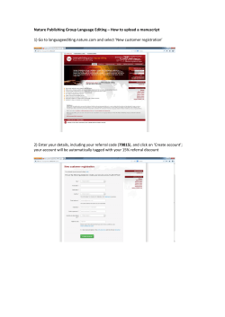


© Copyright 2026