
NAOSITE: Nagasaki University's Academic Output SITE
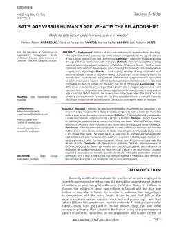
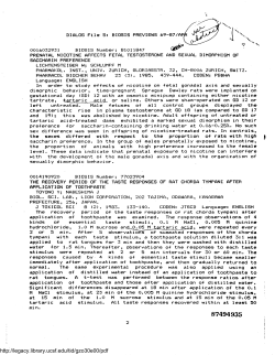
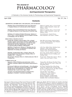
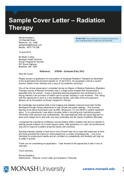
NAOSITE: Nagasaki University's Academic Output SITE
Title
Basic fibroblast growth factor suppresses radiation-induced apoptosis and
TP53 pathway in rat small intestine.
Author(s)
Matsuu-Matsuyama, Mutsumi; Nakashima, Masahiro; Shichijo, Kazuko;
Okaichi, Kumio; Nakayama, Toshiyuki; Sekine, Ichiro
Citation
Radiation research, 174(1), pp.52-61; 2010
Issue Date
2010-07
URL
http://hdl.handle.net/10069/23880
Right
© 2010 by Radiation Research Society.
This document is downloaded at: 2014-10-21T13:54:20Z
http://naosite.lb.nagasaki-u.ac.jp
●
●
O怖da∫Jo“rnal of the Radiation Resea鵬白SOC∫eオy
Basic Rbroblast Growth Factor Suppresses Radiation−lnduced Apoptosis
and TP53 Pathway in Rat Small lntestine
Mutsumi Matsuu−Matsuyama,ら1 Masahiro Nakashima,b Kazuko Shichij o,a Kumio Okaichi,c Toshiyuki Nakayama”
and Ichiro Sekinea
り)iViSiOn Of TUmOr and DiagnOSごiC PathOIOgア, b Ti,∬Ue and Hなt(コpathOIOgy Section, and・1)epar〃nent(∼∫Rα4励0ηβ」()phySieS, A tOmiC・BOmb
Diseaぷε加痂ωρNagaぷ磁University Graduate Sehool〔ゾ肋medica1 Scienceぷ,1−12−4 Sakamoto, Nagasaki 852−8523, Japan
RADIATIoN REsEARcH 174,52−61(2010)
0033−7587/10$15.00
◎2010by Radiation Research Society.
All rights of reproduction in any form reserved.
DOI:10.1667/RR1802.1
Basic Fibroblast Growth Factor Suppresses Radiation−lnduced Apoptosis
and TP53 Pathway in Rat Small lntestine
and Ichiro Sekine”
﹁ペヨ惑︾⋮だ
Mutsumi Matsuu−Matsuyama,a・’ Masahiro Nakashima,6 Kazuko Shich亘o,αKumio Okaichi,c Toshiyuki Nakayama〃
口iViSiOn Of TU%r and DiagnOぷtiC PathOIOgy, b TiSSUe・and・HiSt(卿thOIOgy SeCtiOn, andのepa励2ent()f RadiatiOn B卿吻iCS, AtOmiC・BOmb
1)iぷease Institute,∼〉α9αぷaki乙lniversity(7raduate School 6ゾ・Bio〃zedical Sciences,1−12−45’aka〃10to,ハ「agasaki 852・・8523,」砲αη
radiation resu.lts in rapid apoptosis of the stem cells in
Grow伍Factor S叩presses Radiation・・lnduced Apoptosis and
process of active cell death that involves gross morpho−
TP53 Pathway in Rat SmalHntestine. Radiat. Res.174,52−61
logical alterations including condensation of nuclear
the j〔加nal crypt(2,3). Apoptosis is a programmed
(2010).
The effect of basic fibroblast growth factor(bFGF)was
chromatin, compaction of cytoplasmic organelles, mem−
brane blebbing, and cellular fragmentation into apo−
studied in radiation−induced apoptosis in rat jejunal crypt cel霊s.
ptotic bodies(1,4).
Six−week−old male Wistar rats were administered 4 mglkg
1ア53(fbrmerly known as p53)is a tumor suppressor
bFGF intraperitoneally 25 h befOre receiving 8 Gy whole−body X
gene and is one of the primary cellular factors
rays. The jejunum was removed for analysis from time O to 120 h
determining the nature of growth arrest and/or cell death
after irradiation. Villus length in co耐ml rats declined steadily
after exposure to ionizing radiation(5−8). TP53 plays an
u耐il 72 h, while in bFGF−treated rats the Villi were l皿ger than
important role in damage surveillance and as such has
been dubbed the guardian of the gellome(9). Upon DNA
damage, TP53 accumulation is increased and translocat−
in the controls until 48 h. Crypt lengths were similar to vmi.
bFGF treatment increased Ki−67−positive ceUs in the jejenal
crypt at O,24 and 48 h. The treatment with bFGF reduced the
ed to the nucleus where it binds to DNA, acting to
number of apoptotic cells per jejunal crypt to 23%and 10%of
regulate the transcription of a number of genes including
the control values at 3 and 6 h, respectively, and increased
numbers of mitotic cells significantly at 48 and 72 h. bFGF
α)K7VIA(fbrmerly known as p21wAF茎/cIPI)and P UMA.
decreased the Ievels of TP53, CDKNIA, Puma and Cleaved
caspase 3 at 3 h as detecte勒y Westem blot analyses. Our
CDKN l A is a cydin−dependen.t kinase(CDK)inhibitory
protein that appears to be a critical component of Gl
results s紐99est that bFGF protected against acute radiation−
arrest(10,11). PUMA, which is induced by TP53, is a key
induced in▲ury by supPressing the crypt apoptotic cells including
mediator in the TP53−dependent apoptosis (12−14).
tlle stem ceils and promoted crypt ce皿proliferation. The
1)UMA encodes a BH3−only protein member of the Bcl−
inhibition of apoptosis thus might be related to s叩pression of
2family and binds Bcl−2 and Bc1−xL, localizes to the
the TP53 pathway. ◎2010 by Radiati皿R〔se3rch s㏄iety
mitochondria, and promotes cytochrome c release and
apoptosis. Depending on the cellular context, the
outcome of a TP53 response results in either cell cyc▲e
INTRODUCTION
arrest or apoptosis. Caspase 3 is one of the primary
executioners of apoptosis and is necessary fbr the
High doses of ionizing radiation damage intestinal
cleavage of a large number of proteins and margination
crypt cells and result in functional changes. These
deleterious effects, including malabsorption, are seen
of apoptosis−associated chromatin, DNA fねgmentation,
and nuclear collapse during apoptosis(15).
clinically as acu.te small bowel reactions. Crypt distur−
Basic fibroblast growth factor(bFGF, also known as
bance results from cell loss by apoptosis and destruction
FGF−2)was initially regarded as a potent angiogenic
of the stem cells that are responsible fbr repopulating the
factor because it induces endothelial cell proliferation,
lining of the gut. Cells in the stem cell region of the gut
migration and smooth muscle cell proliferation(16,η).
crypt are arranged hierarchically and are rapidly
bFGF also improved wound healing in patients with
superficial or deep second−degree burns and healing of
1Address fbr correspondence:Division of Tumor and Diagnostic
Pathology Atomic Bomb D輌sease Institute, Nagasaki University
pressure ulcers in clinical trials (18, 19). In the
Graduate School of Biomedical Sciences 1−12−4 Sakamoto, Nagasaki
gastrointestinal tract, bFGF enhances epithelial cell
852−8523,Japan, e−mail:mutsumi@nagasaki−u.ac.jp.
proliferation and restitution as well as stem cell survival
52
︾ク㍉
q 兎で〆﹀ 内7’”ぺ
ミ proliferating Populations (1). Exposure to ionizing
Matsuu−Mats町ama, M., Nakashima, M., Shichilo, K,
Okaichi, K., Nakayama, T. and Sekine,1. Basic Fibroblast
bFGF SUPPRBSSES INTESTINAL APOPTOSIS AFTER IRRADIATION
53
after radiation inj ury to the intestine(20,21). Okunieff
aluminum負lters at a dose rate of O.8903 Gy/min. The radiation
et al. suggested that the LD50/30(7.6 Gy, bone marrow
exposure was unilateral. Two rats were treated simultaneously while
syndrome end point)and LD50/6(12.5 Gy, gastrointes−
they were allowed to move freely in a cardboard box. A single dose of
8Gy was given. Nonirradiated rats(time O samples)were handled
tinal syndrome)of C3H mice after whole−body irradi−
ation were increased by pretreatment with FGF2(22).
identically.
Paris et al. also suggested that bFGF protected C57BL/6
Histologieal Methods and A∬e∬〃lent〈ゾApoptosi,ぷand・Mitosis
mice from death by the GI syndrome caused by whole−
body radiation doses up to 18 Gy(23). It was also
To obta輌n samples, rats were k輌11ed humanely by deep anesthesia at
suggested that bFGF exerted a benef玉cial effect on
radiation−induced enterocolitis(24). Pretreatment with
segments of proximal j{)junuM were obtained from the ligament of
Trietz. Tissue samples were fixed overnight in 4%neutral buffered
FGF−2 has been reported to protect against radiation−
formaldehyde. After fixation, the jejunum was cut longitudinally and
induced apoptosis of crypt cells (22), although its
processed f()r embedding in paraf日n blocks. Thereafter,3一μm sections
mechanism of ef琵ct was not determined.
We reported previously that agents such as伝mented
milk kefir(25), sucralfate(26)and polaprezinc(27)can
serve as radioprotectors when administered prior to
irradiation to suppress radiation−induced apoptosis in
rat colon or small intestine. Sucralfate, an aluminum
3,6,24,48,72and 120 h after 8 Gy irradiation. Six−centimeter
were cut and stained with hematoxylin and eosin(H&E). Three to six
animals were used fbr each group and each data point。
Villus Iength, defined as the length from the apex of the brush
border to the base of the crypt in the jejunum, and crypt depth(along
the long axis of the elliptical crypt)were measured using a 100×
magnification stage micrometer. The lengths of more than five
random villi or crypts were measured, and the measurements were
averaged.
hydroxide complex of sulfated sucrose (26), and
Scoring of apoptosis and mitosis was restricted to goo(l longitu−
polaprezinc, a chelate compound consisting of zinc ion
dinal sections of the crypt in which the base of the crypt was aligned
and L−carnosine (127), are anti−ulcer agents with a
protective effect against radiation−induced inj ury in the
with all of the other crypt bases and showed the crypt lumen. More
rat colon or small intestine after decreases in TP53
accumu.lation, CDKNlA expression and BAX/BCL−2
than 30 good longitudinal sections were selected per animal, and
apoptotic cells and mitotic cells were scored in each crypt in H&E−
stained sections at 400×magnification by Iight microscopy as
described previously(25−27).
ratio or BAX expression.
In this study the effect of bFGF on acute radiation−
induced injury was studied in a rat model in which
apoptosis was induced by 8 Gy X rays in j(加nal crypt
cells. To clarify the mechanism of bFGF in radiation−
TUNEL Staining
Identi丘cation of apoptosis was confirmed using a TUNBL
technique(Apoptag Peroxidase In Situ Apoptosis Detection Kit,
Chemicon, Temecula, CA)that detects apoptosis−associated DNA
induced apoptosis, the location of the ceUs displaying
fragmentation by labeling of 3’・・OH termini with digoxigenin
Cleaved caspase 3 expression was examilled by imlnu−
nohistochemical analysis, and the effect of bFGF on
nucleotides using terminal deoxynucleotidyl trans飴rase(28). Sect輌ons
TP53, CDKNIA, PUMA and Cleaved caspase 3 was
examined by Westem blot analysis.
were deparaf資nized in xylene, rehy(lrated in decreasing concentra・・
tions of ethanol, and digested in proteinase K(36μg/ml)f()r 15min at
room temperature. Endogenous peroxidase was blocked with 3%
hydrogen peroxide. Terminal deoxynucleotidyl transferase(T(IT)in
reaction buffer (containing a f玉xed concentration of digoxigenin−
labeled nucleotides)was applied to sections fbr l h at 37°C followed
MATERIALS AND METHODS
by placemeRt of slides in StopハVash buffer f()r 30 min. Apoptotic cells
メη加∼α1ぷand Trea〃nent{ゾわFGF
were detected after incubation in the 3,3’−diaminobenzidine(DAB)
chromogen(Dako, Carpinteria, CA)f()r approximately 6 min and
Six−week−old male Wistar rats(200−250 g)were purchased from
counterstaining with Methyl GreeR(Sigma, St. Louis, MO).
Charles River Japan(Atsugi, Japan). The rats were housed in groups of
two per cage in an air−conditioned room at 24°C(lights on from 7 a.m、
Immunohistochemis句・
to g p.m。)and were allowed free access to food(1aboratory chow F2,
Japan CLEA, Tokyo, Japan)and tap water at the Laboratory An㎞al
ImmuRohistochemical staining was perf()rmed fbr Ki−67 and
Center of Nagasaki University. All animals were kept in a specific−
αeaved caspase 3. Paraffin sections were prepared and deparaf日一
pathogen−free fac最ity at the Animal Center in accordance with the rules
11ized. The sections were placed in O.Ol moYliteτ citrate buffer(pH 6.0)
and regulations of the lnstitutioital Animal Care and Use Co麺ttee.
and pretreated with microwave heating for antigen retrieva1. The
bFGF(gift of Kaken Pharmaceutical Co. Ltd., Tokyo, Japan)was
sections were reacted with O.3%H202 in deionized water for 30 min to
diluted with distilled water to a concentration ofO.5 mg/mi immediately
inhibit endogenous peroxidase activity. The sections were incubated
before injection. bFGF was given intraperitoneally to rats at a dose of
with anti−rat Ki−67 monoclonal antibody(MIB−5)(Dako Cytomation
4mg/kg 25 h befbre total−body irradiation. The control rats were
Denmark A/S, Denmark)and Cleaved caspase 3(Cell Signaling
injected only with saline. In the bFGF dose−respollse studies,1mg/kg
Technology Inc.)diluted 1:25 and 1:200, respectively, in ChemMateTM
an(至2 mg/kg were administered intraperitoneal豆y 25 h befbre
antibody di短ent(Dako)over night at 4°C. After washing with PBS,
irradiation. Three to six animals were used in each group.
the sections were incubated with biotinylated anti−rabbit and anti−
Irradiation
mouse immunoglobuliRs for 30 min and then reacted with strepta−
vidin corU◆ugated to horseradish peroxidase fbr 30 面n using an
LSAB−2 system−HRP fbr use on rat specimens(Dako)according to
Irradiation was perf()rmed between 9:00 a.m. and 12:00 p.m. Rats
the manufacturer’s instruct輌ons. Antibody binding was visualized by
received whole−body X irradiation from a Toshiba ISOVOLT TITAN
32X−ray,200 kV,15 mA apparatus with O.5−mm aluminum十5−mm
incubation with 3,3’−diaminobenzidine(DAB)chromogen(Dako).
HematOXylin WaS USed f()r RUCIear COUnterStaining.
54
MATSUU−MATSUYAMA ET AL.
Cells showing immunoreactivity fOr Cleaved caspase 3 were scored
A 1.2
on a cell location basis within the half二crypts of the j〔)junum according
1
to the method of Wilson et al.(29)at 400×magnification using light
rats輌n every group. The cell numbers ill the half−crypts were also
determ輌ne(重.
Western Blotting
The jejunum tissues ofcontrol and bFGF−treated rats were removed
at 3,6 and 24 h after 8 Gy irradiation and frozen immediately. The
︵Eε︶旧=旧﹀+oぷ磁仁Φ﹂
microscopy. A minimum of 30 half−crypts were counted from three
0.8
0.6
’ウ
0.4
冷・
0.2
tiss促s were then suspended in RIPA buffer(50 mM Tris,150 mM
NaCl,1%sodium deoxycholate and O.05%SDS, pH 7.4), pulverized
0
on ice, and su句ected to three f士eeze−thaw cycles(30). The insoluble cell
20
40 60 80 100
120
140
Tirne after irradiation(h)
the protein concentrations were(ieterm輌館d using a protein assay
reagent(Bio−R.ad Laboratories, Hercules, CA). Protein samples(30 pg)
were su句ected to l O%or 15%SDS−polyacrylam輌de gel electrophoresis
and then trans琵rred electrophoretica夏ly to Hybond ECL Nitrocellulose
(Oncogene Science Inc., Un輌ondale, NY), rabbit polyclonal anti−
CDKN l A(Santa Cruz Biotechnology, Santa Cruz, CA), Puma(Cell
Signaling Technology Inc., Beverly, MA)and Cleaved caspase 3(Cell
Signaling Technology Inc.)antibody or rabbit polyclonal anti−actin
antibody(Sigma). This was f()llowed with a horseradish peroxidase−
conjugated anti−mouse IgG antibody (Zymed Labs. Inc., San
Francisco, CA)or a horseradish peroxidase−conj◆ugated anti−rabbit
IgG(Amersham). Chemiluminescence(ECL PIus, Amersha⇒was
used to analyze the levels of protein according to the manufacturer’s
protocol. Blots were exposed to Hyperfilm ECL(Amersham). NIH
B
1
︵仁≧ε︶==こ三葛c3
Membranes(Amersham, Arlington Heights, IL). Membranes were
incubated with mouse monoclonal ant輌一TP53(Pab421)antibody
1.2
0.8
0.6
O.4
0.2
0
0 20 40 60 80 100 120 140
IInage 1.61 software was used in]gieasuring the densities of each of the
Time after irradiation(h)
protein bands. The互evel of protein after irradiation was determhled
FIG.1. Comparison of the lengths of villi(panel A)and crypts
using the level of act輌n as a standard.
(panel B)after 8 Gy irradia£ion in rats treated or uRtreated with
bFGF. Data are the means±SEM of three to six rats per data point.
Statistical Evaluation〔of Data
*P<0.05,**Pく0.01and***P<0.001 compared to control rats.
All values were expressed as the means±SEM of results obtained
from three to six animals per data point. Differences between groups
bFGF(Fig.2F)were higher in number than in control rats
were examined using Student’s t test. A P<O.05 was considered to be
砿Oh(Fig.2A)and continued to be higher at 24(Fig.2G)
StatiStiCally Significant.
and 48 h(Fig.2H). At the same time, the Ki−67−positive
cells in the j()junum of c皿trol rats decreased at 24 h
RESULTS
琢c但μFGF・η舵鋤gth〔)f Villi and鋤pts and
Pro1φ螂oη訊抗α磁θ4吻∧lo噺α4漉4 Rατぷ
(Fig.2B), and only a few positive cells were seen at 48 h
(Fig.2C). Regenerated crypt cells appeared at 72 h
(Fig.2D). At 72 and 120 h, there was no difference between
control and bFGF−treated rats(Fig.2D alld I, E and J).
To evaluate the effect of bFGF on radiation−induced
ir巾ry in the rat small intestine, the lengths of villi and
Effects〔ゾbFGF・n・Radiati・n−lnduced Ap〔卿ぷ」ぷ翻
crypts were measured in samples taken f士om O to 120 h
Mi◆tosis〔ゾσアμωゐ
a負er irradiation. The length of villi in control rats
decreased from O to 72 h and then showed an increase
Figure 3A shows histological sections of拘unal crypts
at 120 h(Fig.1A). The length of crypts in control rats
丘om control and bFGF・・treated rats stained with
showed decreases at 24 h and 48 h and then increased by
TUNEL. At 3 h, a large number of apoptotic cells were
120h(Fig.田). For rats treated with bFGF, the villi were
observed in the j()j unal crypts of control rats・ In
significantly longer than controls at O, 6 and 48 h
contrast, only a small number of apoptotic cells were
(Fig.1A). The crypts of bFGF−treated rats were signif三
seen in the j(加nal crypts of bFGF−treated rats.
icantly longer than controls at O,3,6and 24 h(Fig.1B).
These results indicate that bFGF can protect against
Panels B and C of Fig.3compare the apoptotic and
mitotic indices m the j句unum detected by H&E staining
acute radiation−indu.ced injury in the rat j()ju.num.
in control an.d bFGF−treated rats up to 120 h. The
To investigate the effect of bFGF on proliferation in
apoptosis index in the j(加num peaked in control rats by
the拘unum after irradiation, immunohistochemical
3 h and then declined precipitously within 24 h.
staining fOr the Ki−67 antigen was perfbrmed. The Ki−
Pretreatment with bFGF significantly reduced the
67−positive㏄11s in the j句unal crypts of rats treated with
number of apoptotic j()junal crypt cells at 3 and 6 h to
・づ×
0
debris was removed by centrifugation. Supernatants were collected and
55
∨ ト
夢 ▲
羅装醤・
膓讃 i 、砂. ← 、
鷲
bFGF SUPPRESSES INTESTINAL APOPTOSIS AFTER IRRADIATION
1
1
W〆 ^
句俘
当還
ぞ
/﹂“
パ゜
「
…
撒ぽ
㌔♪遵ほ浮畠才
§ 董、㌔‘ーピ
、
蓮﹁
竃
㍉オ,︽,’e 、小
﹂
謄
⊇難購
、f,一
S
〆万 ,人
ぶ▽当
磯・
、
・4毒
㍉
、“
磁
ぱ〆 ㎡
ず∴
’
’,
パζ、
㌧
㌻
FIG.2. Immunohistochemical staining with anti−Ki−67 monoclonal antibody. Jejunum ofcontrol(upper row)and bFGF(lower row)−treated
rat at O(panels A and F),24(panels B and G),48(panels C and H),72(panels D and I)and 120 h(panels E and J)after 8 Gy irradiation.
Original magnification 100×.
22.6%and l O%of the levels in control rats, respectively.
There was no difference in the apoptosis index between
control and bFGF−treated rats from 24 h to 120 h. There
immunohistochemical staining is shown in Fig.4. The
distribution of positive cell locations is shown in
Fig.4A−D. The time courses of the frequencies of
was no difference in the background(O h)levels of
Cleaved caspase 3−positive cells in half−crypts of control
apoptosis in the two groups(Fig.3B).
and bFGF−treated rats are shown in Fig.4E. Cleaved
The mitotic index in the jejunum of control rats
decreased sharply at 3 and 6 h, in contrast to the
caspase 3 expression was localized to the nucleus of the
crypt cells after irradiation. There was no expression of
apoptosis index. This was fbllowed by a gradual increase
Cleaved caspase 3 in the nonirradiated control or
at 72 h that then continued皿til 120 h. The mitotic
bFGF−treated rats. The expression of Cleaved caspase
index of bFGF−treated rats was higher than that of
3 in the irradiated jejunum was increased strongly at 3 h
control rats at and after 48 h. Significant differences in
(Fig.4B), decreased slightly at 6 h(Fig.4C), and then
the mitotic index were noted between control and
declined at 24 h(Fig.4D)in the bottom half of the
crypt, while that of bFGF−treated rats showed no
increase at any time after irradiation. There was a
signi丘cant difference at 3 and 6 h(」P<0.05)between
bFGF−treated rats at 48 and 72 h(Fig.3C).
im〃iunohis・toche〃zical Res・ults・プ6γCleaved Caspase 3
The expression of Cleaved caspase 3 in拘unal crypt
non−bFGF−treated and bFGF−treated rats(Fig.4E).
The half−crypt cell numbers of control and bFGF−
cells of control and bFGF−treated rats detected by
treated rats are shown in Table 1. The half−crypt ce11
56
MATSUU−MATSUYAMA ET AL.
A
。脚婿§騰
㌢辱熟罷,
ご∴.1㌧.£∴㌧.、.…㌦、,’・・寸
ゴ∴∵や口\・
fi
←
癬繍罐
癒翻議、
−一
0
40
20
60
o:ト’bFGF
80
100
120
140
Time after irradiation(h)
6 5 4 3 2 ﹂1 0
十controI
昔eo﹂oαω=80習出E↑o﹂ΦエεコZ
C
戸0 4﹁ 3 9﹄ −
0 0 ︵U O O O
且eo﹂oO茎80旧ぢ且oα句℃﹂ΦΩ∈コZ
B
’
’
’
’
’
’
’
’
’
’
,’
’
’
’
’
’
’
,”
**,/
’
’
.」ゴ
一一
* ,〆
Sθ一一control
,’”
,’
,’
一⇔一一一一一一一
oコr
−一
o:}一’bFGF
’
0
20
40
60
80
100
120
140
Time afヒer irradiation(h)
FIG.3. Panel A:Histological sections of small intestine in control rat and bFGF−treated rat at 3 h after 8 Gy irradiation stained by the
TUNEL method(original maglli丘cation 400×). Apoptosis index(panel B)and mitotic index(panel C)of j(junal crypt cells in control rats and
bFGF−treated rats after 8 Gy irradiation. Data are the means±SEM of three to丘ve rats per data point. More than 30 crypts were analyzed per
an㎞a1.*P<0.05,**、P<0.01 and***P<0.001 compared to control rats.
numbers of bFGF−treated rats were significantly higher
then decreased at 6 and 24 h. A significant difference was
compared to those of non−bFGF−treated rats at each
observed between control and bFGF−treated rats, at 3 h
time after irradiation.
(P<0.05)(Fig.6A). The expression of CDKNlA in
Dependence(ゾ.Rα∂吻γ・tec肋ηげ加e吻α1(]rypts on
control rats increased at 3,6and 24 h. There was a
significant di脆rence between control and bFGF−treated
1)ose()f bFGF
rats at 3 h(P<0.05)(Fig.6B). The expression ofPuma in
control rats increased at 3 and 6 h and then decreased at
Figure 5 shows the apoptosis index at 6 h after 8 Gy
irradiation in control rats and in rats treated with 1,2
and 4 mg/kg of bFGF. There was no difference between
control rats and bFGF−treated rats at l mg/kg.
However, suppression of the apoptosis index was clearly
shown in rats treated with 2 and 4 mg/kg bFGF.
Wes・tern Blo ttカzgノ∼)r TP53, CDKハηノt, Puma and Cleaved
24h. A significant differences were noted between control
and bFGF−treated rats at 3 h(P<0.Ol)(Fig.5C). The
amo皿t of Cleaved caspase 3 in control rats increased at
both 3 and 6 h and then decreased to O.7−fbld at 24 h. A
significant difference was noted between control and
bFGF−treated rats at 3 h(P<0.05)(Fig.6D). Taken
together, these results indicated that b FGF pretreatment
inhibited radiation−induced TP53 accumulation and its
Caspase 3
downstream induction of CDKNlA, Puma and Cleaved
To determine how bFGF treatment was interfering
caspase 3;this was consistently seen at 3 h.
with the pathways leading to radiation−induced apoptosis
in the rat拘unum, the levels of TP53, CDKN l A, Puma
DISCUSSION
and Cleaved caspase 3 were examined by Western blot
analyses;kinetic diagrams are shown in Fig.6. TP53
Radiation enteritis occurs during radiotherapy fbr
accumulation in control rats increased 2.3−fbld at 3 h and
many pelvic cancers and intra−abdominal organs such as
57
bFGF SUPPRESSES INTESTINAL APOPTOSIS AFTER IRRADIATION
bFGF
c°事 電・.p
‘ i
1’
む
、 :i
一一●一一control Oh
吉60
} ■一⊂トbFGFOh
↓血工_α蜘血血田皿皿
0
怐。−control 3h
−[)・一 bFGF 3h
書40
20
0
一■
5
崖
40
衷︸﹀。=ΦコぴΦよ
邑
一...b 80
]]
衷︸>uもコσΦ﹂匹
60
cl
Bloo
80」
5
10 15 20
25
Cell position
Φ i
9 40]
皇
20
0
5
0
30
25
10 15 20
Cell positiOR
60
50
40
30
20
5
25
10 15 20
30
Cell position
十control
﹂,ー、i,ー.﹂ー,⋮、,⋮1﹁ー1‘
−[}−bFGF24h
一ローbFGF6h
40・e
。↓
﹁.⋮⋮﹂﹁ー﹂ 言60!
⊆ {
+control 6h
0
宝︸差Φu皇出8△
十control 24h
ケ ト f
7幅.≧b、sL−.t藁
20」
E南ω皇§琶⑩Φ0
三1:]
、 ♂. 毒
60一
20
30
㍗目
い 噸
蝶‖
撃撃
A
24h
菖「人’ひ∵だ掌禰
藷藷螂
・、
k,1Lぷ..s」φ三、、1、
100
望 ﹂ ︼ ≧ ≦きS]
Vバ.
で ヘ ピ
} {
竃。二_鋪
﹁、二﹂.°°渉層.、 ご▲,
.{
璽嘆蓑讃墾竃
鳥賠麟蕊
Cleaved caspase 3
6h
弩,..﹃・馳 、
・
3h
Oh
24h
6h
3h
Oh
[︻
Control
・…
香cbFGF
10 *
051015202530 0 5 10152025
Cell position Time afterirradiation(h)
FIG.4. The expression of Cleaved caspase 3 in jejunal crypt cells of control and bFGF−treated rats detected by immunohistochemical staining
after exposure to 8 Gy radiation(original magnification 200×). The distribution of the locations of Cleaved caspase 3−positive cells in half−crypts
of control and bFGF−treated rats after exposure to 8 Gy is shown in panels A−D. The time course of the percentage of Cleaved caspase 3−positive
cells in half−crypts of control and bFGF−treated rats is shown in panel E. Data are means±SEM f()r three rats per experimental group. At least
30 half−crypts were scored from each rat.*P<0.05 compared to control rats.
rectum. Although ionizing radiation affects other
intra−abdominal organs, the most radiosensitive is the
Time after irradiation
Oh
3h
6h
24h
Contro1
bFGF
25.9±0.5
24.7±0.9
23.4±0.4
14.1±0.3
28.3±O.5b
28.1 ± 1.la
28.4± 1.le
l8.3 ± 1.6a
30
25
b20
0
1 4■‘
The Half−Crypt Ce皿Numbers after lrradiation
35
5050
TABLE l
40
且
small intestine. Ionizing radiation causes mucosal
﹂Φαω一一Φoo旧甘且o合↑o﹂Φ﹄εコZ
cervix, endometrium, ovary, bladder, prostate and
0
1 2
4
bFGF(mg/㎏)
No tes. Data are means±SEM from pooled data of three counts
FIG.5. Effect of the dose of bFGF on apoptosis index of j ej unal
from each of three rats per experimental group. At least 30 half−crypts
crypt cells 6 h after 8 Gy irradiation. Data are the means±SEM of
were scored from each rat. aP<0.05, bP<0.01 and cP<0.001
three or f()ur rats per data point. More than 30 crypts were selected
compared to control rats.
f()ranalysis per animaL***P〈0.001 compared to control rats.
58
MATSUU−MATSUYAMA ET AL.
A
Oh 3h
TP53
鍵滋
・難
Actin
6h 24h
難灘鑛
鑛譲
B
bFGF
Control
CDKNlA
灘雛灘灘、
ActiR
該ぷ難1懇 欝 診難馨t”t
0
oo
き
0
●
O.5
0
5 10 1S 20 25
5 10 15 20
Control bFGF
Oh 3 h
24h
タ ttt
蓬
Actin
.離繋
.灘 灘難難..難籔 .灘
が ’
搬・繋灘
覇騨
繍※
け が w。
雛診懇\7 叢 葱 杉灘彩t。、 谷難・灘∧
D
C皿trol
叢
’ttt
Actin
難講諺垂嚢妻羅su蕪 譲]垂 ・萎羅灘難難霧壕鍵。・
馨繋難難鍵燃一_ 難 繋獲 縫 彩凝 ;bUt 難
仁o層Φ﹂妥ΦεΦぢ﹂αΦ︾る一Φ匡
25
Time after irradiation {h)
富Φ給音巴冨詫ぷ9
エ ハじ
︵<Σ2︸
5 10 15 20
・難籔嚢難i馨舞碧襲羅饗馨◇霧 霧2紗灘⑳灘総灘葵購総簸.
caspase 3
※×”
3 に﹂ へ∠ CJ 4⊥ ︻﹂ ︵U
きL。⋮Φ包×Φεo苫aΦ﹀[一亘岳
0
bFGF
Oh 3h 6h 24h Oh 3h 6h 24h
Cleaved
嚢
萎難峯轟
24h Oh 3h 6h
灘、
PUMA
6h
25
Time after irradiation{h)
Ti me after irradiation {h》
C
灘燈灘劾 雀懸i
口
1
該灘
*
…手・一.“
ロ ロ 2
へ5 ロ﹂ 亀∠ ■5 1 5 ︵U
コ む
富臣ヒ
o“一ψ3旨×Φ三Φ苫﹂αΦ﹀蔦一Φ匡
2.5
24h
難鍵懸嚢饗難
難灘鷺馨難難 講礁轟難蒙
=o層Φ﹂受Φ忘Φぢ包Φ≧完一Φ匡
3
1.5
bFGF
Oh 3h 6h 24h Oh 3h 6h
Oh 3h 6h 24h
︵<回蔓e︸
磯難蕪
Control
3
十CORtrol
2.5
…{:}・・bFGF
2
1.5
1
’”
p・…ip・…・・……・……__._
0.5
0
0
5 10 15 20
25
Time after irradiation (h)
FIG.6. Western blot analysis of the amounts ofTP53, CDKN l A, PUMA and Cleaved caspase 3 in the jejunum of control and bFGF−treated
rats. The levels of TP53(panel A), CDKN I A(panel B), PUMA(panel C)and Cleaved caspase 3(panel D)were quantified by densitometric
analysis. Each of the protein levels is expressed as a ratio of nonirradiated rats(O h)to coRtrol rats. Data are means±SEM of three to f()ur
separate experiments with two rats per data point.*P<0.05 and**1)<O.Ol compared to control rats.
damage in the gastrointestinal epithelium comprised of
intestine from 48 to 120 h at doses above 8 Gy, which
destruction of crypt cells occurring by the induction of
produced moderate to severe injury to the gastrointes−
apoptosis, decrease in villus height and number,
tinal epithelium, resulting in progressive loss of viable
ulceration and necrosis(31). In the previous study, we
regenerative crypts and compromising the epithelial
noted that losses of weight and appetite ill spontane−
barrier(21). FGF−2 markedly enhanced survival of crypt
ously hypertensive rats were greater than in Wistar−
stem cells when mice received a single dose of rhFGF−2
Kyoto rats after 7.5 Gy X irradiation and suggested that
(4μg/g)25hbefbreγirradiation at doses 13.8 Gy. They
this radiation sickness may be due to injury to the
gastrointestinal epithelium (32). The protection of
radiation−induced apoptosis of intestine may ▲ead to
the improvement of radiation sickness.
suggested that FGF−2 could cause cell cycle arrest,
allowing time fbr repair to occur in otherwise replicating
cell populations, and that FGF−2 might enhance crypt
survival by protecting stem cells from undergoing
Houchen et al. suggested that the expression of FGF−
apoptosis. Okunieff et al. reported that bFGF given
2mRNA and protein began to increase at 12 h afterγ
24hbefore whole−body irradiation with 7−18 Gy
irradiation, and peak levels were observed in the small
protects bowel crypts and a possible intestinal syndrome
bFGF SUPPRESSES INTESTINAL APOPTOSIS AFTER IRRADIATION
59
(22).The protective mechanism may lie in inhibition of
irradiated rat jejunum(Fig.6). We also demonstrated
radiation−induced apoptosis of crypt cells and may
include radioprotection of the vasculature as welL In
this study it was clearly demonstrated that bFGF
treatment can protect the rat small intestine from
that bFGF treatment inhibited the expression of
Cleaved caspase 3 in the crypt, including stem cell
regiOn, With OUr immUnOhiStOChemiCal indingS(Fig.4).
The results fbr Cleaved caspase 3 in the Western blot
analysis(Fig.6D)concurred with the results of time
radiation−induced injury at an X−ray dose of 8 Gy,
which is above the LD50130(5 Gy)of Wistar rats and is
course frequency detected by immunohistochemical
the lowest dose that produced injury to the gastromtes−
analysis(Fig.4E). Merritt et a乙observed a rapid
tinal epithelium. Our results showed that pretreatment
(4.5h)elevation of TP53 protein in the proliferative
compartment of the crypts where cells underwent death
with bFGF inhibited radiation−induced apoptosis of
crypt cells, promoted crypt cell proliferation, and
decreased the level of TP53 and the downstream proteln.s
after irradiation.
Our stu.dy showed that crypt depth and villus length
were significantly increased in irradiated rats by bFGF
treatment(Fig.1A, B). Ki−67−positive cells in the j()junal
crypt were increased by bFGF treatment(Fig.2F)
compared with their nonirradiated controls at time O
by apoptosis after 8 Gy irradiation. They fbund a totaI
repression of apoptosis in the small intestinal crypt
epithelia of TP53−deficient mice 4.5 h after 8 Gy
irradiation(34,35). Komarova et al. showed that
pifithrin−alpha, a chemical inhibitor of TP53, blocked
radiation−induced apoptosis of the gastrointestinal
epithelium, b山it did not alter lethality from the
gastrointestinal syndrome(36,37). In our study, the
(Fig.2A). This indicated that bFGF treatment promot−
su.ppression of the TP53 pathway by bFGF treatment
ed crypt proliferation. The increase of proliferating crypt
may have been a result of the inhibition of radiation−
cells after bFGF treatment may be related to a change in
induced apoptosis. Our analysis, however, was restricted
radiation sensitivity. The numbers of Ki−67−positive cells
in the jejunal crypt of bFGF−treated rats were much
to 8 Gy. Further studies are needed to determine
whether the same mechanisms occur at higher doses of
larger than those of control rats at 24 and 48 h after
radiation(i.e., at or above the LD5016). The expression of
irradiation(Fig.2G, H), suggesting that bFGF treat−
CDKNlA at O h showed no difference between control
ment may promote proliferation of the surviving cells
after irradiation. The treatment with bFGF significantly
reduced the apoptosis index at 3 and 6 h(Fig.3A, B).
However, the mitotic index of bFGF−treated rats
increased from 48 h after irradiation(Fig.3C). The
and bFGF−treated rats(Fig.6B). These data provide no
evidence to support the hypothesis that bFGF modified
radiosensitivity through GI arrest as reported previously
by Houchen et al.(21).
In conclusion, our results suggest that bFGF protects
radioprotective effect of bFGF may involve a suppres−
against acute radiation−induced injury in the rat jejunum
sion of the crypt apoptotic cells including the stem cells
by supPressing the apoptotic cells in the crypt, including
and promotion of crypts cell proliferation.
the stem cells, and by accelerating crypt cell prolifera−
To clarify the location.s of the changes in Cleaved
tion. The mechanism of the protective effect of bFGF
caspase 3, an immunohistochemical staining with
Cleaved caspase 3 was perfbrmed (Fig.4). bFGF
agains£radiation−induced apoptosis may be depend斑t
treatment inhibited the expression of Cleaved caspase
studies of the mechanism are needed. bFGF may reduce
3,which in control rats is expressed in the cells near the
gastrointestinal tract damage during abdominal radia−
tion therapy fbr gastrointestinal, genitourinary and
gynecological tumors. Therapeutic effects have been
reported fOr the treatment of experimental dextran
base of the crypt, especially around cell positions 4−6, in
the putative stem cell region(33). The Ilumber of half−
crypt cells in bFGF−treated rats was significantly higher
compared to c国trols at each time after irradiation
(Table 1). The increase of cell numbers in the crypt by
bFGF treatment may result in the increase of the length
of jeljunal crypts and villi・
The greatest suppression of radiation−induced apop−
tosis was observed whell rats received a single dose of 2
0r 4 mg/kg bFGF. The dose of 4 mg/kg bFGF was the
same dose used by Houchen et al.(21). Both 2 and 4 mg/
on suppression of the TP53 pathway. However, further
sulfate sodium(D SS)−induced colitis and trinitrobenzene
sulfonic acid(TNBS)−induced colitis by the rectal
administration of bFGF(38). Similarly, topical admin−
istration of bFGF by enema may be more effective and
less hazardous fOr the treatment of radiation colitis.
Further studies are needed to investigate the therapeutic
efficacy of such an apProach.
kg of bFGF had the same effect, but l mg/kg bFGF had
no protective effect against radiation−induced apoptosls
(Fig.5).
Westem blot analysis showed that bFGF pretreat−
ACKNOWLEDGMENTS
This work was supported by the Global COE Program丘om the
Ministry of Education, Culture, Sports, Science aRd Technology,
ment resuked in a decrease in the accumulation of TP53
Japan. We wish to thank the staff of Biomedical Research Center
as well as a decrease in the induction of the CDKN l A,
(BRC), Center for Frontier Life Sciences of Nagasaki University fbr
Puma and Cleaved caspase 3 relative to the control
eXCellent aRimal Care.
60
MATSUU−MATSUYAMA ET AL.
Received:March 24,2009; accepted February 24,2010;published
20.
21.
REFERENCES
1
(1999).
22.
(1994).
C.S. Potten,」. W. Wilson and C. Booth, Regulation and
Radiat. Res.150,204211 (1998).
23.
significance of apoptosis iR the stem cells of the gastrointestinal
epithelium. Stem Cells 15,82−93(1997).
4
Endothelial apoptosis as the pr輌mary lesioll initiating intestinal
radiation damage in m輌ce. Scienee 293,293−297(2001).
i
24.
kinetics. Br./. Cancer 26,239−257(1972).
5
M.B. Kastan,0. Onyekwere, D. S輌dransky, B. Vogelstein and
RW. Craig,]Participation of p53 protein in the cellular response
applied science. Eur.」. Surg. Suppl.582,85−89(1998).
25.
S.J. Kuerbitz, B. S. Plunkett, W. V。 Walsh and M. B. Kastan,
Ichiro, The protective effect of」ferm頭ted milk kefir on radiatioR−
induced apoptosis in colonic crypt cells of rats.」. Radiat. Rθぷ.44,
R.B. Tishler, S. K。 Calderwood, C. N. Coleman and B. D. Price,
111−115(2003).
26.
treatment with chemotherapeutic and DNA damaging agents.
radiation−induced apoptosis in rats.」. Radiat.」Res.47,1−8(2006).
Caneer Res.53,2212−2216(1993).
Q.zhan, F. carrier and A.」. Fomace, Induction of cellular p53
27.
activity by DNA−damaging agents and growth arrest. Mol. Ce〃
Biol.13,4242−4250(1993).
9
28.
and B. Vogelstein, WAFl/CIPl ls induced in p53−mediated Gl
arrest and apoptosis. Cancer R〈ls.54,1169−1174(1994).
29.
0’Connor, Relationships between G I arrest and stability of the
12.
30.
accum.ulation in various organs of rats after whole−body
1671−1674(1996).
M.J. Ausserlechner, J. M. Adams and A. Strasser, p53−and
31.
Crescenti and E. S. Rivera, Mechanisms underlying the
J.Yu, Z. Wang, K. W. Kinzler, B. Vogelstein and L Zhang,
radioprotective effect of histamine on small intestine. Int.1.
PUMA mediates the apoptotic response to p53 in colorectal
Radiat. Biol.83,653−663(2007).
cancer cells. Proe. Natl. Acad. Sci. 乙なSン4100,1931−1936(2003).
32.
14.
J.R. Jeffers, E. PargaAas, Y. Lee, C. Yang, J. Wang, J. Brennan,
nervous system to whole−body X irradia£ion. Radiat. Res. 163,
137−143(2005).
dependent apoptotic pathways.(]ancer Cell 4,321−328(2003).
E.A. Slee, C. Adrain and S.」. Martin, Executioner caspase 3,−6,
33.
and −7 perfo rm distinct, non−redundant roles during the
demolition phase of apoptosis.」. Biol. Chem.276,7320−7326
353,821−830(i998).
34.
C.Basilico and D. Moscatelli, The FGF family of growth factors
and oncogenes.ノ4dv. Caneer Res.59,115−165(1992).
17.
S.M. Schwartz and L. L輌aw, Growth control and morphogenesis
in the development and pathology of arteries./. Cardiovasc.
Pharmacol.21(SupPl.1), S31−S49(1993).
18.
gastrointestinal tract of normal and p53−deficient mice. Cancer
Res.54,614−6 i 7(1994).
35.
recombinant bovine basic fibroblast growth factor fbr second−
W.G. Payne, D. E. Ochs, D. D. Meltzer, D. P. Hill,
R.J. Mannari, L. E. Robson and M. C. Robson, Long・・term
outcome study of growth factor−treated pressure ulcers。.4m.」.
Surg.181,81−86(2001).
A.」.Merritt, T. D. Allen, C. S. Potten and J. A. Hickman,
Apoptosis in small intestinal epithelia from p53−null mice:
evid斑ce fbr a delayed, p53−independent G2/M−associated cell
Randomised placebo−controlled trial of use of topical
19.
A.J. Merritt, C. S. Potten, C.」. Kemp, J. A。 Hic㎞an,
A.Balmain, D. P. Lane and P. A. Hall, The role of p53 in
spontaneous and radiation−induced apoptosis in the
X.Fu, Z. Sh斑, Y. Ch頭, J. Xie, Z. Guo, M. Zhang and Z. Sheng,
degree burns.、乙ancet 352,1661−1664(1998).
C.S. Potten, Stem cells in gastro輌ntestinal epithelium:numbers,
characteristics and death. Phil. Trans. R.5「oc. Lond. Biol.5「cj.
(2001).
16.
M.Matsuu, K. Shichijo, Y。 Ikeda, M. Ito, S. Naito, K. Okaichi,
M.Nakashima, T. Nakayama and I. Sekine, Sympathetic
hyperfunction causes increased sensitivity of the autonomic
K.H. MacLean, J. Han, T. Chittenden and G. P. Zambetti,
Puma is an essential mediator of P53−dependeitt an(i in−
15.
V.A. Medina, M. Croci, N. A. Mohamad, N. Massari,
G.Garba血o, G. P. Cricco, M. A. Nunez, G. A. Martin, E. J. V.
prote輌ns puma and noxa. Science 302,1036−1038(2003).
13.
X.Wang, H. Matsumoto, K. Okaichi and T. Ohnishi, p53
exposure to low−dose X−ray irradiation. Anticancer Res.16,
A.Villunger, E. M. Michalak, L. Coultas, F. Mullauer, G. Bock,
drug−induced apoptot輌c responses mediated by BH3・・oltly
」.W. Wilson, D. M. Pritchard, J. A. Hic㎞an and C. S. Potten,
Radiation−induced p53 and p21wAF”i/clPI expression in the murine
intestinal epithelium. Am./. PathoL 153,899−909(1998).
1.Bae, S. Fan, K. Bhatia, K. W. Kohn, A. J. Fomace and P. M.
p53 and p21 Cip 1冊1 proteins拓llowing ga㎜a−irradiation of
human lymphoma cells. Cancer Res.55,2387−2393(1995).
J.H. Wijsman, R. R.. Jonker, R. Keijzer, C. J. Van De Velde,
C。」.Comelisse and J. H. Van Dierendonck, A new method to
detect apoptosis iR paraffin sections:in situ end−labeling of
fragmented DNA.」. Histoehem.(lytochem.41,7−12(1993).
W.S. El−Deiry, J. W. Harper, P. M.0’Connor, V. E. Velculescu,
C.E. Canman, J. Jackman, J. A. Pietenpol, M. Burrell, D. E. Hill
11.
M.Matsuu−Matsuyama, K. Shichijo, K. Okaichi, T. Nakayama,
M.Nakashima, T. Uemura, D. Niino and I. Sekine, Protection
by polaprezinc against radiation−induced apoptosis in rat jejunal
crypt cells.」. Radiat.、Res.49,341−347(2008).
D.P. Lane, p53 guardian of the genome. Nature 358,15−16
(1992).
10.
M.Matsuu−Matsuyama, K. Shichijo, K. Okaichi, K. Ish五, C. Y. Wen,
E.Fukuda, T. Nakayama, M. Nakashima, Y. Okumura and I.
Sekifle, Sucralfate protects intestinal epithelial cells from
Increases in sequence specific DNA binding by p53 following
8
M.Matsuu, K. Shic助o, K. Okaichi, C. Y. Wen, E.匝kuda,
M.Nakashima, T. Nakayama, S. Shirahata, S. Tokumaru and S.
Wild−type p53 is a cell cycle check point determinant following
irradiation. Proc.∧latl.ノl ead. Sci. USン189,7491−7495(1992).
7
S.Szabo, Z. Sandor, A. Vincze, Z. Gombos, A. Mohiuddin and
T.Viravathana, Radiation−induced enterocolitis:Basic and
to DNA damage. Cancer Res.51,6304−6311(1991).
6
F。Par輌s, Z. Fuks, A. Kang, P. Capodieci, G. Juan, D. Ehleiter,
A.Haimovitz−Friedman, C. Cordon−Cardo and R. Kolesnick,
」.F. Kerr, A. H. Wyllie and A. R. Currie, Apoptosis:Abasic
biological phenomenon with wide ranging implications in tissue
P.Okunieff, M. Mester,」. Wang, T. Maddox, X. Gong, D. Tang,
M.Coffee and I. Dlng, In vivo radioprotective effects of
angiogenic growth血ctors on the small bowel of C3H mice.
C.S. Pottel1, Extreme sensitivity of some intestinal crypt cells to
X and gamma irradiation. Nature 269,518−521(1977).
3
C.W. Houchen, R. J. George, M. A. Sturmoski and S. M. Cohn,
FGF−2 enhances intestinal stem cell survival and its expression is
induced after radiation i円ury..4m.」. Physiol.276, G249−G258
」.F. Kerr, C. M. Winterf()rd and B. V. Harmon, Apoptosis. Its
significance in cancer and cancer therapy. Cancer 73,2013−2026
2
A.U. Dignass, S. Tsunekawa and D. K. Podolsky, Fibrob丑ast
growth factors modulate intestinal epithelial cell growth and
migration.(7astroenterology 106,1254−1262(1994).
online:April 27,2010
death after ga】㎜a−irradiat輌on. Oncogene 14,2759−2766(1997).
36.
E.A. K.omarova, R. V. Kondratov, K. Wang, K. Christov,
T.V. Golovkina, J. R. Goldblum and A. V. Gudkov, Dual effect
of p53 0n radiation sensitivity in vivo: P53 promotes
helnatopo輌etic injury, but protects from gastro−intestinal
syndrome in m輌ce. Oncogene 23,3265−3271(2004).
bFGF SUPPRESSES INTESTINAL APOPTOSIS AFTER IRRADIATIOIN
61
37.P。 G. Komarov, E. A. Komarova, R. V. Kondratov, K.
3&M.Matsuura, K. Okazaki, A. Nishio, H. Nakase, H. Tamaki, K.
Christov−Tselkov, J. S. CooR, M、 V. Chernov and A. V.
Gudkov, A chemical illhibitor of p53 that protects mice from
the side effects of cancer therapy. Scienee 285, 1733−1737
Uchida, T. Nishi, M. Asada, K. Kawasaki and T. Chiba,
Therapeutic effects of rectal administration of basic fibroblast
(1999).
128,975−986(2005).
growth factor on experimental mu血e colitis・Gastroenterology


© Copyright 2026